Prevention of adhesion to prosthetic mesh: comparison of ... · fektlerinin polipropilen yama ile...
6
377 Turkish Journal of Trauma & Emergency Surgery Experimental Study Deneysel Çalışma Ulus Travma Acil Cerrahi Derg 2011;17 (5):377-382 Prevention of adhesion to prosthetic mesh: comparison of oxidized generated cellulose, polyethylene glycol and hylan G-F 20 Prostetik yamaya karşı adezyonun önlenmesi: Okside rejenere selüloz, polietilen glikol ve hylan G-F 20’nin karşılaştırılması Ediz ALTINLI, 1 Aziz SÜMER, 2 Neşet KÖKSAL, 1 Ender ONUR, 3 Serkan SENGER, 1 Ersan EROĞLU, 1 Atilla ÇELİK, 1 Gülistan GÜMRÜKÇÜ 4 Departments of 1 General Surgery, 4 Pathology, Haydarpaşa Numune Training and Research Hospital, Istanbul; 2 Department of General Surgery, Yüzüncü Yıl University, Faculty of Medicine, Van; Department of General Surgery, Fatih Sultan Mehmet Training and Research Hospital, Istanbul, Turkey. Haydarpaşa Numune Eğitim ve Araştırma Hastanesi, 1 Genel Cerrahi Kliniği, 4 Patoloji Bölümü, İstanbul; 2 Yüzüncü Yıl Üniversitesi, Tıp Fakültesi, Genel Cerrahi Kliniği, Van; 3 Fatih Sultan Mehmet Eğitim ve Araştırma Hastanesi, Genel Cerrahi Kliniği, İstanbul. Correspondence (İletişim): Aziz Sümer, M.D. Yüzüncü Yıl Üniversitesi, Tıp Fakültesi, Genel Cerrahi Anabilim Dalı, Van, Turkey. Tel: +90 - 505 - 925 71 42 e-mail (e-posta): azizsü[email protected] AMAÇ Bu çalışmanın amacı, hayvan modelinde karın duvarı de- fektlerinin polipropilen yama ile tamirinden sonra oluşan adezyon formasyonu, fibrozis ve enflamasyon üzerine ok- side rejenere selüloz, polietilen glikol ve hylan G-F 20’nin etkilerini araştırmaktır. GEREÇ VE YÖNTEM Kırk sıçan dört gruba ayrıldı ve karın duvarı defekti oluş- turuldu. Defektler sırası ile grup I, II, III ve IV olacak şe- kilde; sadece polipropilen yama (kontrol grubu), polipropi- len yama ve adezyon bariyeri olarak hylan G-F 20, polipro- pilen yama ve adezyon bariyeri olarak okside rejenere se- lüloz, polipropilen yama ve adezyon bariyeri olarak polie- tilen glikol kullanılarak onarıldı. Sıçanlar 14. günde öldü- rüldü. BULGULAR Makroskopik adezyon açısından karşılaştırıldıklarında kontrol gurubu ile adezyon bariyeri kullanan gruplar ara- sında istatistiksel olarak anlamlı fark bulundu. Kontrol grubunda ileri derecede fibroblast proliferasyonu ve po- lietilen glikol grubunda hafif fibroblast proliferasyonu gö- rüldü. SONUÇ Polietilen glikol etkili bir adezyon bariyeridir. Son dönem- de laparoskopik cerrrahi birçok alanda standart metot ha- line gelmiştir. Polietilen glikol laparoskopik düzeneği sa- yesinde hasarlı yüzey üzerine uygulama kolaylılığı sağla- maktadır. Anahtar Sözcükler: Karın cerrahisi; adezyon bariyeri; koruma. BACKGROUND The aim of this study was to investigate the impact of oxi- dized generated cellulose, polyethylene glycol and hylan G-F 20 on adhesion formation, fibrosis and inflammation after repair of abdominal wall defect with polypropylene mesh in an animal model. METHODS Forty rats were divided into four groups and abdominal wall defect was established. The defect was repaired with polypropylene mesh alone (control group), polypropylene mesh and hylan G-F 20 as adhesion barrier, polypropylene mesh and oxidized generated cellulose as adhesion barrier, or polypropylene mesh and polyethylene glycol as adhesion barrier in Groups I, II, III, and IV, respectively. Rats were sacrificed on the 14th day in all groups. RESULTS A comparison of the groups in terms of macroscopic ad- hesion scores revealed statistically significant differences between the groups using an adhesion barrier and the con- trol group. Severe fibroblast proliferation was seen in the control group and mild fibroblast proliferation was seen in polyethylene glycol group. CONCLUSION Polyethylene glycol is an effective adhesion prevention barrier. Laparoscopic surgery has become the standard method in most of the surgical field. With its laparoscopic apparatus, polyethylene glycol allows easy application on the damaged surface. Key Words: Abdominal surgery; adhesion barrier; prevention. doi: 10.5505/tjtes.2011.93195
Transcript of Prevention of adhesion to prosthetic mesh: comparison of ... · fektlerinin polipropilen yama ile...

377
Turkish Journal of Trauma & Emergency Surgery
Experimental Study Deneysel Çalışma
Ulus Travma Acil Cerrahi Derg 2011;17 (5):377-382
Prevention of adhesion to prosthetic mesh: comparison of oxidized generated cellulose,
polyethylene glycol and hylan G-F 20
Prostetik yamaya karşı adezyonun önlenmesi: Okside rejenere selüloz, polietilen glikol ve hylan G-F 20’nin karşılaştırılması
Ediz ALTINLI,1 Aziz SÜMER,2 Neşet KÖKSAL,1 Ender ONUR,3 Serkan SENGER,1 Ersan EROĞLU,1 Atilla ÇELİK,1 Gülistan GÜMRÜKÇÜ4
Departments of 1General Surgery, 4Pathology, Haydarpaşa Numune Training and Research Hospital, Istanbul; 2Department of General Surgery, Yüzüncü Yıl University, Faculty of Medicine, Van; Department of General
Surgery, Fatih Sultan Mehmet Training and Research Hospital, Istanbul, Turkey.
Haydarpaşa Numune Eğitim ve Araştırma Hastanesi, 1Genel Cerrahi Kliniği, 4Patoloji Bölümü, İstanbul;
2Yüzüncü Yıl Üniversitesi, Tıp Fakültesi, Genel Cerrahi Kliniği, Van; 3Fatih Sultan Mehmet Eğitim ve Araştırma Hastanesi,
Genel Cerrahi Kliniği, İstanbul.
Correspondence (İletişim): Aziz Sümer, M.D. Yüzüncü Yıl Üniversitesi, Tıp Fakültesi, Genel Cerrahi Anabilim Dalı, Van, Turkey.Tel: +90 - 505 - 925 71 42 e-mail (e-posta): azizsü[email protected]
AMAÇBu çalışmanın amacı, hayvan modelinde karın duvarı de-fektlerinin polipropilen yama ile tamirinden sonra oluşan adezyon formasyonu, fibrozis ve enflamasyon üzerine ok-side rejenere selüloz, polietilen glikol ve hylan G-F 20’nin etkilerini araştırmaktır.
GEREÇ VE YÖNTEMKırk sıçan dört gruba ayrıldı ve karın duvarı defekti oluş-turuldu. Defektler sırası ile grup I, II, III ve IV olacak şe-kilde; sadece polipropilen yama (kontrol grubu), polipropi-len yama ve adezyon bariyeri olarak hylan G-F 20, polipro-pilen yama ve adezyon bariyeri olarak okside rejenere se-lüloz, polipropilen yama ve adezyon bariyeri olarak polie-tilen glikol kullanılarak onarıldı. Sıçanlar 14. günde öldü-rüldü.
BULGULARMakroskopik adezyon açısından karşılaştırıldıklarında kontrol gurubu ile adezyon bariyeri kullanan gruplar ara-sında istatistiksel olarak anlamlı fark bulundu. Kontrol grubunda ileri derecede fibroblast proliferasyonu ve po-lietilen glikol grubunda hafif fibroblast proliferasyonu gö-rüldü.
SONUÇPolietilen glikol etkili bir adezyon bariyeridir. Son dönem-de laparoskopik cerrrahi birçok alanda standart metot ha-line gelmiştir. Polietilen glikol laparoskopik düzeneği sa-yesinde hasarlı yüzey üzerine uygulama kolaylılığı sağla-maktadır.Anahtar Sözcükler: Karın cerrahisi; adezyon bariyeri; koruma.
BACKGROUNDThe aim of this study was to investigate the impact of oxi-dized generated cellulose, polyethylene glycol and hylan G-F 20 on adhesion formation, fibrosis and inflammation after repair of abdominal wall defect with polypropylene mesh in an animal model.
METHODSForty rats were divided into four groups and abdominal wall defect was established. The defect was repaired with polypropylene mesh alone (control group), polypropylene mesh and hylan G-F 20 as adhesion barrier, polypropylene mesh and oxidized generated cellulose as adhesion barrier, or polypropylene mesh and polyethylene glycol as adhesion barrier in Groups I, II, III, and IV, respectively. Rats were sacrificed on the 14th day in all groups.
RESULTSA comparison of the groups in terms of macroscopic ad-hesion scores revealed statistically significant differences between the groups using an adhesion barrier and the con-trol group. Severe fibroblast proliferation was seen in the control group and mild fibroblast proliferation was seen in polyethylene glycol group.
CONCLUSIONPolyethylene glycol is an effective adhesion prevention barrier. Laparoscopic surgery has become the standard method in most of the surgical field. With its laparoscopic apparatus, polyethylene glycol allows easy application on the damaged surface.Key Words: Abdominal surgery; adhesion barrier; prevention.
doi: 10.5505/tjtes.2011.93195
Ulus Travma Acil Cerrahi Derg
Adhesions are the fibrous bands formed between the body structures and neighboring organs. They typically form from inflammation and after surgical traumas. The adhesions arising after abdominal opera-tions rank first among the problems with which mod-ern surgery has to cope. Independent of the methods used, after each abdominal operation, intraabdominal adhesion formations emerge. The postoperative in-traabdominal adhesion rates range between 64-97%. Following open gynecological interventions, this ra-tio may increase up to 97%.[1-7] The postoperative adhesions are an important problem for surgeons at reoperation owing to the increased access time into the abdominal cavity, difficulties during exploration and injuries to the adjacent organs. In 1998 in the United States, the cost of treatment for preventing forma-tion of adhesions and complications was 1.6 billion dollars. In the U.S. alone, 400,000 adhesion-related operations per year are applied, oriented to complica-tions formed due to adhesion. The defects constituted on the abdominal wall cannot be closed primarily. Un-der these conditions, the usage of prosthetic material is essential. To date, many prosthetic materials have been developed and used in the repair of incisional hernia. Multifilament polyester mesh, double filament polypropylene mesh and polytetrafluoroethylene mesh are some of these.[8-10] For preventing the formation of postoperative adhesions, the benefits of physical membranes have been shown in many experimental studies.[11-13] It is difficult to apply adhesion barriers directly on a damaged surface. The physical barriers suitable for usage especially during laparoscopic op-eration are limited.[14]
Oxidized regenerated cellulose (Interceed®) has a beneficial effect on adhesions by forming a physical separation of adjacent peritoneal surfaces.[15] Poly-ethylene glycol (SprayGel®) consists of two synthetic liquid precursors that, when mixed, rapidly cross-link to form a solid, flexible, absorbable hydrogel.[16] hylan G-F 20 (Synvisc®) is a high-molecular-weight, reticu-lated hyaluronic acid.[17]
The aim of the present study was to evaluate whether adhesions due to intraperitoneal mesh can be prevented with the use of physical barriers such as oxidized generated cellulose, polyethylene glycol and hylan G-F 20.
MATERIALS AND METHODSThis study was performed in the Haydarpaşa Nu-
mune Training and Research Hospital Animal Re-search Laboratory and was approved by Ethical Com-mittee of Haydarpaşa Numune Training And Research Hospital (4/15/2004, no. 10).
In the study, 40 male Wistar Albino rats weigh-ing approximately 250-300 g were used. The animals
were cared for according to the principles of the Na-tional Institutes of Health publication “Guide for Care and Use of Laboratory Animals,” revised 1996.
Surgical ProcedureFollowing anesthesia application of intramuscu-
lar ketamine (50 mg/kg) injection to the rats, a 2 cm midline skin incision was done. After the skin inci-sion, a 2x2 cm full layer defect was performed on the abdominal wall of the rats, and the defect was repaired by mesh materials 2.5 by 2.5 cm in diameter. After polypropylene mesh was fixed to the abdominal wall with 4/0 polypropylene suture unilaterally, anti-adhe-sive materials were placed under the mesh, respective-ly, and the free polypropylene mesh edges were fixed to abdominal wall with 4/0 polypropylene suture. Rats were divided into four groups of 10 rats each as fol-lows:
Group I: Polypropylene mesh only (control group)
Group II: Polypropylene mesh and hylan G-F 20 as adhesion barrier
Group III: Polypropylene mesh and oxidized re-generated cellulose as adhesion barrier
Group IV: Polypropylene mesh and polyethylene glycol as adhesion barrier
The surgical procedure was done under a semi-sterile condition. Different surgeons performed the first and second laparotomies. En bloc removal of mesh and adhesions with any visceral organ was done for all groups, and the samples were preserved in 10% formol solution for histopathological assessment.
Evaluation of Adhesion FormationAdhesion formation was evaluated macroscopical-
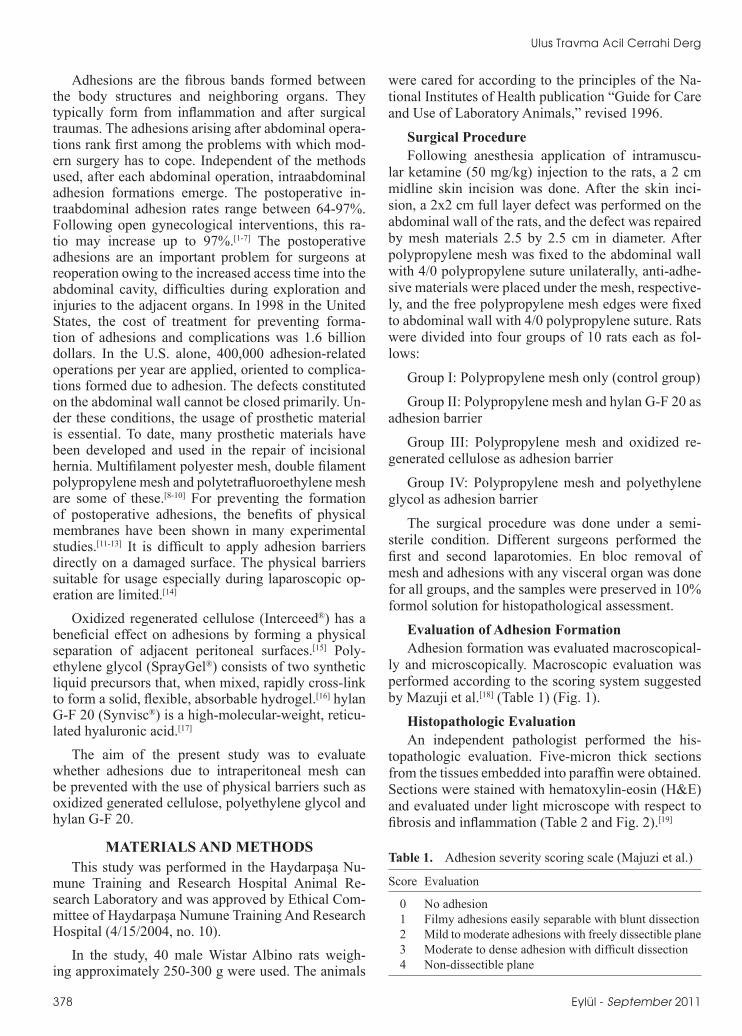
ly and microscopically. Macroscopic evaluation was performed according to the scoring system suggested by Mazuji et al.[18] (Table 1) (Fig. 1).
Histopathologic EvaluationAn independent pathologist performed the his-
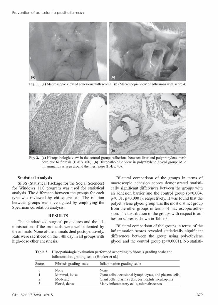
topathologic evaluation. Five-micron thick sections from the tissues embedded into paraffin were obtained. Sections were stained with hematoxylin-eosin (H&E) and evaluated under light microscope with respect to fibrosis and inflammation (Table 2 and Fig. 2).[19]
378 Eylül - September 2011
Table 1. Adhesion severity scoring scale (Majuzi et al.)
Score Evaluation
0 No adhesion 1 Filmy adhesions easily separable with blunt dissection 2 Mild to moderate adhesions with freely dissectible plane 3 Moderate to dense adhesion with difficult dissection 4 Non-dissectible plane
Prevention of adhesion to prosthetic mesh
Statistical AnalysisSPSS (Statistical Package for the Social Sciences)
for Windows 11.0 program was used for statistical analysis. The difference between the groups for each type was reviewed by chi-square test. The relation between groups was investigated by employing the Spearman correlation analysis.
RESULTSThe standardized surgical procedures and the ad-
ministration of the protocols were well tolerated by the animals. None of the animals died postoperatively. Rats were sacrificed on the 14th day in all groups with high-dose ether anesthesia.
Bilateral comparison of the groups in terms of macroscopic adhesion scores demonstrated statisti-cally significant differences between the groups with an adhesion barrier and the control group (p<0.004, p<0.01, p<0.0001), respectively. It was found that the polyethylene glycol group was the most distinct group from the other groups in terms of macroscopic adhe-sion. The distribution of the groups with respect to ad-hesion scores is shown in Table 3.
Bilateral comparison of the groups in terms of the inflammation scores revealed statistically significant differences between the group using polyethylene glycol and the control group (p<0.0001). No statisti-
Cilt - Vol. 17 Sayı - No. 5 379
Table 2. Histopathologic evaluation performed according to fibrosis grading scale and inflammation grading scale (Hooker et al.)
Score Fibrosis grading scale Inflammation grading scale
0 None None 1 Minimal, loose Giant cells, occasional lymphocytes, and plasma cells 2 Moderate Giant cells, plasma cells, eosinophils, neutrophils 3 Florid, dense Many inflammatory cells, microabscesses
Fig. 1. (a) Macroscopic view of adhesions with score 0. (b) Macroscopic view of adhesions with score 4.
Fig. 2. (a) Histopathologic view in the control group: Adhesions between liver and polypropylene mesh pore due to fibrosis (H-E x 400). (b) Histopathologic view in polyethylene glycol group: Mild inflammation is seen around the mesh pore (H-E x 40).
(a)
(a) (b)
(b)

Ulus Travma Acil Cerrahi Derg
cal difference was found between the other adhesion barriers and the control group. The distribution of the groups as per the inflammation scores is shown in Table 4.
Bilateral comparison of the groups in terms of the fibrosis score showed a statistically significant dif-ference between the polyethylene glycol group and control group (p<0.001). No statistical difference was found between the other adhesion barriers and the con-trol group. The distribution between groups is shown in Table 5.
DISCUSSIONAdhesion formation after abdominopelvic proce-
dures has an impact upon patient morbidity, success of subsequent surgical procedures and costs to the health care system in general. In the current approach,
in order to prevent adhesions, peritoneal damage should be reduced during the operation, inflammatory response should be reduced, coagulation formation should be prevented, and fibrinolysis has to be stim-ulated. An ideal physical membrane barrier should not affect wound healing or stimulate fibrosis forma-tion and should be effective in the presence of blood and foreign material.[14] Despite the development of minimally invasive techniques in many procedures and the ultimate decrease in trauma during operations, the technique alone does not effectively eliminate ad-hesion formation.[16]
While using Prolene mesh, contact between the mesh and the visceral organs leads to a severe adhe-sion formation. In the study of Felemovicius et al.,[20] an abdominal defect of 2.5 cm was made in three groups comprised of 20 rats each, and those defects
380 Eylül - September 2011
Table 3. Comparison of the groups in terms of macroscopic adhesion severity score
Adhesion severity score Groups
Control Hylan G-F 20 Oxidized Regenerated Polyethylene glycol Cellulose (n=10) (n=10) (n=10) (n=10)
0 – – – 3 (30%) 1 – 1 (10%) – 5 (50%) 2 – 5 (50%) 4 (40%) 2 (20%) 3 3 (30%) 4 (40%) 5 (50%) – 4 7 (70%) – 1 (10%) – Total 10 10 10 10
Table 4. Comparison of the groups in terms of inflammation score
Inflammation score Groups
Control Hylan G-F 20 Oxidized Regenerated Polyethylene glycol Cellulose (n=10) (n=10) (n=10) (n=10)
0 – – – – 1 – 3 (30%) 3 (30%) 8 (80%) 2 6 (60%) 6 (60%) 6 (60%) 2 (20%) 3 4 (40%) 1 (10%) 1 (10%) – Total 10 10 10 10
Table 5. Comparison of the groups in terms of fibrosis score
Fibrosis score Groups
Control Hylan G-F 20 Oxidized Regenerated Polyethylene glycol Cellulose (n=10) (n=10) (n=10) (n=10)
0 – – – – 1 – 3 (30%) 2 (20%) 8 (80%) 2 5 (50%) 5 (50%) 7 (70%) 2 (20%) 3 5 (50%) 2 (20%) 1 (10%) – Total 10 10 10 10

were repaired by Prolene, Sepramesh, and Sepramesh + Seprafilm, respectively. Adhesion signs were ob-served by electron microscopy in all 20 rats who re-ceived Prolene mesh. Similarly, we determined Grade 3 and 4 adhesions in 10 out of 10 rats in our control group. There was a statistically significant difference between the Prolene group and polyethylene glycol and hylan G-F 20 groups with regard to macroscopic adhesion grades.
To our knowledge, there is no study focusing on the usage of hylan G-F 20 as an adhesion barrier in intraabdominal adhesions in the current literature. In our study, hylan G-F 20 was used as an adhesion bar-rier by laying it underneath the polypropylene mesh. According to the statistical analysis, adhesion grade was 2 in one (10%), 3 in five (50%) and 4 in four (40%) subjects in the hylan G-F 20 group. In terms of macroscopic adhesion grade, there was a statistically significant difference between the hylan G-F 20 and Prolene groups.
The TC-7 (oxidized regenerated cellulose) bar-rier has been shown to provide significant reductions in the severity, incidence and width of postoperative adhesion formation.[21,22] Reid et al.[15] conducted a prospective clinical study in which they evaluated 40 female patients with a history of adhesiolysis be-cause of ovarian adhesions or cystectomy due to ovar-ian cyst. They covered both of the ovaries with TC-7, but sprayed heparin solution over one of them. The second-look laparoscopy demonstrated adhesion in 21 (52.5%) of 40 patients in the TC-7 + heparin spray group and in 26 (65%) of 40 patients in the TC-7 group, and use of heparin in conjunction with TC-7 was shown to exhibit no statistically significant differ-ence. In our study, based on the macroscopic scores, we determined a statistically significant difference between the TC-7 and polypropylene groups. We also determined a statistically significant difference be-tween the polyethylene glycol and TC-7 groups; the difference between TC-7 and hylan G-F 20 was not statistically significant.
Polyethylene glycol is a nontoxic and non-migrating adhesion barrier, which is used during laparoscopic and open surgical procedures due to its strong adhesive properties and easy-to-apply nature. It has an air pump and an apparatus that are particu-larly convenient for laparoscopic surgery. There is no risk of viral transmission via synthetic pieces and the polyethylene-based hydrogel does not carry any infec-tion potential. Moreover, owing to its methylene blue kit, it contributes to the visualization of the damaged surface by staining it blue.[23-25]
Dunn et al.[23] divided 16 rats with cecum abrasion into control and polyethylene glycol groups, while splitting 20 New Zealand rabbits with induced uterine
horn abrasion again into control and polyethylene gly-col groups. They treated the abrasion site with poly-ethylene glycol in the treatment groups. An abrasion between the cecum and lateral wall was determined in 7 of 8 rats in the control group and in 1 of 8 rats in the polyethylene group with cecum abrasion model. Adhesion was found to cover more than 50% of the uterine horn in 8 of 10 rabbits in the control group and in 2 of 10 rabbits in the polyethylene glycol group with uterine horn abrasion. Polyethylene glycol was observed to cause a significant reduction in the inci-dence of adhesion formation.
Metler et al.[25] conducted a study on 64 patients by dividing them into two groups and comparing the control group (n=30) treated solely with surgery and the study group treated with surgery + polyeth-ylene glycol. Open and laparoscopic surgeries were performed for leiomyoma or leiomyomatous uter-ine lesions. Laparoscopic surgery was applied on 82.4% (n=28) of the study group and 76.7% (n=23) of the control group. Mean duration for application of polyethylene glycol barrier was 3.7 minutes, and the average amount of polyethylene glycol used for each patient was 1.9 kits. Adhesion formation in the secondary laparotomies was statistically significantly lower in the study group than in the control group.
In this study, polyethylene glycol, sprayed under-neath the mesh during closure of the induced ventral defect with polypropylene mesh, was found to provide statistically significant reductions in macroscopic ad-hesion formation compared with the hylan G-F 20 and TC-7 applications. Based on the evaluations focused on macroscopic adhesions, the difference between the polyethylene glycol group and control group was found to be highly statistically significant. The high-est difference was observed between the polyethylene glycol and control groups. The difference between the polyethylene glycol group and the TC-7 group in terms of macroscopic adhesion score was highly significant as well. Moreover, the difference between polyethylene glycol and hylan G-F 20 was statistically significant. Macroscopically, the polyethylene glycol group appeared to have the least amount of adhesion formation compared with the other groups, and this difference was evaluated to be statistically significant. Ozmen et al.[26] found that using sodium hyaluronate reduces the incidence and severity of abdominal adhe-sions following laparoscopic mesh insertion.
In conclusion, although hylan G-F 20 is used for cartilage repair of the joints, it can also be used as an antiadhesive barrier after abdominal operations. Poly-ethylene glycol was an effective adhesion prevention barrier, and results seem to be at least comparable with those of other products. Polyethylene glycol is a reli-able and easily applied adhesion barrier, and reduces
Prevention of adhesion to prosthetic mesh
Cilt - Vol. 17 Sayı - No. 5 381

adhesion formation after open and laparoscopic sur-gery.
REFERENCES1. Szabo A, Haj M, Waxsman I, Eitan A. Evaluation of sepra-
film and amniotic membrane as adhesion prophylaxis in mesh repair of abdominal wall hernia in rats. Eur Surg Res 2000;32:125-8.
2. Moreira H Jr, Wexner SD, Yamaguchi T, Pikarsky AJ, Choi JS, Weiss EG, et al. Use of bioresorbable membrane (sodium hyaluronate + carboxymethylcellulose) after controlled bow-el injuries in a rabbit model. Dis Colon Rectum 2000;43:182-7.
3. Risberg B. Adhesions: preventive strategies. Eur J Surg Sup-pl 1997;577:32-9.
4. Becker JM, Dayton MT, Fazio VW, Beck DE, Stryker SJ, Wexner SD, et al. Prevention of postoperative abdominal ad-hesions by a sodium hyaluronate-based bioresorbable mem-brane: a prospective, randomized, double-blind multicenter study. J Am Coll Surg 1996;183:297-306.
5. Menzies D, Ellis H. Intestinal obstruction from adhesions--how big is the problem? Ann R Coll Surg Engl 1990;72:60-3.
6. Ray NF, Larsen JW Jr, Stillman RJ, Jacobs RJ. Economic impact of hospitalizations for lower abdominal adhe-siolysis in the United States in 1988. Surg Gynecol Obstet 1993;176:271-6.
7. Hershlag A, Diamond MP, DeCherney AH. Adhesiolysis. Clin Obstet Gynecol 1991;34:395-402.
8. Dinsmore RC, Calton WC Jr, Harvey SB, Blaney MW. Pre-vention of adhesions to polypropylene mesh in a traumatized bowel model. J Am Coll Surg 2000;191:131-6.
9. Oncel M, Remzi FH, Senagore AJ, Connor JT, Fazio VW. Application of Adcon-P or Seprafilm in consecutive lapa-rotomies using a murine model. Am J Surg 2004;187:304-8.
10. Johns DA, Ferland R, Dunn R. Initial feasibility study of a sprayable hydrogel adhesion barrier system in patients un-dergoing laparoscopic ovarian surgery. J Am Assoc Gynecol Laparosc 2003;10:334-8.
11. Günerhan Y, Caglayan K, Sumer A, Koksal N, Altınlı E, Onur E, et al. The efficacy of carboxymetylcellulose for prevention adhesion formation after thyroid region surgery. Kafkas Uni Vet Fak Der 2009;5:785-9.
12. Rodgers KE, Johns DB, Girgis W, Campeau J, diZerega GS. Reduction of adhesion formation with hyaluronic acid after peritoneal surgery in rabbits. Fertil Steril 1997;67:553-8.
13. Wiseman DM, Gottlick-Iarkowski L, Kamp L. Effect of dif-ferent barriers of oxidized regenerated cellulose (ORC) on cecal and sidewall adhesions in the presence and absence of bleeding. J Invest Surg 1999;12:141-6.
14. Zhou J, Elson C, Lee TD. Reduction in postoperative adhe-sion formation and re-formation after an abdominal opera-tion with the use of N, O - carboxymethyl chitosan. Surgery 2004;135:307-12.
15. Reid RL, Hahn PM, Spence JE, Tulandi T, Yuzpe AA, Wise-man DM. A randomized clinical trial of oxidized regenerated cellulose adhesion barrier (Interceed, TC7) alone or in com-bination with heparin. Fertil Steril 1997;67:23-9.
16. Mettler L, Audebert A, Lehmann-Willenbrock E, Schive K, Jacobs VR. Prospective clinical trial of SprayGel as a barrier to adhesion formation: an interim analysis. J Am Assoc Gy-necol Laparosc 2003;10:339-44.
17. Kahan A, Lleu PL, Salin L. Prospective randomized study comparing the medicoeconomic benefits of Hylan GF-20 vs. conventional treatment in knee osteoarthritis. Joint Bone Spine 2003;70:276-81.
18. Mazuji MK, Kalambaheti K, Pawar B. Prevention of adhe-sions with polyvinylpyrrolidone. Preliminary report. Arch Surg 1964;89:1011-5.
19. Hooker GD, Taylor BM, Driman DK. Prevention of adhesion formation with use of sodium hyaluronate-based bioresorb-able membrane in a rat model of ventral hernia repair with polypropylene mesh--a randomized, controlled study. Sur-gery 1999;125:211-6.
20. Felemovicius I, Bonsack ME, Hagerman G, Delaney JP. Pre-vention of adhesions to polypropylene mesh. J Am Coll Surg 2004;198:543-8.
21. Saravelos HG, Li TC. Physical barriers in adhesion preven-tion. J Reprod Med 1996;41:42-51.
22. Azziz R. Microsurgery alone or with INTERCEED Ab-sorbable Adhesion Barrier for pelvic sidewall adhesion re-formation. The INTERCEED (TC7) Adhesion Barrier Study Group II. Surg Gynecol Obstet 1993;177:135-9.
23. Dunn R, Lyman MD, Edelman PG, Campbell PK. Evalua-tion of the SprayGel adhesion barrier in the rat cecum abra-sion and rabbit uterine horn adhesion models. Fertil Steril 2001;75:411-6.
24. Ferland R, Mulani D, Campbell PK. Evaluation of a sprayable polyethylene glycol adhesion barrier in a porcine efficacy model. Hum Reprod 2001;16:2718-23.
25. Mettler L, Audebert A, Lehmann-Willenbrock E, Schive-Peterhansl K, Jacobs VR. A randomized, prospective, con-trolled, multicenter clinical trial of a sprayable, site-specific adhesion barrier system in patients undergoing myomecto-my. Fertil Steril 2004;82:398-404.
26. Ozmen MM, Aslar AK, Terzi MC, Albayrak L, Berberoğlu M. Prevention of adhesions by bioresorbable tissue barrier following laparoscopic intraabdominal mesh insertion. Surg Laparosc Endosc Percutan Tech 2002;12:342-6.
382 Eylül - September 2011
Ulus Travma Acil Cerrahi Derg