Pneumothorax in Neonates - repositorio-aberto.up.pt · 2015/2016 Íris Anabela Santos Silva...
25
2015/2016 Íris Anabela Santos Silva Pneumothorax in Neonates março, 2016
Transcript of Pneumothorax in Neonates - repositorio-aberto.up.pt · 2015/2016 Íris Anabela Santos Silva...
2015/2016
Íris Anabela Santos Silva
Pneumothorax in Neonates
março, 2016
Mestrado Integrado em Medicina
Área: Pediatria
Tipologia: Dissertação
Trabalho efetuado sob a Orientação de:
Doutora Maria Hercília Ferreira Guimarães Pereira Areias
E sob a Coorientação de:
Dr. Gustavo Marcondes Duarte Rocha
Trabalho organizado de acordo com as normas da revista:
Journal of Pediatric and Neonatal Individualized Medicine
Íris Anabela Santos Silva
Pneumothorax in Neonates
março, 2016
flr,onroITTüÚF TÀCULOÀDE DE MEDI(INÂLa:;l uNrvr*srôÀD! Do ponro
urlrD40! ÇuÊÊ cúLÂqPRoJEÍo oe opçÂo
Projeto de Opção do 6o ano - DrcrlmçÃo or ItrrcnrDADE
Eu, Íris Anabela Santos Silva, abaixo assinado, no mecanográflco 201008519, estudante do 60 ano
do Ciclo de Estudos Integrado em Medicina, na Faculdade de Medicina da Universidade do Porto,
declaro ter atuado com absoluta integridade na elaboração deste projeto de opção.
Neste sentido, confirmo que nÃO incorri em plágio (ato pelo qual um indivíduo, mesmo por omissão,
assume a autoria de um determinado trabalho intelectual, ou paftes dele). Mais declaro que todas as
frases que retirei de trabalhos anteriores pedencentes a outros autores, foram referenciadas, ou
redigidas com novas palavras, tendo colocado, neste caso, a citação da fonte bibliográfica.
Faculdade de Medicina da Universidade do Porto, 2310312076
Assinatura conforme cartão de identificação:
úr, ' -*l- [,, § |,c'. 5;1, ç
IUponro Projecto de Opção do 60 ano - DrcunnçÃo or RepnoouçÃo]FMUP TACULDÀDE DE MEDICINÀÍ. i ururv:nsrolor Do PoRao
UNI§ADE CURRICULARPROJETo DE oPçÃo
NOME
Anabela Santos Silva
NUMERO DE ESTUDANTE DATA DE CONCLUSAO
201 00851 I
orsrcruaÇÃo on ÁRrR Do PRoJEcro
Pediatria - Neonatologia
TITULO OISSEnUçAOTMONOGRAFIA (riscar o que não interessa)
Pneumothorax in Neonates
ORIENTADOR
Maria Hercília Ferreira Guimarães Pereira Areias
COORIENTADOR (se aplicável)
Gustavo Marcondes Duafte Rocha
ASSINALE APENAS UMA DAS OPÇOES:
Faculdade de Medicina da Universidade do Porto, 23103120L6
É euronrzRoR n nrenoouçÃo TNTEGRAL DESTE TRABALHo AeENAS eARA EFErros oe rruvesrrclçÃo,
MEDIANTE DECLARAÇÃO ESCRITA DO INTERESSADO, QUE A TAL SE COMPROMETE. XÉ RutoRrzRoR a nrenoouçÃo pARcrAL DESTE TRABALHo (rNDrcAR, cASo rAL sr:R rurcrssÁRro, No
rqÁxrr'ro or pÁcrruns, tusrRRÇôrs, Gúrrcos, ETc.) ApENAS pARA EFErros DE TNVESTTcAçÃo, rueomruru
orcmnaçÃo EScRrrA Do TNTERESSADo, euE A TAL sE coMpRoMETE.tr
DE AcoRDo coM A LEGISLAÇÃo EM vIGoR, (INDICAR, cASo rAL se:R ruecrssÁRro, No uextuo oe pÁGtNRs,
tLustnRçôrs, GnÁrrcos, rrc.; rlÃo É prnurrlol R nrRnoouçÃo DE euALeuER rARTE DEsrE TRABALHo. tr
Assinatura conforme cartão de identifica çao, úoiS À.* qL" 0.. 5n ,,#y, S;lrrq
Pneumothorax in Neonates
Íris Silva1, Filipa Flôr-de-Lima2,Gustavo Rocha2, Inês Alves1, Hercília Guimarães1,2
1 Faculty of Medicine of Porto University, Porto, Portugal
2 Neonatal Intensive Care Unit, Department of Pediatrics, Centro Hospitalar São João. Alameda
Professor Hernâni Monteiro, 4200-319 Porto, Portugal
Abstract
Introduction: Pneumothorax occurs more frequently in the neonatal period than in any other
period of life and is associated with increased mortality and morbidity. Several risk factors for
pneumothorax, including respiratory pathology, invasive and non-invasive respiratory support,
and predictors of mortality have been described.
Objective: To evaluate the prevalence, to assess risk factors and to describe the clinical
characteristics, management and outcome of newborn infants with pneumothorax as well as
identify predictors of mortality in these newborns.
Methods: This retrospective case-control study included all newborns hospitalized in the NICU
of Centro Hospitalar São João, Porto, Portugal, between 2003 and 2014, with the diagnosis of
pneumothorax. A control group was selected among the newborns without pneumothoraces.
The collected data included: demographics and perinatal data, pneumothorax characteristics,
classification, treatment and clinical outcomes.
Results: Our study included 240 neonates (80 with pneumothoraces and 160 controls), of whom
145 were male (60.4 %). Median gestational age was 37 (24-40) weeks and median birthweight,
2612.5 (360-4324) grams. The prevalence in our NICU was 1.5 %. Pneumothorax was
significantly associated with RDS (p=0.010) and TTN (p<0.001). Invasive mechanical
ventilation (p=0.016) and FiO2>0.4 (p=0.003), were independent risk factors for the -
development of pneumothoraces. The mortality rate was 13.8%. Hypotension, mechanical
ventilation and thoracentesis followed by a chest tube insertion were found to be predictors of
mortality in newborns having pneumothoraces, but pneumothorax was not.
Conclusion: Pneumothorax is relatively frequent in the neonatal intensive care unit. Its risk
factors and predictors of mortality should be known in order to prevent and treat this critical
situation. Pneumothorax itself was not a predictor of mortality, probably due to the adequate
and prompt management used in the NICU.
Keywords: Pneumothorax, newborn, risk factors, mechanical ventilation, neonatal intensive
care, mortality.
Introduction
Pneumothorax occurs more frequently in the neonatal period than in any other period of
life and is associated with increased mortality and morbidity. It begins with the rupture of an
over-distended alveoli. The air escapes along the perivascular connective tissue sheath into the
pleural space, causing pneumothorax and less commonly pneumomediastinum,
pneumopericardium, subcutaneous emphysema, and pneumoperitoneum, altogether known as
air leak syndromes. [1,2]
Pneumothorax is a relatively frequent critical situation in the neonatal intensive care
unit. Symptomatic pneumothorax occurs in about 0.05% to 0.1% of all live births and in very
low birthweight infants this rate can achieve 3.8 % to 9%. [3,4]
Several risk factors for pneumothorax have been described and include immaturity,
respiratory distress syndrome (RDS), invasive and non-invasive respiratory support,
chorioamnionitis, among others. For moderate-late preterm infants risk factors also include
higher birth weight, male gender, and rupture of membranes longer than 24 hours. [5]
Common clinical manifestations of pneumothorax are tachypnea, flaring, hypoxemia
and hypercapnea. In some cases, there is a mediastinal shift that compromises the
cardiovascular system and carries a significant risk of an impaired outcome and death. Tension
pneumothorax causes a rise in intrapleural pressure and subsequent lung collapse, as well as
impaired venous return, systemic hypotension and cardiac arrest. It is, therefore, essential to
recognize these risk infants in order to prevent and treat properly this critical situation. [1,4]
The diagnosis of pneumothorax relies on clinical judgment, transillumination and chest
radiogram. [6]
The treatment of neonatal pneumothorax is not fully defined. Three approaches are the
common practice in NICUs. The expectant approach for small and asymptomatic
pneumothorax, and active intervention to a significant one, such as needle (14–16 G) aspiration
and thoracic drainage.[7,8] Needle aspiration is an option in cases of mild to moderate
pneumothorax when the infant is hemodynamically stable. In hypertensive pneumothoraces the
common therapeutic approach is chest tube placement. [1]
The aim of this study was to evaluate the prevalence, to identify risk factors and to
describe the clinical characteristics, management and outcome of newborn infants with
pneumothorax as well as to identify the predictor factors of mortality in these newborns.
Material and methods
This retrospective case control study included all newborns hospitalized in the NICU (a
level III unit with 17 beds and 450 admissions per year and) of Centro Hospitalar São João,
Porto, between 1st january 2003 and 31st december 2014, with the diagnosis of pneumothorax.
All the data were collected from the hospital electronic clinical database and medical
records of the patients. A control group was selected among the newborns without
pneumothoraces admitted to the NICU in the same years, gender, gestational age (more or less 2
weeks) and birthweight (more or less 500 g). These data includes: information regarding
pregnancy and delivery (gestational age, multiple births, type of deliver), gender, birth weight,
Apgar score at 1 and 5 minutes, the need of resuscitation, antenatal corticotherapy, need for
surfactant, oxygen therapy, nasal continuous positive airway pressure (NCPAP), conventional
mechanical ventilation (CMV) and high frequency oscillation ventilation (HFOV) use .
Pneumothorax characteristics were also obtained (day of life and the duration of the
pneumothorax) and classified as spontaneous (primary or secondary), iatrogenic (positive
airway pressure or surgery), hypertensive and persistent. Data about treatment, neonatal
morbidity and mortality were also collected. Pneumothoraces were diagnosed by chest
radiography and the type of pneumothorax was assessed according to clinical setting and
medical history. [1]
Gestational age was assessed by post-menstrual age, ultrasound examination or the New
Ballard Score (in the absence of obstetrical indexes). [9, 10] Small for gestational age was
defined as a birth weight below the 3rd centile of Fenton`s growth charts. [11] Our NICU has a
protocol for positive pressure ventilation that includes different ventilatory strategies according
to different lung diseases and favors the use of permissive hypercapnea. For the very low birth
weight infants NCPAP just after birth is the preferable mode of ventilation. [12] NCPAP is done
using Infant Flow (Pulmocor, USA), for invasive mechanical ventilation we use Babylog
(Drager, Germany). HFOV is performed as rescue ventilation (Sensor Medics®, USA).
Sepsis was considered in the presence of a positive blood culture, combined with
clinical and laboratory parameters. [13] Intraventricular hemorrhage (IHV) was defined
according to Papile [14] and Volpe [15] (before and after 2010, respectively), intraventricular
bleeding with ventricular dilatation is classified as IHV 3 and with parenchymal involvement as
IHV 4. Retinopathy of prematurity (ROP) was diagnosed and classified according to the
International Classification of Retinopathy of Prematurity revised. [16] Patent ductus arteriosus
(PDA) was diagnosed according to SIBEN consensus. [17] Necrotizing enterocolitis (NEC) was
defined by clinical findings and radiological features, according to the modified Bell criteria.
[18] Cystic periventricular leukomalacia (PVL) was diagnosed by ultrasound. [19]
Respiratory distress syndrome was defined based on the European guidelines [20] and
bronchopulmonary dysplasia (BPD) according to the NIH Consensus definition. [21] Transient
tachypnea of the newborn (TTN) was diagnosed according to the criteria of Machado and Fiori.
[22] Pneumonia was diagnosed based on combination of clinical, radiological and laboratory
parameters. [23] Meconium aspiration syndrome (MAS) was characterized based on Shahed A.
Criteria. [24] Pulmonary hypoplasia (PH) was defined according on clinical, radiologic, and
pathologic criteria. [25]
Hypotension was defined according Cayabyab et al. [26]
The statistical analysis was performed using SPSS for Windows, version 23.
Categorical variables were evaluated by absolute and relative frequencies and continuous
variables were characterized by median (minimum-maximum). All variables were compared
between neonates with (cases) or without (controls) pneumothorax. Chi-Squared or Fisher’s
exact test were used to compare categorical variables and Mann Whitney-U test (non-parametric
test) to compare continuous variables. Risk factors for pneumothorax were evaluated by a
multivariate analysis by logistic regression. Statistically significance was considered for a p
value less than 0.05.
Results Our study included 240 neonates (80 with pneumothoraces and 160 controls), of whom
145 were males (60.4 %). Their median gestational age was 37 (24-40) weeks and the median
birthweight was 2612.5 (360-4324) grams. The prevalence in our NICU was 1.5 %. .The
analysis of demographic and perinatal data of both groups showed statistically significant
differences in Apgar score at 5th minute (p = 0.002) and in resuscitation at birth (p < 0.001),
Table 1.
Table 2 describes the characterization and treatment of pneumothoraces. They were
spontaneous (primary or secondary to lung disease) in 46 (57.5%) neonates and iatrogenic in 34
(42.5%). Fifty seven (71.3%) were hypertensive and 2 (2.5%) persistent. We observed 37
(46.3%) pneumothoraces in the right lung, 35 (43.8%) in the left and 8 (10%) were bilateral.
Oxygen therapy was done in 68 (85%) neonates, NCPAP in 15 (18.8%) and IMV in 60 (75%)
newborns. Thoracentesis was performed in 10 (12.5%) pneumothoraces, but 7 of them also had
to be drained. To treat the pneumothoraces, 57 (71.3%) neonates needed drains and 3 (3.8%)
thoracentesis.
In Table 3 we present the clinical and management data in cases and controls. There
were statistically significant differences between both groups concerning respiratory pathology
(pneumonia, MAS, RDS, pulmonary hypoplasia, transient tachypnea of the newborn). IVH ≥3
grade, PDA, cystic PVL and hypotension were also more frequent in newborns having
pneumothorax. Use of oxygen therapy (p<0.001), NCPAP (p<0.001) and mechanical ventilation
(p<0.001) also revealed a significant increase in pneumothorax cases. Statistical significance
between the two groups was also found for FiO2>0.4 (p < 0.001). The association with
pneumothorax was found for major congenital malformations: congenital cardiopathy (CHD)
and diaphragmatic hernia. The average of days of hospitalization was 9.5 in pneumothorax
group and 4 in controls (p<0.001). The prevalence of death was 24 (30%) in cases and 9 (5.6%)
in controls (p < 0.001).
The two respiratory pathologies, RDS (OR 3.512, 95% CI 1.353-9.118, p=0.010) and
TTN (OR 8.677, 95% CI 2.752-27.354, p<0.001) were significantly associated with
pneumothorax. Mechanical ventilation was also significantly associated with pneumothorax
(OR 3.824, 95% CI 1.285-11.376, p<0.016), as was FiO2>0.4 (OR 5.614, 95%CI 1.828-17.238,
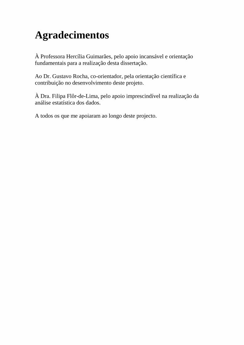
p=0.003), Table 4. Pneumothorax was not identified as a predictor of death OR 1.284, 95% CI
0.409-4.032, p=0.669). Hypotension, thoracentesis and mechanical ventilation were found to be
predictors of death in newborns with pneumothoraces, Table 5.
Discussion
In this study we aimed to compare clinical characteristics between neonates with and
without pneumothoraces and to identify risk factors of pneumothoraces and predictors of death
in these patients.
As expected, there were no significant differences between cases and controls, for
gender, gestational age and birth weight. Statistically significant differences in Apgar score at
5th minute occurred due to the need of resuscitation at birth, which was found to be a risk factor
(p < 0.001). According to clinical practice, the use of invasive ventilation with endotracheal
intubation in neonatal resuscitation is less and less used and replaced by noninvasive ventilation
with the minimum effective pressure. [12] The type of delivery had no influence in
pneumothorax occurrence, results that are consistent with others. [2, 27] Antenatal steroids use
wasn´t associated with pneumothorax (p=0.976), data according with other studies. [28]
The treatment of pneumothorax is not fully defined. In this study we had 80
pneumothoraces, spontaneous in 46 (57.5%) neonates and iatrogenic in 34 (42.5%), but only 68
(85%) received oxygen therapy, because pneumothoraces with very small size in chest x-ray
solved spontaneously. As described in literature, other option for treatment is the expectant
management for non hypertensive pneumothoraces in patients who are ventilated [3], and this
approach was done in 15 (18.8%) neonates with NCPAP and in 60 (75%) with CMV. The use
of needle aspiration and chest tube are common options for the treatment. Thoracentesis was
made in 10 (12.5%) pneumothoraces, however this treatment wasn´t effective in seven of them.
So, thoracentesis only treated 3 (3.8%) non hypertensive pneumothoraces. It is known that,
frequently, neonates treated with needle aspiration, can require subsequent chest-tube insertion.
[29] In our study 57 (71.3%) neonates needed drains to be effectively treated.
As described in Table 3, there was a statistical significant difference between cases and controls
concerning respiratory pathology. When adjusted by multivariate analyses, RDS showed an
OR=3.512 and TTN was OR=8.677, which means that they were positively related with
pneumothorax, results comparable with other studies. [2,28] BPD was not statistically different
between the two groups, as observed in other studies. [28,30] The risk of developing
pneumothorax was also increased in newborns with other pathologies (IVH ≥3 grade, PDA,
cystic PVL and hypotension), showing that pneumothoraces can occur in patients with more
severe diseases. As we expected the administration of surfactant was more frequent in patients
with pneumothoraces, probably due to the lung pathology associated with cases. In the present
study, oxygen therapy, NCPAP and CMV were related with the development of pneumothorax.
Linear regression analyses indicate that neonates who received IMV had a probability 3.8 times
higher to develop pneumothorax and those who received oxygen therapy with a FiO2>0.4 had
an increased risk of pneumothorax 5.6 times higher. Terzic et al. showed that infants who
needed higher FiO2 were more likely to develop pneumothorax. [28] These results suggest that
high pressures in NCPAP and CMV and a high FiO2 should be avoided. Congenital heart
disease and diaphragmatic hernia were also statistical significant risk factors for pneumothorax,
probably because of the pathophysiological disturbances of these pathologies or their surgical
repair. Congenital diaphragmatic hernia is a risk factor for pulmonary hypoplasia once it
occupies space, prevents lung expansion and causes a change in the thoracic cavity. [31]
Neonates with pneumothorax had a prolonged stay in NICU, an average of 9.5 days. Possibly
they had more associated pathology and needed treatment.
We found that 30% of cases died comparing to 5.6% of controls (p<0.001).
Hypotension, mechanical ventilation and thoracentesis associated with chest tube insertion were
found to be predictors of mortality in newborns with pneumothoraces. These neonates had a bad
prognosis since the beginning. However, in our study, pneumothorax was not identified as a
predictor of mortality as had been shown by others [2,6] The good strategy and appropriate
management used in the NICU was probably the explanation for this result.
The limitations of this study are to be a retrospective one, to have a small size sample
and to represent a single center.
Conclusion
Pneumothorax is relatively frequent in the neonatal intensive care unit. Its risk factors
(RDS, TTN, invasive mechanical ventilation and a FiO2>0.4 and predictors of mortality
(hypotension, mechanical ventilation and thoracentesis followed by a chest tube insertion)
should be well known, in order to prevent and treat this critical situation.
Pneumothorax itself was not a predictor of mortality, probably due to the adequate and prompt
management used in the NICU.
Funding: None Conflict of interest: None
References
[1] Neonatal Respiratory Disorders, Moira A Crowley. Fanaroff and Martin's Neonatal-Perinatal
Medicine: Diseases of the Fetus and Infant, J. Martin Richard, Fanaroff Avroy A., Walsh
Michele C. Elsevier Saunders, 10ª edition/vol.2, pp 1113-36
[2] Duong HH, Mirea L, Shah PS, Yang J, Lee SK, Sankaran K.
Pneumothorax in neonates: Trends, predictors and outcomes. J Neonatal Perinatal Med. 2014;
7(1):29-38.
[3] Kitsommart R, Martins B, Bottino MN, Sant'Anna GM. Expectant
management of pneumothorax in preterm infants receiving assisted ventilation: report of 4 cases
and review of the literature. Respir Care. 2012 May; 57(5):789-93.
[4] Smith J, Schumacher RE, Donn SM, Sarkar S. Clinical course of symptomatic spontaneous
pneumothorax in term and late preterm newborns: report from a large cohort. Am J
Perinatol. 2011 Feb; 28(2):163-8.
[5] Andrew A. Colin, Cynthia McEvoy, Robert G. Castile. Respiratory Morbidity and Lung
Function in Preterm Infants of 32 to 36 Weeks' Gestational Age. Pediatrics, July 2010, 126(1): 115–
128.
[6] Ozer EA, Ergin AY, Sutcuoglu S, Ozturk C, Yurtseven A. Is pneumothorax size on chest x-
ray a predictor of neonatal mortality? Iran J Pediatr. 2013 Oct; 23(5):541-5
[7] MacDuff A., Arnold A., Harvey J.; et al. Management of spontaneous pneumothorax:
British Thoracic Society pleural disease guideline 2010. Thorax 2010; 65:ii18-ii31.
[8] Pocivalnik M, Meheden SV, Griesmaier E, Trawöger R, Kiechl-Kohlendorfer U, Pichler
G, Reiterer F, Urlesberger B. Pneumothorax during mechanical ventilation--therapeutic options
in term and preterm neonates. Klin Pediatr. 2013 Dec; 225(7):389-93.
[9] MacDonald H, American Academy of Pediatrics. Committee on F, Newborn. Perinatal care
at the threshold of viability. Pediatrics. 2002; 110(5):1024-7.
[10] Ballard JL, Khoury JC, Wedig K, Wang L, Eilers-Walsman BL, Lipp R. New Ballard
Score, expanded to include extremely premature infants. J Pediatr. 1991; 119(3):417-23.
[11] Fenton, T.R. and J.H. Kim, A systematic review and meta-analysis to revise the Fenton
growth chart for preterm infants. BMC Pediatr, 2013. 13: p. 59.
[12] Tavares PN, Flor-de-Lima F., Soares H., Guimarães H. Early nCPAP versus intubation in
very low birth weight infants. Journal of Pediatric and Neonatal Individualized Medicine 2013;
2(2):e020201.
[13] Vergnano S., Menson E., Kennea N., et al. Neonatal infections in England: the NeonIN
surveillance network, Arch Dis Child Fetal Neonatal, Ed. 2011 Jan; 96(1):F9-F14.
[14] Papile LA, Burstein J, Burstein R, Koffler H. Incidence and evolution of subependymal
and intraventricular hemorrhage: a study of infants with birth weights less than 1,500 gm. J
Pediatr. 1978; 92(4):529-34.
http://www.ncbi.nlm.nih.gov/pubmed/?term=Sutcuoglu%20S%5BAuthor%5D&cauthor=true&cauthor_uid=24800014
[15] Volpe JJ. Neurology of the newborn. 4th ed. Philadelphia: W.B. Saunders; 2001. xiii, 912
p. p.
[16] International Committee for the Classification of Retinopathy of, P., The International
Classification of Retinopathy of Prematurity revisited. Arch Ophthalmol, 2005. 123(7): p. 991-
9.
[17] Golombek SG, Sola A, Baquero H, Borbonet D, Cabanas F, Fajardo C, Goldsmit G, Lemus
L, Miura E, Pellicer A, Perez JM, Rogido M, Zambosco G, van Overmeire B, Primer Grupo de
Consenso Clinico S. First SIBEN clinical consensus: diagnostic and therapeutic approach to
patent ductus arteriosus in premature newborns. Anales de pediatria. 2008; 69(5):454-81.
[18] Walsh MC, Kliegman RM. Necrotizing enterocolitis: treatment based on staging criteria.
Pediatr Clin North Am. 1986; 33(1):179-201.
[19] Kuk, J.Y., et al., Optimal time interval between a single course of antenatal corticosteroids
and delivery for reduction of respiratory distress syndrome in preterm twins. Am J Obstet
Gynecol, 2013, 209(3): p. 256 e1-7.
[20] Sweet DG, Carnielli V, Greisen G, Hallman M, Ozek E, Plavka R, Saugstad OD, Simeoni
U, Speer CP, Vento M, Halliday HL, European Association of Perinatal M. European consensus
guidelines on the management of neonatal respiratory distress syndrome in preterm infants--
2013 update. Neonatology. 2013; 103(4):353-68.
[21] Jobe AH, Bancalari E. Bronchopulmonary dysplasia. Am J Respir Crit Care Med. 2001;
163(7):1723-9.
[22] Machado LU, Fiori HH, Baldisserotto M, Ramos Garcia PC, Vieira AC, Fiori RM.
Surfactant deficiency in transient tachypnea of the newborn. J Pediatr. 2011; 159(5):750-4.
[23]Nissen, M.D., Congenital and neonatal pneumonia. Paediatr Respir Rev, 2007; 8(3): p. 195-
203.
[24] Shahed A., Dargaville P., Ohlsson A., Soll R. Surfactant for meconium aspiration
syndrome in term and late preterm infants, Cochrane Database Syst Rev. 2014, december
2014;12:CD002054.
[25] Laudy J., Tibboel D., Robben S., Krijger R., Ridder M., Wladimiroff J. Prenatal Prediction of
Pulmonary Hypoplasia: Clinical, Biometric, and Doppler Velocity Correlates. Pediatrics, February
2002, volume 109 / issue 2.
[26] Cayabyab R., McLean C W and Seri I. Definition of hypotension and assessment of
hemodynamics in the preterm neonate. Journal of Perinatology (2009) 29, S58–S62.
[27] Basua S., Kumara A, Gupta AK. Complications associated with neonatal resuscitation.
Resuscitation, 2009, 80:4-5.
[28] Terzic S., Heljic S., Panic J., Sadikovic M., Maksic H. Pneumothorax in premature infants
with respiratory distress syndrome: focus on risk factors. Journal of Pediatric and Neonatal
Individualized Medicine 2016; 5(1):e050124.
[29] Litmanovitz I, Carlo WA. Expectant management of pneumothorax in ventilated neonates.
Pediatrics. 2008; 122(5): e975-9.

[30] Bhatia R, Davis PG, Doyle LW, Wong C, Morley CJ. Identification of pneumothorax in
very preterm infants. J Pediatr. 2011;159(1):115-20.e1.
[31] Delgado-Peña YP, Torrent-Vernetta A, Sacoto G, de Mir-Messa I, Rovira-Amigo
S, Gartner S, Moreno-Galdó A, Molino-Gahete JA, Castillo-Salinas F. Pulmonary hypoplasia:
An analysis of cases over a 20-year period. An Pediatr (Barc). 2015 Nov 25. pii: S1695-
4033(15)00405-1.
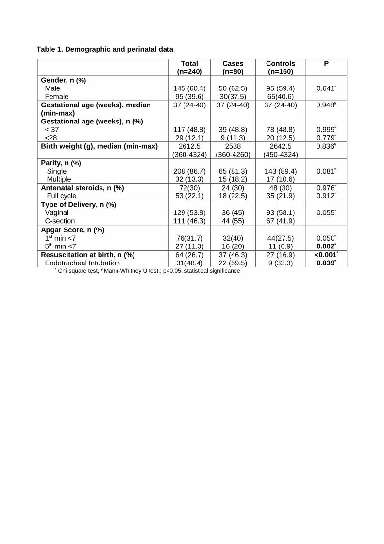
Table 1. Demographic and perinatal data
Total (n=240)
Cases (n=80)
Controls (n=160)
P
Gender, n (%) Male Female
145 (60.4) 95 (39.6)
50 (62.5) 30(37.5)
95 (59.4) 65(40.6)
0.641*
Gestational age (weeks), median (min-max) Gestational age (weeks), n (%) < 37 <28
37 (24-40)
117 (48.8) 29 (12.1)
37 (24-40)
39 (48.8) 9 (11.3)
37 (24-40)
78 (48.8) 20 (12.5)
0.948¥
0.999*
0.779*
Birth weight (g), median (min-max)
2612.5 (360-4324)
2588 (360-4260)
2642.5 (450-4324)
0.836¥
Parity, n (%) Single Multiple
208 (86.7) 32 (13.3)
65 (81.3) 15 (18.2)
143 (89.4) 17 (10.6)
0.081*
Antenatal steroids, n (%) Full cycle
72(30) 53 (22.1)
24 (30) 18 (22.5)
48 (30) 35 (21.9)
0.976*
0.912*
Type of Delivery, n (%) Vaginal C-section
129 (53.8) 111 (46.3)
36 (45) 44 (55)
93 (58.1) 67 (41.9)
0.055*
Apgar Score, n (%) 1st min <7 5th min <7
76(31.7) 27 (11.3)
32(40) 16 (20)
44(27.5) 11 (6.9)
0.050* 0.002*
Resuscitation at birth, n (%) Endotracheal Intubation
64 (26.7) 31(48.4)
37 (46.3) 22 (59.5)
27 (16.9) 9 (33.3)
<0.001*
0.039* * Chi-square test, ¥ Mann-Whitney U test.; p<0.05, statistical significance
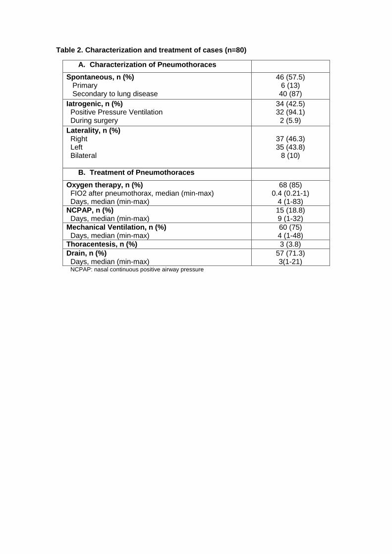
Table 2. Characterization and treatment of cases (n=80)
A. Characterization of Pneumothoraces
Spontaneous, n (%) Primary Secondary to lung disease
46 (57.5) 6 (13) 40 (87)
Iatrogenic, n (%) Positive Pressure Ventilation During surgery
34 (42.5) 32 (94.1) 2 (5.9)
Laterality, n (%) Right Left Bilateral
37 (46.3) 35 (43.8)
8 (10)
B. Treatment of Pneumothoraces
Oxygen therapy, n (%) FIO2 after pneumothorax, median (min-max) Days, median (min-max)
68 (85) 0.4 (0.21-1)
4 (1-83)
NCPAP, n (%) Days, median (min-max)
15 (18.8) 9 (1-32)
Mechanical Ventilation, n (%) Days, median (min-max)
60 (75) 4 (1-48)
Thoracentesis, n (%) 3 (3.8)
Drain, n (%) Days, median (min-max)
57 (71.3) 3(1-21)
NCPAP: nasal continuous positive airway pressure
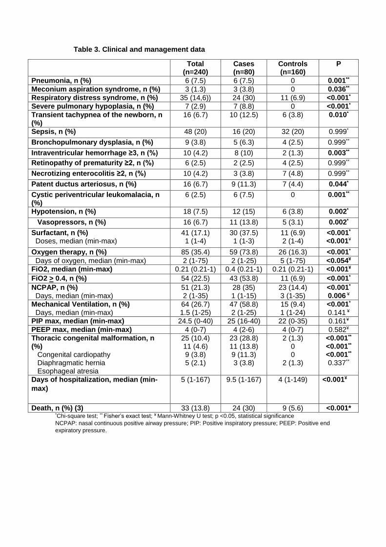
Table 3. Clinical and management data
Total (n=240)
Cases (n=80)
Controls (n=160)
P
Pneumonia, n (%) 6 (7.5) 6 (7.5) 0 0.001**
Meconium aspiration syndrome, n (%) 3 (1.3) 3 (3.8) 0 0.036**
Respiratory distress syndrome, n (%) 35 (14,6)) 24 (30) 11 (6.9) <0.001*
Severe pulmonary hypoplasia, n (%) 7 (2.9) 7 (8.8) 0 <0.001*
Transient tachypnea of the newborn, n (%)
16 (6.7) 10 (12.5) 6 (3.8) 0.010*
Sepsis, n (%) 48 (20) 16 (20) 32 (20) 0.999*
Bronchopulmonary dysplasia, n (%) 9 (3.8) 5 (6.3) 4 (2.5) 0.999**
Intraventricular hemorrhage ≥3, n (%) 10 (4.2) 8 (10) 2 (1.3) 0.003**
Retinopathy of prematurity ≥2, n (%) 6 (2.5) 2 (2.5) 4 (2.5) 0.999**
Necrotizing enterocolitis ≥2, n (%) 10 (4.2) 3 (3.8) 7 (4.8) 0.999**
Patent ductus arteriosus, n (%) 16 (6.7) 9 (11.3) 7 (4.4) 0.044*
Cystic periventricular leukomalacia, n (%)
6 (2.5) 6 (7.5) 0 0.001**
Hypotension, n (%) 18 (7.5) 12 (15) 6 (3.8) 0.002*
Vasopressors, n (%) 16 (6.7) 11 (13.8) 5 (3.1) 0.002*
Surfactant, n (%) Doses, median (min-max)
41 (17.1) 1 (1-4)
30 (37.5) 1 (1-3)
11 (6.9) 2 (1-4)
<0.001*
<0.001¥
Oxygen therapy, n (%) 85 (35.4) 59 (73.8) 26 (16.3) <0.001*
Days of oxygen, median (min-max) 2 (1-75) 2 (1-25) 5 (1-75) <0.054¥
FiO2, median (min-max) 0.21 (0.21-1) 0.4 (0.21-1) 0.21 (0.21-1) <0.001¥
FiO2 > 0.4, n (%) 54 (22.5) 43 (53.8) 11 (6.9) <0.001*
NCPAP, n (%) Days, median (min-max)
51 (21.3) 2 (1-35)
28 (35) 1 (1-15)
23 (14.4) 3 (1-35)
<0.001*
0.006 ¥
Mechanical Ventilation, n (%) Days, median (min-max)
64 (26.7) 1.5 (1-25)
47 (58.8) 2 (1-25)
15 (9.4) 1 (1-24)
<0.001*
0.141 ¥
PIP max, median (min-max) 24.5 (0-40) 25 (16-40) 22 (0-35) 0.161¥
PEEP max, median (min-max) 4 (0-7) 4 (2-6) 4 (0-7) 0.582¥
Thoracic congenital malformation, n (%) Congenital cardiopathy Diaphragmatic hernia Esophageal atresia
25 (10.4) 11 (4.6) 9 (3.8) 5 (2.1)
23 (28.8) 11 (13.8) 9 (11.3) 3 (3.8)
2 (1.3) 0 0
2 (1.3)
<0.001**
<0.001**
<0.001**
0.337**
Days of hospitalization, median (min-
max)
5 (1-167) 9.5 (1-167) 4 (1-149)
<0.001¥
Death, n (%) (3) 33 (13.8) 24 (30) 9 (5.6) <0.001* *Chi-square test; ** Fisher’s exact test; ¥ Mann-Whitney U test; p <0.05, statistical significance
NCPAP: nasal continuous positive airway pressure; PIP: Positive inspiratory pressure; PEEP: Positive end
expiratory pressure.
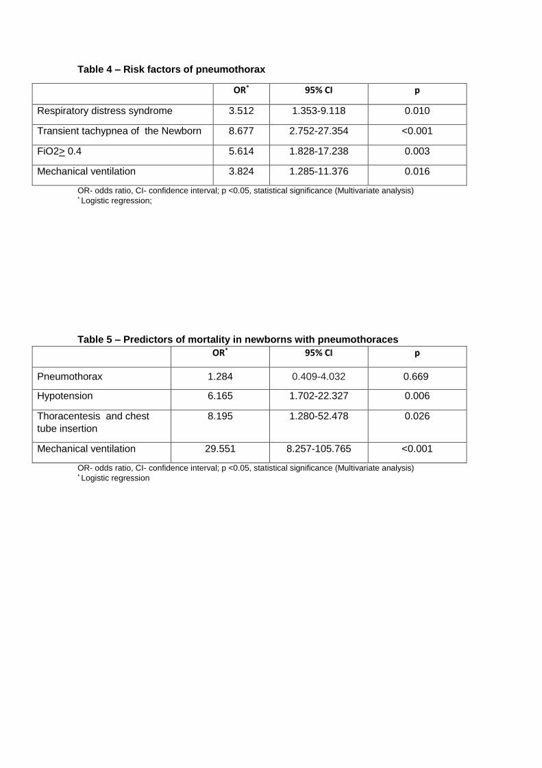
Table 4 – Risk factors of pneumothorax
OR* 95% CI p
Respiratory distress syndrome 3.512 1.353-9.118 0.010
Transient tachypnea of the Newborn 8.677 2.752-27.354 <0.001
FiO2> 0.4 5.614 1.828-17.238 0.003
Mechanical ventilation 3.824 1.285-11.376 0.016
OR- odds ratio, CI- confidence interval; p <0.05, statistical significance (Multivariate analysis) * Logistic regression;
Table 5 – Predictors of mortality in newborns with pneumothoraces
OR* 95% CI p
Pneumothorax 1.284 0.409-4.032 0.669
Hypotension 6.165 1.702-22.327 0.006
Thoracentesis and chest
tube insertion
8.195 1.280-52.478 0.026
Mechanical ventilation 29.551 8.257-105.765 <0.001
OR- odds ratio, CI- confidence interval; p <0.05, statistical significance (Multivariate analysis) * Logistic regression
Agradecimentos
À Professora Hercília Guimarães, pelo apoio incansável e orientação
fundamentais para a realização desta dissertação.
Ao Dr. Gustavo Rocha, co-orientador, pela orientação científica e
contribuição no desenvolvimento deste projeto.
À Dra. Filipa Flôr-de-Lima, pelo apoio imprescindível na realização da
análise estatística dos dados.
A todos os que me apoiaram ao longo deste projecto.
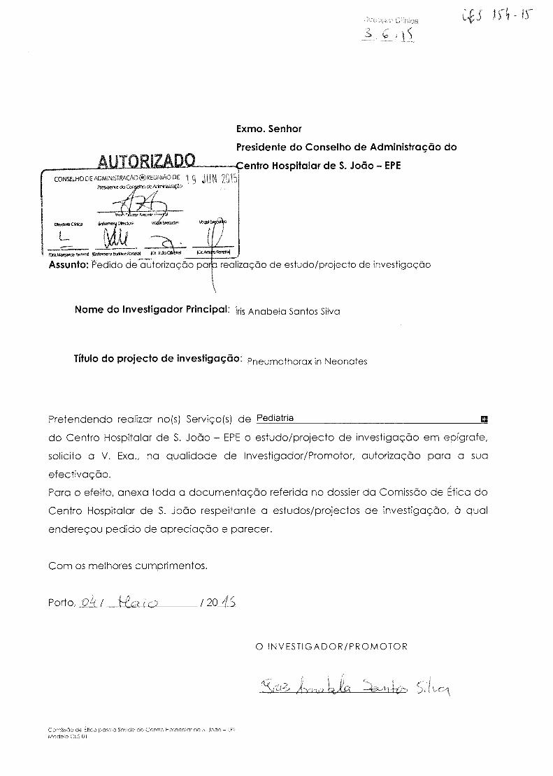
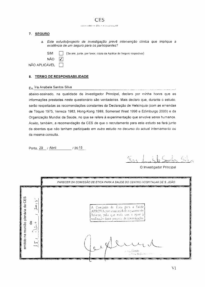
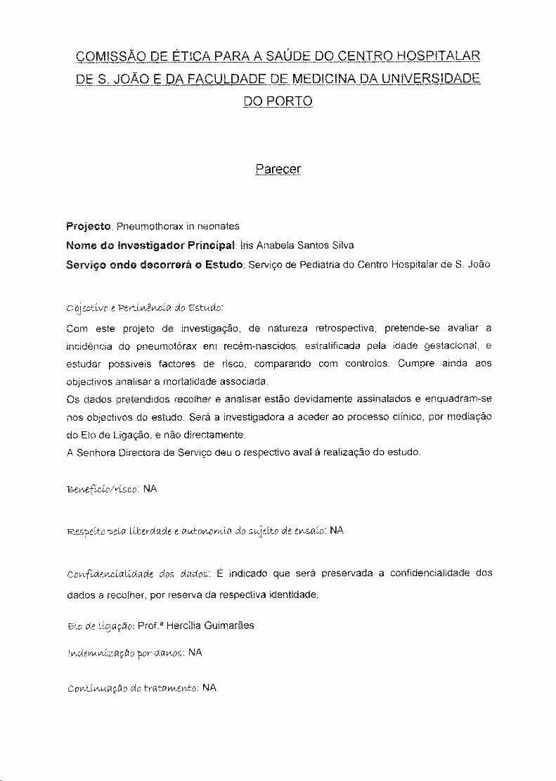
ANEXOS
Parecer da Comissão de Ética para a Saúde e
Autorização do Conselho de Administração do
Centro Hospitalar de S. João
Normas da revista
Journal of Pediatric and Neonatal Individualized Medicine
(JPNIM)
Author Guidelines
You can upload your manuscript files online (please note that, to submit articles to JPNIM's website, authors must first be registered as both readers and authors; if you experience
problems in submitting a manuscript after registering with the site, please read the step-by-step guide in this pdf) or email them to the Editor-in-Chief Vassilios Fanos ([email protected]).
Papers must be written in English or Italian and must be original (not published elsewhere in whole or in part).
Text
Please submit the text in a Word file.
Papers length should be generally around 20,000 characters.
All acronyms in the text should be expanded at first mention, followed by the abbreviation in parentheses.
Please use the following font: Times New Roman, 11 pt.
For paragraph formatting, please use single spacing and full justification.
Do not use boldface or underline character formatting; use italics just for technical terms or not English words. Use quotation marks just for quotations or to underline a particular word meaning.
Do not insert footnotes. Divide the text in paragraphs and assign a title to each part.
Abstract and keywords
For every article, authors should send:
an abstract of 250-300 words, and
6 keywords.
Abstract and keywords must be in English also for Italian articles.
Figures and tables
Figures (graphs, charts, photographs, and illustrations) and tables should be submitted separately from the text file:
for graphs and charts, use Excel files;
for photographs and illustrations, use JPEG, PNG or TIFF files (or, at least, PowerPoint);
for tables, use Excel or Word files.
Please only use the following fonts in figures: Helvetica or Arial.
Authors should quote figures and tables in the text and should number them in the order in which
they appear in the text. Each figure and table should be accompanied by a short description.
Figures and tables must be original.
Please note that editors and the publisher could evaluate the sent files overall and decide to modify the number of figures and tables.
Journal of Pediatric and Neonatal Individualized Medicine
(JPNIM)
Videos, audios and 3D illustrations
Starting from July 2014, we intend to accept also 3D illustrations, audios and videos (e.g., with slide presentations or demonstrations of clinical procedures) to integrate (as PDF-embedded multimedia) into each kind of article, and we are planning to start a new Video series, featuring the explanation of medical procedures. You are welcome to send your contributions for evaluation (see more here).
If you plan to send a video, please make sure to follow these guidelines:
file format: .mp4 (H.264 encoded);
dimensions: 645x360 or smaller.
Videos, audios and 3D illustrations must be original.
References
Please list the references in order of citation in the text, in square brackets.
For each entry, please clearly indicate the following data: names of all the authors, title of the article/book, publication year. Moreover, for journal articles, indicate journal title, volume, issue, first and last page of the article; for websites, indicate the last access; for books, indicate the book publisher and its head office. If you want to quote a chapter within a book, please add information on the chapter (title and authors). Examples:
article (see and follow Pub Med citations): Mishra J, Dent C, Tarabishi R, Mitsnefes MM, Ma Q,
Kelly C, Ruff SM, Zahedi K, Shao M, Bean J, Mori K, Barasch J, Devarajan P. Neutrophil gelatinase-associated lipocalin (NGAL) as a biomarker for acute renal injury after cardiac surgery. Lancet. 2005;365(9466):1231-8.
book: Cowan CP, Cowan PA. When partners become parents: the big life change for couples. New
York: Basic Books, 1992.
chapter within a book: Eyben E. Fathers and sons. In: Rawson B (Ed.). Marriage, divorce and children in ancient Rome. Oxford: Clarendon Press, 1991.
website: http://guidance.nice.org.uk/CG54, last access: April 2012.
Author Listing
For each author, please list the following items: name, surname, institutional affiliations.
To facilitate the publisher’s communication with authors, please list also the e-mail addresses.
Further requirements
Please indicate a conflict of interest statement and a funding acknowledgement statement.
In case of human experimentation, please indicate which ethical standards were followed and write an informed consent statement.
In experiments on animals, please indicate an animal rights statement.
Copyright agreement
Please note that before the article final publication the corresponding author will have to send a
signed copyright agreement, where he/she agree to transfer the copyright to the journal’s publisher. The copyright agreement can be read here.
Submission Preparation Checklist
As part of the submission process, authors are required to check off their submission's compliance with all of the following items, and submissions may be returned to authors that do not adhere to these guidelines.

1. ----- NB: Please note that, to submit articles to JPNIM's website, authors must first
be registered as both readers and authors; if you experience problems in submitting a manuscript after registering with the site, please read the step-by-step guide in this pdf. -----
2. I acknowledge that publishing in JPNIM is free of charge: no payments are required from the Authors.
3. I have read the Editorial Policies. 4. The submission has not been previously published, nor is it before another journal for
consideration (or an explanation has been provided in Comments to the Editor). 5. Authorship credit shall be based on 1) substantial contributions to conception and design,
acquisition of data, or analysis and interpretation of data; 2) drafting the article or revising it critically for important intellectual content; and 3) final approval of the version
to be published. Each and every person listed as an author meets conditions 1, 2 and 3. Acquisition of funding, collection of data, or general supervision of the research group alone does not constitute authorship.
6. All contributors who do not meet the criteria for authorship are to be listed in an Acknowledgments section at the end of the article. Financial and material support are also acknowledged.
7. No conflicts of interest exist or are clearly indicated at the end of the article. 8. The text adheres to the stylistic and bibliographic requirements outlined in the Author
Guidelines, which is found in About the Journal. 9. The text contains a conflict of interest statement and a funding acknowledgement
statement. In case of human experimentation, the text explains which ethical standards were followed and contains an informed consent statement. In experiments on animals,
the text contains an animal rights statement. 10. I authorise the processing of my personal information under the Italian Legislative Decree
n° 196/2003. 11. I acknowledge that before the article final publication I will have to send a signed
copyright agreement, where I agree to transfer the copyright to the journal’s publisher. The copyright agreement can be read here.