
Chap44 Diabetes insipidus and aquaporin (ADH, aldosterone and ACTH)
Plant Cell Physiol. 46(9): 1568–1577 (2005)
doi:10.1093/pcp/pci172, available online at www.pcp.oupjournals.org
JSPP © 2005
Identification of 33 Rice Aquaporin Genes and Analysis of Their Expression and Function
Junko Sakurai 1, 3, *, Fumiyoshi Ishikawa 2, Tomoya Yamaguchi 1, Matsuo Uemura 3 and
Masayoshi Maeshima 2
1 CO2 and Temperature Research Laboratory, National Agricultural Research Center for Tohoku Region, Morioka, 020-0198 Japan
2 Laboratory of Cell Dynamics, Graduate School of Bioagricultural Sciences, Nagoya University, Nagoya, 464-8601 Japan 3 United Graduate School of Agricultural Sciences and Cryobiosystem Research Center, Faculty of Agriculture, Iwate University, Morioka, 020-
8550 Japan
;
Plant aquaporins form a large protein family includ-
ing plasma membrane-type (PIPs) and tonoplast-type
aquaporins (TIPs), and facilitate osmotic water transport
across membranes as a key physiological function. We iden-
tified 33 genes for aquaporins in the genome sequence of
rice (Oryza sativa L. cv. Nipponbare). We investigated their
expression levels in leaf blades, roots and anthers of rice
(cv. Akitakomachi) using semi-quantitative reverse tran-
scription–PCR (RT–PCR). At both early tillering (21 d
after germination) and panicle formation (56 d) stages, six
genes, including OsPIP2;4 and OsPIP2;5, were expressed
predominantly in roots, while 14 genes, including OsPIP2;7
and OsTIP1;2, were found in leaf blades. Eight genes, such
as OsPIP1;1 and OsTIP4;1, were evenly expressed in leaf
blades, roots and anthers. Analysis by stopped-flow spec-
trophotometry revealed high water channel activity when
OsPIP2;4 or OsPIP2;5 were expressed in yeast but not
when OsPIP1;1 or OsPIP1;2 were expressed. Furthermore,
the mRNA levels of OsPIP2;4 and OsPIP2;5 showed a clear
diurnal fluctuation in roots; they showed a peak 3 h after
the onset of light and dropped to a minimum 3 h after the
onset of darkness. The mRNA levels of 10 genes including
OsPIP2;4 and OsPIP2;5 markedly decreased in roots dur-
ing chilling treatment and recovered after warming. The
changes in mRNA levels during and after the chilling treat-
ment were comparable with that of the bleeding sap vol-
ume. These results suggested the relationship between the
root water uptake and mRNA levels of several aquaporins
with high water channel activity, such as OsPIP2;4 and
OsPIP2;5.
Keywords: Aquaporin — Gene expression — Rice — Water
permeability.
Abbreviations: NIP, Nod26-like intrinsic protein; PIP, plasma
membrane intrinsic protein; RT–PCR, reverse transcription–PCR; SIP,
small and basic intrinsic protein; TIP, tonoplast intrinsic protein.
Introduction
Aquaporins are membrane proteins that facilitate water
transport across the membranes in various microorganisms,
animals and plants (King et al. 2004). Intensive research
has revealed the involvement of aquaporins in plant growth
and water relationships (see reviews by Maurel et al. 2002,
Tyerman et al. 2002). Plant aquaporins are distributed in vari-
ous plant tissues relating to water transport, cell differentiation
and enlargement. Some aquaporins were also expressed in
motor cells (Moshelion et al. 2002), guard cells (Kaldenhoff et
al. 1995, Sarda et al. 1997) and reproductive organs (Dixit et al.
2001, O’Brien et al. 2002, Bots et al. 2005a, Bots et al. 2005b).
Studies using mutant lines of aquaporins clearly demonstrated
the involvement of aquaporins in plant–water relationships; for
example, Ma et al. (2004) showed that reduction of TIP1;1
expression levels in Arabidopsis thaliana using the RNA inter-
ference method caused death of the plant, in the case of serious
damage. In addition, gene expression and protein accumula-
tion of aquaporins in response to various environmental
stresses and hormones have been reported in several plants
(Maurel et al. 2002, Tyerman et al. 2002). Furthermore, it has
been reported recently that some types of plant aquaporin could
transport not only water but also various small molecules such
as glycerol, urea (Maurel et al. 2002, Gaspar et al. 2003, Liu et
al. 2003), ammonia (Niemietz and Tyerman 2000, Loqué et al.
2005) and CO2 (Uehlein et al. 2003, Hanba et al. 2004). These
studies are developing our knowledge on the physiological
meanings of aquaporins in plants.
Recent genome sequencing projects have revealed that
aquaporins constitute a large gene family in plants; A. thaliana
and Zea mays have 35 and 33 genes, respectively (Chaumont et
al. 2001, Johanson et al. 2001). These aquaporins have been
classified into four major subfamilies referred to as plasma
membrane intrinsic proteins (PIPs), tonoplast intrinsic proteins
(TIPs), Nod26-like intrinsic proteins (NIPs) and small and
basic intrinsic proteins (SIPs). Although recent studies pro-
vided information on the physiological function of several of
those aquaporin members, the number of aquaporins examined
in detail is still limited.
* Corresponding author: E-mail, [email protected]; Fax, +81-19-641-7794.
1568
Expression and function of rice 33 aquaporin 1569
We have been conducting a series of experiments to eluci-
date the physiological relevance of aquaporins in rice, which is
an important major crop. Although several aquaporin genes
have already been identified and characterized in rice (Liu et al.
1994, Malz and Sauter 1999, Li et al. 2000, Lian et al. 2004,
Takahashi et al. 2004), a complete set of rice aquaporins is
unclear. In the present study, we identified 33 genes for
aquaporins in the rice genome sequence and investigated their
expression profiles in rice plant organs. We also studied the
expression profile in roots during a light/dark cycle and chill-
ing treatment. Furthermore, we expressed typical members of
rice PIPs in yeast cells and determined the water channel activ-
ity of the membrane accumulating these aquaporins by
stopped-flow spectrophotometry. From these results, we dis-
cuss the function of aquaporins in rice roots.
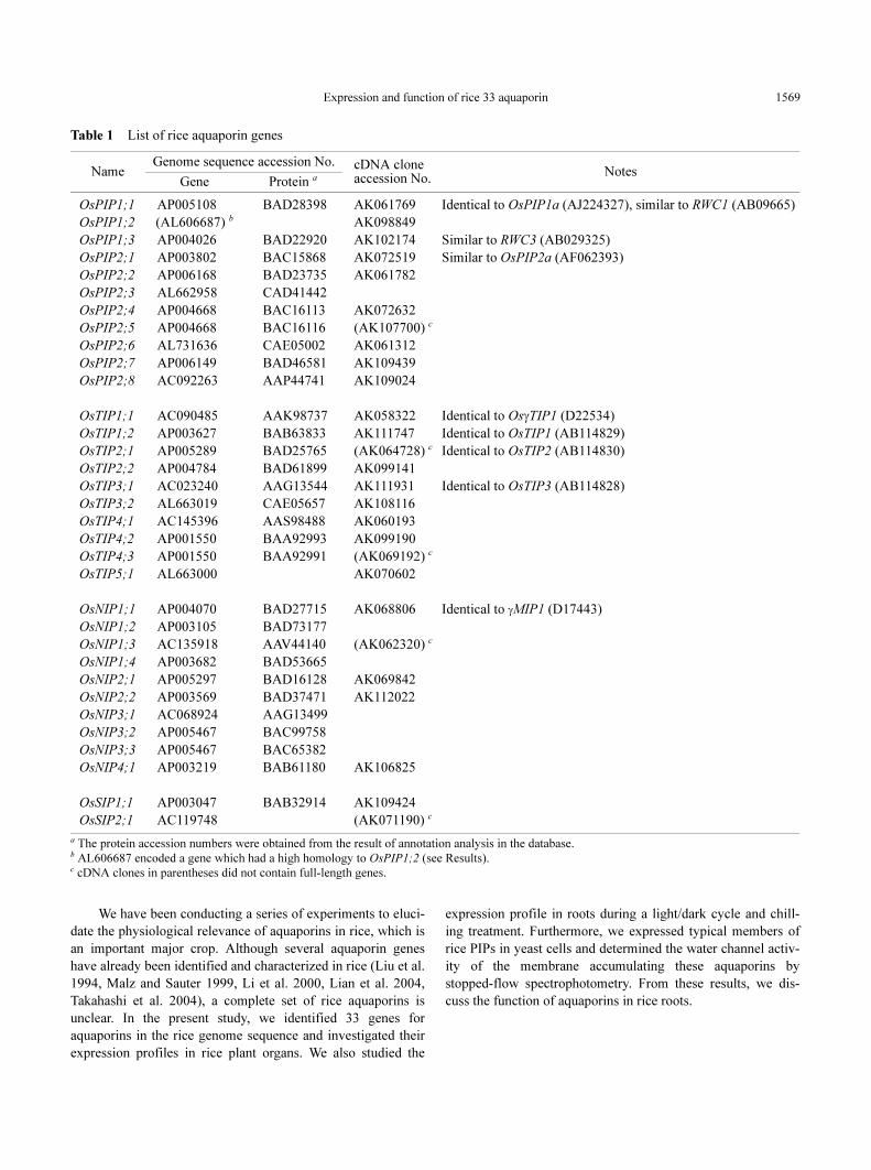
Table 1 List of rice aquaporin genes
a The protein accession numbers were obtained from the result of annotation analysis in the database.b AL606687 encoded a gene which had a high homology to OsPIP1;2 (see Results).c cDNA clones in parentheses did not contain full-length genes.
NameGenome sequence accession No. cDNA clone
accession No.Notes
Gene Protein a
OsPIP1;1 AP005108 BAD28398 AK061769 Identical to OsPIP1a (AJ224327), similar to RWC1 (AB09665)
OsPIP1;2 (AL606687) b AK098849
OsPIP1;3 AP004026 BAD22920 AK102174 Similar to RWC3 (AB029325)
OsPIP2;1 AP003802 BAC15868 AK072519 Similar to OsPIP2a (AF062393)
OsPIP2;2 AP006168 BAD23735 AK061782
OsPIP2;3 AL662958 CAD41442
OsPIP2;4 AP004668 BAC16113 AK072632
OsPIP2;5 AP004668 BAC16116 (AK107700) c
OsPIP2;6 AL731636 CAE05002 AK061312
OsPIP2;7 AP006149 BAD46581 AK109439
OsPIP2;8 AC092263 AAP44741 AK109024
OsTIP1;1 AC090485 AAK98737 AK058322 Identical to OsγTIP1 (D22534)
OsTIP1;2 AP003627 BAB63833 AK111747 Identical to OsTIP1 (AB114829)
OsTIP2;1 AP005289 BAD25765 (AK064728) c Identical to OsTIP2 (AB114830)
OsTIP2;2 AP004784 BAD61899 AK099141
OsTIP3;1 AC023240 AAG13544 AK111931 Identical to OsTIP3 (AB114828)
OsTIP3;2 AL663019 CAE05657 AK108116
OsTIP4;1 AC145396 AAS98488 AK060193
OsTIP4;2 AP001550 BAA92993 AK099190
OsTIP4;3 AP001550 BAA92991 (AK069192) c
OsTIP5;1 AL663000 AK070602
OsNIP1;1 AP004070 BAD27715 AK068806 Identical to γMIP1 (D17443)
OsNIP1;2 AP003105 BAD73177
OsNIP1;3 AC135918 AAV44140 (AK062320) c
OsNIP1;4 AP003682 BAD53665
OsNIP2;1 AP005297 BAD16128 AK069842
OsNIP2;2 AP003569 BAD37471 AK112022
OsNIP3;1 AC068924 AAG13499
OsNIP3;2 AP005467 BAC99758
OsNIP3;3 AP005467 BAC65382
OsNIP4;1 AP003219 BAB61180 AK106825
OsSIP1;1 AP003047 BAB32914 AK109424
OsSIP2;1 AC119748 (AK071190) c
Expression and function of rice 33 aquaporin1570
Results
Identification and nomenclature of rice aquaporin genes
We surveyed the genes for aquaporins in the genome data-
base of rice (cv. Nipponbare). These genes were selected from
a large number of the candidate sequences including both
genome and cDNA sequences. The overlapping sequences
were removed and finally 33 genes were selected, as shown in
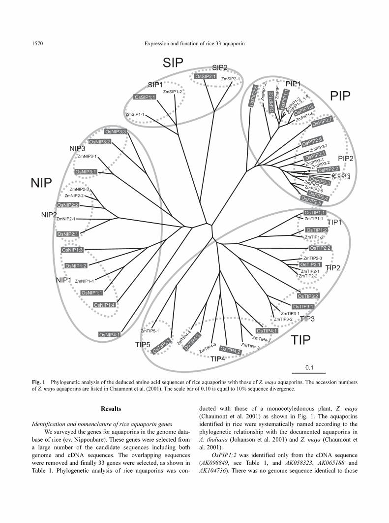
Table 1. Phylogenetic analysis of rice aquaporins was con-
ducted with those of a monocotyledonous plant, Z. mays
(Chaumont et al. 2001) as shown in Fig. 1. The aquaporins
identified in rice were systematically named according to the
phylogenetic relationship with the documented aquaporins in
A. thaliana (Johanson et al. 2001) and Z. mays (Chaumont et
al. 2001).
OsPIP1;2 was identified only from the cDNA sequence
(AK098849, see Table 1, and AK058323, AK065188 and
AK104736). There was no genome sequence identical to those
Fig. 1 Phylogenetic analysis of the deduced amino acid sequences of rice aquaporins with those of Z. mays aquaporins. The accession numbers
of Z. mays aquaporins are listed in Chaumont et al. (2001). The scale bar of 0.10 is equal to 10% sequence divergence.
Expression and function of rice 33 aquaporin 1571
cDNAs. However, the genomic DNA AL606687 contained a
fairly similar sequence; it had a single base insertion and a few
base substitutions when compared with AK098849. Thus, we
named AL606687 and AK098849 as the same aquaporin gene
OsPIP1;2.
Although several other genes encode the partial sequences
of aquaporins (data not shown), they were considered to be
non-functional pseudo-genes. This is mainly because they
lacked some parts of characteristic sequences conserved in the
aquaporin family, such as a transmembrane domain, when we
analyzed their transmembrane topologies (data not shown).
Phylogenetic analysis of rice aquaporins
We classified the rice aquaporins into four subfamilies as
same as those of Z. mays (Chaumont et al. 2001) and A.
thaliana (Johanson et al. 2001) (Fig. 1). Rice had 11 PIPs, 10
TIPs, 10 NIPs and two SIPs. The OsPIP members had a rela-
tively high sequence similarity of 58.5–92.7%, while the mem-
bers of the OsTIP, OsNIP and OsSIP subfamilies had a
somewhat variable sequence similarity. For example, the se-
quence similarity among the OsTIP members was 35.8–72.0%.
The OsPIP subfamily was divided into two groups, PIP1
and PIP2 (Fig. 1). OsPIP2;8 had a unique sequence and formed
a long branch in the tree. However, we provisionally assigned
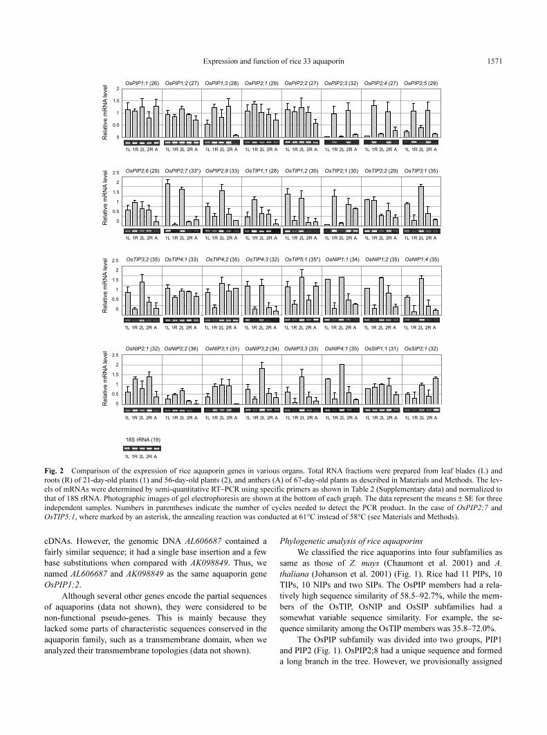
Fig. 2 Comparison of the expression of rice aquaporin genes in various organs. Total RNA fractions were prepared from leaf blades (L) and
roots (R) of 21-day-old plants (1) and 56-day-old plants (2), and anthers (A) of 67-day-old plants as described in Materials and Methods. The lev-
els of mRNAs were determined by semi-quantitative RT–PCR using specific primers as shown in Table 2 (Supplementary data) and normalized to
that of 18S rRNA. Photographic images of gel electrophoresis are shown at the bottom of each graph. The data represent the means ± SE for three
independent samples. Numbers in parentheses indicate the number of cycles needed to detect the PCR product. In the case of OsPIP2;7 and
OsTIP5;1, where marked by an asterisk, the annealing reaction was conducted at 61°C instead of 58°C (see Materials and Methods).
Expression and function of rice 33 aquaporin1572
OsPIP2;8 as a member of the PIP2 group because its sequence
was similar to that of OsPIP2 members but not OsPIP1
members.
The OsTIP subfamily consisted of five groups, OsTIP1,
OsTIP2, OsTIP3, OsTIP4 and OsTIP5, which had two, two,
two, three and one member, respectively. The OsNIP sub-
family consisted of four groups, OsNIP1, OsNIP2, OsNIP3 and
OsNIP4, which had four, two, three and one member, respec-
tively. The OsSIP subfamily had only two members and their
sequence similarity is low (33%).
Organ-specific expression of rice aquaporin genes
The mRNA levels of 32 aquaporin genes were quantified
by reverse transcription–PCR (RT–PCR) and compared in
some organs (Fig. 2). The expression profiles varied with
aquaporins. The mRNA levels of OsPIP1;3, OsPIP2;3,
OsPIP2;4, OsPIP2;5, OsTIP2;1 and OsNIP2;1 were higher in
roots than in leaf blades. In particular, OsPIP2;3, OsPIP2;4
and OsPIP2;5 were expressed predominantly in roots at both
early tillering and panicle formation stages. In contrast, higher
levels of the mRNA of 14 aquaporin genes (OsPIP2;7,
OsPIP2;8, OsTIP1;2, OsTIP3;1, OsTIP3;2, OsTIP4;2,
OsTIP4;3, OsTIP5;1, OsNIP1;1, OsNIP1;2, OsNIP1;4,
OsNIP3;2, OsNIP3;3 and OsNIP4;1) were detected in leaf
blades than in roots. Several genes (OsPIP1;1, OsPIP1;2,
OsPIP2;1, OsPIP2;2, OsPIP2;6, OsTIP2;2, OsTIP4;1 and
OsSIP1;1) were expressed almost equally in both roots and
leaf blades.
The expression profile of some genes, such as OsTIP1;1,
OsNIP2;2, OsNIP3;1 and OsSIP2;1, differed with the stages,
i.e. early tillering or panicle formation. For example, OsTIP1;1
was highly expressed in roots at the early tillering stage; how-
ever, the level decreased to 43% at the panicle formation stage.
Interestingly, the relative mRNA levels of OsPIP1;1, OsTIP4;
2, OsTIP5;1 and OsSIP2;1 were also abundant in anthers in the
heading stage.
Osmotic water permeabilities of OsPIP1;1, OsPIP1;2, OsPIP2;
4 and OsPIP2;5
To measure the osmotic water transport activity of rice
aquaporins by the yeast expression system, we expressed them
in Saccharomyces cerevisiae strain BJ5458, which lacks vacu-
olar proteases, to avoid degradation of translation products. It
has been confirmed that this strain lacks functional aquaporins
(Suga and Maeshima 2004).
Rice aquaporins OsPIP1;1, OsPIP1;2, OsPIP2;4 and
OsPIP2;5 were tagged with a c-myc epitope at the N-termini
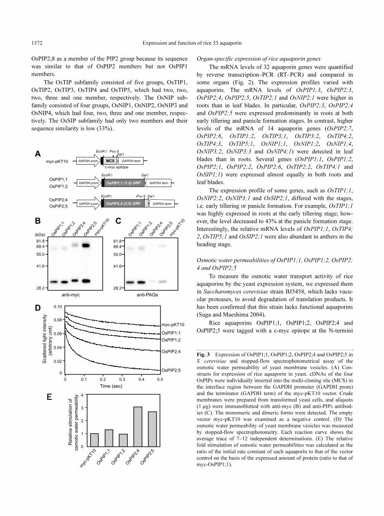
Fig. 3 Expression of OsPIP1;1, OsPIP1;2, OsPIP2;4 and OsPIP2;5 in
S. cerevisiae and stopped-flow spectrophotometrical assay of the
osmotic water permeability of yeast membrane vesicles. (A) Con-
structs for expression of rice aquaporin in yeast. cDNAs of the four
OsPIPs were individually inserted into the multi-cloning site (MCS) in
the interface region between the GAPDH promoter (GAPDH prom)
and the terminator (GAPDH term) of the myc-pKT10 vector. Crude
membranes were prepared from transformed yeast cells, and aliquots
(1 µg) were immunoblotted with anti-myc (B) and anti-PIPs antibod-
ies (C). The monomeric and dimeric forms were detected. The empty
vector myc-pKT10 was examined as a negative control. (D) The
osmotic water permeability of yeast membrane vesicles was measured
by stopped-flow spectrophotometry. Each reaction curve shows the
average trace of 7–12 independent determinations. (E) The relative
fold stimulation of osmotic water permeabilities was calculated as the
ratio of the initial rate constant of each aquaporin to that of the vector
control on the basis of the expressed amount of protein (ratio to that of
myc-OsPIP1;1).
Expression and function of rice 33 aquaporin 1573
(Fig. 3A). This c-myc epitope has been reported to have no
effect on water channel activity of recombinant aquaporin
(Suga and Maeshima 2004). Protein accumulation in the yeast
membranes was detected at the calculated molecular sizes of
monomers (31–32 kDa) and dimers (63–69 kDa) by immunob-
loting with an anti-myc antibody (Fig. 3B). The expressed rice
aquaporins in yeast were also detected by the anti-PAQs anti-
body, which recognizes most PIP members in radish (Ohshima
et al. 2001) (Fig. 3C). This antibody showed only qualitative
information, because the immunoreactivity of the antibody
depends on the amino acid sequences of aquaporin antigens.
No immunostained band was observed in the membranes pre-
pared from yeast cells expressing the vacant vector.
The osmotic water permeability of membrane vesicles
was measured with a stopped-flow light scattering spectro-
photometer. The swelling rate of vesicles in the hypotonic solu-
tion was monitored as a decrease in the scattered light intensity
(Fig. 3D). The vacant vector showed a slow influx of water into
the membrane vesicles and we thought that this was the basal
activity of the yeast membrane preparation. Fig. 3E shows the
relative stimulation of osmotic water permeability of yeast
membranes on the basis of the expressed amount of aquaporin.
Considering the protein expression levels, the myc-tagged
OsPIP1;1 and OsPIP1;2 did not stimulate water permeability
much compared with myc-OsPIP2;4 and OsPIP2;5.
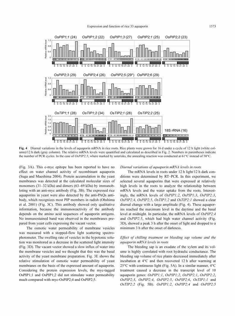
Diurnal variations of aquaporin mRNA levels in roots
The mRNA levels in roots under 12 h light/12 h dark con-
ditions were determined by RT–PCR. In this experiment, we
selected several aquaporins that were expressed at relatively
high levels in the roots to analyze the relationship between
mRNA levels and the water uptake from the roots. Interest-
ingly, the mRNA levels of OsPIP1;2, OsPIP1;3, OsPIP2;3,
OsPIP2;4, OsPIP2;5, OsTIP1;2 and OsTIP2;1 showed a clear
diurnal change with a large amplitude (Fig. 4). These aquapor-
ins reached the maximum level in the daytime and the basal
level at midnight. In particular, the mRNA levels of OsPIP2;4
and OsPIP2;5, which had high water channel activity (Fig.
3D), showed a peak 3 h after the onset of light and dropped to a
minimum 3 h after the onset of darkness.
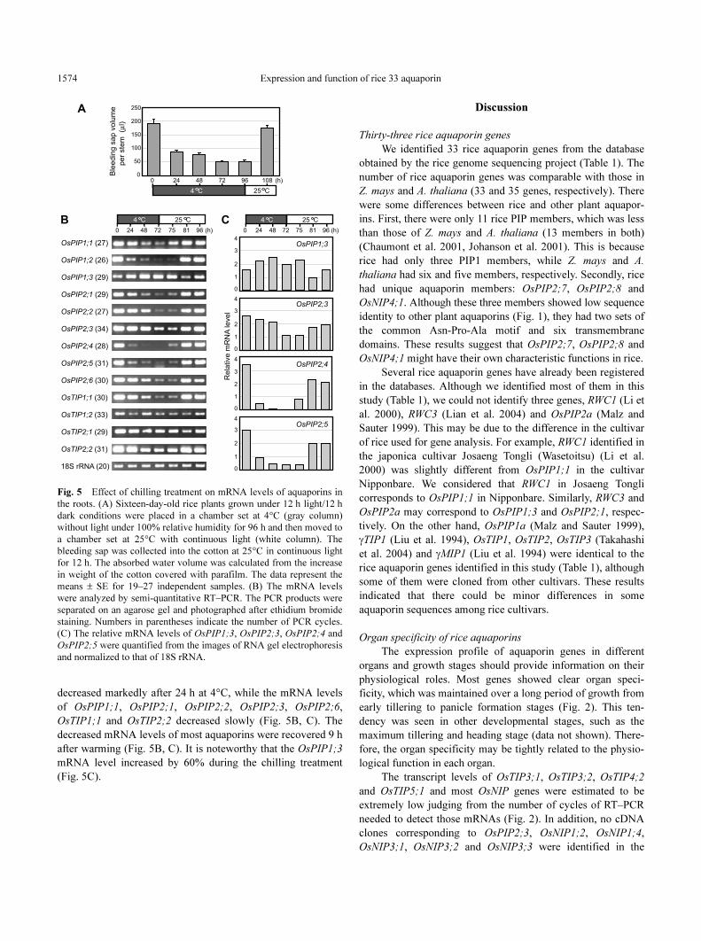
Effect of chilling treatment on bleeding sap volume and the
aquaporin mRNA levels in roots
The bleeding sap is an exudate of the xylem and its vol-
ume is highly correlated with root hydraulic conductance. The
bleeding sap volume of rice plants decreased immediately after
incubation at 4°C and then recovered 12 h after warming at
25°C with continuous light (Fig. 5A). In a similar manner, 4°C
treatment caused a decrease in the transcript level of 10
aquaporin genes: OsPIP1;1, OsPIP1;2, OsPIP2;1, OsPIP2;2,
OsPIP2;3, OsPIP2;4, OsPIP2;5, OsPIP2;6, OsTIP1;1 and
OsTIP2;2 (Fig. 5B). OsPIP1;2, OsPIP2;4 and OsPIP2;5
Fig. 4 Diurnal variations in the levels of aquaporin mRNA in rice roots. Rice plants were grown for 16 d under a cycle of 12 h light (white col-
umn)/12 h dark (gray column). The relative mRNA levels were quantified and calculated as described in Fig. 2. Numbers in parentheses indicate
the number of PCR cycles. In the case of OsPIP2;5, where marked by asterisks, the annealing reaction was conducted at 61°C instead of 58°C.
Expression and function of rice 33 aquaporin1574
decreased markedly after 24 h at 4°C, while the mRNA levels
of OsPIP1;1, OsPIP2;1, OsPIP2;2, OsPIP2;3, OsPIP2;6,
OsTIP1;1 and OsTIP2;2 decreased slowly (Fig. 5B, C). The
decreased mRNA levels of most aquaporins were recovered 9 h
after warming (Fig. 5B, C). It is noteworthy that the OsPIP1;3
mRNA level increased by 60% during the chilling treatment
(Fig. 5C).
Discussion
Thirty-three rice aquaporin genes
We identified 33 rice aquaporin genes from the database
obtained by the rice genome sequencing project (Table 1). The
number of rice aquaporin genes was comparable with those in
Z. mays and A. thaliana (33 and 35 genes, respectively). There
were some differences between rice and other plant aquapor-
ins. First, there were only 11 rice PIP members, which was less
than those of Z. mays and A. thaliana (13 members in both)
(Chaumont et al. 2001, Johanson et al. 2001). This is because
rice had only three PIP1 members, while Z. mays and A.
thaliana had six and five members, respectively. Secondly, rice
had unique aquaporin members: OsPIP2;7, OsPIP2;8 and
OsNIP4;1. Although these three members showed low sequence
identity to other plant aquaporins (Fig. 1), they had two sets of
the common Asn-Pro-Ala motif and six transmembrane
domains. These results suggest that OsPIP2;7, OsPIP2;8 and
OsNIP4;1 might have their own characteristic functions in rice.
Several rice aquaporin genes have already been registered
in the databases. Although we identified most of them in this
study (Table 1), we could not identify three genes, RWC1 (Li et
al. 2000), RWC3 (Lian et al. 2004) and OsPIP2a (Malz and
Sauter 1999). This may be due to the difference in the cultivar
of rice used for gene analysis. For example, RWC1 identified in
the japonica cultivar Josaeng Tongli (Wasetoitsu) (Li et al.
2000) was slightly different from OsPIP1;1 in the cultivar
Nipponbare. We considered that RWC1 in Josaeng Tongli
corresponds to OsPIP1;1 in Nipponbare. Similarly, RWC3 and
OsPIP2a may correspond to OsPIP1;3 and OsPIP2;1, respec-
tively. On the other hand, OsPIP1a (Malz and Sauter 1999),
γTIP1 (Liu et al. 1994), OsTIP1, OsTIP2, OsTIP3 (Takahashi
et al. 2004) and γMIP1 (Liu et al. 1994) were identical to the
rice aquaporin genes identified in this study (Table 1), although
some of them were cloned from other cultivars. These results
indicated that there could be minor differences in some
aquaporin sequences among rice cultivars.
Organ specificity of rice aquaporins
The expression profile of aquaporin genes in different
organs and growth stages should provide information on their
physiological roles. Most genes showed clear organ speci-
ficity, which was maintained over a long period of growth from
early tillering to panicle formation stages (Fig. 2). This ten-
dency was seen in other developmental stages, such as the
maximum tillering and heading stage (data not shown). There-
fore, the organ specificity may be tightly related to the physio-
logical function in each organ.
The transcript levels of OsTIP3;1, OsTIP3;2, OsTIP4;2
and OsTIP5;1 and most OsNIP genes were estimated to be
extremely low judging from the number of cycles of RT–PCR
needed to detect those mRNAs (Fig. 2). In addition, no cDNA
clones corresponding to OsPIP2;3, OsNIP1;2, OsNIP1;4,
OsNIP3;1, OsNIP3;2 and OsNIP3;3 were identified in the
Fig. 5 Effect of chilling treatment on mRNA levels of aquaporins in
the roots. (A) Sixteen-day-old rice plants grown under 12 h light/12 h
dark conditions were placed in a chamber set at 4°C (gray column)
without light under 100% relative humidity for 96 h and then moved to
a chamber set at 25°C with continuous light (white column). The
bleeding sap was collected into the cotton at 25°C in continuous light
for 12 h. The absorbed water volume was calculated from the increase
in weight of the cotton covered with parafilm. The data represent the
means ± SE for 19–27 independent samples. (B) The mRNA levels
were analyzed by semi-quantitative RT–PCR. The PCR products were
separated on an agarose gel and photographed after ethidium bromide
staining. Numbers in parentheses indicate the number of PCR cycles.
(C) The relative mRNA levels of OsPIP1;3, OsPIP2;3, OsPIP2;4 and
OsPIP2;5 were quantified from the images of RNA gel electrophoresis
and normalized to that of 18S rRNA.
Expression and function of rice 33 aquaporin 1575
database (Table 1). These results indicate that the expression of
these genes might be extremely low or limited to specific tis-
sues in rice plants.
OsPIP2;4 and OsPIP2;5 have high water channel activity
By the yeast heterologous expression system and stopped-
flow spectrophotometric analysis, we found that OsPIP2;4 and
OsPIP2;5 had significant osmotic water channel activity (Fig.
3E). In contrast, OsPIP1;1 and OsPIP1;2 showed no signifi-
cant activity. Judging from the high expression of OsPIP2;4
and OsPIP2;5 in roots (Fig. 2), both OsPIP2 members might
play a crucial role as water channels in roots.
The low and high water channel activities in PIP1 and
PIP2 members, respectively, were reported for other plants
(Chaumont et al. 2000, Moshelion et al. 2002, Fetter et al.
2004, Suga and Maeshima 2004). Suga and Maeshima (2004)
demonstrated that the valine residue in loop E of radish PIP2
members is essential for the water channel activity. Radish
PIP1 members have an isoleucine residue at the corresponding
site. Similarly, isoleucine and valine residues were conserved
in all members of the OsPIP1 and OsPIP2 group, respectively,
except for OsPIP2;8. We should examine whether the mem-
bers of OsPIP1 group facilitate the transport of other substrates
and whether their water channel function is regulated by post-
translational modification.
Response to chilling treatment and diurnal variation of rice
aquaporins
One of the aims of the present work is to examine the
involvement of rice aquaporins in water uptake from the roots
under low temperature conditions. Therefore, we analyzed the
relationship between bleeding sap volume, which is highly cor-
related with root hydraulic conductivity, and mRNA levels of
rice aquaporins during chilling treatment. The change in the
bleeding sap volume during and after chilling treatment was
closely correlated with changes in the expression of aquaporin
genes, especially genes for functional water channels, such as
OsPIP2;4 and OsPIP2;5 (Fig. 5A, B, C). The down-regulation
of PIP gene expression in roots was also reported for Z. mays
(Aroca et al. 2005) and for rice (Li et al. 2000). On the other
hand, Aroca et al. (2005) reported that the protein levels of Z.
mays PIP members increased during chilling treatment. There-
fore, further studies, such as analysis at the protein level,
should be conducted to understand the physiological function
of OsPIP2;4 and OsPIP2,5 in water uptake during chilling
treatment.
We also found a diurnal variation in the mRNA levels of
rice aquaporin in roots. OsPIP2;4 and OsPIP2;5 mRNA levels
in roots varied diurnally with a large amplitude (Fig. 4). The
diurnal changes in the mRNAs and proteins have also been
reported for aquaporins of other plants such as Lotus japonicus
(Henzler et al. 1999), Hordeum vulgare (Katsuhara et al. 2003)
and Z. mays (Lopez et al. 2003, Lopez et al. 2004). Lopez et al.
(2003) revealed that the protein levels of Z. mays ZmPIP2
members, but not ZmPIP1s, in roots were correlated closely
with the diurnal variation in root water flux. ZmPIP2;1 and
ZmPIP2;5 showed high water channel activities (Lopez et al.
2003), while ZmPIP1 members had low water channel activi-
ties (Chaumont et al. 2000, Gaspar et al. 2003). Therefore,
Lopez et al. (2003) concluded that ZmPIP2 members might
contribute to diurnal water transport in roots. These observa-
tions in conjunction with the present results underline the
importance of members of the PIP2 group for diurnal water
movement in plant roots.
Materials and Methods
Identification and phylogenetic analysis of rice aquaporin genes
Aquaporin genes from the genome sequence (O. sativa L. cv.
Nipponbare) were identified by BLAST searches on the NCBI data-
base (http://www.ncbi.nlm.nih.gov/BLAST/) and the Rice Genome
Research Program (RGP; http://rgp.dna.affrc.go.jp/) based on the
sequence similarity with aquaporins of A. thaliana (Johanson et al.
2001) and Z. mays (Chaumont et al. 2001). The phylogenetic analysis
was conducted for their deduced amino acid sequences using the
Clustal W program (Thompson et al. 1994) and the results were dis-
played using the TreeView program (Page 1996). The transmembrane
topology of the rice aquaporins was predicted by the ConPred II
method (Arai et al. 2004, http://bioinfo.si.hirosaki-u.ac.jp/~ConPred2/).
Plant materials and growth conditions
Rice (cv. Akitakomachi) seeds were germinated in the dark for
3 d at 25°C and grown in a growth chamber under 12 h light/12 h dark
(light period; 450 µmol s–1 m–2) and at day/night temperatures of 25/
20°C at a relative humidity of 75%. Plants were grown in tap water for
the first 5 d, and then were supplied continuously with fresh culture
solution at a slow rate. The culture solution contained 10 ppm nitro-
gen (NH4NO
3), phosphorus (NaH
2PO
4), potassium (K
2SO
4), calcium
(CaCl2), magnesium (MgSO
4), 0.4 ppm iron (Fe(III)-EDTA) and
0.1 ppm manganese (MnCl2), pH 5.0. Leaf blades and roots were har-
vested from 21-day-old plants (early tillering stage) and 56-day-old
plants (panicle formation stage). Leaf blades were harvested from all
tillers of 21-day-old plants and from the upper three leaves of 56-day-
old plants. Roots were collected from the apical half of the roots.
Anthers were harvested from 67-day-old plants (heading stage, a few
days before anthesis). For analysis of diurnal change in the levels of
aquaporin mRNAs, total RNA fractions were obtained from 16-day-
old plants.
Chilling treatment of plants and measurements of bleeding sap volume
Sixteen-day-old rice plants cultivated under 12 h light/12 h dark
conditions were placed in a chamber set at 4°C without light under
100% relative humidity for 96 h to avoid the stresses of drought and
light. Then plants were moved to a chamber set at 25°C with continu-
ous light. The bleeding sap was collected into the cotton for 12 h from
the stem cut off 3–4 cm above the soil surface at 25°C in continuous
light to avoid the effect of diurnal variation of water uptake from roots.
The bleeding sap volume was calculated from the increase in the
weight of the cotton covered with parafilm. For analysis of aquaporin
mRNAs, plants were chilled for 72 h and then moved to the continu-
ous light chamber at 25°C.
RNA extraction and semi-quantitative RT–PCR
Tissues of rice plants were frozen in liquid nitrogen and ground
in a mortar with a pestle. RNA was extracted from frozen powder of
Expression and function of rice 33 aquaporin1576
the tissue with the RNeasy Plant Mini kit (Qiagen K.K., Tokyo,
Japan). For RT–PCR, the first strand cDNA was synthesized using
ReverTra Ace (Toyobo Co., Ltd., Osaka, Japan). PCR was performed
using AmpliTaq GOLD (ABI, Foster City, CA, USA) and the primers
listed in Table 2 (Supplementary data). All primers were designed
based on the sequences of the 3′-untranslated regions in each
aquaporin gene and to have similar Tm
values (59.3 ± 1.2°C). The PCR
conditions used were 94°C for 30 s, 58°C for 30 s and 72°C for 1 min.
In the case of some aquaporins, the annealing temperature was set at
61°C to prevent the amplification of non-specific PCR products. The
reaction was repeated for 16–36 cycles to obtain an appropriate
amount of DNA. The conditions and cycle numbers were determined
to avoid the saturation of DNA amplification. The obtained DNA was
subjected to agarose gel electrophoresis and stained with ethidium bro-
mide. The signal intensity of the stained bands was photographed by a
charge-coupled device (CCD) camera and analyzed by the NIH Image
program (http://rsb.info.nih.gov/nih-image). The fact that there was no
contamination of genomic DNA in the cDNA samples was confirmed
by PCR using the primer sets listed in Table 2 (Supplementary data).
Expression of rice aquaporin genes in yeast
EcoRI–SalI or EcoRI–PvuII fragments of rice aquaporin cDNA
(OsPIP1;1 and OsPIP1;2, EcoRI–SalI; OsPIP2;4 and OsPIP2;5,
EcoRI–PvuII) were amplified by RT–PCR with gene-specific primers
(Table 2, Supplementary data) and LA Taq (TAKARA SHUZO Co.,
LTD., Kyoto, Japan) using total RNA of the rice roots as a template.
Reverse primers included an additional 3′-non-coding region (8–17
bases), because the open reading frames of rice aquaporins were quite
similar to each other. The obtained fragments were inserted into the
yeast expression vector myc-pKT10 (Tanaka et al. 1990, Suga and
Maeshima 2004) (see Fig. 3A). This vector includes a c-myc epitope
sequence at the down-stream region of the GAPDH promoter. After
confirming the DNA sequences, the obtained plasmid was introduced
into S. cerevisiae strain BJ5458, which is deficient in major vacuolar
proteinases and functional aquaporins (Suga and Maeshima 2004).
Transformed yeast, which was selected using URA3 (orotidine-
5′-phosphate decarboxylase), was grown in AHCW/Glc plates [0.17%
yeast nitrogen base without amino acid, 0.5% ammonium sulfate
(Difco), 1% casamino acid, 0.002% adenine sulfate, 0.002%
tryptophan, 50 mM potassium phosphate, pH 5.5, 2% glucose and 2%
agar] as described previously (Nakanishi et al. 2001, Suga and
Maeshima 2004). Accumulation of the transformed aquaporins was
confirmed by immunoblotting using anti-myc (9E10) (Nacalai Tesque
Inc., Osaka, Japan) and anti-PAQs antibodies, which recognize most
isoforms of PIP1s and PIP2s of radish (Ohshima et al. 2001). The
expression level of each aquaporin protein was calculated from the sig-
nal intensity of the stained bands by anti-myc antibody (Fig. 3C) using
the NIH Image program.
Determination of the osmotic water permeability of membranes
The osmotic water permeability of membranes was measured by
a stopped-flow spectrophotometer (model SX18MV, Applied Photo-
physics Surrey, UK) as described previously (Ohshima et al. 2001,
Suga and Maeshima 2004). Yeast membrane vesicles (0.5 mg ml–1)
containing each rice aquaporin in a 0.45 M mannitol solution were
quickly mixed with an equal volume of 0.1 M mannitol solution. The
membrane suspension medium contained 0.45 M mannitol, 90 mM
KCl, 1 mM EDTA and 20 mM Tris–HCl, pH 7.2. The light-scattering
assay was carried out at 10°C for 7–12 times and the average was
calculated for each membrane sample. The initial rate constants were
calculated from the lines between 0 and 10 ms. The relative fold
stimulation of osmotic water permeabilities was determined by the
ratio of the initial rate constant of each aquaporin to that of vector con-
trol on the basis of the expressed amount of protein (ratio to that of
myc-OsPIP1;1).
Supplementary material
Supplementary material mentioned in the article is available to
online subscribers at the journal website www.pcp.oupjournals.org.
Acknowledgments
We are grateful to Drs. Masumi Okada and Mari Murai for their
valuable discussions throughout this work. We also thank Katsuhiro
Nakayama for his technical advice. This work was supported, in part,
by Grants-in-Aid for Scientific Research from the Ministry of Educa-
tion, Sports, Culture, Science and Technology of Japan to J.S. (No.
17780196) and M.M. (13CE2005 and 14COEA04).
References
Arai, M., Mitsuke, H., Ikeda, M., Xia, J.-X., Kikuchi, T., Satake, M. and
Shimizu, T. (2004) ConPred II: a consensus prediction method for obtaining
transmembrane topology models with high reliability. Nucleic Acids Res. 32:
W390–W393.
Aroca, R., Amodeo, G., Fernández-Illescas, S., Herman, E.M., Chaumont, F.
and Chrispeels, M.J. (2005) The role of aquaporins and membrane damage in
chilling and hydrogen peroxide induced changes in the hydraulic conduct-
ance of maize roots. Plant Physiol. 137: 341–353.
Bots, M., Feron, R., Uehlein, N., Weterings, K., Kaldenhoff, R. and Mariani, T.
(2005a) PIP1 and PIP2 aquaporins are differentially expressed during tobacco
anther and stigma development. J. Exp. Bot. 56: 113–121.
Bots, M., Vergeldt, F., Wolters-Arts, M., Weterings, K., Van As, H. and Mariani,
C. (2005b) Aquaporins of the PIP2 class are required for efficient anther
dehiscence in tobacco. Plant Physiol. 137: 1049–1056.
Chaumont, F., Barrieu, F., Jung, R. and Chrispeels, M.J. (2000) Plasma mem-
brane intrinsic proteins from maize cluster in two sequence subgroups with
differential aquaporin activity. Plant Physiol. 122: 1025–1034.
Chaumont, F., Barrieu, F., Wojcik, E., Chrispeels, M.J. and Jung, R. (2001)
Aquaporins constitute a large and highly divergent protein family in maize.
Plant Physiol. 125: 1206–1215.
Dixit, R., Rizzo, C., Nasrallah, M. and Nasrallah, J. (2001) The Brassica MIP-
MOD gene encodes a functional water channel that is expressed in the stigma
epidermis. Plant Mol. Biol. 45: 51–62.
Fetter, K., Wider, V.V., Moshelion, M. and Chaumont, F. (2004) Interactions
between plasma membrane aquaporins modulate their water channel activity.
Plant Cell 16: 215–228.
Gaspar, M., Bousser, A., Sissoëff, I., Roche, O., Hoarau, J. and Mahé, A. (2003)
Cloning and characterization of ZmPIP1-5b, an aquaporin transporting water
and urea. Plant Sci. 165: 21–31.
Hanba, Y.T., Shibasaka, M., Hayashi, Y., Hayakawa, T., Kasamo, K., Terashima,
I. and Katsuhara, M. (2004) Overexpression of the barley aquaporin HvPIP2;
1 increases internal CO2 conductance and CO
2 assimilation in the leaves of
transgenic rice plants. Plant Cell Physiol. 45: 521–529.
Henzler, T., Waterhouse, R.N., Smyth, A.J., Carvajal, M., Cooke, D.T.,
Schäffner, A.R., Stuedle, E. and Clarkson, D.T. (1999) Diurnal variations in
hydraulic conductivity and root pressure can be correlated with the expression
of putative aquaporins in the roots of Lotus japonicus. Planta 210: 50–60.
Johanson, U., Karlsson, M., Johansson, I., Gustavsson, S., Sjövall, S. Fraysse,
L., Weig, A.R. and Kjellbom, P. (2001) The complete set of genes encoding
major intrinsic proteins in Arabidopsis provides a framework for a new
nomenclature for major intrinsic proteins in plants. Plant Physiol. 126: 1358–
1369.
Kaldenhoff, R., Kölling, A., Meyers, J., Karmann, U., Ruppel, G. and Richter,
G. (1995) The blue light-responsive AthH2 gene of Arabidopsis thaliana is
primarily expressed in expanding as well as in differentiating cells and
encodes a putative channel protein of the plasmalemma. Plant J. 7: 87–95.
Katsuhara, M., Koshio, K., Shibasaka, M. and Kasamo, K. (2003) Expression of
an aquaporin at night in relation to the growth and root water permeability in
barley seedlings. Soil Sci. Plant Nutr. 49: 883–888.
Expression and function of rice 33 aquaporin 1577
King, L.S., Kozono, D. and Agre, P. (2004) From structure to disease: the evolv-
ing tale of aquaporin biology. Natl. Rev. Mol. Cell Biol. 5: 687–698.
Li, L.-G., Li, S.-F., Tao, Y. and Kitagawa, Y. (2000) Molecular cloning of a
novel water channel from rice: its products expression in Xenopus oocytes
and involvement in chilling tolerance. Plant Sci. 154: 43–51.
Lian, H.-L., Yu, X., Ye, Q., Ding, X.-S., Kitagawa, Y., Kwak, S.-S., Su, W.-A.
and Tang, Z.-C. (2004) The role of aquaporin RWC3 in drought avoidance in
rice. Plant Cell Physiol. 45: 481–489.
Liu, L.-H., Ludewig, U., Gassert, B., Frommer, W.B. and von Wirén, N. (2003)
Urea transport by nitrogen-regulated tonoplast intrinsic proteins in
Arabidopsis. Plant Physiol. 133: 1220–1228.
Liu, Q., Umeda, M. and Uchiyama, H. (1994) Isolation and expression analysis
of two rice genes encoding the major intrinsic protein. Plant Mol. Biol. 26:
2003–2007.
Lopez, F., Bousser, A., Sissoëff, I., Gaspar, M., Lachaise, B., Hoarau, J. and
Mahé, A. (2003) Diurnal regulation of water transport and aquaporin gene
expression in maize roots: contribution of PIP2 proteins. Plant Cell Physiol.
44: 1384–1395.
Lopez, F., Bousser, A., Sissoëff, I., Hoarau, J. and Mahé, A. (2004) Characteri-
zation in maize of ZmTIP2-3, a root-specific tonoplast intrinsic protein exhib-
iting aquaporin activity. J. Exp. Bot. 55: 539–541.
Loqué, D., Ludewig, U., Yuan, L. and von Wirén, N. (2005) Tonoplast intrinsic
proteins AtTIP2;1 and AtTIP2;3 facilitate NH3 transport into the vacuole.
Plant Physiol. 137: 671–680.
Ma, S., Quist, T.M., Ulanov, A., Joly, R. and Bohnert, H.J. (2004) Loss of TIP1;
1 aquaporin in Arabidopsis leads to cell and plant death. Plant J. 40: 845–859.
Malz, S. and Sauter, M. (1999) Expression of two PIP genes in rapidly growing
internodes of rice is not primarily controlled by meristem activity or cell
expansion. Plant Mol. Biol. 40: 985–995.
Maurel, C., Javot, H., Lauvergeat, V., Gerbeau, P., Tournaire, C., Santoni, V. and
Heyes, J. (2002) Molecular physiology of aquaporins in plants. Int. Rev.
Cytol. 215: 105–148.
Moshelion, M., Becker, D., Biela, A., Uehlein, N., Hedrich, R., Otto, B., Levi,
H., Moran, N. and Kaldenhoff, R. (2002) Plasma membrane aquaporins in the
motor cells of Samanea saman: diurnal and circadian regulation. Plant Cell
14: 727–739.
Nakanishi, Y., Saijo, T., Wada, Y. and Maeshima, M. (2001) Mutagenic analysis
of functional residues in putative substrate-binding site and acidic domains of
vacuolar H+-pyrophosphatase. J. Biol. Chem. 276: 7654–7660.
Niemietz, C.M. and Tyerman, S.D. (2000) Channel-mediated permeation of
ammonia gas through the peribacteroid membrane of soybean nodules. FEBS
Lett. 465: 110–114.
O’Brien, M., Bertrand, C. and Matton, D.P. (2002) Characterization of a fertili-
zation-induced and developmentally regulated plasma-membrane aquaporin
expressed in reproductive tissues, in the wild potato Solanum chacoense Bitt.
Planta 215: 485–493.
Ohshima, Y., Iwasaki, I., Suga, S., Murakami, M., Inoue, K. and Maeshima, M.
(2001) Low aquaporin content and low osmotic water permeability of the
plasma and vacuolar membranes of a CAM plant Graptopetalum paraguay-
ense: comparison with radish. Plant Cell Physiol. 42: 1119–1129.
Page, R.D.M. (1996) TREEVIEW: an application to display phylogenetic trees
on personal computers. CABIOS 12: 357–358.
Sarda, X., Tousch, D., Ferrare, K., Legrand, E., Dupuis, J.M., Casse-Delbart, F.
and Lamaze, T. (1997) Two TIP-like genes encoding aquaporins are
expressed in sunflower guard cells. Plant J. 12: 1103–1111.
Suga, S. and Maeshima, M. (2004) Water channel activity of radish plasma
membrane aquaporins heterologously expressed in yeast and their modifica-
tion by site-directed mutagenesis. Plant Cell Physiol. 45: 823–830.
Takahashi, H., Rai, M., Kitagawa, T., Morita, S., Masumura, T. and Tanaka, K.
(2004) Differential localization of tonoplast intrinsic proteins on the mem-
brane of protein body type II and aleurone grain in rice seeds. Biosci. Bio-
technol. Biochem. 68: 1728–1736.
Tanaka, K., Nakafuku, M., Tamanoi, F., Kaziro, Y., Matsumoto, K. and Toh-e,
A. (1990) IRA2, a second gene of Saccharomyces cerevisiae that encodes a
protein with a domain homologous to mammalian ras GTPase-activating pro-
tein. Mol. Cell. Biol. 10: 4303–4313.
Thompson, J.D., Higgins, D.G. and Gibson, T.J. (1994) CLUSTAL W: improv-
ing the sensitivity of progressive multiple sequence alignment through
sequence weighting, position-specific gap penalties and weight matrix choice.
Nucleic Acids Res. 22: 4673–4680.
Tyerman, S.D., Niemietz, C.M. and Bramley, H. (2002) Plant aquaporins: multi-
functional water and solute channels with expanding roles. Plant Cell
Environ. 25: 173–194.
Uehlein, N., Lovisolo, C., Siefritz, F. and Kaldenhoff, R. (2003) The tobacco
aquaporin NtAQP1 is a membrane CO2 pore with physiological functions.
Nature 425: 734–737.
(Received June 19, 2005; Accepted July 12, 2005)


![123456åŠ lmn&mo˝ ]˚˛ - ROIS · ¸BioHackathon RDFsummit z“”D]˚˛Q˚ ... (2015).Published online 2014 Dec 9. doi: 10.1093/bioinformatics/btu743 12. Yuichi Kodama,](https://static.fdocument.pub/doc/165x107/60440d6dcaa40e7b14398b46/123456-lmnmo-rois-biohackathon-rdfsummit-zaoeadq-.jpg)


![New arXiv:2005.12906v1 [astro-ph.GA] 25 May 2020 · 2020. 5. 28. · arXiv:2005.12906v1 [astro-ph.GA] 25 May 2020 Publ. Astron. Soc. Japan (2020) 00(0), 1–14 doi: 10.1093/pasj/xxx000](https://static.fdocument.pub/doc/165x107/60740828df63e8151a11a3ac/new-arxiv200512906v1-astro-phga-25-may-2020-2020-5-28-arxiv200512906v1.jpg)

![arXiv:1806.00486v2 [astro-ph.GA] 8 Apr 2019 · 2019-04-09 · arXiv:1806.00486v2 [astro-ph.GA] 8 Apr 2019 Publ. Astron. Soc. Japan (2018) 00(0), 1–23 doi: 10.1093/pasj/xxx000 1](https://static.fdocument.pub/doc/165x107/5f88c049fb05b573e16250ea/arxiv180600486v2-astro-phga-8-apr-2019-2019-04-09-arxiv180600486v2-astro-phga.jpg)