Number 6. May 2019 - E-MJA
39
www.e-mja.finki.ukim.mk Number 6. May 2019
Transcript of Number 6. May 2019 - E-MJA

www.e-mja.finki.ukim.mk
Number 6. May 2019

Macedonian Journal of AnaesthesiaA Journal on Anaesthesiology, Resuscitation, Analgesia and Critical Care
Editor-in-ChiefMirjana Shosholcheva
Deputy Editors:Andrijan Kartalov
Biljana Kuzmanovska
Associate EditorMarija Jovanovski-Srceva
Editorial Board:
Marija Sholjakova (Macedonia)Nikola Jankulovski (Macedonia) Karin Khuenl-Brady (Austria)Quirino Piacevoli (Italy)Zorka Nikolova-Todorova (Macedonia) Radmilo Jankovic (Serbia) Olegs Sabelnikovs (Latvia)Jannicke Mellin-Olsen (Norway) Meral Kanbak (Turkey)Nebojsa Ladjevich (Serbia)Zoran Karadjov (Macedonia)Hristo Bozov (Bulgaria)
Zuhal Aykaç (Turkey)Katarina Sakic (Hrvatska) Jasmina Jakupovich-Smajich (BIH)Jasminka Nancheva (Macedonia)Vojislava Neskovic (Serbia)Daniela Miladinova (Macedonia)Jordan Nojkov (Macedonia)Paul Zilberman (Israel) Antigona Hasani (Kosovo)Biljana Shirgovska (Macedonia) Atanas Sivevski (Macedonia)Hülya Bilgin (Turkey)
Production Editor Vanja Gievski
Publisher:Department of Anaesthesia and Reanimation, Faculty of Medicine, “Ss. Cyril and Methodius”
University, Skopje, Macedonia
Proofreading Daniela Nacevska Stojcevska
Printed by ASKOLOR

CONTENT
EDITORIALTHE SURGICAL SMOKE IN OPERATING THEATRES -UNDERESTIMATED THREAT TO HEALTH WORKERS . . . . . . . . . . . . . 7Gjorgjev Dragan
ORIGINAL ARTICLEARTERIAL BLOOD GAS ALTERATIONS IN RETROPERITONEAL AND TRANSPERITONEAL LAPAROSCOPY . . . . . . . . . . . . . . . . . . . . . . . 10Gavrilovska-Brzanov A, Shosholcheva M, Kuzmanovska B, Kartalov A, Mojsova – Mijovska M, Jovanovski Srceva M, Panovska Petrusheva A, Kokareva A, Stavridis S, Gjorchevska E, Brzanov N
ORIGINAL ARTICLETHE PREDICTIVE VALUE OF SERUM LACTATE ON THE OUTCOME AFTER MAJOR ABDOMINAL SURGERY . . . . . . . . . . . . . . 16Angjushev D, Srceva Jovanovski M, Panovska Petruseva A, Kotevska-Angjushev M, Kokareva A, Mojsova Miovska M, Peev I
ORIGINAL ARTICLECOMPUTER TOMOGRAPHIC (CT) GUIDED PERCUTANEOUS KIDNEY BIOPSY – A NEW METHOD INTRODUCTION . . . . . . . . . . . . . 27 Stojkoski A, Nikolov GI
ORIGINAL ARTICLETHE EFFECTS OF TWO THERMAL INSULATION METHODS ON THE POSTOPERATIVE LACTATE LEVELS, SHIVERING AND PATIENT’S THERMAL COMFORT . . . . . . . . . . . . . . . . . . . . . . . . . . . 30Kuzmanovska B, Kartalov A, Shosholcheva M, Sholjakova M, Tolevska M, Srceva Jovanovski M, Gavrilovska Brzanov A

Number 6. May 2019 | 7 |
EDITORIAL ISSN 2545-4366 | UDK: 616-089.16:613.71 617.5-7:613.71
THE SURGICAL SMOKE IN OPERATING THEATRES -UNDERESTIMATED THREAT TO HEALTH WORKERS
Gjorgjev Dragan1, 2
1Institute of Public Health of the Republic of North Macedonia2Ss Cyril and Methodius, Faculty of Medicine, Skopje
IntroductionThe indoor air quality especially in the health facilities is still not properly regulated issue not only in our country, but in the world as well. The issue of exposure of health care workers and air quality in operating rooms has been a cause for concern for more than three decades. Modern surgical techniques employ a variety of electro-surgical dissection devices that provide means for efficient tissue dissection and maintenance of hemostasis. The primary mechanism to achieve hemostasis and tissue dissection during surgical procedures is by heat-producing devices. These devices include monopolar and bipolar electrocautery, ultrasonic scalpels and a variety of lasers. The creation of surgical smoke is a consequence of tissue dissection with these devices. As heat is generated by the device, the effects of tissue result in the gaseous byproduct of surgical smoke containing cellular content and debris that is released into the air. In fact, surgical smoke results from rupture of cell membranes and vaporization of the intracellular contents (1). The contents of surgical smoke have been described, at the very least as being a nuisance, and at worst - carcino-genic (2). Research studies have confirmed that this smoke plume can also contain toxic gases and vapors such as benzene, hydrogen cyanide, and formaldehyde, bioaerosols, dead and live cellular material (including blood fragments), and viruses. It has been also demonstrated that electrosur-gical devices may produce high quantities of ultrafine particles (UFP) and fine particles (FP) with diameters mostly in the range from 0.01 μm up to 1 μm. Larger particle diameters are also being produced, and particle peak concentrations are just close to the target tissue. Because of the high velocities, the airborne contaminants in the smoke can be spread by convection and diffusion, quite far from the target tissue all around the operating theatre (OT) in a relatively short time (3). The concentration levels remain elevated throughout the use of the electrosurgery unit (ESU).
Risks Exposure - Health EffectsIn the past it was felt that only team members at the direct surgery site were exposed. However, research has proved that all members of the surgical team are exposed to similar level of surgical smoke. A recent report by the Occupational Safety and Health Administration (OSHA) estimated that almost 500,000 healthcare workers, including surgeons, nurses, anesthesiologists, surgical technologists and others, are exposed to laser or electrosurgical smoke. At high concentrations, the smoke causes ocular and upper respiratory tract irritation in healthcare personnel and creates visual problems for the surgeons. The smoke has unpleasant odors and has been shown to have mutagenic potential (1).
REVIEWNOVEL INDICATION FOR ATORVASTATIN: CHRONIC SUBDURAL HEMATOMA . . . . . . . . . . . . . . . . . . . . . . . . . . . . . . . . . . . . . . . . 35Angjushev D, Srceva-Jovanovski M, Panovska-Petruseva A, Kotevska-Angjushev M, Leshi A, Kokareva A, Demjanski, Trposka A, Peev I, Klisevska I
CASE REPORTSEIZURES AFTER PEDIATRIC CARDIAC SURGERY(REPORT OF THREE CASES) - WHAT CAN WE CONCLUDE?. . . . . . . . . . . . . . . . . 42Mandzukovska H, Jovanovski-Srceva M, Stevic M
ORIGINAL ARTICLEIS THERE AN IDEAL IRRIGATION FLUID FOR PERCUTANEOUS NEPHROLITHOTOMY? . . . . . . . . . . . . . . . . . . . . . . . . 49Panovska-Petrusheva A, Kuzmanovska B, Jovanovski-Srceva M, Gavrilovska-Brzanov A, Zafirova D, Golubikj-Nichevska S, Peev I
ORIGINAL ARTICLEDORSAL TENOSYNOVECTOMY OF THE RHEUMATOID WRIST - 1 YEAR FOLLOW UP . . . . . . . . . . . . . . . . . . . . . . . . . . . . . . . . . . . 55M. Foteva
CASE REPORTRARE PRESENTATION OF TRIANGULAR INTERMUSCULAR SPACE SENTINEL LYMPH NODE IN TRUNCAL MELANOMA OF THE BACK . . . . . . . . . . . . . . . . . . . . . . . . . . . . . . . . . . . . . . . . . . . . . . . . . . 62Stojanoski S, Nikolovska B, Manevska N, Makazlieva T, Pejkova S, Miladinova D
GUIDELINES FOR AUTHORS . . . . . . . . . . . . . . . . . . . . . . . . . . . . . . . . . . . . . . 72

| 8 |
Macedonian Journal of Anaesthesia
Number 6. May 2019 | 9 |
Airborne particles with a diameter less than 10 μm are inhalable and may deposit in the respiratory tract, while particles with a diameter less than 2.5 μm precipitate in the alveolar region of the lungs and thus could induce more adverse effect. Very fine particles which have a diameter of less than 0.1 μm can penetrate more deeply in the respiratory system. They have a high deposition rate in the low respiratory tract, as stated by, and thus a higher potential than larger particles in causing health risks. The potential health risks related to the exposures and inhalation of surgical smoke have been linked to acute adverse health effects in exposed health-care workers, including: eye, nose and throat irritation, headache, cough and nasal congestion. Furthermore, surgical smoke has been shown to induce acute and chronic inflammatory changes (e.g. emphysema, asthma, chronic bronchitis) in the respiratory tract of animal models. Still, scientific data on long-term effects of exposure to surgical smoke are unsystematic and scarce (3).
ProtectionPreventive measures, i.e., local exhaust ventilation (LEV) devices and personal protective equip-ment, such as protective masks, can be used in OTs to limit inhalation and exposure to surgical smoke. The operating room air exchanges through the general air circulation is recommended to be maintained at a minimum of 15 exchanges per hour in U.S. hospitals. All rooms should be maintained at positive pressures. It was confirmed that LEV devices, used as close as possible to the airborne contaminant source, can provide an effective smoke evacuation. Fixed or portable evacuator equipment is often present in OTs and ESTs often do have even their own integrated smoke evacuation systems. Unfortunately, since the LEV devices could be large and loud, and therefore seldom used, leave the healthcare workers exposed to smoke hazard (3).
Surgical masks could also support the protection from aerosolized contents of surgical smoke. However, the particulate filtering efficiency differs between masks in respect to particulate size. Standard surgical masks adopted as PPE by surgical teams are ineffective in filtering the UFP and the smallest FP fraction of surgical smoke (77 percent of particulate matter in smoke is 1.1 micrometers and smaller). High filtration surgical masks, although offering more effective smoke protection, are not user-friendly and may increase the personnel’s discomfort (3). The N95 masks provide the greatest level of mask filtration, and the same require individual fitting for optimal performance. These masks give 95% filtration of particles in the 0.1-0.3 μm, however, it is incapable of filtering all UFPs.
The engineering control of airborne contamination represents the preferred approach to mitigate workplace exposure and hazards; and a well-designed and adequately performing OT general ventilation system seems to be the main way of reducing the smoke concentration and the surgical team exposure. The vertical unidirectional airflow systems, characterized by large airflow volumes, always offer better ventilation performance and cleaner air conditions, both in the personnel breathing zone and inside the critical zone occupied by instrumentation and medical staff near to the surgical table (3).
Finally, education of the periopertive staff regarding the health risks from surgical smoke is of great importance. It will definitely increase the compliance with the smoke evacuation procedures to be taken in the OT (4).
As far back as 1996, the National Institute for Occupational Safety and Health (NIOSH) in US identified surgical smoke as a hazard. In a survey fielded in 2011, NIOSH found out that the best practices to minimize exposure to surgical smoke had not been universally implemented and that local exhaust ventilation (LEV), a widely recommended engineering control, was not commonly used in surgical settings. A number of countries around the world have enacted laws or regulations to eliminate or contain surgical smoke.
In US, the Centers for Disease Control (CDC) has recommended a number of the best prac-tices, including: employers should use LEV for all procedures where surgical smoke is generated; smoke evacuators should be used in situations where considerable plume is generated; surgical staff should be educated about the hazards of surgical smoke and trained on methods to minimize exposure prior to working in areas where surgical smoke is generated. The Association of peri Operative Registered Nurses (AORN) in its 2017 AORN Guideline for Surgical Smoke Safety has recommended the health care organization to provide a surgical smoke free environment (5).
On a number of occasions OSHA has reiterated that the management of surgical smoke is a healthcare workers safety issue. But so far, OSHA has not set standards or adopted regulations to protect healthcare personnel and patients from surgical plume or mandated the use of surgical smoke evacuation equipment in health care facilities throughout the country (1).
ConclusionsSurgical smoke is primarily a work environment quality and a health safety problem. It has been linked to adverse acute health effects in exposed healthcare workers. There is a lack of standards and precise guidelines for this issue. The monitoring of the surgical smoke generated by ESTs in terms of UFP concentration in the critical area around surgical tables may give a reasonable value of the exposure level to which medical staff is exposed during real surgeries. Medical staff involved in the surgical processes should be more aware of how their behavior can affect the surgical smoke dispersion and control, and must use LEV and personal protection equipment.
References1. Occupational Safety and Health Administration (OSHA). Laser/Electrosurgery Plume.
Available online: https://www.osha.gov/SLTC/etools/hospital/surgical/surgical.html (ac-cessed on 30 September 2018).
2. Watson D. Surgical Smoke: What Do We Know. Available from: https://www.buffalofilter.com/files/7414/2912/2631/Surgical_Smoke_Plume.pdf (accessed on 30 September 2018).
3. Romano F. et al Electrosurgical Smoke: Ultrafine Particle Measurements and Work Environment Quality in Different Operating Theatres Int. J. Environ. Res. Public Health 2017, 14, 137.
4. Tan and Russell: Surgical plume and its implications: A review of the risk and bar Journal of Perioperative Nursing, Vol. 30 [2018], Iss. 4, Art. 2.
THE SURGICAL SMOKE IN OPERATING THEATRES -UNDERESTIMATED THREAT TO HEALTH WORKERS

| 10 |
Macedonian Journal of Anaesthesia
Number 6. May 2019 | 11 |
ARTERIAL BLOOD GAS ALTERATIONS IN RETROPERITONEAL AND TRANSPERITONEAL LAPAROSCOPY
Gavrilovska-Brzanov A1, Shosholcheva M2, Kuzmanovska B1, Kartalov A1, Mojsova – Mijovska M1, Jovanovski Srceva M1, Panovska Petrusheva A1, Kokareva A1, Stavridis S3, Gjorchevska E1, Brzanov N4
1 University Clinic for Traumatology, Orthopedic Diseases, Anesthesiology, Reanimation and Intensive Care Medicine and Emergency department, Faculty of Medicine, University “Ss Cyril and Methodius”, Skopje, Republic of North Macedonia
2 University Clinic for General Surgery “St. Naum Ohridski” Faculty of Medicine, University “Ss Cyril and Methodius”, Skopje, Republic of North Macedonia
3 University Clinic for Urology, Faculty of Medicine, University “Ss Cyril and Methodius”,Skopje, Republic of North Macedonia4 University Clinic for Abdominal Surgery, Faculty of Medicine, University “Ss Cyril and Methodius”, Skopje, Republic of North Macedonia
ABSTRACTBackground: Due to its numerous benefits laparoscopic surgery become very popular
among physicians, hospitals and patients nowadays. In the urologic pathology laparoscopy can be performed with retroperitoneal or transperitoneal approach. Insufflation of CO2 for achieving visibility in both of the approaches can be absorbed in the vessels and can lead to alterations in arterial blood gasses.
Material and Method: Study population was elective urologic patients scheduled for laparo-scopic surgery. Investigated arterial blood gas variables were determined in three time points: T0 before induction – basal, T1 after one hour of CO2 insufflation, and T2 at the end of the surgery.
Results: Alterations in arterial blood gasses were seen in T1 and T2 for PaO2 in retroperito-neal vs transperitoneal group 173.3 ± 19 vs 196.6 ± 29 (p < 0.003) and 95.5 ± 5.4 vs 101.1 ± 8.2 (p < 0.001). The PaCO2 was also statistically significant in second observed time point T1 in retroperitoneal vs transperitoneal group 45.9 ± 4.1 vs 38.2 ± 0.3 (p < 0.002).
Conclusion: The findings that we have presented can suggest that both approaches are safe although hypercarbia is observed in retroperitoneal group.
Key Words: arterial blood gasses, retroperitoneal laparoscopy, transperitoneal laparoscopy, urologic laparoscopy.
Corresponding author: Aleksandra Gavrilovska-Brzanov, University Clinic for Anesthesia, Reanimation and Intensive Care, Skopje, Republic of North Macedonia
ORIGINAL ARTICLE ISSN 2545-4366 | UDK: 616.6-089.819-032:616.23]:612.23
Introduction:Due to its numerous benefits laparoscopic surgery became very popular, clinically applicable and universally accepted among physicians, patients and hospitals (1). The advantages over open surgery are: small incision, less postoperative pain, superior cosmetic results, brief recov-ery, fewer postoperative complications, decreased length of hospital stay and lower mortality (2). On the other side, laparoscopy requires insufflation of carbon dioxide (CO2) and creating pneumoperitoneum for achieving satisfactory visibility and further alterations in position from supine to Trendelenburg (3). There is a wide field of urologic interventions that can be performed laparoscopically either through retroperitoneal or transperitoneal approach (4). Retroperitoneal approach for laparoscopy was started 1979, but due to the inability to create a satisfactory pneumoperitoneum, the same was abandoned and it was only restored after Gaur announced his creative balloon technique of dissection of the retroperitoneal space previous to CO2 insuf-flation (5, 6). While retroperitoneal approach for laparoscopy may have some advantages; like secure port placement and decreased manipulation with abdominal vessels, on the other hand, it can be challenging due to limited working space, port closeness, higher CO2 insufflation for creating pneumoperitoneum and achieving better visibility and bigger Trendelenburg position which require superior anesthesia management and aggressive mechanical ventilation (MV) (7). Due to its high solubility in the blood, CO2 can enhance alterations in arterial blood gasses (ABG). Therefore, the aim of our study was to compare the alterations in ABG occurring during transperitoneal or retroperitoneal laparoscopic urological intervention.
Material and Methods:This prospective non- randomized study was performed on elective urological patients, accord-ing the American Society of Anesthesiologists physical status - classification status (ASA) I/II, scheduled for urological laparoscopic intervention in the University Clinic for Anesthesia, Reanimation and Intensive Care and University Clinic for Urology - Clinical Center “Mother Theresa” for the period from January until December 2018. All morbidly obese patients with body mass index (BMI) more than 30, where excluded from the study, other exclusion criteria were cardiac or respiratory insufficiency and renal or liver dysfunction. Each patient signed Informed Consent before enrolment in the study.
All patients underwent standard preoperative evaluations and physical status check-ups. For premedication, patients received diazepam 5mg orally night before surgery and in the morning of surgery. In the operation theatre standard monitoring was placed and radial artery cannula-tion was done. Induction in anesthesia was with midazolam 1 or 2 mg, fentanyl 2-10 mcg/ kg, propofol 1-2 mg/ kg, rocuronium 0,6 mg/ kg. After 2 minutes patients were intubated and placed on MV. Pressure was controlled/ volume guarantied with PEEP 5cm H2O and 50% mix of air/oxygen, changes in respirator rates and tidal volume were done when decreased oxygen satu-ration, increased PIP or increased end expiratory CO2 (Et CO2) were observed. Hemodynamic

| 12 |
Macedonian Journal of Anaesthesia
Number 6. May 2019 | 13 |
parameters were recorded during whole time of surgery and ABG analyses were investigated at three time points: T0 before induction – basal, T1 after one hour of CO2 insufflation, and T2 at the end of the surgery.
Statistical analysis was done with STATISTICA version 10; IBM SPSS 20.0. For quantitative variables data are presented as mean and standard deviation (SD), for categorical variables as number and percentage. For analysis, Analysis of Variance U test and Post hoc Tukey HSD test were used. P value of less than <0.05 was considered statistically significant.
Results:A total of 138 patients were operated laparoscopically during the observed period. Only 57 patients from them meet the inclusion criteria and were enrolled in the study. From the other 81 excluded patients: 54 were without ABG analysis, 24 didn’t complied with the inclusion criteria, 1 patient refused to participate in the study and 2 patients were converted to open surgery. In Figure 1 the flow chart diagram of the patients is presented.
Figure 1. Study’s participants flow diagram
Retroperitoneal group (n=26)
Transperitoneal group (n=31)
Additionally excludedConverted to open surgery (n=2)
Excluded (n=79)Not meeting inclusion criteria (n=24)Not obtained ABG (n=54)Consent not provided (n=1)
Assessed for eligibility (n=138)
Evaluated (n=59)
Total studied (n=57)
In Table 1, we present the demographic characteristics and characteristic of the interventions in both retroperitoneal and transperitoneal group.
Table 1. Demographic characteristics and characteristics of the surgery.Variables Retroperitoneal group (n=26) Transperitoneal group (n=31) Gender (Male/Female) 16 / 10 19/12Age (years) 44.6 ± 11.5 46.3 ±15.63BMI (normal 18.5-24.9) (overweight 25-29.9)
18 208 11
Insufflation time (minutes) 105 ± 80.11 107 ± 77.33Surgery time (minutes) 159.3 ± 79.06 168.1 ± 58.54
Data presented as mean and SD.
After CO2 insufflation and pneumoperitoneum created in every patient from both groups, EtCO2 was increased and MV was adjusted according to the changes in order to maintain EtCO2
in normal ranges. The ABG samples collected over the three investigated time points intervals were analyzed with Siemens rapid point 500 ABG analyzer over 10 minutes period after as-sembling. There was significant difference between the observed partial pressure of oxygen and partial pressure of carbon dioxide in the observed groups in investigated time points. The PaO2
in retroperitoneal vs transperitoneal group was statistically significant in T1 173.3 ± 19 vs 196.6 ± 29 (p < 0.003) and in T2 95.5 ± 5.4 vs 101.1 ± 8.2 (p < 0.001). The PaCO2 was also statistically significant in second observed time point T1 in retroperitoneal vs transperitoneal group 45.9 ± 4.1 vs 38.2 ± 0.3 (p < 0.002). The data obtained in ABG analysis are presented in Table 2.
Table 2. Arterial blood gas analyses.Variables Investigated times Retroperitoneal (n=26) Transperitoneal group (n=31) P value
SaO2%T0 94.2 ± 1.65 95.1 ± 1.41 > 0.05T1 97.7 ± 1.07 98.1 ± 0.5 > 0.05T2 94.6 ± 1.4 95.1 ± 1.76 > 0.05
PaO2
T0 95.6 ± 5.1 94.1 ± 6.7 > 0.05T1 173.3 ± 19 196.6 ± 29 < 0.05T2 95.5 ± 5.4 101.1 ± 8.2 < 0.05
PaCO2
T0 35.8 ± 2.3 35.1 ± 2.3 > 0.05T1 45.9 ± 4.1 38.2 ± 0.3 < 0.05T2 40.1 ± 3.2 37.01 ± 3.4 > 0.05
PhT0 7.41 ± 0.02 7.41 ± 0.03 > 0.05T1 7.31 ± 0.04 7.39 ± 0.05 > 0.05T2 7.35 ± 0.05 7.35 ± 0.03 > 0.05
Data presented as mean and SD, SaO2 % - oxygen saturation, PaO2 – partial pressure of oxygen, PaCO2 – partial pressure of carbon dioxide.
Observed hemodynamic parameters are shown in Table 3. We observed the heart rate, sys-tolic and diastolic blood pressure. There wasn’t significance in the observed parameters in the investigated time points between groups. Only one patient in the transperitoneal group developed subcutaneous emphysema.
Table 3. Hemodynamic parameters.Variables Investigated times Retroperitoneal (n=26) Transperitoneal group (n=31) P value
HRT0 90.7 ± 12 85.6 ± 11.3 > 0.05T1 71.2 ± 8.5 71.2 ± 7.0 > 0.05T2 69.9 ± 13.5 66.3 ± 10.4 > 0.05
SKPT0 146.5 ± 11.2 145.7 ± 17.2 > 0.05T1 125.5 ± 10.1 122.7 ± 9.3 > 0.05T2 120.7 ± 11.5 119.5 ± 10.0 > 0.05
DKPT0 85.1 ± 9.2 87.2 ± 10 > 0.05T1 76.7 ± 11.9 80.4 ± 14 > 0.05T2 77.4 ± 12.4 76.3 ± 8.0 > 0.05
Data presented as mean and SD, HR – heart rate, SKP – systolic blood pressure, DKP – diastolic blood pressure.
ARTERIAL BLOOD GAS ALTERATIONS IN RETROPERITONEAL AND TRANSPERITONEAL LAPAROSCOPY

| 14 |
Macedonian Journal of Anaesthesia
Number 6. May 2019 | 15 |
Discussion:Insufflation of CO2 in the retroperitoneal or intraperitoneal cavity creates pneumoperitoneum and increases the intraabdominal pressure. Increased intra-abdominal pressure has influence on every organ and organ system in the body (1,8-11). Intraabdominal pressure moves the diaphragm cephalic and compresses the thoracic cavity leading to decreased compliance and increased resistance, lower functional residual capacity to the lung leading to deteriorated gas exchange (11, 12). Furthermore, the gas exchange is deteriorated from the insufflated CO2 that is absorbed in the blood leading to ventilation mismatch, hypoxia, hypercarbia and ABG alterations (8, 10).
There is still ongoing debate if the retroperitoneal or transperitoneal laparoscopic approach is associated with greater CO2 absorption. In our study, the investigated alterations in ABG analyses in the second time point or one hour after insufflation of CO2, showed that PaO2 is significantly decreased in retroperitoneal group, compared to transperitoneal group and on the other hand, PaCO2 is increased in the retroperitoneal group in comparison to the transperitoneal group. Further on, PaO2 was significantly decreased in the third investigated time point. These results from our evaluation are similar to the results presented from Shah and colleagues in their study of 45 patients whereby they conclude that position of patients was the superior factor that interfered with the ABG changes (8). Another study from Wolf and coauthors, conducted in 63 laparoscopic urological interventions, showed higher CO2 absorption when compared the retroperitoneal to transperitoneal approach, and also showed that retroperitoneal group had higher risk for developing subcutaneous emphysema (13). Additionally, in other prospective study on three groups with 10 patients in each of them: retroperitoneal nephrectomies, laparo-scopic cholecystectomies and control group of open orthopedic surgeries had similar results to our findings. They believe that due to cutting up areolar retroperitoneal tissue, retroperitoneal group has higher CO2 absorption (14). Contrary, there are studies that do not show increased CO2 absorption in retroperitoneal laparoscopies - one is the study of Ng et al., which includes prospective evaluation of 51 patients (15).
As for hemodynamic parameters (heart rate, systolic and diastolic blood pressure) our study didn’t show any statistically significant results between groups. However, in the literature there are presented findings similar and contrary to ours (1, 8, 16). We believe that this is due to the fact that CO2 insufflation can provoke hemodynamic changes depending on volume status, an-esthesia management, patient’s position and the level of intraabdominal pressure that occurred from CO2 insufflation. The different interaction among these factors can provoke diverse out-comes in different patients.
Conclusion:Urological laparoscopy can be performed through retroperitoneal and transperitoneal approach. The findings that we have presented can suggest that both approaches are safe although hyper-carbia is observed in retroperitoneal group. Moreover, maybe this study can obtain information
about the secure approach in compromised patients and can increase the awareness of the anes-thesiologists for careful observation of these patients.
References:1. Shobhana G, Gadani H, Mita P. Comparative Clinical Study Of Preinsufflation Versus
Postdesufflation Arterial Blood Gas Analysis In Laproscopic Surgeries. The Internet Journal of Anesthesiology. 2009;1:25-29.
2. Hamilton BD, Gatti JM, Cartwright PC, et al. Comparison of laparoscopic versus open nephrectomy in the pediatric population. J Urol. 2000;163(3):937–9.
3. Iwasaka H, Miyakawa H, Yamamoto H, Kitano K, et al. Respiratory mechanics and ar-terial blood gases during and after laparoscopic cholecystectomy. CAN J ANESTHE 1996;43(2):129-133.
4. Garg M, Singh V, Sinha RJ, et al. Prospective randomized comparison of transperitoneal vs retroperitoneal laparoscopic simple nephrectomy. Urology.2014;84(2):335-39.
5. Gaur DD. Laparscopic operative retroperitoneoscopy : Use of a new device. J Urol 1992; 148 : 1137-39.
6. Gaur DD, Agarwal DK, Purohit KC. Retroperitoneal laparoscopic nephrectomy: Initial case report. J Urol 1993; 149 : 103-5.
7. Taue R, Izaki H, Koizumi T, et al. Transperitoneal versus retroperitoneal laparoscopic radical nephrectomy: a comparative study. Int J Urol. 2009; 16(3):263-67.
8. Shah R, Nama R, Butala B, et al. Comparison of Physiological Changes between Transperitoneal and Retroperitoneal Approach for Urologic Laparoscopic Surgery. Journal of Clinical and Diagnostic Research. 2018 Apr, Vol-12(4): UC04-UC07.
9. Kuzmanovska B, Kartalov A. Brain oxygenation during laparoscopy. Epilepsy and neurol-ogy 2010;37/38:33-37.
10. Kuzmanovska B, Kartalov A, Srceva M. Correlation of the variations of the mean arterial pressure, heart rate, oxygenation of the blood and oxygenation of the brain during laparos-copy. Medicus 2012; 12:125-128.
11. Gavrilovska-Brzanov A, Shosholcheva M, Nikolova Z, et al. Evaluation of the effect of increased intraabdominal pressure on the visceral perfusion. Medicus 2013; 18(2):37-49.
12. Gavrilovska-Brzanov A, Nikolova Z, Jankulovski N, et al. Evaluation of the Effects of Elevated Intra-abdominal Pressure on the Respiratory Mechanics in Mechanically Ventilated Patients. Maced J Med Sci.2013;6:261-265.
13. Wolf JS Jr, Monk TG, McDougall EM, et al. The extraperitoneal approach and subcutaneous emphysema are associated with greater absorption of carbon dioxide during laparoscopic renal surgery. J Urol. 1995; 154(3):959-63.
14. Streich B, Decailliot F, Perney C, et al. Increased carbon dioxide absorption during retro-peritoneal laparoscopy. Br J Anaesth. 2003; 91(6):793-96.
15. Ng CS, Gill IS, Sung GT, et al. Retroperitoneoscopic surgery is not associated with increased carbon dioxide absorption. J Urol. 1999; 162(4):1268-72.
16. Baird JE, Granger R, Klein R, et al. The effects of retroperitoneal carbon dioxide insufflation on haemodynamics and arterial carbon dioxide. Am J Surg. 1999; 177(2):164-66.
ARTERIAL BLOOD GAS ALTERATIONS IN RETROPERITONEAL AND TRANSPERITONEAL LAPAROSCOPY

| 16 |
Macedonian Journal of Anaesthesia
Number 6. May 2019 | 17 |
THE PREDICTIVE VALUE OF SERUM LACTATE ON THE OUTCOME AFTER MAJOR ABDOMINAL SURGERY
Angjushev D1, Srceva Jovanovski M1, Panovska Petruseva A1, Kotevska-Angjushev M2, Kokareva A1, Mojsova Miovska M1, Peev I3
1 University Clinic for Anesthesia, Reanimation and Intensive Care, Skopje, Republic of North Macedonia
2 Department of cardiology, City General Hospital”8th of September” Skopje, Republic of North Macedonia
3 University Clinic for Plastic and Reconstructive Surgery, Skopje, Republic of North Macedonia
ABSTRACTIntroduction: Anesthesiologist’s primary objective is the early recognition and prompt
treatment of the inadequate tissue perfusion. Multiple studies have reported higher hospital morbidity and mortality in patients with increased lactate levels due to hypoperfusion. Our aim was to define the predictive value of lactates on the outcome after major abdominal surgery.
Methods: Lactate level were measured at 3 time intervals during and after open major abdominal surgery. Patients were assigned to different study groups depending on the level of lactates on the first postoperative day into two groups. Standard group (n=30) included patients whose lactate values were ≤2.2 mmol/l, and Lactate group (n=30) that included patients whose lactate values were >2.2 mmol/l. Postoperatively we followed the both groups for: morbidity, recovery of peristaltic, length of the hospital stay and mortality.
Results: Lactates increased constantly during and after the operation. Patients in the high Lactate group had more postoperative complications, significantly longer time to the first bowel movement and significantly longer hospital stay.
Conclusion: Lactate value on the first postoperative day can have predictive value for com-plications and adverse outcome after major abdominal surgery.
Key words: abdominal surgery, complications, lactates.Corresponding author: Darko Angjushev, University Clinic for Anesthesia, Reanimation
and Intensive Care, Skopje, Republic of North Macedonia, e-mail: [email protected]
ORIGINAL ARTICLE ISSN 2545-4366 | UDK: 616.153.472.3:617.55
IntroductionMajor abdominal operations are one of the most frequently performed procedures in both elective and urgent settings. Despite the recent progress that has been made in anesthetic techniques and the introduction of new protocols for perioperative care in patients undergoing major abdominal surgery, postoperative complications in these types of surgeries still remain a leading cause of in-hospital morbidity and mortality.
Several factors were identified that have impact on increased morbidity and mortality af-ter major abdominal surgery (type and duration of surgery, surgical technique and anesthetic techniques) on one side, and correlated to these factors on the other side, is the possibility of developing serious negative fluid balance and inflammatory response to surgical trauma in the perioperative period (1).
It seems that the prolonged presence of inadequate tissue perfusion and hypo oxygenation of the organs, play important role in the incidence of postoperative complications (2,3,4). Jhanji et all. demonstrated that the impairment of microvascular perfusion both prior and after major abdominal surgery results with higher rate of major postoperative complications (5).
Anesthesiologist’s primary objective is the early recognition and prompt treatment of the inadequate tissue perfusion and oxygenation. This is the reason for the ongoing search after reliable parameters and markers that can provide us with early alarm for both general and/or segmental tissue hypo perfusion.
The current practice clearly shows that even when the standard intraoperative hemodynamic monitoring is showing normal range of vital functions, the existence of segmental tissue hypoper-fusion cannot be excluded (6,7,8). This represents a hidden danger that will result with delayed recovery of the abdominal organs in the postoperative period and a higher incidence of complica-tions. However, many of the studied monitoring techniques and parameters are either too invasive or inquorate for daily practice. Proposed techniques regarding the monitoring of the abdominal organ perfusion are: gastric tonometry – pHi, central venous saturation – ScvO2 and serum lactate (9,10).
Lactate in the human body is produced from the pyruvate enzymatic reduction by lactate dehydrogenase (11). The serum lactate levels are the result of the cellular production and the clearance by enzymatic activity. Daily production of lactate in a normal adult is around 1,500 mmol and blood lactate levels are maintained less than 2 mmol/L. However, in critically ill con-ditions with hypoperfusion and hypoxia, pyruvate is accumulated rapidly and the metabolism is almost entirely shifted to lactate production (12).
Both experimental (13) and clinical studies (14, 15) have emphasized tissue hypoxia, char-acterized by disbalance between global oxygen delivery and tissue consumption, as a primary cause for the increased lactate levels. Multiple studies have reported that critically ill patients with increased lactate levels have higher in-hospital mortality.
These findings strongly support the therapy aimed to increasing oxygen delivery and/or decreasing oxygen demand in patients with increased lactate levels (16,17,18).

| 18 |
Macedonian Journal of Anaesthesia
Number 6. May 2019 | 19 |
It is clear that a single measurement of lactate is a static variable and can only serve as a risk-stratification biomarker. So, the aim of our study was to define the predictive value of the trend of serum lactate level on the outcome in a complex clinical setting of major open abdominal surgery.
Additionally, the secondary objective was to determine the association between the postop-erative values of serum lactate levels at different time points with the parameters that defined the outcome of surgery in our study.
Material and MethodThe study was prospective, randomized clinical research that was conducted at the University Clinic of TOARILUC, at the Clinical Centre “Mother Theresa” in Skopje. The study was ap-proved by the Ethics Committee of the Medical Faculty in Skopje. All patients signed Informed Consent before being recruited to participate in the study.
The study included 60 consecutive patients undergoing elective major abdominal surgery. Major abdominal surgery referred to the operation of the abdominal organs with classical lapa-rotomy that includes bowel resection and surgical anastomosis, that lasts more than 60 minutes and requires prolonged period for postoperative recovery of the patients (>10 days). (19). All patients included in the study were 18-70 years old and ASA 1 or 2 according to the American Society of Anesthesiologist.
In the study, patients with history of coronary ischemic disease, heart failure, renal failure, liver failure and diabetes mellitus were not included, as well as the patients in whom perioperative-ly inoperable state (palliative surgery) was found and massive intraoperative bleeding occurred.
Preoperative assessment and preparation of the patients, intraoperative treatment protocol as well as fluid administration protocol and postoperative treatment until discharge from hospital were identical in both groups and the same were in accordance to the standard practice for this type of surgery in our institution.
The assignment of the patients to one of the two study groups was done after the end of the study period and statistical analysis of the received data, according the measured lactate level of every single patient. The study period for each patient was defined from the day of surgery (day 0) until the discharge of the patient from hospital.
Serum lactate (mmol/l) during the study period was measured from a peripheral venous blood sample at 3 time intervals, as follows: 1) immediately after the induction in general anesthesia, before the first surgical incision; 2) after the end of surgery and awakening of the patient from general anesthesia (in the post anesthesia recovery room) and 3) the next morning (day 1) in the surgical ward (or surgical ICU). The value of serum lactates was not used as a trigger parameter in the patient treatment protocol.
All patients that on the day 1 had lactate ≤2.2 mmol/l were assigned to the Standard group, and all the patients that at the same time interval had lactate level >2.2 mmol/l were assigned to the Lactate group. Both groups were consisted of per 30 patients each. This practice in the
interpretation of the results regarding the level of venous lactates is in correlation with the ex-periences and recommendations from several other studies (20,21).
Standard group (n=30) – value of the lactate was ≤2.2 mmol/l;Lactate group (n=30) – value of the lactate was >2.2 mmol/l.Lactate clearance (as a reliable parameter of the capability of the patient to improve the
oxygen delivery and match the increased consumption of oxygen by the tissues) was calculated by the difference between the 2nd and the 3rd lactate value.
During the study period the following parameters were being examined and analyzed:• MAP (mean arterial pressure), hearth rate and urinary output;• Serum lactate (mmol/l) at 3 time intervals;• time (postoperative) to first auscultatory signs of bowel movement;• postoperative complications (surgical and nonsurgical) until discharge from hospital;• total time of hospital treatment (from the day of surgery until discharge from hospital).
Perioperative ProtocolThe assessment and the preparation of the patients as well as the anesthetic protocol and fluid administration protocol were standardized in all 60 patients, in accordance to the protocol for this type of operations at our Clinic. All patients received standard bowel cleansing procedure the day before surgery and fasted from solid food and fluids from midnight until the morning of surgery. The morning before the surgery (day 0) all patients received 500ml of Ringer lactate i.v. in order to compensate for the negative fluid balance due to bowel emptying procedure and the fasting period.
After the patient entered the operation room, large bore peripheral venous cannula was in-troduced and patient was monitored using the standard noninvasive hemodynamic monitoring. In all patients according the level of surgical incision, the epidural catheter was introduced at the lower thoracic segment of the spine.
The induction of general anesthesia and the mechanical ventilation were standardized in all 60 patients. Patients were given i.v. doses of dormicum, fentanyl, propofol, rocuronium and Sevoflurane®. The protocol of perioperative analgesia was a combination of systemic i.v. anal-gesia and epidural analgesia. During the operation diuretics were avoided in order to enable the adequate interpretation of the monitoring of the urine output.
All 60 patients received the standardized fluid management protocol during and after the operation. All patients received 12-15ml/kg/h for the first hour and 10-12ml/kg/h thereafter until the end of surgery. Within the intra and postoperative period patients were continuously monitored (MAP, hearth rate, saturation, urine output). In case of signs of hemodynamic in-stability at any time during operation, besides the basal Ringer lactate infusion, bolus dose of 3ml/kg of HES (hydroxyethyl starch) was given. If the patient’s hemodynamic response was not sufficient enough, additional boluses of HES were given. Otherwise the basal infusion of Ringer was continued. In order to avoid fluid overload of the patients, intraoperative bleeding
THE PREDICTIVE VALUE OF SERUM LACTATE ON THE OUTCOME AFTER MAJOR ABDOMINAL SURGERY
| 20 |
Macedonian Journal of Anaesthesia
Number 6. May 2019 | 21 |
was compensated with colloid infusion in volume ratio 1:1. If the intraoperative bleeding was massive and exceeded 500ml, patients were excluded from further participation in the study and blood loss was compensated with blood products.
In the postoperative period patients were monitored in the post anesthesia recovery room, and after the period of hemodynamic stabilization were transferred to the surgical ward. The treatment of the patients in the postoperative period until discharge from hospital was standard-ized in all patients and in accordance to the standard protocol in our institution. In the recovery room lactate level was measured in venous blood sample (2nd time interval). Another lactate measurement was done the next morning on the surgical ward (3rd time interval). All patients received i.v. crystalloids, gastro-protective and antiemetic medications, analgesics, antibiotics and standard dose of low-molecular heparin.
In the postoperative period, until the discharge from hospital, the following parameters were being followed:
• time (in hours) to the first auscultatory signs of bowel movement (after the end of the operation);
• major complications: surgical (infectious complications, bowel anastomosis leakage, sur-gical wound complications and wound dehiscence) and also nonsurgical complications;
• total length of hospital treatment (from the day of operation as day 0 until the discharge from hospital).
ResultsThe total number of 60 patients were included in the study. After the statistical analysis of the data according to Pearson Chi-square=0,00 and p>0,05 there was no statistically significant difference between the two groups in the distribution of the gender and the age. (Table 1)
In the Standard group the minimum age was 43 years and maximum age was 69 years while in the Lactate group minimum age was 34 years and maximum age was 70 years. For Z= -0,66 and p>0,05, there was no significant difference in the age distribution between the groups.
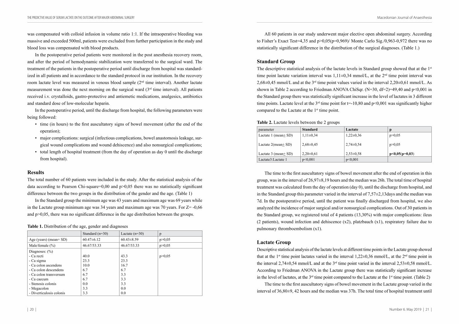
Table 1. Distribution of the age, gender and diagnosesStandard (n=30) Lactate (n=30) p
Age (years) (mean+ SD) 60.47±6.12 60.43±8.59 p>0,05Male/female (%) 46.67/53.33 46.67/53.33 p>0,05Diagnoses: (%)- Ca recti- Ca sigma- Ca colon ascendens- Ca colon descendens- Ca colon transversum- Ca caecum- Stenosis colonis- Megacolon - Diverticulosis colonis
40.023.310.06.76.76.70.03.33.3
43.323.316.76.73.33.33.30.00.0
p>0,05
All 60 patients in our study underwent major elective open abdominal surgery. According to Fisher’s Exact Test=4,35 and p>0,05(p=0,969)/ Monte Carlo Sig./0,963-0,972 there was no statistically significant difference in the distribution of the surgical diagnoses. (Table 1.)
Standard GroupThe descriptive statistical analysis of the lactate levels in Standard group showed that at the 1st time point lactate variation interval was 1,11±0,34 mmol/L, at the 2nd time point interval was 2,68±0,45 mmol/L and at the 3rd time point values varied in the interval 2,20±0,61 mmol/L. As shown in Table 2 according to Friedman ANOVA ChiSqr. (N=30, df=2)=49,40 and p<0,001 in the Standard group there was statistically significant increase in the level of lactates in 3 different time points. Lactate level at the 3rd time point for t=-10,80 and p<0,001 was significantly higher compared to the Lactate at the 1st time point.
Table 2. Lactate levels between the 2 groups parameter Standard Lactate pLactate 1 (mean+ SD)
Lactate 2(mean+ SD)
Lactate 3 (mean+ SD)
1,11±0,34
2,68±0,45
2,20±0,61
1,22±0,36
2,74±0,54
2,53±0,58
p>0,05
p>0,05
p<0,05(p=0,03)Lactate3:Lactate 1 p<0,001 p<0,001
The time to the first auscultatory signs of bowel movement after the end of operation in this group, was in the interval of 26,97±8,19 hours and the median was 26h. The total time of hospital treatment was calculated from the day of operation (day 0), until the discharge from hospital, and in the Standard group this parameter varied in the interval of 7,57±2,13days and the median was 7d. In the postoperative period, until the patient was finally discharged from hospital, we also analyzed the incidence of major surgical and/or nonsurgical complications. Out of 30 patients in the Standard group, we registered total of 4 patients (13,30%) with major complications: ileus (2 patients), wound infection and dehiscence (x2), platzbauch (x1), respiratory failure due to pulmonary thrombоеmbolism (x1).
Lactate GroupDescriptive statistical analysis of the lactate levels at different time points in the Lactate group showed that at the 1st time point lactates varied in the interval 1,22±0,36 mmol/L, at the 2nd time point in the interval 2,74±0,54 mmol/L and at the 3rd time point varied in the interval 2,53±0,58 mmol/L. According to Friedman ANOVA in the Lactate group there was statistically significant increase in the level of lactates, at the 3rd time point compared to the Lactate at the 1st time point. (Table 2)
The time to the first auscultatory signs of bowel movement in the Lactate group varied in the interval of 36,80±9, 42 hours and the median was 37h. The total time of hospital treatment until
THE PREDICTIVE VALUE OF SERUM LACTATE ON THE OUTCOME AFTER MAJOR ABDOMINAL SURGERY

| 22 |
Macedonian Journal of Anaesthesia
Number 6. May 2019 | 23 |
discharge from hospital among this group varied in the interval of 8,60±1,94 days with the me-dian time of 8d. In the Lactate group in the postoperative period, we registered total of 7 patients (23,30%) with major complications: bowel anastomosis leakage (2 patients), surgical wound infection and wound dehiscence (x4), pneumonia (x2), sepsis (x1), mesenteric thrombosis (x1).
Comparative Analysis between the 2 GroupsThe comparative analysis showed that patients in the Lactate group had higher average levels of lactates at all 3 time points compared to the Standard group. (Table 2)
The time to the first bowel movement (recovery of peristalsis) for Z=-3,99 and p<0,001 was significantly longer in the Lactate group. The total time of hospital treatment was also signifi-cantly longer in the Lactate group for Z=-2,83. (Table 3)
In the Lactate group 7 patients (23,33%) developed major complications compared to 4 patients (13,33%) in the Standard group. (Table 3) During the period of hospitalization (until the discharge of hospital), 1 patient (3,33%) died in each of the two study groups. The reasons of the lethal outcome were sepsis and mesenteric thrombosis respectively.
Table 3. Parameters in the postoperative period in the 2 study groupsParameter Standard Lactate p
Bowel movement(hours) (M+ SD) 26,97±8,19 36,80±9,42 p<0,001
Length of treatment(days) (M+ SD) 7,57±2,13 8,60±1,94 p<0,01
Complications(number/%) 4 (13,33%) 7 (23,33%) p>0,05
Predictive Value of LactatesThe further statistical analysis determined the predictive value of the serum lactates in terms
of power to predict the incidence of complications or the adverse outcome after major surgery. For this purpose ENTER method of statistical analysis was employed and the results showed that in both study groups the greatest significance for prediction of complications and adverse outcome after surgery had the level of lactate at the 3rd time point (the day 1 after surgery). In the Lactate group for 95% C.I. for EXP(B):1,18-Wald=4,07 and p<0,05 (p=0,04), the predictive value of serum lactate at the 3rd time point was statistically significant. Both the predictive values of lactate at the 1st (Wald=0,42 p>0,05 (p=0,52)) and the 2nd time point (Wald=0,11 p>0,05(p=0,74)) were not statistically significant.
In both groups the level of lactates at the 3rd time point was also found to have strong pre-dictive value for the prediction of the total length of hospitalization. In the Lactate group for R=0,65 (F=6,22; p=0,002) Durbin–Watson=2,2, the predictive value of lactates was found to be strong and significant.
Discussion The rationale use of lactates in daily clinical practice lays in the pathophysiological understand-ing of the segmental tissue hypo-perfusion and reperfusion. The disbalance between the global oxygen delivery and the current tissue consumption, leads to the state known as “oxygen debt”. If not corrected, this state will lead to a decrease in the mitochondrial oxidative phosphorylation and the cell’s energy production will entirely depend on anaerobic glycolysis (11, 22).
Della Rocca et al. in 2014 analyzed the pathophysiology of the hypo-perfusion and reper-fusion of the abdominal organs and proposed that the success of the perioperative strategy for the fluid administration during major surgery, can be improved by introduction of systematic monitoring of the serum lactates (7).
Other authors proposed that serum lactates measured at different time points during surgery should be used as target parameters in the goal directed protocol for fluid administration. Wenkui et al. in their clinical study from 2010 studied the population of patients that underwent major abdominal surgery. They concluded that in the group of patients where lactates were used as a target parameter in the fluid administration protocol, the incidence of postoperative complications was significantly lower. These authors proposed that systematic following of the lactate level during and after the operation should be part of our daily practice because it reduces the possi-bility of inadequate tissue perfusion and hypo oxygenation. (23) The famous study by Rivers and colleagues proposed the implementation of early goal-directed therapy, aimed at improving the oxygen delivery and maintaining the hemodynamic stability in patients with severe sepsis and increased lactate levels (24).
In our clinical study we measured the level of lactates at 3 different time points during and after open major abdominal surgery. Patients were divided into 2 groups depending on the level of lactates on the day 1 after surgery. Patients with lactate value >2.2mmol/l at this time point were considered high lactate patients and were assigned to Lactate group and other patients with lactate ≤2.2mmol/l were assigned to the Standard group.
The comparative analysis of the lactates showed that despite having higher value on the day 1 (2,20±0,61versus 2,53±0,58 mmol/L) patients in the Lactate group also had higher average levels of lactates at all 3 time points compared to the Standard group. Immediately after surgery, the ratio was 2,68±0,45 versus 2,74±0,54 mmol/L. High postoperative levels of lactates at the 2nd time point, were expected considering the potential of the open abdominal surgery to cause significant inflammatory stress response. However, our further analysis showed that patients in the Standard group showed greater ability to adequately correct these high levels of lactates (from 2,68±0,45to 2,20±0,61mmol/l on the day 1) while in the Lactate group this parameter showed that on day 1 after surgery the lactates were still high (2,74±0,54 to 2,53±0,58 mmol/L).
With further analysis of the received data we wanted to determine what the real predictive value of the level of lactate for complications or adverse outcome of surgery is. We’ve found that in both study groups the greatest significance for the prediction of complications is held
THE PREDICTIVE VALUE OF SERUM LACTATE ON THE OUTCOME AFTER MAJOR ABDOMINAL SURGERY

| 24 |
Macedonian Journal of Anaesthesia
Number 6. May 2019 | 25 |
by the level of lactate at the 3rd time point (the next day after surgery). In the Lactate group the predictive value of serum lactate at the 3rd time point was also statistically significant (p<0,05). The same parameter was also found to have strong predictive value for the prediction of the total length of hospitalization. In the Lactate group the predictive value of lactates was found to be strong and statistically significant (p=0,002).
These results of our study were confirmed in the recent meta-analysis done by Som et al. published in 2017. After analyzing the results of 41 clinical studies, authors concluded that patients with lower lactates in the postoperative period had significantly lower incidence of complications probably due to the improved tissue micro perfusion and oxygenation of the organs (25).
Our goal was also to determine the association between the lactate level and the parameters that defined the outcome of surgery in our study (the recovery of organ function, the length of hospital stay, incidence of postoperative complications and in-hospital mortality). The patients in our study with higher level of lactates (Lactate group) had significantly prolonged recovery of peristalsis expressed through the time to first bowel movement (26,97±8,19 vs 36,80±9,42 hours) (p<0,001). The length of hospitalization was also significantly longer within the Lactate group (7,57±2,13 vs 8,60±1,94 days) (p<0,01).
In a multicenter study from 2007 Donati et al. were investigating the effects of the goal directed treatment protocol during major abdominal surgery on the outcome. They came to a conclusion that in the groups of patients where the treatment was directed toward supernormal values of lactates, ScvO2 and O2ER (O2 extraction rate), the result was significantly shorter length of hospital stay (11,3 vs 13,4 days; p<0,05) and there was reduction of the incidence of postop-erative complications (11,8% vs 29,8%; p<0,05). Authors of this study showed that ScvO2 and lactates can serve as an early markers of tissue hypo-perfusion (26).
Regarding the incidence of complications in our study, more patients in the high lactate group had some form of major complication [4 (13,33%) vs 7 patients (23,33%)]. This difference, however, was not statistically significant.
Della Rocca et al. in an article from 2014 analyzed the literature and available clinical studies and came to a conclusion that standard hemodynamic monitoring does not provide sufficient information to create adequate perioperative fluid treatment protocol (7). In high risk patients and during major surgery procedures administration of fluids and vasopressors should be directed toward predefined hemodynamic parameters of global stability and segmental tissue perfusion and oxygenation. The target of such approach is to optimize the oxygen delivery according to the current needs and consumption, and to avoid the occurrence of “oxygen debt” in the tissues.
Our clinical research, however, has several important limitations. The study population of patients was relatively small and this type of study should be repeated in a large cohort of patients, if possible in a multicenter setting. We included 60 consecutive patients that underwent major abdominal surgery and the duration of the study was limited until the fulfillment of this number. The potential strength of the study is that the whole population of patients received the same
perioperative treatment in terms of preparation and fluid administration protocol. Randomization was not necessary and potential of bias or the researchers’ influence on the results and the out-come was avoided. Only after the study period and during the analysis patients were subdivided into two groups. Another potential limitation lays in the type of monitoring we used. Besides the noninvasive hemodynamic monitoring, we’ve decided to include lactates as a marker of global and regional tissue perfusion. In further studies other potential parameters of hemodynamic stability (ScvO2, ERO2, pHi and others) can be investigated alongside with lactates. Esophageal Doppler monitoring can also be included as a guidance tool in the fluid management protocol in the treatment of critically ill patients.
In conclusion this study demonstrates that postoperative lactate value on the first postopera-tive day holds significant predictive value for postoperative complications and adverse outcome after major abdominal surgery. Patients with serum lactate level >2.2mmol/l obtained on day 1, had prolonged time of recovery of peristalsis and this value of lactate also had the best power to predict the higher incidence of complications and longer length of hospital stay. We believe that this result offers only a small contribution to the understanding of the complicated patho-physiology of the major surgery settings and can serve as a base for further larger clinical trials.
Declaration of InterestsThe authors declare no conflict of interests.
References:1. Weiser T, Regenbogen S, Thompson K, et al.: An estimation of the global volume of surgery:
a modelling strategy based on available data. Lancet 2008;372:139-44.2. Teboul J-L., Monnet X.: Assessment of volume responsiveness. Crit Care Med 2009 Vol.
37, No.3. 3. Singh S., Kuschner W. and Lighthall G.: Perioperative intravascular fluid assessment and
monitoring: a narrative review of established and emerging techniques. Anesth research and practise vol.2011 ID 231493
4. Wax D.B., Schaner D. et al.: Intraoperative factors associated with development of surgical site infections after colorectal surgery. The Internet J of Anesth. 2009 Vol 20 No 1 ISSN: 1092-406X.
5. Jhanji S, Lee C, Watson D, et al.: Microvascular flow and tissue oxygenation after major abdominal surgery: association with post-operative complications. Intensive Care Med 2009;35:671.
6. Mark A. Hamilton et al.: Perioperative fluid management: progress despite lingering con-troversies. Cleveland Clin J 2009: vol76, supplement 4.
7. Della Rocca G., Pompei L.: Goal directed therapy in anesthesia: any clinical impact or just a fashion. Minerva anesth 2011; 77:545-53.
8. Strunden M., Heckel K., Goetz A., et al.: Perioperative fluid and volume management: physiological basis, tools and strategies. Annals of Intensive Care 2011,1:2.
9. Marik P., Monnet X., Teboul J-L.: Hemodynamic parameters to guide fluid therapy. Annals of Intensive Care 2011, 1:1.
10. Cannesson Maxime et al.: Noninvasive and Continuous fluid responsiveness monitoring with pleth variability index (PVI). Br J Anaesth. 2008; 101(2):200-62.
THE PREDICTIVE VALUE OF SERUM LACTATE ON THE OUTCOME AFTER MAJOR ABDOMINAL SURGERY

| 26 |
Macedonian Journal of Anaesthesia
Number 6. May 2019 | 27 |
11. Brooks G.: Lactate shuttles in nature. Biochem Soc Trans 2002; 30:258–264.12. Levy B, Sadoune L, Gelot A, et al: Evolution of lactate/pyruvate and arterial ketone body
ratios in the early course of catecholaminetreated septic shock. Crit Care Med 2000; 28:114–119.
13. Cain SM. Oxygen delivery and uptake in dogs during anemic and hypoxic hypoxia. J Appl Physiol 1977;42:228–234.
14. Friedman G., De Backer D., Shahla M., et al..: Oxygen supply dependency can characterize septic shock. Intensive Care Med 1998; 24:118–123.
15. Ronco J., Fenwick J., Tweeddale M., et al. Identification of the critical oxygen delivery for an-aerobic metabolism in critically ill septic and nonseptic humans. JAMA 1993;270:1724–1730.
16. Haidl F., Brabrand M., Henriksen D. et al.: Lactate is associated with increased 10-day mortal-ity in acute medical patients: a hospital-based cohort study. Eur J Emerg Med 2015;22:282–4.
17. Barfod C., Lundstrøm L., Lauritzen M. et al. : Peripheral venous lactate at admission is associated with in-hospital mortality, a prospective cohort study. Acta Anaesthesiol Scand 2015;59: 514–23.
18. Pedersen M., Brandt V., Holler J. et al. Lactate level, aetiology and mortality of adult patients in an emergency department: a cohort study. Emerg Med J 2015;32:678–84.
19. Ramirez J., Blasco J., Roig J., et al.: Enhanced recovery in colorectal surgery: a multicentre study. BMC surg 2011, 11:9.
20. Creagh-Brown B., De Silva A., Ferrando-Vivas P., et al.: Relationship Between Peak Lactate and Patient Outcome Following High-Risk Gastrointestinal Surgery: Influence of the Nature of Their Surgery: Elective Versus Emergency. Crit Care Med. 2016 May;44(5):918-25
21. Jansen T., van Bommel J, Woodward R, et al.:Association between blood lactate levels, Sequential Organ Failure Assessment subscores, and 28-day mortality during early and late intensive care unit stay: a retrospective observational study. Crit Care Med. 2009 Aug;37(8):2369-74.
22. Philp A., Macdonald A., Watt P.: Lactate-A signal coordinating cell and systemic function. J Exp Biol 2005; 208:4561–4575.
23. Wenkui Y., Ning L., Jianfeng G., et al.: Restricted perioperative fluid administration adjusted by serum lactate level improved outcome after major elective surgery for gastrointestinal malignancy. Surgery 2010 Apr;147(4):542-52.
24. Rivers E., Nguyen B., Havstad S., et al.: Early goal-directed therapy in the treatment of severe sepsis and septic shock. N Engl J Med 2001;345:1368–1377.
25. Som A., Maitra S., Bhattacharjee S., et al: Goal directed fluid therapy decreases postoperative morbidity but not mortality in major noncardiac surgery: a metaanalysis and trial sequential analysis of randomized controlled trials. J Anesth (2017) 31:66–81.
26. Donati A., Loggi S., Jean-Charles Preiser J-C.,et al: Goal-directed Intraoperative therapy reduces morbidity and length of hospital stay in highrisk surgical patients. CHEST 2007; 132:1817–1824.
ORIGINAL ARTICLE ISSN 2545-4366 | UDK: 616.61-076-073.756.8:004
COMPUTER TOMOGRAPHIC (CT) GUIDED PERCUTANEOUS KIDNEY BIOPSY – A NEW METHOD INTRODUCTION
Stojkoski A1, Nikolov GI2
1 University Institute of Radiology, Skopje, Republic of North Macedonia2University Clinic of Nephrology, Faculty of Medicine, Clinical Campus Mother Theresa
Introduction of the Method, History, Relevance and Report of the Cases
ABSTRACTSince its introduction, in the 1950s, the kidney biopsy became a gold standard in the diag-
nosis of the kidney diseases which provides information that lead to appropriate treatment and management of these diseases (1, 2). In our center, the ultrasound-guided percutaneous renal biopsy (PRB) is the most often performed by nephrologists even for pediatric patients. Here we report an introduction of a new method of a computed tomographic (CT) guided kidney biopsy technique done for the first time at our University clinical campus “Mother Theresa” in Skopje.
Corresponding author: Igor G. Nikolov, University Clinic of Nephrology, Clinical Campus Mother Theresa, str. Mother Theresa 19, Skopje 1000, Republic of North Macedonia. Email: [email protected]
THE PREDICTIVE VALUE OF SERUM LACTATE ON THE OUTCOME AFTER MAJOR ABDOMINAL SURGERY

| 28 |
Macedonian Journal of Anaesthesia
Number 6. May 2019 | 29 |
IntroductionKidney biopsy as a technique provides adequate histological diagnosis. Furthermore, histologic diagnosis of the kidney diseases may provide very useful information for the disease and pa-tient’s benefits are enormous. The first and the most important benefit is that the right diagnosis provides correct, pointed treatment and disease management.
On the other hand, it should be emphasized that the risks of kidney biopsy in several situations may overcome the benefits in context of the patient’s safety and need to have histologic diagnosis. Several anatomic anomalies, or kidney cysts in the lower renal part may present contraindication for kidney biopsy (1). Moreover, atrophic kidneys with very small cortices or very tin parenchyma, or horseshoe kidney, in some patients may be also a reason for contraindication of a kidney biopsy.
Due to the controversies (in the benefits versus contraindications and risks) for kidney biop-sies, the literature and the clinicians have involved novel techniques for safer, secure and confi-dent approaches. Imaged guided PR Biopsies have been involved in the novel data as minimally invasive technique for having right histological diagnose. Furthermore, Computer Tomographic (CT) guided PRB is the most novel approach that has more benefits then ultrasound guided PRB as an alternative biopsy technique when we deal with patients with a systemic disease or when high morbidity and mortality might be expected (2,3,4). However, this technique even though used in centers worldwide has never been done at our Clinical Campus “Mother Theresa” in Skopje.
Introduction of the Method, Relevance through Report of the Cases and DiscussionHereby, we report the first two cases of CT guided kidney biopsies performed at the Institute of Radiology in our clinical campus after Informed Consent was obtained. We report the technique done in two male patients 24 and 43 years old, where the first try for PRB was without success and the decision for CT guided kidney biopsies was done. The literature reported, that CT bi-opsies may be used as a primary imaging modality or may be preferred in some patients, like obese patients, patients with complicated anatomies, and those for whom kidney visualization with ultrasound is difficult (1- 4).
For both patients, and as standardized literature protocol before kidney biopsies was per-formed, a complete blood count was obtained. Patients were reviewed for medicaments that may cause bleeding (like certain anticoagulants or antiplatelet agents, as well as non-steroidal drugs and anti-inflammatory agents). Moreover, hemostasis and ratio/prothrombin time, activated partial thromboplastin time were determined. We also followed serum creatinine and serum urea levels.
For one patient we have obtained an intravenous line, preintervention due to his anxiety and fear and just for case tif anxiolytics and sedatives should be given. For this aspect of this method, it has been previously reported that CT guided kidney biopsy may be useful in anxious and uncooperative patients as well as in pediatric patients. It is possible to be used in patients that may also require anxiolytics or general anesthesia to safely perform the procedure (4).
For the technique introduction: In patients we localized the kidneys by ultrasound, and the skin overlying was prepared in a sterile manner. Local anesthetic was used (in our case lidocaine 1%) which was injected in the depth to the kidney surface. Afterwards we performed a CT-guided biopsy, with a siring loaded gun using 16G biopsy needle. We obtained 2 or 3 tissue carrots which were used for Immunofluorescence and histologic analysis of the examples. Literature reveals that this is a standardized technique only in different protocols different number of specimen carrot are obtained (3).
After the intervention, patients were prescribed 6-12 hours bed rest and in that time vital parameters were followed for a period of 24 hours. Blood parameters were obtained 8 hours after the PRB, as well as urine specimen which was analyzed for hematuria. Afterwards, both patients went uneventfully and 24 hours later were discharged. Literature reveals that as a protocol blood specimen, blood count and vital signs should be monitored for the first 24 hours on regular basis due to the possibility of hematuria, renal rupture, hematoma and several other complications that must not be overviewed (4).
In these two patients we have observed no complications and in both cases we have obtained a sufficient number (>10) of visible glomerulus. The results for both patients came after two weeks and we had confirmed diagnosis which we could not do with PBN.
We presented this two first cases in order to implement new method and to emphasize that sometimes this is the method of choice for diagnosis and should be implemented in many plac-es as standard. In the conclusion we can underline that nephrologists in many centers are also competent at biopsy specimen division and processing as well as in our center.
Furthermore, from the aspect of pathology, for this method, we would underline that it is particularly important for centers that send biopsy specimens to pathology laboratories and to stress that specimens for light microscopy, and immunofluorescence, as well as electron micros-copies requires special processing and fixation methods.
In here, collaboration and nephrologists’ inputs on the basis of the biopsy indication can ensure proper specimen division for optimum diagnostic and prognostic yield.
Conflict of Interest:Authors deny any conflict of interest.
References1. Korbet SM: Percutaneous renal biopsy. Semin Nephrol 22: 254–267, 2002 [PubMed: 12012311].2. Burstein DM, Korbet SM, Schwartz MM: The use of the automatic core biopsy system in
percutaneous renal biopsies: A comparative study. Am J Kidney Dis 22: 545–552, 1993 [PubMed: 8213794].
3. Sateriale M, Cronan JJ, Savadler LD: A 5-year experience with 307 CT-guided renal biop-sies: Results and complications. J Vasc Interv Radiol 2: 401–407, 1991 [PubMed: 1799787].
4. Margaryan A, Perazella MA, Mahnensmith RL, et al. Experience with outpatient computed tomographic-guided renal biopsy. Clin Nephrol 74: 440–445, 2010 [PubMed: 21084047].
COMPUTER TOMOGRAPHIC (CT) GUIDED PERCUTANEOUS KIDNEY BIOPSY – A NEW METHOD INTRODUCTION

| 30 |
Macedonian Journal of Anaesthesia
Number 6. May 2019 | 31 |
ORIGINAL ARTICLE ISSN 2545-4366 | UDK: 616.153.472.3:616-089.16
THE EFFECTS OF TWO THERMAL INSULATION METHODS ON THE POSTOPERATIVE LACTATE LEVELS, SHIVERING AND PATIENT’S THERMAL COMFORT
Kuzmanovska B1, Kartalov A1, Shosholcheva M2, Sholjakova M 1, Tolevska M1, Srceva Jovanovski M1, Gavrilovska Brzanov A1
1 University Clinic for Traumatology, Orthopedic Diseases, Anesthesiology, Reanimation and Intensive Care Medicine and Emergency Department, Faculty if Medicine, University “Ss Cyril and Methodius”, Skopje, Republic of North Macedonia
2 University Clinic for General Surgery “St. Naum Ohridski” Faculty of Medicine, University “Ss Cyril and Methodius”, Skopje, Republic of North Macedonia
ABSTRACT Introduction: Surgical patients are at risk of developing hypothermia during perioperative
period. The aim of this study is to compare the effects of two thermal insulation methods on postoperative lactate levels, shivering and patient’s thermal comfort.
Material and Method: Sixty patients, ASA group 1 and 2, selected for elective inguinal hernia repair under spinal anesthesia were randomly divided into 3 groups: inflatable blanket heating group (group A, n=20), warmed infusion irrigation group (group B, n= 20), and control group (group C, n= 20). Blood samples were obtained immediately after the surgery for lactate levels, and shivering and patient’s thermal comfort were recorded during the first and second postoperative hour.
Results: Lactate levels although higher in the control group compared to the groups A and B, did not show significant difference. Patients’ thermal comfort and occurrence of postoperative shivering were different in all three groups.
Conclusion: Inflatable heating blanket and warmed infusion irrigation are effective methods in preventing perioperative hypothermia.
Key words: hypothermia, shivering, thermal comfort.Corresponding author: Biljana Kuzmanovska, University Clinic for Traumatology,
Orthopedic Diseases, Anesthesiology, Reanimation and Intensive Care Medicine and Emergency Department, Faculty if Medicine, University “Ss Cyril and Methodius”, Skopje, Republic of North Macedonia.
IntroductionAs stated by American Society of Anesthesiologist (ASA), every patient subjected to anesthesia should have temperature monitoring due to suspected, intended or anticipated changes in body temperature (1).
Hypothermia is defined as patient’s core temperature below 36.0°C, and preoperatively it is assumed that approximately 20% of the patients acquired inadvertent hypothermia (2).
Normal temperature range is between 36.5°C to 37.5°C, during anesthesia it can decline below 35.0°C. On one hand, hypothermia is generated by the cold operation theater environ-ment, and on the other hand, by anesthesia modified thermoregulation mechanisms, anesthesia generated peripheral vasodilatation, intravenous fluids temperature and the field of operation (3).
Hypothermia leads to increased perioperative morbidity, expanded surgical site infections, and increased surgical bleeding. Hypothermia shifts the oxyhemoglobin dissociation curve to the left, thus decreasing the available oxygen to the tissues, and along with shivering which occurs as a result of heat production and goes along with oxygen consumption that promote anaerobic metabolism, leads to increased lactates and lactic acidosis respectively. Lactic acidosis promotes coagulopathy. Furthermore, hypothermia decreases the drug metabolism and alters the drug effects. Additionally, with all these probable complications, hypothermia might increase the length of hospital stay and might lead to lower patient’s satisfaction (4-7).
Due to everything above mentioned it is essential to prevent inadvertent perioperative hy-pothermia, moreover this inadvertent hypothermia should differ from the intentional induction in hypothermia for medical reasons, which is not subject to our investigations.
Despite this knowledge, application of warming strategies remains not consistent.The aim of our study is to compare the effect of two thermal insulation methods on periopera-
tive hypothermia by comparing postoperative lactate level, shivering and patients’ thermal comfort.
Material and MethodsIn this prospective randomized controlled study, we enrolled sixty patients scheduled for in-guinal hernia repair under spinal anesthesia. Study was conducted in the University Clinic for Anesthesia, Reanimation and Intensive Care and in the University Clinic for Digestive Surgery - Clinical Center “Mother Theresa”, Skopje, North Macedonia. Ethical approval was obtained from the Ethical Committee of Chair for Anesthesiology with Resuscitation and Pain Therapy, Medical Faculty, Ss Cyril and Methodius University, Skopje. Written consent to participate in the study was obtained from each patient. All patients that met the inclusion criteria: ASA 1 and 2, scheduled for elective inguinal hernia repair and above 18 years of age were enrolled in the study. Exclusion criteria was the contraindication for spinal anesthesia.
Patients were divided into three groups: inflatable blanket heating group A (n=20), warmed infusion irrigation group B (n=20), and control group C (n=20), who did not received any of aforementioned warming methods.

| 32 |
Macedonian Journal of Anaesthesia
Number 6. May 2019 | 33 |
All patients underwent standardized spinal anesthesia protocol.Perioperative monitoring of the patients was standardized; EKG, non-invasive blood pressure,
peripheral oxygen saturation and near- core temperature monitoring (axillary) in two measure-ment times, 30 minutes in anesthesia, and immediately upon entering the post anesthesia care unit (PACU).
In the group A, an inflatable blanket with temperature of 38 degrees Celsius air was placed after the spinal anesthesia was conducted.
In the group B patients warmed crystalloid infusion at temperature of 38 degrees Celsius were placed in the peripheral line, with infusion rate of 15 ml/kg/hour. Patients in the control group C didn’t receive either one of the above mentioned warming measures.
After the surgery blood samples were obtained for assessing the lactate levels. Shivering and thermal comfort were recorded during the first and the second postoperative hour.
Thermal comfort was assessed with two proposed answers from which the patients had to choose: “cold” or “pleasantly warm”.
Statistical analysis was performed with IBM SPSS (20.0) program. Data were reported as median and ranges, and categorical variables were expressed as percentage. Statistical signifi-cance was indicated with p <0.05.
Results: Demographics: Sixty patients, ASA physical status 1 and 2, aged between 20 and 65 years, median age 46 +/_ 1 yrs. Male 50, female 10.
Lactate levels ranged from 0.5 to 1.8 mmol/L in all groups. Medium lactate level in group A was 0.9 mmol/L, in group B was 1.10 mmol/L, while in the control group was 1.13 mmol/. (Table 1)
There was no statistically significant difference (p > 0.05) among the groups, with p = 0.09 between group A and B, and p=0.06 between group A and C, and p= 0.84 between group B and C.
Table 1: Median Lactate levels in group A, B and C. Group A ( n=20) B (n=20) C (n=20)Median Lactate levels ( mmol/L) 0.9 1.10 1.13
No statistically significant difference in postoperative lactate levels between all three groups.
Near - core temperature:There is statistically significant difference in median near-core temperatures (axillary) be-
tween group A and group C, and between group B and group C (p<0.05) in both measurement times, but there is no statistically significant difference between group A and B (>0.05) in both measurement times.
Shivering:
Table 2. Incidence of shivering among the patients of all three groups in the first and the second postoperative hour.Group A (n=20) B (n=20) C ( n=20)First postoperative hour 2 (10%) 4 (20%) 7 (35%)Second postoperative hour 0 0 0
Thermal comfort:
Table 3. Thermal comfort of the patients in group A, B and C.Group A (n=20) B (n=20) C ( n=20)Cold 5 (25%) 5 (25%) 15 (75%)Pleasantly warm 15 (75%) 15 (75%) 5 (25%)
DiscussionThe prevention and treatment of perioperative hypothermia is an important part of anesthesia care. Understanding the principles of human body thermoregulation, mechanisms that lead to perioperative hypothermia, and adverse effects of hypothermia is of essential importance for anesthesia providers.
Temperature monitoring is a cornerstone of perioperative thermal care. Core temperature is temperature of deep thoracic, abdominal and central nervous system. It is usually 2- 4 degrees warmer than the skin temperature. Core temperature is measured at the esophagus, bladder, na-sopharynx, pulmonary artery, etc. Near-core temperature is the temperature measured in axils, rectum or oral cavity, and is commonly used during regional anesthesia. In this study, we mon-itored body temperature axillary, and found that there is no statistically significant difference in the measured near-core body temperature in between the groups of patients that were subject to either of thermal insulation methods, while, there is statistically significance between both group of patients that received thermal insulation, compared to control group of patients, with no thermal insulation at all.
Thermal comfort, described as pleasantly warm, was reported in higher percentage in both group A and B, while higher percentage of the control group described their thermal comfort as “cold”.
Shivering was observed and noted in higher percentage in the control group compared to groups A and B, in the first postoperative hour. In the second postoperative hour, no shivering was observed in either of the three groups.
There was no statistically significant difference in serum lactate levels between group A, B and C. Some authors report higher levels of serum lactate levels in patients that didn’t receive any perioperative heating (8). Serum lactate levels are primary markers of tissue (hypo) perfusion,
THE EFFECTS OF TWO THERMAL INSULATION METHODS ON THE POSTOPERATIVE LACTATE LEVELS, SHIVERING AND PATIENT’S THERMAL COMFORT

| 34 |
Macedonian Journal of Anaesthesia
Number 6. May 2019 | 35 |
and when interpreting lactates in a hypothermia context, careful consideration should be taken about patient’s volemic status, blood loss and duration of surgery.
To avoid perioperative hypothermia, some authors suggest preoperative heating of the pa-tients with warm air inflatable blanket 30 minutes before the surgery (3,8).
ConclusionBoth inflatable blanket and warmed infusion irrigation are good thermal insulation methods to prevent perioperative inadvertent hypothermia.
References1. American Society of Anesthesiologists. 2010. Standards for basic anesthetic monitoring.
http://www.asahq.org/For-Healthcare-Professionals/∼/media/For%20Members/documents/Standards%20Guidelines%20Stmts/Basic%20Anesthetic%20Monitoring%202011.ashx. Accessed February, 2019.
2. Kurz A. Physiology of thermoregulation. Best Pract Res Clin Anaesthesiol. 2008;22(4):627–644.3. Frank SM, Fleisher LA, Breslow MJ, et al. Perioperative maintenance of normo-
thermia reduces the incidence of morbid cardiac events. A randomized clinical trial. JAMA. 1997;277(14):1127–1134.
4. Hart S, Bordes B, Hart J, et al. Unintended Perioperative Hypothermia. Ochsner J. 2011 Fall; 11(3): 259–270.
5. Schmied H, Kurz A, Sessler DI, et al. Mild hypothermia increases blood loss and transfusion requirements during total hip arthroplasty. Lancet. 1996;347 (8997):289–292.
6. Peng RY, Bongard FS. Hypothermia in trauma patients. J Am Coll Surg. 1999;188(6):685–696.7. NICEPerioperative hypothermia (inadvertent): the management of inadvertent periopera-
tive hypothermia in adults, NICE Clinical Guideline 29, 2008 London National Institute for Health and Clinical Excellence Available from www.nice.org.uk/CG065 (accessed February, 2019).
8. Zheng X et al. Effects of perioperative thermoregulation on patient’s body temperature, pe-ripheral circulation and blood coagulation time in patients undergoing elective vertical hemi laryngectomy. Lin Chung Er Bi Yan Hou Tou Jing Wai Ke Za Zhi. 2017; 31 (14): 1113-1115.
NOVEL INDICATION FOR ATORVASTATIN: CHRONIC SUBDURAL HEMATOMA
Angjushev D1, Srceva-Jovanovski M1, Panovska-Petruseva A1, Kotevska-Angjushev M2, Leshi A1, Kokareva A1, Demjanski 1, Trposka A1, Peev I3, Klisevska I1
1University Clinic for Anesthesia, Reanimation and Intensive Care, Skopje, R. N. Macedonia2 Department of cardiology, City General Hospital”8th of September” Skopje3University Clinic For Plastic and Reconstructive Surgery, Skopje
ABSTRACTChronic subdural hematoma, one of the most common neurosurgical diseases, is the most
commonly found in elderly patients who have experienced some type of head trauma. The in-cidence is expected to double by the year 2030, owing to the growing aging of the population. Surgery has long been the treatment of choice, but it carries a significant risk of recurrence, complications and death. Nonsurgical treatments are very limited and ineffective. It has been demonstrated that statins could improve chronic subdural hematoma outcome by enhancing angiogenesis and reducing local inflammation associated with chronic subdural hematoma. Several recent clinical studies suggest that oral atorvastatin, a member of statins, is beneficial in the management of chronic subdural hematoma, by reducing the size of hematomas and by improving the clinical outcomes. Atorvastatin is a safe, effective non-surgical alternative for chronic subdural hematoma.
Keywords: atorvastatin, chronic subdural hematoma, conservative therapy, statinsCorresponding author: Darko Angjushev, University Clinic for Anesthesia, Reanimation
and Intensive Care, Skopje, R.N. Macedonia, email: [email protected].
REVIEW ISSN 2545-4366 | UDK: 616.831.957-003.215-085.272.4THE EFFECTS OF TWO THERMAL INSULATION METHODS ON THE POSTOPERATIVE LACTATE LEVELS, SHIVERING AND PATIENT’S THERMAL COMFORT

| 36 |
Macedonian Journal of Anaesthesia
Number 6. May 2019 | 37 |
IntroductionChronic subdural hematoma (CSDH) is a common neurosurgical disease, mostly found in elderly patients. It is often associated with head trauma. CSDH is one of the most common intracra-nial hematomas. The incidence is 58/100.000 persons a year in people of 70 years of age and older (1). The incidence is expected to double by the year 2030, due to the increasing usage of anticoagulants and continuous aging of the population. It is expected to become the most com-mon adult brain disease requiring neurosurgical intervention (2). In patients without history of trauma, the use of antiplatelet or anticoagulant therapy is often present at the time of diagnosis, which represents a common risk factor for development of CSDH. This is also a serious surgical challenge for the treatment of CSDH(3,4). Symptomatic patients with confirmed hematoma on radiological imaging are usually treated surgically (5). The surgical drainage remains the pre-ferred treatment of small symptomatic and large asymptomatic hematomas (2,6). After surgical evacuation of CSDH, the recurrence rate is still relatively high (7–20%) requiring to repeat surgery (6). Independent of the treatment management and strategy, the mortality rate has been reported to be high, 32% in elderly patients and up to 38% in 90 years or older patients (7,8). Surgery also carries a considerable risk of complications (pneumonia, peri-operational infection and pulmonary edema) following the surgery (9).
Surgical intervention remains the treatment of choice, although the outcome of surgical treatment is not always satisfactory, and may be complex in old patients, especially if they are treated with anticoagulants. Therefore, the effective and safe alternative to surgery is highly recommended. Conservative treatment could be a good choice in some situations, for example, in patients who have high perioperative risk or are refusing operation. Conservative management is best reserved for patients with small non-space-occupying hematomas (2,10,11).
Atorvastatin, as a member of the statins, has been recently investigated in the management of CSDH. Recently in animals and in clinical studies atorvastatin has been reported to be safe and effective to CSDH treatment (12-20). The best candidates are asymptomatic or mildly symptom-atic CSDH patients with small-sized hematomas, as a monotherapy or as an adjunct to surgery.
In this review, our aim was to provide practical information for neurosurgeons and anes-thetists about this potentially new treatment for CSDH with atorvastatin. We summarized the current knowledge on this issue, to drive further research on this topic.
Conservative Therapy with Statins for CSDHStatins, 3-hydroxy-3-methylglutaryl coenzyme A (HMG-CoA) reductase inhibitors are approved for reducing low-density lipoprotein cholesterol levels. Recent clinical studies showed that statins could improve CSDH outcome due to its effect of regulation of angiogenesis and inflammation, by enhancing angiogenesis and reducing inflammation associated with CSDH (9).
The underlying pathogenesis in the formation of CSDH is localized inflammation and im-paired angiogenesis in the neomembrane of the hematoma. From the immature vessels of the
neomembrane may be induced further blood leakage. Localized inflammation further worsens the repair of vessel leakage (9). The formation of mature vessels at the neomembrane of the hematoma reduces vessel leakage and on that way subdural hematoma progression is prevented. Also, the formation of mature vessels was associated with subdural hematoma absorption (21).
Statins are enhancing angiogenesis by mobilizing circulating endothelial progenitor cells which promotes the vessel repairs (22-25). Vascular endothelial growth factor (VEGF) is found to be at very high level in hematoma, but also in the serum of the patients with CSDH (26). VEGF when present at very high level, inhibits the maturation of new vessels (27). Statins also inhibits VEGF and reduces the inflammation in the vessel wall by reducing tumor necrotic factor (TNF-α) (28,29).
Clinical Studies with AtorvastatinRecently in clinical studies atorvastatin, a member of statins, has been reported to be effective in the treatment of CSDH(15,16,17,18). In animal models and in clinical studies , it has been shown that atorvastatin prevents the formation and promotes rapid absorption of the hematoma by blocking inflammation and immature angiogenesis (12-20).
The reduced size of subdural hematoma with atorvastatin was first described by Li et al. and Wang et al. in animal models (mice) (12, 13). A low dose of atorvastatin (3 mg/kg/day) was found to have antiangiogenic and local anti-inflammatory effects in mice (14).
Very few clinical studies recently published, have investigated the role of oral atorvastatin in CSDH management (15-20). All of these studies (15,17,18,19,20) were performed in China and one (16) in Hong Kong, China.
Wang et al.(18) in a prospective study, were analyzing the effects of atorvastatin as a con-servative treatment of CSDH. In a group of 23 patients oral atorvastatin was given in a daily dose of 20 mg for 1–6 months. They reported that after 4 weeks of treatment with atorvastatin, only 1 patient underwent surgery due to neurological deterioration with an increasing volume of hematoma. The remaining 22 patients had improved neurological status, and did not experi-ence any recurrence during the 36 months of follow-up. Radiological imaging showed that in 4 patients the hematomas were absorbed completely 3 months after the atorvastatin treatment, while the other 18 patients after 6 months were presented with no hematoma by CT or MRI. Adverse effects of atorvastatin were not documented (18).
Xu et al analyzed the treatment effects of oral atorvastatin as a monotherapy and as an adju-vant drug to traditional surgical therapy(15). Atrovastatin in dose of 20 mg once a day was given for 1–6 months. 7 patients were treated conservatively with atorvastatin as a monotherapy. In all 7 patients a significant reduction of the size of hematoma was recorded after 1 month, and all hematomas had disappeared after 6 months (picture 1). None of the patients in this group required surgical treatment. When atorvastatin was used complementary to surgery, a significant difference in the clinical outcome was seen in the group that received atorvastatin as an adjuvant drug to surgery (15).
NOVEL INDICATION FOR ATORVASTATIN: CHRONIC SUBDURAL HEMATOMA

| 38 |
Macedonian Journal of Anaesthesia
Number 6. May 2019 | 39 |
Picture 1. CT brain scan of a 70 year old patient with chronic subdural hematoma on atorvastatin treatment.
Findings from another very recent study in patients with chronic subdural hematoma demon-strate that the administration of atorvastatin as an adjuvant treatment to surgery was associated with lower risk of CSDH recurrence rate(17). Also Chan et al. in a study on 24 patients reported reduced need for surgical intervention in patients on atorvastatin (16).
A multicentre, randomized, controlled clinical ATOCH trial from China evaluating the effi-cacy and safety of atorvastatin as a conservative treatment of CSDH was conducted by the Dr. Jiang and colleagues (19,20). This trial was performed on 196 patients with CT-confirmed or MRI-confirmed CSDHs in 25 neurosurgery centers in China between 2014 and 2015, with the results published in July 2018. Every day for 8 weeks, 98 patients received 20 mg of oral atorvastatin and 98 received placebo. The change in hematoma volume was measured by CT. Neurological function was assessed using the Barthel Index and Markwalder grading scale/ Glasgow Coma Scale. Excluded from the study were patients who needed emergency surgery, had high risk of cerebral herniation, were allergic to statins, or had previous or current bleeding problems, cancer, or multiple organ failure. This Chinese study showed that patients on atorvastatin had a larger decrease in hematoma volume, significantly improved neurologic function and quality of life, and reduced need for surgery in patients with CSDH, compared to the patients taking placebo. This study confirmed that this treatment is safe and effective for patients who have CSDH with mild clinical symptoms, smaller hematoma, and lower risk of cerebral herniation (19,20).
In this review, all of the included studies have used atorvastatin 20mg/ day as the interven-tion. Currently this dosage is used for 1-3 (15,16,19,20) or up to 6 months (18). This low dosage was chosen because it is the dose used for patients with hyperlipidemia associated with minimal
adverse effects (30,31). Atorvastatin at a higher dose (80mg) was reported to increase the risk of intracerebral hemorrhage (32). According to the data from Stroke Prevention by Aggressive Reduction in Cholesterol Levels trial, the risk is increased in patients with uncontrolled hyperten-sion and with low body weight (14). All studies in this review have reported no related adverse events to treatment to atorvastatin.
The data of current published studies are also raising an important question as to whether statins in general or atorvastatin in particular is efficient in the CSDH management.
LimitationsSo far all studies were performed in China, so the results cannot be representative for global population. In order to generalize the conclusions, more clinical studies in other countries are needed.
ConclusionAtorvastatin is a safe, and effective conservative therapy of chronic subdural hematomas. It is also a cost-effective alternative to surgery (19,20). Potential candidates for treatment with ator-vastatin are patients with small-sized hematomas, who are asymptomatic or with mild clinical symptoms and have lower risk of cerebral herniation. The main option for atorvastatin treatment is currently oral intake of a 20mg atorvastatin daily for 1-6 months. According to the results of current published studies, atorvastatin has been clinically efficient, in reducing hematoma vol-ume of CSDH (15,16,18,19,20), decreasing the recurrence rate (15) and reduction of the need for surgical intervention (16,19,20) in patients on atorvastatin.
More clinical studies in other countries are needed in order to make the results and conclu-sions representative for global population. Also clinical studies for the effectiveness of other statins for treatment of CSDH are needed.
Reference1. Kudo H, Kuwamura K, Izawa I, et al. Chronic subdural hematoma in elderly people:
present status on Awaji Island and epidemiological prospect. Neurol Med Chir (Tokyo). 1992;32(4):207–9.
2. Santarius T, Kirkpatrick PJ, Kolias AG, et al. Working toward rational and evidence-based treatment of chronic subdural hematoma. Clin Neurosurg. 2010;57:112–22.
3. Chen JC, Levy ML. Causes, epidemiology, and risk factors of chronic subdural hematoma. Neurosurg Clin N Am. 2000;11(3):399–406.
4. De Bonis P et al. Antiplatelet/anticoagulant agents and chronic subdural hematoma in the elderly. PLoS One. 2013;8(7):e68732.
5. Kolias AG, Chari A, Santarius T, Hutchinson PJ. Chronic subdural haematoma: modern management and emerging therapies. Nat Rev Neurol. 2014;10(10):570–8
6. Soleman J, Lutz K, Schaedelin S, Mariani L, Fandino J. Use of Subperiosteal Drain Versus Subdural Drain in Chronic Subdural Hematomas Treated With Burr-Hole Trepanation: Study Protocol for a Randomized Controlled Trial. JMIR Res Protoc. 2016;5(2):e38.
NOVEL INDICATION FOR ATORVASTATIN: CHRONIC SUBDURAL HEMATOMA

| 40 |
Macedonian Journal of Anaesthesia
Number 6. May 2019 | 41 |
7. Miranda LB, Braxton E, Hobbs J, QuigleyMR.Chronic subdural hematoma in the elderly: not abenign disease. J Neurosurg. 2011;114(1):72-76.
8. Lee L, Ker J, Ng HY, et al. Outcomes of chronic subdural hematoma drainage in nonage-narians and centenarians: a multicenter study. J Neurosurg.2016;124(2):546-551.
9. Zou H, Zhu XX, Ding YH, Zhang GB, Geng Y, Huang DS.Statins in conditions other than hypocholesterolemic effects for chronic subdural hematoma therapy, old drug, new tricks? Oncotarget. 2017 Apr 18;8(16):27541-27546.
10. Suzuki J, Takaku A. Nonsurgical treatment of chronic subdural hematoma. J Neurosurg. 1970; 33(5):548–53.
11. Kolias AG, Chari A, Santarius T,et al. Chronic subdural haematoma: modern management and emerging therapies. Nat Rev Neurol. 2014;10(10):570–8
12. Li T, Wang D, Tian Y, et al. Effects of atorvastatin on the inflammation regulation and elim-ination of subdural hematoma in rats. J Neurol Sci. 2014;341:88-96.
13. Wang D, Li T, Wei H, et al. Atorvastatin enhances angiogenesis to reduce subdural hematoma in a rat model. J Neurol Sci. 2016;362:91-99.
14. Araujo FA, Rocha MA, Mendes JB, et al. Atorvastatin inhibits inflammatory angiogenesis in mice through down regulation of VEGF, TNFalpha and TGF-beta1. Biomed Pharmacother. 2010; 64:29-34.
15. Xu M, Chen P, Zhu X, et al. Effects of atorvastatin on conservative and surgical treatments of chronic subdural hematoma in patients. WorldNeurosurg 2016;91:23–8.
16. Chan DY, Chan DT, Sun TF, et al. The use of atorvastatin for chronic subdural haematoma: a retrospective cohort comparison study. Br JNeurosurg 2017;31:72–7.
17. Tang R, Shi J, Li X, et al.Effects of Atorvastatin on Surgical Treatments of Chronic Subdural Hematoma. World 2018 Sep;117:e425-e429.
18. Wang D, Li T, Tian Y, et al. Effects of atorvastatin on chronic subdural hematoma: a prelim-inary report from three medical centers. J Neurol Sci. 2014;336(1-2):237–42.
19. Jiang R, Wang D, Poon WS, et al. Effect of ATorvastatin On Chronic subdural Hematoma (ATOCH): a study protocol for a randomized controlled trial. Trials 2015;16:528.
20. Jiang R, et al “Safety and efficacy of atorvastatin for chronic subdural hematoma in Chinese patients: A randomized clinical trial” JAMA Neurol 2018;
21. Wang D, Jiang R, Liu L, et al. Membrane neovascularization and drainage of subdural he-matoma in a rat model. J Neurotrauma 2010;27:1489–98.
22. Sobrino T et al. Increased levels of circulating endothelial progenitor cells in patients with ischaemic stroke treated with statins during acute phase. Eur J Neurol. 2012;19(12):1539–46.
23. Liu Y et al. Beneficial effects of statins on endothelial progenitor cells. Am J Med Sci. 2012;344(3):220–6.
24. Zhu JH et al. Statins contribute to enhancement of the number and the function of endothelial progenitor cells from peripheral blood. Sheng Li XueBao.2004;56(3):357–64.
25. Dimmeler S et al. HMG-CoA reductase inhibitors (statins) increase endothelial progenitor cells via the PI 3-kinase/Akt pathway. J Clin Invest.2001;108(3):391–7.
26. Hohenstein A et al. Increased mRNA expression of VEGF within the hematoma and im-balance of angiopoietin-1 and -2 mRNA within the neomembranes of chronic subdural hematoma. J Neurotrauma. 2005;22(5):518–28.
27. Nagy JA et al. VEGF-A induces angiogenesis, arteriogenesis, lymphangiogenesis, and vas-cular malformations. Cold Spring Harb SympQuant Biol. 2002;67:227–37.
28. Araujo FA et al. Atorvastatin inhibits inflammatory angiogenesis in micethrough down regulation of VEGF, TNF-alpha and TGF-beta1. BiomedPharmacother. 2010;64(1):29–34.
29. Buttmann M et al. Atorvastatin partially prevents an inflammatory barrier breakdown of
cultured human brain endothelial cells at a pharmacologically relevant concentration. J Neurochem. 2007;102(4):1001–8.
30. Bakker-Arkema RG, Davidson MH, GoldsteinRJ, et al. Efficacy and safety of a new HMG-CoAreductase inhibitor, atorvastatin, in patients with hypertriglyceridemia. JAMA. 1996;275(2):128-133.
31. Cilla DD Jr, Whitfield LR, Gibson DM, et al. Multiple-dose pharmacokinetics, pharmacody-namics, and safety of atorvastatin, an inhibitor of HMG-CoA reductase, in healthy subjects. Clin Pharmacol Ther. 1996;60(6):687-695.
32. Pandit AK, Kumar P, Kumar A, et al. High-dose statin therapy and risk of intracerebral hemorrhage: a meta-analysis. Acta Neurol Scand 2016;134:22–8.
НОВА ИНДИКАЦИЈА НА АТОРВАСТАТИН: ХРОНИЧЕН СУБДУРАЛЕН ХЕМАТОМ
АПСТРАКТХроничниот субдурален хематом, едно од најчестите неврохируршки заболувања е
најчесто присутно кај постарата популација кои имаат или доживеале траума на главата. До 2030 година инциденцата се очекува двојно да се зголеми поради континуираното стареење на популацијата. Долго време хируршкиот третман е терапија од избор, но има значаен ризик за рецидиви, компликации и смрт. Опциите за нехируршки третман се ограничени и неефективни. Покажано е дека статините го подобруваат исходот на хроничен субдурален хематом преку засилување на ангиогенезата и намалување на локалната инфламација асоцирана со хроничен субдурален хематом. Неколку клинички студии сугерираат дека оралниот аторвастатин, кој припаѓа на статините е корисен во тераписки менаџмент на хроничен субдурален хематом, преку намалување на големината на хематомите и преку подобрување на клиничкиот исход. Аторвастатин е безбедна, ефективна нехируршка алтернатива во терапија на хроничен субдурален хематом.
Клучни зборови: аторвастатин, хроничен субдурален хематом, конзервативна терапија, статини.
NOVEL INDICATION FOR ATORVASTATIN: CHRONIC SUBDURAL HEMATOMA

| 42 |
Macedonian Journal of Anaesthesia
Number 6. May 2019 | 43 |
CASE REPORT ISSN 2545-4366 | UDK: 616.12-089-06
SEIZURES AFTER PEDIATRIC CARDIAC SURGERY(REPORT OF THREE CASES) - WHAT CAN WE CONCLUDE?
Mandzukovska H1, Jovanovski-Srceva M2, Stevic M3
1Medical Faculty Skopje, University ”Ss Cyril and Methodius”, Skopje; University Clinic for Children’s Diseases- Skopje, Republic of North Macedonia
2 Medical Faculty Skopje, University ” Ss Cyril and Methodius”, Skopje; University Clinic for Anesthesia Reanimation and Intensive Care, Skopje, Republic of North Macedonia
3 Medical Faculty, University of Belgrade, Serbia; University Children’s Hospital Belgrade, Serbia
ABSTRACTSeizures are complications that can occur after cardiac surgery of congenital heart disease.
Their incidence is overall low. Enthought poorly reported in the literature their notification, early treatment and follow up is essential for further child neurological development. Furthermore, factors that lead to seizures after cardiac surgeries are additionally reported with controversies. While some authors discuss that the diagnosis and the type of surgery are essential for their oc-currence other corelate them to prolonged duration of cardiopulmonary bypass (CPB), x-cross of aorta, prolonged hypothermia or duration of mechanical ventilation and intensive care unit (ICU) stay.
In this article, we present series of cases of children who had seizures after cardiac surgery, where we made a comparation of several factors like diagnoses, duration of cardiopulmonary bypass and age, as possible factors for seizure occurrence.
Key words: cardiac surgery, children, postoperative treatment, seizuresCorresponding author: Hristina Mandzukovska, PhD, MD; University Clinic for Children’s
Diseases - Skopje, +38976462902.
IntroductionChildren and infants with congenital heart diseases (CHD), who are undergoing surgery (for birth defect reconstruction) are at increased risk for perioperative and postoperative mortality and morbidity. Not rarely, cardiac surgery has been reviled as an important threat, that lead to different degree of nervous system injuries and clinically evident seizures (as a sign of brain damage) have been reported in less than 20% of these children postoperatively. [1,2,3].
When talking about the causes, why these seizures occur, different factors have been report-ed and are indicated as triggers: focal or global ischemia, air emboli, hypoperfusion, metabolic disturbances, drug combinations etc. [4,5]. Despite these pathophysiological findings, dependent perioperative factors like cardiopulmonary bypass (CPB), duration of cross clamp of aorta and hypothermia are reported as additional cause for postoperative seizures in this population [6,7].
We report series of three cases of operated infants who were hospitalized at the Intensive Care Unit (ICU) in whom seizures occurred after cardiac surgery. In all infants we analyzed the age, gender, diagnosis, duration of CPB, cross-clamp of aorta in minutes, duration of hypothermia in minutes and several other factors and discuss about them as possible factors that may lead to seizure occurrence.
SERIES OF CASES REPORTFirst CaseAt the Clinic for Pediatric diseases in Skopje, a 20-day-old male neonate, weighted 2.8 kg (birth weight 2.5 kg), was transferred from other secondary private hospital for irregular breathing, followed with long apneic periods and vital signs: NIBP 43/22; HR – 90 /min; SpO2 < 80%. He was intubated at admission and mechanical ventilation was initiated.
During ICU stay a large number of routine and non-routine investigations were made. Coartation of aorta was confirmed on the echocardiogram and surgery was scheduled. At that time in North Macedonia regular neonatal cardio-surgery team was not established, so the patient was stabilized, onset of prostaglandin therapy was initiated, and operation was done two weeks after admitting.
Perioperative period was relatively stable with duration of CPB of 120 minutes, duration of the cross clamping of aorta for 60 minutes and duration of hypothermia over 55 minutes. Surgery was finished after 240 minutes and the child was transferred from the operating room to the ICU, sedated for routine follow up.
Postoperatively in the first 12 hours, prolonged tonic-clonic seizures (lasted over 20 minutes) occurred. Anticonvulsive medicaments were immediately started, and sedation doses were in-creased. With the help of EEG temporal focus for seizures was confirmed and computer tomog-raphy (CT) of the brain was indicated. Brain CT revealed intracranial bleeding and consecutive hydrocephalus that was surgically treated with implantation of Pudens valve.
The patient stayed in the ICU for 10 days and after the first 24 hours postoperatively, clini-cally seizures were not manifested (See Table No.1 and 2.) Patient underwent further 3th, 6th and 9th month after surgery neurodevelopment follow up, according to the clinic’s protocol.

| 44 |
Macedonian Journal of Anaesthesia
Number 6. May 2019 | 45 |
Second Case A 4-months old female infant, weighted 5.0 kg (birth weight 2.9 kg) was admitted to surgery for correction of Tetralogy of Fallot. At the admission before the operation, infant’s general condition was NIBP 81/48 mmHg; HR – 100bpm. and SpO2 < 85%.
The operation started routinely, but after one-hour, unexpected arrhythmia was manifested which prolonged the time of operation. However, the duration of CPB was 80 minutes, cross clamping of aorta was 48 minutes and duration of hypothermia was over 40 minutes.
16 hours after transferring to the ICU department, simple/complex partial seizures were clinically manifested and confirmed with EEG monitoring. Immediately anticonvulsive therapy was applicated. The patient stayed in the ICU for one week. (See Table No.1 and 2). Patient underwent further 3th, 6th and 9th month after surgery neurodevelopment follow up, according to the clinic protocol.
Third CaseWe report a 2.5 months infant undergoing cardiac surgery for Double outlet right ventricle – DORV. Preoperative general condition was relatively stable. At the admission patient’s general condition was NIBP 72/42 mmHg; HR – 110 bpm and SpO2 < 80%.
Operation was started with routine anesthetic protocol, the infant was stable, but 30 min-utes after the beginning of the operation, extreme non-responding hypotension was manifested (vasoplegia) and inotropic support had to be initiated with (Dobutamine and Dopamine). Due to the instable state of the patient, the surgery was prolonged (CPB lasted 56 minutes, cross clamping of aorta 35 minutes and hypothermia over 30 minutes). After the surgery patient was routinely transferred to the ICU.
14 hours after the surgery, simple/complex partial seizures clinically were manifested and confirmed with EEG monitoring. Anticonvulsive therapy was started. Period of ICU stay was 5 days. (See Table No.1 and 2.). Patient underwent further 3th, 6th and 9th month after surgery neurodevelopment follow up, according to the clinic’s protocol.
Table No.1. Demographic data and perioperative characteristics of the patients and procedures
First Patient Second Patient Third PatientPerioperative proceduresGender Male Female MaleAge (months) 1.5 4 2.5
Diagnosis Coartation of aorta Tetralogia of Fallot Double outlet right ventricle
Cross-clamp of aorta (min) 60 48 35CPB (min) 120 80 56Duration of hypothermia (min) 45 40 30
Table No. 2. Characteristics of seizures and clinical diagnostic measuresTime of
occurrence (h)The type of the
seizure EEG monitoring CT brain postoperative following
First patient 12 tonic-clonic positive intracranial
bleeding good condition
Second patient 16 simple/
complex partial positive / good condition
Third patient 14 simple/
complex partial positive / good condition
DiscussionSeizures as a neurological disorder can clinically occur after cardiac surgery and are considered as a biomarker for the extent of the brain damage during surgery. Their confirmation is usually achieved by bedside EEG monitoring as a part of standard post cardiac surgery protocol in many surgical centers. [8, 9]
The extension of the brain damage and clinically manifested seizures are very difficult to assess and to diverse the factors that triggered the brain ischemia during cardiac surgery. Many perioperative procedures like long duration of the aorta clamping, the duration of the CPB, prolonged hypother-mia, duration of mechanical ventilation and ICU stay, interfere and complicate the overall picture (7,10,11). Due to these difficulties, clinicians and researchers implement novel strategies for brain protection and early neurological defects detection in children undergoing cardiac surgery (12, 13, 14).
In this article we present a series of three operated infants who manifested seizures in the early postoperative period. When we analyzed the three patient’s demographic data and diagnosis, we find out that all patients were with different diagnosis, were at different age and underwent different type of surgery. In first case the age of operated infant was 1.5 months, in second case was 4 months and in third case the age of the infant was 2.5 months, two of them were males and one female.
Similarly, in the studies of Gaynor JW.et al. and Maryam Y. et al. (where demographic data was elaborated in correlation to postoperative seizure occurrence), gender, age and race were not emphasized as factors for postoperative seizures occurrence (5,10). However, their studies included larger number of participants which make their studies more relevant when discussing.
Additionally, our analyzed case reports corresponded to the results from the above citied authors where authors concluded that postoperative seizures occurrence is the most likely to be found in patients who were undergoing surgery for complex heart diseases defects (5,10). Our three cases had complex congenital heart diseases (DORV, TOF CoAo) which were also reported as a factor for postoperative and even perioperative seizures in the study of Gunn, J.K where periop-erative amplitude-integrated EEG was used during and post cardiac defect reconstruction (15).
Many authors, when speaking for perioperative procedures (duration of cardiopulmonary bypass, cross - clamp of aorta and duration of hypothermia) emphasized that these procedures might be or are the most important factors for postoperative seizure occurrence (10,15,16,17).
SEIZURES AFTER PEDIATRIC CARDIAC SURGERY(REPORT OF THREE CASES) - WHAT CAN WE CONCLUDE?

| 46 |
Macedonian Journal of Anaesthesia
Number 6. May 2019 | 47 |
For our presented cases, we assumed that these perioperative factors are the triggers for postoperative seizures. In addition, for the first case the duration of CPB was prolonged to 120 minutes, followed by prolonged duration of hypothermia of 45 minutes and cross-clamp of aorta for one hour. Compared to the second case, where, duration of CPB was 80 minutes; hypother-mia for 40 minutes and cross – clamp of aorta for 48 minutes. On the other side for the third case perioperative procedures (duration of CPB for 56 min. duration of hypothermia of 30 min. and cross-clamp of aorta for 35 min.) lasted shorter, compared to the previous two cases. Even thought our cases showed different duration of perioperative procedures, in all three patients seizures occurred. Our explanation is that even thought all patients had complex cardiac abnor-malities to be reconstructed, all of them at some extend had prolonged perioperative procedures and additionally their preoperative condition (due to the heart disease) was not equal.
The Boston Circulatory Arrest Study and the study of Newburger, J. W. confirmed that duration of operative procedures especially prolonged hypothermia and the presence of com-plex heart diseases are independent factors for postoperative seizure occurrence (16,17). Their findings were confirmed when they compared hypothermic circulatory arrest versus low-flow cardiopulmonary bypass in infants’ heart surgery. Unfortunately, in all our patients we did not compare any of their methods, so we cannot make equivalence of our analyses to theirs, but we can make independent correlation and similarity to their results. Additionally, working in differ-ent methodology and with lower number of patients, Maryam Y. and the coworkers confirmed that delayed sternal closure and prolonged duration of hypothermia were predictors of seizure occurrence in early postoperative period (10).
While some clinicians and researchers were involved in finding the reason why postoperative seizures after cardiac surgery in children occur, others employed themselves in describing the correlation between occurrence of seizures and later neurodevelopmental dysfunction (impaired executive function, inattention and impulsive behavior, and impaired language and social skills) as major subject for further knowledge (17,18,19).
In previously citied “The Boston Circulatory Arrest Study”, the postoperative seizures (ton-ic-clonic, simple/ complex partial) occurrence were related to worse neuropsychologic outcomes, including lower scores on reading and math composites, general memory index, executive func-tion, and visual special testing in further life of the children (19,20).
In our analyzes of the cases, we analyzed the time of occurrence, the type of the seizure and the methods of diagnosing and treatment. For all three patients, postoperative seizures occurred only in the first 24 hours postoperatively. For the first case seizures onset occurred early in the first 12 hours post-operativity and EEG monitoring confirmed tonic-clonic seizures (that lasted over 20 minutes). In the second and in the third case, EEG monitoring confirmed simple/complex partial type of seizures with the onset in the first 16 hours and 14 hours in respect to the cases. Literature reported that in several studies most seizures occurred 13 to 36 hours after surgery with the me-dian seizure onset time of 18 hours after surgery which is similar to ours analyzed results (10,17).
Our article has several limitations. It is presented as series of case reports, it has small num-ber of patients included, it doesn’t have extra conclusive data or results, but still gives a good opportunity to discuss a part of the medicine that is rarely reported in the literature. This article can be an initiation to further controlled randomized studies to investigate the occurrence of seizures after cardiac surgery from several aspects. Additionally, our article may open the door for implementing some standards and recommendations of several prominent associations for preventing and improving postoperative morbidity.
Conclusion Our series of case reports even though a small, confirmed that seizures occur postoperatively, after cardiac surgery during the first 24 hours in patients with different demographic data and different clinical characteristics. However, the larger studies need to be evaluated and studied, so the data can be confirmed.
Conflict of Interest: All authors denied conflict of interest
Authors ContributionsHM contributed in conception, gathering of the data and writing some parts of the article.MJS contributed in the design, interpretation of the gathered results and writing some parts of the article.MS contributed in analyzing the data, correction of some parts of the article.
Referenses1. Abend NS, Dlugos D, Clancy RR. A review of long term EEG monitoring in critically ill
children with hypoxic ischemic brain encephalopathy, congenital heart disease, ECMO, and stroke. J ClinNeurophysiol. 2013;30: 134-42.
2. Clancy RR, McGaurn SA, Wernovsky G, et al. Risk of seizures in survivors of newborn heart surgery using deep hypothermic circulatory arrest. Pediatrics. 2003; 111:592-601.
3. Chock VY, Reddy VM, Bernstein D, et al. Neurologic events in neonates treated surgically for congenital heart disease. J Perinatol. 2006; 26:237-42.
4. Gunn JK, Beca J, Penny DJ, et al. Amplitude-integrated electroencephalography and brain injury in infants undergoing Norwood-type operations. Ann Thorac Surg. 2012; 93:170-6.
5. Gaynor JW, Nicolson SC, Jarvik GP, et al. Increasing duration of deep hypothermic circula-tory arrest is associated with an increased incidence of postoperative electroencephalographic seizures. J Thorac Cardiovasc Surg. 2005; 130:1278-86.
6. Clancy RR, Sharif U, Ichord R, et al. Electrographic neonatal seizures after infant heart surgery. Epilepsia. 2005; 46:84-90.
7. Schmitt B, Finckh B, Christen S, et al. Electroencephalographic changes after pediatric cardiac surgery with cardiopulmonary bypass: is slow wave activity unfavorable? Pediatr Res. 2005; 58:771-8.
8. J. William Gaynor, Gail P. Jarvik,Judy Bernbaum, et al. The relationship of postoperative electrographic seizuresto neurodevelopmental outcome at 1 year of age afterneonatal and infant cardiac surgeryJ Thorac Cardiovasc Surg 2006;131:181-9.
SEIZURES AFTER PEDIATRIC CARDIAC SURGERY(REPORT OF THREE CASES) - WHAT CAN WE CONCLUDE?

| 48 |
Macedonian Journal of Anaesthesia
Number 6. May 2019 | 49 |
9. McBride MC, Laroia N, Guillet R. Electrographic seizures in neonates correlate with poor neurodevelopmental outcome. Neurology2000;55:506–13.
10. Maryam Y. Naim, J. William Gaynor, Jodi Chen, et al. Subclinical seizures identified by postoperative electroencephalographic monitoring are common after neonatal cardiac sur-gery The J of Thoracic and cardiovascular surgery 2015 Volume 150 Issue 1 p 169-180.
11. J. William Gaynor, Gail P. Jarvik, Marsha Gerdes, et al. Postoperative electroencephalo-graphic seizures are associated with deficits in executive function and social behaviors at 4 years of age following cardiac surgery in infancy J Thorac Cardiovasc Surg 2013;146:132-9.
12. Bellinger DC, Wypij D, Kuban KC, et al. Developmental and neurological status of chil-dren at 4 years of age after heart surgery with hypothermic circulatory arrest or low-flow cardiopulmonary bypass. Circulation.1999;100:526–32.
13. Andropoulos DB, Mizrahi EM, Hrachovy RA, et al. Electroencephalographic seizures af-ter neonatal cardiac surgery with high-flow cardiopulmonary bypass. AnesthAnalg. 2010; 110:1680–1685.
14. S.L. Helmers, D. Wypij, J.E. Constantinou, J.W. et al. Perioperative electroencephalograph-ic seizures in infants undergoing repair of complex congenital cardiac defectsElectroenceph.ClinNeurophysiol, 1997 102 pp. 27-36.
15. Gunn, J.K., Beca, J., Hunt, R.W., Olischar, M., and Shekerdemian, L.S. Perioperative amplitude-integrated EEG and neurodevelopment in infants with congenital heart disease. Intensive Care Med. 2012; 38: 1539–1547.
16. Ungerleider RM., Gaynor JW. The Boston Circulatory arrest study: An analyzis.JThorac Cardiovasc Surg.2003; 126:1385-96.
17. Newburger, J.W., Jonas, R.A., Wernovsky, et al. A comparison of the perioperative neuro-logic effects of hypothermic circulatory arrest versus low-flow cardiopulmonary bypass in infant heart surgery. N Engl J Med. 1993; 329: 1057–1064.
18. Shillingford AJ, Glanzman MM, Ittenbach RF, et al. Inattention, hyperactivity, and school performance in a population of school-age children with complex congenital heart disease. Pediatrics. 2008;121:e759–67.
19. Andropoulos DB, Mizrahi EM, Hrachovy RA, et al. Electroencephalographic seizures af-ter neonatal cardiac surgery with high-flow cardiopulmonary bypass. AnesthAnalg. 2010; 110:1680–5.
20. Bellinger DC, Wypij D, Rivkin MJ, et al. Adolescents with d-transposition of the great arteries corrected with the arterial switch procedure: neuropsychological assessment and structural brain imaging. Circulation. 2011; 124:1361–9.
ORIGINAL ARTICLE ISSN 2545-4366 | UDK: 616.61-003.7-089.878
IS THERE AN IDEAL IRRIGATION FLUID FOR PERCUTANEOUS NEPHROLITHOTOMY? A COMPARISON BETWEEN NORMAL SALINE AND DISTILLED WATER
Panovska-Petrusheva A1, Kuzmanovska B1, Jovanovski-Srceva M1, Gavrilovska-Brzanov A1, Zafirova D1, Golubikj-Nichevska S1, Peev I2
1University Clinic for Anesthesia, Reanimation and Intensive Care, Skopje, North Macedonia2University Clinic For Plastic and Reconstructive Surgery, Skopje
ABSTRACTObjective: Percutaneous nephrolithotomy (PCNL) is a minimally-invasive procedure for
removing renal stones. One of the commonest complications is systemic absorption of the irri-gation fluid used during the procedure. In this study, we’ve compared the two most commonly used irrigation fluids, normal saline and distilled water, as well as the changes they may cause in the electrolyte and hematological status postoperatively.
Materials and Methods: This study included 80 patients scheduled for elective PCNL, divided into two equal groups, Group I (n=40), in which normal saline was used, and Group II (n=40), in which distilled water was used as an irrigation fluid. All PCNL procedures were performed under general endotracheal anesthesia. The serum values of sodium, potassium, calcium, hemoglobin, hematocrit and creatinine were compared preoperatively, 1 hour and 24 hours postoperatively, between the two groups.
Results: In Group II, 8 patients had sodium serum values lower than 130mEq/L. There were no significant changes in the values of serum potassium, calcium and hemoglobin in both groups, pre and postoperatively. The serum values of creatinine postoperatively were higher in 7 patients in both groups, compared to their preoperative values. In Group II, 9 patients had lower values of hematocrit during the 1st hour postoperatively. The mean values of serum sodium and haematocrit changes were statistically significant in Group II.
Conclusion: Normal saline may be safer to use for PCNL compared to distilled water, since it causes less disturbances in the hemodynamic status and serum electrolyte levels.
Coresponding author: Aleksandra Panovska-Petrusheva, University Clinic for Anesthesia, Reanimation and Intensive Care, Skopje, North Macedonia
SEIZURES AFTER PEDIATRIC CARDIAC SURGERY(REPORT OF THREE CASES) - WHAT CAN WE CONCLUDE?

| 50 |
Macedonian Journal of Anaesthesia
Number 6. May 2019 | 51 |
IntroductionOne of the commonest chronic kidney conditions is nephrolithiasis, and it has been reported that its prevalence and incidence is increasing globally (1,2). Over the years, many techniques have been developed in order to achieve the best outcome in removing the renal stones, one of which is percutaneous nephrolithotomy (PCNL). PCNL is a method that was first used in the 1970’s and 1980’s, but it is currently being re-established as a procedure of choice for patients with renal stones larger than 2cm in size, kidney anomalies, staghorn stones, and stones that do not respond to other methods of treatment (3,4). During PCNL, the patient is first set in the lithotomy position, and a ureteric catheter is being placed, through which the contrast is being inserted. After that, the patient is set in the prone position, the depth of the kidneys is being measured under x-rays, the contrast is inserted, and a percutaneous access is made under a narrow angle, followed by stone fragmentation, using a pneumatic lithotripter (5). During this procedure, much like the transurethral endoscopic procedures, a constant irrigation is needed, by using fluids such as normal saline, distilled water, Ispirol, glycine, sorbitol and mannitol, from a height of 60-80cm above the patient’s head. Even though Ispirol is the most adequate irrigation fluid, urologists the most often use distilled water and normal saline. PCNL can be done under general or regional anesthesia, depending upon the surgeon’s and anesthesiologist’s training and experience, as well as the patients themselves.
Even though, it is considered to be a minimally-invasive and generally safe procedure, it is associated with several complications, as any surgical intervention (4,8,9). One of the most challenging complication, from an anesthesiologist’s point of view, is systemic absorption of the irrigation fluid used during the procedure. It may lead to fluid overload and severe changes in the electrolytes’ levels (5). Therefore, there have been many studies comparing two or more irrigation fluids. We have compared the two most commonly used irrigation fluids, normal saline and distilled water, and their effects on the electrolyte status and hemodynamic changes.
ObjectivesSince PCNL has not been performed for many years at our University Hospital, and it has recently become popular among the urologists, we wanted to compare the effects of normal saline and distilled water, as the most commonly used irrigation fluids, in order to be able to prevent and deal with the complications that each of them may cause. Our goal was to evaluate the changes of the electrolyte levels and hematological status postoperatively.
Materials and Methods This study included 80 patients, aged 28-61, scheduled for elective PCNL. The patients were divided into two equal groups, each consisting of 40 patients, Group I and Group II. The pa-tients in Group I underwent PCNL during which normal saline was used for irrigation, and the patients in Group II underwent PCNL during which distilled water was used for irrigation.
All PCNL procedures were performed under general endotracheal anesthesia. Every patient underwent preoperative anesthetic evaluation, and routine examinations were performed. Two hours before surgery, our patients received 5mg of diazepam per os. The basic monitoring was conducted before and during surgery (ECG, SpO2, noninvasive blood pressure, heart rate and EtCO2). The general anesthesia was inducted with midazolam (0.02-0.04 mg/kg) propofol (1.5-2 mg/kg), fentanyl (1.5-2 mcg/kg) and rocuronium bromide (0.6 mg/kg). After the endotracheal intubation, anesthesia was maintained with adequate doses of propofol and additional doses of rocuronium bromide, and intraoperative analgesia was maintained by using fentanyl and/or non-opioid drugs. The mechanical ventilation was pressure controlled, volume guarantee to maintain EtCO2 between 35-40 mm Hg.
Pre and postoperatively, blood samples from each of the patients were taken. We compared the serum values of sodium, potassium, calcium, hemoglobin, hematocrit and creatinine preop-eratively, 1 hour and 24 hours postoperatively, between the two groups.
Statistical AnalysisAll data was analyzed by using statistical version SPSS 16. The comparative analysis between the two groups was made with Chi-square/Fisher’s exact test. The comparative analysis before and after the surgical procedure was made with analysis of variance (ANOVA). All the data were normally distributed and presented as mean values with standard deviation. Value of p<0.05 was considered statistically significant.
ResultsThe basic characteristics of the patients in both groups were similar (Table 1). Mean age of the patients was 39.45 years in Group I and 38.34 years in Group II. The male/female ratio was 25/15 in Group I and 21/19 in Group II. The mean duration of irrigation was 60.45 minutes in Group I and 57.80 minutes in Group II. The mean volume of irrigation fluid was 8.19 L in Group I and 8.60 L in Group II.
Table 1. Patients’ basic characteristicsVariables Normal saline (n=40) Distilled water (n=40) pAgeMean±SD (yrs) 39.45±14.60 38.34±12.40
0.76Range (yrs) 28–60 29–61Male/Female 25/15 21/19Duration of irrigationMean±SD (min) 60.45±18.67 57.80±17.90
0.19Range (min) 48–145 40–130Irrigation volume (Litre)Mean±SD (Litre) 8.19±4.36 8.60±5.23
0.85Range (Litre) 7.89–14.50 8.21–13.70
IS THERE AN IDEAL IRRIGATION FLUID FOR PERCUTANEOUS NEPHROLITHOTOMY?

| 52 |
Macedonian Journal of Anaesthesia
Number 6. May 2019 | 53 |
The levels of sodium, potassium, calcium, creatinine, hemoglobin and hematocrit preoper-atively, and 1/24-hours postoperatively are shown in Table 2.
Table 2. Pre and postoperative serum values of biochemical parameters
Variables (Mean±SD) Before operation Postop.1 hour Postop. 24 hours p
Normal saline irrigation
Sodium (mEq/L) 140± 4.6 141.23± 3.35 138.33± 4.53 0.22
Potassium (mEq/L) 4.33± 0.62 4.34± 0.26 4.38± 0.48 0.47
Calcium (Total) (mg/dL) 10.28± 1.65 10.39± 1.46 10.12± 1.44 0.59
Creatinine (mg/dL) 2.19± 0.64 2.36± 0.35 2.02± 0.22 0.19
Hemoglobin (g/dL) 12.93± 1.54 11.11± 1.23 11.35± 1.54 0.11
Hematocrit (%) 37.33± 3.9 35.23± 3.0 36.23± 3.89 0.28
Distilled water irrigation
Sodium (mEq/L) 138.20± 3.54 135.10± 4.0 135.60± 3.81 0.02
Potassium (mEq/L) 3.87±0.34 3.54±0.34 3.56±0.54 0.03
Calcium (Total) (mg/dL) 10.14± 1.53 10.25± 1.52 10.21± 1.80 0.22
Creatinine (mg/dL) 2.42± 0.53 2.36± 1.44 2.13 ± 1.22 0.35
Hemoglobin (g/dL) 13.09± 1.37 11.62± 1.67 11.73± 1.59 0.17
Hematocrit (%) 36.45± 3.0 32.66± 3.37 33.28± 3.72 0.02
In Group II, 8 patients had sodium serum values lower than 130mEq/L. The changes of po-
tassium values were not significant in both groups, 1 and 24 hours postoperatively. There were no significant changes in the values of serum calcium and hemoglobin in both groups, pre and postoperatively. The serum values of creatinine postoperatively were higher in 7 patients (3 in Group I and 4 in Group II), compared to their preoperative values. In Group II, 9 patients had lower values of hematocrit during the 1st hour postoperatively, compared to the preoperative values in these patients (32.66 ± 3.37 vs 36.45 ± 3.0, p<0.02). The mean values of serum sodi-um and hematocrit changes were statistically significant in Group II (p=0.02 vs 0.03). (Table 3)
Table 3. Mean changes in pre and postoperative serum values of biochemical parameters
Variables Mean change (before/after) Normal saline (n=40) Distilled water (n=40) p
Sodium (mEq/L) −0.41±3.56 −1.80±3.66 0.03
Potassium (mEq/L) −0.25±0.56 −0.22±0.45 0.16
Calcium (mg/dL) −0.34±0.21 −0.16±0.44 0.13
Hemoglobin (g/dL) −0.67±0.88 −0.72±0.84 0.23
Hematocrit (%) −0.21±0.43 −0.18±0.34 0.04
Serum creatinine (mg/dL) −0.12±0.20 −0.13±0.23 0.24
DiscussionPCNL is currently one of the most used procedures for removing renal stones, since it is consid-ered to cause fewer traumas to the surrounding tissue (1). However, one of the most common and potentially dangerous complication is systemic absorption of the irrigation fluid, which may lead to fluid overload, subsequently - prompt hemodilution and electrolyte disorders (5). Depending on the hemodynamic status and the volume of absorption the symptoms may vary, from nausea, vomiting, confusion, dizziness, headache, visual disturbances, hypo/hypertension, reflex brady-cardia, weakness, muscle twitching to, by far the most serious complications, congestive heart failure, pulmonary edema and cerebral edema and coma (5,6,7,9,10,11). Therefore, the type and amount of irrigation fluid, and the duration of irrigation should be taken into account. Hypo-osomolar solutions of glycine, mannitol, sorbitol and glucose used to be the most commonly used irrigation fluids, until many researchers proved that they are not so safe to use, so it has been suggested that normal saline should be used, and that distilled water is a safe alternative to it (12).
In our study, we wanted to compare the effects on the electrolyte and hematological status of both normal saline and distilled water after PCNL procedure, and to determine whether one is safer to use than the other. We did not notice significant changes in the levels of potassium, calcium and hemoglobin postoperatively in neither group. The creatinine levels were higher in 7 patients (3 in Group I and 4 in Group II), which may be due to the surgical stress, without correlation to the type of irrigation fluid (5). There were significant changes in the mean values of serum sodium and hematocrit postoperatively in Group II, especially in the 1st hour, which we attributed to the characteristics of distilled water, more specifically its lower osmolarity than normal saline, which causes it to enter the cells more easily, hence the hyponatremia and he-modilution. Fortunately, none of the patients developed any of the symptoms mentioned above.
A few authors have compared the post-PCNL effects of normal saline and distilled water. Aghamir et al and Hosseini et al noticed that distilled water is safe to use and that there is no difference in the outcome between both irrigation fluids (12,13). However, Purkait et al con-ducted a study on renal failure patients that underwent PCNL, comparing the effects of normal saline and distilled water, and came to the conclusion that in patients with renal failure it is safer to use normal saline, since there were significant changes in sodium, potassium and hematocrit levels after using distilled water for irrigation (5).
In conclusion, even PCNL, as a minimally-invasive procedure, has its own risks, from both surgical and anesthesiological aspect. By comparing the effects of the two most often used irri-gation fluids for this procedure, normal saline and distilled water, we have concluded that normal saline may be safer to use for PCNL. Distilled water is associated with lower serum sodium and hematocrit levels, which may lead to serious postoperative complications, while normal saline causes less disturbances in the hematological status and serum electrolyte levels, leading to less postoperative complications and more rapid recovery.
IS THERE AN IDEAL IRRIGATION FLUID FOR PERCUTANEOUS NEPHROLITHOTOMY?

| 54 |
Macedonian Journal of Anaesthesia
Number 6. May 2019 | 55 |
References:1. 1. Worcester EM, Coe FL. Nephrolithiasis. Prim Care. 2008;35(2):369-91, vii.2. 2. Romero V, Akpinar H, Assimos DG. Kidney stones: a global picture of prevalence, inci-
dence, and associated risk factors. Rev Urol 2010; 12:e86-96.3. 3. Malik I, Wadhwa R. Percutaneous Nephrolithotomy: Current Clinical Opinions and
Anesthesiologists Perspective. Anesth Res and Pract 2016; vol. 2016, Article ID 9036872.4. 4. E. Taylor, J. Miller, T. Chi, et al. Complications associated with percutaneous nephroli-
thotomy. Translat Androl and Urol 2012; vol. 1, no. 4, pp. 223–228.5. 5. Purkait B, Kumar M, Bansal A, et al. Is normal saline the best irrigation fluid to be used
during percutaneous nephrolithotomy in renal failure patient? A prospective randomized controlled trial. Turk J Urol 2016;42(4):267-271.
6. 6. Gehring, H, Nahm, W, Zimmermann, K, et al. Irrigating fluid absorption during percutaneous nephrolithotripsy. Acta Anaesth Scand 1999; 43: 316-321. doi:10.1034/j.1399-6576.1999.430312.x.
7. 7. Khoshrang, H, Falahatkar, S, Ilat, S, et al. Comparative study of hemodynamics electrolyte and metabolic changes during prone and complete supine percutaneous nephrolithotomy. Nephro-urol month 2012, 4(4), 622-8.
8. 8. Preminger GM, Assimos DG, Lingeman JE, et al. Chapter 1: AUA guideline on man-agement of staghorn calculi: diagnosis and treatment recommendations. J Urol 2005;173: 1991-2000.
9. 9. Rudnick DM, Stoller ML. Complications of percutaneous nephrostolithotomy. Can J Urol 1999;6: 872-875.
10. 10. Gupta K, Rastogi B, Jain M, et al. Electrolyte changes: An indirect method to assess irrigation fluid absorption complications during transurethral resection of prostate: A pro-spective study. Saudi J Anaesth. 2010;4(3):142-6.
11. 11. Hahn, RG. Fluid absorption in endoscopic surgery. Brit J Anesth 2016. Vol 96 , Issue 1 , 8 – 20.
12. 12. Aghamir SM, Alizadeh F, Meysamie A, et al. Sterile water versus isotonic saline solution as irrigation fluid in percutaneous nephrolithotomy. J Urol. 2009; 6:249–53.
13. 13. Hosseini MM, Hassanpour A, Manaheji F, et al. Percutaneous nephrolithotomy: is dis-tilled water as safe as saline for irrigation?. Urol J. 2014 Jul 8;11(3):1551-6.
DORSAL TENOSYNOVECTOMY OF THE RHEUMATOID WRIST - 1 YEAR FOLLOW UP
M. FotevaUniversity Clinic for Orthopedic Surgery - Skopje
ABSTRACT Wrist involvement is very common in rheumatoid arthritis, with more than 2/3 of the patients
having at least some wrist symptoms within the first 2 years of diagnosis. This number increases to more than 90% by 10 years. Dorsal wrist synovectomy and/or extensor tendon tenosynovec-tomy are often the first surgical procedures to be considered in patients with persistent synovitis of the wrist. In the period between 2012 and 2015, we have performed operation on 20 wrists in 13 patients with RA, who underwent dorsal tenosynovectomy of the wrist. Preoperatively and postoperatively at 6 months and 12 months, all patients underwent physical (Mayo wrist score) and radiographic examination (Larsen grading) of the wrist. The patients showed significant improvement in the clinical ranking 6 months postoperatively with sustained improvement at 12 months. The Larsen grade showed no significant difference for the three time points. Our data suggest that wrist synovectomy is a successful treatment for wrist synovitis in RA, and that it is particularly effective at reducing pain and improving function, and thus allowing patients to have early return to work. No synovectomy is complete and definitive, and it cannot correct fixed deformities, and that is why this procedure is reserved for those without significant bone destruction, free of carpal collapse or subluxation, but having significant synovitis and pain not responding to systemic medications.
Key words: dorsal tenosynovectomy, rheumatoid arthritis, rheumatic wrist, wrist.Corresponding author: Marta Foteva, University Clinic for Orthopedic Surgery – Skopje.
ORIGINAL ARTICLE ISSN 2545-4366 | UDK: 616.727.4-018.36-002.77-089.87IS THERE AN IDEAL IRRIGATION FLUID FOR PERCUTANEOUS NEPHROLITHOTOMY?

| 56 |
Macedonian Journal of Anaesthesia
Number 6. May 2019 | 57 |
IntroductionRheumatoid arthritis (RA) is a chronic inflammatory disease characterized by joint swelling,
joint tenderness, and destruction of synovial joints, leading to severe disability and premature mortality 1. Medical management can slow the natural disease progression, but despite the early aggressive therapy and modern medications, many patients experience persistent and progressive disease. Wrist involvement is very common with more than 2/3 of the patients having at least some wrist symptoms within the first 2 years of diagnosis. This number increases to more than 90% by 10 years2. The wrist is affected by three pathologic processes: cartilage degradation, ligamentous laxity and synovial hypertrophy with erosion3. Cartilage is destroyed by the chemical effects of lysosomal enzymes and cytokine - activated neutrophils. Synovial tissue hypertrophy is invading areas of increased vascularity first. Eventually, all joints and tendons may become diseased. The natural progression of RA of the wrist leads to carpal collapse with dorsal interca-lated segmental instability (DISI) or volar intercalated segmental instability (VISI), distal ulnar instability (caput ulnae syndrome), involvement of distal radioulnar joint (DRUJ), radiolunate and radioscaphoid joints and, ultimately, pancarpal arthritis4.
Surgical interventions can improve hand and wrist function for many rheumatoid patients5. There are many surgical options depending on the extent of rheumatic changes in the wrist and the functional status of the patient. The so - called prophylactic surgical procedures are aimed to prevent the further damage to the tendons and joints. Dorsal wrist synovectomy and/or extensor tendon tenosynovectomy are often the first surgical procedures to be considered in patients with persistent synovitis of the wrist. Dorsal tenosynovectomy should alleviate pain, improve wrist and hand function, and also decrease the incidence of extensor tendon ruptures.
Material and MethodIn the period between 2012 and 2015, we have performed operation on 20 wrists in 13 pa-tients with RA, who underwent dorsal tenosynovectomy of the wrist. All of the patients had chronic synovitis of the wrist, longer than 6 months despite rheumatologic treatment (Image 1). Preoperatively all patients underwent physical examination of the wrist and hand using the Mayo wrist score6 (Table 1). Routine radiographic assessment included anteroposterior and lateral radiographic views of the wrist. Radiographic staging of each joint in the wrist was performed preoperatively, as described by Larsen et al.7 (Table 2).
Image 1. Lateral view of a wrist with chronic synovitis, typically just distal to the extensor retinaculum.
Operative technique: 8,9,10 We use the classic operative technique as described by many authors, with minor adjustments: The arm is put in a tournique, and regional intravenous anestesia (RIVA) is applied. We use a dorsal midline longitudinal skin incision extending from the mid-metacarpus to 5-6 cm proximal to the wrist. Skin is carefuly manipulated, because of its fragility in RA patients. We continue with disection, being very careful not to damage the radial and ulnar sensory nerves, coursing just external to the retinacular plane. The extensor retinaculum is incised longitudinally in the second compartment and the retinacular septae are divided to expose the second, third, and fourth extensor compartment. Then, we performed synovectomy of the extensor carpi radialis lon-gus, extensor carpi radials brevis, extensor pollicis longus, extensor indicis proprius and the four extensor digitorum communis tendons. For synovectomy of the extensor digiti quinti and extensor carpi ulnaris, separate incisions are made in the retinaculum of the fifth and sixth compartment. Lister’s tubercle should be smoothed to the dorsum of the distal radius, to prevent possible extensor tendon rupture. All bony prominences that may cause tendon abrasion should be removed. Then, the extensor retinacular flaps from the second, third and fourth compartments are transposed deep to the extensor tendons and repaired to the site of the original division. Thus, a smooth surface is formed over which extensor tendons can glide. This diminishes the chance of recurrent synovitis and tendon rupture. Postoperatively, a supportive hand dressing holding the wrist is applied and the hand is elevated. Early gentle active finger motion is encouraged at 24 hours in the initial dressing. The sutures are removed at two-three weeks and wrist active exercises are added. Postoperatively all patients underwent physical and radiographic examination of the wrist at 6 month and 12 months.
Table 1. Mayo wrist scoreComponents Points Definition
Pain25 20 10 0
No pain Mild, occasional Moderate (tolerable)Severe to intolerable
Functional status25 20 15 0
Return to work Restricted employmentAble to work, unemployed Unable to work
Range of motion
2520151050
Total motion Percentage of normal ≥ 120° 90-100 100-119 80-89 90-99 70-79 60-89 50-69 30-59 25-49 0-29 0-24
Grip Strength25151050
Percentage of normal 90-100 75-89 50-74 25-49 0-24
DORSAL TENOSYNOVECTOMY OF THE RHEUMATOID WRIST - 1 YEAR FOLLOW UP

| 58 |
Macedonian Journal of Anaesthesia
Number 6. May 2019 | 59 |
Table 2. Larsen radiographic grading of the rheumatoid jointsStage Definition
0 Normal conditions1 Soft tissue swelling, periarticular osteoporosis, and slight joint space narrowing2 Definite early abnormality, one or several small erosions3 Medium destructive abnormality, marked erosion4 Severe destructive abnormality, large erosion5 Mutilating abnormality (the original articular surfaces have disappeared), gross deformity
Results The mean age of our patients was 47.7 years (range 37-58). Mean time from diagnosis of RA was 5,2 years (range 3-9 years). All wrists were free of collapse or subluxation, with significant synovitis and pain, slight join space narrowing and rare small erosions (Larsen 1 and 2). The mean preopera-tive duration of the wrist synovitis was 13 months (range 8-18), with some of the patients not being sure of the exact date of the beginning of the synovitis. Using the Mayo wrist score, we examined the patients at three time points: preoperatively, and postoperatively at 6 months and 12 months. According to the score we’ve ranked them into four clinical categories: excellent, good, satisfactory and poor. The difference in the ranking for the analyzed time points was tested using Wilcoxon pairs matched test. This test showed significant difference in the ranking for the three time points that were tested (figure 1). The patients showed significant improvement in the clinical ranking 6 months postoperatively, with sustained improvement at 12 months. Only one patient experienced worsening in the range of movement and ranked as satisfactory at 12 months (from good at 6 months).
Chart 1. Preoperative clinical ranking
Chart 2. Clinical ranking 6 months postoperatively
Chart 3. Clinical ranking 12 months postoperatively
The Larsen grade showed no significant difference for the three time points. Only in two wrists, 12 months postoperatively there were medium destructive abnormalities and marked erosions (Larsen stage 3). There were no tendon ruptures in the operated wrists in the examined period.
Discussion Our data suggest that wrist synovectomy is a successful treatment for wrist synovitis in RA, and that it is particularly effective at reducing pain and improving function, and thus allowing patients to have early return to work. No synovectomy is complete and definitive, and it cannot correct fixed deformities, and that is why this procedure is reserved for those without significant bone destruction, free of carpal collapse or subluxation, but having significant synovitis and pain not responding to systemic medications. There is some controversy regarding the effectiveness of synovectomy. Several authors have reported that wrist synovectomy provides consistent and dramatic pain relief and maintains grip strength with a lower probability of tendon rupture, even if halting of the disease process is not demonstrated.11,13 But, some investigators claim that pain relief occurs at the expense of loss of joint motion and also the effect is ultimately transitory, with recurrence depending on the activity of the underlying cause of the arthritis14. In our study pain relief and improved function persisted after 12 months. In this period there was no loss in the range of motion, except for one patient. Also, there were no tendon ruptures. In our opinion this is making the surgery worthwhile.
We should not forget that patients should undergo thorough assessment, including a full his-tory of disease activity, current medication, and previous surgical procedures. It is important to make consultation with the treating rheumatologist to weigh the risks and benefits of continuing perioperative medications. Anesthetic consultation is often necessary, especially in patients with cervical spine instability.
ConclusionThis procedure is effective and even if it doesn’t halt the progress of the disease, it brings com-fort and reduces pain in the patients with RA of the wrist, improving wrist and hand function. It diminishes the extensor tendon rupture and slows the progression of the RA in the wrist. Even if
DORSAL TENOSYNOVECTOMY OF THE RHEUMATOID WRIST - 1 YEAR FOLLOW UP

| 60 |
Macedonian Journal of Anaesthesia
Number 6. May 2019 | 61 |
this procedure is more than 70 years in use, it withstood the test of time and is still widely used because of the many benefits it offers.
References:1. 2010 Rheumatoid Arthritis Classification Criteria. Arthritis and Rheumatism. Vol 62, No.
9, 2010: 2569-2581. 2. Hamalainen M, Kammonen M, Lehtimaki M, et al: Epidemiology of wrist involvement in
rheumatoid arthritis. Rheumatol 1992; 17:1-7.3. Shapiro JS: The wrist in rheumatoid arthritis. Hand Clin 1996; 12:477-495.4. Hindley CJ, Stanley JK.: The rheumatoid wrist: patterns of disease progression. A review
of 50 wrists. J Hand Surg Br. Aug; 1991;16(3):275-9.5. Flury MP, Herren DB, Simmen BR. Rheumatoid arthritis of the wrist. Classification related
to the natural course. Clin Orthop Relat Res. 1999; Sep (366):72-7.6. Amadio PC, Berquist TH, Smith DK, et al. Scaphoid malunion. J Hand Surg [Am]. 1989
Jul; 14(4):679-87.7. Larsen A, Dale K, Eek M. Radiographic evaluation of rheumatoid arthritis by standard
reference films. Acta Radiol Diagn; 1977; 18:481–91. 8. Tubiana R. Technique of dorsal synovectomy on the rheumatoid wrist. Ann Chir Main.
1990; 9:138–45. 9. Brumfield R, Kuchner SH, Gellman H, et al. Results of dorsal wrist synovectomy in the
rheumatoid hand. J Hand Surg [Am] 1990; 15:733–5. 10. Thirupathi RG, Ferlic DC, Clayton ML. Dorsal wrist synovectomy in rheumatoid arthritis—a
long-term study. J Hand Surg [Am] 1983; 8:848-856.11. Allieu Y, Canovas F. Conservative surgery of the dorsal rheumatoid wrist. Tech in hand and
upper extrem. Surgery. 2002; Vol. 6, 1: 42-48. 12. Canale ST. In Campbell’s operative orthopaedics. 10th Ed. St Louis, MO: Mosby; 2003.
p. 3729-3734.13. Shapiro JS, Rodts T, Labanauskas I, et al. Early synovectomy versus late arthroplasty of the
rheumatoid wrist. A comparative study. J Hand Surg 1985; 10A:430.14. Merle M. Surgery of the wrist and hand in rheumatoid arthritis. Bull Acad Natl Med. 2009
Jan; 193(1):63-78; discussion 78-9.
ДОРЗАЛНА ТЕНОСИНОВЕКТОМИЈА КАЈ РЕВМАТИЧНА КИТКА - ЕДНОГОДИШНО СЛЕДЕЊЕ
АПСТРАКТКитката е многу често зафатена при ревматоиден артрит. Кај повеќе од 2/3 од
пациентите има некакви симптоми во китката во првите 2 години од поставената дијагноза. Овој број се зголемува на повеќе од 90% во следните 10 години. Дорзална синовектомија на китката и/или теносиновектомија на екстензорните тетиви се често првите хируршки интервенции кои се земаат предвид кај пациенти со персистентен синовит на китката. Во периодот од 2012 до 2015 година, опериравме 20 китки кај 13 пациенти со дорзална теносиновектомија на китката. Предоперативно и постоперативно на 6 месеци и на 12 месеци, сите пациенти беа подложени на клинички (со Мајо скор за китка) и радиолошки преглед (Ларсен оценување) на китката. Пациентите покажаа сигнификантно подобрување во клиничкиот ранг 6 месеци постоперативно, со задржано подобрување и по 12 месеци. Ларсен оценувањето не покажа сигнификантна разлика за трите временски точки. Нашите податоци укажуваат дека синовектомија на китката е успешен третман за синовит на китката кај ревматоиден артритис, и дека е особено ефективна за намалување на болката и подобрување на функцијата, а со тоа им се овозможува на пациентите брзо да се вратат на работа. Ниедна синовектомија не е комплетна и дефинитивна и не може да ги коригира фиксираните деформитети и затоа оваа процедура е резервирана за оние пациенти без значителна коскена деструкција, без карпален колапс или сублуксација, само со значителен синовит и болка кои не реагираат на системски медикаменти.
DORSAL TENOSYNOVECTOMY OF THE RHEUMATOID WRIST - 1 YEAR FOLLOW UP

| 62 |
Macedonian Journal of Anaesthesia
Number 6. May 2019 | 63 |
CASE REPORT ISSN 2545-4366 | UDK: 616.5-006.81:616.428-076
RARE PRESENTATION OF TRIANGULAR INTERMUSCULAR SPACE SENTINEL LYMPH NODE IN TRUNCAL MELANOMA OF THE BACK
Stojanoski S1, Nikolovska B2, Manevska N1, Makazlieva T1, Pejkova S2, Miladinova D1
1 Institute of Pathophysiology and Nuclear Medicine ”Acad. Isak S. Tadzer” University Ss Cyril and Methodius Skopje, Republic of North Macedonia
2 University Clinic for Plastic and Reconstructive Surgery
ABSTRACTBackground: Sentinel lymph node biopsy procedure in malignant melanoma is considered
as a staging modality and significantly decreases surgical morbidity and improves patient staging. The status of triangular intermuscular space lymph nodes gains an increased predictive role in grading truncal melanomas of the back and modulation of postoperative therapeutic protocols. If positive, almost always the same are associated with worse disease outcome. Nevertheless, the clinical significance of triangular intermuscular space lymph node micro-metastases and their management, up to date, remains poorly characterized, especially related to the ipsilateral or contralateral axillary node basin.
Aim: The aim of this study is to present a rare case of male patient clinically diagnosed as stage II nodal melanoma of the back with concomitant scintigraphic detection of triangular intermuscular space and contralateral axillary sentinel lymph nodes.
Methods: Dual method of planar and SPECT/CT scintigraphic sentinel lymph node detection with 99mTc-SENTI-SCINT, intraoperative gamma probe detection and radioguided surgery were used.
Case report: We present a case of clinically diagnosed stage II nodal melanoma of the back with concomitant scintigraphic detection of intermuscular space and contralateral axillary sentinel lymph nodes. Histopathological evaluation of the extirpated nodes, performed by hematoxylin and eosin staining and immunohistochemistry section analyses revealed micro-metastases in the intermuscular space nodes and no metastatic involvement of the contralateral axillary node.
Conclusions: Detection of intermuscular space lymph node metastases improves the N (nodal) grading of malignant melanoma by selecting a high risk subgroup of patients with no metastatic involvement of the ipsilateral or contralateral axillary node basin.
Keywords: gamma detection probe, intermuscular space lymph nodes, malignant melanoma, sentinel lymph node.
Corresponding author: Stojanoski S, Institute of Pathophysiology and Nuclear Medicine ”Acad. Isak S. Tadzer” University Ss Cyril and Methodius Skopje, Republic of North Macedonia
IntroductionMalignant melanoma is considered to be responsible for the majority of skin cancer deaths, even though it represents only 4-5% of all skin tumors. The standard surgical approach is wide local excision of the primary tumor, but the loco - regional or distant lymph node metastases are the most common sites of disease recurrence (1).
Sentinel lymph nodes (SLN), also found in the literature as “guardian” nodes, are the first echelon nodes of the lymphatic pathway of malignant melanoma where metastatic cells might possibly migrate. Localization and thorough pathohistological evaluation of the SLNs, with immense sensitivity and specificity, contributes to conclusive information about the tumor’s lymphatic spread (metastases / micro-metastases / isolated tumor cells) and also facilitates the outlining of the tumor’s lymphatic drainage. If the detected SLNs are free of metastatic cells, sentinel lymph node biopsy (only the SLN removal) is implemented. But, if metastases are iden-tified, nodal up staging (from N0 to N1) is mandatory and radical complete lymphadenectomy in the specific drainage basin is performed, thus also permitting subsequent adequate postoperative patient’s treatment (2).
The radionuclide procedure for SLN localization, extirpation and microscopic assessment is a minimally invasive operative procedure, but simultaneously an immensely accurate evaluation technique for lymph node metastatic infiltration. Previously unexpected and / or aberrant patterns of lymphatic drainage can also be revealed, especially in truncal melanomas of the back (3).
The triangular intermuscular space (TIS), which is located lateral to the scapula and which receives drainage from the back is one such rare lymphatic drainage basin. This space is bounded by the teres major inferiorly, the infraspinatus, teres minor, and subscapularis superiorly, and the long head of the triceps laterally. Several previous scientific studies have identified the TIS as a potential drainage basin for melanomas of the back. Uren et al. described such drainage in approximately 11% of patients with melanomas on the back (4).
Recently, the status of TIS lymph nodes increments its anticipating aspect in malignant mela-noma staging and alteration of the postoperative therapeutic protocols. If found to be positive for metastatic involvement, they are usually associated with worse disease outcome and therapeutic protocol redefinition. Nevertheless, visualization of the triangular intermuscular space lymph nodes with the technique of SLN localization is not very common, in only 11% of the cases; and the clinical implication of metastases / micro-metastases in this lymphatic drainage basin, the association between TIS and axillary SLNs and the definitive surgical management of patients with positive TIS SLNs has not yet been precisely defined (1,5).
Material and MethodsThe radionuclide SLN localization technique includes preoperative intradermal/ peritumoral injection (3-4h before the operation) of 4 mCi (148 MBq) 99mTc-SENTI-SCINT, divided into 4 isolated doses (each dose activity 1 mCi/37MBq) inserted into 4 different peritumoral/ perioperative locations.

| 64 |
Macedonian Journal of Anaesthesia
Number 6. May 2019 | 65 |
ALARA principles for radionuclide doses (as low as reasonably achievable) were fully applied in order to gain the optimal diagnostic information with the lowest ionizing burden to the patient (2).
SENTI-SCINT is a MEDI-RADIOPHARMA LTD Hungary’s commercial kit, which contains human serum albumin nano-sized colloid particles with diameter of 100-600 nm in a form of sterile lyophilized powder. The particles were labelled with 99mTc-pertechnetate (2).
Standard quality control included ascendant paper chromatography. The maximum injected volume at single site did not exceed 0.2-0.3 ml. In case of intravascular application, the SLN localization method is considered unsuccessful (2).
The first (dynamic) phase acquisition included (30 minutes; 30 frames, 60 seconds per frame, 256 × 256 matrix) followed by static AP, LL (lateral) and AO (oblique) positions (600 seconds per position) at 30 minutes, 1 hour and 2 hours after dose application. For the dynamic and static images we used the dual head gamma camera Mediso DHV Nucline Spirit. Cobalt flood source Featherlite Co57 MED 3709 for body contour drawing and spatial positioning was also used. SLN localization, preoperative quantification and intraoperative radio-guided surgery was performed with gamma detection probe EUROPROBE I SYSTEM CE 0459. Postoperative counts quantification of the extirpated SLN, basin evaluation and possible additional secondary echelon SLNs (each lymph node with over 10% of the activity of the isolated primary echelon SLN) or remnant tissue detection were also performed (2).
SPECT/CT study was performed after the planar scintigraphy using Optima NM/CT 640 GE Healthcare dual detector/4-slice CT gamma camera (20 minutes, step and shoot method, LEHR collimator, 60 views, angle of rotation 360o, angle of position 6o, matrix size 128x128x16). Images were reconstructed with slice thickness 2 mm and slice spacing 2 mm with matrix size 512x512 (6,7).
The histopathological microscope analysis was performed by routine hematoxylin and eosin (H & E) staining and immunohistochemistry section analyses (antibody markers S-100, HMB 45 and Melan A), using the technique Avidin Biotin Immunoperoxidase complex and the EnVision (Dako, Denmark) visualization system. Melanoma cell infiltrations of 2 mm and larger were considered as metastasis, deposits of 0,2-2 mm were considered as micro-metastases (MM) and clusters below 0,2 mm were considered as isolated tumor cells (ITC) (8).
18F-FDG PET/CT whole body scan was performed 1 month after the operation (60 minutes post iv application of 387 MBq 18F-FDG on SIEMENS Biograph mCT, slice thickness 5mm, standard uptake value of the liver for comparison SUV max 2,6).
Case reportWe present a case of a 68 years old male patient clinically diagnosed as stage II nodal melano-ma of the back, T1b-T2+ (1-4mm), N0, M0 (clinical TNM). The patient underwent diagnostic dermoscopy of suspicious mole (nevus) of the back, with consecutive biopsy and the results confirmed presence of peripheral malignant melanoma cells. Tumor markers (S – 100) were negative. Clinical diagnosis of stage II malignant melanoma of the back was established.
Ultrasonography of both axillary pits was performed and neither enlarged nor suspicious lymph nodes were detected. Prior the operative procedure the patient was admitted for scintigraphic SLN detection. We used the dual method for lymphoscintigraphic SLN detection (planar and SPECT/CT technique) using 99mTc-SENTI-SCINT.
The results revealed double drainage of the radiotracer, simultaneously towards the contralat-eral axillary pit and towards the ipsilateral TIS basin. The dynamic phase analysis and the planar images reconstruction were able to detect 4 SLNs, the first one in the region of the contralateral axillary pit and 3 SLNs in the ipsilateral TIS. (Figure 1 and Figure 2).
Figure 1. Dynamic phase of lymphoscintigraphy – PA (postero-anterior) position (presentation of lymphatic drain-age pathways towards the contralateral axillary pit and the ipsilateral TIS basin)
Figure 2. Planar static images – AP (antero-posterior) position (presentation of the axillary SLN and the TIS SLNs)
RARE PRESENTATION OF TRIANGULAR INTERMUSCULAR SPACE SENTINEL LYMPH NODE IN TRUNCAL MELANOMA OF THE BACK

| 66 |
Macedonian Journal of Anaesthesia
Number 6. May 2019 | 67 |
The planar images gave diagnostic information about the lymphatic drainage basins only. SPEC/CT study was performed to confirm the exact anatomical localization of the detected SLNs and to allow the surgeon to plan the optimal surgical approach (Figure 3a and 3b and Figure 4a and 4b).
Figure 3a. SPECT/CT study presenting the exact localization of the axillary SLN (left axilla)
Figure 3b. CT localization of the axillary SLN (left axilla)
Figure 4a. SPECT/CT study presenting the exact localization of the TIS SLNs (right TIS)
Figure 4b. CT localization of the TIS SLNs (right TIS)
Radio-guided surgical procedure was performed and all SLNs, the left axillary and the ones in the TIS lymphatic drainage region, were extirpated. The primary melanoma was also removed with wide excision. Pathohistological analysis presented neither metastases nor micro-metasta-ses, nor ITC in the axillary SLN and only micro-metastases > 0.2 mm in one of the 3 extirpated TIS SLNs. The final postoperative histopathological evaluation confirmed these results and
RARE PRESENTATION OF TRIANGULAR INTERMUSCULAR SPACE SENTINEL LYMPH NODE IN TRUNCAL MELANOMA OF THE BACK

| 68 |
Macedonian Journal of Anaesthesia
Number 6. May 2019 | 69 |
the immunohistochemical findings presented S - 100+, HMB 45+, Melan A+. Radical axillary lymphadenectomy was not performed, considering the fact that the SLN in the left axillary region was negative for metastases, and there was no drainage detected in the right axillary pit.
18F-FDG PET/CT scan was performed, and no pathological tracer uptake was noted (Figure 5)
Figure 5. PET/CT postoperative scan (no pathological tracer uptake)
Taking this into consideration, and also the negative tumor markers and negative postopera-tive ultrasonography results, the postoperative TNM classification (UICC 8 – 2017) was: Breslow 2 (1,2 mm), Clark 4 (infiltration into reticular dermis), pT2b, N2a M0, L1, V0, R0; stage IIIb.
Molecular analysis was performed and the patient was found to be BRAF V600 (+). Since one of the TIS SLNs was positive for micro-metastases, the patient was admitted to the University Clinic of Radiotherapy and Oncology for further oncologic adjuvant treatment.
DiscussionSLN scintigraphic localization method was initiated as a radio-guided technique for clear cut detec-tion of presence or absence of metastases/ micro-metastases/ isolated tumor cells, in the locoregional or further away distant lymphatic drainage basins in malignant melanoma patients with previous clinically and ultrasonographically negative lymph nodes, preoperatively staged as T1b-T2+ (1-4mm), N0, M0 (2). Truncal melanomas, especially the ones located in the upper back region usually present metastatic spread to lymph nodes of contra or ipsilateral axillary lymphatic basins and/or to the cervical lymph nodes. It the recent years, as the technique gains routine application and especially after the introduction of the SPECT/CT modality, the TIS lymphatic basin is frequently considered to be as an additional lymphatic drainage pathway for upper back truncal melanomas (9). The Sydney Melanoma Unit reported a rate between 11 – 19% incidence of TIS SLNs presentation when lymphoscintigraphy technique was applied preoperatively (10). Thus, the histopathological microscopic status of the detected TIS SLNs contributes in the nodal (TNM) malignant melanoma staging and the postoperative therapeutic approach modulation. If found to be positive for metasta-ses/ micro-metastases, this lymph nodes correlate with worse disease outcome, N0 to N+ upstaging, more aggressive melanoma variants with higher rate of lymphovascular invasion and are usually considered as an independent factor for lower 5 years survival rate and/ or disease free period.
Previous scientific research in the field of truncal melanomas of the back indicates different possible combinations of lymphatic drainage and consecutive metastatic involvement (11,12). The TIS lymphatic drainage basin may be the only site of SLNs presentation, either ipsi - or contralateral to the primary tumor (melanoma) localization, but concomitant TIS and axillary and/or inguinal SLNs presentation is also possible. Even though seldom, interval SLNs presentation must not be excluded a possibility. This combinations only widen the spectrum of possibilities for lymphatic drainage and SLNs metastatic involvement for truncal malignant melanomas of the back and make it more complicated for the surgeon to predict the directions of the metastatic cells spread without the help of lymphoscintigraphic SLN localization technique.
Previously published scientific data confirms that all patients with SLNs in the TIS and the axilla have drainage through the TIS to the axilla, demonstrating that the TIS nodes and axillary nodes are closely linked. Because the TIS nodes are likely upstream of the axillary nodes, it is not adequate to perform biopsy only on the axillary nodes when a TIS SLN exists (13, 1). Our patient had a rare case of clinically diagnosed stage II nodal melanoma of the back with concomitant scintigraphic detection of 3 ipsilateral triangular intermuscular space SLNs (one harbouring micro-metastases and two secondary nodes without metastatic involvement) and one contralateral axillary sentinel lymph node (without metastatic involvement).
Patients with truncal melanomas of the back that are being diagnosed with positive TIS sentinel lymph nodes and negative axillary lymph nodes have similar 10 years survival rate as patients diagnosed with positive axillary lymph nodes only (14). This fact favors the necessity of TIS lymph node detection and the possible drainage towards these lymphatic pathways must always be considered as a possibility in order not to avoid a high risk subgroup of patients with axillary negative and TIS positive lymph nodes. Such was the case with our patient where the histopathologically positive TIS SLN detection contributed into N0 to N+ (N2a) upstage and final stage IIIb patient classification. In this manner, the diagnosis of positive TIS sentinel lymph nodes, using the SLN localization technique, improves the nodal staging of malignant melanomas of the back and contributes into adequate patient postoperative overall TNM staging.
The TIS SLN can be difficult for identification by using only planar 2D (two dimensional) lymphoscintigraphy, especially in the anterior views. TIS node usually overlaps the axillary space on anterior and posterior views, but using the lateral views, it will be posteriorly located whereas axillary nodes will be positioned anteriorly. Also, the TIS node may be close to the site of the primary tumor, and the dominant injection site radioactivity may obscure the TIS hot spot if not carefully sought on posterior, lateral or oblique views (1). Oblique views may also contribute in their identification, but 3D SPECT/CT technique is superior not only in detection, but also in precise anatomical localization and much better spatial resolution when compared to planar scintigraphy. In this manner, the surgeon can plan the best operative approach for minimally invasive radio-guided surgical procedure and also explore the TIS with the handheld gamma probe intraoperatively.
RARE PRESENTATION OF TRIANGULAR INTERMUSCULAR SPACE SENTINEL LYMPH NODE IN TRUNCAL MELANOMA OF THE BACK

| 70 |
Macedonian Journal of Anaesthesia
Number 6. May 2019 | 71 |
The TIS nodes are usually few in number and very localized. Their removal is not expected to cause lymphedema of an extremity or other morbidity, thus, a complete dissection of the TIS lymphatic basin at the time of TIS SLN biopsy would eliminate the potential need for reoperation if micro/ metastatic disease was confirmed on histopathology. The TIS lymphatic drainage basin has communication with the axilla by intervening lymphatic channels, so melanomas draining to the TIS may also drain to the axilla (15, 1). This fact raises the dilemma of possible primary melanoma drainage towards the TIS lymph nodes and remains as a scientific challenge in future as a hypothesis about TIS lymph nodes being “sentinel nodes” for the ipsilateral axillary lymph nodes. In such cases, the concomitant ipsilateral axillary lymph nodes may be considered as secondary echelon nodes from the same lymphatic drainage pathway.
It is also possible, on the other hand, that a lymphatic drainage channel to the axilla may travel beside a channel to a TIS node or completely bypass the TIS basin, in which case the axillary node could be considered as the SLN. Finally, if there are separate channels to the TIS and to the axilla, we can confirm that it is a case of two separate lymphatic drainage pathways/ basins and any detected SLNs in each of the basins must be considered as a separate sentinel lymph node (1).
The dilemma if the positive TIS lymph nodes correlate with the status of the ipsilateral axil-lary lymph nodes is current clinical scientific issue (16, 1). In our case, both TIS and contralateral axillary SLNs were detected, the axillary being negative on histopathology analysis, and one of the 3 detected TIS being positive on micro-metastasis. No ipsilateral axillary SLN was detected. We did not perform ipsilateral axillary radical dissection and we based our decision on the fact that only micro-metastatic involvement of the TIS SLN was confirmed on histopathology. In this manner, the preoperative normal tumor marker values and ultrasonographic findings, no lym-phoscintigraphic ipsilateral axillary SLN detection and postoperative normal PET/CT scan and tumor marker values confirmed our decision. According to the scientific data, if TIS lymph nodes are positive for micro-metastases and ipsilateral axillary nodes are not detected, radical axillary dissection could be avoided, but with reasonable consideration to observe the axilla. The extent of the surgical procedure concerning the ipsi- or contralateral axilla should be based only on the status of the axillary lymph nodes detected by SLN biopsy technique. In patients in whom a pos-itive TIS SLN is identified, performing a complete dissection of the TIS basin is recommended. If simultaneously, the SLN and non SLNs in the TIS basin contain metastatic (macro metastatic) disease, and no SLN is detected in the ipsilateral axillary basin, it might be reasonable to consider either ipsilateral axillary dissection (but only if suspicious lymph nodes are detected on US / CT / MRI) or frequent ipsilateral axilla observation for future possible disease recurrence.
Conclusion The clinical significance of the positive TIS sentinel lymph nodes has not yet been clearly and precisely defined. Further in depth analysis on larger multicenter patient series will immensely contribute to resolving this clinical dilemma. Up to date, we can conclude that the diagnosis of the
TIS SLNs improves nodal malignant melanoma staging and overall TNM staging by detecting a subgroup of high risk patients with ipsilateral axillary negative and possible TIS positive lymph nodes. The clinical decision for the surgical treatment of the contralateral axilla should be based solely on the axillary lymph node status even if the TIS nodes are positive for metastases. The clinical decision for the surgical treatment of the ipsilateral axilla should be based either on the axillary SLN status if detected, or if not detected, on the extent of metastatic disease involvement of the TIS SLNs and TIS non SLNs.
References1. Hennessy SA, Dengel LT, Hranjec T, et al. A Triangular Intermuscular Space Sentinel Node
in Melanoma: Association With Axillary Lymphatic Drainage. Ann Surg Oncol. 2010; 17(9): 2465–2470.
2. Stojanoski S, Ristevska N, Pop Gjorceva D et al. Internal mammary sentinel lymph nodes in breast cancer-effects on disease prognosis and therapeutic protocols. Mac J Med Sci. 2015; 3(1): 139-142.
3. Noveski L, Dzonov B, Stojmenski S, et al. Sentinel lymph node mapping in patients with melanoma. Acta morphol. 2012; 9 (2): 34-37.
4. Uren RF, Howman-Giles R, Thompson JF. Patterns of lymphatic drainage from the skin in patients with melanoma. J Nucl Med 2003; 44:570–582.
5. Noveski L et al. Survival and prognosis of patients with cutaneous melanoma on extremities. JMS 2018:1(1):38-45.
6. Stojanoski S, Ristevska N, Pop Gjorceva D et al. Sentinel lymph node detection in breast cancer-first experience. Contributions. 2015; 36(1): 145-151.
7. Kondov B, Milenkovikj Z, Spirovski Z, et al. Application of sentinel lymph node detection in axilla in patients surgically treated for early breast cancer. Pysioacta. 10 (2), 2016: 53-62.
8. Stojanoski S, Manevska N, Antovic S, et al. Sentinel lymph node detection in colorectal cancer-first experience. Mac J Med Sci. 2017; 5(6): 744-750.
9. Herbert GS, Beshlian KM. The triangular intermuscular space as a site of lymph node me-tastasis in melanoma of the back. Ann Plastic Surg 2010; 64:52–54.
10. Uren RF. Lymphatic drainage of the skin. Ann Surg Oncol 2004; 11:179S–185S.11. Roozendaal GK, de Vries JDH, van Poll D. Sentinel node outside lymph node basins in
patients with melanoma. Br J Surg 2001; 88:305–308.12. Thompson JF, Uren RF. Lymphatic mapping in management of patients with primary cuta-
neous melanoma. Lancet 2005; 6:877–885.13. El Muntasar A, Oudit D. Lymphoscintigraphy mapping of truncal malignant melanoma:
A study of 212 patients at the Christie NHS Foundation Trust. J Plast Reconstr Aesthet Surg. 2017; 70(1): 37-39.
14. Bertolli E et al. Validation of a Nomogram for Non-sentinel Node Positivity in Melanoma Patients, and Its Clinical Implications: A Brazilian-Dutch Study. Ann Surg Oncol. 2019; 26(2): 395-405.
15. Leong SP et al. Intraoperative Imaging with a Portable Gamma Camera May Reduce the False-Negative Rate for Melanoma Sentinel Lymph Node Surgery. Ann Surg Oncol. 2018; 25(11): 3326-3333.
16. Jaukovic L, Rajović M, Kandolf Sekulovic L, et al. Time to Procedure, Nuclear Imaging and Clinicopathological Characteristics as Predictive Factors for Sentinel Lymph Node Metastasis in Cutaneous Melanoma: A Single-Center Analysis. Ann Plast Surg. 2018; 81(1): 80-86.
RARE PRESENTATION OF TRIANGULAR INTERMUSCULAR SPACE SENTINEL LYMPH NODE IN TRUNCAL MELANOMA OF THE BACK

| 72 |
Guidelines for Authors Macedonian Journal of Anaesthesia
Number 6. May 2019 | 73 |
Guidelines for Authors
Macedonian Journal of Anaesthesia (MJA) is a scientific journal of the Macedonian Society of Anaesthesia (MSA) and Macedonian Society of Critical Care Medicine (MSCCM). The aim of this specialized medical journal is to speed and promote scientific achievements, novelties’, clinical experience’s, reviews, controversial topics in anesthesia, reanimation and intensive care.
The Journal is published twice a year (April and November), but additional supplements might be published when needed. MJA publishes original (professional and scientific) articles, review articles, case reports, therapeutic and technological innovation, discussions, critics, impressions from meetings, information for international conferences and reviews of new books or variate.
Manuscripts that are published should have not been published previously. Manuscripts that have been previously published only in form of abstracts are eligible for publishing in the journal but should be followed by additional letter send to the Editor, where the abstract details are noted (abstract number, which book of proceeding or doi, date and place).
The authors are responsible for respecting the ethical guidelines for medical researches, as well as for all that is explained, attitudes, analyses and shown results.
The redaction sends the manuscripts to anonymous reviewing process from international or domestic reviewers and the Editors determine the definitive categorization of the manuscript. Once reviewed manuscript together with the reviewers’ remarks is send back to the corresponding author, after what the author is omitted to deliver the final form of the manuscript to the Editorial board within one week.
Editorial board has the right to publish the manuscripts according to the reviewer’s priority. The order in which the manuscripts are published does not reflect the scientific value of the arti-cle. The Editorial board keeps the rights to make changes to the manuscript to fulfil the criteria.
MANUSCRIPT PREPARATION
Manuscript should be sent together with the accompanying letter from the corresponding authors where declaration that the text has not been published previously is signed. Additional conflict of interests and confirmation by all the authors should be declared in this letter (example: Annex no.1).
The guidelines for authors adhere to the uniform Requirements for Manuscripts submitted to Biomedical Journals:www.nlm.nih.gov.
Language and style of the manuscripts should be clear, simple to according the language, anesthesiological and medical taxonomy.
The manuscript has to be written in English, followed by an abstract in Macedonia (after the references section).
Manuscripts should be written in Microsoft Word (*.doc format) with Times New Roman font and size 12. Margins on left, up and bottom should be 3cm and right margin should be 2,5cm.
the inline space should be 2. Do not use Bold or Italic letters for the whole text (only for parts that have to be emphasized). Manuscript should not exceed 10 pages (without the references).
Abbreviations and correct medical terms should be used according to the International Committee of Editors of Medical Journals (http://www.icmje.org). Use only standard abbrevia-tions; use of nonstandard abbreviations can be confusing to readers. Avoid abbreviations in the title of the manuscript. The spelled-out abbreviation followed by the abbreviation in parenthesis should be used on first mention unless the abbreviation is a standard unit of measurement.
MANUSCRIPT SECTIONS
• Manuscript should be organized in:• Title page• Text organized in IMRaD • Acknowledgments• Authors Contribution• References• Abstract in Macedonian only (For Macedonian Natives)Review articles, case reports, therapeutic and technological innovation, discussions, critics,
impressions from meetings, information for international conferences and reviews of new books or variate may be written in different sequences and manners.
TITLE PAGE
The title of the manuscript written in CAPITAL LETTERS. Authors Surname and Name initial ( Jovanov.J), without academic or other titles.Name and address of the institution where the authors come from whit the subscribed digits Abstract in English. Abstract should include up to 250 words and should contain goals of
the paper, important elements from the methodology, concisely displayed results and conclusion. Each abstract at the end must have Key words: in alphabetical order.
TEXT
• Introduction, • Matherial and Method,• Results • Discussion• Conclusion Review articles, case reports, therapeutic and technological innovation, discussions, critics,
impressions from meetings, information for international conferences and reviews of new books or variate may be written in different sequences and manners.

| 74 |
Guidelines for Authors Macedonian Journal of Anaesthesia
Number 6. May 2019 | 75 |
Introduction section should include a literature overview in relevance to the elaborated problem. In this sections 3-5 key references are citied and this section should not be longer than 2 pages.
Material and method sections includes detailed description of the performances in the research as well as the statistical analyses used. This section should include: time during what the research was conducted, type of the study, place of where the research was undertaken, randomization or stratification used (clear description of the examined groups), exclusion and inclusion criteria, method, analysis types, apparatus and instruments used and referent values of the examined features (in SI-International System units).
Results are displayed in simple manner with text, images, tables and charts that are submit-ted in the text where author wants to stand, titled and numbered appropriately. Additionally, on separate document all carts images and tables are send together with the manuscript.
Title and the number of the charts and tables are placed above them while the explanations, abbreviations and comments are placed below. Images title and number is placed below and the image should include proper explanation.
Discussion section emphasize the key finding of the actual research and compares these result to other relevant literature data.
Conclusion section should not include more than 150 words and shoul be drown from the relevant elaborated results.
Acknowledgment and Author contributions sections are displayed after the conclusion and before the reference section.
REFFERENCES
This sections include only the citied references. The references are listed in order of ap-pearance in the paper and the citation is standard numbers enclosed in small brackets in the same line with the text ( ).
For each reference if more than three authors appear provide the names of the first three authors and followed by et al.
Examples:Journal references:
Nirmala BC, Kumari G. Foot drop after spinal anaesthesia: a rare complication. Indian J Anaesth. 2011; 55: 78–79.
Lynch EP, Lazor MA, Gellius JE, et al. The Impact of Posoperative Pain on the Development of Postoperative Delirium. Anesth Analg 1998; 86:781-785.
2. Journal supplements:AzmanJ, Frkovic V, Bilic L, et al. Korelacija I regresija. Acta Med Croat 2006;60 (suppl I):81-89.
3. BooksBrown, D.L. Spinal, epidural, and caudal anesthesia. In R.D. Miller Miller’s Anesthesia, 6th
edition. Philadelphia: Elsevier Churchill Livingstone; 2005.p 98-198
4. Doctoral or master thesisJelisavac Cosic S.Urokinazni I tkivni aktivator plazminogena i njihov inhibitor u raku dojke
(Master thesis).Zagreb: Farmaceutsko-biohemijski fakultet 2004, p.50
5. Electronic referenceDag Stat. Mackinnon A. Available from :http//www.mhri.cdu.au/biostats.Accessed May 5th 2006.Webster NR. The anaesthetist as peri-operative physician.Anaesthesia. http://dx.doi.
org/10.1046/j.1365- 2044.2000.01722.x
References used from abstracts are marked as (abstr)., and from letters with (letter)
АПСТРАКТ НА МАКЕДОНСКИ
Macedonian abstract should include title (in capital letters) and all the needed features as the English abstract only written in Macedonian with Times New Roman, font size 12 with Macedonian support in Microsoft Word.
Prepared manuscript should be submitted electronically to [email protected].
All manuscripts that don’t fulfil the above criteria will not be accepted for reviewing
Accompany letterANNEX no.1
I ___________________. Here by declare that the article ____________________ (NAME OF
THE ARTICLE) has not been previously published (fully or partialy) previously.
We autors____________________________________________________________________
_____________________________(FULL NAME AND SURNAMES OF THE AUTORS AND
SIGNATURES) are responsible for the etic, profesional and scientific content of the study.
I ________________________( THE FIRST AUTHOR FULL NAME) declare Conflict of
interest or declare non Conflict of interest.
______________________________(FIRST AND SURNAME OF THE CORESPONDING
AUTHOR, …….. (ADRESS), ………,….. (TELEPHONE NUMBER) ……., E-MAIL