NOTES ON THE GREGARINES IN JAPAN 2.petit.lib.yamaguchi-u.ac.jp/G0000006y2j2/file/6450/... ·...
7
Bulletin of the Faculty of Education, Yamaguchi University. V61 NOTES ON THE GREGARINES IN JAPAN 2 Neoschneideria douxi (Hesse) i.s found from Dixa sp. in By エし HYoma HosHIDE and Kazumi HosHIDE* (Received September 28,1968) Acephaline』gregarine which has segmentation in deutomeri in Yamaguchi, Japan. The mosquito larvae are occasionally ca in small streams from winter till spring. They are easily foun body color and of characteristic habit of sitting on somethin bending the abdomen in the shape U,Almost of the larvae are para Habitat and Parasitic ratio. About 90%of the larvae coUected near around Miyano, Yamag 1967,were Iodged by this species. The number of parasites o not so large and 30r 4 was common, however, more than 30 para 「 host’s intestine at a time. If one puts the whole djgestive tract between two plates of sl under microscope, one can easily recognise the position and parasite generally sticks to the wall of middle or posterior regio Cephalins. In young stage.of cephalins they stick their epimerite into the The smallest cephalin observed was 20μin length. The body り three parts, protomerite, deutomerite and epimerite. In thi noticeable in the deutomerite. When the cephalins grow up to be measured more than 30μi deutomerite is separated into four loges with three clear septa( Measurements of one of them are shown ln microns:Total l 32,protomerite length 8. protomerite width 5, deutomerite width 7. The body is elongate ovoidal in shape. The protomerite an epimerite at the anterior end. The deutomerlte being elong The second loge is the Iargest part of the body and a spherical last and fourth loge is small, terminating in a well rounded ext ’ Zoological lnstitute, Faculty of Science, Hokkaido University, Sap p一 45 一一
Transcript of NOTES ON THE GREGARINES IN JAPAN 2.petit.lib.yamaguchi-u.ac.jp/G0000006y2j2/file/6450/... ·...

Bulletin of the Faculty of Education, Yamaguchi University. V61. 18, Pt. 2.
NOTES ON THE GREGARINES IN JAPAN 2.
Neoschneideria douxi (Hesse) i.s found from Dixa sp. in Japan.
By
エし
HYoma HosHIDE and Kazumi HosHIDE*
(Received September 28,1968)
Acephaline』gregarine which has segmentation in deutomerite is found from dixa sp.
in Yamaguchi, Japan. The mosquito larvae are occasionally captured in paddy fields or
in small streams from winter till spring. They are easily found because of their blackish
body color and of characteristic habit of sitting on something at the water edge and of
bending the abdomen in the shape U,Almost of the larvae are parasitized by this gregarine.
Habitat and Parasitic ratio.
About 90%of the larvae coUected near around Miyano, Yamaguchi City last winter,
1967,were Iodged by this species. The number of parasites obtained from one host was
not so large and 30r 4 was common, however, more than 30 parasites rarely live in the 「
host’s intestine at a time.
If one puts the whole djgestive tract between two plates of slide glass and examines it
under microscope, one can easily recognise the position and state of the parasite. The
parasite generally sticks to the wall of middle or posterior region of the intestine(Fig.m).
Cephalins.
In young stage.of cephalins they stick their epimerite into the wall of the host’s gut.
The smallest cephalin observed was 20μin length. The body was already divided into
り three parts, protomerite, deutomerite and epimerite. In this stage no separatlon ls
noticeable in the deutomerite.
When the cephalins grow up to be measured more than 30μin the body Iength the
deutomerite is separated into four loges with three clear septa(Fig. b).
Measurements of one of them are shown ln microns:Total length excepting epimerite
32,protomerite length 8. protomerite width 5, deutomerite length 24 and deutomerite
width 7. The body is elongate ovoidal in shape. The protomerite is elongate conical with
an epimerite at the anterior end. The deutomerlte being elongate ovoidal has four loges.
The second loge is the Iargest part of the body and a spherical nucleus situates in it. The
last and fourth loge is small, terminating in a well rounded extremity.
’ Zoological lnstitute, Faculty of Science, Hokkaido University, Sapporo.
p一 45 一一

Notes on the Gregaarines in Japan 2.
The epimerite is a corolla-shape organella attached on the top of protomerite with a
short stalk. The corolla is furnished with 16 hooks around it. The epimerite measures 5pt
jn height and 6pt in width.
As the cephalins grow older, they measure 150-200!t’in length and become an
elongate cylindrical form (Fjg. c, j and k).
Measurements of some cephalins of this stage with all dimensjons expressed respective-
ly in microns as follows.
Table
Total length (TL)
Protomerite length (LP)
Deutomerite length (LD)
Protomerite width (WP)
Deutomerite width (WD)
Ratio of LP:TL
WP: WD
Nucleus size
About the cephalin which is
1.
114
12
103
15
i6
1 : 9.5
1 : 1.0
10×12
148pt in length ,
each in microns as follows : protomerite length
45 and IVth 23. The anterior three are cylindrical and
last one is subglobular. At the septa excepting
deutomerite, any distinct constriction is not visible.
The epimerite is alike a corolla of flower as that of the younger
but is much developed. The hooks around the corolla are counted
is a shallow depression at the top of the corolla and in
is often discernib】e. The corolla measures 10-12ノ‘in diameter
Sporadins .
The epimerite persists long after the animals have compieted their developement. The
matured sporadins are set free jn the gut lumen losing their epimerjte (Fjg. a and 1).
The sporadins are solitary and elongate cylindrical. The largest one measured 250!t in
length, 25/・{ in width. They are generally about 200pt in length and 20-23pt in width.
The average ratio of protomerite length:total legth =: 1:12 and protomerite width:
deutomerite width = 1:1.2.
The protomerite is almost globular and is broadly rounded at apex. The septum
between the protomerite and deutomerite is flat, but there is a distinct constriction here.
The deutomerite is elongate cylindrical. lt is widest through the middle and tapers
gradually from here to the ’posterior end. Near the end it often widens slightly again and
terminates in a broadly roundeq extremity. The deutomerite is separated into four or
-46一噌
148 150 178 16 16 17 134 134 164 16 16 18 18 19 19 1 : 9.3 1 : 9.4 1 : 10.5
1 : 1.1 1 : 1.2 1 : 1.0
11x14 11×15 13x16
the separated parts of body measure
16, DeutQmerite lst loge 22, Ilnd 42, IIIrd
almost’the same in width but [the
the one between the protomerite and
one described above
from 18 to 20. There
the center of which a small style
,a in diameter (Fig. d).
Hyoma HosHiDE and Kazumi HosHiDE
rarely five loges with septum.
The ectoplasm is fairly thick at the protomerite, it measures about 3pt in thickness,
while at the deutomerite it is “thin. Fine longitudinal strjations are well discernible in the
cpjcyte. The endoplasm is dense and is light yellowish brown in color. There are very
fjne granules near the septum between protomerite and deutomerite and near around the
nucleus. Other regions contain much larger granules vvhich are well dyed with Lugol’s
solution
The nucleus situates in the second loge of the deutomerite. lt is ellipsoidal or rarely is
spherical. lt measures 16-18pt x 11-13”t . The longitudinal axis of the nucleus js parallel
to that of the body. One nucleolus is visible.
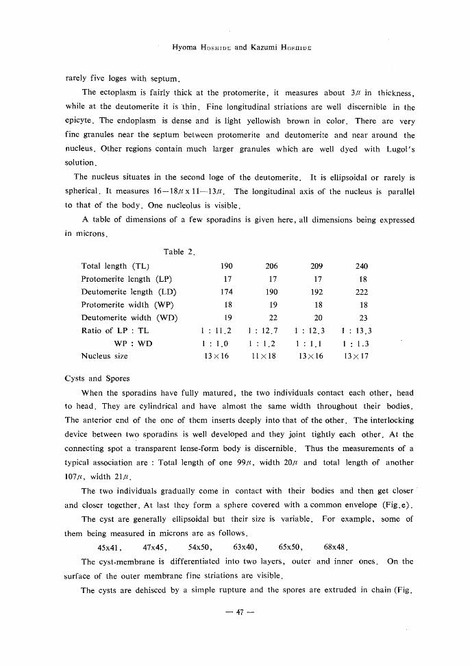
A table of dimensions of a few sporadins is given here, all dimensions being expressed
ln mlcrons ・
Table 2 .
Total length (TL)
Protomerite length (LP)
Deutomerite length (LD)
Protomerjte wjdth (WP)
Deutomerite width (WD)
Ratio of LP:TL
WP: WD
Nucleus size
190
17
174
18
19
1 : 11.2
1:1.0
13×16
206
17
190
19
22
1 : 12.7
1:1211x18
209
17
192
18
20
1 : 12.3
1 : 1.1
.
13x16
240
18
222
18
23
1 : 13.3
1 : 1.3
13x17
Cysts and Spores
When the sporadjns have fully matured, the two individuals contact each other, head
to head. They are cylindrical and have almost the same width throughout their bodies.
The anterior end of the one of them inserts deeply into that of the other. The interlocking
device between two sporadins is well developed and they joint tightly each other. At the
connecting spot a transparent lense-form body is discernible. Thus the measurements of a
typical associatjon are : Total length of one 99/t, width 20/t and total length of another
107!t, width 21/t.
The two individualti gradually come in contact with their bodies and then get closer
and closer together. At last they form a sphere covered wjth a common envelope (Fig.e).
The cyst are generally ellipsoidal but their size is variable. For example, some of
them being measured in microns are ag. follows.
45x41, 47x45, 54x50, 63x40, 65x50, 68x48.
The cyst-membrane iS differentiated into two layers, outer’ anq jpner ones. On the
surface of the outer membrane fine striations are visible.
The cysts are dehisced by a simple rupture and the spores are extruded in chain (Fig.
一 47 一一
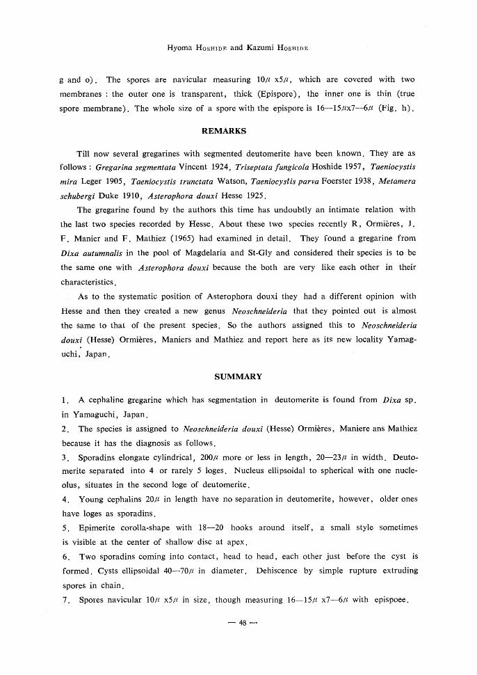
Hyoma HosHiDE and Kazumi HosHiDE
gand o). The spores are navicular measuring 10pt xspt, which are covered with two
membranes;the outer one is transparent, thick (Epispore), the jnner one is thin (true
spore membrane). The whole size of a spore with the epispore is 16-15ptx7-6zt (Fig, h).
REMARKS
Till now several gregarines with segmented deutomerite have been known. They are as
follows:Gregarina segmentata Vincent 1924, Triseptata fungicota H:oshide 1957, Taeniocアst’3
mira Leger 1905, Taeniocアstis〃unctata Watson, Taeniocアstis parva Foerster 1938,.Metamera
3c乃〃bergi Duke 1910, Asterophora 40〃xi Hesse 1925.
The gregarine found by the authors this time has undoubtly an intimate relation with
the last two species recorded by Hesse. About these two species recently R, Ormi6res,」.
F.Manier and F. Mathiez(1965)had examined in detail. They found a gregarine from
Dixa autu〃inal’s in the pool of Magdelaria and St-Gly and considered their species is to be
the same one with 1dsterophora do〃κ’because the both are very like each other in their
characteristics.
As to the systematic position of Asterophora douxi they had a different opinion with
Hesse and then they created a new genusハieosehneideria that they pointed out is almost
the same to that of the present species. So the authors assigned this to ハleoschne’deria
douxi(Hesse)Ormiらres, Maniers and Mathiez and report here as its new locality Yamag一 ゆ
uchi, Japan.
SUMMARY
1. A cephaline gregarine which has segmentation in deutomerite is found from Dixa sp.
in Yamaguchi, Japan.
2. The species is assigned to Neoschneideria douxi (Hesse) Ormieres, Maniere ans Mathiez.
because it has the diagnosis as follows.
3. Sporadins elongate cylindrical, 200pt more or less in length, 20-23pt in width. Deuto-
merite separated into 4 or rarely 5 loges. Nucleus ellipsoidal to spherical with one nucle-
olus, situates in the second loge of deutomerite.
4. Young cephalins 20pt in length have no separation in deutomerite, however, older ones
haye lpges as sPoradins.
5. Epimerite corolla-shape with 18-20 hooks around itself, a small style sometimes
is visible at the center of shallow disc at apex.
6. Two sporadins coming into contact, head to head, each other just before the cyst is
formed. Cysts ellipsojdal 40-70!-t in diameter. Dehiscence by simple rupture extruding
spores in chain.
7. Spores navicular 10!t x5!t in size, though measuring 16-15/t x7-6/t with epispoee.
p一 48 一

Notes on the Gregarines in Japan 2.
REFERENCES
i. Duke. L.:(1910) Some observation on a new Gregarine (Metamera schubergi) n. gen., n.
sp. Quart. J. Mjcr. Sc.,55:261-286.
2. Foerster, H.:(1938) Gregarinen in schlesischen lnsekten. Zeit. f. Parasit., 10:157-209,
644一一674.
3. Hesse, E., (1925) Deux nowvelles Gregarines segmentees. C. R. A. F. A. S., 49:403-409.
4. Hoshjde, H.: (1957一一一58) Studies on the cephaline Gregarines of Japan II. Bull. Fac. Educ.
Yamaguchi Uni., 7. Part II:45一一一109, 35一一一101.
5. Kamm, M. W.:(1922) Studies in Gregarines II. lllinojs Biol. Monogr.,7:1- r
6. L6ger, L. : (1892) Recherches sur les Gregarines. Tabl. Zool.. 3: 1-182.
7. L6ger, L.:(1906) Etude sur Taeniocystis m ira Leger, Gregarine metamerique. Arch. f.
protist., 7 : 307-329.
8. Nieschulz, O. : (1924) Eine neue Gregarjnengattung fur Schneideria metamorphose Nowling
(1922). (Paraschneideria) Zool. Anz., 60 : 149-150.
9. Ormieres, R., Manier, J. F. et Mathiez, F.: (1965) Proposition de Neosehneideria n., gen.
pour les Gregarines metamerisees parasites des larves de Dixa (Diptera-Culicidae) .
Ann. Psrasitol.,40 :249-254.
10. Vjncent, M. : (1924) on a new Gregarine, Gregarina segmentata n. sp. an intestinal parasite
of Cis bidentatus Oliv. .(Coleoptera) Parasitology, 16 : 196一一一一302.
EXPLANATION OF PLATE 1.
a:A full grown sporadjn.
b : A small cephalin.
c : A fairly large cephalin.
d : Anterior portion of a cephalin.
e: Pair of sporadjns attached to each other, head to head.
f : Two sporadins just previous,to cyst formation.
g: A cyst, in the process of extruding spores in chains.
h : Several navicular spores with epispore.
PLATE II.
i : A young cephalin, stained with Lugol’s solution.
j : A cephlin detached from the host’s gut wall.
k : A cephalin sticking to the host.
1 : A sporadjn, four loges of the deutomerite are visible.
m : A part of the host’s intestin, in which two parasites are seen through the wall of the
intestine .
n:Two pairs of sporadins, in the process of cyst formation.
o: Exudation of spores from the ripe cyst.
一49一

Hyoma HosHiDE and Kazumi HosHiDE
PLATE 1
モ に
1◎’/..・i
凸
馬
一一
b
”5ノし。
繰
∂o/t
e lolk
a丁む
d 5ノ丸
f 諒
㊥◎レ⑨
oo 頂
h
-50一
9 誘

Notes on the Gregarines in Japan 2.
PLATE ll
i’ /
愉こも’1:・’
K’ s♂.毛君㍉’
睾seA’ ..
コ ロ ロ ロ し
驚 醗・・激鹸・・ t ’;N.
轟轟磁声鳳.ご=二『訟溢歪.} ..;,ご,・.
waノ墜盤虹詮、翫
t t
i . . 幽1.一離 ・・㌔h;ノ_.馬.ゴψ講
一一 51 一一