Neuromagnetic Decoding of Simultaneous Bilateral Hand ... · BRAIN-MACHINE interface (BMI) for...
10
IEEE TRANSACTIONS ON NEURAL SYSTEMS AND REHABILITATION ENGINEERING, VOL. 26, NO. 6, JUNE 2018 1301 Neuromagnetic Decoding of Simultaneous Bilateral Hand Movements for Multidimensional Brain– Machine Interfaces Abdelkader Nasreddine Belkacem , Shuichi Nishio, Takafumi Suzuki, Hiroshi Ishiguro, and Masayuki Hirata Abstract — To provide multidimensional control, we describe the first reported decoding of bilateral hand movements by using single-trial magnetoencephalography signals as a new approach to enhance a user’s ability to interact with a complex environment through a multidimen- sional brain–machine interface. Ten healthy participants performed or imagined four types of bilateral hand move- ments during neuromagnetic measurements. By applying a support vector machine (SVM) method to classify the four movements regarding the sensor data obtained from the sensorimotor area, we found the mean accuracy of a two-class classification using the amplitudes of neuromag- netic fields to be particularly suitable for real-time appli- cations, with accuracies comparable to those obtained in previous studies involving unilateral movement. The sensor data from over the sensorimotor cortex showed discrim- inative time-series waveforms and time-frequency maps Manuscript received August 28, 2017; revised February 13, 2018 and April 24, 2018; accepted May 8, 2018. Date of publication May 15, 2018; date of current version June 6, 2018. This work was supported in part by the ImPACT Program of the Council for Science, Technology and Innovation (Cabinet Office, Government of Japan), in part by the “Development of BMI Technologies for Clinical Application” through the Strategic Research Program for Brain Sciences by AMED, in part by the “Research and Development of Technologies for High Speed Wireless Communication From Inside to Outside of the Body and Large Scale Data Analyses of Brain Information and Their Application for BMI” from NICT, and in part by KAKENHI funded by the Japan Society for the Promotion of Science under Grant 26282165. (Corresponding author: Masayuki Hirata.) A. N. Belkacem is with the Global Center for Medical Engineer- ing and Informatics, Endowed Research Department of Clinical Neu- roengineering, Osaka University, Osaka 565-0871, Japan (e-mail: [email protected]). S. Nishio is with Hiroshi Ishiguro Laboratories, Advanced Telecommu- nications Research Institute International, Kyoto 619-0288, Japan. T. Suzuki is with the Center for Information and Neural Networks, National Institute of Information and Communications Technology, Osaka 565-0871, Japan. H. Ishiguro is with the Department of System Innovation, Graduate School of Engineering Science, Osaka University, Osaka 560-8531, Japan. M. Hirata is with the Global Center for Medical Engineering and Infor- matics, Endowed Research Department of Clinical Neuroengineering, Osaka University, Osaka 565-0871, Japan, and also with the Department of Neurosurgery, Graduate School of Medicine, Osaka University, Osaka 565-0871, Japan (e-mail: [email protected]). This paper has supplementary downloadable material available at http://ieeexplore.ieee.org, provided by the author. Digital Object Identifier 10.1109/TNSRE.2018.2837003 in the bilateral hemispheres according to the four tasks. Furthermore, we used four-class classification algorithms based on the SVM method to decode all types of bilateral movements. Our results provided further proof that the slow components of neuromagnetic fields carry sufficient neural information to classify even bilateral hand move- ments and demonstrated the potential utility of decoding bilateral movements for engineering purposes such as mul- tidimensional motor control. Index Terms— Android, bilateral movements, brain- machine interface, magnetoencephalography, motor imagery, SVM classification, voluntary motor control. I. I NTRODUCTION A BRAIN-MACHINE interface (BMI) for motor control is a system based on decoding brain activity data from the motor cortex in the human forebrain to offer an alternative to physiological motor pathways. Such a system is able to send commands from the brain to a machine by using motor signals generated via voluntary motor control or motor imagery tasks in subjects performing certain body movements or imagining these movements without using the brain’s normal output path- ways of peripheral nerves and muscles, respectively. The brain signals for BMIs are measured with either invasive or nonin- vasive techniques. For decades, most studies have focused on discriminating unilateral actual/imagined movements such as right or left hand/finger/toe grasping and tongue movement [1]–[3]. This decoding of unilateral movements has been well studied and used in controlling machines (computers and cursors [4]–[7], robots and prostheses [8]–[10], etc.) to achieve a high relia- bility in noninvasive BMIs on the basis of electroencephalog- raphy (EEG) and magnetoencephalography (MEG) signals. Regarding the cortical activation of unilateral movements, pre- vious studies have shown that in both brain hemispheres there are typical movement-related cortical potentials or fields as well as certain oscillatory changes in the frequency bands, such as the alpha (8-13 Hz), beta (13-30 Hz) and gamma (>30 Hz) bands. Generally, a diffuse power decrease in the alpha and beta bands has been observed in the sensorimotor area, whereas a more focal power increase in the gamma band 1534-4320 © 2018 IEEE. Translations and content mining are permitted for academic research only. Personal use is also permitted, but republication/redistribution requires IEEE permission. See http://www.ieee.org/publications_standards/publications/rights/index.html for more information.
Transcript of Neuromagnetic Decoding of Simultaneous Bilateral Hand ... · BRAIN-MACHINE interface (BMI) for...
IEEE TRANSACTIONS ON NEURAL SYSTEMS AND REHABILITATION ENGINEERING, VOL. 26, NO. 6, JUNE 2018 1301
Neuromagnetic Decoding of SimultaneousBilateral Hand Movements for
Multidimensional Brain–Machine Interfaces
Abdelkader Nasreddine Belkacem , Shuichi Nishio, Takafumi Suzuki,Hiroshi Ishiguro, and Masayuki Hirata
Abstract— To provide multidimensional control, wedescribe the first reported decoding of bilateral handmovements by using single-trial magnetoencephalographysignals as a new approach to enhance a user’s ability tointeract with a complex environment through a multidimen-sional brain–machine interface. Ten healthy participantsperformed or imagined four types of bilateral hand move-ments during neuromagnetic measurements. By applyinga support vector machine (SVM) method to classify thefour movements regarding the sensor data obtained fromthe sensorimotor area, we found the mean accuracy of atwo-class classification using the amplitudes of neuromag-netic fields to be particularly suitable for real-time appli-cations, with accuracies comparable to those obtained inprevious studies involving unilateral movement. The sensordata from over the sensorimotor cortex showed discrim-inative time-series waveforms and time-frequency maps
Manuscript received August 28, 2017; revised February 13, 2018 andApril 24, 2018; accepted May 8, 2018. Date of publication May 15,2018; date of current version June 6, 2018. This work was supportedin part by the ImPACT Program of the Council for Science, Technologyand Innovation (Cabinet Office, Government of Japan), in part by the“Development of BMI Technologies for Clinical Application” throughthe Strategic Research Program for Brain Sciences by AMED, in partby the “Research and Development of Technologies for High SpeedWireless Communication From Inside to Outside of the Body and LargeScale Data Analyses of Brain Information and Their Application for BMI”from NICT, and in part by KAKENHI funded by the Japan Society for thePromotion of Science under Grant 26282165. (Corresponding author:Masayuki Hirata.)
A. N. Belkacem is with the Global Center for Medical Engineer-ing and Informatics, Endowed Research Department of Clinical Neu-roengineering, Osaka University, Osaka 565-0871, Japan (e-mail:[email protected]).
S. Nishio is with Hiroshi Ishiguro Laboratories, Advanced Telecommu-nications Research Institute International, Kyoto 619-0288, Japan.
T. Suzuki is with the Center for Information and Neural Networks,National Institute of Information and Communications Technology, Osaka565-0871, Japan.
H. Ishiguro is with the Department of System Innovation, GraduateSchool of Engineering Science, Osaka University, Osaka 560-8531,Japan.
M. Hirata is with the Global Center for Medical Engineering and Infor-matics, Endowed Research Department of Clinical Neuroengineering,Osaka University, Osaka 565-0871, Japan, and also with the Departmentof Neurosurgery, Graduate School of Medicine, Osaka University, Osaka565-0871, Japan (e-mail: [email protected]).
This paper has supplementary downloadable material available athttp://ieeexplore.ieee.org, provided by the author.
Digital Object Identifier 10.1109/TNSRE.2018.2837003
in the bilateral hemispheres according to the four tasks.Furthermore, we used four-class classification algorithmsbased on the SVM method to decode all types of bilateralmovements. Our results provided further proof that theslow components of neuromagnetic fields carry sufficientneural information to classify even bilateral hand move-ments and demonstrated the potential utility of decodingbilateral movements for engineering purposes such as mul-tidimensional motor control.
Index Terms— Android, bilateral movements, brain-machine interface, magnetoencephalography, motorimagery, SVM classification, voluntary motor control.
I. INTRODUCTION
ABRAIN-MACHINE interface (BMI) for motor control isa system based on decoding brain activity data from the
motor cortex in the human forebrain to offer an alternative tophysiological motor pathways. Such a system is able to sendcommands from the brain to a machine by using motor signalsgenerated via voluntary motor control or motor imagery tasksin subjects performing certain body movements or imaginingthese movements without using the brain’s normal output path-ways of peripheral nerves and muscles, respectively. The brainsignals for BMIs are measured with either invasive or nonin-vasive techniques.
For decades, most studies have focused on discriminatingunilateral actual/imagined movements such as right or lefthand/finger/toe grasping and tongue movement [1]–[3]. Thisdecoding of unilateral movements has been well studied andused in controlling machines (computers and cursors [4]–[7],robots and prostheses [8]–[10], etc.) to achieve a high relia-bility in noninvasive BMIs on the basis of electroencephalog-raphy (EEG) and magnetoencephalography (MEG) signals.Regarding the cortical activation of unilateral movements, pre-vious studies have shown that in both brain hemispheres thereare typical movement-related cortical potentials or fields aswell as certain oscillatory changes in the frequency bands, suchas the alpha (8-13 Hz), beta (13-30 Hz) and gamma (>30 Hz)bands. Generally, a diffuse power decrease in the alphaand beta bands has been observed in the sensorimotor area,whereas a more focal power increase in the gamma band
1534-4320 © 2018 IEEE. Translations and content mining are permitted for academic research only. Personal use is also permitted,but republication/redistribution requires IEEE permission. See http://www.ieee.org/publications_standards/publications/rights/index.html for more information.
1302 IEEE TRANSACTIONS ON NEURAL SYSTEMS AND REHABILITATION ENGINEERING, VOL. 26, NO. 6, JUNE 2018
has been observed in the motor area. Furthermore, previousnoninvasive EEG or MEG studies have demonstrated that thedecoding accuracy of a 2-class classification for two types ofvoluntary or imaginary body movements is significantly higherthan that expected by chance. The accuracy has been foundto vary from 60% to 80% using a sensor or source decodinglevel [4]–[19]. In contrast, the mean multi-class classificationaccuracy of movement decoding that has been achieved fromthe same part of the body has been less than or approximatelyequal to 60% in most BMI studies [20]–[23].
A dimension of control or degree of freedom is a criticalissue for achieving a high performance in motor BMIs in acomplex real-time interaction in a more realistic environment.In this case, decoding bilateral hand movements or handgestures may be an initial step toward increasing dimensional-ity and achieving a continuous and multidimensional controlof multiple devices (e.g., wheelchair and robotic arm). Thistype of multidimensional BMI system would require higherdimensional control and naturalistic BMI tasks. In addition,unilateral and bilateral movements activate different partsof the brain [24]–[28]. When patients perform or imaginebilateral movements during rehabilitation exercise sessions,patients activate more areas of the brain and therefore maxi-mize the neuroplastic benefits [29]. Furthermore, simultaneousbilateral movements (e.g., contralateral and ipsilateral handmovements) require a greater overall activation of musclecontraction and therefore probably require the firing of agreater number of cortical cells [24], [25]. Because the topog-raphy of brain activity is well known regarding unilateralmovements [30], [31], it is of interest to clarify the spa-tiotemporal frequency distribution of neural activity duringsimultaneous bilateral hand movements. In addition, peoplesimultaneously use their hands bilaterally for doing differenttasks in daily life. However, to date, analyzing human brainactivity during simultaneous bilateral hand movements has notbeen reported in detail. This type of analysis may offer a betterunderstanding of the neural correlates of bilateral hand move-ments, which remain unclear. Only a few studies based onfunctional magnetic resonance imaging (fMRI) measurementshave offered a better understanding of the neural correlates ofuni- and bi-manual finger presses [32] and bimanual anti-phaseand in-phase movements [33]. Furthermore, decoding bilateralmovements would also provide multidimensional controlin multifunctional augmentative and alternative communi-cation systems. Regarding unilateral movements, previousBMI studies based on sensorimotor amplitudes and rhythmshave used electroencephalograms (EEGs), magnetoencephalo-grams (MEGs), or electrocoticograms (ECoGs) to distinguishbetween the actual/imagined movement and the rest state(e.g., right hand grasping vs. relaxing position of the lefthand (no motion), and left hand grasping vs. relaxing positionof the right hand). Only a few studies have added a case ofbilateral movements, such as closing (grasping) bilateral handsor simultaneously moving bilateral fingers [21], [23]–[26].None of these studies have attempted to decode four typesof simultaneous bilateral hand movements (e.g., R-Closevs. L-Open: closing a right hand vs. opening a left hand,R-Open vs. L-Close: opening a right hand vs. closing a left
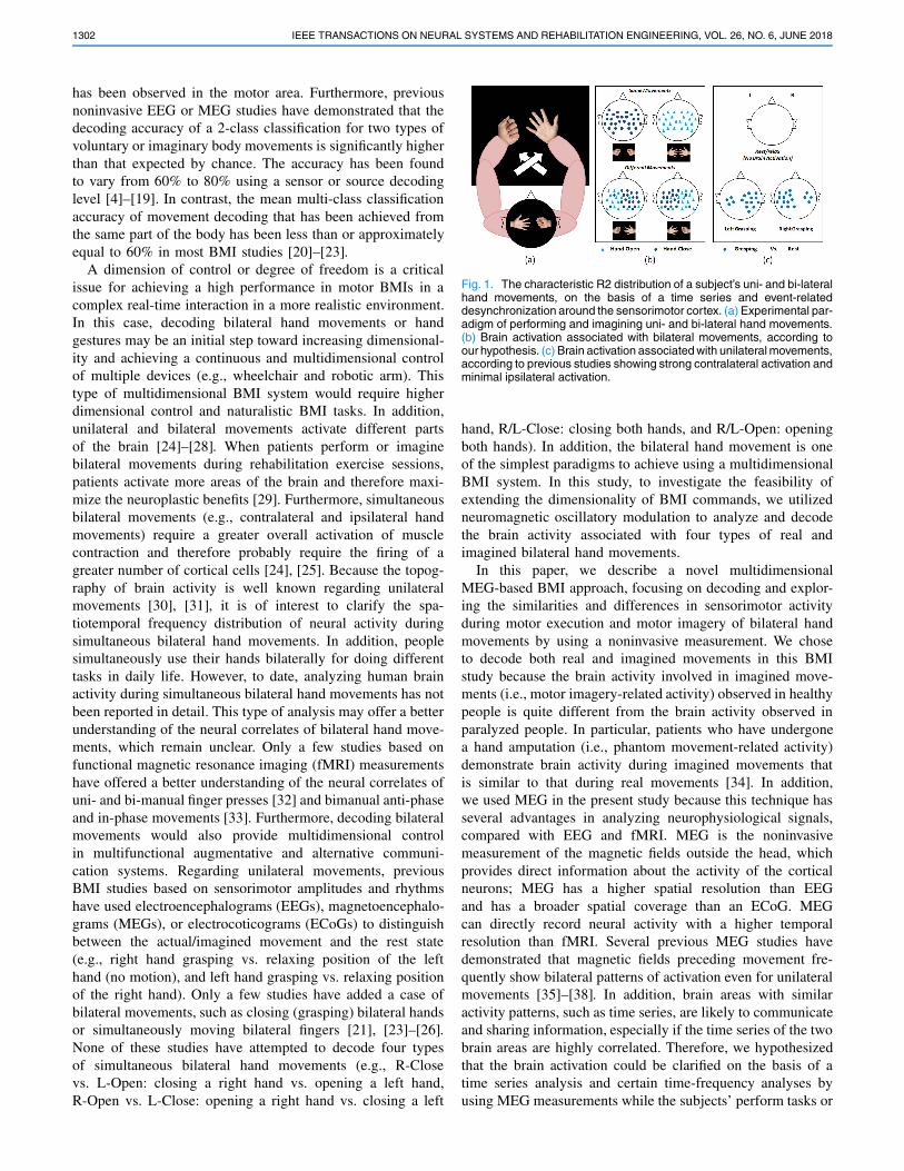
Fig. 1. The characteristic R2 distribution of a subject’s uni- and bi-lateralhand movements, on the basis of a time series and event-relateddesynchronization around the sensorimotor cortex. (a) Experimental par-adigm of performing and imagining uni- and bi-lateral hand movements.(b) Brain activation associated with bilateral movements, according toour hypothesis. (c) Brain activation associated with unilateral movements,according to previous studies showing strong contralateral activation andminimal ipsilateral activation.
hand, R/L-Close: closing both hands, and R/L-Open: openingboth hands). In addition, the bilateral hand movement is oneof the simplest paradigms to achieve using a multidimensionalBMI system. In this study, to investigate the feasibility ofextending the dimensionality of BMI commands, we utilizedneuromagnetic oscillatory modulation to analyze and decodethe brain activity associated with four types of real andimagined bilateral hand movements.
In this paper, we describe a novel multidimensionalMEG-based BMI approach, focusing on decoding and explor-ing the similarities and differences in sensorimotor activityduring motor execution and motor imagery of bilateral handmovements by using a noninvasive measurement. We choseto decode both real and imagined movements in this BMIstudy because the brain activity involved in imagined move-ments (i.e., motor imagery-related activity) observed in healthypeople is quite different from the brain activity observed inparalyzed people. In particular, patients who have undergonea hand amputation (i.e., phantom movement-related activity)demonstrate brain activity during imagined movements thatis similar to that during real movements [34]. In addition,we used MEG in the present study because this technique hasseveral advantages in analyzing neurophysiological signals,compared with EEG and fMRI. MEG is the noninvasivemeasurement of the magnetic fields outside the head, whichprovides direct information about the activity of the corticalneurons; MEG has a higher spatial resolution than EEGand has a broader spatial coverage than an ECoG. MEGcan directly record neural activity with a higher temporalresolution than fMRI. Several previous MEG studies havedemonstrated that magnetic fields preceding movement fre-quently show bilateral patterns of activation even for unilateralmovements [35]–[38]. In addition, brain areas with similaractivity patterns, such as time series, are likely to communicateand sharing information, especially if the time series of the twobrain areas are highly correlated. Therefore, we hypothesizedthat the brain activation could be clarified on the basis of atime series analysis and certain time-frequency analyses byusing MEG measurements while the subjects’ perform tasks or
BELKACEM et al.: NEUROMAGNETIC DECODING OF SIMULTANEOUS BILATERAL HAND MOVEMENTS 1303
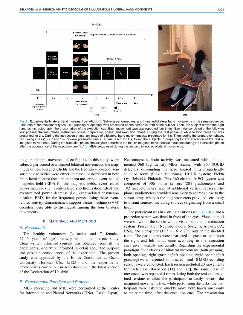
Fig. 2. Experimental bilateral hand movement paradigm. (a) Subjects performed real and imagined bilateral hand movements in the same sequence.First, one of the movement types, i.e., grasping or opening, was presented on the screen in front of the subject. Then, the subject moved the righthand as instructed upon the presentation of the execution cue. Each movement type was repeated four times. Each trial consisted of the followingfour phases; the rest phase, instruction phase, preparation phase, and execution phase. During the rest phase, a white fixation cross “+” waspresented for 3 s. During the instruction phase, an image of a bilateral hand movement was presented for 1 s. Then, during the preparation phase,two timing cues (“> <” and “><”) were presented one at a time, each for 1 s, to aid the subjects in preparing for the execution of the real orimagined movements. During the execution phase, the subjects performed the real or imagined movement as requested during the instruction phaseafter the appearance of the execution cue “×.” (b) MEG setup used during the real and imagined bilateral movements.
imagine bilateral movements (see Fig. 1). In this study, whensubjects performed or imagined bilateral movements, the mag-nitude of neuromagnetic fields and the frequency power of sen-sorimotor activities were either increased or decreased in bothbrain hemispheres; these phenomena are termed event-relatedmagnetic field (ERF) for the magnetic fields, event-relatedpower increase (i.e., event-related synchronization, ERS) andevent-related power decrease (i.e., event-related desynchro-nization, ERD) for the frequency power. Using these event-related activity characteristics, support vector machine (SVM)decoders were able to distinguish among the four bilateralmovements.
II. MATERIALS AND METHODS
A. Participants
Ten healthy volunteers (3 males and 7 females,22-45 years of age) participated in the present study.Clear written informed consent was obtained from all theparticipants, who were informed in detail about the purposeand possible consequences of the experiment. The presentstudy was approved by the Ethics Committee at OsakaUniversity Hospital (No. 15123), and the experimentalprotocol was carried out in accordance with the latest versionof the Declaration of Helsinki.
B. Experimental Paradigm and Protocol
MEG recording and MRI were performed at the Centerfor Information and Neural Networks (CiNet, Osaka, Japan).
Neuromagnetic brain activity was measured with an aug-mented 360 high-density MEG scanner with 360 SQUIDdetectors surrounding the head housed in a magneticallyshielded room (Elekta Neuromag TRIUX system; ElektaOy, Helsinki, Finland). This 360-channel MEG system wascomposed of 306 planar sensors (204 gradiometers and102 magnetometers) and 54 additional vertical sensors. Theplanar gradiometers provided sensitivity to sources close to thesensor array, whereas the magnetometers provided sensitivityto distant sources, including sources originating from a smallhead.
The participant was in a sitting position (see Fig. 2(b)), and aprojection screen was fixed in front of the eyes. Visual stimuliwere shown on the screen with a visual stimulus presentationsystem (Presentation, Neurobehavioral Systems, Albany, CA,USA) and a projector (12.5 × 16 × 20��) outside the shieldedroom. The participants were instructed to grasp or open boththe right and left hands once according to the executioncues given visually and aurally. Regarding the experimentalparadigm, four classes of bilateral movements (both grasping,both opening, right grasping/left opening, right opening/leftgrasping) were presented on the screen, and 10 MEG recordingsessions were conducted. Each session included 20 movementsfor each class. Based on [11] and [12], the same class ofmovement was repeated 4 times during both the real and imag-ined sessions to allow the participants to easily perform theimagined movements (i.e., while performing the tasks, the par-ticipants were asked to quickly move both hands once-onlyat the same time, after the execution cue). The presentation
1304 IEEE TRANSACTIONS ON NEURAL SYSTEMS AND REHABILITATION ENGINEERING, VOL. 26, NO. 6, JUNE 2018
order of the movement types was randomized. The participantswere instructed to perform the imagined or actual movementswithout moving any other body parts, to avoid both muscle andhead motion artifacts. In addition, the experimental paradigmand protocol were designed to enable the participants to kines-thetically perform the indicated motor imagery task rather thanrelying on visual imagery, while avoiding any motion duringthe imagery task. To reduce fatigue, we asked participants totake 10-min to 20-min breaks between the first five randomsessions and the rest sessions. The schematic of the overallexperimental paradigm is shown in Fig. 2(a).
Furthermore, a 3 T structural MRI scan was performedto more clearly obtain detailed images of the head andbrain structures in slices for each subject. During the MEGrecording and MRI scanning, we measured certain referencepoints. These reference points were used to co-register theMEG sensor positions to voxel coordinates of the anatomicaldata. An iterative closest points (ICP) algorithm was usedto refine this sensor/anatomy registration. The recorded MRIdata were converted using FreeSurfer software (MartinosCenter software, http://surfer.nmr.mgh.harvard.edu/) into aformat suitable for uploading on the Brainstorm software(http://neuroimage.usc.edu/brainstorm), and we then alignedthe MEG data with the individual anatomical data. The acqui-sition of individual anatomical MRIs of participants in thisstudy is desirable because the source localization is part ofthe planned analysis.
C. Data Acquisition and PreprocessingNeuromagnetic activity was sampled at 1000 Hz, after
which the temporal extension of the signal space separa-tion method (tSSS) was used to suppress noise and artifactsgenerated by the sensors and sources of interference locatedvery close to the MEG sensors. Because the spatial filteringused in this study was based directly on Maxwell’s equations,the operation can be called Maxwell filtering; hence, we usedMaxFilter 2.2 software to calculate the tSSS. A bandpassfilter from 0.5 to 100 Hz and a 60-Hz notch filter wereapplied using Brainstorm software [39] to reduce some DCand high frequency noises and eliminate AC line noise.In addition, muscular activities were recorded using 8 elec-tromyogram (EMG) electrodes positioned on the flexor andextensor pollicis longus to measure hand flexion and extensionduring the performance of bilateral movements. Thus, to obtainthe exact onset of the execution of real movement, we usedEMG signals as a trigger instead of using the onset time, onthe basis of the execution of the visual stimulus “×”. Afterthe presentation of the execution cue, robust EMG amplitudeswere observed during the real movement but not during theimagined movement (see Supplementary Fig. 2). For voluntarymovements, 100 epochs per task were extracted in referenceto the MEG onsets. Each epoch had duration of 2 s, including1 s of pre-onset and 1 s of post-onset. We performed thesame protocol for the imagined movements in reference tothe onsets of the visual and auditory stimuli. We calculatedthe average of 100 epochs for each bilateral movement typeby using Brainstorm software. To clarify the brain activationassociated with bilateral hand movements, we applied the
continuous wavelet transform (Morlet wavelet) to plot the2D time-frequency spectrogram of the average of each typeof hand movement over the sensorimotor cortex area. Then,we analyzed the spatiotemporal frequency patterns over thehead (not only over the sensorimotor area) by calculating the2D time-frequency representation by using all the MEG sen-sors. The event-related power decreases/increases in the MEGsignals were calculated after computing the 2D time-frequencyspectrogram using Brainstorm functions during physical motorexecution and mental motor imagery, which is a widely usedtechnique in BMI studies. The frequency power of the eventperiods was averaged across 100 trials before calculating theERD/ERS using a Morlet wavelet transform. For the SVMclassification, we used a single-trial for the binary and multi-class decoding of bilateral movements using MATLAB 2016asoftware (Mathworks, Natick, MA, USA), which was extendedto distinguish among the four classes using a multi-SVM.
D. Classification of Bilateral Hand Movements
In this study, the sensor-based BMI analysis took advan-tage of the event-related phenomenon observed at spe-cific MEG sensors located over the sensorimotor cortexduring real or imagined bilateral movements. To calculatethe classification accuracy over all the subjects, we usedbinary SVM classifiers with the data from 114 MEGsensors (see Supplementary Fig. 1) after the preprocessingphase. For feature extraction, we used the MEG amplitudesof the movement-related cortical fields from single trialsof 0 to 500 ms (i.e., from the EMG onset for real movementsand from the presentation of the execution cue for imaginedmovements) for each MEG sensor around the sensorimotorcortex for each bilateral hand movement. To reduce the featurevector dimension, mean amplitude of single trial for each MEGsensor around the sensorimotor was used instead of using thewhole time series which means the feature vector was theamplitude-averaged activity within the temporal segment of[0 500] ms. Thus, we calculated the average of the ampli-tudes in each trial during a 500-ms interval for 114 MEGsensors (the sensors were determined empirically according tothe time-frequency analysis within/across subjects). A featureselection algorithm (sequential forward feature selection)was applied to avoid the high multicollinearity between the114 values of the feature vector (see Fig. 3). We randomly splitthe MEG data (100 trials for each bilateral movement type)to 75% training data and 25% testing data so that the testingdataset was independent of the training dataset. Then we run5-fold cross-validation approach in 75% of the trials in order toget the best features which refer to the most informative MEGsensors. Finally, we tested SVM model with the rest of 25% oftrials. For the multi-class classification, we used two differentalgorithms: the hierarchical SVM algorithm [40], a decisiontree with an SVM classifier at each node, and the multi-SVM(one against all [41]). We specified a Gaussian RBF kernel forthe decision function of the SVM classifiers.
For the statistical analysis, two-sample t-tests were con-ducted to determine whether the classification accuracysignificantly exceeded chance levels (50% for the 2-class
BELKACEM et al.: NEUROMAGNETIC DECODING OF SIMULTANEOUS BILATERAL HAND MOVEMENTS 1305
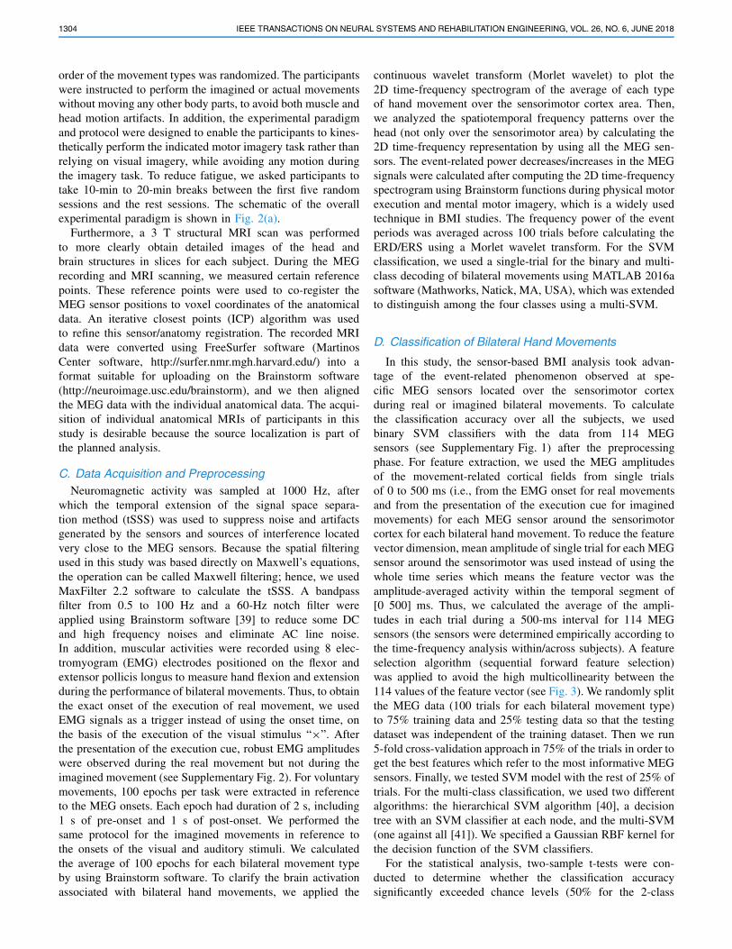
Fig. 3. Topography of the number of times a MEG sensor is selectedas significant feature using sequential feature selection method forSVM classification across the cohort (114-MEG-selsected sensors sur-rounded by black borders were empirically selected in this study fromboth hemispheres above the sensorimotor cortex). See Supplemen-tary Figure 1 for more details about MEG sensors’ labels. The color-map (blue-red) is based on the number of subjects.
classification and 25% for the 4-class classification) with athreshold of p < 0.05. Non-parametric statistical tests, suchas t-tests or two-way analysis of variance (ANOVA), and amultiple comparisons test of means using Tukey’s methodwere used not only to confirm that the decoding performancesignificantly exceeded chance level but also to examine thesimilarities and differences in decoding between the bilateralmovement types and participants and between the real andimagined movements.
III. RESULTS
A. Time-Frequency Spectrograms of Bilateral HandMovements by Using a Wavelet Method
To better understand brain activation related to bilateralmovements, we analyzed the spatiotemporal frequency pat-terns by calculating the time-frequency spectrograms by usinga Morlet wavelet transform at each of the MEG sensors overthe sensorimotor area.
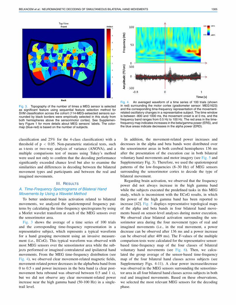
Fig. 3 shows the average of a time series of 100 trialsand the corresponding time-frequency representation in arepresentative subject, which represents a typical waveformfor a hand grasping movement using an invasive measure-ment (i.e., ECoG). This typical waveform was observed withmost MEG sensors over the sensorimotor area while the sub-jects performed or imagined contralateral and ipsilateral handmovements. From the MEG time-frequency distribution (seeFig. 4), we observed clear movement-related magnetic fields,movement-related power decreases in the alpha/beta band from0 to 0.5 s and power increases in the beta band (a clear post-movement beta rebound was observed between 0.5 and 1 s),but we did not observe any clear movement-related powerincrease near the high gamma band (50-100 Hz) in a single-trail level.
Fig. 4. An averaged waveform of a time series of 100 trials (shownin red) surrounding the motor cortex (gradiometer sensor: MEG1623)and the corresponding time-frequency representation of the movement-related oscillatory changes in a representative subject. The time windowis between -800 and 1000 ms, the movement onset is at 0 ms, and thefrequency band ranges from 0.5 Hz to 100 Hz. The red area in the time-frequency map indicates increases in the beta/gamma power (ERS), andthe blue areas indicate decreases in the alpha power (ERD).
In addition, the movement-related power increases anddecreases in the alpha and beta bands were distributed overthe sensorimotor areas in both cerebral hemispheres 136 msafter the presentation of the execution cue in both bilateralvoluntary hand movements and motor imagery (see Fig. 5 andSupplementary Fig. 3). Therefore, we used the spatiotemporalpatterns of the low-frequencies (8–30 Hz) of MEG sensorssurrounding the sensorimotor cortex to decode the type ofbilateral movement.
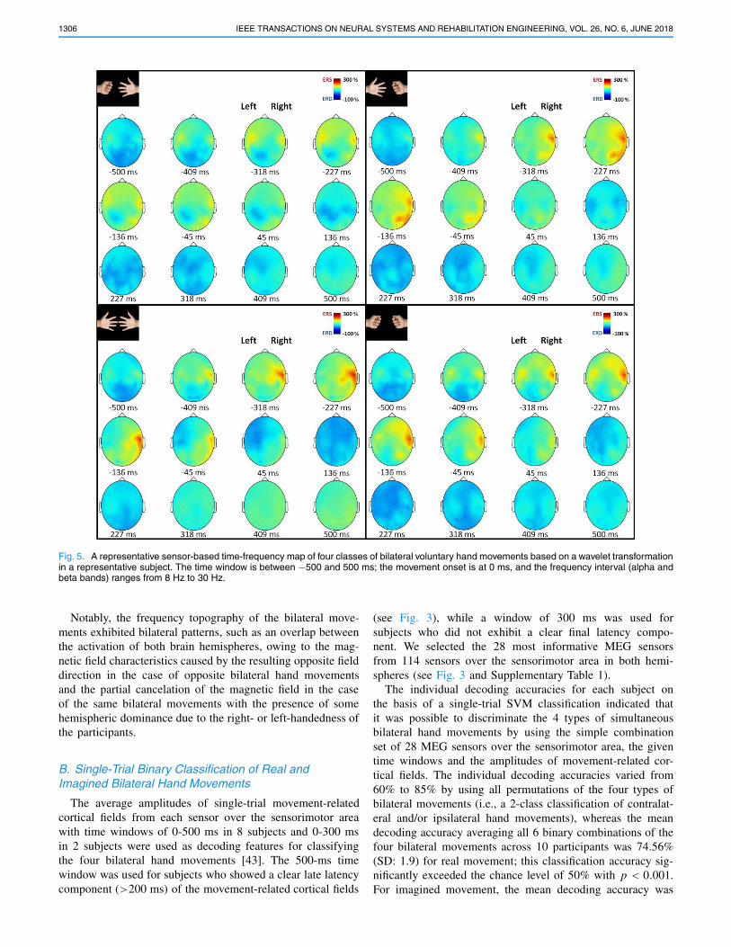
Regarding brain activation, we observed that the frequencypower did not always increase in the high gamma bandwhile the subjects executed the predefined tasks in this MEGstudy, which is inconsistent with the ECoG results, in whichthe power of the high gamma band has been reported toincrease [42]. Fig. 5 displays representative topological mapsof the alpha and beta bands in four bilateral hand move-ments based on sensor-level analyses during motor execution.We observed clear bilateral activation surrounding the sen-sorimotor area during the four movement types of real andimagined movements (i.e., in the real movement, a powerdecrease can be observed after 136 ms and a power increasecan be observed after 409 ms). The F-values of the multiplecomparison tests were calculated for the representative sensor-based time-frequency map of the four classes of bilateralvoluntary hand movements (see Fig. 6). Then, we calcu-lated the group average of the sensor-based time-frequencymap of the four bilateral hand classes across subjects (seeSupplementary Figs. 4-11). A clear power increase/decreasewas observed in the MEG sensors surrounding the sensorimo-tor area in all four bilateral hand classes across subjects in bothreal and imagined movements. Based on these observations,we selected the most relevant MEG sensors for the decodingphase.
1306 IEEE TRANSACTIONS ON NEURAL SYSTEMS AND REHABILITATION ENGINEERING, VOL. 26, NO. 6, JUNE 2018
Fig. 5. A representative sensor-based time-frequency map of four classes of bilateral voluntary hand movements based on a wavelet transformationin a representative subject. The time window is between −500 and 500 ms; the movement onset is at 0 ms, and the frequency interval (alpha andbeta bands) ranges from 8 Hz to 30 Hz.
Notably, the frequency topography of the bilateral move-ments exhibited bilateral patterns, such as an overlap betweenthe activation of both brain hemispheres, owing to the mag-netic field characteristics caused by the resulting opposite fielddirection in the case of opposite bilateral hand movementsand the partial cancelation of the magnetic field in the caseof the same bilateral movements with the presence of somehemispheric dominance due to the right- or left-handedness ofthe participants.
B. Single-Trial Binary Classification of Real andImagined Bilateral Hand Movements
The average amplitudes of single-trial movement-relatedcortical fields from each sensor over the sensorimotor areawith time windows of 0-500 ms in 8 subjects and 0-300 msin 2 subjects were used as decoding features for classifyingthe four bilateral hand movements [43]. The 500-ms timewindow was used for subjects who showed a clear late latencycomponent (>200 ms) of the movement-related cortical fields
(see Fig. 3), while a window of 300 ms was used forsubjects who did not exhibit a clear final latency compo-nent. We selected the 28 most informative MEG sensorsfrom 114 sensors over the sensorimotor area in both hemi-spheres (see Fig. 3 and Supplementary Table 1).
The individual decoding accuracies for each subject onthe basis of a single-trial SVM classification indicated thatit was possible to discriminate the 4 types of simultaneousbilateral hand movements by using the simple combinationset of 28 MEG sensors over the sensorimotor area, the giventime windows and the amplitudes of movement-related cor-tical fields. The individual decoding accuracies varied from60% to 85% by using all permutations of the four types ofbilateral movements (i.e., a 2-class classification of contralat-eral and/or ipsilateral hand movements), whereas the meandecoding accuracy averaging all 6 binary combinations of thefour bilateral movements across 10 participants was 74.56%(SD: 1.9) for real movement; this classification accuracy sig-nificantly exceeded the chance level of 50% with p < 0.001.For imagined movement, the mean decoding accuracy was
BELKACEM et al.: NEUROMAGNETIC DECODING OF SIMULTANEOUS BILATERAL HAND MOVEMENTS 1307
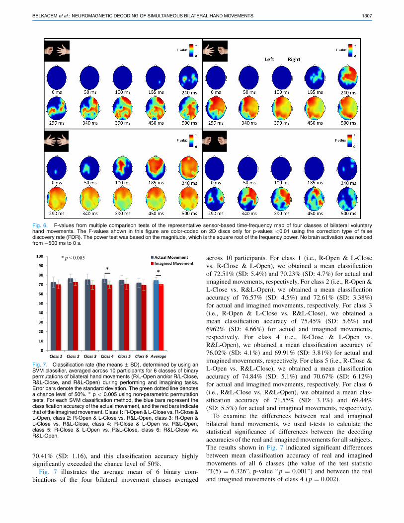
Fig. 6. F-values from multiple comparison tests of the representative sensor-based time-frequency map of four classes of bilateral voluntaryhand movements. The F-values shown in this figure are color-coded on 2D discs only for p-values <0.01 using the correction type of falsediscovery rate (FDR). The power test was based on the magnitude, which is the square root of the frequency power. No brain activation was noticedfrom −500 ms to 0 s.
Fig. 7. Classification rate (the means ± SD), determined by using anSVM classifier, averaged across 10 participants for 6 classes of binarypermutations of bilateral hand movements (R/L-Open and/or R/L-Close,R&L-Close, and R&L-Open) during performing and imagining tasks.Error bars denote the standard deviation. The green dotted line denotesa chance level of 50%. ∗ p < 0.005 using non-parametric permutationtests. For each SVM classification method, the blue bars represent theclassification accuracy of the actual movement, and the red bars indicatethat of the imagined movement. Class 1: R-Open & L-Close vs. R-Close &L-Open, class 2: R-Open & L-Close vs. R&L-Open, class 3: R-Open &L-Close vs. R&L-Close, class 4: R-Close & L-Open vs. R&L-Open,class 5: R-Close & L-Open vs. R&L-Close, class 6: R&L-Close vs.R&L-Open.
70.41% (SD: 1.16), and this classification accuracy highlysignificantly exceeded the chance level of 50%.
Fig. 7 illustrates the average mean of 6 binary com-binations of the four bilateral movement classes averaged
across 10 participants. For class 1 (i.e., R-Open & L-Closevs. R-Close & L-Open), we obtained a mean classificationof 72.51% (SD: 5.4%) and 70.23% (SD: 4.7%) for actual andimagined movements, respectively. For class 2 (i.e., R-Open &L-Close vs. R&L-Open), we obtained a mean classificationaccuracy of 76.57% (SD: 4.5%) and 72.61% (SD: 3.38%)for actual and imagined movements, respectively. For class 3(i.e., R-Open & L-Close vs. R&L-Close), we obtained amean classification accuracy of 75.45% (SD: 5.6%) and6962% (SD: 4.66%) for actual and imagined movements,respectively. For class 4 (i.e., R-Close & L-Open vs.R&L-Open), we obtained a mean classification accuracy of76.02% (SD: 4.1%) and 69.91% (SD: 3.81%) for actual andimagined movements, respectively. For class 5 (i.e., R-Close &L-Open vs. R&L-Close), we obtained a mean classificationaccuracy of 74.84% (SD: 5.1%) and 70.67% (SD: 6.12%)for actual and imagined movements, respectively. For class 6(i.e., R&L-Close vs. R&L-Open), we obtained a mean clas-sification accuracy of 71.55% (SD: 3.1%) and 69.44%(SD: 5.5%) for actual and imagined movements, respectively.
To examine the differences between real and imaginedbilateral hand movements, we used t-tests to calculate thestatistical significance of differences between the decodingaccuracies of the real and imagined movements for all subjects.The results shown in Fig. 7 indicated significant differencesbetween mean classification accuracy of real and imaginedmovements of all 6 classes (the value of the test statistic“T(5) = 6.326”, p-value “p = 0.001”) and between the realand imagined movements of class 4 (p = 0.002).
1308 IEEE TRANSACTIONS ON NEURAL SYSTEMS AND REHABILITATION ENGINEERING, VOL. 26, NO. 6, JUNE 2018
We used a two-way ANOVA to evaluate the decodingperformance and to compare the classification results amongthe six classes across 10 participants. For the real move-ments, no significant differences were observed between par-ticipants (F(9,59) = 1.27, p = 0.28), and no significantdifferences were observed between classes (F(5,59) = 1.55,p = 0.195). Using a multiple comparisons test of the means,we observed that no classes had significantly different means,except for classes 2 and 6, the means of which were signifi-cantly different when using the Tukey-Kramer test for the typeof critical value. For the imagined movements, no significantdifferences were observed using a two-way ANOVA betweenclasses (F(5,59) = 0.76, p = 0.583), but we observedsignificant differences between participants (F(9,59) = 2.78,p = 0.011). These significant differences may be related to theperformance of each participant on the imagery tasks. Usinga multiple comparisons test of means with the Tukey-Kramermethod, we observed that no classes had means that weresignificantly different.
C. Multi-Class Decoding of Real and ImaginedBilateral Hand Movements
Using single-trial multi-class decoding algorithms basedon an SVM classifier, we sought to distinguish betweenthe four types of bilateral hand movements. We found thatthe individual decoding accuracy of real movements largelyexceeded the chance level (i.e., 25%), by using a hierarchicalSVM (p = 1.292e-10) and a multi-SVM (p = 9.296e-10).For the imagined movements, we found that the individualdecoding accuracy also exceeded the chance level, by usinga hierarchical SVM (p = 9.204e-09) and a multi-SVM(p = 1.237e-07). The average decoding accuracy for allthe subjects was approximately 50%, which is approximatelytwice the chance level in both scenarios (performing andimagining bilateral movements).
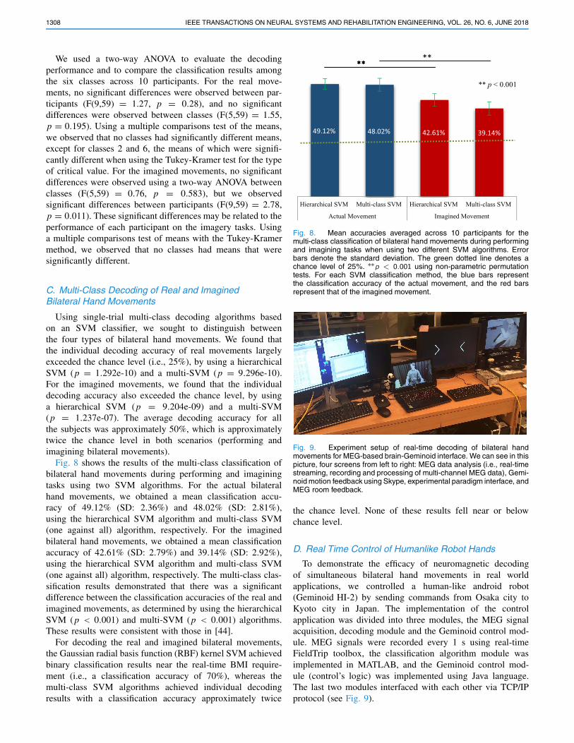
Fig. 8 shows the results of the multi-class classification ofbilateral hand movements during performing and imaginingtasks using two SVM algorithms. For the actual bilateralhand movements, we obtained a mean classification accu-racy of 49.12% (SD: 2.36%) and 48.02% (SD: 2.81%),using the hierarchical SVM algorithm and multi-class SVM(one against all) algorithm, respectively. For the imaginedbilateral hand movements, we obtained a mean classificationaccuracy of 42.61% (SD: 2.79%) and 39.14% (SD: 2.92%),using the hierarchical SVM algorithm and multi-class SVM(one against all) algorithm, respectively. The multi-class clas-sification results demonstrated that there was a significantdifference between the classification accuracies of the real andimagined movements, as determined by using the hierarchicalSVM (p < 0.001) and multi-SVM (p < 0.001) algorithms.These results were consistent with those in [44].
For decoding the real and imagined bilateral movements,the Gaussian radial basis function (RBF) kernel SVM achievedbinary classification results near the real-time BMI require-ment (i.e., a classification accuracy of 70%), whereas themulti-class SVM algorithms achieved individual decodingresults with a classification accuracy approximately twice
Fig. 8. Mean accuracies averaged across 10 participants for themulti-class classification of bilateral hand movements during performingand imagining tasks when using two different SVM algorithms. Errorbars denote the standard deviation. The green dotted line denotes achance level of 25%. ∗∗p < 0.001 using non-parametric permutationtests. For each SVM classification method, the blue bars representthe classification accuracy of the actual movement, and the red barsrepresent that of the imagined movement.
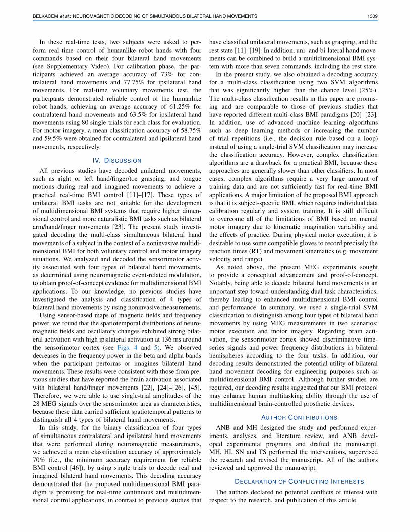
Fig. 9. Experiment setup of real-time decoding of bilateral handmovements for MEG-based brain-Geminoid interface. We can see in thispicture, four screens from left to right: MEG data analysis (i.e., real-timestreaming, recording and processing of multi-channel MEG data), Gemi-noid motion feedback using Skype, experimental paradigm interface, andMEG room feedback.
the chance level. None of these results fell near or belowchance level.
D. Real Time Control of Humanlike Robot Hands
To demonstrate the efficacy of neuromagnetic decodingof simultaneous bilateral hand movements in real worldapplications, we controlled a human-like android robot(Geminoid HI-2) by sending commands from Osaka city toKyoto city in Japan. The implementation of the controlapplication was divided into three modules, the MEG signalacquisition, decoding module and the Geminoid control mod-ule. MEG signals were recorded every 1 s using real-timeFieldTrip toolbox, the classification algorithm module wasimplemented in MATLAB, and the Geminoid control mod-ule (control’s logic) was implemented using Java language.The last two modules interfaced with each other via TCP/IPprotocol (see Fig. 9).
BELKACEM et al.: NEUROMAGNETIC DECODING OF SIMULTANEOUS BILATERAL HAND MOVEMENTS 1309
In these real-time tests, two subjects were asked to per-form real-time control of humanlike robot hands with fourcommands based on their four bilateral hand movements(see Supplementary Video). For calibration phase, the par-ticipants achieved an average accuracy of 73% for con-tralateral hand movements and 77.75% for ipsilateral handmovements. For real-time voluntary movements test, theparticipants demonstrated reliable control of the humanlikerobot hands, achieving an average accuracy of 61.25% forcontralateral hand movements and 63.5% for ipsilateral handmovements using 80 single-trials for each class for evaluation.For motor imagery, a mean classification accuracy of 58.75%and 59.5% were obtained for contralateral and ipsilateral handmovements, respectively.
IV. DISCUSSION
All previous studies have decoded unilateral movements,such as right or left hand/finger/toe grasping, and tonguemotions during real and imagined movements to achieve apractical real-time BMI control [11]–[17]. These types ofunilateral BMI tasks are not suitable for the developmentof multidimensional BMI systems that require higher dimen-sional control and more naturalistic BMI tasks such as bilateralarm/hand/finger movements [23]. The present study investi-gated decoding the multi-class simultaneous bilateral handmovements of a subject in the context of a noninvasive multidi-mensional BMI for both voluntary control and motor imagerysituations. We analyzed and decoded the sensorimotor activ-ity associated with four types of bilateral hand movements,as determined using neuromagnetic event-related modulation,to obtain proof-of-concept evidence for multidimensional BMIapplications. To our knowledge, no previous studies haveinvestigated the analysis and classification of 4 types ofbilateral hand movements by using noninvasive measurements.
Using sensor-based maps of magnetic fields and frequencypower, we found that the spatiotemporal distributions of neuro-magnetic fields and oscillatory changes exhibited strong bilat-eral activation with high ipsilateral activation at 136 ms aroundthe sensorimotor cortex (see Figs. 4 and 5). We observeddecreases in the frequency power in the beta and alpha bandswhen the participant performs or imagines bilateral handmovements. These results were consistent with those from pre-vious studies that have reported the brain activation associatedwith bilateral hand/finger movements [22], [24]–[26], [45].Therefore, we were able to use single-trial amplitudes of the28 MEG signals over the sensorimotor area as characteristics,because these data carried sufficient spatiotemporal patterns todistinguish all 4 types of bilateral hand movements.
In this study, for the binary classification of four typesof simultaneous contralateral and ipsilateral hand movementsthat were performed during neuromagnetic measurements,we achieved a mean classification accuracy of approximately70% (i.e., the minimum accuracy requirement for reliableBMI control [46]), by using single trials to decode real andimagined bilateral hand movements. This decoding accuracydemonstrated that the proposed multidimensional BMI para-digm is promising for real-time continuous and multidimen-sional control applications, in contrast to previous studies that
have classified unilateral movements, such as grasping, and therest state [11]–[19]. In addition, uni- and bi-lateral hand move-ments can be combined to build a multidimensional BMI sys-tem with more than seven commands, including the rest state.
In the present study, we also obtained a decoding accuracyfor a multi-class classification using two SVM algorithmsthat was significantly higher than the chance level (25%).The multi-class classification results in this paper are promis-ing and are comparable to those of previous studies thathave reported different multi-class BMI paradigms [20]–[23].In addition, use of advanced machine learning algorithmssuch as deep learning methods or increasing the numberof trial repetitions (i.e., the decision rule based on a loop)instead of using a single-trial SVM classification may increasethe classification accuracy. However, complex classificationalgorithms are a drawback for a practical BMI, because theseapproaches are generally slower than other classifiers. In mostcases, complex algorithms require a very large amount oftraining data and are not sufficiently fast for real-time BMIapplications. A major limitation of the proposed BMI approachis that it is subject-specific BMI, which requires individual datacalibration regularly and system training. It is still difficultto overcome all of the limitations of BMI based on mentalmotor imagery due to kinematic imagination variability andthe effects of practice. During physical motor execution, it isdesirable to use some compatible gloves to record precisely thereaction times (RT) and movement kinematics (e.g. movementvelocity and range).
As noted above, the present MEG experiments soughtto provide a conceptual advancement and proof-of-concept.Notably, being able to decode bilateral hand movements is animportant step toward understanding dual-task characteristics,thereby leading to enhanced multidimensional BMI controland performance. In summary, we used a single-trial SVMclassification to distinguish among four types of bilateral handmovements by using MEG measurements in two scenarios:motor execution and motor imagery. Regarding brain acti-vation, the sensorimotor cortex showed discriminative time-series signals and power frequency distributions in bilateralhemispheres according to the four tasks. In addition, ourdecoding results demonstrated the potential utility of bilateralhand movement decoding for engineering purposes such asmultidimensional BMI control. Although further studies arerequired, our decoding results suggested that our BMI protocolmay enhance human multitasking ability through the use ofmultidimensional brain-controlled prosthetic devices.
AUTHOR CONTRIBUTIONS
ANB and MH designed the study and performed exper-iments, analyses, and literature review, and ANB devel-oped experimental programs and drafted the manuscript.MH, HI, SN and TS performed the interventions, supervisedthe research and revised the manuscript. All of the authorsreviewed and approved the manuscript.
DECLARATION OF CONFLICTING INTERESTS
The authors declared no potential conflicts of interest withrespect to the research, and publication of this article.
1310 IEEE TRANSACTIONS ON NEURAL SYSTEMS AND REHABILITATION ENGINEERING, VOL. 26, NO. 6, JUNE 2018
REFERENCES
[1] G. Pfurtscheller, C. Neuper, and J. Kalcher, “40-Hz oscillations dur-ing motor behavior in man,” Neurosci. Lett., vol. 164, pp. 179–182,Dec. 1993.
[2] G. Pfurtscheller, C. Brunner, A. Schlögl, and F. H. Lopes da Silva,“Mu rhythm (de)synchronization and EEG single-trial classification ofdifferent motor imagery tasks,” NeuroImage, vol. 31, no. 1, pp. 153–159,2006.
[3] C. Neuper, G. R. Müller-Putz, R. Scherer, and G. Pfurtscheller, “Motorimagery and EEG-based control of spelling devices and neuroprosthe-ses,” Prog. Brain Res., vol. 159, pp. 393–409, 2006, doi: 10.1016/S0079-6123(06)59025-9.
[4] T. M. Vaughan et al., “The wadsworth BCI research and developmentprogram: At home with BCI,” IEEE Trans. Neural Syst. Rehabil. Eng.,vol. 14, no. 2, pp. 229–233, Jun. 2006.
[5] J. R. Wolpaw, N. Birbaumer, D. J. McFarland, G. Pfurtscheller, andT. M. Vaughan, “Brain–computer interfaces for communication andcontrol,” Clin. Neurophysiol., vol. 113, pp. 767–791, Jun. 2002.
[6] N. Birbaumer, “Breaking the silence: Brain–computer interfaces (BCI)for communication and motor control,” Psychophysiology, vol. 43,pp. 517–532, Nov. 2006.
[7] B. Blankertz, G. Dornhege, M. Krauledat, K.-R. Müller, and G. Curio,“The non-invasive berlin brain–computer interface: Fast acquisitionof effective performance in untrained subjects,” NeuroImage, vol. 37,pp. 539–550, Aug. 2007.
[8] R. Fukuma et al., “Closed-loop control of a neuroprosthetic hand bymagnetoencephalographic signals,” PLoS ONE, vol. 10, p. e0131547,Jul. 2015.
[9] R. Fukuma et al., “Real-time control of a neuroprosthetic hand bymagnetoencephalographic signals from paralysed patients,” Sci. Rep.,vol. 6, Feb. 2016, Art. no. 21781.
[10] L. R. Hochberg et al., “Neuronal ensemble control of prosthetic devicesby a human with tetraplegia,” Nature, vol. 442, no. 7099, pp. 164–171,Jul. 2006.
[11] G. Pfurtscheller, “Enhancement of sensorimotor rhythms with motorimagery,” Appl. Psychophysiol. Biofeedback, vol. 31, p. 351, Dec. 2006.
[12] G. Pfurtscheller and C. Neuper, “Future prospects of ERD/ERS in thecontext of brain–computer interface (BCI) developments,” Event-Rel.Dyn. Brain Oscillations, vol. 159, pp. 433–437, 2006.
[13] F. Popescu, S. Fazli, Y. Badower, B. Blankertz, and K.-R. Müller, “Singletrial classification of motor imagination using 6 Dry EEG electrodes,”PLoS ONE, vol. 2, p. e637, Jul. 2007.
[14] L. R. Hochberg et al., “Reach and grasp by people with tetraplegiausing a neurally controlled robotic arm,” Nature, vol. 485, pp. 372–375,May 2012.
[15] J. L. Collinger et al., “High-performance neuroprosthetic control by anindividual with tetraplegia,” Lancet, vol. 381, pp. 557–564, Feb. 2013.
[16] T. Yanagisawa et al., “Real time cortical control of a prosthetic handusing electrocorticogram (ECoG),” Neurosci. Res., vol. 65, p. S49,Jan. 2009.
[17] T. Yanagisawa et al., “Real-time control of a prosthetic handusing human electrocorticography signals,” J. Neurosurg., vol. 114,pp. 1715–1722, Jun. 2011.
[18] L. Qin, L. Ding, and H. He, “Motor imagery classification by meansof source analysis for brain computer interface applications,” J. NeuralEng., vol. 2, pp. 65–72, Dec. 2004.
[19] A. Vallabhaneni and B. He, “Motor imagery task classification forbrain computer interface applications using spatiotemporal principlecomponent analysis,” Neurol. Res., vol. 26, pp. 282–287, Apr. 2004.
[20] H. Sugata et al., “Neural decoding of unilateral upper limb movementsusing single trial MEG signals,” Brain Res., vol. 1468, pp. 29–37,Aug. 2012.
[21] K. LaFleur, K. Cassady, A. Doud, K. Shades, E. Rogin, and B. He,“Quadcopter control in three-dimensional space using a noninvasivemotor imagery-based brain–computer interface,” J. Neural Eng., vol. 10,no. 4, p. 046003, Aug. 2013.
[22] M. Nakamura et al., “Categorical discrimination of human body partsby magnetoencephalography,” Frontiers Hum. Neurosci., vol. 9, p. 609,Nov. 2015.
[23] J. Meng, S. Zhang, A. Bekyo, J. Olsoe, B. Baxter, and B. He,“Noninvasive electroencephalogram based control of a robotic arm forreach and grasp tasks,” Sci. Rep., vol. 6, Dec. 2016, Art. no. 38565.
[24] L. Deecke et al., “Hemispherical differences of cerebral potentialspreceding right and left unilateral and bilateral finger movementsin right-handed subjects,” Pflügers Arch.-Eur. J. Physiol., p. R74,Mar. 1978.
[25] R. Kristeva, E. Keller, L. Deecke, and H. H. Kornhuber, “Cerebralpotentials preceding unilateral and simultaneous bilateral fingermovements,” Electroencephalogr. Clin. Neurophysiol., vol. 47, no. 2,pp. 229–238, 1979.
[26] R. Kristeva, D. Cheyne, W. Lang, G. Lindinger, and L. Deecke,“Movement-related potentials accompanying unilateral and bilateralfinger movements with different inertial loads,” Electroencephalogr.Clin. Neurophysiol., vol. 75, pp. 410–418, May 1990.
[27] R. Kristeva, D. Cheyne, and L. Deecke, “Neuromagnetic fieldsaccompanying unilateral and bilateral voluntary movements:Topography and analysis of cortical sources,” Electroencephalogr.Clin. Neurophysiol., vol. 81, pp. 284–298, Aug. 1991.
[28] R. Kristeva, D. Cheyne, and L. Deecke, “Human neuromagnetic fieldsassociated with unilateral and bilateral voluntary movements,” Int.J. Psychophysiol., vol. 11, pp. 48–49, Jul. 1991.
[29] A. R. Luft et al., “Repetitive bilateral arm training and motor cortexactivation in chronic stroke: A randomized controlled trial,” JAMA,vol. 292, pp. 1853–1861, Oct. 2004.
[30] H. Sugata et al., “Alpha band functional connectivity correlates with theperformance of brain–machine interfaces to decode real and imaginedmovements,” Frontiers Hum. Neurosci., vol. 8, p. 620, Aug. 2014.
[31] A. Ikeda et al., “Movement-related potentials associated withbilateral simultaneous and unilateral movements recorded from humansupplementary motor area,” Electroencephalogr. Clin. Neurophysiol.,vol. 95, pp. 323–334, Nov. 1995.
[32] J. Diedrichsen, T. Wiestler, and J. W. Krakauer, “Two distinct ipsilateralcortical representations for individuated finger movements,” CerebralCortex, vol. 23, pp. 1362–1377, Jun. 2013.
[33] T. Wu, L. Wang, M. Hallett, K. Li, and P. Chan, “Neural correlates ofbimanual anti-phase and in-phase movements in Parkinson’s disease,”Brain, vol. 133, pp. 2394–2409, Aug. 2010.
[34] A. Guillot, F. Di Rienzo, T. MacIntyre, A. Moran, and C. Collet,“Imagining is not doing but involves specific motor commands: Areview of experimental data related to motor inhibition,” FrontiersHum. Neurosci., vol. 6, p. 247, Sep. 2012.
[35] L. Kauhanen et al., “EEG and MEG brain-computer interface fortetraplegic patients,” IEEE Trans. Neural Syst. Rehabil. Eng., vol. 14,no. 2, pp. 190–193, Jun. 2006.
[36] S. Lehericy et al., “Correspondence between functional magneticresonance imaging somatotopy and individual brain anatomy of thecentral region: Comparison with intraoperative stimulation in patientswith brain tumors,” J. Neurosurg., vol. 92, pp. 589–598, Apr. 2000.
[37] C. D. Alberstone et al., “Magnetic source imaging and brain surgery:Presurgical and intraoperative planning in 26 patients,” J. Neurosurg.,vol. 92, pp. 79–90, Jan. 2000.
[38] P. Hlustík, A. Solodkin, R. P. Gullapalli, D. C. Noll, and S. L. Small,“Somatotopy in human primary motor and somatosensory handrepresentations revisited,” Cerebral Cortex, vol. 11, pp. 312–321,Apr. 2001.
[39] F. Tadel, S. Baillet, J. C. Mosher, D. Pantazis, and R. M. Leahy, “Brain-storm: A user-friendly application for MEG/EEG analysis,” Comput.Intell. Neurosci., vol. 2011, no. 2011, pp. 879716-1–879716-13, 2011.
[40] E. Dong et al., “Classification of multi–class motor imagery with anovel hierarchical SVM algorithm for brain⣓computer interfaces,”Med. Biol. Eng. Comput., vol. 55, no. 10, pp. 1809–1818, 2017.
[41] L. Bottou et al., “Comparison of classifier methods: A case study inhandwritten digit recognition,” in Proc. 12th IAPR Int. Conf. PatternRecognit. Conf. B, Pattern Recognit. Neural Netw., vol. 2, 1994,pp. 77–82.
[42] K. J. Miller, G. Schalk, E. E. Fetz, M. den Nijs, J. G. Ojemann,and R. P. N. Rao, “Cortical activity during motor execution, motorimagery, and imagery-based online feedback,” Proc. Nat. Acad. Sci.USA, vol. 107, pp. 4430–4435, Mar. 2010.
[43] H. Sugata et al., “Movement-related neuromagnetic fields andperformances of single trial classifications,” Neuroreport, vol. 23,pp. 16–20, Jan. 2012.
[44] H. Sugata, M. Hirata, T. Yanagisawa, K. Matsushita, S. Yorifuji, andT. Yoshimine, “Common neural correlates of real and imaginedmovements contributing to the performance of brain–machineinterfaces,” Sci. Rep., vol. 6, Apr. 2016, Art. no. 24663.
[45] W. Yi, S. Qiu, H. Qi, L. Zhang, B. Wan, and D. Ming, “EEG featurecomparison and classification of simple and compound limb motorimagery,” J. Neuroeng. Rehabil., vol. 10, p. 106, Oct. 2013.
[46] G. Pfurtscheller et al., “Human brain-computer interface,” in MotorCortex in Voluntary Movements: A Distributed System for DistributedFunctions (Frontiers in Neuroscience), E. Vaadia and A. Riehle, Eds.Boca Raton, FL, USA: CRC Press, 2005, pp. 405–441.