Neurology in Practice - Hong Kong University Press · 2017-07-08 · Cranial Nerve Disorders 30 4....
46
Neurology in Practice Fifth Edition Y. L. Yu 余毓靈 MD (HK), FRCP, FRCPE, FRACP, FHKCP, FHKAM (Medicine) J. K. Y. Fong 方嘉揚 MBBS (HK), FRCP, FRCPE, FHKCP, FHKAM (Medicine) S. L. Ho 何樹良 MD (Wales), FRCP, FRCPE, FRCPG, FHKCP, FHKAM (Medicine) R. T. F. Cheung 張德輝 MBBS (HK), PhD (W Ont), FRCP, FRCPE, FRCPG, FHKCP, FHKAM (Medicine) K. H. Chan 陳灌豪 MD (HK), PhD (HK), FRCPG, FHKCP, FHKAM (Medicine)
Transcript of Neurology in Practice - Hong Kong University Press · 2017-07-08 · Cranial Nerve Disorders 30 4....
Neurology in Practice
Fifth Edition
Y. L. Yu 余毓靈MD (HK), FRCP, FRCPE, FRACP, FHKCP, FHKAM (Medicine)
J. K. Y. Fong 方嘉揚MBBS (HK), FRCP, FRCPE, FHKCP, FHKAM (Medicine)
S. L. Ho 何樹良MD (Wales), FRCP, FRCPE, FRCPG, FHKCP, FHKAM (Medicine)
R. T. F. Cheung 張德輝MBBS (HK), PhD (W Ont), FRCP, FRCPE, FRCPG, FHKCP, FHKAM (Medicine)
K. H. Chan 陳灌豪MD (HK), PhD (HK), FRCPG, FHKCP, FHKAM (Medicine)
Hong Kong University PressThe University of Hong KongPokfulam RoadHong Kongwww.hkupress.org
© 2017 Hong Kong University Press
ISBN 978-988-8390-73-1 (Paperback)
All rights reserved. No portion of this publication may be reproduced or transmitted in any form or by any means, electronic or mechanical, includ-ing photocopy, recording, or any information storage or retrieval system, without prior permission in writing from the publisher.
British Library Cataloguing-in-Publication DataA catalogue record for this book is available from the British Library.
10 9 8 7 6 5 4 3 2 1
Printed and bound by Paramount Printing Co., Ltd. in Hong Kong, China
Foreword to the Second Edition viPreface to the Fifth Edition viiiPreface to the First Edition ixList of Abbreviations xiAbout the Authors xvi
1. Approach to Neurological Diagnosis 12. Neurodiagnostic Tests 173. Cranial Nerve Disorders 304. Headache 485. Disorders of Cerebrospinal Fluid Dynamics 596. Cerebrovascular Disease 647. Epilepsy 908. Movement Disorders 1119. Demyelinating Diseases of the Central Nervous
System 13410. Autoimmune Disorders of the Nervous System 14811. Dementia 16312. Impaired Consciousness and Brain Death 17513. Infections of the Central Nervous System 18514. Spinal Cord Disorders 21315. Peripheral Neuropathy 22616. Myopathy 24817. Systemic Disease and Neurotoxicity 26218. Brain Tumours 28219. Neurorehabilitation 29020. Common Medicolegal Issues in Neurology 297
Further Reading 309Index 310
Contents

Y. L. Yu is a neurologist in private practice and is Honorary Clinical Professor at the University of Hong Kong and Honorary Consultant at Hong Kong Sanatorium & Hospital. His previous appointments include Registrar and Senior Registrar at National Hospital for Neurology and Neurosurgery, Queen Square, London, and Reader in Neurology, Department of Medicine, University of Hong Kong.
J. K. Y. Fong is a neurologist in private practice and is Consultant Neurologist at Hong Kong Adventist Hospital. His past appoint-ments include Honorary Clinical Assistant Professor at the University of Hong Kong and Honorary Consultant (Neurology) in the Department of Medicine at Ruttonjee Hospital, Senior Medical Officer at Queen Mary Hospital, and Honorary Research Fellow at UCL Institute of Neurology, Queen Square, London.
S. L. Ho is the Henry G. Leong Professor in Neurology and Division Chief (Neurology) at the University of Hong Kong. He is also Honorary Consultant at Queen Mary Hospital and Tung Wah Hospital. A graduate of the University of Wales College of Medicine, he received his general medical training in Coventry and Manchester, and subsequently training in neurology in Birmingham, England. He was Registrar and Clinical Research Fellow at the Department of Neurology, University of Birmingham.
R. T. F. Cheung is the Lee Man-Chiu Professor in Neuroscience at the University of Hong Kong, Director of Acute Stroke Services at Hong Kong West Cluster, and Honorary Consultant at Queen Mary Hospital and Tung Wah Hospital. His previous appointments
About the Authors

About the Authors xvii
include Clinical Fellow in Neurology at the Department of Clinical Neurological Sciences, University of Western Ontario, and Staff Neurologist of the North American Symptomatic Carotid Endarterectomy Trial, Robarts Research Institute, Ontario.
K. H. Chan is Clinical Associate Professor in the Department of Medicine at the University of Hong Kong and Honorary Consultant at Queen Mary Hospital. His previous appointments include Research Fellow in Autoimmune Neurology at the Mayo Clinic, Mayo Medical School, Minnesota, and Clinical Assistant Professor at the University of Hong Kong.

Neurology is the branch of medical science which deals with the nervous system in both its normal and diseased states. Clinical neurology is the application of the basic neurosciences, in particu-lar neuroanatomy, neurophysiology, and neurochemistry in patient management.
Most students and practitioners tend to shy away from neurol-ogy allegedly because it is perceived to be difficult. In fact, solving a neurological problem can be the most fascinating exercise in detection and logical deduction in clinical medicine. This demands an organized line of thought, a clear plan to be followed, and a specific aim at each stage of the investigation. As long as a proper approach is adopted, neurological diagnosis can be a straightfor-ward and rewarding exercise.
When one approaches a patient with a neurological problem, three vital questions ought to be asked:1. Where is/are the lesion(s)?2. What is/are the probable underlying pathological condition(s)?3. Is the disorder neurological or functional?
History
History taking is not a haphazard activity; it should focus on the three questions. With care, the diagnosis can be made from the history alone in many cases. In others, the history will direct one to focus on certain aspects of neurological examination. This is important, since the patient may not be able to cooperate if one pursues every fine detail of a full neurological examination. In certain diseases, such as epilepsy and headache, the history is crucial for the diagnosis because physical examination and inves-tigation are often negative.
Approach to Neurological Diagnosis
1

2 Neurology in Practice
Relatives or eyewitnesses should be interviewed as far as pos-sible since many patients may not be aware of the incident and symptoms, or are unable to give a full history because of impaired cognition and/or dysphasia.
The history can be unnecessarily lengthy if there is no empha-sis, but details should be obtained in relevant areas. The following items should be covered:• Detailsofthepresentingsymptom• Modeofonset:acute,subacute,insidious• Duration• Courseofillness:static,intermittent,progressive• Associatedsymptoms:positiveandnegative• Possiblecausesorriskfactorsofthedisease• Psychologicalaspects• Functionalstatus:howwellthepatientcopeswiththedisability• Familyhistory• Social(includingoccupational)history
Physical examination
After history taking, one should have a good idea as to which functional aspects of the nervous system are affected, and detailed examination must be directed to the relevant areas. The examina-tion will serve to confirm the diagnosis suggested by the history.
It cannot be over-emphasized that one must be systematic in the neurological examination; otherwise one will get lost or overlook some important tests. A proposed scheme is as follows.
General examination
This includes recognition of abnormal facies and peripheral signs. It may provide clues to the cause, risk factors or associated condi-tions of the neurological disorder. Examples are:• Clubbingandlymphadenopathyincerebralmetastasis• Bloodpressureandheartrhythminsyncope• Goitreandthyroidsignsinmyopathyandneuropathy• Skinrashindermatomyositis• Nailfoldchangesinvasculitis
Approach to Neurological Diagnosis 3
Non-neurological causes of ‘neurological’ symptoms and impor-tant co-morbid conditions may also be identified. Examples are:• Earandhearingabnormalityinpatientswithdizziness• Osteoarthritisofthehipinpatientswithlegweakness
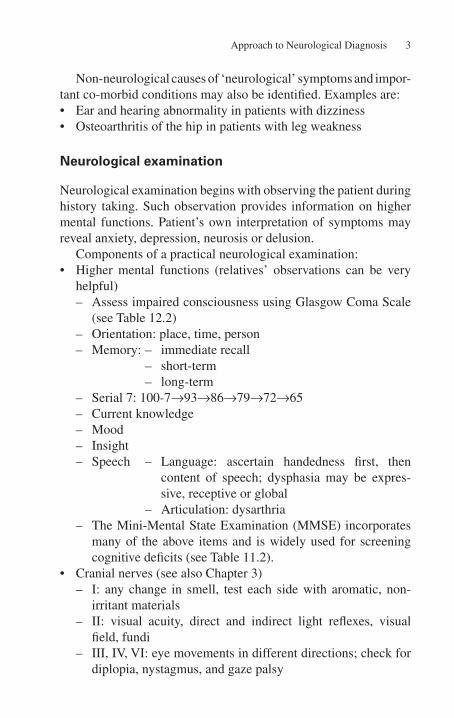
Neurological examination
Neurological examination begins with observing the patient during history taking. Such observation provides information on higher mental functions. Patient’s own interpretation of symptoms may reveal anxiety, depression, neurosis or delusion.
Components of a practical neurological examination:• Highermental functions (relatives’ observations can be very
helpful)– Assess impaired consciousness using Glasgow Coma Scale
(see Table 12.2)– Orientation: place, time, person– Memory: – immediate recall – short-term – long-term– Serial 7: 100-7→93→86→79→72→65– Current knowledge– Mood– Insight– Speech – Language: ascertain handedness first, then
content of speech; dysphasia may be expres-sive, receptive or global
– Articulation: dysarthria– The Mini-Mental State Examination (MMSE) incorporates
many of the above items and is widely used for screening cognitive deficits (see Table 11.2).
• Cranialnerves(seealsoChapter3)– I: any change in smell, test each side with aromatic, non-
irritant materials– II: visual acuity, direct and indirect light reflexes, visual
field, fundi– III, IV, VI: eye movements in different directions; check for
diplopia, nystagmus, and gaze palsy

4 Neurology in Practice
– V: facial sensation to pinprick and light touch in all three divisions of the trigeminal nerve, corneal reflex, power of jaw opening and closure, jaw jerk
– VII: facial symmetry, UMN and LMN facial weakness– VIII: hearing acuity, Weber’s and Rinne’s test (256 Hz tuning
fork)– IX, X: any hoarseness of voice, symmetry of palatal move-
ments, gag reflex– XI: power of sternomastoid and trapezius– XII: any deviation, wasting or fasciculation of tongue
• Motorexaminationofupperandlowerlimbs– Muscle bulk, tone, power (Tables 1.1 and 1.2), tendon
reflexes, plantar response, coordination, gait– Differentiate between UMN and LMN signs– Segmental levels for reflexes: biceps (C5–6), supinator
(C5–6), triceps (C7–8), finger jerks (C8–T1), knee (L3–4), ankle (S1–2)
• Sensationsofupperandlowerlimbs– Pain, temperature, vibration (128 Hz tuning fork), joint
position– Recognize pattern of sensory loss: peripheral nerve vs
dermatome (Figure 1.1)– C5–T1 dermatomes over the upper limb: shoulder (C5),
thumb (C6), middle finger (C7), little finger (C8), inner- upper arm (T1)
– C4 and T2 are contiguous over sternal angle– Over the trunk: nipple (T4), xiphisternum (T7), umbilicus
(T10), symphysis pubis (L1)– L2–S2 over the lower limb: upper outer thigh (L2), lower
inner thigh (L3), inner lower leg (L4), anterior lower leg and foot (L5), lateral lower leg (S1), mid-strip of leg posteriorly (S2)
– S3 over saddle region– S4–5 over perianal region
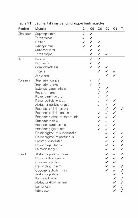
Table 1.1 Segmental innervation of upper limb muscles
Region Muscle C4 C5 C6 C7 C8 T1
Shoulder SupraspinatusTeres minorDeltoidInfraspinatusSubscapularisTeres major
✓
✓
✓
✓
✓
✓
✓
✓
✓
✓
✓
✓
✓
✓ ✓
Arm BicepsBrachialisCoracobrachialisTricepsAnconeus
✓
✓
✓
✓
✓
✓
✓
✓
✓
✓
✓
✓
Forearm Supinator longusSupinator brevisExtensor carpi radialisPronator teresFlexor carpi radialisFlexor pollicis longusAbductor pollicis longusExtensor pollicis brevisExtensor pollicis longusExtensor digitorum communisExtensor indicisExtensor carpi ulnarisExtensor digiti minimiFlexor digitorum superficialisFlexor digitorum profundusPronator quadratusFlexor carpi ulnarisPalmaris longus
✓
✓
✓
✓
✓
✓
✓
✓
✓
✓
✓
✓
✓
✓
✓
✓
✓
✓
✓
✓
✓
✓
✓
✓
✓
✓
✓
✓
✓
✓
✓
✓
✓
✓
✓
✓
✓
✓
✓
✓
✓
✓
✓
✓
✓
✓
✓
✓
✓
Hand Abductor pollicis brevisFlexor pollicis brevisOpponens pollicisFlexor digiti minimiOpponens digiti minimiAdductor pollicisPalmaris brevisAbductor digiti minimiLumbricalsInterossei
✓
✓
✓
✓
✓
✓
✓
✓
✓
✓
✓
✓
✓
✓
✓
✓
✓
✓
✓
✓
✓
✓
✓
✓

Table 1.2 Segmental innervation of lower limb muscles
Region Muscle L1 L2 L3 L4 L5 S1 S2 S3
Hip IliopsoasTensor fascia lataeGluteus mediusGluteus minimusQuadratus femorisGluteus maximusObturator internusPiriformis
✓ ✓ ✓ ✓
✓
✓
✓
✓
✓
✓
✓
✓
✓
✓
✓
✓
✓
✓
✓
✓
✓
✓
✓
Thigh SartoriusPectineusAdductor longusQuadricepsGracilisAdductor brevisObturator externusAdductor magnusAdductor minimusArticularis genusSemitendinosusSemimembranosusBiceps femoris
✓
✓
✓
✓
✓
✓
✓
✓
✓
✓
✓
✓
✓
✓
✓
✓
✓
✓
✓
✓
✓
✓
✓
✓
✓
✓
✓
✓
✓
✓
✓
✓ ✓
Leg Tibialis anteriorExtensor hallucis longusPopliteusPlantarisExtensor digitorum longusSoleusGastrocnemiusPeroneus longusPeroneus brevisTibialis posteriorFlexor digitorum longusFlexor hallucis longusExtensor hallucis brevis
✓
✓
✓
✓
✓
✓
✓
✓
✓
✓
✓
✓
✓
✓
✓
✓
✓
✓
✓
✓
✓
✓
✓
✓
✓
✓
✓
✓
✓
✓
✓
✓
✓
✓
✓
✓
✓
✓
✓
✓
Foot Extensor digitorum brevisFlexor digitorum brevisAbductor hallucisFlexor hallucis brevisLumbricalsAbductor digiti minimiFlexor digiti minimi brevisOpponens digiti minimiQuadratus plantaeInterossei
✓ ✓
✓
✓
✓
✓
✓
✓
✓
✓
✓
✓
✓
✓
✓
✓
✓
✓
✓
✓
✓
✓
✓
✓
✓
✓
✓
✓
✓
✓
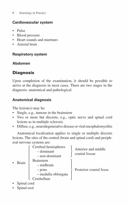
8 Neurology in Practice
Cardiovascular system
• Pulse• Bloodpressure• Heartsoundsandmurmurs• Arterialbruit
Respiratory system
Abdomen
Diagnosis
Upon completion of the examination, it should be possible to arrive at the diagnosis in most cases. There are two stages in the diagnosis: anatomical and pathological.
Anatomical diagnosis
The lesion(s) may be:• Single,e.g.,tumourinthebrainstem• Two or more but discrete, e.g., optic nerve and spinal cord
lesions as in multiple sclerosis• Diffuse,e.g.,neurodegenerativediseaseorviralencephalomyelitis
Anatomical localization applies to single or multiple discrete lesions. The sites of the central (brain and spinal cord) and periph-eral nervous systems are:
• Brain
Cerebral hemispheres– dominant– non-dominant
Anterior and middle cranial fossae
Brainstem– midbrain– pons– medulla oblongata
Cerebellum
Posterior cranial fossa
• Spinalcord• Spinalroot
Approach to Neurological Diagnosis 9
• Plexus–brachial,lumbosacral• Peripheralnerve• Neuromuscularjunction• Muscle
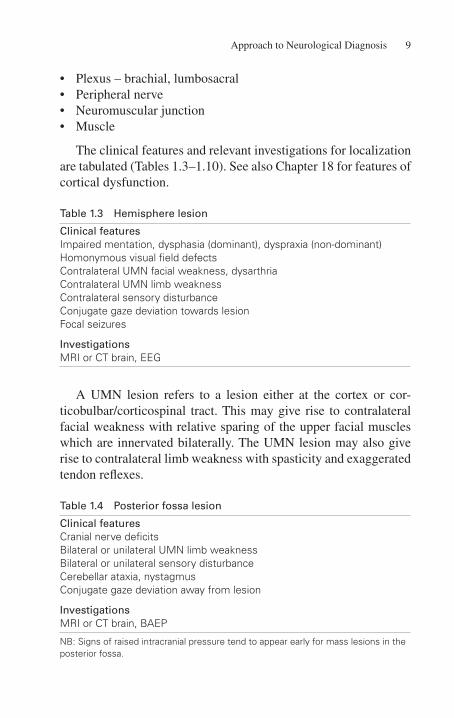
The clinical features and relevant investigations for localization are tabulated (Tables 1.3–1.10). See also Chapter 18 for features of cortical dysfunction.
Table 1.3 Hemisphere lesion
Clinical featuresImpaired mentation, dysphasia (dominant), dyspraxia (non-dominant)Homonymous visual field defectsContralateral UMN facial weakness, dysarthriaContralateral UMN limb weaknessContralateral sensory disturbanceConjugate gaze deviation towards lesionFocal seizures
InvestigationsMRI or CT brain, EEG
A UMN lesion refers to a lesion either at the cortex or cor-ticobulbar/corticospinal tract. This may give rise to contralateral facial weakness with relative sparing of the upper facial muscles which are innervated bilaterally. The UMN lesion may also give rise to contralateral limb weakness with spasticity and exaggerated tendon reflexes.
Table 1.4 Posterior fossa lesion
Clinical featuresCranial nerve deficitsBilateral or unilateral UMN limb weaknessBilateral or unilateral sensory disturbanceCerebellar ataxia, nystagmusConjugate gaze deviation away from lesion
InvestigationsMRI or CT brain, BAEP
NB: Signs of raised intracranial pressure tend to appear early for mass lesions in the posterior fossa.

10 Neurology in Practice
Table 1.5 Spinal cord lesion
Clinical featuresUMN limb weakness below lesion – unilateral or bilateralLMN limb weakness at level of lesion – unilateral or bilateralPattern of sensory deficits – level, glove and stocking, suspended,
dissociatedSphincter disturbance
InvestigationsMRI spine*, XR spine, SEP, CSF analysis
*If MRI not available, myelogram or CT myelogram is an alternative.
A LMN lesion refers to a lesion of the motor neurone or its axons. Depending on the site (brainstem or spinal cord), it may give rise to ipsilateral facial weakness affecting the upper and lower facial muscles or ipsilateral limb weakness with hypotonia and reduced or absent tendon reflexes.
Table 1.6 Spinal root lesion
Clinical featuresSegmental LMN weakness and sensory deficitsAutonomic disturbance
InvestigationsNCS, EMG, SEP, XR spine, MRI spine*, CSF analysis
*If MRI not available, myelogram or CT myelogram is an alternative.
Table 1.7 Plexus lesion
Clinical featuresMulti-segmental LMN weakness and sensory deficitsAutonomic disturbance
InvestigationsNCS, EMG, SEP, MRI, CSF analysis
Approach to Neurological Diagnosis 11
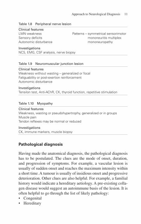
Table 1.8 Peripheral nerve lesion
Clinical featuresLMN weaknessSensory deficitsAutonomic disturbance
Patterns – symmetrical sensorimotor mononeuritis multiplex mononeuropathy
InvestigationsNCS, EMG, CSF analysis, nerve biopsy
Table 1.9 Neuromuscular junction lesion
Clinical featuresWeakness without wasting – generalized or focalFatiguability or post-exertion reinforcementAutonomic disturbance
InvestigationsTensilon test, Anti-AChR, CK, thyroid function, repetitive stimulation
Table 1.10 Myopathy
Clinical featuresWeakness, wasting or pseudohypertrophy, generalized or in groupsMuscle painTendon reflexes may be normal or reduced
InvestigationsCK, immune markers, muscle biopsy
Pathological diagnosis
Having made the anatomical diagnosis, the pathological diagnosis has to be postulated. The clues are the mode of onset, duration, and progression of symptoms. For example, a vascular lesion is usually of sudden onset and reaches the maximum intensity within a short time. A tumour is usually of insidious onset and progressive deterioration. Other clues are also helpful. For example, a familial history would indicate a hereditary aetiology. A pre-existing colla-gen disease would suggest an autoimmune basis of the lesion. It is often helpful to go through the list of likely pathology:• Congenital• Hereditary

12 Neurology in Practice
• Inflammatory:infective,granulomatous,autoimmune• Demyelinating• Vascular• Degenerative• Neoplastic:benign,malignant• Traumatic• Idiopathic
Investigations
After a clinical diagnosis has been arrived at, the next steps are to confirm it and to obtain more information in order to plan the management. Investigations (Tables 1.3–1.10) are organized with these aims in mind. To be cost-effective, they should be specific and relevant. Such targeting can only be achieved with a sound clinical diagnosis.
Common neurological symptoms and their differential diagnosis
Headache (see Chapter 4)
Facial pain (see Chapter 3)
Dizziness
This is a common symptom which may be due to systemic or ves-tibular disturbance. Distinction has to be made from loss or lapse of consciousness. Associated symptoms of vertigo and disequilib-rium, if present, should be elicited.
Systemic disturbancepresyncopehyperventilationanaemiacardiac arrhythmiadrug-induced, e.g., bromocriptine, levodopa, methyldopaelectrolyte disturbance, e.g., hyponatraemia

Approach to Neurological Diagnosis 13
Vestibular disturbancePeripherallabyrinthitisMeniere’s disease
Centralbrainstem/cerebellar lesionvertebrobasilar strokecerebello-pontine angle tumoursdemyelination
Altered consciousness
This state includes complete and partial, prolonged and brief dis-turbance of consciousness. Details of relevant features during the episode should be obtained from the patient or relatives. Many causes of impaired consciousness apply (see Chapter 12).
epileptic seizures, generalized or complex partialstrokecardiogenic syncopemetabolic/endocrine, especially hypoglycaemiaintoxication/drug overdose, e.g., alcohol, substance abuseneoplasticfunctional disorders
Visual impairment (see Chapter 3)
Ptosis
UnilateralIII nerve palsyHorner’s syndromeMG
BilateralMGmyopathy
14 Neurology in Practice
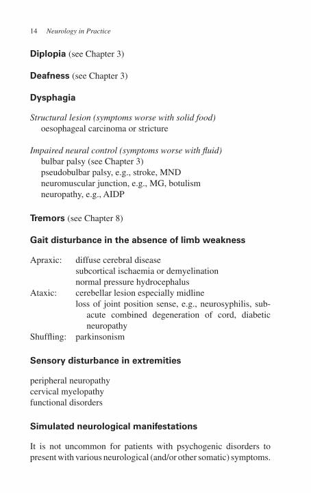
Diplopia (see Chapter 3)
Deafness (see Chapter 3)
Dysphagia
Structural lesion (symptoms worse with solid food)oesophageal carcinoma or stricture
Impaired neural control (symptoms worse with fluid)bulbar palsy (see Chapter 3)pseudobulbar palsy, e.g., stroke, MNDneuromuscular junction, e.g., MG, botulismneuropathy, e.g., AIDP
Tremors (see Chapter 8)
Gait disturbance in the absence of limb weakness
Apraxic: diffuse cerebral disease subcortical ischaemia or demyelination normal pressure hydrocephalusAtaxic: cerebellar lesion especially midline loss of joint position sense, e.g., neurosyphilis, sub-
acute combined degeneration of cord, diabetic neuropathy
Shuffling: parkinsonism
Sensory disturbance in extremities
peripheral neuropathycervical myelopathyfunctional disorders
Simulated neurological manifestations
It is not uncommon for patients with psychogenic disorders to present with various neurological (and/or other somatic) symptoms.
Approach to Neurological Diagnosis 15
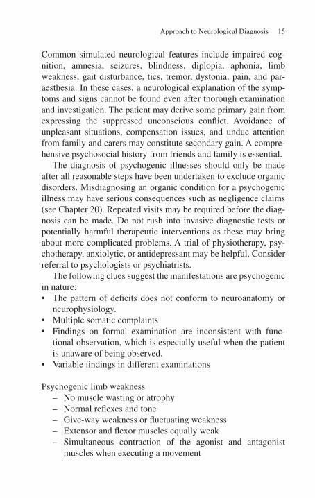
Common simulated neurological features include impaired cog-nition, amnesia, seizures, blindness, diplopia, aphonia, limb weakness, gait disturbance, tics, tremor, dystonia, pain, and par-aesthesia. In these cases, a neurological explanation of the symp-toms and signs cannot be found even after thorough examination and investigation. The patient may derive some primary gain from expressing the suppressed unconscious conflict. Avoidance of unpleasant situations, compensation issues, and undue attention from family and carers may constitute secondary gain. A compre-hensive psychosocial history from friends and family is essential.
The diagnosis of psychogenic illnesses should only be made after all reasonable steps have been undertaken to exclude organic disorders. Misdiagnosing an organic condition for a psychogenic illness may have serious consequences such as negligence claims (see Chapter 20). Repeated visits may be required before the diag-nosis can be made. Do not rush into invasive diagnostic tests or potentially harmful therapeutic interventions as these may bring about more complicated problems. A trial of physiotherapy, psy-chotherapy, anxiolytic, or antidepressant may be helpful. Consider referral to psychologists or psychiatrists.
The following clues suggest the manifestations are psychogenic in nature:• Thepatternofdeficitsdoesnot conform toneuroanatomyor
neurophysiology.• Multiplesomaticcomplaints• Findings on formal examination are inconsistent with func-
tional observation, which is especially useful when the patient is unaware of being observed.
• Variablefindingsindifferentexaminations
Psychogenic limb weakness– No muscle wasting or atrophy– Normal reflexes and tone– Give-way weakness or fluctuating weakness– Extensor and flexor muscles equally weak– Simultaneous contraction of the agonist and antagonist
muscles when executing a movement
16 Neurology in Practice
Psychogenic movement disorders– Abrupt onset, atypical pattern, paroxysmal symptoms– Inconsistency over time– Entrainment of tremor to the examiner’s suggested rate– Spontaneous remissions– Disappearance when distracted– Aggravation during formal examination– Response to placebo, psychotherapy or suggestion
Psychogenic seizure (see Chapter 7)
Always bear in mind the following:• Organicandpsychogenicdisordersmaycoexist.• Deliriumanddementiaareduetoorganicdisorders.• Cerebrovascular disease, brain tumour, epilepsy, Parkinson’s
disease, Alzheimer’s disease, multiple sclerosis, and Huntington’s disease can produce anxiety and depression.
• Substancemisuse(e.g.,alcohol,recreationaldrugs)canleadtopsychogenic and neurological complications.
• Psychosomatic disorders consist of hysteria, somatizationdisorders, somatoform pain disorders, and neurasthenia. They commonly present with fatigue, dizziness, headache or pain attributed to physical illness.
• Apatientwithfactitiousdisorderrepeatedlyinducesthesymp-toms or signs of disease in the absence of any psychiatric or physical disorder.
Spinal cord disorders or myelopathies frequently cause severe and permanent disability because the spinal cord contains the entire motor and sensory systems of the trunk and limbs. Therefore, prompt diagnosis and treatment are essential. In particular, acute spinal cord compression is a neurological emergency.
Classification
Spinal cord disorders may be classified according to their causes. Trauma is the most common cause of myelopathy. For acute non-traumatic myelopathies, transverse myelitis and cord com-pression are common whereas spinal cord stroke is rare. In chronic non-traumatic myelopathies, spondylosis and benign tumours account for most cases. Syringomyelia, multiple sclerosis, degen-erative and paraneoplastic cord lesions should be considered in the differential diagnosis. Subacute combined degeneration of spinal cord and syphilitic myelitis are rare but treatable entities.
Congenital and developmental disorders
Examples are syringomyelia, Arnold-Chiari malformation, and diastematomyelia. These conditions are often associated with spinal bifida.
Infection
Neurotropic viruses, e.g., herpes virus, poliovirus, and rabies may affect the spinal cord and/or the brain. The virus, however, cannot be identified in most cases.
Spinal Cord Disorders14
214 Neurology in Practice
Pyogenic infections of leptomeninges usually affect the sub-arachnoid space around the brain and the spinal cord. Epidural abscess may result from direct or haematogenous spread.
Tuberculous leptomeningitis produces spinal cord ischaemia by inflammatory arteritis. Tuberculous spinal osteitis (Pott’s disease), with its associated psoas abscess and bony destruction, may cause cord compression or ischaemia.
Fungal infections (e.g., cryptococcosis) and parasitic infections (e.g., cysticercosis and hydatid disease) are rare in the spinal cord.
Neoplastic diseases
The lesion may be extradural, intradural-extramedullary, or intramedullary in location. The common benign lesions are neu-rofibroma and meningioma. The malignant lesions are ependy-moma, astrocytoma, and carcinomatous meningeal infiltration. Related entities are paraneoplastic and irradiation myelopathy.
Diseases of the spine
• Spondylosis(vide infra)• In longstanding rheumatoid arthritis, atlantoaxial or subaxial
dislocation may cause cervical cord compression.
Vascular disorders
Diseases of the aorta, vertebral, intercostal, and radicular arteries may present as cord transection or anterior spinal artery syndrome. Acute spinal venous obstruction is rare and usually secondary to conditions such as tumour, septicaemia, and thrombotic diathesis; the prognosis is poor because of extensive haemorrhagic infarc-tion. Vasculitis, e.g., in SLE, PAN, may give rise to acute spinal cord symptoms. AVM may either present with acute or chronic spinal cord features.

Spinal Cord Disorders 215
Demyelination
• Multiplesclerosis• Post-infectiousencephalomyelitis
Degenerative
• Spinocerebellardegeneration• Idiopathicspasticparaparesis
Trauma (see Chapter 20)
Clinical picture and diagnosis
The clinical features depend on the pattern and level of the lesion (Figure 14.1).
Motor deficits
• UMNweaknessbelowthelesion• LMNweaknessatthelevelofthelesion• MixedLMNandUMNweakness,e.g.,upperlimbsincervical
cord lesion• Spinalshockstage:flaccidparalysis
Sensory disturbance
• Spinothalamicsensations:pain,temperature• Posteriorcolumnsensations:vibration,jointposition• Sensorydisturbancemaypresentasasensorylevel,gloveand
stocking distribution, segmental sensory loss, or dissociated sensory loss.
• Painmayarisefromvertebralcollapseormalignantinfiltrationof spinal root (radicular pain) or spinal cord.

Spinal Cord Disorders 217
Autonomic dysfunction
• Bladder:spasticorparalytic• Bowel:incontinence± constipation• Sexualdysfunction
Differential diagnosis
• Guillain-Barré syndrome may present as a rapidly evolvingtetraparesis. Distinguishing features include the presence of bulbar palsy, paresis of extraocular muscles and areflexia.
• Acuteneuropathies:usuallyduetovasculitisortoxin.• Myastheniagravis:nosensorydisturbance;ocularinvolvement
is common.• Acuteocclusionoftheterminalaortamaycauseischaemiaof
the cauda equina or proximal sciatic and femoral nerves. Rarely the spinal cord may be involved. Pale cold skin and loss of pulses in the lower limbs are clues to the diagnosis.
• Occlusionofanteriorcerebralarterymaycausebilateralinfarc-tion of the paracentral lobule, resulting in acute paraplegia, sphincter dysfunction, and loss of position sense with intact pain perception. The last feature distinguishes this entity from acute spinal cord syndrome.
• Falxmeningiomawithpressureeffecton the legareasof thecerebral cortex
Specific investigations
Investigations are guided by the clinical diagnosis, especially for the spinal level where the lesion is likely to be present.• X-rayspine:vertebrallesions,e.g.,collapse,spondylosis• MRIspine:investigationofchoicesincethecord,subarachnoid
space, and adjacent tissues can be clearly visualized. CT spine or myelogram may be indicated if MRI is not available.
• CSFanalysis:usefulininfection,demyelination,andneoplasticmeningitis
NB: LP may cause further deterioration if there is cord compression.

218 Neurology in Practice
SELECTED ENTITIES
Acute spinal cord compression
It is a neurological emergency caused by:• Malignancy:carcinomatousmetastasis,lymphoma,myeloma• Infection:tuberculousorpyogenicabscess,vertebralcollapse• Epiduralhaematoma:spontaneous,traumatic• Acutediscprotrusion
Urgent MRI is indicated to confirm or exclude spinal cord compression.
High-dose IV steroids can be given prior to confirmation with neuroimaging. Upon confirmation, urgent neurosurgical decom-pression or radiotherapy should be arranged. Delay of treatment is associated with poor recovery of function.
Transverse myelitis
The causes include viral infection, SLE, post-infectious demyeli-nation, NMOSD, and MS. Note any history of viral infection or vaccination and previous episodes of neurological deficits. Acute spinal cord compression must be excluded.
MRI is indicated to visualize the lesion and exclude cord compression.
CSF analysis commonly shows lymphocytosis, normal glucose, normal or slightly raised protein. Oligoclonal IgG indicates intrathecal synthesis of IgG; it is a non-specific finding although commonly present in demyelination.
Treatment depends on the underlying cause.
Spinal Cord Disorders 219
Degenerative disease of the spine
If the spinal canal is constitutionally narrow, degenerative tissues of the spine (e.g., osteophytes, discs, ossified posterior longitudi-nal ligament, hypertrophied ligamentum flava) are more likely to cause compression of the spinal cord or roots.
Cervical spondylotic myelopathy (CSM)
Cervical spondylosis as a radiological feature is very common, and CSM is the most common cause of cervical cord lesion in subjects over age 50.
Males are more affected than females. A history of neck injury, neck pain, and stiffness can be elicited in some patients. The symp-tomatology is that of a cervical cord lesion, with or without spinal root lesion.
The mechanisms by which cervical spondylosis brings about cord damage are complex. Compression due to acquired spondy-lotic changes on top of a constitutionally narrow canal (sagittal diameter < 10 mm) is the most important factor. In addition,dynamic factors, viz. friction with osteophytes, pressure from the ligamentum flava, vertebral subluxation, and hyperextension injury also play a part. These mechanisms lead to impairment of microcirculation at the arteriolar level resulting in ischaemic cord damage. The differential diagnosis is that of other cervical cord lesions, e.g., neurofibroma, syringomyelia. Motor neurone disease, at its early stage, may mimic CSM.
Cervical spine radiography provides information on the sagittal diameter of the spinal canal, the presence and degree of spon-dylotic tissues, and the presence of vertebral subluxation. MRI (Figure 14.2) shows the degree and site of spinal cord and root compression and is the investigation of choice. SEP study may document posterior column deficits and is helpful in monitoring progress.

Spinal Cord Disorders 221
Treatment is conservative for patients with mild symptoms and/or a long history with little or no progression. However, surgery is indicated for patients with significant symptomatology or pro-gression, and for those in whom conservative treatment has failed.
Cauda equina compression by lumbar prolapsed intervertebral disc (LPID)
LPID is common but in most cases, it either causes compres-sion of a single spinal root (e.g., sciatica) or no neurological damage. Central protrusion of the disc into the spinal canal occurs uncommonly but may lead to acute or subacute cauda equina com-pression. It occurs more often at L4/5 and L5/S1 levels, hence the lowermost lumbar roots and sacral roots are affected.
The clinical picture is typical, and features include low back pain with or without radiation to both lower limbs; reduced sensa-tion in the buttocks, perineum, and posterior thighs; absent ankle jerks; and sphincter disturbances with urinary retention, constipa-tion and erectile dysfunction. Weakness in the lower limbs, espe-cially of foot movements, may be present.
Diagnosis is made on clinical grounds and confirmed by MRI. Early diagnosis and prompt surgical decompression may partially or completely reverse the deficits.
Intermittent claudication of cauda equina
The underlying pathology is stenosis of the lumbar spinal canal, often due to spondylotic tissues superimposed on a constitution-ally narrow canal.
The clinical features consist of weakness, reduced tendon reflexes, and sensory disturbance in the legs after walking a certain distance. At rest, these features may be absent. The differential diagnosis is peripheral vascular disease in which case the pulses in the legs are weak or absent.

222 Neurology in Practice
X-ray of lumbar spine shows the degenerative bony changes and alignment. MRI confirms the diagnosis and provides informa-tion for surgical management.
Surgical decompression is indicated for patients with severe or progressive symptomatology.
Multiple sclerosis (see Chapter 9)
Syringomyelia
This is a classic example of an intramedullary lesion, with cavita-tion of the central part of the spinal cord, often extending vertically in the central grey matter over many segments. It presents with seg-mental sensory impairment of spinothalamic sensations due to dis-ruption of decussating fibres at the anterior commissure. Extension of the syrinx to the anterior horns causes segmental amyotrophy, whereas extension to the posterior horns causes segmental loss of posterior column sensations. Functional disturbances of the legs and sphincters occur at a late stage.
Syringobulbia is due to extension of syrinx to the brainstem, resulting in lower cranial nerve palsy and disturbance of facial sensations.
About half of all idiopathic cervical syringomyelia cases are associated with type I Arnold-Chiari malformation in which the neck may be short and congenital abnormalities of the cervical spine and base of skull may be present. Secondary syringomyelia may complicate obstruction of the foramen magnum by localized arachnoiditis, cyst or tumour.
X-ray cervical spine and base of skull may show skeletal abnor-malities. In MRI cervical spine (Figure 14.3), the location and extent of the syrinx, as well as cerebellar tonsillar herniation, can be visualized. MRI brain shows syringobulbia and other associated abnormalities.
Decompression of the syrinx is the treatment for symptomatic patients, particularly for those with Arnold-Chiari malformation.

224 Neurology in Practice
Spinocerebellar ataxia (SCA)
In most cases, including the SCA type 3, the inheritance is autoso-mal dominant. Sporadic cases are far less common. The symptoms start in the 30s or earlier. The progression is usually slow, and disability is moderate to severe. Degeneration of corticospinal and spinocerebellar tracts results in cerebellar ataxia and pyramidal signs. Associated features are uncommon and include periph-eral neuropathy, optic neuropathy, cardiomyopathy, and cardiac arrhythmia. Cognition is intact. There is no curative treatment. Genetic diagnosis (including preimplantation) for the affected family is available.
Subacute combined degeneration of spinal cord
This is a classical and treatable condition due to vitamin B12 defi-ciency which may occur in pernicious anaemia, dietary insuffi-ciency, and gastric or ileal resection. The pathological changes are degeneration of the lateral and posterior columns of the spinal cord, and to a lesser extent the brain and peripheral nerves. The patient presents with spastic tetraparesis, impaired posterior column sen-sations, peripheral neuropathy, and occasionally encephalopathy.
The diagnosis is made upon confirmation of vitamin B12 defi-ciency (e.g., megaloblastic anaemia, low serum B12) and exclusion of other spinal cord lesions. The cause of vitamin B12 deficiency should also be elucidated.
Treatment is by parenteral vitamin B12 replacement. Hypokalaemia may develop during treatment. Early treatment confers a better chance of complete recovery.
Paraneoplastic myelopathy
This is not caused by spinal cord compression or invasion by car-cinoma. The clinical syndrome is a rapidly developing non-inflam-matory myelopathy with motor and sensory dysfunction.
Pathologically, there is necrosis of the tracts of the spinal cord without evidence of neoplasm. Anti-tumour antibody cross-reac-tive to the spinal cord has been proposed as the mechanism.
Spinal Cord Disorders 225
Irradiation myelopathy
The lesion evolves over a period of weeks and then becomes per-manent. The total dose, fractionation, and size of radiated field are important factors. In general, fractionated irradiation up to a total dose of 3,500 rad is relatively safe. Irradiation induces oblit-erative endarteritis and thus ischaemic necrosis of the spinal cord tissues. The time interval between irradiation and the first spinal cord symptom varies, but usually ranges from 6–48 months. CSF is either normal or shows a slightly elevated protein level.

abducens nerve (VI) 31f, 38, 39abscess 192–95, 197acalculia 163, 284acanthocytes 120accessory nerve (XI) 31f, 46acetylcholinesterase inhibitors
125, 170donepezil (Aricept) 125, 170,
172galantamine (Reminyl) 170,
172rivastigmine (Exelon) 125,
170, 172acid maltase deficiency 254, 255acoustic neuroma 25, 42, 44, 45,
45t, 285acromegaly 227t, 233, 249t, 261,
265, 269, 288activated factor VII 79acute disseminated
encephalomyelitis (ADEM) 134, 138f, 142–44
acute haemorrhagic leukoencephalitis 142
post-immunization encephalomyelitis 142
post-infectious encephalomyelitis (encephalitis) 142,189, 215
post-organ-transplantation encephalomyelitis 142
acute inflammatory demyelinating polyradiculoneuropathy (AIDP). See Guillain-Barrè syndrome
acute mountain sickness 62acyclovir 197, 202, 203adult metachromatic
leukodystrophy 138agnosia 163, 165, 170, 190, 284agranulocytosis 126AIDS (acquired
immunodeficiency syndrome) 164t, 191, 199, 200t, 204, 208
akathisia 127akinesia 111, 122, 127akinetic mutism 173, 178albinism 41alcuronium 212alien hand syndrome 127Alport’s syndrome 45taltered consciousness 13, 92,
175, 198, 203, 271, 302aluminium 164tAlzheimer’s disease (AD) 16,
117, 163, 163t, 168, 169–71, 244t
amantadine 123, 141, 258
Index
Note: f = figure; t = table

Index 311
aminocaproic acid 80aminoglycosides 45, 45t, 160,
196tamiodarone 114t, 243tamitriptyline 43, 55, 204, 237amoebiasis 190amphotericin B 208amyloid 29, 169aneurysm 20, 49t, 50t, 66f, 67,
68t, 77t, 80, 263angiography 21–22, 50t, 66f, 74,
80, 81, 82, 84t, 85tangioplasty 80, 84t, 89anosmia 32, 283, 301anterior horn cell disease. See
motor neurone disease and spinal muscular atrophy
anterior temporal lobectomy 105anti-acetylcholine receptor
antibody (anti-AChR) 11t, 154, 155, 159
anticholinergics 120, 123, 125, 128, 158, 165, 296
atropine 38t, 158, 182t, 277benzhexol 123benztropine 123, 128orphenidrine 123pralidoxine 277
anti-cholinesterase 158, 161, 162
anticoagulation 77–78, 81, 82, 83t, 85t, 86, 87, 88, 264
heparin 68t, 75, 77t, 239low molecular weight heparin
(LMWH) 75, 87, 239new oral anticoagulants
(NOACs) 87warfarin 68t, 77t, 87, 101
antidepressants 15, 57, 126, 127, 133, 141, 167, 182t, 246, 266
antidiuretic hormone, inappropriate secretion (SIADH) 80, 188, 270, 273
antiepileptic drugs (AEDs) 55, 75, 97–103, 98t, 99t, 100t, 102t, 104–7, 104f, 109, 110
adverse effects 100t, 101carbamazepine 43, 98, 99t,
100t, 101, 102, 107choice 98, 99t, 100clobazam 99t, 102ethosuximide 99t, 101felbamate 99tgabapentin 43, 55, 56, 99t,
100, 100t, 101lamotrigine 99t, 101, 107levetiracetam 55, 99t, 100,
100t, 109oxcarbazepine 99t, 100, 100tphenobarbitone 98, 99t, 100t,
101, 107, 110phenytoin 41, 55, 98, 99t,
100t, 101, 107, 109piracetam 99t, 117pregabalin 43, 99t, 100, 100tprimidone 98, 99ttopiramate 55, 99t, 100, 100t,
101valproate 55, 98, 99t, 100t,
101, 107vigabatrin 99t, 100, 100t, 101zonisamide 100
antihistamines 112, 115, 119, 121, 128
diphenhydramine 128antiphospholipid antibody
syndrome 74, 266antiplatelet agents 78, 83t, 88
aspirin 78, 85, 85t, 86clopidogrel 88dipyridamole 88

312 Index
ticlopidine 88antipsychotics (neuroleptics)
112, 114t, 115, 119, 128, 129, 171, 257, 258, 258t, 266
chlorpromazine 171, 250haloperidol 171thioridazine 171
aphasia (dysphasia) 3, 9t, 69, 70, 72t, 85t, 163, 283, 284, 301
apraxia (dyspraxia) 9t, 69, 85t, 163, 165, 169, 172
Argyll Robertson pupil 37, 38t, 210
Arnold-Chiari malformation 42, 60, 213, 222, 223t
arterial dissection 49t, 50t, 56, 68t, 74, 80–81, 303
arteriovenous malformation (AVM) 21, 50t, 68t, 77t, 81, 91t, 94, 98t
ataxia 9t, 29, 53, 70, 100t, 101, 137, 180, 190, 191
ataxia telangiectasia 119atenolol 55athetosis 114–15, 137atlanto-axial dislocation or
subluxation 119, 267atrial fibrillation 83t, 85t, 86–87,
86tatypical antipsychotics
(neuroleptics) 126, 129, 171, 258
olanzapine 171, 258quetiapine 126, 129, 171, 258risperidone 171, 258
autoimmune encephalitis 148–52antibodies against neuronal
intracellular antigens 152anti-NMDAR 149–51LGI1 antibodies 151
autosomal dominant nocturnal frontal epilepsy syndrome 92
axonotmesis 229azathioprine 157, 159, 260, 267
baclofen 27, 43, 141, 295Balo’s concentric sclerosis 138barbiturates 41, 182t, 278Barthel ADL index 291, 292tBecker muscular dystrophy 249t,
250, 252, 253tBehçet’s disease 267Bell’s palsy 30, 44benign positional vertigo 45benign Rolandic epilepsy 102tbenzodiazepines 20, 41, 102,
103, 107, 113, 278, 280, 306clonazepam 98, 99t, 113diazemuls 109diazepam 109lorazepam 109midazolam 109, 110
benzylpenicillin 196, 196t, 197, 199, 212
β-blocker (e.g., propranolol) 55, 78, 85t, 113, 130, 253
Betaferon 139Binswanger’s disease 171bitemporal hemianopia 33f, 269,
288blepharospasm 118Bornholm disease 260Borrelia burgdorferi 190, 209botulinum toxin 120, 131, 295brachial plexopathy 230–31, 234,
279thoracic outlet syndrome (TOS)
231bradykinesia 111, 121, 124, 131,
205, 257
Index 313
brain death 181–84brainstem auditory evoked
potentials (BAEP) 9t, 25brain tumour 164t, 282–89
astrocytoma 269craniopharyngiomas 286glioma 94, 286, 267fmeningioma 94, 28, 287fneurofibroma 287–88oligodendroglioma 286pituitary tumour 287
Brown-Sèquard syndrome 137, 216f
bulbar palsy 14, 46–47, 147, 217, 239, 273
caeruloplasmin 111, 131, 132candidiasis 200tcarbon tetrachloride 164tcarcinomatous meningitis 47,
214carotid artery stenosis 84t,
88–89carotid endarterectomy 84t, 89carpal tunnel syndrome 227t,
233–34, 233t, 269carpopedal spasms 264catatonia 178–79catechol-O-methyltransferase
(COMT) inhibitor (entacapone) 124
causalgia 227tcentral pontine myelinolysis 25,
134, 138, 146–47, 270, 279cephalosporin, third generation
196, 196tcerebello-pontine angle lesion or
tumour 13, 31, 44, 45cerebral amyloid angiopathy 68tcerebral haemorrhage. See
intracerebral haemorrhage
cerebral oedema 59, 62, 73, 80, 185, 277, 299f, 300
cerebral toxoplasmosis 200t, 208
cerebral ultrasonography 22, 74, 84t
cerebral vasculitis 49t, 138, 167, 167t, 266
cerebral venous thrombosis 49t, 64, 78, 82
cerebrospinal fluid (CSF) 27, 28, 49t, 50t, 59, 60, 61, 62, 63, 188, 189, 189t
cerebro-tendinous xanthomatosis 164t
cerebrovascular disease (CVD). See stroke
cervical dystonia 118Charcot-Marie-Tooth disease. See
HMSNCharcot’s joint 210, 228chemotherapy 272–73chloroquine 243t, 250, 267chorea 114–15
gravidarum (pregnancy) 115Huntington’s 115Sydenham’s 115, 128
choreoathetosis 94, 111, 114–15choroid plexus 59chronic deep brain stimulation
124, 130chronic inflammatory
demyelinating polyradiculoneuropathy (CIDP) 227t, 240–42
Churg-Strauss syndrome 268–69ciliary ganglion 37, 38tcisplatin 273cluster headache (migrainous
neuralgia) 48, 49t, 55–56codeine 103, 278

314 Index
cognitive impairment 163, 273t, 274, 281, 290, 294
colloid cyst 60colour vision 32coma 175–81, 176t, 183fcommon neurological symptoms
12–14complex regional pain syndrome
(reflex sympathetic dystrophy syndrome) 245
compound muscle action potentials (cMAP) 22, 23, 156, 156t
computed tomography (CT) 17, 65f, 66f, 287f, 299f
conduction block 23, 44, 241, 244, 244t
congenital myopathy 248, 249tcopolymer-1 (glatiramer acetate)
140copper 111, 120, 167tcorpus callosotomy 106cortical dysplasia 91, 91tcoxsackievirus 142, 187, 190,
249t, 260cranial mononeuropathy 30, 266cranial or temporal arteritis 30,
49t, 50t, 57–58, 68t, 269cranial polyneuropathy
(polyneuritis cranialis) 30–31
critical illness polyneuropathy 243–44
crocodile tears 44cryptococcosis/cryptococcal
meningitis 186t, 200t, 207–8CT angiography 21, 66fCushing’s syndrome 157, 249t,
261, 269, 288cyclophosphamide 159, 244, 260,
266, 267
cyclosporine 114t, 159, 260cytosine arabinoside 273
dantrolene 141, 258, 295deafness 45, 45t, 46, 254t, 301deep vein thrombosis (DVT) 78,
80dehydroepiandrosterone 266delirium tremens 146, 279delusion 3, 122, 126, 170, 210dementia 163–74, 254tdengue fever 201depression 16, 57, 76, 125, 165,
280, 281depolarizing muscle relaxants
(decamethonium, d-tubocurarine, succinylcholine) 253, 256
dermatomyositis. See polymyositisdetrusor sphincter dyssynergia
137Devic’s disease. See neuromyelitis
optica spectrum disorders, neuromyelitis optica
diabetic neuropathies 14, 236–38, 237t
diabetic amyotrophy 236, 237tdiabetic retinopathy 36f3,4-diaminopyridine 162Di George syndrome 199digoxin 115dimercaprol 276diplopia 3, 14, 38, 39, 40, 53, 70disopyramide 253disorders with anti-myelin
oligodendrocyte glycoprotein (MOG) antibody 146
dizziness 12–13, 100tdopa-decarboxylase inhibitors
(benzerazide, carbidopa) 121–22

Index 315
dopamine agonists 115, 123, 126, 288
apomorphine, pramipexole, ropinirole, rotigotine 123
bromocriptine 12, 258doxycycline 210drop attacks 94, 262drug-induced movement disorders
112, 121, 128–30acute dystonic reactions 128akathisia 128parkinsonism 121, 129tardive dyskinesia 129–30
Duchenne muscular dystrophy 249t, 250, 251–52
dysarthria 3, 9t, 53, 69, 70, 294dysautonomia 227t, 228, 238,
239dyskinesia 111, 122, 123, 124
levodopa-induced 122, 124dysphagia 14, 70, 111, 120, 127,
133, 147, 211, 245, 252dysphasia. See aphasiadyspraxia. See apraxiadysthyroid eye disease 157dystonia 114t, 118–20
Edinger-Westphal nucleus 31f, 37electroencephalogram (EEG) 9t,
24, 25–26, 93t, 94–96, 95f, 96f, 98t, 102t, 104t
ictal EEG 93t, 94, 95fvideo-EEG 26, 95, 104f, 105
electromyography (EMG) 10t, 11t, 23–24
encephalitis 49t, 112, 119, 175, 178, 187–88, 189–91, 189t
arboviruses 189, 204coxsackie 187, 190, 249tcytomegalovirus (CMV) 190,
197, 200t, 238
dengue fever 190, 201enterovirus 187, 190Epstein-Barr virus 188, 190,
200t, 238herpes simplex (HSV) 190,
191, 200t, 201–2, 202fJapanese B 190, 204lethargica 112measles 188, 190mumps 188, 190non-viral 190paramyxovirus 190rabies 190rubella 164t, 190varicella-zoster 190, 200t,
203–4Zika 201
endocrine myopathies 260–61epilepsy 91–110
aura 92complex partial seizures 92,
191psychogenic seizure 16, 93tstatus epilepticus (SE) 107–10,
108tvagal nerve stimulation (VNS)
106ergotamine 54, 56ethambutol 34, 206evoked potentials 24–25experimental allergic
encephalomyelitis 140extracranial-to-intracranial
(EC-IC) bypass 89
facial nerve (VII) 31f, 43–44, 130, 131, 203
famciclovir 204fibromuscular dysplasia 80fluconazole 208flucytosine 208
316 Index
fludrocortisone 126, 127, 237, 263flumazenil 20, 280flunarizine 555-fluorouracil 273folate 107, 167t, 208Foramen of Magendie 59Foramen of Lushka 59fresh frozen plasma 79fronto-temporal dementia 172fundal appearance 35f, 36f
gadolinium 18, 18t, 19, 66f, 137, 284, 287f
gag reflex 4, 46, 75, 177, 182tgait disturbance 14, 163ganciclovir 197gangliosidoses 164tgiant cell arteritis. See cranial or
temporal arteritisGlasgow Coma Scale (GCS) 175,
176t, 301glaucoma 35f, 49tglossopharyngeal nerve (IX) 31f,
46glyceryl trinitrate 56gold 243t, 267Guillain-Barrè syndrome (acute
inflammatory demyelinating polyradiculoneuropathy; AIDP) 217, 227t, 238–40
clinical variants 238–39
Hallervorden-Spatz syndrome 112
hallucination 92, 112, 123, 126, 170, 202, 279, 280, 284
headache 58–59head injury 297–303
cerebral oedema 300intracranial haematoma 298,
299f
post-concussion syndrome 301post-traumatic epilepsy 301primary parenchymal damage
300, 301soft-tissue and skull injuries
298systemic complications 300vascular damage 300
hemiballismus/ballismus 111, 116, 122
hemifacial spasm 44, 94, 130hemiparesis/hemiplegia 70, 193,
295hemisphere lesion 9thepatic encephalopathy 94, 102,
108, 164t, 165, 279hereditary motor sensory
neuropathy (HMSN; Charcot-Marie-Tooth disease) 29, 227t, 228–29, 247
hereditary sensory autonomic neuropathy (HSAN) 228
hereditary spastic paraplegia 119Herxheimer reaction 210hippocampal sclerosis 18, 91,
91tHolmes-Adie pupil 38, 38tHorner’s syndrome 13, 38, 38t,
56, 70, 81, 230human immunodeficiency virus
(HIV)-1 138, 142, 200t, 204–5
human T-lympho-trophic virus (HTLV)-1 138
Huntington’s disease 16, 115, 119, 132–33, 163t
hydrocephalus 49t, 60–61, 167t, 174, 185f
normal pressure (communicating) 14, 27, 61, 112, 164t, 174

Index 317
5-hydroxytryptamine (5HT). See serotonin
hyperacusis 44hyperbaric oxygen 75, 277hyperkinesia 111hyperparathyroidism 249t, 271hypertensive retinopathy 36fhyperventilation 12, 25, 60, 75,
181, 182t, 264hyperventilation syndrome 94hypoglossal nerve (XII) 31f, 47hypokinesia 111hypoparathyroidism,
pseudohypoparathyroidism 115
hypopituitarism 164t
ichthammol lotions 126idiopathic intracranial hypertension
(benign intracranial hypertension, pseudotumour cerebri) 27, 34, 49t, 62, 284
idiopathic torsion dystonia 119ifosfamide 273immunocompromised host
197–99, 200tantibody deficiency 198asplenism/hyposplenism 198cell-mediated immune
deficiency 199complement system deficiency
198neutropenia 198
indomethacin 57infections of the CNS 185–212infective endocarditis 263–64infliximab 267influenza virus 142, 190inhalational anaesthetic agents
(e.g., halothane, enflurane, isoflurane) 110, 161, 256
interferon alpha 267interferon beta (Avonex,
Betaferon, Rebif) 139interferon gamma 139, 267International League Against
Epilepsy (ILAE) 96, 97finternuclear ophthalmoplegia
136intracerebral haemorrhage (ICH)
49t, 65f, 67, 68t, 78–79bleeding diatheses 79control of blood pressure 78control of raised ICP 79
intracranial hypotension 63intracranial pressure (ICP) 34,
40, 50t, 59, 60, 61, 73, 75, 78, 79, 82, 181, 185f, 238
intravenous immunoglobulin (IVIG) 144, 145, 146, 150, 151, 153, 159, 161, 238, 240, 242, 244, 260, 272
ischaemic core, penumbra 76ischaemic optic neuropathy 34ischaemic stroke 64, 65f, 67–68t,
75, 76, 176, 188acute therapy 76–78acute thrombolysis 76–77, 77tanticoagulation 77–78antiplatelet 78neurosurgery 78
jaw jerk 4, 46jugular foramen syndrome 46juvenile myoclonic epilepsy 90,
92, 102t
Kayser-Fleischer (KF) ring 131Kearn-Sayre syndrome 254, 254tkernicterus 91t, 115Kernig’s sign 187Kugelberg-Welander 247t

318 Index
Kurtzke Expanded Disability Status Scale (EDSS) 134, 135–36t
lacunar stroke 64, 65f, 67, 69L-asparaginase 273Leber’s disease 34Legionella cincinnatiensis 142Lennox-Gastaut syndrome 91,
99tleprosy 42, 227t, 236lesionectomy 105leukoencephalopathy 191, 203,
273, 274levodopa (Madopar, Sinemet)
113, 115, 119, 120, 121–22, 124, 126, 127, 128
Lewy body 121, 164tLhermitte’s phenomenon 136lipohyalinosis 69lithium 56, 243tliver transplantation 132locked-in syndrome 146, 178locus coeruleus 121lumbar puncture (LP) 27–28, 59,
60, 74, 194, 195, 217, 303lumboperitoneal shunt 61lumbosacral plexopathy (diabetes,
malignancy, radiation, retroperitoneal, traumatic) 9, 231–33
Lyme disease 43, 138, 190, 209
macular scar 36fmagnetic resonance imaging
(MRI) 9t, 10t, 17–20, 65f, 74, 77t, 138f, 167t, 199f, 220f, 223f, 287f, 299f
malaria, cerebral 190malignant hyperthermia 256–57,
258t
mannitol 60, 79, 197, 258, 285Marburg’s variant 138medial medullary syndrome 47Meige syndrome 118memantine (Ebixa) 170, 172Meniere’s disease 13, 45, 45tmeningioma 164t, 214, 217,
282t, 284, 286, 287fmeningism 49t, 198, 201, 205meningitis 45t, 49t, 167, 186–89,
189tcarcinomatous 47, 214chronic 167t, 187, 189tcryptococcal 186t, 187, 189,
196tHaemophilus influenzae 186t,
187, 188, 207–8meningococcal 186t, 196t,
199pneumococcal 186t, 187, 189pyogenic 187, 188, 189t, 196streptococcus suis 196t, 199viral (aseptic) 187–88, 197
meralgia paraesthetica 227t, 233t
Mestinon. See pyridostigminemetabolic myopathy 248metabolic syndromes 269–71
diabetes insipidus 270diabetes mellitus 269hypercalcaemia 164t, 271hypernatraemia 270hyperthyroidism 157, 164t,
167, 167thypocalcaemia 164t, 271hypokalaemia 270hyponatraemia 176, 270hypothyroidism 157, 164t,
167, 167t, 176, 260, 270pituitary tumour 269
metamucil 126

Index 319
methanol 34methotrexate 260, 267, 273methyldopa 12, 119, 1241-methyl-4-phenyl-1,2,3,6-
tetrahydropyridine (MPTP) 112
methylphenidate 179, 278metoclopramide 54, 119, 121,
128metronidazole 196t, 197, 212,
243tmicrographia 111migraine 51–55
acephalgic 53aphasic 53aura 52, 53, 55childhood periodic syndromes
53chronic 53complicated 53equivalent 53hemiplegic 53infarction 54menstrual 53, 55ophthalmic 53retinal 53status migranosus 53
migrainous neuralgia. See cluster headache
mild cognitive impairment (MCI) 168–69
minimally conscious state 178Mini-Mental State Examination
(MMSE) 3, 165, 166tmitochondrial encephalomyopathy
28, 117, 157, 250, 254, 254tmitochondrial
encephalomyopathy, lactic acidosis and stroke-like episodes (MELAS) 254, 254t
modafinil 179monoamine oxidase-B inhibitors
(rasagiline, selegiline) 123motor neuron disease (MND)/
amyotrophic lateral sclerosis (ALS) 14, 47, 219, 245–46
motor unit action potentials (MUAP) 23
Moyamoya disease 68t, 82MR angiography 21–22, 66f, 74,
81, 84tMR venography 82multiple sclerosis (MS) 24, 41,
42, 45, 134–41, 144, 164tprimary progressive MS
(PPMS) 135progressive relapsing MS
(PRMS) 135relapsing and remitting MS
(RRMS) 135, 139, 140secondary progressive MS
(SPMS) 135, 139variants 138
multiple sleep latency test (MSLT) 27, 179
multiple subpial transaction 106muscle biopsy 11t, 28muscular dystrophy 248, 249t
Becker muscular dystrophy 249t, 250, 252, 253t
Duchenne muscular dystrophy 249t, 250, 251–52
myasthenia gravis (MG) 14, 23, 40, 153–61, 226, 272
anaesthetic management 161classification 153–54crisis 160–61D-penicillamine 154, 267pregnancy 161repetitive nerve stimulation 23,
155, 156t
320 Index
Tensilon (edrophonium) test 155
myasthenic syndrome (Eaton-Lambert syndrome) 23, 156t, 157, 226, 272
mycophenolate 159, 267Mycoplasma pneumoniae 142myelopathy. See spinal cord
disordersmyoclonus 25, 111, 117, 127,
177t, 180, 272myoclonus epilepsy with ragged
red fibres (MERRF) 254, 254t
myoglobinuric myopathies. See rhabdomyolysis
myokymia 137, 227tmyopathy 11t, 157, 226, 248–61myotonia 248, 252, 253tmyotonia congenita 249t, 253,
253tmyotonic dystrophy 252myotonic syndromes 253
naloxone 180, 279naratriptan 54narcolepsy 27, 94, 179, 280
cataplexy 94, 179daytime sleepiness 179hypnagogic hallucinations 179sleep paralysis 179
nasopharyngeal carcinoma (NPC) 30, 47, 50t, 193, 259, 274
neoplastic disorders 271–74chemotherapy, complications
272–73paraneoplastic syndrome
271–72radiotherapy, complications
274nerve biopsy 11t, 29
nerve conduction study (NCS) 10t, 11t, 21–22
neuro-acanthocytosis 115neurofibrillary tangles 169neurofibroma/neurofibromatosis
30, 42, 214, 227t, 287–88neurogenic bowel 296neuroleptic malignant syndrome
(NMS) 257–58, 258tneuromelanin 121neuromuscular junction lesion 11tneuromyelitis optica spectrum
disorders (NMOSD) 144–46neuromyelitis optica (Devic’s
disease) 134, 125, 128, 132, 144
neuromyotonia 227t, 256neuropathic bladder 296neuropraxia 229neurorehabilitation 83, 290–96
CNS plasticity 293neurotmesis 229neurotoxicology 275–81
alcohol 84t, 91t, 98t, 114t, 164t, 182t, 227t, 243t, 258t, 278–79
amphetamine 68t, 280, 281arsenic 243t, 275benzodiazepines 278, 280cannabis 278, 281carbon monoxide 112, 175,
276–77ciguatera poisoning 277cocaine 68t, 117, 280ecstasy (MDMA) 278, 281environmental neurotoxins
275–76heroin 231, 243t, 278, 279lead 34, 164t, 227t, 273, 275lysergic acid diethylamide
(LSD) 281

Index 321
manganese 112, 164t, 276marine toxins 277–78mercury 114t, 164t, 243t, 276pesticides 277puffer fish (tetrodotoxin)
poisoning 277–78shellfish 278solvents 279–80substance abuse 279–81
n-hexane 34, 227t, 243t, 276nifedipine 253nimodipine 80non-steroidal anti-inflammatory
drugs (NSAIDs) 54, 55, 57, 204, 237, 240, 267
nystagmus 3, 9t, 40–42, 44, 70, 101, 137
obstructive sleep apnoea 264–65ocular myopathy 40oculogyric crisis 128oculomotor nerve (III) 31f, 38,
39olfactory nerve (I) 31f, 32oligoclonal bands (OB) 137,
143olivopontocerebellar degeneration
112, 115, 119optic atrophy 35f, 37, 269optic chiasm lesions 33f, 269optic nerve (II) 31f, 32–37optic neuritis 24, 33f, 49t, 136,
143optic neuropathy 34, 37otitis media 45totosclerosis 45t
pachymeningitis 60pallidotomy 124Pancoast’s tumour 38, 230pancuronium 212, 258
papilloedema 28, 34, 35f, 37, 59, 176t, 193, 210, 238, 241, 264
paramyotonia congenita 249t, 253, 253t, 256
paraneoplastic neuropathy (subacute sensory, sensorimotor) 242–43
paranoia 165, 169, 281paraplegia 119, 217, 295parasomnia 27parkinsonism 14, 111–13, 119Parkinson-plus syndromes 112,
126–28corticobasal ganglionic
degeneration (CBGD) 112, 117, 127, 163t
multisystem atrophy (MSA) 112, 126–27, 163t
progressive supranuclear palsy (Steele-Richardson-Olzewski syndrome) 112, 127, 163t
Parkinson’s disease 112, 121–26, 163t
drug therapy 121–26motor fluctuations 122non-motor features 124–26surgical procedures 124
paroxysmal hemicrania 49t, 56–57
pathological diagnosis 11–12penciclovir 204penicillamine (D-penicillamine)
132, 154, 160, 267, 276periodic leg movements 27periodic paralysis 248, 249t,
255–56familial hyperkalaemic
(Gamstorp’s disease) 255–56
322 Index
familial hypokalaemic 255secondary hypokalaemic 255
peripheral nerve lesion 11tperipheral neuropathy 226–47
acute injuries 229critical illness 243–44entrapment 233–36, 267hereditary 228–29multifocal motor 244paraneoplastic 242–43plexopathy 230–32, 266toxic or drug-induced 243,
243tpersistent vegetative state 177–78phaeochromocytoma 49tphenothiazines 133Pick’s disease 115, 163tpizotifen 55plasmapheresis/plasma exchange
139, 144, 145, 146, 150, 151, 153, 159, 160, 161, 238, 240, 242, 244, 260, 272
plexus lesion 10tpolyarteritis nodosa (PAN) 30,
138, 214, 268polycythaemia, polycythaemia
rubra vera 64t, 115, 164t, 167, 264
polymyalgia rheumatica 58, 249tpolymyositis, dermatomyositis 2,
157, 226, 249t, 259–60, 272polysomnography 27, 265porphyria 164t, 227fpositron emission tomography
(PET) 20–21, 285posterior fossa lesion 9tpost-herpetic neuralgia 49t, 203postural hypotension 122, 123,
125t, 126, 227t, 262–63postural instability 111, 121, 205primitive reflexes 163, 172, 283
prion diseases. See spongiform encephalopathies
procainamide 161, 253progressive multifocal
leukoencephalopathy (PML) 163t, 191, 200t
progressive myoclonic epilepsy 25, 91, 117
propofol 110protamine sulphate 79pseudobulbar palsy 14, 46, 172,
246, 273pseudodementia 167, 172pseudoparathyroidism 105pseudoparkinsonism 102pseudotumour cerebri. See
idiopathic intracranial hypertension
psychogenic movement disorders 16, 111
psychogenic weakness 15, 228psychosis 125, 133ptosis 13pulmonary embolism 78pulvinar sign 173punch-drunk syndrome 112pupil disorders 37–38pyridostigmine (Mestinon) 158pyridoxine 132pyrimethamine 208
quinine sulphate 161, 253
radioisotope studies 20–21radionecrosis 274, 285radiosurgery 43, 285, 288radiotherapy 218, 271, 274, 285,
286, 288, 289Ramsay-Hunt syndrome 43, 203reactive seizures 91reflex epilepsy 90

Index 323
reflex sympathetic dystrophy syndrome. See complex regional pain syndrome
restless legs 227tretinal artery occlusion 35fretinal vein occlusion 35fretinitis pigmentosa 35fretrobulbar neuritis 35rhabdomyolyis (myoglobinuric
myopathies) 108t, 248, 250, 256, 257, 258, 258t
rheumatoid arthritis/disease 214, 227t, 267
rigidity, cogwheel or plastic 111, 121, 124, 127, 131
risus sardonicus 211rotenone 112
sarcoidosis 138Schilder’s disease (myelinoclastic
diffuse sclerosis) 138schizophrenia 165, 179, 281sciatic nerve injury 306–7seborrheic dermatitis 126selective serotonin reuptake
inhibitors 179selenium sulphide 126senokot 126serotonin 51, 281severe combined
immunodeficiency 199simulated neurological
manifestation 14–16single photon emission computed
tomography (SPECT) 20Sjögren’ssyndrome138,268sleep apnoea 27, 179, 252, 264–65sleeping sickness, American or
African 190somatosensory evoked potentials
(SEP) 10t, 24–25
spasticity 9, 27, 140, 187, 295spina bifida 60spinal cord disorders (myelopathy)
213–25, 228, 295acute compression 218autonomic dysfunction 217cauda equina compression
216f, 221cervical spondylotic myelopathy
(CSM) 24, 219–21, 220fcongenital and developmental
213demyelination 215diseases of the spine 214infection 213–14intermittent claudication
221–22irradiation 225lumbar prolapsed intervertebral
disc (LPID) 221myelitis 218neoplastic 214paraneoplastic 224spinocerebellar ataxia
(SCA)/spinocerebellar degeneration 119, 163t, 224, 228
subacute combined degeneration 224
syringomyelia/syringobulbia 24, 38, 47, 213, 222, 223f, 235, 265, 305
trauma 304–5vascular 214
spinal cord/root lesion 10tspinal muscular atrophy 246–47,
247tspirochaetal diseases 209–11
general paralysis of the insane (GPI) 211
Lyme disease 43, 138, 190, 209
324 Index
meningovascular syphilis 211neurosyphilis 115, 138, 163t,
167t, 209–11tabes dorsalis 201, 211
spongiform encephalopathies (prion diseases) 173–74
bovine spongiform encephalopathy 173
Creutzfeldt-Jakob disease (CJD) 115, 117, 163t, 167t, 173–74, 191
variant (vCJD) 163t, 173startle syndrome 94, 117statins 84t, 88, 250steroids (corticosteroids,
dexamethasone, methylprednisolone, prednisolone) 56, 58, 60, 63, 139, 159, 202, 218, 257, 260, 266, 267, 269
Stevens-Johnson syndrome 102stiff-person syndrome 111, 119,
152–53stroke (cerebrovascular disease)
64–89cardiogenic causes, embolic
67t, 84–87, 85t, 86tlocation of intracranial
haemorrhage 68trisk factors 83ttypes and subtypes 64
subacute inflammatory demyelinating polyneuropathy (SIDP) 242
subacute sclerosing panencephalitis (SSPE) 163t, 191–92
subarachnoid haemorrhage (SAH) 49t, 60, 67, 70, 79–80, 303
sub-hyaloid haemorrhage 36fsubstantia nigra 121
sudden unexpexted death in epilepsy (SUDEP) 91
sulphadiazine 208syncope 2, 13, 93t, 262, 263systemic lupus erythematosus
(SLE) 115, 138, 191, 214, 218, 227t, 241, 244t, 265–66
systemic sclerosis 268
Takayasu disease 68tTensilon test 11ttension headache 48, 57teratogenicity 100ttetanus 211–12tetrabenazine 112, 115, 116, 129thalamotomy 116, 124thiamine 109, 147, 180thiopentone 110thymectomy 158, 159, 260thymoma 158tics 15, 111, 116–17
Gilles de la Tourette’s syndrome 116
tissue plasminogen activator (tPA) 76, 77t
Tolosa-Hunt Syndrome 31, 49t, 56
toxic epidermal necrolysis 100ttranexamic acid 80transient global amnesia 94, 262transient ischaemic attacks (TIA)
64, 81, 82, 83t, 86t, 89transverse myelitis 134, 138, 143,
144, 203, 213, 218tremor 111, 113, 114t, 131
cerebellar 113, 114tdystonic 113, 114tessential 114t, 130kinetic 114tmetabolic 114tphysiological 114t

Index 325
psychogenic 114tresting 111, 113, 114trubral 113, 114tthyrotoxicosis 114t
triethylene tetramine dihydrochloride (trientine) 132
trigeminal nerve (V) 31f, 42trigeminal neuralgia (tic
doloureux) 42–43, 137, 141trinucleotide repeats 132triptans (sumatriptan, zolmitriptan)
54, 56trismus 211trochlear nerve (IV) 31f, 38, 39tuberculosis of the CNS 205–7
tuberculoma 206–7, 206ttuberculous meningitis (TBM)
44, 47, 188, 200t, 205–6, 206t, 214
tuberculous spinal osteitis (Pott’s disease) 214
typhus, scrub typhus 190
Uhthoff’s phenomenon 136uraemia 101, 165, 227t, 266
vagus nerve (X) 31f, 46–47valaciclovir 204vancomycin 196, 196tvaricella-zoster virus 142, 203–4vascular dementia 64, 163, 163t,
171–72ventriculoperitoneal shunt 61verapamil 56, 57
vertebral artery dissection 49t, 80–81
vertigo 44–45vestibular migraine 45vestibular neuronitis 45vestibulocochlear nerve (VIII)
51f, 44–45visual acuity 33visual evoked potentials (VEP)
24, 37visual field defects 33, 33fvisual pathway 32, 33fvitamin B1 deficiency 164tvitamin B12 deficiency 138, 164t,
167, 167t, 227tvitamin K 79
Wallerian degeneration 229Waterhouse-Friederichsen
syndrome 199Weber’s test 4, 45Wegener’s granulomatosis 30,
268–69Werdnig-Hoffman disease 247tWernicke’s encephalopathy 146,
147, 279whiplash injury 305Whipple’s disease 164tWilson’s disease 112, 115, 119,
120, 131–32, 164t, 167tscreening of family members
132writer’s cramp 118
zinc 126, 132