Morphiceptin Analogues Containing a Dipeptide Mimetic Structure: An Investigation on the Bioactive...
11
Morphiceptin Analogues Containing a Dipeptide Mimetic Structure: An Investigation on the Bioactive Topology at the μ-Receptor Paolo Grieco, # Laura Giusti, § Alfonso Carotenuto, # Pietro Campiglia, # Vincenzo Calderone, § Teresa Lama, # Isabel Gomez-Monterrey, # Gianpaolo Tartaro, ‡ Maria R. Mazzoni, § and Ettore Novellino* ,# Dipartimento di Chimica Farmaceutica e Tossicologica, Universita ` degli Studi di Napoli “Federico II”, I-80131 Napoli, Italy, Dipartimento di Psichiatria, Neurobiologia, Farmacologia e Biotecnologie, Universita ` di Pisa, Pisa, Italy and Dipartimento di Patologia Testa, Collo, Cavo Orale e Comunicazione Audio Verbale, Seconda Universita ` di Napoli, Napoli, Italy Received July 29, 2004 We describe the design, the conformational behavior, and the biological activity at the μ-opioid receptor of new morphiceptin analogues. In these analogues a recently described dipeptide mimetic structure replaces both the N- and the C-terminal Xaa-Pro dipeptide of morphiceptin. Conformational investigation on the most active analogue, compared to the parent peptide, indicates a high degree of structural tolerance within the μ-opioid receptor binding site. In fact, our results indicate that only the location and the relative orientation of the side chains of the aromatic pharmacophoric residues represent the indispensable structural features for μ-receptor binding. To reach such topological arrangement, opioid peptides can adopt different conformations and configurations. In particular, opioid peptides bearing a proline residue as spacer between the two aromatic residues can adopt, in the active state, both cis and trans configurations at the Tyr 1 -Pro 2 amide bond, each of them with the appropriate backbone and side chains orientations. Introduction Opioids have always attracted increasing research interest primarily for their powerful pain relieving properties and their potential for recreational abuse. Opioids exert their biological actions through three types of receptors, identified as μ, κ, and δ. 1,2 Develop- ment of both peptide 3 and nonpeptide 4 selective receptor ligands and the recent cloning of opioid receptors have provided efficient tools for the functional studies of these receptors. In particular, experiments of μ-opioid receptor knockout mice have demonstrated that this receptor mediates most of the opioid actions, including analgesia, tolerance, and reward. 5 Therefore, selective ligands for this receptor subtype could be very useful as drug candidates. Many natural and synthetic opioid tetrapeptides show both high affinity and specificity for μ-receptor. In this context, morphiceptin (H-Tyr-Pro-Phe-Pro-NH 2 ) 6 and the endomorphins [endomorphin-1 (H-Tyr-Pro-Trp-Phe- NH 2 ) and endomorphin-2 (H-Tyr-Pro-Phe-Phe-NH 2 )] 7 are opioid peptide agonists with high selectivity for μ opioid receptors. These peptides contain a Pro residue in position 2, and consequently cis/trans isomerization occurs at the Tyr 1 -Pro 2 peptide bond. 8-10 In fact, previ- ous results of 1 H NMR spectroscopic studies performed on morphiceptin and endomorphins in aqueous and DMSO solutions indicated the existence of a cis/trans equilibrium with a predominance of the trans isomer in all cases. 8-11 The cis/trans configuration around the Tyr 1 -Pro 2 amide bond is of particular significance for morphiceptin and endomorphins bioactivity because this bond is located within the biologically important N- terminal tripeptide message sequence. 12 To elucidate the active configuration around this peptide bond, extensive conformational studies have been performed on the endogenous peptides morphiceptin, endomor- phins, and their analogues. 8-11,13-18 Briefly, while for morphiceptin there is general consensus about a Pro 2 cis configuration in the bioactive structure, 14 Pro 2 con- figuration in endomorphin-1 bioactive structure has been reported both as trans 9,15 and cis. 11 Similarly, endomorphin-2 active derivatives were shown to possess both trans 17 and cis 16,18 configuration. Furthermore, morphiceptin has a second proline residue at position 4; therefore, the cis/trans isomerization occurs at both Tyr 1 -Pro 2 and Phe 3 -Pro 4 peptide bonds. 8 In an attempt to obtain additional information on the importance of cis/trans isomerization at proline peptide bonds of morphiceptin and on the overall structural requirements of μ-opioid selective ligands, we synthe- sized and biologically evaluated new analogues in which a previously reported dipeptide mimetic structure 19 has been introduced in the peptide sequence (Figure 1). Since in this dipeptide mimetic the amide bond is replaced by a single rotable bond between the C-1′ and the C-2 atom of the pseudo-dipeptide, no cis-trans isomerization can occur. We replaced, one by one, both the N- and the C-terminal Xaa-Pro dipeptide of mor- phiceptin with the corresponding dipeptide mimetic obtaining the compounds 2 and 3, respectively (Figure 1). Successively, following the biological results which indicate that only the replacement of the C-terminal dipeptide is tolerated at the μ-opioid receptor, we tested two further analogues in which the pseudo-Phe residue of 3 was replaced with a pseudo-D-Phe or a pseudo-Trp * Corresponding author: Prof. Ettore Novellino, Dipartimento di Chimica Farmaceutica e Toss., Universita ` di Napoli “Federico II”, Via D. Montesano, 49, 80131, Napoli, Italy. Phone: +39-081-678646, Fax: +39-081-678644, e-mail: [email protected]. # Universita ` degli Studi di Napoli “Federico II”. § Universita ` di Pisa. ‡ Seconda Universita ` di Napoli. 3153 J. Med. Chem. 2005, 48, 3153-3163 10.1021/jm040867y CCC: $30.25 © 2005 American Chemical Society Published on Web 04/02/2005
Transcript of Morphiceptin Analogues Containing a Dipeptide Mimetic Structure: An Investigation on the Bioactive...

Morphiceptin Analogues Containing a Dipeptide Mimetic Structure: AnInvestigation on the Bioactive Topology at the µ-Receptor
Paolo Grieco,# Laura Giusti,§ Alfonso Carotenuto,# Pietro Campiglia,# Vincenzo Calderone,§ Teresa Lama,#Isabel Gomez-Monterrey,# Gianpaolo Tartaro,‡ Maria R. Mazzoni,§ and Ettore Novellino*,#
Dipartimento di Chimica Farmaceutica e Tossicologica, Universita degli Studi di Napoli “Federico II”, I-80131 Napoli, Italy,Dipartimento di Psichiatria, Neurobiologia, Farmacologia e Biotecnologie, Universita di Pisa, Pisa, Italy and Dipartimento diPatologia Testa, Collo, Cavo Orale e Comunicazione Audio Verbale, Seconda Universita di Napoli, Napoli, Italy
Received July 29, 2004
We describe the design, the conformational behavior, and the biological activity at the µ-opioidreceptor of new morphiceptin analogues. In these analogues a recently described dipeptidemimetic structure replaces both the N- and the C-terminal Xaa-Pro dipeptide of morphiceptin.Conformational investigation on the most active analogue, compared to the parent peptide,indicates a high degree of structural tolerance within the µ-opioid receptor binding site. Infact, our results indicate that only the location and the relative orientation of the side chainsof the aromatic pharmacophoric residues represent the indispensable structural features forµ-receptor binding. To reach such topological arrangement, opioid peptides can adopt differentconformations and configurations. In particular, opioid peptides bearing a proline residue asspacer between the two aromatic residues can adopt, in the active state, both cis and transconfigurations at the Tyr1-Pro2 amide bond, each of them with the appropriate backbone andside chains orientations.
Introduction
Opioids have always attracted increasing researchinterest primarily for their powerful pain relievingproperties and their potential for recreational abuse.Opioids exert their biological actions through threetypes of receptors, identified as µ, κ, and δ.1,2 Develop-ment of both peptide3 and nonpeptide4 selective receptorligands and the recent cloning of opioid receptors haveprovided efficient tools for the functional studies of thesereceptors. In particular, experiments of µ-opioid receptorknockout mice have demonstrated that this receptormediates most of the opioid actions, including analgesia,tolerance, and reward.5 Therefore, selective ligands forthis receptor subtype could be very useful as drugcandidates.
Many natural and synthetic opioid tetrapeptides showboth high affinity and specificity for µ-receptor. In thiscontext, morphiceptin (H-Tyr-Pro-Phe-Pro-NH2)6 andthe endomorphins [endomorphin-1 (H-Tyr-Pro-Trp-Phe-NH2) and endomorphin-2 (H-Tyr-Pro-Phe-Phe-NH2)]7
are opioid peptide agonists with high selectivity for µopioid receptors. These peptides contain a Pro residuein position 2, and consequently cis/trans isomerizationoccurs at the Tyr1-Pro2 peptide bond.8-10 In fact, previ-ous results of 1H NMR spectroscopic studies performedon morphiceptin and endomorphins in aqueous andDMSO solutions indicated the existence of a cis/transequilibrium with a predominance of the trans isomerin all cases.8-11 The cis/trans configuration around the
Tyr1-Pro2 amide bond is of particular significance formorphiceptin and endomorphins bioactivity because thisbond is located within the biologically important N-terminal tripeptide message sequence.12 To elucidatethe active configuration around this peptide bond,extensive conformational studies have been performedon the endogenous peptides morphiceptin, endomor-phins, and their analogues.8-11,13-18 Briefly, while formorphiceptin there is general consensus about a Pro2
cis configuration in the bioactive structure,14 Pro2 con-figuration in endomorphin-1 bioactive structure hasbeen reported both as trans9,15 and cis.11 Similarly,endomorphin-2 active derivatives were shown to possessboth trans17 and cis16,18 configuration. Furthermore,morphiceptin has a second proline residue at position4; therefore, the cis/trans isomerization occurs at bothTyr1-Pro2 and Phe3-Pro4 peptide bonds.8
In an attempt to obtain additional information on theimportance of cis/trans isomerization at proline peptidebonds of morphiceptin and on the overall structuralrequirements of µ-opioid selective ligands, we synthe-sized and biologically evaluated new analogues in whicha previously reported dipeptide mimetic structure19 hasbeen introduced in the peptide sequence (Figure 1).Since in this dipeptide mimetic the amide bond isreplaced by a single rotable bond between the C-1′ andthe C-2 atom of the pseudo-dipeptide, no cis-transisomerization can occur. We replaced, one by one, boththe N- and the C-terminal Xaa-Pro dipeptide of mor-phiceptin with the corresponding dipeptide mimeticobtaining the compounds 2 and 3, respectively (Figure1). Successively, following the biological results whichindicate that only the replacement of the C-terminaldipeptide is tolerated at the µ-opioid receptor, we testedtwo further analogues in which the pseudo-Phe residueof 3 was replaced with a pseudo-D-Phe or a pseudo-Trp
* Corresponding author: Prof. Ettore Novellino, Dipartimento diChimica Farmaceutica e Toss., Universita di Napoli “Federico II”, ViaD. Montesano, 49, 80131, Napoli, Italy. Phone: +39-081-678646,Fax: +39-081-678644, e-mail: [email protected].
# Universita degli Studi di Napoli “Federico II”.§ Universita di Pisa.‡ Seconda Universita di Napoli.
3153J. Med. Chem. 2005, 48, 3153-3163
10.1021/jm040867y CCC: $30.25 © 2005 American Chemical SocietyPublished on Web 04/02/2005
residue leading to the compounds 4 and 5, respectively.Finally, conformational preferences of the most activecompound 3 and of the parent morphiceptin 1 wereelucidated by 1H NMR spectroscopy in a membranemimetic environment.
Results
Chemistry. The dipeptide mimetics were synthesizedby a modified Schmidt procedure as we previouslyreported.19 Briefly, the starting Fmoc-(Tyr, or Phe, orD-Phe, or Trp)-H, prepared from the correspondingFmoc-amino acid by reduction of its N,O-dimethyl-hydroxamate derivatives, was added to cysteine hydro-chloride in the presence of potassium hydrogen carbon-ate to provide the thiazolidine derivatives. The resultingcompound mimics a dipeptide with a pseudo-aromaticresidue in position 1 and a pseudo-proline (thiazolidine)in position 2 (Figure 1). Before introduction to the solidphase, the pseudo-dipeptide derivatives were N-Bocprotected using classic methodology. 1H NMR analysisof the crude products showed the presence of twostereoisomers in 4:1 ratio.
Final compounds were synthesized by the solid-phaseapproach using the standard Fmoc methodology in amanual reaction vessel20 (See Experimental Section).The purification was achieved using a semipreparativeRP-HPLC C 18 bonded silica column (Vydac 218TP1010).The purified compounds were 98% pure as determinedby analytical RP-HPLC. The correct molecular weightof the compounds was confirmed by mass spectrometryand amino acid analysis. The analytical data for eachcompound are presented in Table 1.
Biological Activity. Compound biological activitywas tested both in binding and functional assays.Inhibition of [3H]DAMGO binding to rat brain mem-branes was evaluated at various compound concentra-tions. The concentration-response curves were ana-lyzed and compared to the inhibition curve obtained formorphiceptin (Figure 2, panel A). Derived Ki values arereported in Table 2. In functional assays, inhibition of
isolated guinea pig ileum contraction induced by electri-cal stimulation was investigated. The inhibitory effectsof various concentrations of tested compounds werecompared to those obtained using known opioid receptoragonists, such as DAMGO, loperamide, and morphine.The maximal efficacy of all three agonists was ap-
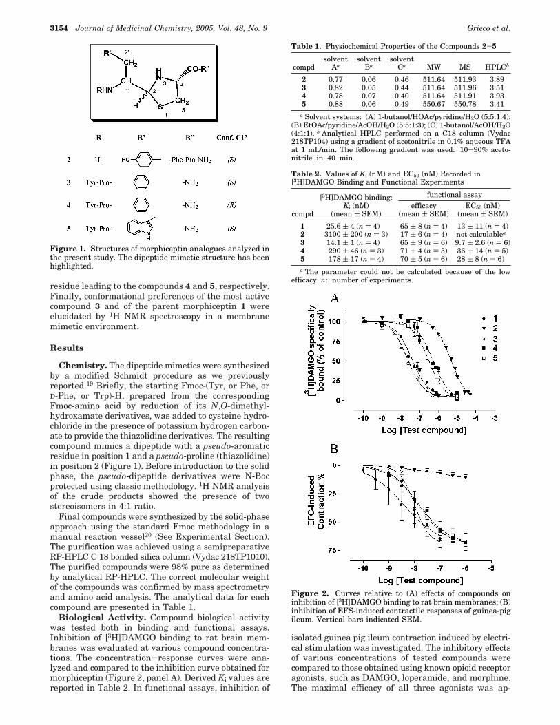
Figure 1. Structures of morphiceptin analogues analyzed inthe present study. The dipeptide mimetic structure has beenhighlighted.
Table 1. Physiochemical Properties of the Compounds 2-5
compdsolvent
Aasolvent
Basolvent
Ca MW MS HPLCb
2 0.77 0.06 0.46 511.64 511.93 3.893 0.82 0.05 0.44 511.64 511.96 3.514 0.78 0.07 0.40 511.64 511.91 3.935 0.88 0.06 0.49 550.67 550.78 3.41
a Solvent systems: (A) 1-butanol/HOAc/pyridine/H2O (5:5:1:4);(B) EtOAc/pyridine/AcOH/H2O (5:5:1:3); (C) 1-butanol/AcOH/H2O(4:1:1). b Analytical HPLC performed on a C18 column (Vydac218TP104) using a gradient of acetonitrile in 0.1% aqueous TFAat 1 mL/min. The following gradient was used: 10-90% aceto-nitrile in 40 min.
Table 2. Values of Ki (nM) and EC50 (nM) Recorded in[3H]DAMGO Binding and Functional Experiments
functional assay
compd
[3H]DAMGO binding:Ki (nM)
(mean ( SEM)efficacy
(mean ( SEM)EC50 (nM)
(mean ( SEM)
1 25.6 ( 4 (n ) 4) 65 ( 8 (n ) 4) 13 ( 11 (n ) 4)2 3100 ( 200 (n ) 3) 17 ( 6 (n ) 4) not calculablea
3 14.1 ( 1 (n ) 4) 65 ( 9 (n ) 6) 9.7 ( 2.6 (n ) 6)4 290 ( 46 (n ) 3) 71 ( 4 (n ) 5) 36 ( 14 (n ) 5)5 178 ( 17 (n ) 4) 70 ( 5 (n ) 6) 28 ( 8 (n ) 6)a The parameter could not be calculated because of the low
efficacy. n: number of experiments.
Figure 2. Curves relative to (A) effects of compounds oninhibition of [3H]DAMGO binding to rat brain membranes; (B)inhibition of EFS-induced contractile responses of guinea-pigileum. Vertical bars indicated SEM.
3154 Journal of Medicinal Chemistry, 2005, Vol. 48, No. 9 Grieco et al.

proximately 65% (efficacy, 61 ( 9, 68 ( 11, and 66 (9% for DAMGO, loperamide, and morphine, respec-tively). The EC50 values for DAMGO, loperamide, andmorphine were 36.2 ( 11.1, 488.9 ( 127.4, and 30.7 (11.2 nM, respectively. Both loperamide and morphineinhibitions were antagonized by naloxone (data notshown). In Figure 2, panel B, the concentration-response curves of morphiceptin and synthesized com-pounds are shown while in Table 2 the derived EC50values are compared to Ki values obtained from bindingassays.
Replacement of Tyr1-Pro2 with the new motif incompound 2 caused a dramatic decrease of drug potencyin inhibiting [3H]DAMGO binding as indicated by 120-fold increase of the Ki value in comparison to that ofmorphiceptin. In functional studies, this compound didnot produce any inhibition of ileum contraction withinthe concentration range tested. In contrast, the com-pound 3 in which Phe3-Pro4 is replaced with the corre-sponding dipeptide mimetic structure was as active asmorphiceptin both in binding and functional assays. Inthe last assay, the compound 3 showed an efficacyparameter comparable to that of the reference agonists,exhibiting the pharmacodynamic profile of full agonist.The activity as opioid receptor agonist of this compoundwas confirmed by the naloxone-induced reversibility ofsmooth muscle contraction inhibition (data not shown).Successively, keeping the dipeptide mimetic moiety atthe C-terminal position, we replaced the pseudo-Pheresidue of 3 with a pseudo-D-Phe or a pseudo-Trpresidue, obtaining the compounds 4 and 5, respec-tively (Figure 1). These substitutions are rationallysupported by the observation that various morphiceptinanalogues, in which the Phe3 was replaced by otheraromatic residues, preserve µ-opioid activity.21 Com-pounds 4 and 5 were 12- and 7-fold less potent inhibitorsof [3H]DAMGO binding to membranes than morphicep-tin. However, both these compounds were slightly lesspotent than morphiceptin in inhibiting ileum smoothmuscle contraction with the pharmacological pattern offull agonists. The specificity of this effect throughactivation of opioid receptors was confirmed by naloxoneantagonism (data not shown). The slight differencesbetween Ki and EC50 values of these compounds mightbe the consequence of the use of tissue preparationsobtained from different animal species.
NMR Analysis. A conformational analysis of mor-phiceptin and of the most active peptide 3 was per-formed using NMR and molecular modeling techniques.To date, many conformational studies have been per-formed to reveal the bioactive conformation of µ-opioidpeptides. However, the conformational flexibility ofopioid peptides has hampered numerous attempts atdetermining the relationship between the solutionconformation and activity using spectroscopic and mod-eling methods. It is known that the conformationalspace of the short opioid peptides is strongly affectedby local environments22 and that is generally poorlydefined in aqueous solution. Since many hormones andneurotransmitters have been found to interact with thephospholipid bilayer and the resulting conformationalpreferences in the membrane-bound state have beencorrelated with their bioactivities,23 we have determinedthe conformational preferences of 3 in SDS micelles andcompared the results with those obtained for the parentpeptide 1.
Compound 3 in SDS Micelles. In the 1D 1H NMRspectrum of 3 in SDS micelles, four distinct sets ofsignals are detected which arise from the cis/transisomerization of the Tyr-Pro peptide bond, and from theinversion of the C-2 chiral center of the thiazolidine ring(Figure 1). Integration of the signals in the 1D spectrumreveals the prevalence of one structure which accountsfor 82% of all the isomers (the populations of the otherisomers are about 7%, 7%, and 4% of the total). All theresonances of this major isomer (3a) were assignedfollowing the Wuthrich procedure24 via the usual sys-tematic application of DQF-COSY,25 TOCSY,26 andNOESY27 experiments with the support of the XEASYsoftware package (Table 3).28 Since only few signalsunambiguously belong to the three minor isomers,complete assignment and structure determination werenot possible in these cases. Partial assignment of the1H NMR resonances of the three minor isomers isreported in the Supporting Information (isomers 3b-d). The following descriptions will be focused on themajor solution isomer 3a.
Sequential NOE connectivities of the Tyr1 HR withthe Pro2 Hδ‘s evidence that Tyr-Pro peptide bond hasthe trans configuration. On the basis of chemical shiftsimilarities with 3a (Supporting Information), the iso-mer 3b (7% of the total population) was tentatively
Table 3. 1H NMR Resonance Assignmentsa of Analyzed Compounds in SDS-d25 200 mM Solution
residueb NH (3JNR, ex, Tc)c CRH (3JRâ)c CâH others
3aTyr1 4.46 (4.0, 10.8) 3.19, 3.00 7.23 (δ); 6.89 (ε)Pro2 4.35 (4.6, 7.6) 1.88, 1.13 1.84, 1.66 (γ); 3.79, 3.39 (δ)ψ-Phe3 7.77 (4.5, s, 2.7) 3.98 (10.5, 4.5) 3.34, 3.03 7.17 (δ); 7.21 (ε)Thiaz 5.58 (C-2); 4.94 (C-4); 3.82, 3.42 (C-5) 7.24 (η)
1 all-transTyr1 4.39 (6.8, 7.0) 2.88 7.06 (δ); 6.79 (ε)Pro2 4.54 (3.6, 8.2) 2.06, 1.87 1.90, 1.72 (γ); 3.67, 3.07 (δ)Phe3 7.50 (7.2, f, 6.0) 4.82 (9.0, 5.5) 3.16, 3.01 7.28 (δ); 7.26 (ε)Pro4 4.35 (4.6, 8.7) 2.22, 1.90 1.86 (γ); 3.71, 3.31 (δ)
1 cis-transTyr1 3.50 (10.5, 4.5) 2.99, 2.87 6.91 (δ); 6.76 (ε)Pro2 3.13 (5.1, 7.2) 1.64 1.51, 1.35 (γ); 3.42, 3.27 (δ)Phe3 8.00 (7.2, f, 6.5) 4.86 (10.3, 5.2) 3.23, 2.95 7.34 (δ); 7.32 (ε)Pro4 4.31 (5.6, 9.2) 2.24 1.95 (γ); 3.81, 3.57 (δ)
a Obtained at 300 K, pH ) 5.5, with TSP (δ 0.00 ppm) as reference shift. Chemical shifts are accurate to (0.02 ppm. b ψ-Phe3: pseudo-Phe3 and Thiaz: thiazolidine ring. c 3J coupling constants in Hz. ex ) HN exchange rate (f, fast; s, slow). Tc ) -∆δ/∆T (ppb/K).
Xaa-Pro Dipeptide Mimetic Analogues of Morphiceptin Journal of Medicinal Chemistry, 2005, Vol. 48, No. 9 3155

indicated as trans; therefore, the overall Pro2 transpopulation of 3 raises to ≈90% of the total. Absoluteconfiguration at C-2 of the thiazolidine ring of 3a(Figure 1) was established on the basis of the observedNOEs. In fact, NOEs with different intensities wereobserved between the methylene protons (H2C-5) of thisring and HC-4 or HC-2 (HC-5lf/HC-4, strong; HC-5hf/HC-4, medium; HC-5lf/HC-2, not observed; HC-5hf/HC-2, weak; where hf and lf mean high field and low field,respectively); furthermore, no NOE was observed be-tween HC-4 and HC-2. This NOE pattern is consistentwith an anti orientation of HC-4 and HC-2 which definesthe S configuration at C-2.
Analysis of short and medium-range NOEs, 3JNH-HRand 3JHR-Hâ coupling constants, HN exchange rates, andtemperature coefficients for exchanging HN’s was usedto characterize main conformational features of 3a(Table 3, and Supporting Information). Backbone con-formation is defined by the angles showed in Figure 3.Since Tyr1 is immediately followed by a proline, angleψ1 rotation is highly restricted to a range of 60-180°.14
Strong sequential NOE from the HR of Pro2 to the HNof the pseudo-dipeptide indicates that the ψ2 angle isrestricted to a range of 60°-180°. Small value of 3JNH-HR(4.5 Hz) and strong intraresidue NOE between the HNand the HC-1′ of the pseudo-Phe3 establishes a nearlygauche orientation of these two protons (φ 3 ≈ -60°).Coupling constant value between HC-1′ and HC-2within the pseudo-dipeptide moiety (3J ) 9.7 Hz) andmedium intensity NOE between the same proton signalsindicate a nearly trans orientation of these two atoms.Finally, slow exchange rate of amide proton of pseudo-Phe3, and a low value of the temperature coefficient forthe same proton (-∆δ/∆T < 3.0 ppb/K), indicate thatsuch proton is engaged in a hydrogen bond, probablystabilizing a turn structure. A turn structure is sup-ported by the observation of weak NOE between HR ofTyr1 and HN of Phe3.
Conformations around the CR-Câ bond (ø1) of the Tyr1
and pseudo-Phe3 residues are illustrated by the New-man projections in Figure 4, where the two â-protonsare distinguished by superscripts R and S (for pro-R andpro-S proton, respectively). The prochiralities of theâ-protons of Tyr1 and pseudo-Phe3 were assigned by acombination of the observed 3JHR-Hâ values and NOEsincluding these â-protons. For the first residue, strongand medium NOEs were observed from the Tyr1 HR andPro2 Hδ’s to the Tyr1 Hâlf which shows a small 3JHR-Hâcoupling constant (4.0 Hz) whereas weak NOEs weremeasured to Tyr1 Hâhf showing a large 3JHR-Hâ value(10.8 Hz). As mentioned above, torsion angle ψ1 isrestricted in a range from 60° to 180°. By use ofmolecular models with ψ1 in this region, the observedNOE between Tyr1 Hâlf and Pro2 Hδ’s allows us toidentify the â-protons as follows: Hâ(R) ) Hâhf andHâ(S) ) Hâlf. Fractions of the three side chains con-formers ø1 ) g-, t, g+ of Tyr1 were estimated byemploying the rotational isomeric state approximation.29
The trans (JT) and gauche (JG) coupling constantsrequired for the analysis were set to 13.85 and 3.55 Hzfollowing Cung et al.30 The calculated ø1
1 fractions (f(g-)) 0.70, f(t) ) 0.04, and f(g+) ) 0.26) indicate a largeprevalence of the g- isomer. For the pseudo-Phe3
residue intraresidue strong NOE was observed from theHR (HR stands for HC-1′ and Hâ for HC-2′) to the Hâhfwhich shows a small 3JHR-Hâ coupling constant (4.5 Hz)whereas medium NOE was measured to Hâlf showinga large 3JHR-Hâ value (10.5 Hz); medium intraresidueNOEs were observed from the HΝ to both Hâ’s; mediumand weak sequential NOEs were observed from HC-2to Hâlf.and Hâhf, respectively. Molecular model withpseudo ψ3 ≈ 180° (see above) allows us to identify theâ-protons as follows: Hâ(R) ) Hâhf and Hâ(S) ) Hâlf.The lacking of JT and JG reference values for thispseudo-Phe3 residue prevented us from estimating theconformer fractions. Anyway, the whole pattern ofdescribed NOE points out to a trans conformation of thepseudo-Phe3 side chain.
NMR-derived constraints for 3a were used as theinput data for a simulated annealing structure calcula-tion as implemented within the standard protocol of theDYANA program.31 The annealing procedure produced200 conformations from which 50 structures werechosen, whose interprotonic distances best fitted NOEderived distances, and then refined through successivesteps of restrained and unrestrained EM calculationsusing the program Discover (Biosym, San Diego, CA).For each peptide, 10 structures satisfying the NMR-derived constraints (violations smaller than 0.10 Å) were
Figure 3. Structures of morphiceptin (1) and its activeanalogue 3. Torsion angles given in the figure define spatialorientation of opioid pharmacophores. C-Terminal (pseudo)dipeptide has been indicated.
Figure 4. Newman projections about the CR-Câ bond in theside chains of the Tyr and Phe residues. The two â-protonsare distinguished by superscripts R and S (for pro-R and pro-Sproton, respectively). For the pseudo-Phe3 residue of 3 thesymbol CO represents the thiazolidine carbon atom C-2.
3156 Journal of Medicinal Chemistry, 2005, Vol. 48, No. 9 Grieco et al.

chosen for further analysis. Structure 3a was welldefined, possessing average rms deviations of the back-bone heavy atoms and of all heavy atoms equal to 0.20and 0.45 Å, respectively (Figure 5, and Table 4). Theanalysis of the secondary structure showed the existenceof an inverse γ-turn motif (C-7 turn) encompassingresidues 1-3. This well-conserved region is stabilizedby a hydrogen bond between pseudo-Phe3 HN and Tyr1
CO. The side chain of Tyr1 and pseudo-Phe3 are alsowell defined showing a preference for g- and transorientations, respectively.
Morphiceptin in SDS Micelles. In the 1D 1H NMRspectrum of 1 in SDS micelles, four distinct sets ofsignals are detected which arise from the cis/transisomerization of the Tyr-Pro and Phe-Pro peptide bonds.Integration of the signals in the 1D spectrum reveals aratio of 60:25:10:5. The major isomer has been assignedto the all-trans structure. The second largest isomeraccounting for 25% adopts a cis configuration aroundthe Tyr-Pro amide bond. Complete resonance assign-ment of the all-trans and of the cis-trans isomers wasachieved (Table 3). Since only few signals unambigu-ously belong to the two minor isomers, assignment andstructure determination were not possible in thesecases, even though their configurations at Pro2 and Pro4
were established through chemical shifts comparisonwith the major isomers (Supporting Information). Allspectral parameters for both all-trans and cis-transisomers are listed in Table 3 and the complete list ofthe NOE contacts are reported in the SupportingInformation. Following similar arguments as those usedfor the isomer 3a we could deduce the conformationalpreferences of both the all-trans and cis-trans isomersof 1. Principal spectral features for the all-trans isomerwere: the overlapping of the resonances of the Tyr1 Hâ’swhich indicates side chain conformational averaging forthis residue; the NOE contacts of Hâ’s and HR of Tyr1
with HN of Phe3 which indicated the presence of a bentstructure about the Pro2 residue; the presence of someNOE contacts between the side chains of residues 1 and3 which indicate their spatial proximity, again indicat-ing the presence of a bent structure in the segment 1-3.The temperature coefficient (-∆δ/∆T ) 6.0 ppb/K) ofthe amide protons of Phe3 and its fast exchange rate inD2O/micelles solution indicate full accessibility of thisproton to the solvent and the absence of a definedsecondary structure.
In the cis-trans isomer, the cis configuration aboutthe Pro2 omega angle was evidenced by a sequentialNOE connectivity between the HR atom signals of Tyr1
and of Pro2 while the NOE connectivities between thePhe3 HR with the Pro4 Hδ’s established the transconfiguration of the Pro4 omega angle. For residuesfollowed by a cis proline residues the ψ angle range isprecluded to 60-180° as for those followed by transprolines.14 Therefore, the Tyr1 and Phe3 residues ofmorphiceptin adopt only the structures with ψ angleswithin this range. Two conformational states with (φ,ψ) ≈ (-75°, 135°) and (φ, ψ) ≈ (-75°, -40°) are probablefor the cis configurational isomers of a proline embeddedin a long peptide sequence.14 Strong sequential NOEfrom the HR of Pro2 to the HN of Phe3 indicates anextended conformation at ψ2 (ψ2 ≈ 135°). NOE patternindicates an extended conformation also for residue 3(φ, ψ) ≈ (-140°, 120°). Finally, the preferred orienta-tions of the side chains of the residues Tyr1 and Phe3
were assigned by a combination of the observed 3JHR-Hâvalues and NOEs including their â-proton signals. Asshown in Table 4, a preferred trans orientation of theside chains of both the aromatic residues is observed.
Structure calculation for all-trans and cis-trans iso-mers were performed as described for compound 3. The10 lowest energy and least violated conformers resultingfrom calculation are reported for both isomers in Figure6, where the mean rms deviations from backbonesuperimposition are 0.40 Å and 0.30 Å for the all-transand cis-trans isomers, respectively. Violations smallerthan 0.15 Å were encountered for both the isomers.Regardless the well-defined structure of these isomers,no standard pattern of secondary structure could beobserved. The side chain conformation is well definedfor the cis-trans isomer (rms deviation from heavyatoms superposition is 0.60 Å), which shows a transorientation of both the Tyr and the Phe side chains. Inthe all-trans isomer the Tyr side chain is flexible andcan adopts both the g- and the g+ conformations whilethe Phe side chain is more defined showing a transorientation. The conformational analysis indicates thatthe separation of the aromatic rings of the tyrosine and
Figure 5. Superposition of the 10 lowest energy conformersof 3a. Structures were superimposed using the backbone heavyatoms. Hydrogen atoms are not shown for clarity, exceptpseudo-Phe3 HN, which is engaged in a hydrogen bond withthe backbone carbonyl oxygen of Tyr1 (broken line).
Table 4. Torsion Angles Defining the PharmacophoreOrientations of Described Compoundsa
angle 3ab EM1c 1 all-transb 1 cis-transb MCc
ψ1 156 ( 6 150 154 ( 6 84 ( 10 60-180ø1
1 g- g- g+, g- t tω1 176 ( 5 180 175 ( 5 0 ( 5 0ψ2 91 ( 5 150 43 ( 8 133 ( 5 150φ3 -70 ( 8d -120 -131 ( 11 -143 ( 9 -120ψ3 39 ( 4d -30 114 ( 11 128 ( 5 130ø1
3 t t, g+ t t ta Angles are in degrees. b Angles value of the mean structure
of the 10 lowest energy conformers for 3a, 1 all-trans, and 1 cis-trans are reported (( standard deviation). c EM1: Endomorphin-1active structure from ref 9 (trans-endomorphin-1 ‘extended struc-ture’). MC: Morphiceptin active structure from ref 14 (topologyI). d Pseudo-φ 3 and pseudo-ψ3 angles.
Xaa-Pro Dipeptide Mimetic Analogues of Morphiceptin Journal of Medicinal Chemistry, 2005, Vol. 48, No. 9 3157
phenylalanine residues, as expressed by the center-to-center distance, is dφ-φ ) 10.5-11.0 Å for the cis-transisomer. For the all-trans isomer dφ-φ ≈ 5.0 and dφ-φ ≈7.5 Å are observed for conformers presenting the ø1
1
angle in the g+ and g- orientation, respectively.
Discussion
Extensive structure-function studies on opioid recep-tor agonists and antagonists have been performed inattempts to understand the conformational require-ments of these ligands for selective interaction with thedifferent opioid receptors.3,4 There is general consensusthat the N-terminal portion of the opioid peptides actsas the message unit and that it contains three phar-macophoric groups: a positive ionizable feature placedon the amino terminal group of Tyr1; two hydrophobicaromatic features positioned onto the Tyr1 aryl ringsand a second aromatic residue in the third or fourthposition. The two aromatic residues are separated byan appropriate spacer. In this context, the Tyr1 residueplays the role of the primary pharmacophore while theorientation of the second aromatic pharmacophore rela-tive to the tyrosine side chain dictates the receptorselectivity. In particular, a relatively large separationof the two aromatic side chains (10-13 Å) is requiredfor the µ-opioid receptor activity of these peptides.13 Inthe µ-selective opioid peptides morphiceptin and endo-morphin-1 and -2 the spacer between the two pharma-cophoric aromatic residues is represented by a prolineresidue. Furthermore, morphiceptin bears a secondproline residue in the fourth position.
To gain information about the structural require-ments of µ-selective opioid peptides, we studied somemorphiceptin derivatives introducing in this endogenoushormone a dipeptide mimetic structure that we haverecently synthesized (Figure 1).19 The rational basis ofthis substitution was the elimination of the amide bondin the dipeptide Xaa-Pro responsible of the cis-transisomerization. In the dipeptide mimetic that we havedeveloped this amide bond is absent, replaced by asingle rotable bond between the C-1′ and the C-2 of thedipeptide mimetic structure.
Biological data (Figure 2, and Table 2) indicate thatwhile the replacement of Tyr1-Pro2 with the correspond-ing pseudo-dipeptide caused almost a complete loss ofbiological activity (compound 2), replacement of residues3-4 with an appropriate pseudo-dipeptide was toleratedat the µ-opioid receptor. In fact, compound 3 showedsimilar activity to morphiceptin both in binding andfunctional assays, while compounds 4 and 5 wereslightly less potent than the parent peptide in functionalassays, even if they showed 12- and 7-fold reducedaffinities, compared to morphiceptin in binding assay.
Hence, the amide bond at the Tyr1-Pro2 appears tobe indispensable for the biological activity of the mor-phiceptin, probably because the conformational restric-tions imposed by the cis or trans configuration of thisbond are necessary to the correct spatial disposition ofthe peptide.
Starting from the biological data, we have studied theconformational behavior of the most active analogue (3)and of the parent peptide morphiceptin (1) in the SDSmembrane-like environment. The use of SDS micellesto study the conformational properties of these peptidesis based on their interaction with a membrane receptor.For peptides acting as ligand of membrane receptors,the use of membrane mimetic media, such as SDS ordodecylphosphocoline, is suggested, hypothesizing amembrane-assisted mechanism of interactions betweenthe peptides and their receptors.23 Exploring the con-formational behavior of peptide hormones in membrane-mimetic environment, we have recently succeeded incorrelating receptor binding affinity of urotensin-IIanalogues with their preferred conformation in an SDSmicelle solution.32 Analogue 3 shows a trans configu-ration at Tyr1-Pro2 amide bond (about 90% of the isomerpopulations) and a well-defined inverse γ-turn structurecentered on Pro2 residue (Figure 5). Side chains of thearomatic residues are also well defined with g- andtrans preferred orientation of the ø1 angles of Tyr1 andpseudo-Phe3, respectively.
As shown in Figure 7, the calculated structure of 3can be efficiently superimposed to the ‘active’ structureof endomorphin-1 described by Podlogar et al.9 This
Figure 6. Superposition of the 10 lowest energy conformers of all-trans (red) and cis-trans (blue) isomers of 1. Structures weresuperimposed using the backbone heavy atoms. Hydrogen atoms are not shown for clarity.
3158 Journal of Medicinal Chemistry, 2005, Vol. 48, No. 9 Grieco et al.
structure, termed ‘trans-endomorphin-1 extended struc-ture’, was derived by the authors from NMR analysisperformed both in DMSO and in water followed by MDcalculations. The authors found that endomorphin-1exists in the cis and trans configuration with respect tothe Tyr1-Pro2 amide bond in approximately 25% and75% populations, respectively. The trans structure wasindicated as the bioactive one considering how it ef-ficiently overlaid with other selective µ ligands, PL-017and D-TIPP. The hypothesis of a trans configuration ofthe Pro-omega bond for the bioactive conformation ofendomorphin-1 was challenged by Fiori et al.11 Theseauthors, analyzing the conformational behavior of endo-morphin-1 in AOT reverse micelles, proposed a differentbioactive model for the peptide. In this model theconfiguration of the Pro-omega bond was cis and the twopharmacophoric aromatic residues were indicated as thefirst (Tyr1) and the fourth (Phe4). In contrast, Podlogaret al.9 indicated the pharmacophoric aromatic residuesas the first (Tyr1) and the third (Trp3) of the endo-morphin-1. In this context, our compound 3 possessesunique structural features which avoid possible ambi-guities. First, it shows an almost pure trans configura-tion at the Tyr1-Pro2 amide bond, which is not the casefor the other µ-opioid selective peptide agonists contain-ing the Tyr1-Pro2 amide bond studied to date. Second,differently from endomorphin-1, compound 3 lacks thearomatic residue in position 4; therefore, the twoaromatic pharmacophoric points are placed onto thearomatic rings of the first and of the third residues.
Interestingly, an inverse γ-turn structure has beenrecently proposed by Toth and co-workers as the favoredconformation of morphiceptin, endomorphin-1 and -2.33
These authors analyzed the conformational behavior ofthese peptides by the simulated annealing method andsolvated molecular dynamic calculations. It is worth tonote that the N-terminal inverse γ-turns were found
only in the Pro2 trans conformers: they were absentfrom the cis conformers.
Regarding the micelle-bound structures of the mor-phiceptin, two major isomers could be observed with theall-trans or the cis-trans configurations at the twoX-Pro amide bonds (Figure 6). The largest differencesbetween the isomers are observed for the relative spatialarrangement of the two aromatic side chains of the Tyrand the Phe residues. The all-trans isomer assumes arather compact structure with small distances, dφ-φ )5.0-7.5 Å, between the two aromatic rings of the Tyrand Phe residues. On the other hand, relatively largevalues of distance (dφ-φ ) 10.5-11.0 Å) are observedfor the minimum energy conformations estimated forthe cis-trans isomer. The results obtained from theconformational analysis leads to the conclusion that onlythe cis-trans isomer displays the relative large separa-tion of Tyr1 and Phe3 side chains which is required forthe µ-opioid receptor activity.13 Interestingly, the struc-tural parameters corresponding to the cis-trans isomer,that we have found, are highly consistent with thetopochemical model proposed by Yamazaki et al.14 forthe morphiceptin bioactive conformation (see Table 4).In fact, this model consists of a cis configuration of Tyr1-Pro2 amide bond and a trans orientation of the Tyr1 aswell as of the Phe3 side chains.
The results obtained for compound 3 and for morphi-ceptin seem to be in contrast since a trans configurationof Tyr1-Pro2 amide bond has been found for the ‘active’structure of the first while a cis configuration at thisbond is required for the latter. To gain insight into thisapparent contradiction, we superimposed the obtainedmicelles bound structure of 3 with that of the parentmorphiceptin. As shown in Figure 8, despite the differ-ent configuration at Pro2 amide bond, the two structuresdisplay similar topological arrangement. In particular,the pharmacophoric distance from Tyr center to thecenter of the Phe aromatic ring is very similar in thetwo analogues, both in the range of 10-11 Å, a result
Figure 7. Superposition of the lowest energy structure of 3a(violet) with that of endomorphin-1 (azure) described byPodlogar et al.9 Structures were superimposed using the heavyatoms of residues 1-3. Hydrogen atoms are not shown forclarity.
Figure 8. Superposition of the lowest energy structure of 3a(violet) with that of the cis-trans isomer of 1 (azure).Structures were superimposed using the side chain heavyatoms of residues 1-3. Hydrogen atoms are not shown forclarity.
Xaa-Pro Dipeptide Mimetic Analogues of Morphiceptin Journal of Medicinal Chemistry, 2005, Vol. 48, No. 9 3159

in accordance with the µ-opioid receptor requirement.13
Also, the corresponding aromatic moieties of the twocompounds have similar spatial orientations. This find-ing agrees with the assumption that the location andthe relative orientation of the aromatic side chains,rather than a peculiar backbone conformation, dictatethe selectivity toward the µ-opioid receptor and that theprincipal role of the other residues is confined to thestabilization of one specific bioactive conformationamong the pool of structures accessible for such short,conformational unrestrained peptides.34
N-Terminal tyrosine residue is a common structuralfeature of opioid peptides. Actually, the hydroxy phenylring and the positively charged nitrogen are a generalrequirement for nearly all of the reported opioid ligandpharmacophores.35 The relative disposition of thesepharmacophoric points is fully defined by ø1 angle ofTyr1. We found different ø1 angle values of Tyr1 forcompound 3 (ø1 ≈ 60°) and morphiceptin (ø1 ≈ 180°).This difference would indicate that the µ-opioid receptorcan accommodate both the g_ and trans tyrosine sidechain conformations, again indicating a high degree ofstructural tolerance within the receptor binding site.
Finally, it is interesting to note the dramatic influenceof the C-terminal residue on the overall conformationalbehavior of the peptides that we investigated. Thisinfluence could explain some results previously reportedin the literature. For instance, the inactivity of the ‘alltrans’ 2-methyl proline derivative of the morphiceptin36
and of other trans constrained pseudo-Pro2 derivatives16
can be explained, admitting that the morphiceptinanalogues with a proline residue at position 4 cannotreach the appropriate active conformation when theyadopt the trans configuration at Pro2 omega bond. Incontrast, above data suggest that the presence of otherC-terminal residues, such as phenylalanine in theendomorphins and our pseudoproline in compound 3,allows the existence of a compatible bioactive conforma-tion also (or only) with the trans Pro2 configuration.Eventually, the influence of the C-terminal residue onthe overall conformation of the opioid peptides are inaccordance with the biological result indicating that thepotency of opioid tetrapeptides containing the Tyr-Pro-Phe-X-NH2 or Tyr-Pro-Trp-X-NH2 sequence is deter-mined by the nature of the fourth residue.37
Conclusions
In conclusion, comparing the biological and confor-mational behavior of some morphiceptin analogues withthe parent peptide, we confirm a high degree of struc-tural tolerance within the µ-opioid receptor binding site.In fact, our results agree with the assumption that onlythe location and the relative orientation of the sidechains of the aromatic pharmacophoric residues repre-sent the indispensable structural feature for µ-receptorbinding. To reach such topological arrangement, thepeptide can adopt different conformations and configu-rations. In particular, opioid peptides bearing a prolineresidue as spacer between the two aromatic residuescan adopt, in active state, both cis and trans configura-tions at the Tyr1-Pro2 amide bond, each of them withthe appropriate backbone and side chain orientations.Our results also point to the importance of the C-terminal residue in dictating the overall conformational
preferences of these peptides. These data shed light onthe topological requirements of the µ-opioid receptor andtherefore might be useful for the design of new ligandsat this receptor of great pharmacological interest.
Experimental Section
Materials. NR-Fmoc-protected amino acids, HBTU, HOBt,and Ring amide resin were purchased from Advanced Chem-Tech (Louisville, KY). Peptide synthesis solvents, reagents, aswell as CH3CN for HPLC were reagent grade and wereacquired from commercial sources and used without furtherpurification unless otherwise noted. FAB-MS analyses wereperformed by MALDI. The purity of the finished peptides waschecked by analytical RP-HPLC using a Shimadzu model CL-10AD VP system with a built-in diode array detector. In allcases, the purity of the finished peptides was greater than 95%as determined by these methods.
[3H]DAMGO (50 Ci/mmol) and [3H]naltrindole (35 Ci/mmol)were purchased from PerkinElmer Life and Analytical Science.Leupeptin, bacitracin, phenylmethanesulfonyl fluoride (PMSF),bestatin and DAMGO were products of Sigma-Aldrich Co.7-Benzylidenenaltrexone (BNTX) was purchased from TocrisCookson (Bristol, UK). Naloxone and loperamide were productsof Alexis Corp. Morphine was purchased from Salars. Otheragents and reagents were from standard commercial sources.
For NMR analysis, 2H2O were obtained from Aldrich (Mil-waukee, WI), 98% SDS-d25 was obtained from CambridgeIsotope Laboratories, Inc. (Andover, MA), [(2,2,3,3-tetra-deuterio-3-(trimethylsilanyl)]propionic acid (TSP) from MSDIsotopes (Montreal, Canada).
General Method for Peptide Synthesis and Purifica-tion. All peptides were synthesized by the solid-phase methodof peptide synthesis and purified by RP-HPLC. The peptideswere synthesized on 0.15 g each of Rink amide resin (substitu-tion 0.7 mmol/g) by manual methods using NR-Fmoc chemistryand an orthogonal side chain protection strategy. The entiresynthesis was performed under argon. The resin was firstswollen in DCM/DMF (1:1) for 2 h, and the following aminoacids or the pseudo-dipeptide were then added to the growingpeptide chain by stepwise addition of NR-Fmoc amino acidsusing standard solid-phase methods. Each coupling reactionwas achieved using a 3-fold excess of each of the amino acid,HBTU, and HOBt in the presence of a 6-fold excess of DIPEAfor 1 h. Deprotection of the NR-Fmoc group was carried out bytreating the protected peptide resin with 25% piperidinesolution in DMF (1 × 4 mL, 20 min). After each coupling anddeprotection, the peptide resin was washed with DMF (3 × 4mL), DCM (3 × 50 mL) and again with DMF. The peptidesequences were thus assembled by alternate cycles of couplingand deprotection. After coupling of the N-terminal amino acid,the N-terminal Fmoc group was deblocked as described above,and the peptide-resin was thoroughly washed with DCM (4 ×25 mL) and dried under argon to yield dried peptide-resin.
The peptide-resin was then cleaved by treating with 4 mLof a solution of Et3SiH (5%), water (5%), and p-thiocresol/p-cresol (0.1%, 1:1) in TFA with shaking at room temperaturefor 3 h. The resin was then removed from the solution byfiltration, and the crude peptide was recovered by precipitationwith cold anhydrous ethyl ether. Centrifugation at 1500g for3 min followed by decantation of the supernatant ether andair-drying of the residue yielded the crude peptide as a whiteto pale beige colored amorphous solid.
Final peptide purification was achieved using a preparativeRP-HPLC Vydac C18 (218TP1520, 15 µm). The peptidesamples were injected onto the column at a concentration of20-30 mg/mL in 20% aqueous CH3CN and were eluted witha CH3CN gradient (10 to 90%) over 40 min at a flow rate of15.0 mL/min, with a constant concentration of TFA (0.1% v/v).The separations were monitored at 230 and 280 nm andintegrated with a Shimadzu diode array detector mod. SPD-M10A VP dual wavelength absorbance detector model UV-D.Fractions corresponding to the major peak were collected,pooled, and lyophilized to yield the final peptides as pure
3160 Journal of Medicinal Chemistry, 2005, Vol. 48, No. 9 Grieco et al.

(>95%) white solids. The analytical data for each compoundare presented in Table 1.
Binding Assay. The preparation of whole brain (minuscerebellum) membranes for opioid δ and µ receptor bindingexperiments was carried as described previously.38 Proteinconcentration was determined by the method of Lowry et al.39
using bovine serum albumin (BSA) as standard.For labeling opioid µ receptors, brain membranes (0.3 mg
of protein) were incubated with 0.7-1 nM [3H]DAMGO in 1mL of 50 mM Tris-HCl, pH 7.4, 5 mM MgCl2 (buffer A)containing 10 µg/mL bacitracin, 2 µg/mL bestatin, 5 µg/mLleupeptin, 4 µg/mL trypsin inhibitor, and 0.1 mM phenyl-methanesulfonyl fluoride (PMSF) at 25 °C for 2 h. Nonspecificbinding was measured in the presence of 10 µM 7-benzyl-idenenaltrexone (BNTX). Incubation was terminated by filtra-tion through GF/C filters (Millipore) previously treated with0.3% polyethyleneimine for more than 1 h. Then filters werewashed three times with 5 mL of ice-cold buffer A, andradioactivity was measured using a liquid scintillation cocktailby a Packard TRI-CARB 1600 scintillation counter (PackardInstrument, Meriden, CT). For labeling opioid δ receptors,binding studies were performed as described by Santagada etal.38 using [3H]naltrindole as radioligand. Compound con-centrations of 1 to 10 µM produced modest reductions of[3H]naltrindole binding to membranes but caused a consistentinhibition of [3H]DAMGO binding. In Table 5, the inhibitoryeffects of 10 µM morphiceptin analogues on [3H]naltrindolebinding are shown. Competition studies were carried out byincubating membranes in buffer A with 0.7 nM [3H]DAMGOand 11 newly synthesized peptides (1 nM to 10 µM).
Functional Assay. The pharmacological procedures werecarried out following the guidelines of the European CouncilDirective 86/609 concerning animal experimentation. For thefunctional study, compounds were tested on electrostimulatedileum of male Dunkin Hartley guinea pigs (300-350 g). Theanimals were sacrificed by cervical dislocation and bled, underlight ether anaesthesia. Segments of ileum (2-3 cm in length)were excised (5-6 cm far from the ileo-cecal valve), freed ofextraneous tissues, and suspended between two platinumelectrodes, under a preload of 0.5 g, in 10 mL organ baths,containing Tyrode saline (composition in mM: 136.80 NaCl,2.95 KCl, 1.80 CaCl2, 1.05 MgSO4‚7H2O, 0.41 NaH2PO4, 11.90NaHCO3, 5.50 glucose), thermostated at 37 °C, and continu-ously bubbled with a mixture of O2 (95%) and CO2 (5%).
After 30-40 min of equilibration time, the electrical fieldstimulation (EFS) started (train duration 100 ms; pulse width1 ms; pulse interval 10 ms; supramaximal voltage 20 V). Whenthe electrically induced contractile spikes reached a stablecontrol width, 3-fold increasing concentrations (0.1 nM to 1µM) of the tested compounds and reference opioid agonists,morphine, DAMGO and loperamide, were added cumulatively.In parallel sets of experiments, compounds were added in thepresence of the opioid antagonist naloxone (10 nM). Changesin isotonic tension were recorded by isotonic transducers(Basile mod. 7006) connected to microdynamometers (Basilemod. 7050).
Biological Data Analysis. 1. Binding Assay. Data wereanalyzed by a nonlinear least-squares fitting, using theGraphPad Prism Version 3.00 computer program. Single- andmultiple-site models were statistically compared to determinethe best fit, and differences between models were tested bycomparing the residual variance using a partial F test and asignificance level of P < 0.05 (GraphPad Prism Version 3.00).The IC50 values obtained from displacement and dilution
curves were converted to Ki values by the Cheng and Prusoffequation40 using the value of 1.3 nM as dissociation constant(KD) for [3H]DAMGO. Values represent the mean ( SEM ofat least three independent experiments.
2. Functional Assay. The inhibitory effects of test com-pounds and of reference agonists were evaluated as percentageof the control width (0 ) no effects; 100 ) full abolition of theEFS-induced contractile spikes). The parameter of agonistefficacy reflected the maximal inhibitory effect recorded, whilethe potency parameter was expressed as EC50, representingthe concentration evoking a half-maximal inhibitory effect.Data, expressed as mean ( SEM for 4-6 experiments, werestatistically analyzed by Student t test and Anova. P valueslower than 0.05 were considered as representative of statisticalsignificance.
NMR Spectroscopy. The samples for NMR spectroscopywere prepared by dissolving the appropriate amount of peptidein 0.45 mL of 1H2O (pH 5.5), 0.05 mL of 2H2O to obtain aconcentration 1-2 mM of peptides, and 200 mM of SDS-d25.NH exchange studies were performed dissolving peptides in0.50 mL of 2H2O and 200 mM of SDS-d25. NMR spectra wererecorded on a VARIAN Unity Inova 500 spectrometer. All thespectra were recorded at a temperature of 300 K. One-dimensional (1D) NMR spectra were recorded in the Fouriermode with quadrature detection, and the water signal wassuppressed by a low-power selective irradiation in the homo-gated mode. 2D DQF-COSY,25 TOCSY,26 and NOESY27 ex-periments were run in the phase-sensitive mode using quadra-ture detection in ω1 by time-proportional phase increase ofinitial pulse. Data block sizes were 2048 addresses in t2 and512 equidistant t1 values. Before Fourier transformation, thetime domain data matrixes were multiplied by shifted sin2
functions in both dimensions. A mixing time of 70 ms was usedfor the TOCSY experiments. NOESY experiments were runat 300 K with mixing times in the range of 150-300 ms. Thespectra were calibrated relative to TSP (0.00 ppm) as internalstandard. The qualitative and quantitative analyses of DQF-COSY, TOCSY, and NOESY spectra, were obtained using theinteractive program package XEASY.28 3JHN-HR and 3JHR-Ηâ
coupling constants were obtained by 1D spectra and bysections of cross-peaks from the resolution enhanced 4K × 2KDQF-COSY spectra. The temperature coefficients of theamide proton chemical shifts were calculated from 1D 1H NMRand 2D DQF-COSY experiments performed at differenttemperatures in the range 300-320 K by means of linearregression.
In the NOESY spectra of both morphiceptin and peptide 3,all the cross-peaks show the same sign of the diagonal peaks,showing that the peptide interaction with the SDS micelles istaking place and that the whole system is moving in solutionwith ωτc . 1.
Structural Determinations and Computational Mod-eling. The NOE-based distance restraints were obtained fromNOESY spectra collected with a mixing time of 200 ms. TheNOE cross-peaks were integrated with the XEASY programand were converted into upper distance bounds using theCALIBA program incorporated into the program packageDYANA.31 Cross-peaks which were overlapped more than 50%were treated as weak restraints in the DYANA calculation.Nonstandard dipeptide mimetic moiety was added to DYANAresidue library using MOLMOL.41 Only NOE-derived con-straints (Supporting Information) were considered in theannealing procedures. For each examined peptide, an ensembleof 200 structures was generated with the simulated annealingstandard protocol of the program DYANA. 50/200 structureswere chosen, whose interprotonic distances best fitted NOEderived distances, and then refined through successive stepsof restrained and unrestrained EM calculations. First, steepestdescents minimizations with distance restraints were per-formed on all structures with the Discover algorithm (Biosym,San Diego) utilizing the consistent valence force field (CVFF).42
A generic distance maximum force constant of 100 kcal/moland an upper distance force constants of 32 kcal/Å2 were used.Minimization proceeded until the change in energy was less
Table 5. Inhibition of [3H]Naltrindole Binding to BrainMembranes by Morphiceptin Analogues
compound (10 µM) % of inhibition (mean ( SEM)a
1 23.6 ( 4 (n ) 4)2 10.2 ( 4 (n ) 3)3 22.7 ( 8 (n ) 3)4 42.2 ( 12 (n )3)5 67.8 ( 14 (n )3)
a n: number of experiments.
Xaa-Pro Dipeptide Mimetic Analogues of Morphiceptin Journal of Medicinal Chemistry, 2005, Vol. 48, No. 9 3161

than 0.05 kcal/mol. This was followed by 3000 steps ofunrestrained steepest descents energy minimization. A distance-dependent dielectric constant equal to 4r was applied toevaluate electrostatic interactions. The minimization loweredthe total energy of the structures; no residue was found in thedisallowed region of the Ramachandran plot. The final struc-tures were analyzed using the InsightII program (Biosym, SanDiego, CA). Graphical representation were carried out withthe InsightII program (Biosym, San Diego, CA). RMS deviationanalysis between energy minimized structures were carriedout with the program MOLMOL.41 The PROMOTIF programwas used to extract details on the location and types ofstructural secondary motifs.43
Acknowledgment. Authors thank Paolo Rovero(University of Florence, Italy) for helpful discussions.Authors also thank David M. Ferguson and GermanaPaterlini (University of Minnesota) who kindly suppliedthe atomic coordinates of the endomorphin-1 calculatedstructure.
Appendix
Abbreviations used for amino acids and designationof peptides follow the rules of the IUPAC-IUB Com-mission of Biochemical Nomenclature in J. Biol. Chem.1972, 247, 977-983. Amino acid symbols denote L-configuration unless indicated otherwise. The followingadditional abbreviations are used: AAA, amino acidanalysis; AOT, bis(2-ethylhexyl)sulfosuccinate sodiumsalt; Boc, tert-butyloxycarbonyl; Bzl, benzyl; tBu, tert-butyl; CH3CN, acetonitrile; DCM, dichoromethane;DIPEA, N,N-diisopropylethylamine; DMF, N,N-di-methylformamide; DQF-COSY, double quantum fil-tered correlated spectroscopy; Et3SiH, triethylsilane;FAB-MS, fast-atom bombardment mass spectrometry;Fmoc, 9-fluorenylmethoxycarbonyl; HOBt, N-hydroxy-benzotriazole; HBTU, 2-(1H-benzotriazole-1-yl)-1,1,3,3-tetramethyluronium hexafluorophosphate; MALDI-TOF, matrix-assisted laser desorption ionization/time-of-flight mass spectrometry; MD, molecular dynamic;NMP, N-methyl pyrrolidinone; NMR, nuclear magneticresonance; NOE, nuclear Overhauser effect; NOESY,nuclear Overhauser enhancement spectroscopy; RP-HPLC, reversed-phase high performance liquid chro-matography; SDS, sodium dodecylsulfate; SPPS, solid-phase peptide synthesis; SPS, solid-phase synthesis;TFA, trifluoroacetic acid; TLC, thin-layer chromatog-raphy; TOCSY, total correlated spectroscopy; Trt, tri-phenylmethyl (trityl); TSP, 3-(trimethylsilanyl)propionicacid.
Supporting Information Available: 1H NMR assign-ments of the minor isomers of compounds 1 and 3. Lists ofNOE used in the structure calculations. This material isavailable free of charge via the Internet at http://pubs.acs.org.
References(1) Lord, J. A. H.; Waterfield, A. A.; Hughes, J.; Kosterlitz, H. W.
Endogenous Opioid Peptides: Multiple Agonists and Receptors.Nature 1977, 267, 495-499.
(2) Kieffer, B. L. Recent Advances in Molecular Recognition andSignal Transduction of Active Peptides: Receptors for OpioidPeptides. Cell. Mol. Neurobiol. 1995, 15, 615-635.
(3) (a) Hruby, V. J.; Agnes, R. S. Conformation-Activity Relation-ships of Opioid Peptides with Selective Activities at OpioidReceptors. Biopolymers 1999, 51, 391-410. (b) Hruby, V. J.;Balse, P. M. Conformational and Topographical Considerationsin Designing Agonist Peptidomimetics from Peptide Leads. Curr.Med. Chem. 2000, 7, 945-970.
(4) (a) Portoghese, P. S. The role of Concepts in Structure ActivityRelationship Studies of Opioid Ligands. J. Med. Chem. 1992,35, 1927-1937. (b) Portoghese, P. S. From Models to Mol-ecules: Opioid Receptor Dimers, Bivalent Ligands, and Selec-tive Opioid Receptor Probes. J. Med. Chem. 2001, 44, 2259-2269.
(5) Matthes, H. W.; Maldonado, R.; Simonin, F.; Valverde, O.; Slowe,S.; Kitchen, I.; Befort, K.; Dierich, A.; Le Meur, M.; Dolle, P.;Tzavara, E.; Hanoune, J.; Roques, B. P.; Kieffer, B. L. Loss ofMorphine-Induced Analgesia, Reward Effect and WithdrawalSymptoms in Mice Lacking the µ-Opioid-Receptor Gene. Nature.1996, 383, 819-23.
(6) Chang, K. J.; Killian, A.; Hazum, E.; Cuatrecasas, P.; Chang, J.K. Morphiceptin (H-Tyr-Pro-Phe-Pro-NH2): a Potent and Spe-cific Agonist for Morphine (µ) Receptors. Science 1981, 212, 75-77.
(7) Zadina, J. E.; Hackler, L.; Ge, L. J.; Kastin, A. J. A Potent andSelective Endogenous Agonist for the µ-Opiate Receptor. Nature1997, 386, 499-502.
(8) Goodman, M.; Mierke, D. Configurations of Morphiceptins by1H and 13C NMR Spectroscopy. J. Am. Chem. Soc. 1989, 111,3489-3496.
(9) Podlogar, B. L.; Paterlini, M. G.; Ferguson, D. M.; Leo, G. C.;Demeter, D. A.; Brown, F. K.; Reitz, A. B. ConformationalAnalysis of the Endogenous µ-Opioid Agonist Endomorphin-1Using NMR Spectroscopy and Molecular Modeling. FEBS Lett.1998, 439, 13-20.
(10) In, Y.; Minoura, K.; Ohishi, H.; Minakata, H.; Kamigauci, M.;Sugiura, M.; Ishida, T. Conformational Comparison of µ-SelectiveEndomorphin-2 with its C-Terminal Free Acid in DMSOSolution, by 1H NMR Spectroscopy and Molecular ModelingCalculation. J. Pept. Res. 2001, 58, 399-412.
(11) Fiori, S.; Renner, C.; Cramer, J.; Pegoraro, S.; Moroder, L.Preferred Conformation of Endomorphin-1 in Aqueous andMembrane-Mimetic Environments. J. Mol. Biol. 1999, 291, 163-175.
(12) Schwyzer, R. ACTH: A Short Introductory Review. Ann. N. Y.Acad. Sci. 1977, 297, 3-26.
(13) Yamazaki, T.; Probstl, A.; Schiller, P. W.; Goodman, M. Biologicaland Conformational Studies of [Val4]Morphiceptin and [D-Val4]-Morphiceptin Analogs Incorporating Cis-2-AminocyclopentaneCarboxylic Acid as a Peptidomimetic for Proline. Int. J. Pep. Prot.Res. 1991, 37, 364-381.
(14) Yamazaki, T.; Ro, S.; Goodman, M.; Chung, N. N.; Schiller, P.W. A Topochemical Approach to Explain Morphiceptin Bio-activity. J. Med. Chem. 1993, 36, 708-719.
(15) Paterlini, M. G.; Avitabile, F.; Ostrowski, B. G.; Ferguson, D.M.; Portoghese, P. S. Stereochemical Requirements for ReceptorRecognition of the µ-Opioid Peptide Endomorphin-1. Biophys.J. 2000, 78, 590-599.
(16) Keller, M.; Boissard, C.; Patiny, L.; Chung, N. N.; Lemieux, C.;Mutter, M.; Schiller, P. W. Pseudoproline-Containing Analoguesof Morphiceptin and Endomorphin-2: Evidence for a Cis Tyr-Pro Amide Bound in the Bioactive Conformation. J. Med. Chem.2001, 44, 3896-3903.
(17) Doi, M.; Asano, A.; Komura, E.; Ueda, Y. The Structure of anEndomorphin Analogue Incorporating 1-Aminocyclohexane-1-Carboxylic Acid for Proline is Similar to the â-turn of Leu-Enkephalin. Biochem. Biophys. Res. Commun. 2002, 297, 138-142.
(18) Okada, Y.; Fujita, Y.; Motoyama, T.; Tsuda, Y.; Toshio, Y.; Li,T.; Sasaki, Y.; Ambo, A.; Jinsmaa, Y.; Bryant, S. D.; Lazarus, L.H. Structural Studies of [2′,6′-Dimethyl-L-tyrosine1]Endomorphin-2 Analogues: Enhanced Activity and Cis Orientation of the Dmt-Pro Amide Bond. Bioorg. Med. Chem. 2003, 11, 1983-1994.
(19) Grieco, P.; Campiglia, P.; Gomez-Monterrey, I.; Novellino, E.Synthesis of Conformationally Constrained â-turn ThiazolidineMimetic. Tetrahedron Lett. 2002, 43, 1197-1199.
(20) Stewart J. M.; Young J. D. In Solid-Phase Peptide Synthesis;Pierce Chemical: Rockford, IL, 1984.
(21) Fichna, J.; Do-Rego, J. C.; Costentin, J.; Chung, N. N.; Schiller,P. W.; Kosson, P.; Janecka, A. Opioid Receptor Binding and inVivo Antinociceptive Activity of Position 3-Substituted Morphi-ceptin Analogs. Biochem. Biophys. Res. Commun. 2004, 320,531-536.
(22) Mosberg, H. I.; Hurst, R.; Hruby, V. J.; Galligan, J. J.; Burks,T. F.; Gee, K.; Yamamura, K. I. [D-Pen2, L-Cys5]Enkephalin-amide and [D-Pen2, D.-Cys5]Enkephalinamide, Conformation-ally Constrained Cyclic Enkephalinamide Analogs with δ Recep-tor Specificity. Biochem. Biophys. Res. Commun. 1982, 106, 506-512.
(23) Moroder, L.; Romano, R.; Guba, W.; Mierke, D. F.; Kessler, H.;Delporte, C.; Winand, J.; Christophe, J. New Evidence for aMembrane Bound Pathway in Hormone Receptor Binding.Biochemistry 1993, 32, 13551-13559.
(24) Wuthrich, K. NMR of Proteins and nucleic acids; John Wiley &Sons: New York, 1986.
3162 Journal of Medicinal Chemistry, 2005, Vol. 48, No. 9 Grieco et al.

(25) (a) Piantini, U.; Sorensen, O. W.; Ernst, R. R. Multiple QuantumFilters for Elucidating NMR Coupling Network. J. Am. Chem.Soc. 1982, 104, 6800-6801. (b) Marion. D.; Wuthrich, K.Application of Phase Sensitive Two-Dimensional CorrelatedSpectroscopy (COSY) for Measurements of 1H-1H Spin-SpinCoupling Constants in Proteins. Biochem. Biophys. Res. Com-mun. 1983, 113, 967-974.
(26) Bax, A.; Davis, D. G. Mlev-17-Based Two-Dimensional Homo-nuclear Magnetization Transfer Spectroscopy. J. Magn. Reson.1985, 65, 355-360.
(27) Jenner, J.; Meyer, B. H.; Bachman, P.; Ernst, R. R. Investigationof Exchange Processes by Two-Dimensional NMR Spectroscopy.J. Chem. Phys. 1979, 71, 4546-4553.
(28) Bartels, C.; Xia, T.; Billeter, M.; Guentert, P.; Wuthrich, K. TheProgram XEASY for Computer-Supported NMR Spectral Analy-sis of Biological Macromolecules. J. Biomol. NMR 1995, 6,1-10.
(29) Pachler, K. G. R. Nuclear Magnetic Resonance Study of SomeR-Amino Acids-II. Rotational Isomerism. Spectrochim. Acta1964, 20, 581-587.
(30) Cung, M. T.; Marraud, M. Conformational Dependence of theVicinal Proton Coupling Constant for the CR-Câ Bond inPeptides. Biopolymers 1982, 21, 953-967.
(31) Guntert P.; Mumenthaler C.; Wuthrich, K.; Torsion AngleDynamics for NMR Structure Calculation With the New Pro-gram DYANA. J. Mol. Biol. 1997, 273, 283-298.
(32) Carotenuto, A.; Grieco, P.; Campiglia, P.; Novellino, E.; Rovero,P. Unraveling the Active Conformation of Urotensin II. J. Med.Chem. 2004, 47, 1652-1661.
(33) Otvos, F.; Kortvelyesi, T.; Toth, G. Structure Activity Relation-ship of Endomorphin-1, Endomorphin-2 and Morphiceptin byMolecular Dynamics Methods. J. Mol. Struct. (THEOCHEM)2003, 666-667, 345-353.
(34) (a) Aubry, A.; Birlirakis, N.; Sakarellos-Daitsiotis, M.; Sakarellos,C.; Marraud, M. A Crystal Molecular Conformation of Leucine-Enkephaline Related to Morphine Molecule. Biopolymers 1989,28, 27-40. (b) Gentilucci, L.; Tolomelli, A. Recent advances in
the Investigation of the bioactive conformation of peptide activeat the µ-opiod receptor. Conformational analysis of Endomor-phins. Curr. Top. Med. Chem. 2004, 4, 105-121.
(35) Huang, G.; Loew, G. Development of a Common 3D Pharma-cophore for Delta-opioid Recognition from Peptides and Non-Peptides Using a Novel Computer Program. J. Comput.-AidedMol. Des. 1997, 11, 21-28.
(36) Nelson, R. D.; Gottlieb, D. J.; Balasubramanian, T. M.; Marshall,G. R. In NIDA Research Monograph; Rapaka, R. S., Barnett,G., Hawks, R. L., Eds.; U.S. Government Printing Office:Washington, DC, 1986; Vol. 69, pp 204-230.
(37) Yang, Y. R.; Chiu, T. H.; Chen, C.-L. Structure-ActivityRelationships of Naturally Occurring and Synthetic OpioidTetrapeptides Acting on Locus Coeruleus Neurons. Eur. J.Pharmacol. 1999, 372, 229-236.
(38) Santagada, V.; Caliendo, G.; Severino, B.; Perissutti, E.; Cec-carelli, F.; Giusti, L.; Mazzoni, M. R.; Salvatori, P. A. Probingthe Shape of a Hydrophobic Pocket in the Active Site of δ-OpioidAntagonists. J. Pept. Sci. 2001, 7, 374-385.
(39) Lowry, O. H.; Rosebrough, N. J.; Farr, A.; Randall, R. J. ProteinMeasurement with Folin Phenol Reagent. J Biol Chem. 1951,193, 265-275.
(40) Cheng, Y. C.; Prusoff, W. H. Relationship between the InhibitionConstant (Ki) and the Concentration of Inhibitor which Causes50% Inhibition (IC50) of an Enzymatic Reaction. Biochem Phar-macol. 1973, 22, 3099-3108.
(41) Koradi, R.; Billeter, M., Wuthrich, K. MOLMOL: A Programfor Display and Analysis of Macromolecular Structures. J. Mol.Graphics 1996, 14, 51-55.
(42) Maple, J.; Dinur, U.; Hagler, A. T. Derivation of Force Fieldsfor Molecular Mechanics and Dynamics from Ab Initio EnergySurface. Proc. Natl. Acad. Sci. U.S.A. 1988, 85, 5350-5354.
(43) Hutchinson, E. G.; Thornton, J. M. PROMOTIF- A Program toIdentify and Analyze Structural Motifs in Proteins. Protein Sci.1996, 5, 212-220.
JM040867Y
Xaa-Pro Dipeptide Mimetic Analogues of Morphiceptin Journal of Medicinal Chemistry, 2005, Vol. 48, No. 9 3163


![Review Article Bioactive Peptides: A Review - BASclbme.bas.bg/bioautomation/2011/vol_15.4/files/15.4_02.pdf · Review Article Bioactive Peptides: A Review ... casein [145]. Other](https://static.fdocument.pub/doc/165x107/5acd360f7f8b9a93268d5e73/review-article-bioactive-peptides-a-review-article-bioactive-peptides-a-review.jpg)