Molecular Insight into the Therapeutic Promise of ...downloads.hindawi.com › journals › omcl ›...
16
Review Article Molecular Insight into the Therapeutic Promise of Targeting APOE4 for Alzheimer’s Disease Abdullah Al Mamun , 1,2 Md. Sahab Uddin , 1,2 Md. Fahim Bin Bashar, 3 Sonia Zaman, 1 Yesmin Begum, 1 Israt Jahan Bulbul, 1 Md. Siddiqul Islam, 1 Md. Shahid Sarwar , 4 Bijo Mathew, 5 Md. Shah Amran, 6 Ghulam Md Ashraf, 7,8 May N. Bin-Jumah , 9 Shaker A. Mousa , 10 and Mohamed M. Abdel-Daim 11,12 1 Department of Pharmacy, Southeast University, Dhaka, Bangladesh 2 Pharmakon Neuroscience Research Network, Dhaka, Bangladesh 3 Department of Pharmacy, University of Development Alternative, Dhaka, Bangladesh 4 Department of Pharmacy, Noakhali Science and Technology University, Noakhali, Bangladesh 5 Division of Drug Design and Medicinal Chemistry Research Lab, Department of Pharmaceutical Chemistry, Ahalia School of Pharmacy, Palakkad, India 6 Department of Pharmaceutical Chemistry, University of Dhaka, Dhaka, Bangladesh 7 King Fahd Medical Research Center, King Abdulaziz University, Jeddah, Saudi Arabia 8 Department of Medical Laboratory Technology, Faculty of Applied Medical Sciences, King Abdulaziz University, Jeddah, Saudi Arabia 9 Department of Biology, College of Science, Princess Nourah bint Abdulrahman University, Riyadh 11474, Saudi Arabia 10 Pharmaceutical Research Institute, Albany College of Pharmacy and Health Sciences, New York, NY 12144, USA 11 Department of Zoology, College of Science, King Saud University, P.O. Box 2455, Riyadh 11451, Saudi Arabia 12 Pharmacology Department, Faculty of Veterinary Medicine, Suez Canal University, Ismailia 41522, Egypt Correspondence should be addressed to Md. Sahab Uddin; [email protected] Received 5 March 2020; Accepted 17 April 2020; Published 15 May 2020 Academic Editor: Luciana Mosca Copyright © 2020 Abdullah Al Mamun et al. This is an open access article distributed under the Creative Commons Attribution License, which permits unrestricted use, distribution, and reproduction in any medium, provided the original work is properly cited. Alzheimer’s disease (AD) is a progressive neurodegenerative disease that causes chronic cognitive dysfunction. Most of the AD cases are late onset, and the apolipoprotein E (APOE) isoform is a key genetic risk factor. The APOE gene has 3 key alleles in humans including APOE2, APOE3, and APOE4. Among them, APOE4 is the most potent genetic risk factor for late-onset AD (LOAD), while APOE2 has a defensive effect. Research data suggest that APOE4 leads to the pathogenesis of AD through various processes such as accelerated beta-amyloid aggregations that raised neurofibrillary tangle formation, cerebrovascular diseases, aggravated neuroinflammation, and synaptic loss. However, the precise mode of actions regarding in what way APOE4 leads to AD pathology remains unclear. Since APOE contributes to several pathological pathways of AD, targeting APOE4 might serve as a promising strategy for the development of novel drugs to combat AD. In this review, we focus on the recent studies about APOE4-targeted therapeutic strategies that have been advanced in animal models and are being prepared for use in humans for the management of AD. 1. Introduction Alzheimer’s disease (AD) is a neurodegenerative disorder in which the death of nerve cells causes memory loss and cogni- tive decline that is serious enough to interfere with daily life [1–4]. Neuropathologically, AD is characterized by the depo- sition of extracellular beta-amyloid (Aβ) as well as increased intracellular neurofibrillary tangles along with the activation Hindawi Oxidative Medicine and Cellular Longevity Volume 2020, Article ID 5086250, 16 pages https://doi.org/10.1155/2020/5086250
Transcript of Molecular Insight into the Therapeutic Promise of ...downloads.hindawi.com › journals › omcl ›...
Review ArticleMolecular Insight into the Therapeutic Promise of TargetingAPOE4 for Alzheimer’s Disease
Abdullah Al Mamun ,1,2 Md. Sahab Uddin ,1,2 Md. Fahim Bin Bashar,3 Sonia Zaman,1
Yesmin Begum,1 Israt Jahan Bulbul,1 Md. Siddiqul Islam,1 Md. Shahid Sarwar ,4
Bijo Mathew,5 Md. Shah Amran,6 Ghulam Md Ashraf,7,8 May N. Bin-Jumah ,9
Shaker A. Mousa ,10 and Mohamed M. Abdel-Daim 11,12
1Department of Pharmacy, Southeast University, Dhaka, Bangladesh2Pharmakon Neuroscience Research Network, Dhaka, Bangladesh3Department of Pharmacy, University of Development Alternative, Dhaka, Bangladesh4Department of Pharmacy, Noakhali Science and Technology University, Noakhali, Bangladesh5Division of Drug Design and Medicinal Chemistry Research Lab, Department of Pharmaceutical Chemistry, Ahalia Schoolof Pharmacy, Palakkad, India6Department of Pharmaceutical Chemistry, University of Dhaka, Dhaka, Bangladesh7King Fahd Medical Research Center, King Abdulaziz University, Jeddah, Saudi Arabia8Department of Medical Laboratory Technology, Faculty of Applied Medical Sciences, King Abdulaziz University,Jeddah, Saudi Arabia9Department of Biology, College of Science, Princess Nourah bint Abdulrahman University, Riyadh 11474, Saudi Arabia10Pharmaceutical Research Institute, Albany College of Pharmacy and Health Sciences, New York, NY 12144, USA11Department of Zoology, College of Science, King Saud University, P.O. Box 2455, Riyadh 11451, Saudi Arabia12Pharmacology Department, Faculty of Veterinary Medicine, Suez Canal University, Ismailia 41522, Egypt
Correspondence should be addressed to Md. Sahab Uddin; [email protected]
Received 5 March 2020; Accepted 17 April 2020; Published 15 May 2020
Academic Editor: Luciana Mosca
Copyright © 2020 Abdullah Al Mamun et al. This is an open access article distributed under the Creative Commons AttributionLicense, which permits unrestricted use, distribution, and reproduction in any medium, provided the original work isproperly cited.
Alzheimer’s disease (AD) is a progressive neurodegenerative disease that causes chronic cognitive dysfunction. Most of the ADcases are late onset, and the apolipoprotein E (APOE) isoform is a key genetic risk factor. The APOE gene has 3 key alleles inhumans including APOE2, APOE3, and APOE4. Among them, APOE4 is the most potent genetic risk factor for late-onset AD(LOAD), while APOE2 has a defensive effect. Research data suggest that APOE4 leads to the pathogenesis of AD throughvarious processes such as accelerated beta-amyloid aggregations that raised neurofibrillary tangle formation, cerebrovasculardiseases, aggravated neuroinflammation, and synaptic loss. However, the precise mode of actions regarding in what way APOE4leads to AD pathology remains unclear. Since APOE contributes to several pathological pathways of AD, targeting APOE4might serve as a promising strategy for the development of novel drugs to combat AD. In this review, we focus on therecent studies about APOE4-targeted therapeutic strategies that have been advanced in animal models and are beingprepared for use in humans for the management of AD.
1. Introduction
Alzheimer’s disease (AD) is a neurodegenerative disorder inwhich the death of nerve cells causes memory loss and cogni-
tive decline that is serious enough to interfere with daily life[1–4]. Neuropathologically, AD is characterized by the depo-sition of extracellular beta-amyloid (Aβ) as well as increasedintracellular neurofibrillary tangles along with the activation
HindawiOxidative Medicine and Cellular LongevityVolume 2020, Article ID 5086250, 16 pageshttps://doi.org/10.1155/2020/5086250
of glia and neuronal death [5–9]. Besides environmental andlifestyle-related risk factors, genetic constituents are alsodeliberated to raise the risk of progressing AD [10, 11].Undeniably, research has detected several loci connectedwith AD, such as both pathogenic and susceptibility genes.AD is categorized into 2 forms according to the age of onset,including early-onset AD (EOAD) that begins in people whoare below 65 years old and it is estimated to be about 1–5% ofall cases, and late-onset AD (LOAD) that starts in peopleaged 65 or above 65 and it is projected to be greater than95% of all cases [12]. There are 3 leading congenital genemutations, such as presenilin-1 (PSEN1), presenilin-2(PSEN2), and amyloid precursor protein (APP), which playa central role in raising the generation of Aβ and have beendirectly related to EOAD [13–15]. On the contrary, LOADhas been detected to be more complicated and is linked toseveral genes with increased vulnerability [16]. Genetically,the apolipoprotein E4 (APOE4) allele is considered as theparamount determinant for LOAD [17–20].
People who inherit one copy of the APOE4 allele maycause a higher risk of raising AD, and people who possesstwo copies of the APOE4 allele are at greater risk of progres-sing AD [21, 22]. The APOE4 gene may also be accompaniedby an earlier onset of memory dysfunction and other symp-toms in comparison with AD patients who do not have thisgene. It is unknown how the APOE4 allele is connected withthe risk of AD. Conversely, the APOE4 gene is also linkedwith a greater number of protein clusters called amyloid pla-ques, which are found in the AD brain tissue [23, 24]. Fur-thermore, an aggregation of amyloid plaques is greatlyresponsible for the death of neurons and the developingsymptoms of AD [25, 26]. It has been found that the interac-tion between herpes simplex virus type 1 (HSV-1) and APOEisoforms indicate a connection between HSV-1 deoxyribonu-cleic acid (DNA) detection in AD tissues and the existence ofthe APOE4 allele [27, 28]. In addition, recent investigationshave shown a potential relationship between APOEisoform-dependent modifications in tau pathology and neu-rodegeneration [29]. Therefore, it is evident that peoplewho have the APOE4 allele inherit a greater risk of progres-sing AD, not the disease itself [30].
Oxidative stress has also been connected with APOE4in AD patients. ApoE4 is linked to higher oxidative stressas well as diminished antioxidant enzyme activity in thehippocampus of AD patients [31–33]. Oxidative stressmarkers such as increased oxidized proteins, glycosylatedproducts, elevated levels of lipid peroxidation, formationof aldehydes, alcohols, ketones, free carbonyls, and choles-tenone, as well as oxidative modifications in ribonucleicacid (RNA) and mitochondrial and nuclear DNA wereobserved in postmortem brain tissue and in peripheral sys-tems such as cells and isolated mitochondria from initialphases of AD and APOE4 carriers [34–51]. On the otherhand, APOE was shown to act as an antioxidant directlyor indirectly against hydrogen peroxide-mediated cytotox-icity in a B12 APOE expressing cell line [52]. Accordingto the study by Hayek et al. [53], the increased levels ofperoxidized plasma low-density lipoproteins in APOE-deficient mice were observed. Furthermore, the levels of
lipid oxidation were considerably elevated in the frontalcortex of AD patients who were heterozygous or homozy-gous APOE4 carriers compared to homozygous APOE3carriers and controls [54]. Although upregulation of cata-lase activity was completely found in the frontal cortextissue of homozygous APOE4 carriers, the activities ofsuperoxide dismutase and glutathione concentrations werenot as different as those from controls [54].
Hitherto, there are no approved drugs directly targetingAPOE4, even though APOE4 was detected about 25 yearsago [55–57]. Hence, due to its genetic predominance, APOEisoforms have turned into an auspicious target for betterunderstanding the pathophysiological pathways of AD, iden-tifying patients who are at greater risk for the progression ofAD and opening a novel therapeutic approach against AD.Moreover, some clinical researches both in animals andhumans have verified that APOE4 remarkably affects the var-ious independent biological pathways in the brain which playa pivotal role in the development of AD [55, 58]. In thisreview, we emphasize the current studies regarding APOE4-targeted promising therapeutic strategies to combat ADpathogenesis.
2. APOE—Polymorphism and Susceptibility toAlzheimer’s Disease
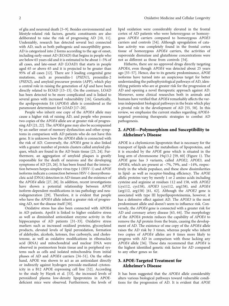
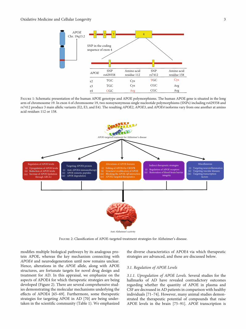
APOE is a chylomicron lipoprotein that is necessary for thetransport of lipids and the metabolism of lipoproteins, andit is encoded by the APOE gene which is situated in thelong arm of chromosome 19q13.2 [59, 60] (Figure 1). TheAPOE gene has 3 variants, called APOE2, APOE3, andAPOE4, which are present at ~7%, 79%, and ~14%, respec-tively in the whole populace, and these show dissimilaritiesin lipid- as well as receptor-binding efficiency. The APOEallelic proteins vary by merely 1 or 2 amino acids includingcysteine and arginine at residues 112 and 158, with APOE2(cys112, cys158), APOE3 (cys112, arg158), and APOE4(arg112, arg158) [61, 62]. Although the APOE2 gene isassociated with type III hyperlipoproteinemia, however, ithas a defensive effect against AD. The APOE3 is the mostpredominant allele and doesn’t seem to influence risk. Con-versely, the APOE4 gene is connected with a greater risk forAD and coronary artery disease [63, 64]. The morphologyof the APOE4 protein reduces the capability of APOE4 toremove the Aβ protein from the brain, causing the develop-ment of AD. The existence of one copy of the APOE4 alleleraises the AD risk by 3 times, whereas people who inherittwo copies of APOE4 alleles are 8 times more likely toprogress with AD in comparison with those lacking anyAPOE4 allele [56]. These data recommend that APOE4 isthe highest identified genetic risk factor for AD comparedto any other genes so far.
3. APOE-Targeted Treatment forAlzheimer’s Disease
It has been suggested that the APOE4 allele considerablyalters various biological pathways toward vulnerable condi-tions for the progression of AD. It is evident that APOE
2 Oxidative Medicine and Cellular Longevity
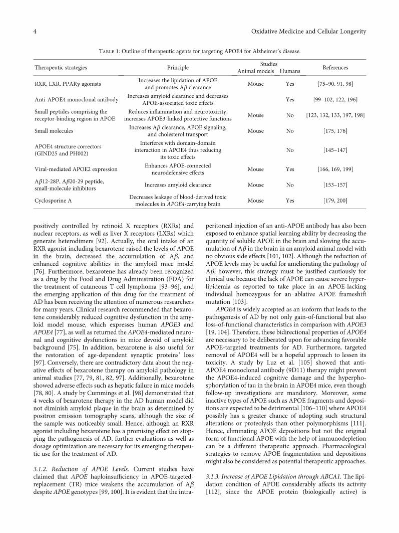
modifies multiple biological pathways by its analogous pro-tein APOE, whereas the key mechanism connecting withAPOE4 and neurodegeneration until now remains unclear.Hence, alterations in the APOE allele, along with APOEstructures, are fortunate targets for novel drug design andtreatment for AD. In this appraisal, we emphasize on theaspects of APOE4 for which therapeutic strategies are beingdeveloped (Figure 2). There are several comprehensive stud-ies demonstrating the molecular mechanisms underlying theeffects of APOE4 [65–69]. Furthermore, some therapeuticstrategies for targeting APOE in AD [70] are being under-taken in the scientific community (Table 1). We emphasized
the diverse characteristics of APOE4 via which therapeuticstrategies are advanced, and these are discussed below.
3.1. Regulation of APOE Levels
3.1.1. Upregulation of APOE Levels. Several studies for thehallmarks of AD have revealed contradictory outcomesregarding whether the quantity of APOE in plasma andCSF are decreased in AD patients in comparison with healthyindividuals [71–74]. However, many animal studies demon-strated the therapeutic potential of compounds that raiseAPOE levels in the brain [75–91]. APOE transcription is
2 41 3
APOESNP
rs429358 Amino acidresidue 112
SNPrs7412
Amino acidresidue 158
ε2ε3ε4
TGCTGCCGC
CysCysArg
TGCCGCCGC
Arg
Cys
Arg
SNP in the codingsequence of exon 4
APOEChr. 19q13.2
Figure 1: Schematic presentation of the human APOE genotype and APOE polymorphisms. The human APOE gene is situated in the longarm of chromosome 19. In exon 4 of chromosome 19, two nonsynonymous single nucleotide polymorphisms (SNPs) including rs429358 andrs7412 produce 3 main allelic variants (E2, E3, and E4). The resulting APOE2, APOE3, and APOE4 isoforms vary from one another at aminoacid residues 112 or 158.
APOE-targeted treatment for Alzheimer’s disease
Anti-Alzheimer’s activity
(ii)(iii)
(i)Regulation of APOE levels
Upregulation of APOE levelsReduction of APOE levelsIncrease of APOE lipidation
through ABCA1
Indirect therapeutic strategiesRegulation of APOE receptorsRestoration of blood-brain barrier
integrity
(i)(ii)
Alterations of APOE featuresEditing of APOE4 by CRISPRStructural modification of APOEBlocking the APOE-A𝛽 interactionAPOE2-targeted therapeutics
(i)(ii)
(iii)(iv)
Targeting APOE4 proteinAnti-APOE4 immunotherapyAPOE mimetic peptidesAPOE degradation
(i)(ii)
(iii)
MiscellaneousTargeting neuroinflammation(i)
(ii)(iii)
Targeting vascular diseasesTargeting transcription
factors
Figure 2: Classification of APOE-targeted treatment strategies for Alzheimer’s disease.
3Oxidative Medicine and Cellular Longevity
positively controlled by retinoid X receptors (RXRs) andnuclear receptors, as well as liver X receptors (LXRs) whichgenerate heterodimers [92]. Actually, the oral intake of anRXR agonist including bexarotene raised the levels of APOEin the brain, decreased the accumulation of Aβ, andenhanced cognitive abilities in the amyloid mice model[76]. Furthermore, bexarotene has already been recognizedas a drug by the Food and Drug Administration (FDA) forthe treatment of cutaneous T-cell lymphoma [93–96], andthe emerging application of this drug for the treatment ofAD has been receiving the attention of numerous researchersfor many years. Clinical research recommended that bexaro-tene considerably reduced cognitive dysfunction in the amy-loid model mouse, which expresses human APOE3 andAPOE4 [77], as well as returned the APOE4-mediated neuro-nal and cognitive dysfunctions in mice devoid of amyloidbackground [75]. In addition, bexarotene is also useful forthe restoration of age-dependent synaptic proteins’ loss[97]. Conversely, there are contradictory data about the neg-ative effects of bexarotene therapy on amyloid pathology inanimal studies [77, 79, 81, 82, 97]. Additionally, bexaroteneshowed adverse effects such as hepatic failure in mice models[78, 80]. A study by Cummings et al. [98] demonstrated that4 weeks of bexarotene therapy in the AD human model didnot diminish amyloid plaque in the brain as determined bypositron emission tomography scans, although the size ofthe sample was noticeably small. Hence, although an RXRagonist including bexarotene has a promising effect on stop-ping the pathogenesis of AD, further evaluations as well asdosage optimization are necessary for its emerging therapeu-tic use for the treatment of AD.
3.1.2. Reduction of APOE Levels. Current studies haveclaimed that APOE haploinsufficiency in APOE-targeted-replacement (TR) mice weakens the accumulation of Aβdespite APOE genotypes [99, 100]. It is evident that the intra-
peritoneal injection of an anti-APOE antibody has also beenexposed to enhance spatial learning ability by decreasing thequantity of soluble APOE in the brain and slowing the accu-mulation of Aβ in the brain in an amyloid animal model withno obvious side effects [101, 102]. Although the reduction ofAPOE levels may be useful for ameliorating the pathology ofAβ; however, this strategy must be justified cautiously forclinical use because the lack of APOE can cause severe hyper-lipidemia as reported to take place in an APOE-lackingindividual homozygous for an ablative APOE frameshiftmutation [103].
APOE4 is widely accepted as an isoform that leads to thepathogenesis of AD by not only gain-of-functional but alsoloss-of-functional characteristics in comparison with APOE3[19, 104]. Therefore, these bidirectional properties of APOE4are necessary to be deliberated upon for advancing favorableAPOE-targeted treatments for AD. Furthermore, targetedremoval of APOE4 will be a hopeful approach to lessen itstoxicity. A study by Luz et al. [105] showed that anti-APOE4 monoclonal antibody (9D11) therapy might preventthe APOE4-induced cognitive damage and the hyperpho-sphorylation of tau in the brain in APOE4 mice, even thoughfollow-up investigations are mandatory. Moreover, someinactive types of APOE such as APOE fragments and deposi-tions are expected to be detrimental [106–110] where APOE4possibly has a greater chance of adopting such structuralalterations or proteolysis than other polymorphisms [111].Hence, eliminating APOE depositions but not the originalform of functional APOE with the help of immunodepletioncan be a different therapeutic approach. Pharmacologicalstrategies to remove APOE fragmentation and depositionsmight also be considered as potential therapeutic approaches.
3.1.3. Increase of APOE Lipidation through ABCA1. The lipi-dation condition of APOE considerably affects its activity[112], since the APOE protein (biologically active) is
Table 1: Outline of therapeutic agents for targeting APOE4 for Alzheimer’s disease.
Therapeutic strategies PrincipleStudies
ReferencesAnimal models Humans
RXR, LXR, PPARγ agonistsIncreases the lipidation of APOE
and promotes Aβ clearanceMouse Yes [75–90, 91, 98]
Anti-APOE4 monoclonal antibodyIncreases amyloid clearance and decreases
APOE-associated toxic effectsYes [99–102, 122, 196]
Small peptides comprising thereceptor-binding region in APOE
Reduces inflammation and neurotoxicity,increases APOE3-linked protective functions
Mouse No [123, 132, 133, 197, 198]
Small moleculesIncreases Aβ clearance, APOE signaling,
and cholesterol transportMouse No [175, 176]
APOE4 structure correctors(GIND25 and PH002)
Interferes with domain-domaininteraction in APOE4 thus reducing
its toxic effectsNo [145–147]
Viral-mediated APOE2 expressionEnhances APOE-connected
neurodefensive effectsMouse Yes [166, 169, 199]
Aβ12-28P, Aβ20-29 peptide,small-molecule inhibitors
Increases amyloid clearance Mouse No [153–157]
Cyclosporine ADecreases leakage of blood-derived toxicmolecules in APOE4-carrying brain
Mouse Yes [179, 200]
4 Oxidative Medicine and Cellular Longevity
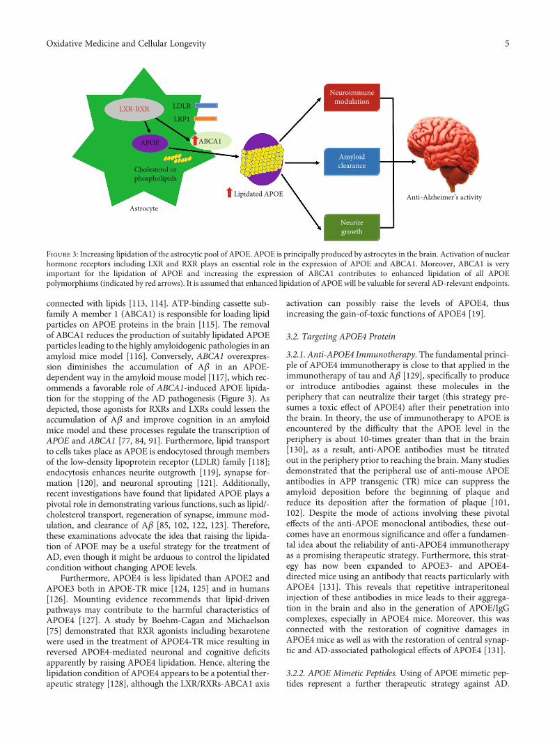
connected with lipids [113, 114]. ATP-binding cassette sub-family A member 1 (ABCA1) is responsible for loading lipidparticles on APOE proteins in the brain [115]. The removalof ABCA1 reduces the production of suitably lipidated APOEparticles leading to the highly amyloidogenic pathologies in anamyloid mice model [116]. Conversely, ABCA1 overexpres-sion diminishes the accumulation of Aβ in an APOE-dependent way in the amyloid mouse model [117], which rec-ommends a favorable role of ABCA1-induced APOE lipida-tion for the stopping of the AD pathogenesis (Figure 3). Asdepicted, those agonists for RXRs and LXRs could lessen theaccumulation of Aβ and improve cognition in an amyloidmice model and these processes regulate the transcription ofAPOE and ABCA1 [77, 84, 91]. Furthermore, lipid transportto cells takes place as APOE is endocytosed through membersof the low-density lipoprotein receptor (LDLR) family [118];endocytosis enhances neurite outgrowth [119], synapse for-mation [120], and neuronal sprouting [121]. Additionally,recent investigations have found that lipidated APOE plays apivotal role in demonstrating various functions, such as lipid/-cholesterol transport, regeneration of synapse, immune mod-ulation, and clearance of Aβ [85, 102, 122, 123]. Therefore,these examinations advocate the idea that raising the lipida-tion of APOE may be a useful strategy for the treatment ofAD, even though it might be arduous to control the lipidatedcondition without changing APOE levels.
Furthermore, APOE4 is less lipidated than APOE2 andAPOE3 both in APOE-TR mice [124, 125] and in humans[126]. Mounting evidence recommends that lipid-drivenpathways may contribute to the harmful characteristics ofAPOE4 [127]. A study by Boehm-Cagan and Michaelson[75] demonstrated that RXR agonists including bexarotenewere used in the treatment of APOE4-TR mice resulting inreversed APOE4-mediated neuronal and cognitive deficitsapparently by raising APOE4 lipidation. Hence, altering thelipidation condition of APOE4 appears to be a potential ther-apeutic strategy [128], although the LXR/RXRs-ABCA1 axis
activation can possibly raise the levels of APOE4, thusincreasing the gain-of-toxic functions of APOE4 [19].
3.2. Targeting APOE4 Protein
3.2.1. Anti-APOE4 Immunotherapy. The fundamental princi-ple of APOE4 immunotherapy is close to that applied in theimmunotherapy of tau and Aβ [129], specifically to produceor introduce antibodies against these molecules in theperiphery that can neutralize their target (this strategy pre-sumes a toxic effect of APOE4) after their penetration intothe brain. In theory, the use of immunotherapy to APOE isencountered by the difficulty that the APOE level in theperiphery is about 10-times greater than that in the brain[130], as a result, anti-APOE antibodies must be titratedout in the periphery prior to reaching the brain. Many studiesdemonstrated that the peripheral use of anti-mouse APOEantibodies in APP transgenic (TR) mice can suppress theamyloid deposition before the beginning of plaque andreduce its deposition after the formation of plaque [101,102]. Despite the mode of actions involving these pivotaleffects of the anti-APOE monoclonal antibodies, these out-comes have an enormous significance and offer a fundamen-tal idea about the reliability of anti-APOE4 immunotherapyas a promising therapeutic strategy. Furthermore, this strat-egy has now been expanded to APOE3- and APOE4-directed mice using an antibody that reacts particularly withAPOE4 [131]. This reveals that repetitive intraperitonealinjection of these antibodies in mice leads to their aggrega-tion in the brain and also in the generation of APOE/IgGcomplexes, especially in APOE4 mice. Moreover, this wasconnected with the restoration of cognitive damages inAPOE4 mice as well as with the restoration of central synap-tic and AD-associated pathological effects of APOE4 [131].
3.2.2. APOE Mimetic Peptides. Using of APOE mimetic pep-tides represent a further therapeutic strategy against AD.
APOE
Cholesterol orphospholipids
ABCA1
LDLR
LRP1
Lipidated APOE
Neuroimmunemodulation
Amyloidclearance
Neuritegrowth
AstrocyteAnti-Alzheimer’s activity
LXR-RXR
Figure 3: Increasing lipidation of the astrocytic pool of APOE. APOE is principally produced by astrocytes in the brain. Activation of nuclearhormone receptors including LXR and RXR plays an essential role in the expression of APOE and ABCA1. Moreover, ABCA1 is veryimportant for the lipidation of APOE and increasing the expression of ABCA1 contributes to enhanced lipidation of all APOEpolymorphisms (indicated by red arrows). It is assumed that enhanced lipidation of APOE will be valuable for several AD-relevant endpoints.
5Oxidative Medicine and Cellular Longevity
Furthermore, these tiny peptides, which either conform to thereceptor-binding APOE domain [132–134] or to a discretedomain of APOE including amphipathic helix domains[134], significantly lessen the neurodegeneration after braininsults [133, 135–138] and defend against tau- and Aβ-medi-ated pathogenesis in TR mice and analogous model domains[132–134]. The fundamental mechanism of the defensiveeffects of these tiny peptides might be owing to their anti-inflammatory effects. Conversely, a study of Vitek et al. [133]revealed that these APOE mimetic peptides were defensiveafter brain insults in both APOE3 and APOE4 mice.
3.2.3. APOE Degradation. APOE4 generates an intermediatemolten globule structure, which makes it less stable whencompared with APOE3 and its C- and N-terminal interactionas described earlier. Furthermore, this domain interaction inneurons makes APOE4 particularly vulnerable to discreteproteases and responsible for the production of C-terminalneurotoxic portions of APOE4 [67, 139–141]. As stress raisesthe neuronal APOE generation, it has been anticipated thatthe raised generation of intraneuronal APOE4 portionsunder stressful states may play a pivotal role in inducingthe pathogenic actions of APOE4 [67, 139–141]. Hence, theneuronal degradation of APOE4 leads to the detection ofthe proteases as well as the advancement of inhibitors againstthem demonstrating an additional strategy for neutralizingthe actions of APOE4.
3.3. Alterations of APOE Features
3.3.1. Editing of APOE4 by CRISPR. The transformation ofthe APOE4 gene to either APOE2 orAPOE3 and the abolitionof the concentration dissimilarity between them would playan important role in solving the crux of the APOE4 problem
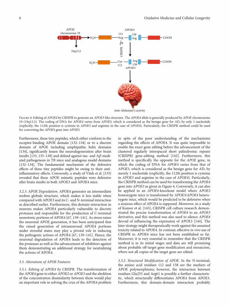
in spite of the poor understanding of the mechanismsregarding the effects of APOE4. It was quite impossible toenable the exact gene editing before the advancement of theclustered regularly interspaced short palindromic repeats(CRISPR) gene-editing method [142]. Furthermore, thismethod is specifically the opposite for the APOE gene, inwhich the coding of DNA for APOE4 varies from that ofAPOE3, which is considered as the benign gene for AD, bymerely 1 nucleotide (explicitly, the 112th position is cysteinein APOE3 and arginine in the case of APOE4). Particularly,the CRISPRmethod can be used for transforming the APOE4gene into APOE3 as given in Figure 4. Conversely, it can alsobe applied in an APOE4-knockout model where APOE3homozygote mice is transformed by APOE3/APOE4 hetero-zygote mice, which would be predicted to be defensive whena noxious effect of APOE4 is supposed. Moreover, in a studyof Komor et al. [143], CRISPR cell culture research demon-strated the precise transformation of APOE4 to an APOE3derivative, and this method was also used to silence APOE4devoid of influencing the expression of APOE3 [144]. Thelater strategy might therapeutically work against the assumedtoxicity related to APOE4. In contrast, effective in vivo use ofCRISPR to APOE4 mice has not been established so far.Moreover, it is very essential to remember that the CRISPRmethod is in its initial stages and data are still promisingabout probable off-target gene modification and mosaicism,where not all copies of the target gene are edited.
3.3.2. Structural Modification of APOE. In the N-terminal,the amino acid residues 112 and 158 are the markers ofAPOE polymorphisms; however, the interaction betweenresidues Glu255 and Arg61 is possibly a further characteris-tic, which structurally differentiates APOE4 from APOE3.Furthermore, this domain-domain interaction probably
19q13.2
APOEchromosome 19
H2N
H2N
112Arg
158Arg COOH
APOE4
CRIS
PR
Cys112
Arg158
COOH
APOE3
Anti-Alzheimer’s activity
Figure 4: Editing of APOE4 by CRISPR to generate an APOE3-like structure. The APOE4 allele is generally produced by APOE chromosome19 (19q13.2). The coding of DNA for APOE4 varies from APOE3, which is considered as the benign gene for AD, by only 1 nucleotide(explicitly, the 112th position is cysteine in APOE3 and arginine in the case of APOE4). Particularly, the CRISPR method could be usedfor converting the APOE4 gene into APOE3.
6 Oxidative Medicine and Cellular Longevity
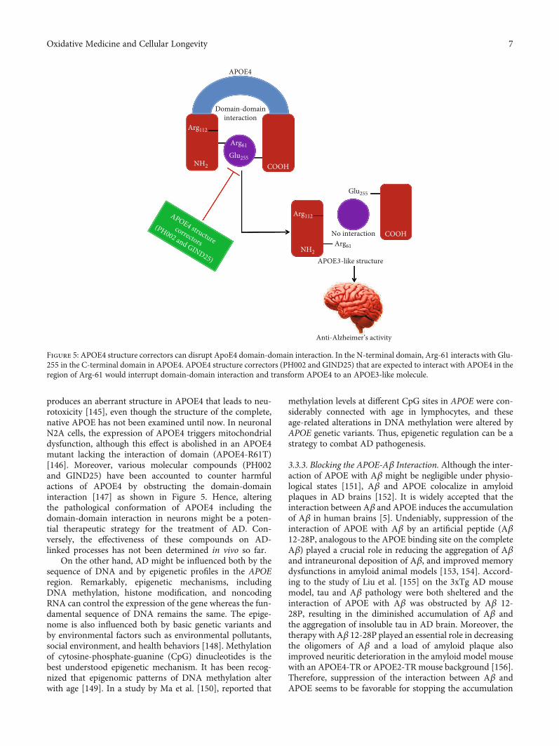
produces an aberrant structure in APOE4 that leads to neu-rotoxicity [145], even though the structure of the complete,native APOE has not been examined until now. In neuronalN2A cells, the expression of APOE4 triggers mitochondrialdysfunction, although this effect is abolished in an APOE4mutant lacking the interaction of domain (APOE4-R61T)[146]. Moreover, various molecular compounds (PH002and GIND25) have been accounted to counter harmfulactions of APOE4 by obstructing the domain-domaininteraction [147] as shown in Figure 5. Hence, alteringthe pathological conformation of APOE4 including thedomain-domain interaction in neurons might be a poten-tial therapeutic strategy for the treatment of AD. Con-versely, the effectiveness of these compounds on AD-linked processes has not been determined in vivo so far.
On the other hand, AD might be influenced both by thesequence of DNA and by epigenetic profiles in the APOEregion. Remarkably, epigenetic mechanisms, includingDNA methylation, histone modification, and noncodingRNA can control the expression of the gene whereas the fun-damental sequence of DNA remains the same. The epige-nome is also influenced both by basic genetic variants andby environmental factors such as environmental pollutants,social environment, and health behaviors [148]. Methylationof cytosine-phosphate-guanine (CpG) dinucleotides is thebest understood epigenetic mechanism. It has been recog-nized that epigenomic patterns of DNA methylation alterwith age [149]. In a study by Ma et al. [150], reported that
methylation levels at different CpG sites in APOE were con-siderably connected with age in lymphocytes, and theseage-related alterations in DNA methylation were altered byAPOE genetic variants. Thus, epigenetic regulation can be astrategy to combat AD pathogenesis.
3.3.3. Blocking the APOE-Aβ Interaction. Although the inter-action of APOE with Aβ might be negligible under physio-logical states [151], Aβ and APOE colocalize in amyloidplaques in AD brains [152]. It is widely accepted that theinteraction between Aβ and APOE induces the accumulationof Aβ in human brains [5]. Undeniably, suppression of theinteraction of APOE with Aβ by an artificial peptide (Aβ12-28P, analogous to the APOE binding site on the completeAβ) played a crucial role in reducing the aggregation of Aβand intraneuronal deposition of Aβ, and improved memorydysfunctions in amyloid animal models [153, 154]. Accord-ing to the study of Liu et al. [155] on the 3xTg AD mousemodel, tau and Aβ pathology were both sheltered and theinteraction of APOE with Aβ was obstructed by Aβ 12-28P, resulting in the diminished accumulation of Aβ andthe aggregation of insoluble tau in AD brain. Moreover, thetherapy with Aβ 12-28P played an essential role in decreasingthe oligomers of Aβ and a load of amyloid plaque alsoimproved neuritic deterioration in the amyloid model mousewith an APOE4-TR or APOE2-TR mouse background [156].Therefore, suppression of the interaction between Aβ andAPOE seems to be favorable for stopping the accumulation
COOH
APOE4
Domain-domaininteraction
NH2Arg61
COOH
Glu255
Glu255
No interaction
APOE3-like structure
Anti-Alzheimer’s activity
Arg112
NH2
Arg112
Arg61
APOE4 structurecorrectors
(PH002 and GIND25)
Figure 5: APOE4 structure correctors can disrupt ApoE4 domain-domain interaction. In the N-terminal domain, Arg-61 interacts with Glu-255 in the C-terminal domain in APOE4. APOE4 structure correctors (PH002 and GIND25) that are expected to interact with APOE4 in theregion of Arg-61 would interrupt domain-domain interaction and transform APOE4 to an APOE3-like molecule.
7Oxidative Medicine and Cellular Longevity
of Aβ despite the APOE polymorphisms, although the phar-macological effect of artificial peptides may vary according tothe APOE polymorphism being targeted. Additionally, Haoet al. [157] demonstrated that Aβ 20–29 peptides mightobstruct the interaction of APOE with Aβ, thus decreasingthe fibrillogenesis as well as cytotoxicity of Aβ in vitro. Cap-tivatingly, immunotherapy of APOE noticeably reduces theaccumulation of Aβ in amyloid mouse models as discussedearlier [101, 102]. Recently, in an in vivo test, using antibod-ies that accept both human APOE3 and APOE4 and thencombining specifically to nonlipidated APOE rather thanlipidated APOE result in diminishing the accumulation ofAβ in a TR mice model [158]. In addition, APOE antisenseoligonucleotides are also used for the lessening of amyloidplaques [159]. It is evident that APOE3 and APOE4 binddiversely and directly to Aβ [125]; however, a study byVerghese et al. [151] revealed that the interaction of APOE4with Aβ might be indirect as well as intervened through athird molecule. Therefore, blocking the interaction betweenAPOE4 and Aβ may be a promising therapeutic strategyfor the treatment of AD.
3.3.4. APOE2-Targeted Therapeutics. The occurrence ofAPOE2 in AD patients is about 2-times lower than the gen-eral populace, and it is connected with less noticeable brainpathology than that found in non-APOE2 AD subjects[160]. The heterozygosity of APOE2 is also linked withlongevity [160] and lessened age-dependent cognitive dys-function [161–164]. In addition, APOE2 increases the neuro-protective activity against AD via various molecularmechanisms [165]. Consequently, in the case of neurodegen-erative disorders that are directly linked with neuronal andsynaptic loss, APOE2 is defensive because of its capabilityto excite the restoration of these pathways. Various investiga-tions recommended that the brain pathological effects ofAPOE4 in TR mice could be counteracted by the injection(intracerebral) of viral vectors expressing APOE2 [166,167], recommending a new anti-APOE4 therapeutic strategy[168]. According to the study of Hu et al. [169], APOE4 ishypolipidated compared with APOE3 and APOE2 is hyperli-pidated in relation to APOE3. Moreover, it is evident thatAPOE2 and APOE4 affect the identical pathway such as lipi-dation of APOE; however, they drive in opposite directions.In contrast, the probability that APOE4 and APOE2 activatethrough diverse nonoverlapping processes with contrastingphysiological outcomes could not be denied.
3.4. Indirect Therapeutic Strategies
3.4.1. Regulation of APOE Receptors. The LDLR family,including LDLR and low-density lipoprotein receptor-related protein 1 (LRP1) [170], plays a central role in mediat-ing the endocytosis of APOE. APOE accelerates the cellularuptake of Aβ via these receptors either by producing thecomplexes of APOE/Aβ or by inhibiting the Aβ interactionthrough competition for the binding receptor. Furthermore,diverse mechanisms including APOE isoform-dependent,lipidation-status-dependent, and concentration-dependentmechanisms [112, 170] are responsible for complicating
these events. Captivatingly, aggregating data have demon-strated the crucial roles of LDLR as well as LRP1 in the clear-ance of Aβ in the brain and the metabolism of lipids [171–174]; however, how APOE mediated these phenotypesremains unclear. Therefore, raising LDLR and LRP1 can beconsidered as a therapeutic strategy to trigger the clearanceprocesses of Aβ in human AD. Undeniably, some com-pounds such as fluvastatin, caffeine, and rifampicin probablyhave defensive actions for the management of AD by mount-ing levels of LRP1 [175, 176]. Moreover, LRP1 and a differentAPOE receptor including APOE receptor 2 (APOER2) areplaying key roles in controlling synaptic activities [170,177]. On the other hand, a large extracellular matrix glyco-protein, for example, reelin, improves the activity of synapticglutamate receptors via APOER2, and APOE4 interrupts thisprocess by damaging the recycling process of APOER2 [178].A study by Gilat-Frenkel et al. [69] showed that the levels ofAPOER2 in the brain hippocampus are also decreased partic-ularly in APOE4-TR mice. Hence, controlling the levels ofAPOER2 can serve as an APOE-driven anti-AD treatment,even though additional researches are required.
3.4.2. Restoration of Blood-Brain Barrier Integrity. Accordingto the study of Bell et al. [179], the expression of APOE4 inastrocytes activated the proinflammatory cyclophilin A-(CyA-) nuclear factor κB-matrix metalloproteinase 9(MMP-9) processes in brain pericytes, thus resulting in thedegradation of the tight junction protein and the breakdownof the blood-brain barrier (BBB) in an animal model. Prom-inently, these pathologies of BBB in APOE4-TR mice wererestored by the treatment of cyclosporine A [179]. Age-dependent BBB breakdown as determined by the QAlb indexalong with raised CyA and active levels of MMP-9 were iden-tified in CSF from cognitively regular APOE4 carriers [179].Therefore, cyclosporine Amay be considered as a therapeuticagent for the treatment of AD, whereas it is still debatablewhether APOE4-driven BBB breakdown is adequate tochange the worldwide homeostatic capability of BBB leadingto the pathogenesis of AD [180, 181].
3.5. Miscellaneous
3.5.1. Targeting Neuroinflammation. Various inflammation-related targets have been recommended, such as microglia,where the current detection of gene expression patternsconnected with diverse phases of the activation of microgliapresents new targets wherein microglial activation can becontrolled [182, 183] and which have been demonstrated tobe useful in neurodegeneration-linked models [184, 185].Furthermore, these advancements and the relationship ofAPOE4 with raised neuroinflammation recommend thatinflammation-linked therapies can be specifically favorablein APOE4 carriers. Conversely, neuroinflammation is adouble-edged sword, supposed to be defensive at initialphases and pathological at consequent chronic phases. Theuse of APOE4- and AD-linked immunotherapeuticapproaches is thereby anticipated to be dependent on theinflammatory reaction by which AD patients are treated. Inaddition, this may differ in diverse brain regions. Therefore,
8 Oxidative Medicine and Cellular Longevity
novel hallmarks that detect the phase and brain area of neu-roinflammation are required to solve this matter.
3.5.2. Targeting Vascular Diseases. There are some vascularrisk factors including atherosclerosis, diabetes, and hyperten-sion that raise the AD risk [186, 187]. Furthermore, APOE4is connected with the raised risk for atherosclerosis and vas-cular dementia [188, 189] as well as with damaged vascula-ture integrity and BBB [189]; therefore, it is suggested thatthe role of APOE4 in the pathogenesis of AD may be medi-ated partly by a vascular constituent. Hence, the detectionof the molecules by which the AD-linked vascular effects ofAPOE4 are driven, and which can, therefore, be consideredas an AD-APOE4 vascular therapeutic approach, remainspresently unclear [186]. Conversely, as significant featuresof vascular diseases could be treated pharmacologically andby lifestyle changes [190], such strategies are anticipated tolessen the role of vascular and APOE4/vascular pathologyto AD.
3.5.3. Targeting Transcription Factors. Generally, the patho-logical mechanisms of APOE4 are mediated extracellularlyor through membrane transport and cytosolic pathways,but it has currently been recommended that APOE4 also goesthrough nuclear translocation and it combines particularlywith high affinity to copious DNA sites [191]. Most of thesesites are located in promoter areas, recommending thatAPOE4 can work as a transcription factor for a lot of diversegenes such as autophagy and growth factor-related genes[192, 193]. A study by Rohn [194] demonstrated that APOE4is contained in the nucleus and this pathway is associatedwith a particular proteolytic APOE4 degradation. Further-more, these investigations and outcomes suggested thatAPOE4 combines with the genes’ promoters involved indiverse pathways that are linked with aging and AD [195]contributing to the exciting recommendation that APOE4could serve as a transcription factor. There are many queriesthat remain to be investigated, including in what way APOEescapes the endoplasmic reticulum and is transferred to thenucleus as well as what is the influence of this mechanismcompared with other pathological pathways.
4. Conclusions
In this review, we discuss several APOE4-mediated strategies,varying from the APOE gene to the APOE protein and itsinteracting molecules not only in cellular but also in animalmodel systems. These investigational strategies have beenadvanced to act against the pathological effects of APOE4 inthe animal model. The role of APOE4 in raising AD risk ismultifaceted, linking a wide range of cell types and activities,which needs to be considered for APOE-mediated drugdevelopment. Currently, there is no established proof forthe human APOE4-mediated therapeutic investigations andit is expected that developments in animal studies will offerauspicious opportunity for the advancement of novelAPOE-targeted treatment for AD.
Conflicts of Interest
The authors proclaim no conflict of interest.
Authors’ Contributions
AAM and MSU conceived the original idea and designed theoutlines of the study. AAM, MSU, and MFBB prepared thedraft of the manuscript. AAM and MSU prepared the figuresfor the manuscript. SZ, YB, IJB, MSI, MSS, BM, MSA, andGMA participated in the literature review of the manuscript.MNB-J, SAM, and MMA-D participated in the revision andimproved the manuscript. All authors have read andapproved the final manuscript.
Acknowledgments
This work was funded by the Deanship of Scientific Researchat Princess Nourah bint Abdulrahman University throughthe Fast-Track Research Funding Program.
References
[1] M. S. Uddin, A. A. Mamun, Z. K. Labu, O. Hidalgo-Lanussa,G. E. Barreto, and G. M. Ashraf, “Autophagic dysfunction inAlzheimer’s disease: cellular and molecular mechanisticapproaches to halt Alzheimer’s pathogenesis,” Journal of Cel-lular Physiology, vol. 234, no. 6, pp. 8094–8112, 2018.
[2] A. Al Mamun and M. S. Uddin, “KDS2010: a potent highlyselective and reversible MAO-B inhibitor to abate Alzhei-mer’s disease,” Combinatorial Chemistry & High ThroughputScreening, vol. 23, 2020.
[3] M. S. Uddin, A. A. Mamun, S. Takeda, M. S. Sarwar, andM. M. Begum, “Analyzing the chance of developing dementiaamong geriatric people: a cross-sectional pilot study in Ban-gladesh,” Psychogeriatrics, vol. 19, no. 2, pp. 87–94, 2018.
[4] M. F. Hossain, M. S. Uddin, G. M. S. Uddin et al., “Melatoninin Alzheimer’s disease: a latent endogenous regulator of neu-rogenesis to mitigate Alzheimer’s neuropathology,” Molecu-lar Neurobiology, vol. 56, no. 12, pp. 8255–8276, 2019.
[5] A. Serrano-Pozo, M. P. Frosch, E. Masliah, and B. T. Hyman,“Neuropathological alterations in Alzheimer disease,” ColdSpring Harbor Perspectives in Medicine, vol. 1, no. 1, articlea006189, 2011.
[6] M. S. Uddin, A. al Mamun, M. T. Kabir et al., “Nootropic andanti-Alzheimer’s actions of medicinal plants: molecularinsight into therapeutic potential to alleviate Alzheimer’sneuropathology,” Molecular Neurobiology, vol. 56, no. 7,pp. 4925–4944, 2019.
[7] M. T. Kabir, M. Abu Sufian, M. S. Uddin et al., “NMDAReceptor Antagonists: Repositioning of Memantine as a Mul-titargeting Agent for Alzheimer's Therapy,” Current Pharma-ceutical Design, vol. 25, no. 33, pp. 3506–3518, 2019.
[8] M. S. Uddin, M. T. Kabir, P. Jeandet et al., “Novel Anti-Alzheimer’s Therapeutic Molecules Targeting Amyloid Pre-cursor Protein Processing,” Oxidative Medicine and CellularLongevity, vol. 2020, Article ID 7039138, 19 pages, 2020.
[9] M. A. Rahman, M. R. Rahman, T. Zaman et al., “Emergingpotential of naturally occurring autophagy modulatorsagainst neurodegeneration,” Current Pharmaceutical Design,vol. 26, no. 7, pp. 772–779, 2020.
9Oxidative Medicine and Cellular Longevity
[10] M. S. Uddin, A. A. Mamun, M. Jakaria et al., “Emergingpromise of sulforaphane-mediated Nrf2 signaling cascadeagainst neurological disorders,” Science of The Total Environ-ment, vol. 707, article 135624, 2020.
[11] M. S. Uddin, M. T. Kabir, D. Tewari, B. Mathew, andL. Aleya, “Emerging signal regulating potential of small mol-ecule biflavonoids to combat neuropathological insults ofAlzheimer's disease,” Science of The Total Environment,vol. 700, article 134836, 2020.
[12] C. Reitz and R. Mayeux, “Alzheimer disease: epidemiology,diagnostic criteria, risk factors and biomarkers,” BiochemicalPharmacology, vol. 88, no. 4, pp. 640–651, 2014.
[13] J. Hardy, “Amyloid, the presenilins and Alzheimer's disease,”Trends in Neurosciences, vol. 20, no. 4, pp. 154–159, 1997.
[14] H. M. Lanoiselée, G. Nicolas, D. Wallon et al., “APP, PSEN1,and PSEN2 mutations in early-onset Alzheimer disease: agenetic screening study of familial and sporadic cases,” PLoSMedicine, vol. 14, no. 3, article e1002270, 2017.
[15] A. Al Mamun, M. S. Uddin, M. T. Kabir et al., “Exploringthe promise of targeting ubiquitin-proteasome system tocombat Alzheimer’s disease,” Neurotoxicity Research, pp.1–10, 2020.
[16] T. Jiang, J.-T. Yu, Y. Tian, and L. Tan, “Epidemiology and eti-ology of Alzheimer’s disease: from genetic to non-genetic fac-tors,” Current Alzheimer Research, vol. 10, no. 8, pp. 852–867,2013.
[17] D. Harold, R. Abraham, P. Hollingworth et al., “Genome-wide association study identifies variants at CLU andPICALM associated with Alzheimer's disease,” Nature Genet-ics, vol. 41, no. 10, pp. 1088–1093, 2009.
[18] J.-C. Lambert, European Alzheimer's Disease Initiative(EADI), C. A. Ibrahim-Verbaas et al., “Meta-analysis of74,046 individuals identifies 11 new susceptibility loci forAlzheimer's disease,” Nature Genetics, vol. 45, no. 12,pp. 1452–1458, 2013.
[19] C. C. Liu, T. Kanekiyo, H. Xu, and G. Bu, “Apolipoprotein Eand Alzheimer disease: risk, mechanisms and therapy,”Nature Reviews Neurology, vol. 9, no. 2, pp. 106–118, 2013.
[20] M. T. Kabir, M. S. Uddin, M.M. Begum et al., “Cholinesteraseinhibitors for Alzheimer’s disease: multitargeting strategybased on anti-Alzheimer’s drugs repositioning,” CurrentPharmaceutical Design, vol. 25, no. 33, pp. 3519–3535, 2019.
[21] S. Montufar, C. Calero, R. Vinueza et al., “Associationbetween the APOE ε4 allele and late-onset Alzheimer’s dis-ease in an Ecuadorian mestizo population,” InternationalJournal of Alzheimer's Disease, vol. 2017, pp. 1–9, 2017.
[22] M. S. Uddin, D. Tewari, A. Al Mamun et al., “Circadian andsleep dysfunction in Alzheimer’s disease,” Ageing ResearchReviews, vol. 60, article 101046, 2020.
[23] D. B. Carter, “The interaction of Amyloid-β with ApoE,”Sub-Cellular Biochemistry, vol. 38, pp. 255–272, 2005.
[24] M. Safieh, A. D. Korczyn, and D. M. Michaelson, “ApoE4: anemerging therapeutic target for Alzheimer’s disease,” BMCMedicine, vol. 17, no. 1, p. 64, 2019.
[25] M. S. Uddin, M. F. Hossain, A. Al Mamun et al., “Exploringthe multimodal role of phytochemicals in the modulation ofcellular signaling pathways to combat age-related neurode-generation,” Science of The Total Environment, vol. 725, arti-cle 138313, 2020.
[26] M. S. Uddin, M. T. Kabir, M. H. Rahman et al., “Exploring themultifunctional neuroprotective promise of rasagiline deriva-
tives for multi-dysfunctional Alzheimer’s disease,” CurrentPharmaceutical Design, vol. 26, 2020.
[27] R. F. Itzhaki, W. R. Lin, D. Shang, G. K. Wilcock, B. Faragher,and G. A. Jamieson, “Herpes simplex virus type 1 in brainand risk of Alzheimer's disease,” The Lancet, vol. 349,no. 9047, pp. 241–244, 1997.
[28] M. Linard, L. Letenneur, I. Garrigue, A. Doize, J.-F. Dartigues,and C. Helmer, “Interaction between APOE4 and herpes sim-plex virus type 1 in Alzheimer’s disease,” Alzheimer's &Dementia, vol. 16, no. 1, pp. 200–208, 2020.
[29] E. J. Koller, E. Gonzalez de la Cruz, M.Weinrich et al., “Intra-cerebral expression of AAV-APOE4 is not sufficient to alterTau burden in two distinct models of tauopathy,” MolecularNeurobiology, vol. 57, no. 4, pp. 1986–2001, 2020.
[30] A. Hanson, S. Craft, and W. Banks, “The APOE genotype:modification of therapeutic responses in Alzheimer’s dis-ease,” Current Pharmaceutical Design, vol. 21, no. 1,pp. 114–120, 2014.
[31] C. Ramassamy, D. Averill, U. Beffert et al., “Oxidative InsultsAre Associated with Apolipoprotein E Genotype in Alzhei-mer's Disease Brain,” Neurobiology of Disease, vol. 7, no. 1,pp. 23–37, 2000.
[32] M. S. Uddin and A. B. Upaganlawar, Oxidative Stress andAntioxidant Defense: Biomedical Value in Health and Dis-eases, Nova Science Publishers, Hauppauge, NY, USA, 2019.
[33] M. S. Uddin andM. T. Kabir, “Oxidative stress in Alzheimer’sdisease: molecular hallmarks of underlying vulnerability,” inBiological, Diagnostic and Therapeutic Advances in Alzhei-mer's Disease, G. Ashraf and A. Alexiou, Eds., pp. 91–115,Springer, Singapore, 2019.
[34] M. A. Smith, M. Rudnicka-Nawrot, P. L. Richey et al., “Car-bonyl-related posttranslational modification of neurofila-ment protein in the neurofibrillary pathology of Alzheimer’sdisease,” Journal of Neurochemistry, vol. 64, no. 6,pp. 2660–2666, 1995.
[35] D. A. Butterfield, F. Di Domenico, and E. Barone, “Elevatedrisk of type 2 diabetes for development of Alzheimer disease:a key role for oxidative stress in brain,” Biochimica et Biophy-sica Acta - Molecular Basis of Disease, vol. 1842, no. 9,pp. 1693–1706, 2014.
[36] E. Kosenko, I. Solomadin, L. Tikhonova, V. Reddy, G. Aliev,and Y. Kaminsky, “Pathogenesis of Alzheimer disease: roleof oxidative stress, amyloid-β peptides, systemic ammoniaand erythrocyte energy metabolism,” CNS & NeurologicalDisorders Drug Targets, vol. 13, no. 1, pp. 112–119, 2014.
[37] R. J. Castellani, P. I. Moreira, G. Perry, and X. Zhu, “The roleof iron as a mediator of oxidative stress in Alzheimer disease,”BioFactors, vol. 38, no. 2, pp. 133–138, 2012.
[38] X. Zhu, A. K. Raina, H. G. Lee et al., “Oxidative stress andneuronal adaptation in Alzheimer disease: the role of SAPKpathways,” Antioxidants & Redox Signaling, vol. 5, no. 5,pp. 571–576, 2003.
[39] W. R. Markesbery, “The role of oxidative stress in Alzheimerdisease,” Archives of Neurology, vol. 56, no. 12, pp. 1449–1452, 1999.
[40] D. Praticò, “Oxidative stress hypothesis in Alzheimer’s dis-ease: a reappraisal,” Trends in Pharmacological Sciences,vol. 29, no. 12, pp. 609–615, 2008.
[41] S. Schippling, A. Kontush, S. Arlt et al., “Increased lipopro-tein oxidation in Alzheimer’s disease,” Free Radical Biology& Medicine, vol. 28, no. 3, pp. 351–360, 2000.
10 Oxidative Medicine and Cellular Longevity
[42] M. A. Smith, R. K. Kutty, P. L. Richey et al., “Hemeoxygenase-1 is associated with the neurofibrillary pathologyof Alzheimer’s disease,” The American Journal of Pathology,vol. 145, no. 1, pp. 42–47, 1994.
[43] C. Ramassamy, P. Krzywkowski, D. Averill et al., “Impact ofapoE deficiency on oxidative insults and antioxidant levelsin the brain,” Molecular Brain Research, vol. 86, no. 1-2,pp. 76–83, 2001.
[44] M. J. Picklo, T. J. Montine, V. Amarnath, and M. D. Neely,“Carbonyl Toxicology and Alzheimer's Disease,” Toxicologyand Applied Pharmacology, vol. 184, no. 3, pp. 187–197, 2002.
[45] C. N. Bassett and T. J. Montine, “Lipoproteins and lipid per-oxidation in Alzheimer’s disease,” The Journal of Nutrition,Health & Aging, vol. 7, pp. 24–29, 2003.
[46] P. Mecocci, M. F. Beal, R. Cecchetti et al., “Mitochondrialmembrane fluidity and oxidative damage to mitochondrialDNA in aged and AD human brain,”Molecular and ChemicalNeuropathology, vol. 31, no. 1, pp. 53–64, 1997.
[47] M. A. Lovell andW. R. Markesbery, “Oxidative DNA damagein mild cognitive impairment and late-stage Alzheimer’s dis-ease,” Nucleic Acids Research, vol. 35, no. 22, pp. 7497–7504,2007.
[48] M. A. Lovell and W. R. Markesbery, “Oxidatively modifiedRNA in mild cognitive impairment,”Neurobiology of Disease,vol. 29, no. 2, pp. 169–175, 2008.
[49] M. A. Bradley-Whitman, M. D. Timmons, T. L. Beckett, M. P.Murphy, B. C. Lynn, and M. A. Lovell, “Nucleic acid oxida-tion: an early feature of Alzheimer’s disease,” Journal of Neu-rochemistry, vol. 128, no. 2, pp. 294–304, 2014.
[50] L. Migliore, I. Fontana, F. Trippi et al., “Oxidative DNA dam-age in peripheral leukocytes of mild cognitive impairmentand AD patients,” Neurobiology of Aging, vol. 26, no. 5,pp. 567–573, 2005.
[51] R. Sultana, P. Mecocci, F. Mangialasche, R. Cecchetti,M. Baglioni, and D. A. Butterfield, “Increased protein andlipid oxidative damage in mitochondria isolated from lym-phocytes from patients with Alzheimer’s disease: insights intothe role of oxidative stress in Alzheimer’s disease and initialinvestigations into a potential biomarker for this DementingDisorder,” Journal of Alzheimer's Disease, vol. 24, no. 1,pp. 77–84, 2011.
[52] M. Miyata and J. D. Smith, “Apolipoprotein E allele-specificantioxidant activity and effects on cytotoxicity by oxidativeinsults and β-amyloid peptides,” Nature Genetics, vol. 14,no. 1, pp. 55–61, 1996.
[53] T. Hayek, J. Oiknine, J. G. Brook, and M. Aviram, “Increasedplasma and lipoprotein lipid peroxidation in apo E-deficientmice,” Biochemical and Biophysical Research Communica-tions, vol. 201, no. 3, pp. 1567–1574, 1994.
[54] C. Ramassamy, D. Averill, U. Beffert et al., “Oxidative damageand protection by antioxidants in the frontal cortex of Alzhei-mer’s disease is related to the apolipoprotein E genotype,”Free Radical Biology & Medicine, vol. 27, no. 5-6, pp. 544–553, 1999.
[55] C. G. Fernandez, M. E. Hamby, M. L. McReynolds, andW. J. Ray, “The role of APOE4 in disrupting the homeo-static functions of astrocytes and microglia in aging andAlzheimer’s disease,” Frontiers in Aging Neuroscience,vol. 11, p. 14, 2019.
[56] E. Corder, A. Saunders, W. Strittmatter et al., “Gene dose ofapolipoprotein E type 4 allele and the risk of Alzheimer’s dis-
ease in late onset families,” Science, vol. 261, no. 5123,pp. 921–923, 1993.
[57] W. J. Strittmatter, A. M. Saunders, D. Schmechel et al., “Apo-lipoprotein E: high-avidity binding to beta-amyloid andincreased frequency of type 4 allele in late-onset familial Alz-heimer disease,” Proceedings of the National Academy of Sci-ences of the United States of America, vol. 90, no. 5, pp. 1977–1981, 1993.
[58] H. Kim, J. Yoo, J. Shin et al., “Modelling APOE ɛ3/4 allele-associated sporadic Alzheimer’s disease in an induced neu-ron,” Brain, vol. 140, no. 8, pp. 2193–2209, 2017.
[59] R. W. Mahley and S. C. Rall Jr., “APOLIPOPROTEINE: farmore than a lipid transport protein,” Annual Review of Geno-mics and Human Genetics, vol. 1, no. 1, pp. 507–537, 2000.
[60] M. S. Uddin and M. T. Kabir, “Emerging signal regulatingpotential of genistein against Alzheimer’s disease: a promis-ing molecule of interest,” Frontiers in Cell and DevelopmentBiology, vol. 7, pp. 1–12, 2019.
[61] J. E. Hixson and D. T. Vernier, “Restriction isotyping ofhuman apolipoprotein E by gene amplification and cleavagewith HhaI,” Journal of Lipid Research, vol. 31, no. 3,pp. 545–548, 1990.
[62] M. S. Uddin, M. M. Rhman, M. Jakaria et al., “Estrogen sig-naling in Alzheimer’s disease: molecular insights and thera-peutic targets for Alzheimer’s dementia,” MolecularNeurobiology, pp. 1–17, 2020.
[63] Q. Chen, S. E. Reis, C. M. Kammerer et al., “APOE polymor-phism and angiographic coronary artery disease severity inthe Women's Ischemia Syndrome Evaluation (WISE) study,”Atherosclerosis, vol. 169, no. 1, pp. 159–167, 2003.
[64] C. A. R. Martins, A. Oulhaj, C. A. de Jager, and J. H.Williams,“APOE alleles predict the rate of cognitive decline in Alzhei-mer disease: a nonlinear model,” Neurology, vol. 65, no. 12,pp. 1888–1893, 2005.
[65] S. B. Sando, S. Melquist, A. Cannon et al., “APOE ε4 lowersage at onset and is a high risk factor for Alzheimer’s disease;a case control study from central Norway,” BMC Neurology,vol. 8, no. 1, 2008.
[66] Z. S. Nagy, M. M. Esiri, K. A. Jobst et al., “Influence of theapolipoprotein E genotype on amyloid deposition and neuro-fibrillary tangle formation in Alzheimer's disease,” Neurosci-ence, vol. 69, no. 3, pp. 757–761, 1995.
[67] F. M. Harris, W. J. Brecht, Q. Xu, R. W. Mahley, andY. Huang, “Increased tau phosphorylation in apolipoproteinE4 transgenic mice is associated with activation of extracellu-lar signal-regulated kinase: modulation by zinc,” The Journalof Biological Chemistry, vol. 279, no. 43, pp. 44795–44801,2004.
[68] R. E. Bennett, T. J. Esparza, H. A. Lewis et al., “Human apo-lipoprotein E4 worsens acute axonal pathology but not amy-loid-β immunoreactivity after traumatic brain injury in3xTG-AD mice,” Journal of Neuropathology and Experimen-tal Neurology, vol. 72, no. 5, pp. 396–403, 2013.
[69] M. Gilat-Frenkel, A. Boehm-Cagan, O. Liraz, X. Xian, J. Herz,and D. Michaelson, “Involvement of the Apoer2 and Lrp1receptors in mediating the pathological effects of ApoE4in vivo,” Current Alzheimer Research, vol. 11, no. 6,pp. 549–557, 2014.
[70] M. S. Uddin, M. T. Kabir, A. Al Mamun, M. M. Abdel-Daim,G. E. Barreto, and G. M. Ashraf, “APOE and Alzheimer’s dis-ease: evidence mounts that targeting APOE4 may combat
11Oxidative Medicine and Cellular Longevity
Alzheimer’s pathogenesis,” Molecular Neurobiology, vol. 56,no. 4, pp. 2450–2465, 2019.
[71] P. Talwar, J. Sinha, S. Grover et al., “Meta-analysis of apolipo-protein E levels in the cerebrospinal fluid of patients withAlzheimer's disease,” Journal of the Neurological Sciences,vol. 360, pp. 179–187, 2016.
[72] E. Martínez-Morillo, O. Hansson, Y. Atagi et al., “Total apo-lipoprotein E levels and specific isoform composition in cere-brospinal fluid and plasma from Alzheimer’s disease patientsand controls,” Acta Neuropathologica, vol. 127, no. 5,pp. 633–643, 2014.
[73] R. Simon, M. Girod, C. Fonbonne et al., “Total ApoE andApoE4 isoform assays in an Alzheimer’s disease case-control study by targeted mass spectrometry (n = 669): a pilotassay for methionine-containing proteotypic peptides,”Molecular & Cellular Proteomics, vol. 11, no. 11, pp. 1389–1403, 2012.
[74] W. E. Heywood, D. Galimberti, E. Bliss et al., “Identificationof novel CSF biomarkers for neurodegeneration and theirvalidation by a high-throughput multiplexed targeted proteo-mic assay,”Molecular Neurodegeneration, vol. 10, no. 1, 2015.
[75] A. Boehm-Cagan and D. M. Michaelson, “Reversal of apoE4-driven brain pathology and behavioral deficits by bexaro-tene,” The Journal of Neuroscience, vol. 34, no. 21,pp. 7293–7301, 2014.
[76] P. E. Cramer, J. R. Cirrito, D. W. Wesson et al., “ApoE-directed therapeutics rapidly clear β-amyloid and reverse def-icits in AD mouse models,” Science, vol. 335, no. 6075,pp. 1503–1506, 2012.
[77] A. R. Price, G. Xu, Z. B. Siemienski et al., “Comment on“ApoE-directed therapeutics rapidly clear β-amyloid andreverse deficits in AD mouse models.”,” Science, vol. 340,no. 6135, p. 924–d, 2013.
[78] M. Tachibana, M. Shinohara, Y. Yamazaki et al., “Rescuingeffects of RXR agonist bexarotene on aging-related synapseloss depend on neuronal LRP1,” Experimental Neurology,vol. 277, pp. 1–9, 2016.
[79] K. D. LaClair, K. F. Manaye, D. L. Lee et al., “Treatment withbexarotene, a compound that increases apolipoprotein-E,provides no cognitive benefit in mutant APP/PS1 mice,”Molecular Neurodegeneration, vol. 8, no. 1, p. 18, 2013.
[80] L. M. Tai, K. P. Koster, J. Luo et al., “Amyloid-β pathologyand APOE genotype modulate retinoid X receptor agonistactivity in vivo,” The Journal of Biological Chemistry,vol. 289, no. 44, pp. 30538–30555, 2014.
[81] I. Tesseur, A. C. Lo, A. Roberfroid et al., “Comment on"ApoE-Directed therapeutics rapidly Clear β-Amyloid andreverse deficits in AD mouse Models",” Science, vol. 340,no. 6135, p. 924–e, 2013.
[82] K. Veeraraghavalu, C. Zhang, S. Miller et al., “Comment on“ApoE-directed therapeutics rapidly clear β-amyloid andreverse deficits in AD mouse models.”,” Science, vol. 340,no. 6135, p. 924, 2013.
[83] M. P. Burns, L. Vardanian, A. Pajoohesh-Ganji et al., “Theeffects of ABCA1 on cholesterol efflux and Abeta levelsin vitro and in vivo,” Journal of Neurochemistry, vol. 98,no. 3, pp. 792–800, 2006.
[84] J. J. Donkin, S. Stukas, V. Hirsch-Reinshagen et al., “ATP-binding cassette transporter A1 mediates the beneficial effectsof the liver X receptor agonist GW3965 on object recognitionmemory and amyloid burden in amyloid precursor protein/-
presenilin 1 mice,” The Journal of Biological Chemistry,vol. 285, no. 44, pp. 34144–34154, 2010.
[85] R. P. Koldamova, I. M. Lefterov, M. Staufenbiel et al., “Theliver X receptor ligand T0901317 decreases amyloid βproductionin vitroand in a mouse model of alzheimer's dis-ease,” The Journal of Biological Chemistry, vol. 280, no. 6,pp. 4079–4088, 2005.
[86] B. T. Casali, A. W. Corona, M. M. Mariani, J. C. Karlo,K. Ghosal, and G. E. Landreth, “Omega-3 fatty acids augmentthe actions of nuclear receptor agonists in a mouse model ofAlzheimer’s disease,” The Journal of Neuroscience, vol. 35,no. 24, pp. 9173–9181, 2015.
[87] D. R. Riddell, H. Zhou, T. A. Comery et al., “The LXR agonistTO901317 selectively lowers hippocampal Aβ42 andimproves memory in the Tg2576 mouse model of Alzhei-mer's disease,” Molecular and Cellular Neurosciences,vol. 34, no. 4, pp. 621–628, 2007.
[88] T. Vanmierlo, K. Rutten, J. Dederen et al., “Liver X receptoractivation restores memory in aged AD mice without reduc-ing amyloid,” Neurobiology of Aging, vol. 32, no. 7, pp. 1262–1272, 2011.
[89] L. Escribano, A. M. Simón, E. Gimeno et al., “Rosiglitazonerescues memory impairment in alzheimer's transgenic mice:mechanisms involving a reduced amyloid and tau pathol-ogy,” Neuropsychopharmacology, vol. 35, no. 7, pp. 1593–1604, 2010.
[90] R. Skerrett, M. P. Pellegrino, B. T. Casali, L. Taraboanta, andG. E. Landreth, “Combined liver X receptor/peroxisomeproliferator-activated receptor γ agonist treatment reducesamyloid β levels and improves behavior in amyloid precursorprotein/presenilin 1 mice,” The Journal of Biological Chemis-try, vol. 290, no. 35, pp. 21591–21602, 2015.
[91] Q. Jiang, C. Y. D. Lee, S. Mandrekar et al., “ApoE promotesthe proteolytic degradation of Aβ,” Neuron, vol. 58, no. 5,pp. 681–693, 2008.
[92] C. Hong and P. Tontonoz, “Liver X receptors in lipid metab-olism: opportunities for drug discovery,” Nature ReviewsDrug Discovery, vol. 13, no. 6, pp. 433–444, 2014.
[93] N. Mehta, A. S. Wayne, Y. H. Kim et al., “Bexarotene is activeagainst subcutaneous panniculitis-like T-cell lymphoma inadult and pediatric populations,” Clinical Lymphoma, Mye-loma & Leukemia, vol. 12, no. 1, pp. 20–25, 2012.
[94] S. Gregoriou, D. Rigopoulos, C. Stamou, V. Nikolaou, andG. Kontochristopoulos, “Treatment of mycosis fungoideswith bexarotene results in remission of diffuse plane xantho-mas,” Journal of Cutaneous Medicine and Surgery, vol. 17,no. 1, pp. 52–54, 2013.
[95] J. J. Scarisbrick, S. Morris, R. Azurdia et al., “U.K. consensusstatement on safe clinical prescribing of bexarotene forpatients with cutaneous T-cell lymphoma,” The British Jour-nal of Dermatology, vol. 168, no. 1, pp. 192–200, 2013.
[96] L. Väkevä, A. Ranki, and S. Hahtola, “Ten-year experience ofbexarotene therapy for cutaneous T-cell lymphoma in Fin-land,” Acta Dermato-Venereologica, vol. 92, no. 3, pp. 258–263, 2012.
[97] N. F. Fitz, A. A. Cronican, I. Lefterov, and R. Koldamova,“Comment on “ApoE-directed therapeutics rapidly clear β-amyloid and reverse deficits in AD mouse models”,” Science,vol. 340, no. 6135, p. 924, 2013.
[98] J. L. Cummings, K. Zhong, J. W. Kinney et al., “Double-blind,placebo-controlled, proof-of-concept trial of bexarotene in
12 Oxidative Medicine and Cellular Longevity
moderate Alzheimer’s disease,” Alzheimer's Research & Ther-apy, vol. 8, no. 1, p. 4, 2016.
[99] N. Bien-Ly, A. K. Gillespie, D. Walker, S. Y. Yoon, andY. Huang, “Reducing human apolipoprotein E levels attenu-ates age-dependent Aβ accumulation in mutant human amy-loid precursor protein transgenic mice,” The Journal ofNeuroscience, vol. 32, no. 14, pp. 4803–4811, 2012.
[100] J. Kim, H. Jiang, S. Park et al., “Haploinsufficiency of humanAPOE reduces amyloid deposition in a mouse model of amy-loid-β amyloidosis,” The Journal of Neuroscience, vol. 31,no. 49, pp. 18007–18012, 2011.
[101] F. Liao, Y. Hori, E. Hudry et al., “Anti-ApoE antibody givenafter plaque onset decreases Aβ accumulation and improvesbrain function in a mouse model of Aβ amyloidosis,” TheJournal of Neuroscience, vol. 34, no. 21, pp. 7281–7292, 2014.
[102] J. Kim, A. E. M. Eltorai, H. Jiang et al., “Anti-apoE immuno-therapy inhibits amyloid accumulation in a transgenic mousemodel of Aβ amyloidosis,” The Journal of Experimental Med-icine, vol. 209, no. 12, pp. 2149–2156, 2012.
[103] A. C. Y. Mak, C. R. Pullinger, L. F. Tang et al., “Effects of theabsence of apolipoprotein E on lipoproteins, neurocognitivefunction, and retinal function,” JAMA Neurology, vol. 71,no. 10, pp. 1228–1236, 2014.
[104] G. Bu, “Apolipoprotein E and its receptors in Alzheimer's dis-ease: pathways, pathogenesis and therapy,” Nature ReviewsNeuroscience, vol. 10, no. 5, pp. 333–344, 2009.
[105] I. L. O. Luz, A. Nakaryakov, N. I. Smorodinsky, andD.Michaelson, “Anti-apoE4 immunotherapy: a novel approachto the treatment of Alzheimer’s disease (AD),” Journal ofMolecular Neuroscience, vol. 51, pp. S75–S76, 2013.
[106] M. A. Marques, P. A. Owens, and K. A. Crutcher, “Progresstoward identification of protease activity involved in proteol-ysis of apolipoprotein E in human brain,” Journal of Molecu-lar Neuroscience, vol. 24, no. 1, pp. 073–080, 2004.
[107] M. Tolar, M. A. Marques, J. A. K. Harmony, and K. A.Crutcher, “Neurotoxicity of the 22 kDa thrombin-cleavagefragment of apolipoprotein E and related synthetic peptidesis receptor-mediated,” The Journal of Neuroscience, vol. 17,no. 15, pp. 5678–5686, 1997.
[108] S. Chang, T. Ma, R. D. Miranda, M. E. Balestra, R.W. Mahley,and Y. Huang, “Lipid- and receptor-binding regions of apoli-poprotein E4 fragments act in concert to cause mitochondrialdysfunction and neurotoxicity,” Proceedings of the NationalAcademy of Sciences of the United States of America,vol. 102, no. 51, pp. 18694–18699, 2005.
[109] D. A. Elliott, K. Tsoi, S. Holinkova et al., “Isoform-specificproteolysis of apolipoprotein-E in the brain,” Neurobiologyof Aging, vol. 32, no. 2, pp. 257–271, 2011.
[110] N. Bien-Ly, Y. Andrews-Zwilling, Q. Xu, A. Bernardo, C.Wang,and Y. Huang, “C-terminal-truncated apolipoprotein (apo) E4inefficiently clears amyloid-beta (Aβ) and acts in concert withAβ to elicit neuronal and behavioral deficits in mice,” Proceed-ings of the National Academy of Sciences of the United States ofAmerica, vol. 108, no. 10, pp. 4236–4241, 2011.
[111] D. M. Hatters, N. Zhong, E. Rutenber, and K. H. Weisgraber,“Amino-terminal domain stability mediates apolipoprotein Eaggregation into neurotoxic fibrils,” Journal of MolecularBiology, vol. 361, no. 5, pp. 932–944, 2006.
[112] T. Kanekiyo, H. Xu, and G. Bu, “ApoE and Aβ in Alzheimer’sdisease: accidental encounters or partners?,” Neuron, vol. 81,no. 4, pp. 740–754, 2014.
[113] R. W. Mahley and Y. Huang, “Small-molecule structure cor-rectors target abnormal protein structure and function: struc-ture corrector rescue of apolipoprotein E4-associatedneuropathology,” Journal of Medicinal Chemistry, vol. 55,no. 21, pp. 8997–9008, 2012.
[114] P. S. Hauser, V. Narayanaswami, and R. O. Ryan, “Apolipo-protein E: from lipid transport to neurobiology,” Progress inLipid Research, vol. 50, no. 1, pp. 62–74, 2011.
[115] S. E. Wahrle, H. Jiang, M. Parsadanian et al., “ABCA1 isrequired for normal central nervous system apoE levelsand for lipidation of astrocyte-secreted apoE,” The Journalof Biological Chemistry, vol. 279, no. 39, pp. 40987–40993,2004.
[116] S. E. Wahrle, H. Jiang, M. Parsadanian et al., “Deletion ofAb-ca1Increases Aβ deposition in the PDAPP transgenic mousemodel of Alzheimer disease,” The Journal of Biological Chem-istry, vol. 280, no. 52, pp. 43236–43242, 2005.
[117] S. E. Wahrle, H. Jiang, M. Parsadanian et al., “Overexpressionof ABCA1 reduces amyloid deposition in the PDAPP mousemodel of Alzheimer disease,” The Journal of Clinical Investi-gation, vol. 118, pp. 671–682, 2008.
[118] M. E. Harris-White and S. A. Frautschy, “Low density lipo-protein receptor-related proteins (LRPs), Alzheimer’s andcognition,” Current Drug Targets. CNS and Neurological Dis-orders, vol. 4, no. 5, pp. 469–480, 2005.
[119] B. P. Nathan, S. Bellosta, D. A. Sanan, K. H. Weisgraber,R. W. Mahley, and R. E. Pitas, “Differential effects of apolipo-proteins E3 and E4 on neuronal growth in vitro,” Science,vol. 264, no. 5160, pp. 850–852, 1994.
[120] D. H. Mauch, K. Nägler, S. Schumacher et al., “CNS synapto-genesis promoted by glia-derived cholesterol,” Science,vol. 294, no. 5545, pp. 1354–1357, 2001.
[121] B. Teter, P. T. Xu, J. R. Gilbert, A. D. Roses, D. Galasko, andG.M. Cole, “Human apolipoprotein E isoform-specific differ-ences in neuronal sprouting in organotypic hippocampal cul-ture,” Journal of Neurochemistry, vol. 73, no. 6, pp. 2613–2616, 1999.
[122] D. M. Michaelson, “APOE ε4: the most prevalent yet under-studied risk factor for Alzheimer’s disease,” Alzheimer's &Dementia, vol. 10, no. 6, pp. 861–868, 2014.
[123] D. O. Osei-Hwedieh, M. Amar, D. Sviridov, and A. T. Rema-ley, “Apolipoprotein mimetic peptides: mechanisms of actionas anti-atherogenic agents,” Pharmacology & Therapeutics,vol. 130, no. 1, pp. 83–91, 2011.
[124] K. L. Youmans, L. M. Tai, E. Nwabuisi-Heath et al., “APOE4-specific changes in Aβ accumulation in a new transgenicmouse model of Alzheimer disease,” The Journal of BiologicalChemistry, vol. 287, no. 50, pp. 41774–41786, 2012.
[125] L. M. Tai, T. Bilousova, L. Jungbauer et al., “Levels of solubleapolipoprotein E/amyloid-β (Aβ) complex are reduced andoligomeric Aβ increased with APOE4 and Alzheimer diseasein a transgenic mouse model and human samples,” The Journalof Biological Chemistry, vol. 288, no. 8, pp. 5914–5926, 2013.
[126] A. J. Hanson, J. L. Bayer-Carter, P. S. Green et al., “Effect ofapolipoprotein E genotype and diet on apolipoprotein E lipi-dation and amyloid peptides: randomized clinical trial,”JAMA Neurology, vol. 70, no. 8, pp. 972–980, 2013.
[127] M. Buttini, H. Akeefe, C. Lin et al., “Dominant negativeeffects of apolipoprotein E4 revealed in transgenic modelsof neurodegenerative disease,” Neuroscience, vol. 97, no. 2,pp. 207–210, 2000.
13Oxidative Medicine and Cellular Longevity

[128] L. M. Tai, S. Mehra, V. Shete et al., “Soluble apoE/Aβ complex:mechanism and therapeutic target for APOE4-induced ADrisk,” Molecular Neurodegeneration, vol. 9, no. 1, p. 2, 2014.
[129] A. Bittar, U. Sengupta, and R. Kayed, “Prospects for strain-specific immunotherapy in Alzheimer's disease and tauopa-thies,” NPJ Vaccines, vol. 3, no. 1, p. 9, 2018.
[130] R. W. Mahley, “Central nervous system lipoproteins,” Arte-riosclerosis, Thrombosis, and Vascular Biology, vol. 36, no. 7,pp. 1305–1315, 2016.
[131] I. Luz, O. Liraz, and D. M. Michaelson, “An anti-apoE4 spe-cific monoclonal antibody counteracts the pathologicaleffects of apoE4 in vivo,” Current Alzheimer Research,vol. 13, no. 8, pp. 918–929, 2016.
[132] K. Ghosal, A. Stathopoulos, D. Thomas, D. Phenis, M. P.Vitek, and S. W. Pimplikar, “The apolipoprotein-E-mimeticCOG112 protects amyloid precursor protein intracellulardomain-overexpressing animals from Alzheimer’s disease-like pathological features,” Neurodegenerative Diseases,vol. 12, no. 1, pp. 51–58, 2013.
[133] M. P. Vitek, D. J. Christensen, D. Wilcock et al., “APOE-mimetic peptides reduce behavioral deficits, plaques and tan-gles in Alzheimer’s disease transgenics,” NeurodegenerativeDiseases, vol. 10, no. 1-4, pp. 122–126, 2012.
[134] W. Wang and X. Zhu, “HDLmimetic peptides affect apolipo-protein E metabolism: equal supplement or functionalenhancer?,” Journal of Neurochemistry, vol. 147, no. 5,pp. 580–583, 2018.
[135] D. T. Laskowitz, P. Song, H. Wang et al., “Traumatic braininjury exacerbates neurodegenerative pathology: improve-ment with an apolipoprotein E-based therapeutic,” Journalof Neurotrauma, vol. 27, no. 11, pp. 1983–1995, 2010.
[136] X. Yao, M. P. Vitek, A. T. Remaley, and S. J. Levine, “Apolipo-protein mimetic peptides: a new approach for the treatment ofasthma,” Frontiers in Pharmacology, vol. 3, p. 37, 2012.
[137] H. Wang, L. G. Anderson, C. D. Lascola et al., “Apolipopro-teinE mimetic peptides improve outcome after focal ische-mia,” Experimental Neurology, vol. 241, pp. 67–74, 2013.
[138] C. R. White, D. W. Garber, and G. M. Anantharamaiah,“Anti-inflammatory and cholesterol-reducing properties ofapolipoprotein mimetics: a review,” Journal of Lipid Research,vol. 55, no. 10, pp. 2007–2021, 2014.
[139] W. J. Brecht, F. M. Harris, S. Chang et al., “Neuron-spe-cific apolipoprotein E4 proteolysis is associated withincreased tau phosphorylation in brains of transgenicmice,” The Journal of Neuroscience, vol. 24, no. 10,pp. 2527–2534, 2004.
[140] Y. Huang, X. Q. Liu, T. Wyss-Coray, W. J. Brecht, D. A.Sanan, and R. W. Mahley, “Apolipoprotein E fragments pres-ent in Alzheimer’s disease brains induce neurofibrillarytangle-like intracellular inclusions in neurons,” Proceedingsof the National Academy of Sciences of the United States ofAmerica, vol. 98, no. 15, pp. 8838–8843, 2001.
[141] F. M. Harris, W. J. Brecht, Q. Xu et al., “Carboxyl-terminal-truncated apolipoprotein E4 causes Alzheimer’s disease-likeneurodegeneration and behavioral deficits in transgenicmice,” Proceedings of the National Academy of Sciences ofthe United States of America, vol. 100, no. 19, pp. 10966–10971, 2011.
[142] J. A. Doudna and E. Charpentier, “The new frontier ofgenome engineering with CRISPR-Cas9,” Science, vol. 346,no. 6213, p. 1258096, 2014.
[143] A. C. Komor, Y. B. Kim, M. S. Packer, J. A. Zuris, and D. R.Liu, “Programmable editing of a target base in genomicDNA without double-stranded DNA cleavage,” Nature,vol. 533, no. 7603, pp. 420–424, 2016.
[144] R. Rabinowitz, A. Kadair, T. Ben-Zur, D. Michaelson, andD. Offen, “ApoE4 allele specific knockout using a syntheticCas9 variant as a potential gene therapy approach for Alzhei-mer's disease,” Cytotherapy, vol. 21, no. 5, article e7, 2019.
[145] N. Zhong, K. Scearce-Levie, G. Ramaswamy, and K. H. Weis-graber, “Apolipoprotein E4 domain interaction: synaptic andcognitive deficits in mice,” Alzheimers Dement, vol. 4, no. 3,pp. 179–192, 2008.
[146] H.-K. Chen, Z.-S. Ji, S. E. Dodson et al., “Apolipoprotein E4domain interaction mediates detrimental effects on mito-chondria and is a potential therapeutic target for Alzheimerdisease,” The Journal of Biological Chemistry, vol. 286, no. 7,pp. 5215–5221, 2011.
[147] J. Brodbeck, J. McGuire, Z. Liu et al., “Structure-dependentimpairment of intracellular apolipoprotein E4 traffickingand its detrimental effects are rescued by small-moleculestructure correctors,” The Journal of Biological Chemistry,vol. 286, no. 19, pp. 17217–17226, 2011.
[148] R. Cacabelos and C. Torrellas, “Epigenetics of aging and Alz-heimer’s disease: implications for pharmacogenomics anddrug response,” International Journal of Molecular Sciences,vol. 16, no. 12, pp. 30483–30543, 2015.
[149] J. A. Smith, A. L. Zagel, Y. V. Sun et al., “Epigenomic indica-tors of age in African Americans,”Hereditary Genetics, vol. 3,no. 3, 2014.
[150] Y. Ma, C. E. Smith, C. Q. Lai et al., “Genetic variants modifythe effect of age on APOE methylation in the genetics of lipidlowering drugs and diet network study,” Aging Cell, vol. 14,no. 1, pp. 49–59, 2015.
[151] P. B. Verghese, J. M. Castellano, K. Garai et al., “ApoE influ-ences amyloid-β (Aβ) clearance despite minimal apoE/Aβassociation in physiological conditions,” Proceedings of theNational Academy of Sciences of the United States of America,vol. 110, no. 19, pp. E1807–E1816, 2013.
[152] Y. Namba, M. Tomonaga, H. Kawasaki, E. Otomo, andK. Ikeda, “Apolipoprotein E immunoreactivity in cerebralamyloid deposits and neurofibrillary tangles in Alzheimer'sdisease and kuru plaque amyloid in Creutzfeldt-Jakob dis-ease,” Brain Research, vol. 541, no. 1, pp. 163–166, 1991.
[153] M. A. Kuszczyk, S. Sanchez, J. Pankiewicz et al., “Blocking theinteraction between apolipoprotein E and Aβ reduces intra-neuronal accumulation of Aβ and inhibits synaptic degener-ation,” The American Journal of Pathology, vol. 182, no. 5,pp. 1750–1768, 2013.
[154] M. J. Sadowski, J. Pankiewicz, H. Scholtzova et al., “Blockingthe apolipoprotein E/amyloid-beta interaction as a potentialtherapeutic approach for Alzheimer's disease,” Proceedingsof the National Academy of Sciences of the United States ofAmerica, vol. 103, no. 49, pp. 18787–18792, 2006.
[155] S. Liu, A. Breitbart, Y. Sun et al., “Blocking the apolipopro-tein E/amyloid β interaction in triple transgenic mice ame-liorates Alzheimer’s disease related amyloid β and taupathology,” Journal of Neurochemistry, vol. 128, no. 4,pp. 577–591, 2014.
[156] J. E. Pankiewicz, M. Guridi, J. Kim et al., “Blocking theapoE/Aβ interaction ameliorates Aβ-related pathology inAPOE ε2 and ε4 targeted replacement Alzheimer model
14 Oxidative Medicine and Cellular Longevity
mice,” Acta Neuropathologica Communications, vol. 2, no. 1,p. 75, 2014.
[157] J. Hao, W. Zhang, P. Zhang et al., “Aβ20–29 peptide blockingapoE/Aβ interaction reduces full-length Aβ42/40 fibril for-mation and cytotoxicity in vitro,” Neuropeptides, vol. 44,no. 4, pp. 305–313, 2010.
[158] F. Liao, A. Li, M. Xiong et al., “Targeting of nonlipidated,aggregated apoE with antibodies inhibits amyloid accumula-tion,” The Journal of Clinical Investigation, vol. 128, no. 5,pp. 2144–2155, 2018.
[159] T. P. V. Huynh, F. Liao, C. M. Francis et al., “Age-dependenteffects of apoE reduction using antisense oligonucleotides in amodel of β-amyloidosis,” Neuron, vol. 96, no. 5, pp. 1013–1023.e4, 2017.
[160] G. W. Rebeck, M. Kindy, and M. J. LaDu, “Apolipoprotein Eand Alzheimer’s disease: the protective effects of ApoE2 andE3,” Journal of Alzheimer's Disease, vol. 4, no. 3, pp. 145–154, 2002.
[161] B. T. Hyman, T. Gomez-Isla, M. Briggs et al., “Apolipopro-tein E and cognitive change in an elderly population,” Annalsof Neurology, vol. 40, no. 1, pp. 55–66, 1996.
[162] E. L. Helkala, K. Koivisto, T. Hänninen et al., “Memory func-tions in human subjects with different apolipoprotein E phe-notypes during a 3-year population-based follow-up study,”Neuroscience Letters, vol. 204, no. 3, pp. 177–180, 1996.
[163] H. B. Staehelin, P. Perrig-Chiello, C. Mitrache, A. R. Miserez,and W. J. Perrig, “Apolipoprotein E genotypes and cognitivefunctions in healthy elderly persons,” Acta Neurologica Scan-dinavica, vol. 100, no. 1, pp. 53–60, 1999.
[164] R. S. Wilson, J. L. Bienias, E. Berry-Kravis, D. A. Evans, andD. A. Bennett, “The apolipoprotein E varepsilon2 allele anddecline in episodic memory,” Journal of Neurology, Neurosur-gery, and Psychiatry, vol. 73, no. 6, pp. 672–677, 2002.
[165] C. Conejero-Goldberg, J. J. Gomar, T. Bobes-Bascaran et al.,“APOE2 enhances neuroprotection against Alzheimer's dis-ease through multiple molecular mechanisms,” MolecularPsychiatry, vol. 19, no. 11, pp. 1243–1250, 2014.
[166] J.-C. Dodart, R. A. Marr, M. Koistinaho et al., “Gene deliveryof human apolipoprotein E alters brain Aβ burden in amouse model of Alzheimer’s disease,” Proceedings of theNational Academy of Sciences of the United States of America,vol. 102, no. 4, pp. 1211–1216, 2005.
[167] S. Suri, V. Heise, A. J. Trachtenberg, and C. E. Mackay, “Theforgotten APOE allele: A review of the evidence and sug-gested mechanisms for the protective effect of APOE ɛ2,”Neuroscience and Biobehavioral Reviews, vol. 37, no. 10,pp. 2878–2886, 2013.
[168] J. B. Rosenberg, M. G. Kaplitt, B. P. de et al., “AAVrh.10-mediated APOE2 central nervous system gene therapy forAPOE4-associated Alzheimer’s disease,” Human GeneTherapy. Clinical Development, vol. 29, no. 1, pp. 24–47,2018.
[169] J. Hu, C. C. Liu, X. F. Chen, Y. W. Zhang, H. Xu, and G. Bu,“Opposing effects of viral mediated brain expression of apoli-poprotein E2 (apoE2) and apoE4 on apoE lipidation and Aβmetabolism in apoE4-targeted replacement mice,” MolecularNeurodegeneration, vol. 10, no. 1, p. 6, 2015.
[170] D. M. Holtzman, J. Herz, and G. Bu, “Apolipoprotein E andapolipoprotein E receptors: normal biology and roles in Alz-heimer disease,” Cold Spring Harbor Perspectives in Medicine,vol. 2, no. 3, article a006312, 2012.
[171] T. Kanekiyo, J. Zhang, Q. Liu, C. C. Liu, L. Zhang, and G. Bu,“Heparan sulphate proteoglycan and the low-density lipo-protein receptor-related protein 1 constitute major pathwaysfor neuronal Amyloid-β uptake,” The Journal of Neurosci-ence, vol. 31, no. 5, pp. 1644–1651, 2011.
[172] Q. Liu, J. Trotter, J. Zhang et al., “Neuronal LRP1 knockout inadult mice leads to impaired brain lipid metabolism and pro-gressive, age-dependent synapse loss and neurodegenera-tion,” The Journal of Neuroscience, vol. 30, no. 50,pp. 17068–17078, 2010.
[173] J. Kim, J. M. Castellano, H. Jiang et al., “Overexpression oflow-density lipoprotein receptor in the brain markedlyinhibits amyloid deposition and increases extracellular Aβclearance,” Neuron, vol. 64, no. 5, pp. 632–644, 2009.
[174] T. Kanekiyo, J. R. Cirrito, C.-C. Liu et al., “Neuronal clearanceof amyloid-β by endocytic receptor LRP1,” The Journal ofNeuroscience, vol. 33, no. 49, pp. 19276–19283, 2013.
[175] M. Shinohara, N. Sato, H. Kurinami et al., “Reduction of brainβ-amyloid (Aβ) by fluvastatin, a hydroxymethylglutaryl-CoAreductase inhibitor, through increase in degradation of amy-loid precursor protein C-terminal fragments (APP-CTFs)and Aβ clearance,” The Journal of Biological Chemistry,vol. 285, no. 29, pp. 22091–22102, 2010.
[176] H. Qosa, A. H. Abuznait, R. A. Hill, and A. Kaddoumi,“Enhanced brain amyloid-β clearance by rifampicin and caf-feine as a possible protective mechanism against Alzheimer’sdisease,” Journal of Alzheimer's Disease, vol. 31, no. 1,pp. 151–165, 2012.
[177] C. Lane-Donovan, G. T. Philips, and J. Herz, “More than cho-lesterol transporters: lipoprotein receptors in CNS functionand neurodegeneration,” Neuron, vol. 83, no. 4, pp. 771–787, 2014.
[178] Y. Chen, M. S. Durakoglugil, X. Xian, and J. Herz, “ApoE4reduces glutamate receptor function and synaptic plasticityby selectively impairing ApoE receptor recycling,” Proceed-ings of the National Academy of Sciences of the United Statesof America, vol. 107, no. 26, pp. 12011–12016, 2010.
[179] R. D. Bell, E. A. Winkler, I. Singh et al., “Apolipoprotein Econtrols cerebrovascular integrity via cyclophilin A,” Nature,vol. 485, no. 7399, pp. 512–516, 2012.
[180] W. Alata, Y. Ye, I. St-Amour, M. Vandal, and F. Calon,“Human apolipoprotein E ε4 expression impairs cerebral vas-cularization and blood-brain barrier function in mice,” Jour-nal of Cerebral Blood Flow and Metabolism, vol. 35, no. 1,pp. 86–94, 2014.
[181] N. Bien-Ly, C. A. Boswell, S. Jeet et al., “Lack of widespreadBBB disruption in Alzheimer’s disease models: focus on ther-apeutic antibodies,”Neuron, vol. 88, no. 2, pp. 289–297, 2015.
[182] H. Keren-Shaul, A. Spinrad, A. Weiner et al., “A uniquemicroglia type associated with restricting development ofAlzheimer’s disease,” Cell, vol. 169, no. 7, pp. 1276–1290.e17, 2017.
[183] G. C. Brown and P. H. St George-Hyslop, “Decipheringmicroglial diversity in Alzheimer’s disease,” Science,vol. 356, no. 6343, pp. 1123-1124, 2017.
[184] S. Wattananit, D. Tornero, N. Graubardt et al., “Monocyte-derived macrophages contribute to spontaneous long-termfunctional recovery after stroke in mice,” The Journal of Neu-roscience, vol. 36, no. 15, pp. 4182–4195, 2016.
[185] M. S. Uddin, M. T. Kabir, and A. A. Mamun, “Pharmaco-logical approaches to mitigate neuroinflammation in
15Oxidative Medicine and Cellular Longevity
Alzheimer’s disease,” International Immunopharmacology,vol. 84, article 106479, 2020.
[186] R. D. Bell, “The imbalance of vascular molecules in Alzhei-mer’s disease,” Journal of Alzheimer's Disease, vol. 32, no. 3,pp. 699–709, 2012.
[187] R. Altman and J. C. Rutledge, “The vascular contribution toAlzheimer’s disease,” Clinical Science, vol. 119, no. 10,pp. 407–421, 2010.
[188] T. T. Rohn, “Is apolipoprotein E4 an important risk factor forvascular dementia?,” International Journal of Clinical andExperimental Pathology, vol. 7, no. 7, pp. 3504–3511, 2014.
[189] R. W. Mahley, K. H. Weisgraber, and Y. Huang, “Apolipo-protein E: Structure determines function, from atherosclero-sis to Alzheimer’s disease to AIDS,” Journal of Lipid Research,vol. 50, pp. S183–S188, 2009.
[190] D. A. Davey, “Alzheimer’s disease and vascular dementia: onepotentially preventable and modifiable disease? Part II: man-agement, prevention and future perspective,”Neurodegenera-tive Disease Management, vol. 4, no. 3, pp. 261–270, 2014.
[191] V. Theendakara, C. A. Peters-Libeu, P. Spilman, K. S. Poksay,D. E. Bredesen, and R. V. Rao, “Direct transcriptional effectsof apolipoprotein E,” The Journal of Neuroscience, vol. 36,no. 3, pp. 685–700, 2016.
[192] P. A. Parcon, M. Balasubramaniam, S. Ayyadevara et al.,“Apolipoprotein E4 inhibits autophagy gene productsthrough direct, specific binding to CLEAR motifs,” Alzhei-mers Dement, vol. 14, no. 2, pp. 230–242, 2018.
[193] A. Urfer-Buchwalder and R. Urfer, “Identification of anuclear respiratory factor 1 recognition motif in the apoli-poprotein E variant APOE4 linked to Alzheimer's disease,”Scientific Reports, vol. 7, no. 1, 2017.
[194] T. T. Rohn and Z. D. Moore, “Nuclear localization of apolipo-protein E4: a new trick for an old protein,” International Jour-nal of Neurology and Neurotherapy, vol. 4, no. 2, 2017.
[195] V. Theendakara, C. A. Peters-Libeu, D. E. Bredesen, and R. V.Rao, “Transcriptional effects of ApoE4: relevance to Alzhei-mer’s disease,” Molecular Neurobiology, vol. 55, no. 6,pp. 5243–5254, 2018.
[196] S. Budd Haeberlein, J. O'Gorman, P. Chiao et al., “Clinicaldevelopment of aducanumab, an anti-Aβ human monoclo-nal antibody being investigated for the treatment of early Alz-heimer’s disease,” Journal of Prevention of Alzheimer’sDisease, vol. 4, no. 4, pp. 255–263, 2017.
[197] S. S. Minami, A. Cordova, J. R. Cirrito et al., “ApoE mimeticpeptide decreases Aβ production in vitro and in vivo,”Molec-ular Neurodegeneration, vol. 5, no. 1, p. 16, 2010.
[198] S. P. Handattu, C. E. Monroe, G. Nayyar et al., “In vivo andin vitro effects of an apolipoprotein E mimetic peptide onamyloid-β pathology,” Journal of Alzheimer's Disease,vol. 36, no. 2, pp. 335–347, 2013.
[199] E. Hudry, J. Dashkoff, A. D. Roe et al., “Gene transfer ofhuman Apoe isoforms results in differential modulation ofamyloid deposition and neurotoxicity in mouse brain,” Sci-ence Translational Medicine, vol. 5, no. 212, article212ra161, 2013.
[200] M. Kockx, D. L. Guo, M. Traini et al., “Cyclosporin Adecreases apolipoprotein E secretion from human macro-phages via a protein phosphatase 2B-dependent and ATP-binding cassette transporter A1 (ABCA1)-independentpathway,” The Journal of Biological Chemistry, vol. 284,no. 36, pp. 24144–24154, 2009.
16 Oxidative Medicine and Cellular Longevity