Modulation of pancreatic β-cells in neonatally streptozotocin-induced type 2 diabetic rats by the...
17
This article was downloaded by: [Umeå University Library] On: 10 October 2013, At: 07:33 Publisher: Taylor & Francis Informa Ltd Registered in England and Wales Registered Number: 1072954 Registered office: Mortimer House, 37-41 Mortimer Street, London W1T 3JH, UK Natural Product Research: Formerly Natural Product Letters Publication details, including instructions for authors and subscription information: http://www.tandfonline.com/loi/gnpl20 Modulation of pancreatic β-cells in neonatally streptozotocin-induced type 2 diabetic rats by the ethanolic extract of Momordica charantia fruit pulp Rahman Md Hafizur a , Nurul Kabir a & Sidra Chishti a a Dr Panjwani Center for Molecular Medicine and Drug Research, International Center for Chemical and Biological Sciences, University of Karachi , Karachi 75270 , Pakistan Published online: 13 Feb 2011. To cite this article: Rahman Md Hafizur , Nurul Kabir & Sidra Chishti (2011) Modulation of pancreatic β-cells in neonatally streptozotocin-induced type 2 diabetic rats by the ethanolic extract of Momordica charantia fruit pulp, Natural Product Research: Formerly Natural Product Letters, 25:4, 353-367, DOI: 10.1080/14786411003766904 To link to this article: http://dx.doi.org/10.1080/14786411003766904 PLEASE SCROLL DOWN FOR ARTICLE Taylor & Francis makes every effort to ensure the accuracy of all the information (the “Content”) contained in the publications on our platform. However, Taylor & Francis, our agents, and our licensors make no representations or warranties whatsoever as to the accuracy, completeness, or suitability for any purpose of the Content. Any opinions and views expressed in this publication are the opinions and views of the authors, and are not the views of or endorsed by Taylor & Francis. The accuracy of the Content should not be relied upon and should be independently verified with primary sources of information. Taylor and Francis shall not be liable for any losses, actions, claims, proceedings, demands, costs, expenses, damages, and other liabilities whatsoever or howsoever caused arising directly or indirectly in connection with, in relation to or arising out of the use of the Content. This article may be used for research, teaching, and private study purposes. Any substantial or systematic reproduction, redistribution, reselling, loan, sub-licensing, systematic supply, or distribution in any form to anyone is expressly forbidden. Terms &
Transcript of Modulation of pancreatic β-cells in neonatally streptozotocin-induced type 2 diabetic rats by the...

This article was downloaded by: [Umeå University Library]On: 10 October 2013, At: 07:33Publisher: Taylor & FrancisInforma Ltd Registered in England and Wales Registered Number: 1072954 Registeredoffice: Mortimer House, 37-41 Mortimer Street, London W1T 3JH, UK
Natural Product Research: FormerlyNatural Product LettersPublication details, including instructions for authors andsubscription information:http://www.tandfonline.com/loi/gnpl20
Modulation of pancreatic β-cells inneonatally streptozotocin-induced type2 diabetic rats by the ethanolic extractof Momordica charantia fruit pulpRahman Md Hafizur a , Nurul Kabir a & Sidra Chishti aa Dr Panjwani Center for Molecular Medicine and Drug Research,International Center for Chemical and Biological Sciences,University of Karachi , Karachi 75270 , PakistanPublished online: 13 Feb 2011.
To cite this article: Rahman Md Hafizur , Nurul Kabir & Sidra Chishti (2011) Modulation ofpancreatic β-cells in neonatally streptozotocin-induced type 2 diabetic rats by the ethanolicextract of Momordica charantia fruit pulp, Natural Product Research: Formerly Natural ProductLetters, 25:4, 353-367, DOI: 10.1080/14786411003766904
To link to this article: http://dx.doi.org/10.1080/14786411003766904
PLEASE SCROLL DOWN FOR ARTICLE
Taylor & Francis makes every effort to ensure the accuracy of all the information (the“Content”) contained in the publications on our platform. However, Taylor & Francis,our agents, and our licensors make no representations or warranties whatsoever as tothe accuracy, completeness, or suitability for any purpose of the Content. Any opinionsand views expressed in this publication are the opinions and views of the authors,and are not the views of or endorsed by Taylor & Francis. The accuracy of the Contentshould not be relied upon and should be independently verified with primary sourcesof information. Taylor and Francis shall not be liable for any losses, actions, claims,proceedings, demands, costs, expenses, damages, and other liabilities whatsoever orhowsoever caused arising directly or indirectly in connection with, in relation to or arisingout of the use of the Content.
This article may be used for research, teaching, and private study purposes. Anysubstantial or systematic reproduction, redistribution, reselling, loan, sub-licensing,systematic supply, or distribution in any form to anyone is expressly forbidden. Terms &

Conditions of access and use can be found at http://www.tandfonline.com/page/terms-and-conditions
Dow
nloa
ded
by [
Um
eå U
nive
rsity
Lib
rary
] at
07:
33 1
0 O
ctob
er 2
013

Natural Product ResearchVol. 25, No. 4, February 2011, 353–367
Modulation of pancreatic b-cells in neonatally streptozotocin-induced
type 2 diabetic rats by the ethanolic extract of Momordica charantiafruit pulp
Rahman Md. Hafizur*, Nurul Kabir and Sidra Chishti
Dr Panjwani Center for Molecular Medicine and Drug Research, International Center forChemical and Biological Sciences, University of Karachi, Karachi 75270, Pakistan
(Received 17 November 2009; final version received 9 March 2010)
Effective doses of the Momordica charantia fruit pulp (MCF) ethanolicextract on pancreatic �-cells modulation in neonatally streptozotocin-induced type 2 diabetic rats were studied. Diabetic rats (n¼ 8) were treatedwith MCF extract (400mg kg�1 day�1) or glibenclamide (5mgkg�1) for 28days. Control rats (n¼ 11) and untreated diabetic rats (n¼ 8) received onlywater. Fasting glucose, serum insulin (by ELISA) and �-cell function(HOMA %B by homeostasis model assessment) were measured. �- and�-cells were identified by immunostaining, nuclei by DAPI, and �-cell sizeand number by morphometry. Significant improvement of fasting bloodglucose, serum insulin and �-cell function was observed with the MCFextract for the diabetic rat model. The islet size, total �-cell area andnumber of �-cells were increased to almost double in the diabetic ratstreated with MCF extract as compared to the untreated diabetic rats. Thenumber of �-cells did not change significantly. Insulin granules in �-cellswere notably reduced in diabetic islets as compared to control islets.However, extract-treated diabetic rat �-cells were abundant with insulingranules, which was comparable to non-diabetic control islets. Themodulation of pancreatic �-cells may be involved in the experimentalobservation of anti-diabetic effects of M. charantia extract.
Keywords: neonatally streptozotocin-induced type 2 diabetes; Momordicacharantia; pancreatic islets; �-cells; modulation; immunohistochemistry
1. Introduction
Momordica charantia (karela/bitter melon) is traditionally used for the treatment ofdiabetes in the developing world, particularly in India and Pakistan, which have along history of using herbal remedies for treating diabetes. All parts of the plant(fruit pulp, seed, leaves and the whole plant) have shown hypoglycaemic activity innormal animals (Ali et al., 1993; Bailey, Day, Turner, & Leatherdale, 1985; Cakiciet al., 1994; Day, Cartwright, Provost, & Bailey, 1990; Jayasooriya et al., 2000;Sarkar, Pranava, & Marita, 1996) and anti-diabetic activity in alloxan- (Akhtar,1982; Kar, Choudhary, & Bandyopadhyay, 2003; Pari, Ramakrishnan, &
*Corresponding author. Email: [email protected]
ISSN 1478–6419 print/ISSN 1029–2349 online
� 2011 Taylor & Francis
DOI: 10.1080/14786411003766904
http://www.informaworld.com
Dow
nloa
ded
by [
Um
eå U
nive
rsity
Lib
rary
] at
07:
33 1
0 O
ctob
er 2
013

Venkateswaran, 2001; Rathi, Grover, & Vats, 2002; Singh, Tyagi, & Agarwal, 1989)or streptozotocin (STZ)-induced (Ahmed, Adeghate, Sharma, Pallot, & Singh, 1998;Ahmed, Lakhani, Gillett, John, & Raza, 2001; Bailey et al., 1985; Day et al., 1990;Karunanayake, Jeevathayaparan, & Tennekoon, 1990; Kedar & Chakrabarti, 1982;Sarkar et al., 1996; Sitasawad, Shewade, & Bhonde, 2000) as well as genetic modelsof diabetes (Miura et al., 2001).
The anti-diabetic effect of M. charantia is said to be mediated through an insulinsecretogenic effect or through an influence on enzymes involved in the glucosemetabolism (Ali et al., 1993; Platel & Srinivasan, 1997). It is suggested that viable�-cells capable of secreting insulin are required for M. charantia to exert itshypoglycaemic activity (Karunanayake et al., 1990) and in the animal model, it isnoted that the number of �-cells increases among those treated with M. charantia(Ahmed et al., 1998). Acetone extract of M. charantia fruits has been shown to lowerblood sugar in alloxan diabetic rats through regeneration of pancreatic �-cells in theislets of Langerhans (Singh & Gupta, 2007). Some studies have reported improve-ment in glucose tolerance without any significant alteration in the plasma insulinlevel, suggesting that M. charantia may also have an extrapancreatic effect (Baileyet al., 1985). In some uncontrolled trials, administration of M. charantia in diabeticsubjects either as an aqueous homogenised suspension of the fruit pulp or as fruitjuice have shown a significant improvement in glucose tolerance (Ahmad, Hassan,Halder, & Bennoor, 1999; Welihinda, Karunanayake, Sheriff, & Jayasingheh, 1986).
The hypoglycaemic chemicals of M. charantia are a mixture of steroidal saponinsknown as charantins, insulin-like peptides and alkaloids (Raman & Lau, 1996), andthese chemicals are concentrated in the fruits of M. charantia; therefore, the fruit ofM. charantia has shown a more pronounced hypoglycaemic/anti-diabetic activity(Ali et al., 1993). There are claims and counter claims regarding the blood glucoselowering effect of the seed extract of M. charantia (Ali et al., 1993; Kedar &Chakrabarti, 1982). However, extracts obtained from fruit pulps all showed bloodglucose lowering activity.
Though many scientific articles have described the phytochemical and pharma-cological properties of the plant, the precise cellular mechanism(s) of blood glucoselowering activity of M. charantia is not yet known. In most of these studies, only theblood glucose lowering effect along with some other biochemical parameters areshown, without any direct histological and/or immunohistochemical evidence of theanti-diabetic activity of M. charantia at the cellular level.
In view of the above considerations, this study was designed to examine the effectof an ethanolic extract of M. charantia fruit pulp (MCF) on pancreatic �-cellmodulation of a neonatally STZ-induced type 2 diabetic rat model that closelyparallels the pathological course of diabetes in humans. The possible cellularmechanism(s) of anti-diabetic activity of M. charantia is described using this model.
2. Results
2.1. Effect of STZ on neonate Wistar pups
STZ is widely used in rat pups in doses ranging from 90 to 120mgkg�1 body weightto create features simulating type 2 diabetes (Arulmozhi, Veeranjaneyulu,& Bodhankar, 2004; Bonner-Weir et al., 1981; Can et al., 2004; Goverdhan &
354 R.M. Hafizur et al.
Dow
nloa
ded
by [
Um
eå U
nive
rsity
Lib
rary
] at
07:
33 1
0 O
ctob
er 2
013

Murthy, 2009; Hannan et al., 2003; Jahan et al., 2009; Marathe, Parekar, Shinde, &Rege, 2006; Okyar, Can, Akev, Baktir, & Sutlupinar, 2001; Portha, Levacher, Picon,& Rosselin, 1974). In this study, neonate Wistar pups (2-days-old) were intraper-itoneally injected with STZ, at a dose of 100mgkg�1 body weight. We found thatthis dose of STZ was not lethal to our experimental rat pups.
The mortality in the STZ group was 21.42% (15/70), whereas in the vehicle-treated group it was 14.28% (4/28). In the STZ group, 13 animals died in the firstweek, one animal died in the second week and one animal died in the fourth week,while in the vehicle-treated group, two animals died in the first week and two animalsdied in the third week. The mortality rate in the control group was comparable tothat reported by other researchers (10–30%) (Bonner-Weir et al., 1981), whereas aremarkable reduction was seen in the mortality induced by STZ (30–50%) (Bonner-Weir et al., 1981; Serrades, Bailbe, & Portha, 1989).
2.2. Effects of MCF extracts on OGTT in diabetic rats
The effects of oral administration of three different doses of the MCF extract onblood glucose levels of STZ-induced type 2 diabetic rats challenged with a glucoseload are presented in Figure 1.
It was found that the blood glucose reached peak levels at 30min after theglucose load in all the animals. At 30min, the 400mgkg�1 and 600mgkg�1 doses ofthe MCF extract produced significantly lower blood glucose levels compared to thevehicle. A maximum decrease of 24.5% (p¼ 0.014) was observed with 400mgkg�1
of extract at 30min. The dose of 600mgkg�1 also produced a significant (p¼ 0.019)decrease (23.8%) in the blood glucose at 30min. However, the dose of 200mgkg�1
0
2
4
6
8
10
12
14
16
18
20
–60 –30 0 30 60 90 120Time (min)
Blo
od g
luco
se (
mM
L–1)
Control
DM
+Gb
+MC200
+MC400
+MC600
Figure 1. Effects of different doses of M. charantia extract and glibenclamide on OGTT inneonatally STZ-induced type 2 diabetic rats. Momordica charantia extracts and glibenclamidewere administered orally 45min before glucose (3 g kg�1) loading. Data representmeans� SEM for six rats in each group. DM, diabetic rats treated with glucose only; þGb,diabetic rats treated with glucose plus glibenclamide (5mgkg�1); þMC200, þMC400 andþMC600, diabetic rats treated with glucose plusM. charantia extract of 200, 400, 600mgkg�1,respectively; Control, age-matched, healthy, non-diabetic control treated with only glucose.
Natural Product Research 355
Dow
nloa
ded
by [
Um
eå U
nive
rsity
Lib
rary
] at
07:
33 1
0 O
ctob
er 2
013

did not produce a significant decrease (8.9%) in the blood glucose at 30min.The reference drug glibenclamide (5mg kg�1) caused a significant decrease (29.8%)in the blood glucose levels at 30min compared to the vehicle, which was comparableto the doses of 400mgkg�1 and 600mgkg�1, respectively.
2.3. Chronic effect of daily administration of M. charantia extract on blood glucose
In the type 2 diabetic model rats, fasting blood glucose level was not very high(8.12� 0.24mML�1), indicating the presence of functioning �-cells (Table 1). Therewas a slight change in the fasting blood glucose value in the case of untreateddiabetic rats in the experimental periods. The blood glucose level of the MCF-treateddiabetic rats significantly decreased on day 14 (6.58� 0.18mML�1) with respect tothe value of day 1 (8.24� 0.21mML�1). The result of day 28 (5.54� 0.13mML�1)was found to be statistically significant with respect to the values of both day 1(8.24� 0.21mML�1) and day 14 (6.58� 0.18mML�1), respectively. The standarddrug glibenclamide also lowered the blood glucose level significantly from day 1 today 14 (p50.02) and from day 1 to day 28 (p50.01), respectively.
2.4. Chronic effect of daily administration of M. charantia extract on serum insulin
Fasting serum insulin levels in the diabetic rats were significantly decreased comparedto the control rats at 11 weeks of age (48.18� 6.19 pML�1 vs. 116.16� 15.01 pML�1,p50.001; Table 1). Very little change in the serum insulin levels was observed in thecontrol and diabetic groups during the 11–15 weeks of the experimental periods (1–28days). Interestingly, when the diabetic rats were treated with the MCF extract for 28days, a significant increase in the serum insulin levels was observed compared to theuntreated diabetic rats (83.29� 9.13 pML�1 vs. 44.05� 4.40 pML�1, p¼ 0.008). Thestandard drug glibenclamide increased the serum insulin level significantly from day 1
Table 1. Chronic effect of ethanol extract of M. charantia on fasting blood glucose (mML�1)and fasting serum insulin (pML�1) in type 2 diabetic rats.
Group
Fasting bloodglucose (mML�1)
Fasting seruminsulin (pML�1)
Day 1 Day 14 Day 28 Day 1 Day 28
Control (n¼ 11) 4.08� 0.13 4.32� 0.16 4.19� 0.20 116.16� 15.01 118.96� 19.86Untreateddiabetic (n¼ 8)
8.12� 0.24 8.29� 0.24 8.54� 0.20 48.18� 6.19 44.05� 4.40
MC-treateddiabetic (n¼ 8)
8.24� 0.21 6.58� 0.18* 5.54� 0.13** 46.12� 3.73 83.29� 9.13**
Gb-treateddiabetic (n¼ 8)
8.02� 0.35 6.20� 0.48* 5.55� 0.31** 48.24� 4.24 81.47� 7.62**
Notes: All values were expressed as mean� SEM. n, number of rats in each group; MC, M.charantia; Gb, glibenclamide. Statistical comparisons: MC-treated diabetic vs. untreateddiabetic; Gb-treated diabetic vs. untreated diabetic. *p50.01; **p50.001.
356 R.M. Hafizur et al.
Dow
nloa
ded
by [
Um
eå U
nive
rsity
Lib
rary
] at
07:
33 1
0 O
ctob
er 2
013

to day 28 (p50.02). The increased serum insulin by MCF might be due to themodulation (improved �-cell quantity and/or quality) in the pancreatic �-cells.
2.5. Changes in b-cell function
HOMA %B (�-cell function) was dramatically decreased in the diabetic ratscompared to the control rats (55.4� 5.9% vs. 103.8� 14.8%, p¼ 0.007; Table 2). Nosignificant changes of HOMA %B were observed in the control or the untreateddiabetic rats during the experimental periods. However, significant improvement ofHOMA %B was observed in the MCF extract-treated diabetic rats compared to theuntreated diabetic rats (70.0� 3.4% vs. 52.1� 5.6%, p¼ 0.014). Glibenclamide alsosignificantly improved the HOMA %B in the diabetic rats (72.4� 4.3% vs.48.2� 4.8%). The improvement of HOMA %B by M. charantia might be due tothe modulation of pancreatic �-cells.
2.6. Histological studies on the pancreas
H&E staining revealed that, in the control rats, the islets of Langerhans wereunevenly scattered in the pancreatic tissue, and they were often quite abundantlydistributed and were of varying sizes in the same lobule of the pancreas.
Small and shrunken islets and destruction of �-cells were observed in the STZ-induced diabetic rat. Pancreatic islets of this group revealed significant architecturaldisarray, which also extended into the surrounding exocrine tissue. Anothersignificant change following STZ treatment was the occurrence of peripheralwidening between the pancreatic acinar (exocrine tissue) and the islet cells.
Well-formed small- and medium-sized islets were observed in the MCF extract-treated diabetic rats. Pancreatic islets of M. charantia-treated diabetic rats alsoshowed architectural disarray, but to a lesser extent as compared to the STZ-induceddiabetic rats. Even though there was peripheral widening between the acinar and theislet cells, it was not as prominent as in the STZ-induced diabetic rats. Both acinarand islet components of the pancreas seemed close to each other, as if showing the
Table 2. Chronic effect of M. charantia extract on �-cell function(HOMA %B) in the experimental rats.
Group
�-cell function (HOMA %B)
Day 1 Day 28
Control (n¼ 11) 103.8� 14.8 109.2� 7.6Untreated diabetic (n¼ 8) 55.4� 5.9 52.1� 5.6MC-treated diabetic (n¼ 8) 46.5� 3.9 70.0� 3.4*Gb-treated diabetic (n¼ 8) 48.2� 4.8 72.4� 4.3*
Notes: All values were expressed as mean� SEM. n, number of rats in eachgroup; MC, M. charantia; Gb, glibenclamide. Statistical comparisons: MC-treated diabetic vs. untreated diabetic; Gb-treated diabetic vs. untreateddiabetic. *p50.01.
Natural Product Research 357
Dow
nloa
ded
by [
Um
eå U
nive
rsity
Lib
rary
] at
07:
33 1
0 O
ctob
er 2
013

islet is returning back to its normal structure; this correlated well with the significantdecrease in blood glucose, increase in serum insulin and increase in �-cell function inthe extract-treated group.
2.7. Immunohistochemical studies on the pancreas
A representation of the immunohistochemical features of insulin-expressing �-cellsand glucagon-expressing �-cells along with DIC images in the experimental rats areshown in Figure 2. The results revealed that insulin-positive cells were found in allthree groups. In the islets of the control rat, insulin-expressing cells were the mostabundant and clustered in the core of the islet (Figure 2(b)). Interestingly, total �-cellarea and number of �-cells were notably reduced in the diabetic rats (Figure 2(f)).Large-sized islets were never observed in this diabetic group. In contrast, when thediabetic rats were treated withM. charantia extract for 28 days, the size of total �-cellarea increased to almost double compared to the untreated diabetic rats (Figure 2(j)).
The glucagon-expressing cells were localised to the periphery of the islets in thecontrol rats (Figure 2(c)). In the diabetic rats, the distribution of glucagon-expressingcells was found mostly in the periphery of the islet (Figure 2(g)). However, in somecases of the STZ-induced diabetic rats, many glucagon-positive cells were also seenscattered within the central portion of the islets (data not shown). The M. charantia
Figure 2. Fluorescence micrographs of insulin, glucagon and nuclei along with DIC images incontrol, untreated diabetic and treated diabetic rats. Rat pancreases showing insulin-positivecells in the peripheral and central portions of (b) control, (f ) untreated diabetic and( j) M. charantia-treated diabetic groups; glucagon-positive cells in the peripheral part of theislet of (c) control, (g) untreated diabetic and (k) M. charantia-treated diabetic rats; nuclei(DAPI) stained of the same sections of (d) control, (h) untreated diabetic and (l) M. charantia-treated diabetic rats. The DIC images of the same sections of (a) control, (e) untreated diabeticand (i) M. charantia-treated diabetic rats are also shown. Scale bar in (a)¼ 100 mm.
358 R.M. Hafizur et al.
Dow
nloa
ded
by [
Um
eå U
nive
rsity
Lib
rary
] at
07:
33 1
0 O
ctob
er 2
013
extract had no effect on the distribution of the glucagon-expressing cells in thediabetic rats (Figure 2(k)).
A representation of the nuclei staining of the control (Figure 2(d)), untreated
diabetic (Figure 2(h)) and treated diabetic rats (Figure 2(l)) are also shown forcomparison. The DIC images of the pancreatic section showed significant differences
in the control (Figure 2(a)), untreated diabetic (Figure 2(e)) and MCF-treateddiabetic rats (Figure 2(i)), respectively. The size of the islet was smaller in untreated
diabetic rats compared with control rats, whereas a moderate-size islet was found inthe MCF extract-treated diabetic rats.
The high-resolution �-cells immunohistochemical data revealed that non-diabeticcontrol islets were more fluorescent and brighter, and insulin granules were abundant
in the �-cells, whereas the diabetic islets showed a ground-glass appearance andinsulin granules were notably reduced in the �-cells (data not shown). However,
extract-treated diabetic rat islets showed granulated �-cells comparable to the non-diabetic control rat islets. These results indicate that M. charantia can improve the
quality as well as quantity of �-cells in diabetic rats.
2.8. Morphometric studies on the pancreas
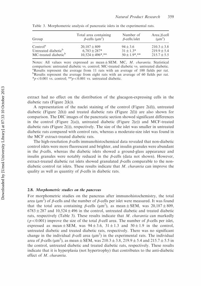
For morphometric studies on the pancreas after immunohistochemistry, the total
area (mm2) of �-cells and the number of �-cells per islet were measured. It was foundthat the total area containing �-cells (mm2), as mean�SEM, was 20,187� 809,
6783� 287 and 10,524� 496 in the control, untreated diabetic and treated diabeticrats, respectively (Table 3). These results indicate that M. charantia can markedly
(p50.001) improve the size of the total �-cell area. The number of �-cells per islet,expressed as mean� SEM, was 94� 3.6, 31� 1.3 and 50� 1.9 in the control,
untreated diabetic and treated diabetic rats, respectively. There was no significantchange in the individual �-cell area (mm2) in the experimental rats. The individual
area of �-cells (mm2), as mean� SEM, was 210.3� 3.8, 219.9� 5.4 and 215.7� 5.5 inthe control, untreated diabetic and treated diabetic rats, respectively. These results
indicate that it is hyperplasia (not hypertrophy) that contributes to the anti-diabeticeffect of M. charantia.
Table 3. Morphometric analysis of pancreatic islets in the experimental rats.
GroupTotal area containing
�-cells (mm2)Number of�-cells/islet
Area/�-cell(mm2)
Controla 20,187� 809 94� 3.6 210.3� 3.8Untreated diabeticb 6,783� 287* 31� 1.3* 219.9� 5.4MC-treated diabeticb 10,524� 496*,** 50� 1.9*,** 215.7� 5.5
Notes: All values were expressed as mean� SEM. MC, M. charantia. Statisticalcomparison: untreated diabetic vs. control; MC-treated diabetic vs. untreated diabetic.aResults represent the average from 11 rats with an average of 100 fields per rat.bResults represent the average from eight rats with an average of 60 fields per rat.*p50.001 vs. control; **p50.001 vs. untreated diabetic.
Natural Product Research 359
Dow
nloa
ded
by [
Um
eå U
nive
rsity
Lib
rary
] at
07:
33 1
0 O
ctob
er 2
013

2.9. Toxicity studies
The acute toxicity results showed no mortality up to a dose of 2000mgkg�1 bodyweight. Serum creatinine was measured as a marker of toxicity to check whether the
extract had any toxic effect on chronic (28 day) administration. The results showedthat there was no significant increase in the serum creatinine level during the
experimental periods (data not shown).
3. Discussion
Three different doses of MCF extract (200, 400 and 600mgkg�1) along with
glibenclamide, a sulphonylurea derivative, were tested for OGTT to determine theeffective dose of M. charantia and to compare the efficacy of M. charantia with a
standard drug. It was found that the dose of 400mgkg�1 produced the highestreduction of blood glucose level at 30min and no appreciable reduction in this level
was observed on further increase of the dose (Figure 1). So the dose 400mgkg�1 waschosen as the most effective dose with which to observe the effects of the extract on
the biochemical and histological studies in the model rats.The standard drug glibenclamide significantly improved glucose tolerance in
diabetic rats, comparable to the dose of 400mgkg�1 of M. charantia extract. In viewof the similarity between the effects of MCF and glibenclamide, it can be suggested
that the mode of action of the MCF extract is probably mediated by an enhancedsecretion of insulin. This assumption was confirmed by our biochemical and histo-
logical examinations, which revealed histological recovery of islets of Langerhans
and cells within the islets of the diabetic rat pancreas.The results of our chronic study demonstrated that the administration of
400mgkg�1 d�1 MCF decreased the fasting blood glucose as well as improved serum
insulin and �-cell function in neonatally STZ-induced type 2 diabetic rats. This effectwas comparable to the standard drug glibenclamide and this phenomenon clearly
indicates that the MCF extract might have insulin-secretagogue activity, which inturn controls the hyperglycaemic state of type 2 diabetes. This is the first study
demonstrating that MCF could improve �-cell function in a type 2 diabetic rat
model, a model which closely parallels the pathological course of diabetes in humans.MCF improved glucose tolerance in type 2 diabetic rats. This effect may be due
to stimulation of insulin release by the pancreas or due to delayed glucose
absorption. Delayed glucose absorption is usually due to inhibition of the enzyme�-glucosidase located at the intestinal surface. MCF did not show �-glucosidaseinhibitory activity in our experiments (data not shown), suggesting that this fruitmay have effect on the modulation of insulin secretion. Insulin releasing activity is
known for this fruit extract, as well as its different fractions (Mamun et al., 2004;
Welihinda, Arvidson, Gylfe, Hellman, & Karlsson, 1982). Whether this insulin-releasing activity is due to the activation of pancreatic �-cells or pancreatic �-cellregeneration is not clear. The results of this study reveal that the lack of increase inthe size of the �-cells suggest that hypertrophy of the �-cells is not involved, whereasthe increase in the number of the �-cells point to hyperplasia of the pancreatic �-cells(Figure 2 and Table 3). Therefore, hyperplasia rather than hypertrophy maycontribute to the anti-diabetic property of this fruit.
360 R.M. Hafizur et al.
Dow
nloa
ded
by [
Um
eå U
nive
rsity
Lib
rary
] at
07:
33 1
0 O
ctob
er 2
013

The results of this study have demonstrated marked changes in the pattern ofdistribution of �- and �-cells in the pancreatic islets of diabetic rats compared to age-matched controls. The relative distribution of these islet cells in the control rats(Figure 2) is similar to the results of the reported study (Ahmed et al., 1998). Therewere, however, significant differences in the distribution pattern of �- and �-cells inthe pancreatic islets of M. charantia-treated and untreated diabetic rats. The numberof �-cells decreased markedly in both M. charantia-treated and untreated diabeticrats compared with the control, but the decrease was much higher in the untreatedrats, coincident with the results of fasting blood glucose and serum insulin (Tables 1and 3). The increase in the quantity of �-cells by M. charantia may be explained bytwo possible mechanisms. First, M. charantia may exert its effect to recover thepartially damaged �-cells, and/or second, it may have the potential to initiateformation of new �-cells in these experimental conditions. Moreover, in addition tothe increase in the number of �-cells, M. charantia significantly improves the qualityof �-cells as well as �-cell function (Table 2).
There is much controversy regarding the changes in the number of �-cells in STZ-induced diabetes. It has been demonstrated that the number of pancreatic �-cells donot change significantly in STZ-induced rats (Papaccio & Mezzogiorno, 1992). Pons& Aoki (1995), however, have reported a remarkable increase in the number of �-cells in diabetic rats. In this study, a significant increase in the number of �-cells inthe diabetic group was found (data not shown). In diabetes, it appears that the �-cellsincrease in number to compensate for the relative decrease in the �-cells. The lack ofeffect of the M. charantia extract on �-cells in the diabetic rat may be due to the factthat it is effective only on �-cells.
There are claims that M. charantia can stimulate insulin secretion (Mamun et al.,2004; Welihinda et al., 1982). It seems therefore that the induction of an increase inthe number of �-cells may be one of the several mechanisms of this fruit material. Itis possible that M. charantia may have initiated �-cell proliferation, since it has beenreported that pancreatic endocrine cells have the potential to proliferate afterinduction of diabetes with STZ (Wang, Bouwens, & Kloppel, 1994). It is alsopossible that the anti-diabetic effect of the M. charantia extract may be due to theimprovement of �-cell quality and �-cell function in this model (Table 2).
In conclusion, our study provides evidence in support of the traditional use ofM. charantia extract as an anti-diabetic remedy. Moreover, our results provideconvincing evidence that the anti-diabetic effect of this fruit is due to the modulation(quantitative and qualitative improvement) of pancreatic �-cells in the diabeticmodel rats tested. Future studies will be needed to elucidate the active principle(s) ofM. charantia responsible for the modulation of pancreatic �-cells.
4. Experimental
4.1. Materials
STZ was obtained from Alexis Biochemical, San Diego, CA, USA. Glibenclamidewas obtained from Sigma (Sigma, St. Louis, MO, USA). Primary antibodies forinsulin (guinea pig polyclonal anti-human insulin) were from Abcam, Cambridge,UK and glucagon (mouse monoclonal anti-glucagon, Clone K79bB10) wasfrom Sigma, St. Louis, MO, USA. The fluorescent secondary antibodies, Texas
Natural Product Research 361
Dow
nloa
ded
by [
Um
eå U
nive
rsity
Lib
rary
] at
07:
33 1
0 O
ctob
er 2
013

Red-donkey anti-guinea pig IgG and Cy2-donkey anti-mouse IgG were obtainedfrom Jackson ImmunoResearch, Baltimore, PA, USA. 40,6-Diamidino-2-phenylin-dole (DAPI) was obtained from Wako Pure Chemical, Osaka, Japan.
4.2. Animals
The experimental design of this study proceeded according to the guidelines for careand management of laboratory animals and was approved by the local animal ethicalcommittee. Wistar rats of either sex from the animal house of the InternationalCenter for Chemical and Biological Sciences, University of Karachi, Pakistan wereused throughout the study.
4.3. Preparation of extract
Fresh fruits of M. charantia (5 kg) were bought from a local market in Karachi,Pakistan. Authentication of the fruit was carried out by Prof. Dr Surayya Khatoon,Chairperson, Department of Botany, University of Karachi, Pakistan. The seed-freefruits (4.2 kg) were sliced into small pieces and soaked in aqueous 80% ethanol(2� 10L) for 7 days each at room temperature. Pooled extracts were filtered,combined and evaporated to dryness under vacuum by using a rotary evaporator.Finally, the crude extract was freeze-dried to give the experimental extract (90 g). Theextract was dissolved in distilled water before administering it to the diabetic rats.
4.4. Toxicity evaluation
The extract ofM. charantia was tested for acute toxicity (if any) in rats. To determinethe acute toxicity, a single oral administration of the M. charantia, at different dosesof the extract (0.25, 0.5, 1.0, 1.5 and 2.0 g kg�1 body weight), was orally administeredto different groups of Wistar rats (n¼ 6) of either sex. Mortality and generalbehaviour of the animals were observed continuously for the initial 4 h andintermittently for the next 6 h and then again at 24, 48 and 72 h following extractadministration.
4.5. Induction of type 2 diabetes
Neonate Wistar pups (2-day-old) were intraperitoneally injected with STZ freshlydissolved in cold citrate buffer (0.1M, pH 4.5) at a dose of 100mgkg�1 body weight,according to Bonner-Weir, Trent, Honey, and Weir (1981) to obtain type 2 modeldiabetic rats. Control pups were injected with an equivalent volume of citrate buffer.The animals were kept under a constant 12 h light–dark cycle, with free access towater and standard rat chow. After 8 weeks of STZ treatment, the animals werechecked for the occurrence of diabetes and those that showed a fasting blood glucoselevel 6.1–9.4mML�1 (in accordance with the type 2 diabetic model) (Bolkent et al.,2005) were monitored for the next 2 weeks to observe the consistency of the bloodglucose level. At 10–11 weeks, an oral glucose tolerance test (OGTT) was performedfor the final screening of type 2 model diabetic rats. For this purpose, rats were keptwithout food overnight (12–14 h) and in the next morning blood glucose was
362 R.M. Hafizur et al.
Dow
nloa
ded
by [
Um
eå U
nive
rsity
Lib
rary
] at
07:
33 1
0 O
ctob
er 2
013

measured at 0 h. Immediately after measuring the blood glucose, glucose was fed at a
dose of 3 g kg�1 body weight and blood glucose was measured at 30, 45, 60, 90 and
120min. The rats having fasting blood glucose levels of 7.1–9.4mML�1 at 0 h and
showing the highest rise to 12.5–25.1mML�1 at 30min, and which returned to their0 h value at 120min, were included in the study. Blood glucose levels were
determined using a glucometer Accu-Chek� Go (Roche Diagnostics, Mannheim,
Germany) at a consistent time of the day.
4.6. OGTT with M. charantia extract in diabetic rats
Thirty diabetic rats were divided into five groups of six rats each. Group 1 treated
with distilled water was used as the negative control. Group 2, treated with
glibenclamide (5mgkg�1), was taken as the positive control. Groups 3–5 served as
treated groups and received the M. charanria extract at doses of 200, 400 and
600mgkg�1, respectively.All the diabetic rats were kept without food overnight (12–14 h) prior to the
OGTT test. Forty-five minutes following the various treatment schedules, each ratwas given an oral glucose load at 3 g kg�1 body weight. All rats were tested for blood
glucose levels at �45min ( just before the administration of the extracts or
glibenclamide), 0min ( just before the oral administration of glucose load) and 30,
60 and 120min after the glucose load. OGTT was also performed in the normal
non-diabetic rats for comparison.
4.7. Chronic extract treatment
The experimental rats were divided into four groups. These comprised of the
untreated diabetics (n¼ 8), the glibenclamide-treated group (n¼ 8), the MCF-treated
diabetics (n¼ 8) and the age-matched non-diabetic control rats (n¼ 11). The extract
was fed orally at a dose of 400mgkg�1 day�1 for 28 days to the MCF-treateddiabetic group. Normal control rats and untreated diabetic rats received only water
in place of the extract. Glibenclamide was given at a dose of 5mg kg�1 for 28 days.
The body weight and fasting blood glucose were determined once a week until the
end of the experimental period.
4.8. Collection of blood samples and estimation of biochemical parameters
For the purpose of biochemical estimation, blood samples were collected by snipping
the tail vein on day 1 and day 28 after overnight fasting. Blood samples were allowed
to clot and serum was separated by centrifugation. Serum samples were frozen and
stored at �30�C for biochemical assay.Serum insulin was measured using an Ultra Sensitive Rat Insulin ELISA kit
(Crystal Chem Inc., IL, USA) on day 1 and day 28 according to the manufacturer’sprotocol. The estimation of serum creatinine was done by alkaline picrate method on
days 1 and 28.
Natural Product Research 363
Dow
nloa
ded
by [
Um
eå U
nive
rsity
Lib
rary
] at
07:
33 1
0 O
ctob
er 2
013

4.9. Assessment of b-cell function
Pancreatic �-cell function (HOMA %B) was measured from fasting glucose(mML�1) and fasting serum insulin (pML�1) concentration by homeostasis modelassessment (HOMA) using HOMA-CIGMA software, where HOMA %B indicates�-cell function (Matthews et al., 1985).
4.10. Tissue processing, histological and immunohistochemical staining
On day 29, i.e. after completion of the 28 days of treatment, the rats were sacrificedfollowing an overnight fast. A portion of the pancreas from the same anatomicallocation (splenic) from each rat was taken and fixed in 10% buffered formalin.Tissues were washed with water, dehydrated with 70%, 90% and 100% 2-propanoland embedded in Paraplast Plus (Carl Roth, Karlsruhe, Germany). Using amicrotome, 7 mm sections were cut and then put on SuperFrost Ultra Plus� slides(Carl Roth, Germany). The slides were baked at 47�C overnight and stored at roomtemperature until further processing.
For histology, four slides (each slide containing two sections) in different planesfrom each rat were deparaffinised in xylene, rehydrated in 100%, 90% and 70%ethanol and washed in water. All the sections were stained for haematoxylin andeosin (H&E). The stained images were acquired using a Nikon 90i Microscope(Nikon, Tokyo, Japan) equipped with a Nikon DXM-1200C digital camera.
For immunohistochemistry, three slides (six sections) from each rat were triplestained for insulin, glucagon and nuclei. For this, sections were deparaffinised,rehydrated and washed in water. All slides were subjected to antigen retrievalprotocols by heat treatment (90�C for 30min) in 0.1M citrate buffer (pH 6.0). Aftercooling to room temperature, each section was incubated with blocking solution(2% normal donkey serum) for 10min at room temperature. Then each section wasincubated with a mixture of guinea pig anti-insulin (1 : 100) and mouse anti-glucagon(1 : 1500) in a dark humidified chamber for 1 h. The slides were washed in threechanges of phosphate buffered saline (PBS) for 5min each. The sections wereincubated with a mixture of Texas Red-conjugated donkey anti-guinea pig IgG(1 : 100) and Cy2-conjugated donkey anti-mouse IgG (1 : 100) for 30min. Finally, thesections were washed with PBS and nuclei staining was done with DAPI (1 : 10,000-fold dilution of a 1mgmL�1 stock solution). After washing, the sections weremounted in Clearmount mounting solution (Zymed Laboratories, San Francisco,CA, USA). The fluorescent images were viewed using a Nikon TE2000E fluorescentmicroscope equipped with a Nikon DS-2MBWc camera in DAPI, fluoresceinisothiocyanate (FITC) and TxRed channel along with differential interferencecontrast (DIC) images. The images were acquired using NIS-Elements image analysissoftware AR 3.0 (Nikon, Japan). No specific immunostaining was observed in thepancreatic tissues when the primary antibodies were omitted. Finally, imageprocessing was performed with Adobe Photoshop CS2.
4.11. Morphometry
NIS-Elements image analysis software AR 3.0 was used for morphometricmeasurements. The �-cell area was measured by acquiring at least 10 adjacent
364 R.M. Hafizur et al.
Dow
nloa
ded
by [
Um
eå U
nive
rsity
Lib
rary
] at
07:
33 1
0 O
ctob
er 2
013

non-overlapping images from each DAPI/insulin stained section (six sections per rat)using a TE2000E microscope with a 10� objective. DAPI/insulin stained imageswere acquired from all visible multicellular islets containing at least 10 cells. Imageswere analysed for �-cell area with NIS-Elements. The total �-cell area wasdetermined on sections stained with insulin antibody and the number of �-cellnuclei in the �-cell area was counted. The total �-cell area was divided by the totalnumber of nuclei to calculate the area of an individual �-cell.
4.12. Statistical analysis
Statistical analyses were performed using the SPSS 12.0 statistical package forWindows (SPSS, Inc., Chicago, IL, USA). All values were expressed asmean�SEM. Statistical difference among the groups was assessed by one-wayANOVA with post-hoc Bonferroni. To compare the data between and withingroups, unpaired and paired t-tests (2-tailed) were performed, respectively.Differences were considered significant at p50.05.
Acknowledgements
We are thankful to the Higher Education Commission (HEC) of Pakistan for financialsupport. We highly appreciate the role of Professor M. Mosihuzzaman for helpful discussions,suggestions, encouragement and critical reading of the manuscript. We would also like tothank Professor Dr Muhammad Iqbal Choudhary for encouragement and critical reading ofthe manuscript. We would like to thank Dr Suad Naheed for helpful discussions. We arethankful to Mr Mumtaz Ali and Mr Sardar Tauseef Shafquat for assistance in the animalhandling and Maryam Bano for glibenclamide experiments.
References
Ahmad, N., Hassan, M.R., Halder, H., & Bennoor, K.S. (1999). Effect of Momordica
charantia (Karolla) extracts on fasting and postprandial serum glucose levels in
NIDDM patients. Bangladesh Medical Research Council Bulletin, 25, 11–13.Ahmed, I., Adeghate, E., Sharma, A.K., Pallot, D.J., & Singh, J. (1998). Effects ofMomordica
charantia fruit juice on islet morphology in the pancreas of the streptozotocin-diabetic
rat. Diabetes Research and Clinical Practice, 40, 145–151.Ahmed, I., Lakhani, M.S., Gillett, M., John, A., & Raza, H. (2001). Hypotriglyceridemic and
hypocholesterolemic effects of anti-diabetic Momordica charantia (karela) fruit extract
in streptozotocin-induced diabetic rats. Diabetes Research Clinical Practice, 51,
155–161.Akhtar, M.S. (1982). Trial of Momordica charantia Linn (karela) powder in patients with
maturity-onset diabetes. Journal of Pakistan Medical Association, 32, 106–107.Ali, L., Khan, A.K., Mamun, M.I., Mosihuzzaman, M., Nahar, N., Nur-e-Alam, et al. (1993).
Studies on hypoglycemic effects of fruit pulp, seed, and whole plant of Momordica
charantia on normal and diabetic model rats. Planta Medica, 59, 408–412.Arulmozhi, D.K., Veeranjaneyulu, A., & Bodhankar, S.L. (2004). Neonatal streptozotocin-
induced rat model of type 2 diabetes mellitus: a glance. Indian Journal of Pharmacology,
36, 217–221.
Natural Product Research 365
Dow
nloa
ded
by [
Um
eå U
nive
rsity
Lib
rary
] at
07:
33 1
0 O
ctob
er 2
013

Bailey, C.J., Day, C., Turner, S.L., & Leatherdale, B.A. (1985). Cerasee, a traditional
treatment for diabetes. Studies in normal and streptozotocin diabetic mice. Diabetes
Research, 2, 81–84.Bolkent, S., Akev, N., Can, A., Bolkent, S., Yanardag, R., & Okyar, A. (2005).
Immunohistochemical studies on the effect of Aloe vera on the pancreatic �-cells in
neonatal streptozotocin-induced type-II diabetic rats. Egyptian Journal of Biology, 7,
14–19.
Bonner-Weir, S., Trent, D.F., Honey, R.N., & Weir, G.C. (1981). Responses of neonatal rat
islets to streptozotocin: limited B-cell regeneration and hyperglycemia. Diabetes, 30,
64–69.
Cakici, I., Hurmoglu, C., Tunctan, B., Abacioglu, N., Kanzik, I., & Sener, B. (1994).
Hypoglycaemic effect of Momordica charantia extracts in normoglycaemic or
cyproheptadine-induced hyperglycaemic mice. Journal of Ethnopharmacology, 44,
117–121.Can, A., Akev, N., Ozsoy, N., Bolkent, S., Arda, B.P., Yanardag, R., et al. (2004). Effect of
Aloe vera leaf gel and pulp extracts on the liver in type-II diabetic rat models. Biological
Pharmacological Bulletin, 27, 694–698.
Day, C., Cartwright, T., Provost, J., & Bailey, C.J. (1990). Hypoglycaemic effect of
Momordica charantia extracts. Planta Medica, 56, 426–429.Goverdhan, P., & Murthy, E.N. (2009). Antidiabetic effect of Cleome aspera in type 2 diabetic
rats. Journal of Pharmacy Research, 2, 1072–1074.Hannan, J.M., Rokeya, B., Faruque, O., Nahar, N., Mosihuzzaman, M., Azad Khan,
A.K., et al. (2003). Effect of soluble dietary fibre fraction of Trigonella foenum graecum
on glycemic, insulinemic, lipidemic and platelet aggregation status of type 2 diabetic
model rats. Journal of Ethnopharmacology, 88, 73–77.Jahan, I.A., Nahar, N., Mosihuzzaman, M., Rokeya, B., Ali, L., Azad Khan, A.K., et al.
(2009). Hypoglycaemic and antioxidant activities of Ficus racemosa Linn. fruits. Natural
Product Research, 23, 399–408.Jayasooriya, A.P., Sakono, M., Yukizaki, C., Kawano, M., Yamamoto, K., & Fukuda, N.
(2000). Effects ofMomordica charantia powder on serum glucose levels and various lipid
parameters in rats fed with cholesterol-free and cholesterol-enriched diets. Journal of
Ethnopharmacology, 72, 331–336.Kar, A., Choudhary, B.K., & Bandyopadhyay, N.G. (2003). Comparative evaluation of
hypoglycaemic activity of some Indian medicinal plants in alloxan diabetic rats. Journal
of Ethnopharmacology, 84, 105–108.Karunanayake, E.H., Jeevathayaparan, S., & Tennekoon, K.H. (1990). Effect of Momordica
charantia fruit juice on streptozotocin-induced diabetes in rats. Journal of
Ethnopharmacology, 30, 199–204.Kedar, P., & Chakrabarti, C.H. (1982). Effects of bittergourd (Momordica charantia) seed and
glibenclamide in streptozotocin-induced diabetes mellitus. Indian Journal of
Experimental Biology, 20, 232–235.Mamun, M.I.R., Nahar, N., Mosihuzzaman, M., Rokeya, B., Ali, L., & Azad Khan, A.K.
(2004). Insulin releasing effects of Momordica charantia fractions on isolated rat islets.
Journal of Bangladesh Chemical Society, 17, 59–64.Marathe, P., Parekar, R., Shinde, S., & Rege, N. (2006). A split dose regimen of streptozotocin
to induce diabetes in a neonatal rat model. Indian Journal of Pharmacology, 38, 432–433.
Matthews, D.R., Hosker, J.P., Rudenski, A.S., Naylor, B.A., Treacher, D.F., & Turner, R.C.
(1985). Homeostasis model assessment: insulin resistance and �-cell function from
fasting plasma glucose and insulin concentrations in man. Diabetologia, 28, 412–419.
Miura, T., Itoh, C., Iwamoto, N., Kato, M., Kawai, M., Park, S.R., et al. (2001).
Hypoglycemic activity of the fruit of the Momordica charantia in type 2 diabetic mice.
Journal of Nutrition Sciences Vitaminology (Tokyo), 47, 340–344.
366 R.M. Hafizur et al.
Dow
nloa
ded
by [
Um
eå U
nive
rsity
Lib
rary
] at
07:
33 1
0 O
ctob
er 2
013

Okyar, A., Can, A., Akev, N., Baktir, G., & Sutlupinar, N. (2001). Effect of aloe vera leaveson blood glucose level in type I and type II diabetic rat models. Phytotherapy Research,15, 157–161.
Papaccio, G., & Mezzogiorno, M. (1992). Morphological aspect of glucagon and somatostatin
islet cells in diabetic biobreeding and low-dose streptozotocin-treated Wistar rats.Pancreas, 4, 289–294.
Pari, L., Ramakrishnan, R., & Venkateswaran, S. (2001). Antihyperglycaemic effect of
Diamed, a herbal formulation, in experimental diabetes in rats. Journal of PharmacyPharmacology, 53, 1139–1143.
Platel, K., & Srinivasan, K. (1997). Plant foods in the management of diabetes mellitus:
vegetables as potential hypoglycaemic agents. Nahrung, 41, 68–74.Pons, P., & Aoki, A. (1995). Differential proliferation of somatostatin and glucagon cells in
rat pancreatic islets submitted to different stimuli. Annals of Anatomy, 177, 221–227.
Portha, B., Levacher, C., Picon, L., & Rosselin, G. (1974). Diabetogenic effect ofstreptozotocinin on the rat during the perinatal period. Diabetes, 23, 888–895.
Raman, A., & Lau, C. (1996). Antidiabetic properties and phytochemistry of Momordicacharantia. Phytomedicine, 2, 349–362.
Rathi, S.S., Grover, J.K., & Vats, V. (2002). The effect of Momordica charantia and Mucunapruriens in experimental diabetes and their effect on key metabolic enzymes involved incarbohydrate metabolism. Phytotherapy Research, 16, 236–243.
Sarkar, S., Pranava, M., & Marita, R. (1996). Demonstration of the hypoglycemic action ofMomordica charantia in a validated animal model of diabetes. Pharmacology Research,33, 1–4.
Serrades, P., Bailbe, D., & Portha, B. (1989). Long-term gliclazide treatment improves thein vitro glucose induced insulin release in rats with type 2 (non-insulin-dependent)diabetes induced by neonatal streptozotocin. Diabetologia, 32, 577–584.
Singh, N., & Gupta, M. (2007). Regeneration of beta cells in islets of Langerhans of pancreas
of alloxan diabetic rats by acetone extract ofMomordica charantia (Linn.) (bitter gourd)fruits. Indian Journal of Experimental Biology, 45, 1055–1062.
Singh, N., Tyagi, S.D., & Agarwal, S.C. (1989). Effects of long term feeding of acetone extract
of Momordica charantia (whole fruit powder) on alloxan diabetic albino rats. IndianJournal of Physiology Pharmacology, 33, 97–100.
Sitasawad, S.L., Shewade, Y., & Bhonde, R. (2000). Role of bittergourd fruit juice in STZ-
induced diabetic state in vivo and in vitro. Journal of Ethnopharmacology, 73, 71–79.Wang, R.N., Bouwens, L., & Kloppel, G. (1994). Beta-cell proliferation in normal and
streptozotocin-treated newborn rats: site, dynamics and capacity. Diabetologia, 37,
1088–1096.Welihinda, J., Arvidson, G., Gylfe, E., Hellman, B., & Karlsson, E. (1982). The insulin-
releasing activity of the tropical plant Momordica charantia. Acta Biologica et MedicaGermanica, 41, 1229–1240.
Welihinda, J., Karunanayake, E.H., Sheriff, M.H., & Jayasinghe, K.S. (1986). Effect ofMomordica charantia on the glucose tolerance in maturity onset diabetes. Journal ofEthnopharmacology, 17, 277–282.
Natural Product Research 367
Dow
nloa
ded
by [
Um
eå U
nive
rsity
Lib
rary
] at
07:
33 1
0 O
ctob
er 2
013