
Microbiologie - University of...
15
11/11/2016 1 Bacteria identification : Media What you have to know about the media • What are the sources of C,H,N,O,P,S? • What type of media is it? • What are the indicators? • What are the selective agents? • They allow the growth of what bacteria? • What are the possible reactions? Ex. MacConkey Agar • Peptone - 17 g • Proteose peptone - 3 g • Lactose - 10 g • Bile salts - 1.5 g • Sodium chloride - 5 g • Neutral red - 0.03 g • Crystal Violet - 0.001 g • Agar - 13.5 g • Sources of C,H,N,O,P,S? • Type of media? • Indicators? • Selective agents? • Allow growth of what bacteria? • Possible reactions?
Transcript of Microbiologie - University of...

11/11/2016
1
Bacteria identification :Media
What you have to know about the media
• What are the sources of C,H,N,O,P,S?
• What type of media is it?
• What are the indicators?
• What are the selective agents?
• They allow the growth of what bacteria?
• What are the possible reactions?
Ex. MacConkey Agar
• Peptone - 17 g
• Proteose peptone - 3 g
• Lactose - 10 g
• Bile salts - 1.5 g
• Sodium chloride - 5 g
• Neutral red - 0.03 g
• Crystal Violet - 0.001 g
• Agar - 13.5 g
• Sources of C,H,N,O,P,S?
• Type of media?
• Indicators?
• Selective agents?
• Allow growth of what bacteria?
• Possible reactions?

11/11/2016
2
Identification :Complex Carbon Sources
Complex Carbon Utilization
• Too large to be transported inside
• Requires exocellular enzymes for the external degradation into smaller units
– Polysaccharides (starch)
– Lipids (triglycerides, etc…)
– Proteins (casein)
– Polynucleotide chain (DNA)
Complex Carbon Sources: Starch
• Media used: Starch Agar
• Detected Enzyme: α-amylase
– cleaves α-1,4 bound between glucose monomers
• Identification: Iodine (halo = starch digestion)

11/11/2016
3
Complex Carbon Sources: Starch
Before iodine addition After iodine addition
Complex Carbon Source: Protein
• Media used: Milk agar
• Detected Enzyme: Caseinase (protease)
– cleaves peptide bounds joining amino acids in the casein protein
• -Identification: clear area (halo) under and surrounding growth
Complex Carbon Source: Protein
11/11/2016
4
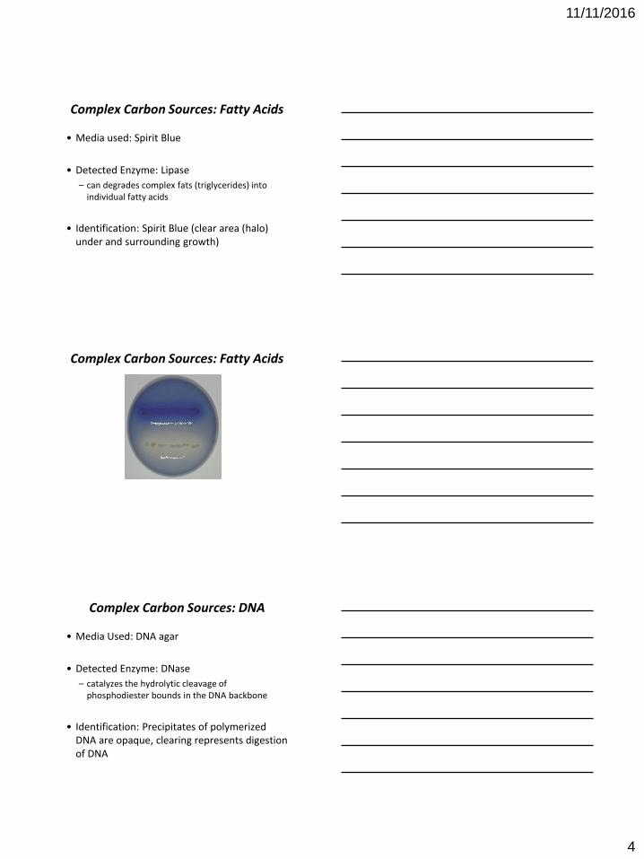
Complex Carbon Sources: Fatty Acids
• Media used: Spirit Blue
• Detected Enzyme: Lipase
– can degrades complex fats (triglycerides) into individual fatty acids
• Identification: Spirit Blue (clear area (halo) under and surrounding growth)
Complex Carbon Sources: Fatty Acids
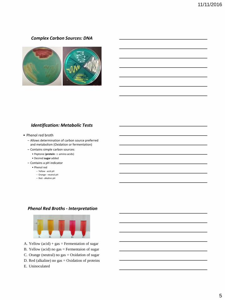
Complex Carbon Sources: DNA
• Media Used: DNA agar
• Detected Enzyme: DNase
– catalyzes the hydrolytic cleavage of phosphodiester bounds in the DNA backbone
• Identification: Precipitates of polymerized DNA are opaque, clearing represents digestion of DNA
11/11/2016
5
Complex Carbon Sources: DNA
Identification: Metabolic Tests
• Phenol red broth
– Allows determination of carbon source preferred and metabolism (Oxidation or fermentation)
– Contains simple carbon sources:
• Peptone (protein amino acids)
• Desired sugar added
– Contains a pH indicator
• Phenol red– Yellow - acid pH
– Orange - neutral pH
– Red - alkaline pH
Phenol Red Broths - Interpretation
A. Yellow (acid) + gas = Fermentation of sugar
B. Yellow (acid) no gas = Fermentaion of sugar
C. Orange (neutral) no gas = Oxidation of sugar
D. Red (alkaline) no gas = Oxidation of proteins
E. Uninoculated
11/11/2016
6
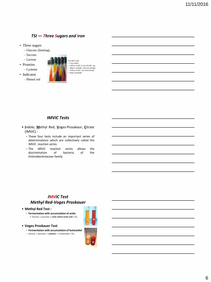
TSI — Three Sugars and Iron
• Three sugars
– Glucose (limiting)
– Sucrose
– Lactose
• Proteins
– Cysteine
• Indicator
– Phenol red
IMViC Tests
• Indole, Methyl Red, Voges-Prosakaur, Citrate(IMViC) :
– These four tests include an important series ofdeterminations which are collectively called theIMViC reaction series
– The IMViC reaction series allows thediscrimination of bacteria of theEnterobacteriaceae family
IMViC Test Methyl Red-Voges Proskauer
• Methyl Red Test :– Fermentation with accumulation of acids:
• Glucose pyruvate lactic and/or acetic acid + CO2
• Voges Proskauer Test – Fermentation with accumulation of butanediol– Glucose pyruvate acetoine 2 butanediol + CO2
- +
- +

11/11/2016
7
Methyl Red Test
• Test for acid accumulation
– Carbon Sources: Glucose and proteins
– Indicator -methyl red; Added after growth
• MR +: red (pH < 5.2)
• MR - : Yellow (pH > 5.2)Neutral Acid
Voges-Proskauer Test
VP + = redVP - = Yellow
Usual results of MR/VP:MR+/VP-; MR-/VP+ MR-/VP-
Reagents VP:
butanediol + -naphthol + KOH + O2 acetoin
Neutral Acid
- +
Acid
produced
No
acetoin
Neutral Acetoin
IMViC: Indole Test
• Principal
– Some microorganisms can metabolize tryptophane by the tryptophanase
TryptophaneTryptophanase
Indole + acide Pyurvic + NH3
Kovac’s reagent
Red color

11/11/2016
8
IMViC Test : Citrate Utilization
• Unique carbon source
– Citrate
• Indicator
– Bromthymol blue
• Citrate utilization generates alkaline end products
– Changes from green to blue
Positive
Klebsiella, Enterobacter
Negative
E. coli
Urea Utilization• Enzyme tested
– Urease
• pH Indicator
– Phenol red (turns pink)
C O + 2 H2O CO2 + H2O + 2 NH3 (NH4)2CO3
H2N
H2N
Ureaammonium
carbonate
(alkaline)Amino acids
PositiveNegative
Urea Utilization – Phenol Red

11/11/2016
9
Ornithine Decarboxylase Assay
• Detects ornithine decarboxylase
– Catalyzes the decarboxylation of ornithine
– Produces diamine putrescine and carbon dioxide
(causes alkaline change)
• Indicator: Brom Cresol purple
– Purple when alkaline or neutral
– Yellow when acid
Ornithine Decarboxylase Assay
• Left: Alkaline with and without ornithine
• Center: Alkaline with ornithine, acidic without
ornithine
• Right: Acidic with and without ornithine
Phenylalanine Slants
• Detects phenylalanine deaminase
• Phenylpyruvic acid reacts with ferric chloride
to produce a green colour

11/11/2016
10
Phenylalanine Slants
A: Positive for phenylpyruvic acid
B: Negative for phenylpyruvic acid
A B
Lysine Agar Slants
• Detects lysine decarboxylase
• Primarily used to detect bacteria in the
Enterobacteriaceae group (for example,
salmonella)
• Indicator: Brom cresol purple
Lysine Agar Slants :Brom Cresol Purple
11/11/2016
11
Lysine Agar Slants
• Purple butt : lysine
decarboxylase positive
• Purple slant: lysine
deamination negative
• Yellow butt: glucose
fermentation
• Red slant: lysine
deamination positive
• Black precipitate:
sulfur reduction
SIM — H2S, Indole and Motility
• Semi-solid medium
– Allows to visualize motility
• Cystein metabolism
CysteineH2S; H2S+ FeSO4 Black precipitate
• Tryptophan metabolism
(A) Tryptophan Indole + NH4 + Pyruvate
(B) Indole + Kovac reagent Red
Non inoculated Non-motile
H2S and motile
Indole+ -

11/11/2016
12
Anaerobic Respiration :Nitrate Reductase
2 H+
2 H+
3 H+ + 3 OH -
3 H2O
2 H+
NO2- + H2O (N = +3) nitrite
NO3- + 2 H+ (N = +5) nitrate
2 e-
2 e-
2 e-
Fp
Fe-S
2 e-
Q
Cyt
b
NADH + H+
FADH2
Nitrate
reductase
Interior
Exterior
Final e- acceptor
NO3- + 2 H+ + 2 e- H2O + NO2
- NO, N2O,
NH2OH,
NH3, N2
nitrate nitrite
Step 1: Test for nitriteNO2
- + sulfanilic acid and alpha naphthylamine HNO2
Nitrate is not reducedNo Nitrite
Yellow
Nitrate is reducedProduction of Nitrite
Red
Nitrate is reduced to nitriteNitrite is reduced
No Nitrite Yellow
Anaerobic Respiration :Nitrate Reductase (con’t)
NO3- + 2 H+ + 2 e- H2O + NO2
- NO, N2O,
NH2OH,
NH3, N2
nitrate nitrite
Step 2: Test for the presence of nitrateNO3
- + Zn (s) NO2-
Nitrate is presentReduction to Nitrite
Red
Nitrate is absentNitrite was reduced
Yellow
Anaerobic Respiration :Nitrate Reductase (con’t)

11/11/2016
13
Oxidase Test : Aerobic RespirationElectron Transport Chain
3 H2O
H+
2 H+
2 H+
3 H+ + 3 OH-
2 H+
H2O
3 H+ + 1/2 O2
2 e-
2 e-
2 e-
Fp
Fe-S
2 e-
Q
Cyt b
Cyt o
NADH + H+
FADH2
interior
exterior
Oxidase Test : Aerobic Respiration
• Cytochrome oxidase catalyzes the reduction of a final electron acceptor, oxygen
• An artifcial e- donor, phenylenediamine, is used to reduce the cytochrome oxidase
• If the enzyme is present, the colorless reagent (reducedstate) will turn blue (oxidized state)
phenylenediamine
Differential Tests for the Identification of Gram Positive Cocci

11/11/2016
14
Blood Hemolysis
• Media used: Blood agar
• Detected Enzyme: hemolysins
• Identification:
– α-hemolysis: greenish hue, partial breakdown of
red blood cells
– β-hemolysis: clearing, breaks down red blood cells
and hemoglobin completely
– γ-hemolysis: no hemolysins
Blood Hemolysis
β
α
γ
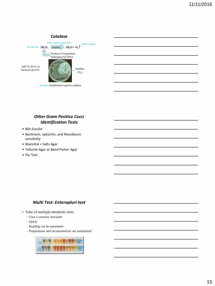
Catalase
• Enzyme found in most organisms living in the presence of oxygen
• Reduces peroxide, which can be damaging to a cell (free radical)
• First step in the discrimination between:
– Micrococcaceae (catalase positive)
– Streptocaccaceae (catalase negative)
11/11/2016
15
Catalase
2H2O2 2H2O + O2 catalase
Product of respiration
Damaging for DNA
Aerobic metabolism requires catalase
bubbles
(O2)
Add 3% H2O2 to
bacterial growth
We add this.
Does bacteria make this?Detect bubbles.
Other Gram Positice Cocci Identification Tests
• Bile-Esculin
• Bacitracin, optochin, and Novobiocin sensitivity
• Mannitol + Salts Agar
• Tellurite Agar or Baird Parker Agar
• Pyr Test
Multi Test: Enteropluri-test
• Tube of multiple metabolic tests
– Uses a constant inoculum
– Quick
– Reading can be automated
– Preparations and inconsistencies are normalized


![RETI di LABORATORI - [Aeronautico] SENS&MICROLAB](https://static.fdocument.pub/doc/165x107/558cd901d8b42a946a8b45a8/reti-di-laboratori-aeronautico-sensmicrolab.jpg)