Microbiological Quality of Soaps and Efficacy of ...library.iugaza.edu.ps/thesis/121275.pdf · ك...
102
Microbiological Quality of Soaps and Efficacy of Antiseptics and Disinfectants Used in Hospitals in Gaza - Palestine المستخدمة في اتلية المطهرلصابون وفعا الميكروبية ل الجودةشفيات قطاع غزة مست- فلسطينAhmad Saleh Auda Salama Supervised by Prof. Dr. Abdelraouf A. Elmanama Prof. of Microbiology A thesis submitted in partial fulfillment of the requirements for the degree of Master of Science in Microbiology November/2016 الج ـ امع ـــــــــس ـة ا ـــــمي ــ ة– غ ــ زةعليات السامي والدراعل شئون البحث ال ك ـ ليـــــ ةعلوم ال ماجستيريقةء الدقحيا اThe Islamic University–Gaza Research and Postgraduate Affairs Faculty of Science Master of Microbilogy
Transcript of Microbiological Quality of Soaps and Efficacy of ...library.iugaza.edu.ps/thesis/121275.pdf · ك...
Microbiological Quality of Soaps and Efficacy of
Antiseptics and Disinfectants Used in Hospitals in
Gaza - Palestine
الجودة الميكروبية للصابون وفعالية المطهرات المستخدمة في فلسطين -مستشفيات قطاع غزة
Ahmad Saleh Auda Salama
Supervised by
Prof. Dr. Abdelraouf A. Elmanama
Prof. of Microbiology
A thesis submitted in partial fulfillment
of the requirements for the degree of
Master of Science in Microbiology
November/2016
زةــغ – ةــالميــــــة اإلســـــــــامعـالج
شئون البحث العلمي والدراسات العليا
العلومة ليــــــك
األحياء الدقيقةماجستير
The Islamic University–Gaza
Research and Postgraduate Affairs
Faculty of Science
Master of Microbilogy
II
Abstract "Microbiological Quality of Soaps and Efficacy of Antiseptics and Disinfectants
Used in Hospitals in Gaza – Palestine" The aim of the present study is a determine microbiological (bacteria and fungi) quality
of antiseptics and soap samples, identify bacteria that contaminate antiseptics and soaps
that used in hospitals in Gaza – Palestine, measure the efficacy of antiseptics on bacteria,
and determine the chemical efficacy of antiseptics and disinfectants.
To determine microbiological (bacteria and fungi) quality of antiseptics and soap samples
and identify bacteria that contaminate antiseptics and soaps that used in hospitals in Gaza
– Palestine, I used plated media method, to measure the efficacy of antiseptics on bacteria,
I used stainless steel cylinder method, and to determine the chemical efficacy of antiseptics
and disinfectants I used pH measurements and chemical concentration test.
The soap results shown as the percentage of samples that complied with the standards
(passed) was 15/15 (100%) in European Gaza Hospital, and the lowest passing result as
1/14 (6.7%) in Kamal Adwan Hospital and the total passing value as 73/105 (69.5%) and
the total failing value as 32/105 (30.5%), and The results showed that the percentage of
contaminated samples by bacteria and fungi was 18/105 (17.1%) and total percentage
value was 32/105 (30.5%), the most common contaminant was coliform 13/105 (13.4%),
Pseudomonas spp. 12/105 (11.4%) and Bacillus spp. 6/105 (5.7%). The contamination
with yeast 11/105 (10.5%) was more than the mold 7/105 (6.7%), and The pH test results
showed that the average of pH of soap samples in hospitals indicate the highest passing
value as 18/20 (90%) in Nasser Hospital, the lowest passing value as 0/15 (0%) in
European Gaza hospital, and the total passing value as 43/105 (41%), and the total failing
value as 62/105 (59%).
The antiseptics/disinfectants results shown as the average of zone of inhibition is 28.2 mm
for E. coli, 20.8 mm for P. aeruginosa, and 20.7mm for S. aureus, the total percentage
passing samples by concentration is 140/233 (60.09%) and the percentage of failing
sample in all hospitals is 93/233 (39.91%), and the highest percentage of passing results
is 79.66% for Chlorhexidine, and lowing percentage of passing results is 48.28% for
Chlorine, and in pH parameter that total percentage of passing results is 141/144 (97.9%),
and that the total percentage of falling results is 3/144 (2.1%).
In conclusions, the results of the present study show that more than half samples collected
from biggest central hospital in Gaza stripe failed in tests, and the percentage of fail in
Antiseptics/Disinfectants samples were 40.3%, and in soap samples were 74.3%.this
results may become a potential human health hazard, the main causes were using locally
manufactured soaps and disinfectants and over dilution the antiseptics/disinfectants before
using.
Keywords: microbiological quality, effectiveness, antiseptics, disinfectants, soap,
hospitals in Gaza, Palestine, S. aureus, P. aeruginosa, E. coli.
III
الملخص "فلسطين –الجودة الميكروبية للصابون وفعالية المطهرات المستخدمة في مستشفيات قطاع غزة "
لملوثة ا( للمطهرات وللصابون، ولتحديد البكتيريا وفطرياتتهدف هذه الدراسة لتحديد الجودة الميكروبية )بكتيريا ، لقياس فعالية المطهرات على البكتيريافلسطين، وكذلك –لهذه المواد المستخدمة في مستشفيات قطاع غزة
وتهدف أيضا لتحديد الفعالية الكيميائية للمطهرات والمعقمات المستخدمة.المواد وتحديد أنواع البكتيريا الملوثة لهذه ،لتحديد الجودة الميكروبية )بكتيريا وفطريات( للمطهرات والصابون
ت طريقة زراعة األطباق البكتيرية. ولقياس فعالية فلسطين استخدم –في مستشفيات قطاع غزة المستخدمة المعقمات على البكتيريا استخدمت طريقة اسطوانات الستانلس ستيل، ولتحديد الفعالية الكيميائية للمطهرات
.التركيز الكيميائياختبار والمعقمات استخدمت اختبار قياس درجة الحموضة و ( في %6.7في مستشفى غزة األوروبي، وأدنى نسبة نجاح ) (%100نتائج الصابون أعلى نسبة نجاح )أظهرت
(، وتظهر النتائج %30.5( ونسبة الرسوب الكلي )%69.5) كانت نسبة النجاح الكليمستشفى كمال عدوان، و ث (، وأكثر مسببات التلو %30.5كلية بلغت ) ( وبنسبة%17.1أن نسبة العينات الملوثة بالبكتيريا والفطريات )
%( 10.5(. والتلوث بالخمائر )%5.7(، والعصية النيابية )%11.4ئفة الزنجارية )ا(، الز %13.4ة )كانت القولوني( في مستشفى %90) (، وفي اختبار درجة الحموضة كانت أعلى نسبة نجاح%6.7أكثر من التلوث باألعفان )وبنسبة رسوب (%41( في مستشفى غزة األوروبي، بنسبة نجاح عامة بلغت )%0ناصر، وأقل نسبة نجاح )
(.%59عامة بلغت )لم م 20.8ملم لإلشريكية القولونية، و 28.2أن متوسط منطقة التثبيط بلغت أظهرت نتائج المطهرات والمعقمات
ز الكيميائيفي اختبار التركينسبة نجاح العينات كانت ملم للمكورة العنقودية الذهبية. و 20.7للزائفة الزنجارية، و ( للكلورهكسدين، وأقل %79.7(، وكانت أعلى نسبة نجاح )%39.9) ة الفشل في العيناتكانت نسب%( و 60.1)
( ونسبة الرسوب %97.9وفي اختبار درجة الحموضة كانت نسبة النجاح الكلي )( للكلور، %48.3نسبة نجاح ) (.%2.1الكلي )
عة من أكبر المستشفيات في االستنتاجات فإن نتائج هذه الدراسة تظهر أنه أكثر من نصف العينات المجمو المركزية في قطاع غزة قد رسبت في االختبارات وأن نسبة الرسوب في عينات المطهرات والمعقمات بلغت
(. وهذه النتائج قد تشكل خطرا عل صحة االنسان، األسباب %74.3%( وفي عينات الصابون بلغت )40.3) يز المطهرات والمعقمات قبل استخدامها.ترك وتقليلالرئيسية هي استخدام الصابون المصنع محليا،
ين، الجودة الميكروبية، الفعالية، المطهرات، المعقمات، الصابون، مستشفيات قطاع غزة، فلسط كلمات مفتاحية:
المكورة العنقودية الذهبية، الزائفة الزنجارية، اإلشريكية القولونية.
IV
:قال اهلل تعالى
نت أ نك ا إ ن لمت ع ال م ا إ ن ل لم ع ل ك ان بح وا س ال "ق
يم"ك الح يم ل الع
(32: البقرة )
They said: "Glory to Thee, of knowledge We have none, save what
Thou Hast taught us: In truth it is Thou Who art perfect in
knowledge and wisdom."
(Al-Baqarah: 32)
V
Acknowledgment
All the praises and thanks are for Almighty Allah the most gracious and merciful, for
helping me in completion of this study.
I would like to express my gratitude to all people who have contributed to this work,
in particular, I would like to thank:
Special thanks and greatly gratitude to my supervisor Dr. Abdelraouf Elmanama
for his professionalism, encouragement and enthusiastic guidance. He is a true
researcher driven by curiosity and with never ending energy. Without his stimulating,
critical discussions, comments, support and great help, this work would not have
been completed.
Special thanks and greatly gratitude to Mr. Hashem Arafa and Saleh Al-taweel for
ther help, encouragement, patience and supported this work in a variety of ways.
To all workers at the Public Health Laboratory for Food and Water, Gaza for all
the help.
Finally, I thank the countless people who contributed to this research. Anyone who
helped me in any form.
VI
Table of Contents
Contents Declaration .................................................................................................................. I
Abstract ...................................................................................................................... II
Acknowledgment ........................................................................................................ V
Table of Contents ..................................................................................................... VI
List of Figures ............................................................................................................. X
List of Abbreviations ............................................................................................... XI
Chapter 1 Introduction .............................................................................................. 1
1.1 Overview ............................................................................................................. 2
1.2 Objectives ........................................................................................................... 5
1.2.1 General Objective ............................................................................................ 5
1.2.2 Specific Objectives .......................................................................................... 5
1.3 Significance ........................................................................................................ 5
Chapter 2 Literature review ...................................................................................... 7
2.1 Soap .................................................................................................................... 8
2.2 Antiseptic and Disinfectant ................................................................................. 9
2.2.1 Commonly Used Antiseptic and Disinfectant in Gaza .................................. 10
2.2.1.1 Alcohols: ..................................................................................................... 10
2.2.1.2 Halogens ..................................................................................................... 11
2.2.1.3 Quaternary Ammonium .............................................................................. 13
2.2.1.4 Biguanides .................................................................................................. 13
2.3 Definition of Activity ........................................................................................ 14
2.4 Factors effects on Antiseptics and disinfectants activity .................................. 15
2.4.1 Concentration ................................................................................................. 16
2.4.2 Contact time ................................................................................................... 17
2.4.3 The effect of pH ............................................................................................. 18
2.4.4 Organic load or soiling .................................................................................. 18
2.4.5 Temperature ................................................................................................... 19
2.4.6 Neutralizing Agents ....................................................................................... 19
2.4.7 The surface ..................................................................................................... 20
2.4.8 Different types of microorganisms ................................................................ 20
2.4.9 Bacterial phenotype ....................................................................................... 21
2.4.10 The number of microorganisms ................................................................... 22
2.5 Limitations in the use of Antiseptics and disinfectants .................................... 22
2.6 Mechanism of Action of Common Antiseptics and Disinfectants ................... 24
2.6.1: Mechanism of Action of Alcohols ................................................................ 26
2.6.2 Mechanism of Action of Halogens ................................................................ 26
2.6.3 Mechanism of Action of Quaternary Ammonium Compounds ..................... 28
2.6.4 Mechanism of Action of Biguanides ............................................................. 28
2.7 Mechanism of Microorganism Resistance ........................................................ 31
2.8 Nosocomial Infection ........................................................................................ 31
2.8.1 From Exposure to Infection ........................................................................... 32
2.8.2 Source of Infection ......................................................................................... 33
VII
2.8.3Nosocomial Infection Pathogens .................................................................... 34
2.8.3.1 Conventional pathogens .............................................................................. 34
2.8.3.2Conditional pathogens ................................................................................. 34
2.8.3.3Opportunistic pathogens .............................................................................. 35
2.9 Reference Studies ............................................................................................. 35
2.9.1 Bacterial contamination in liquid soap for hospital and public use ............... 35
2.9.2 Gram-negative bacteria .................................................................................. 37
2.9.3 Bar or liquid Soap .......................................................................................... 41
2.9.4 Strain of bacteria capable of metabolizing anionic detergent ........................ 42
Chapter 3 Methodology ............................................................................................ 44
3.1 Source and Number of Sample ......................................................................... 45
3.2 Sample Collection ............................................................................................. 45
3.3 Media and Reagents .......................................................................................... 45
3.3.1 Media ............................................................................................................. 45
3.3.2 Reagents and Identification Systems ............................................................. 46
3.4 Equipment, Glassware and Disposables ........................................................... 46
3.5 Quality of Soap and Efficacy of Antiseptics and Disinfectants ........................ 47
3.5.1 Microbiological Quality of Soap ................................................................... 47
3.5.2 Microbiological Efficacy of Antiseptics and Disinfectants ........................... 48
3.5.2.1 Microorganisms used .................................................................................. 48
3.5.2.2 Stainless steel cylinder method ................................................................... 48
3.5.3 Chemical Efficacy of Antiseptics and Disinfectants ..................................... 49
3.5.3.1 pH measurements ........................................................................................ 49
3.5.3.2 Concentration of Hypochlorous acid .......................................................... 49
3.5.3.3 Concentration of Ethyl Alcohol .................................................................. 50
3.5.3.4 Concentration of Povidone Iodine .............................................................. 51
3.5.3.5 Concentration of Chlorhexidine Gluconate ................................................ 51
3.5.3.6 Concentration of Cetrimide ........................................................................ 51
3.6 Data Analysis .................................................................................................... 51
Chapter 4 Results ...................................................................................................... 52
4.1 Distribution of the Samples .............................................................................. 53
4.2 Microbiological quality of Soaps ...................................................................... 54
4.3 Antiseptics/Disinfectants Tests ......................................................................... 57
4.4 The Relationship between Concentration and S. aureus, P. aeruginosa, and E.
coli. 66
4.5 The relationship between pH and S. aureus, P. aeruginosa, and E. coli. .......... 66
4.6 The percentage of total passed for all types of samples in all tests at each
hospital .................................................................................................................... 67
Chapter 5 Discussion ................................................................................................ 69
5.1 The percentage of soap and Anti/Dis based on various microbiological and
chemical tests in seven hospitals ............................................................................ 70
5.2 Distribution of tested samples according to hospital ........................................ 71
5.3 The percentage of pass and fail results of soap in hospitals ............................. 72
5.3.1 Beit Hanoun ................................................................................................... 72
5.3.2 Kamal Adwan ................................................................................................ 72
5.3.3 Al-Shifa .......................................................................................................... 72
5.3.4 Al-Aqsa .......................................................................................................... 72
VIII
5.3.5 Nasser ............................................................................................................. 72
5.3.6 European Gaza ............................................................................................... 73
5.3.7 Abu Yousef Al Najjar .................................................................................... 73
5.4 Soap .................................................................................................................. 73
5.5 Antiseptics and Disinfectants ............................................................................ 74
5.5.1 Microbiological test ....................................................................................... 74
5.5.2 Concentration and pH tests ............................................................................ 74
5.6 Relationship between the Concentration and pH of Anti/Dis and zone of
inhibition of S. aureus, P. aeruginosa, and E. coli. ................................................ 75
Chapter 6 Conclusions and Recmmendations ........................................................ 76
6.1 Conclusion ........................................................................................................ 77
6.2 Recommendations ............................................................................................. 78
Referencs .................................................................................................................... 80
IX
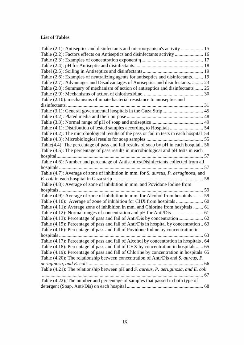
List of Tables
Table (2.1): Antiseptics and disinfectants and microorganism's activity .................. 15
Table (2.2): Factors effects on Antiseptics and disinfectants activity ....................... 16
Table (2.3): Examples of concentration exponent η .................................................. 17
Table (2.4): pH for Antiseptic and disinfectants ........................................................ 18
Tabel (2.5): Soiling in Antiseptics and disinfectants ................................................. 19
Table (2.6): Examples of neutralizing agents for antiseptics and disinfectants ......... 19
Table (2.7): Advantages and Disadvantages of Antiseptics and disinfectants. ......... 23
Table (2.8): Summary of mechanism of action of antiseptics and disinfectants ....... 25
Table (2.9): Mechanisms of action of chlorhexidine. ................................................ 30
Table (2.10): mechanisms of innate bacterial resistance to antiseptics and
disinfectants. .............................................................................................................. 31
Table (3.1): General governmental hospitals in the Gaza Strip ................................. 45
Table (3.2): Plated media and their purpose .............................................................. 48
Table (3.3): Normal range of pH of soap and antiseptics .......................................... 49
Table (4.1): Distribution of tested samples according to Hospitals ........................... 54
Table (4.2): The microbiological results of the pass or fail in tests in each hospital 54
Table )4.3(: Microbiological results for soap samples .............................................. 55
Table)4.4(: The percentage of pass and fail results of soap by pH in each hospital .. 56
Table )4.5(: The percentage of pass results in microbiological and pH tests in each
hospital ....................................................................................................................... 57
Table )4.6(: Number and percentage of Antiseptics/Disinfectants collected from all
hospitals ..................................................................................................................... 57
Table )4.7(: Average of zone of inhibition in mm. for S. aureus, P. aeruginosa, and
E. coli in each hospital in Gaza strip ......................................................................... 58
Table )4.8(: Average of zone of inhibition in mm. and Povidone Iodine from
hospitals ..................................................................................................................... 59
Table )4.9(: Average of zone of inhibition in mm. for Alcohol from hospitals ........ 59
Table )4.10(: Average of zone of inhibition for CHX from hospitals ...................... 60
Table )4.11(: Average zone of inhibition in mm. and Chlorine from hospitals ........ 61
Table )4.12(: Normal ranges of concentration and pH for Anti/Dis .......................... 61
Table )4.13(: Percentage of pass and fail of Anti/Dis by concentration .................... 62
Table )4.15(: Percentage of pass and fall of Anti/Dis in hospital by concentration .. 63
Table )4.16(: Percentage of pass and fall of Povidone Iodine by concentration in
hospitals ..................................................................................................................... 63
Table )4.17(: Percentage of pass and fall of Alcohol by concentration in hospitals . 64
Table )4.18(: Percentage of pass and fail of CHX by concentration in hospitals ...... 65
Table )4.19(: Percentage of pass and fall of Chlorine by concentration in hospitals 65
Table )4.20(: The relationship between concentration of Anti/Dis and S. aureus, P.
aeruginosa, and E. coli .............................................................................................. 66
Table )4.21(: The relationship between pH and S. aureus, P. aeruginosa, and E. coli
................................................................................................................................... 67
Table )4.22(: The number and percentage of samples that passed in both type of
detergent (Soap, Anti/Dis) on each hospital .............................................................. 68
X
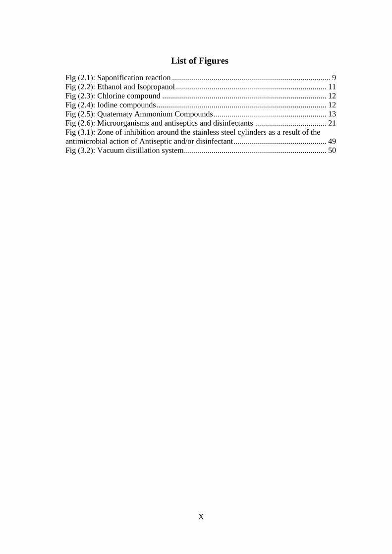
List of Figures
Fig )2.1(: Saponification reaction ................................................................................ 9
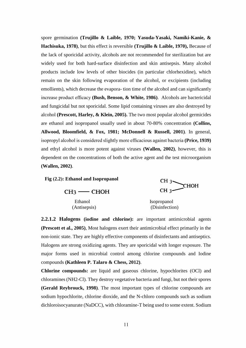
Fig )2.2(: Ethanol and Isopropanol ............................................................................ 11
Fig )2.3(: Chlorine compound ................................................................................... 12
Fig )2.4): Iodine compounds ...................................................................................... 12
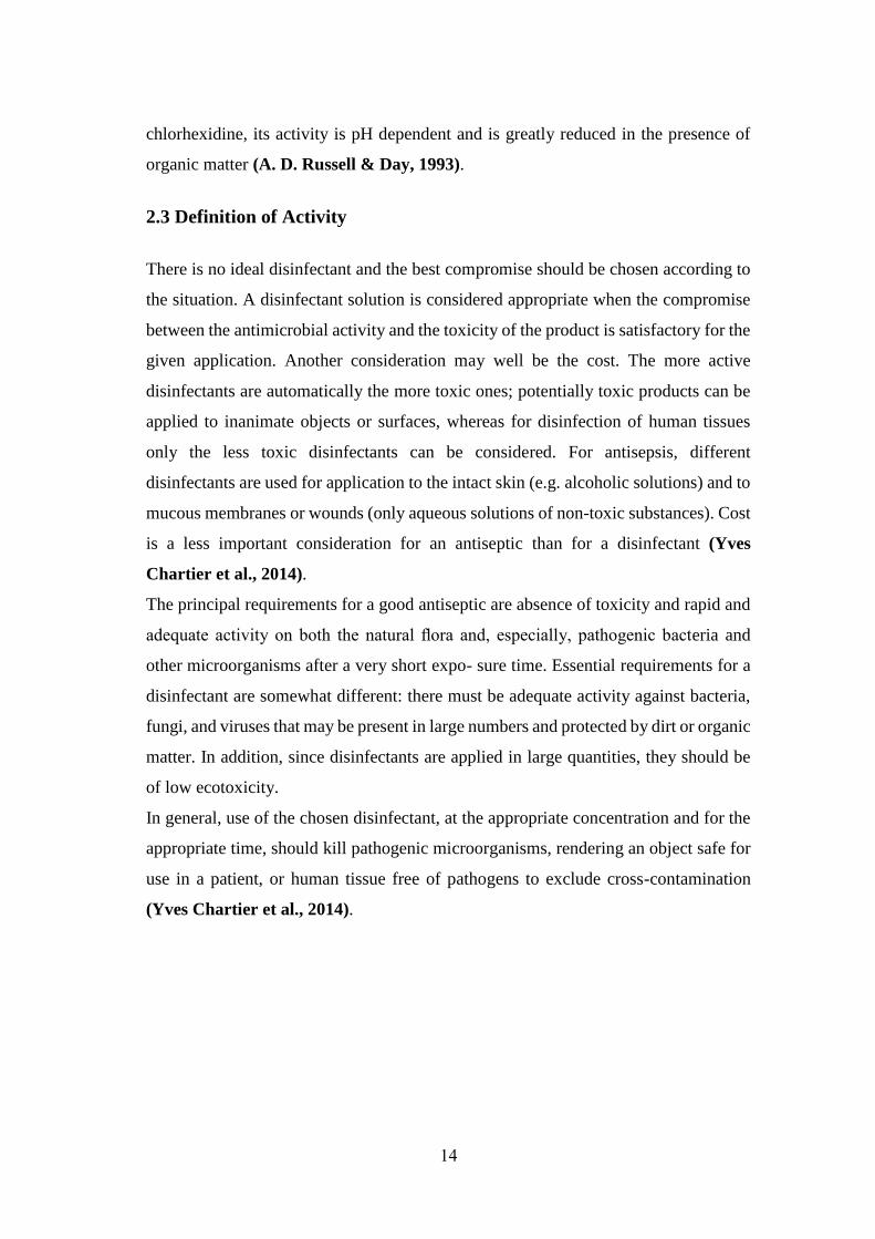
Fig (2.5): Quaternaty Ammonium Compounds ......................................................... 13
Fig (2.6): Microorganisms and antiseptics and disinfectants .................................... 21
Fig (3.1): Zone of inhibition around the stainless steel cylinders as a result of the
antimicrobial action of Antiseptic and/or disinfectant ............................................... 49
Fig (3.2): Vacuum distillation system ........................................................................ 50
XI
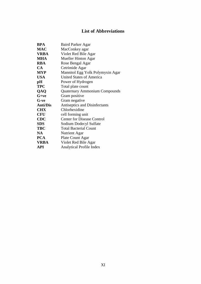
List of Abbreviations
BPA Baird Parker Agar
MAC MacConkey agar
VRBA Violet Red Bile Agar
MHA Mueller Hinton Agar
RBA Rose Bengal Agar
CA Cetrimide Agar
MYP Mannitol Egg Yolk Polymyxin Agar
USA United States of America
pH Power of Hydrogen
TPC Total plate count
QAQ Quaternary Ammonium Compounds
G+ve Gram positive
G-ve Gram negative
Anti/Dis Antiseptics and Disinfectants
CHX Chlorhexidine
CFU cell forming unit
CDC Center for Disease Control
SDS Sodium Dodecyl Sulfate
TBC Total Bacterial Count
NA Nutrient Agar
PCA Plate Count Agar
VRBA Violet Red Bile Agar
API Analytical Profile Index

Chapter 1
Introduction
2
Chapter 1
Introduction
1.1 Overview
Microbiological quality is the acceptability of a product lot based on the absence or
presence of a number of microorganisms, including parasites and/or a quantity of their
toxins/metabolites per unit of mass, volume, area or lot (Cordier, 2004).
Soap is a salt of a fatty acid. Soaps are mainly used as surfactants for washing, bathing,
and cleaning, yet they are also used in textile spinning, as they are important
components of lubricants. Soaps for cleansing are obtained by treating vegetable or
animal oils and fats with a strongly alkaline solution (Cavitch, 1995).
Soap has a little value as an antiseptic, but it does have an important function in the
mechanical removal of microbes through scrubbing. The skin normally contains dead
cells, dust, dried sweat, microbes, and oily secretions from oil glands. Soap breaks the
oily film into tiny droplets, a process called emulsification, and the water and soap
together lift up the emulsified oil and debris and float them away as the lather is washed
off. In this sense, soaps are good degerming agents (Degermation refers to the process
of mechanically removing microbes from the skin) (Alberts, Wilson, & Hunt, 2008;
Tortora, Funke, & Case, 2013).
Medical applications quite often require sterility, especially with regard to invasive
instruments such as scalpels, clamps, dental hand tools, and the like, this absolute level
of microbial control is often unwarranted and perhaps even unwanted. In many cases,
it is remarkably important to focus on reducing the size of a microbial population, or
its microbial load. Sanitization refers to any cleansing technique that removes debris,
microorganisms, and toxins, and in this way reduces the potential for infection and
spoilage. Soaps and detergents are the most commonly employed sanitizers. It is
important to note that sanitization is often preferable to sterilization. (Kathleen P.
Talaro & Chess, 2012).
There are many types of microorganisms isolated from soaps in hospitals and public
places such as Pseudomonas aeruginosa, Escherichia coli, Acinetobacter baumanii,
3
Proteus penneri, Flavimonas oryzihabitans, Enterobacter aerogenes, Enterobacter
cloacae, Staphylococcus aureus, Klebsiella oxytoca, Klebsiella pneumoniae, Serratia
marcescens, Citrobacter koseri, candida (J. A. Caetano, Lima, Di Ciero Miranda,
Serufo, & Ponte, 2011; M. Chattman, S. L. Gerba, & C. P. Maxwell, 2011; V.
Chaturvedi & Kumar, 2011; S. M. H. Zeiny, 2009).
Efficacy is the ability to produce a desired or intended result (Stevenson, 2010).
Antibiotics are defined as naturally occurring substances which inhibit or destroy
selective bacteria or other microorganisms, generally at low concentrations.
Antiseptics are biocides or products that destroy or inhibit the growth of
microorganisms in or on living tissue and disinfectants are similar but generally are
products or biocides that are used on inanimate objects or surfaces. Sterilization refers
to a physical or chemical process that completely destroys or removes all microbial
life, including spores (Sanitization reduces microbial numbers on inanimate objects to
safe levels by physical or chemical means.). Preservation is the prevention of
multiplication of microorganisms in formulated products, including pharmaceuticals
and foods. A number of biocides are also used for cleaning purposes; cleaning in these
cases refers to the physical removal of foreign material from a surface (Seymour
Stanton Block, 2001a).
The Food and Drug Administration in the United States regulates the formulation,
manufacture, and use of antiseptics and germicides because these agents involve direct
human exposure and contact (Madigan, Martinko, Bender, Buckley, & Stahl,
2012).
Antisepsis include preparing the skin before surgical incisions with iodine compounds,
swabbing a wound with hydrogen peroxide, and ordinary hand washing with a
germicidal soap (Kathleen Park Talaro & Rhoads, 2012). Disinfectants can be
sporostatic but are not necessarily sporicidal (Johnston, Lambert, Hanlon, &
Denyer, 2002).
Antiseptics and disinfectants are used extensively in hospitals and other health care
settings for a variety of topical and hard-surface applications. They are an essential
part of infection control practices and aid in the prevention of nosocomial infections
4
(Control & Epidemiology, 1996; William A Rutala, 1996). Mounting concerns over
the potential for microbial contamination and infection risks in food and general
consumer markets have also led to increased use of antiseptics and disinfectants by the
public. A wide variety of active chemical agents (or “biocides”) are found in these
products, many of which have been used for hundreds of years for antisepsis and
disinfection (S.S. Block, 1991). In general, antiseptics and disinfectants have a broader
spectrum of activity than antibiotics, and, while antibiotics tend to have specific
intracellular targets, antiseptics and disinfectants may have multiple targets. The
widespread use of antiseptic and disinfectant products has prompted some speculation
on the development of microbial resistance, in particular cross- resistance to antibiotics
(McDonnell & Russell, 2001).
There has been a dramatic increase in the usage of chemical biocides (i.e. disinfectants
and antiseptics) in the food, water and pharmaceutical industries, and in the healthcare
and domiciliary environments. The need to reduce and control nosocomial infection
(Favero, 2002) and to improve product quality and overall hygiene, for example, in
the hospitals (Solveig Langsrud, Maan Singh Sidhu, Even Heir, & Holck, 2003)
health authorities and the public. Public knowledge in particular and a better
commitment to overall hygiene (Bloomfield, 2002) have contributed to the increased
usage of antiseptics and disinfectants in the home environment (Levy, 2001). Protocols
for testing the antimicrobial efficacy of disinfectants and antiseptics are essential to
provide reliable information on the efficacy of an antimicrobial product and provide
assurance for the end users (J. Holah, 2003).
It is important to note that many of these antiseptics and disinfectants may be used
singly or in combination in a variety of products which vary considerably in activity
against microorganisms. Antimicrobial activity can be influenced by many factors
such as formulation effects, presence of an organic load, synergy, temperature,
dilution, and test method (Fraise, Maillard, & Sattar, 2012).
The concept of testing or checking disinfectants is very old. Robert Koch described a
disinfectant test in the article (Über Desinfektion), in 1881 (Gerald Reybrouck,
1998). Testing disinfectant helps to find the cause of the spread of the infection
(Wadhwa et al., 2007). Disinfectants should remove or inactivate known or possible
5
pathogens from inanimate objects (Gerald Reybrouck, 1998), hospitals should have
their inbuilt test method that can be easily applicable (Hume et al., 2009) (Akinsanya,
1993).
An unpublished study in Gaza strip investigated the contamination of liquid soaps in
hospitals. It showed alarming results with high percentage of the tested liquid soap
samples was found to be contaminated with coliforms (29%), a considerable
percentage of samples contained yeast (21%) and molds (5%).
1.2 Objectives
1.2.1 General Objective
To assess the microbiological quality and effectiveness of antiseptics and soap used in
hospitals in Gaza – Palestine.
1.2.2 Specific Objectives
1. To determine microbiological (bacteria and fungi) quality of antiseptics,
disinfectants and soap samples.
2. To identify bacteria that contaminate antiseptics, disinfectants and soaps that
used in hospital in Gaza – Palestine
3. To measure the efficacy of antiseptics and disinfectants on bacteria.
4. To determine the chemical concentration of antiseptics and disinfectants.
1.3 Significance
Hospitals should exert their utmost efforts to prevent contamination to avoid health
care associated infection (HAI), which is becoming a major threat because of the high
possibility of the spread of multiple drug resistant pathogens in hospitals. Therefore,
soaps, detergents, and antiseptics are commonly used in hospital to minimize risks of
pathogens transmission. As with any commercial products, these materials may be
6
contaminated during manufacturing, processing, storage and use, thus, complicating
the process and increasing risks of HAI.
Determination of the microbiological quality of antiseptics and soaps in general
hospitals in the Gaza strip would generate the first original data and shed light on the
general quality. This is of utmost need, because most of these soaps are manufactured
locally with no standards or guidelines to ensure their quality. Antiseptics are imported
as concentrates and are diluted locally, no data available on the effectiveness of such
products. This study will be the first to tackle this issue.
It is expected that the results of this cross sectional study would help the hospital`s
infection control committees in reviewing the process of purchasing such products and
the policy of testing, use and storage. In general, hospitals would benefit from this
study and is expected to contribute to the reduction of HAI. The local industry will
also benefit from the findings of this study.
7
Chapter 2
Literature review
8
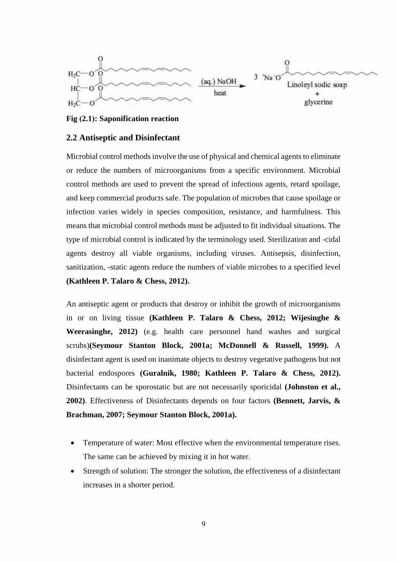
2.1 Soap
A substance used with water for washing and cleaning, made of a compound of natural
oils or fats with sodium hydroxide or another strong alkali. Typically having perfume
and coloring added (Oxford University Press.). They are defined as salts formed
through the reaction of fatty acids obtained from vegetal and animal fats with metals
or basic radicals (sodium, potassium, ammonia etc.), and exert detergent action, i.e.
they permit the removal of dirt, remains and viable (non-colonizing) microorganisms
(J. A. Caetano et al., 2011). Chemically, soap is a salt of a fatty acid (McNaught,
Wilkinson, & International Union of Pure and Applied Chemistry., 1997). Soaps
are mainly used as surfactants for washing, bathing, and cleaning, but they are also
used in textile spinning and are important components of lubricants. Soaps for
cleansing are obtained by treating vegetable or animal oils and fats with a strongly
alkaline solution. Fats and oils are composed of triglycerides; three molecules of fatty
acids are attached to a single molecule of glycerol. The alkaline solution, which is
often called lye (although the term "lye soap" refers almost exclusively to soaps made
with sodium hydroxide), brings about a chemical reaction known as saponification. In
this reaction, the triglyceride fats are first hydrolyzed into free fatty acids, and then
these combine with the alkali to form crude soap, an amalgam of various soap salts,
excess fat or alkali, water, and liberated glycerol (glycerin) (Cavitch, 1995). Soaps are
key components of most lubricating greases, which are usually emulsions of calcium
soap or lithium soaps and mineral oil. These calcium- and lithium-based greases are
widely used. Much other metallic soap is also useful, including those of aluminum,
sodium, and mixtures of them. Such soaps are also used as thickeners to increase the
viscosity of oils. In ancient times, lubricating greases were made by the addition of
lime to olive oil (Bohnet, 2003).
9
Fig ) 2.1(: Saponification reaction
2.2 Antiseptic and Disinfectant
Microbial control methods involve the use of physical and chemical agents to eliminate
or reduce the numbers of microorganisms from a specific environment. Microbial
control methods are used to prevent the spread of infectious agents, retard spoilage,
and keep commercial products safe. The population of microbes that cause spoilage or
infection varies widely in species composition, resistance, and harmfulness. This
means that microbial control methods must be adjusted to fit individual situations. The
type of microbial control is indicated by the terminology used. Sterilization and -cidal
agents destroy all viable organisms, including viruses. Antisepsis, disinfection,
sanitization, -static agents reduce the numbers of viable microbes to a specified level
(Kathleen P. Talaro & Chess, 2012).
An antiseptic agent or products that destroy or inhibit the growth of microorganisms
in or on living tissue (Kathleen P. Talaro & Chess, 2012; Wijesinghe &
Weerasinghe, 2012) (e.g. health care personnel hand washes and surgical
scrubs)(Seymour Stanton Block, 2001a; McDonnell & Russell, 1999). A
disinfectant agent is used on inanimate objects to destroy vegetative pathogens but not
bacterial endospores (Guralnik, 1980; Kathleen P. Talaro & Chess, 2012).
Disinfectants can be sporostatic but are not necessarily sporicidal (Johnston et al.,
2002). Effectiveness of Disinfectants depends on four factors (Bennett, Jarvis, &
Brachman, 2007; Seymour Stanton Block, 2001a).
Temperature of water: Most effective when the environmental temperature rises.
The same can be achieved by mixing it in hot water.
Strength of solution: The stronger the solution, the effectiveness of a disinfectant
increases in a shorter period.
10
Duration of exposure: The longer a disinfectant remains on the surface the more
effective it will be.
Cleanliness of surface: The most important thing to remember is that the
disinfectant works the best on clean surface.
Sanitization refers to a physical or chemical process that completely destroys or
removes all microbial life, including spores and reduces microbial numbers on
inanimate objects to safe levels (Seymour Stanton Block, 2001a; Kathleen P. Talaro
& Chess, 2012).
Preservation is the prevention of multiplication of microorganisms in formulated
products, including pharmaceuticals and foods (Seymour Stanton Block, 2001a;
McDonnell & Russell, 1999).
Degermation refers to the process of mechanically removing microbes from the skin
(Kathleen P. Talaro & Chess, 2012).
Microbial death is defined as the permanent loss of reproductive capability in
microorganisms (Kathleen P. Talaro & Chess, 2012).
Antimicrobials are described according to their ability to destroy or inhibit microbial
growth. Microbicidal agents cause microbial death. They are described by what they
are -cidal for: sporocides, bactericides, fungicides, viricides (Kathleen P. Talaro &
Chess, 2012).
2.2.1 Commonly Used Antiseptic and Disinfectant in Gaza
Four major groups of antiseptics and disinfectants
2.2.1.1 Alcohols: are among the most widely used disinfectants and antiseptics
(Seymour Stanton Block, 2001a; Wallen, 2002). They are colourless hydrocarbons
with one or more hydroxyl functional groups. Alcohols exhibit rapid broad-spectrum
antimicrobial activity against vegetative bacteria (including mycobacteria), viruses,
and fungi but are not sporicidal. They are, however, known to inhibit sporulation and
11
spore germination (Trujillo & Laible, 1970; Yasuda-Yasaki, Namiki-Kanie, &
Hachisuka, 1978), but this effect is reversible (Trujillo & Laible, 1970), Because of
the lack of sporicidal activity, alcohols are not recommended for sterilization but are
widely used for both hard-surface disinfection and skin antisepsis. Many alcohol
products include low levels of other biocides (in particular chlorhexidine), which
remain on the skin following evaporation of the alcohol, or excipients (including
emollients), which decrease the evapora- tion time of the alcohol and can significantly
increase product efficacy (Bush, Benson, & White, 1986). Alcohols are bactericidal
and fungicidal but not sporicidal. Some lipid containing viruses are also destroyed by
alcohol (Prescott, Harley, & Klein, 2005). The two most popular alcohol germicides
are ethanol and isopropanol usually used in about 70-80% concentration (Collins,
Allwood, Bloomfield, & Fox, 1981; McDonnell & Russell, 2001). In general,
isopropyl alcohol is considered slightly more efficacious against bacteria (Price, 1939)
and ethyl alcohol is more potent against viruses (Wallen, 2002), however, this is
dependent on the concentrations of both the active agent and the test microorganism
(Wallen, 2002).
Fig )2.2(: Ethanol and Isopropanol
Ethanol Isopropanol
(Antisepsis) (Disinfection)
2.2.1.2 Halogens (iodine and chlorine): are important antimicrobial agents
(Prescott et al., 2005), Most halogens exert their antimicrobial effect primarily in the
non-ionic state. They are highly effective components of disinfectants and antiseptics.
Halogens are strong oxidizing agents. They are sporicidal with longer exposure. The
major forms used in microbial control among chlorine compounds and Iodine
compounds (Kathleen P. Talaro & Chess, 2012).
Chlorine compounds: are liquid and gaseous chlorine, hypochlorites (OCl) and
chloramines (NH2-Cl). They destroy vegetative bacteria and fungi, but not their spores
(Gerald Reybrouck, 1998). The most important types of chlorine compounds are
sodium hypochlorite, chlorine dioxide, and the N-chloro compounds such as sodium
dichloroisocyanurate (NaDCC), with chloramine-T being used to some extent. Sodium
12
hypochlorite solutions are widely used for hard-surface disinfection (household
bleach) and can be used for disinfecting spillages of blood containing human
immunodeficiency virus or HBV. NaDCC can also be used for this purpose and has
the advantages of providing a higher concentration of available chlorine and being less
susceptible to inactivation by organic matter. In water, sodium hypochlo- rite ionizes
to produce Na1 and the hypochlorite ion, OCl2, which establishes an equilibrium with
hypochlorous acid, HOCl (Ascenzi, 1995). Between pH4 and 7, chlorine exists
predominantly as HClO, the active moiety, whereas above pH9, OCl2 predominates.
Although chlorine compounds have been predominantly used as hard-surface
disinfectants, novel acidified sodium chlorite (a two-component system of sodium
chlorite and mandelic acid) has been described as an effective antiseptic (McDonnell
& Russell, 1999).
Fig )2.3(: Chlorine compound
Chlorine compound
(Disinfection)
Iodine compounds: have the broadest spectrum of all topical anti-infectives with
action against bacteria, fungi, viruses, spores, protozoa and yeast. Iodine is used
mainly as a skin antiseptic. But less reactive than chlorine, iodine is rapidly
bactericidal, fungicidal, tuberculocidal, virucidal, and sporicidal (Seymour Stanton
Block, 2001a). Although aqueous or alcoholic (tincture) solutions of iodine have been
used for 150 years as antiseptics, they are associated with irritation and excessive
staining. In addition, aqueous solutions are generally unstable; in solution, at least
seven iodine species are present in a complex equilibrium, with molecular iodine (I2)
being primarily responsible for antimictrobial efficacy (Seymour Stanton Block,
2001a).
Fig )2.4): Iodine compounds
Iodine compounds
(Antisepsis, Cleaning)
13
2.2.1.3 Quaternary Ammonium Compounds (QAC): Surface-active agents
(surfactants) have two regions in their molecular structures, one a hydrocarbon, water-
repellent (hydrophobic) group and the other a water-attracting (hydrophilic or polar)
group. Depending on the basis of the charge or absence of ionization of the hydrophilic
group, surfactants are classified into cationic, anionic, nonionic, and ampholytic
(amphoteric) compounds. Of these, the cationic agents, as exemplified by quaternary
ammonium compounds (QACs), are the most useful antiseptics and disinfectants
(Hugo, 1971). They are sometimes known as cationic detergents. QACs have been
used for a variety of clinical purposes (e.g., preoperative disinfection of unbroken skin,
application to mucous membranes, and disinfection of noncritical surfaces). In
addition to having antimicrobial properties, QACs are also excellent for hard surface
cleaning and deodorization. have positively charged quaternary nitrogen and a long
chain hydrophobic aliphatic chain (Prescott et al., 2005). They are used as low level
disinfectants (McDonnell & Russell, 2001). If used in medium concentrations, they
are effective against some Gram-positive bacteria, viruses, fungi and algae. In low
concentrations, they have microbistatic effect. QACs are ineffective against the
tubercle bacillus, the hepatitis virus, Pseudomonas and spores at any concentration
(Kathleen P. Talaro & Chess, 2012).
Fig (2.5): Quaternaty Ammonium Compounds
Quaternary Ammonium Compounds (QACs)
(Antisepsis, cleaning, Disinfection, preservation)
2.2.1.4 Biguanides: three substances under this type are Chlorhexidine, Alexidine
and polymeric biguanides
Chlorhexidine is probably the most widely used biocide in antiseptic products, in
particular in handwashing and oral products but also as a disinfectant and preservative.
This is due in particular to its broad-spectrum efficacy, substantivity for the skin, and
low irritation. Of note, irritability has been described and in many cases may be
product specific (Seymour Stanton Block, 2001a). Despite the advantages of
14
chlorhexidine, its activity is pH dependent and is greatly reduced in the presence of
organic matter (A. D. Russell & Day, 1993).
2.3 Definition of Activity
There is no ideal disinfectant and the best compromise should be chosen according to
the situation. A disinfectant solution is considered appropriate when the compromise
between the antimicrobial activity and the toxicity of the product is satisfactory for the
given application. Another consideration may well be the cost. The more active
disinfectants are automatically the more toxic ones; potentially toxic products can be
applied to inanimate objects or surfaces, whereas for disinfection of human tissues
only the less toxic disinfectants can be considered. For antisepsis, different
disinfectants are used for application to the intact skin (e.g. alcoholic solutions) and to
mucous membranes or wounds (only aqueous solutions of non-toxic substances). Cost
is a less important consideration for an antiseptic than for a disinfectant (Yves
Chartier et al., 2014).
The principal requirements for a good antiseptic are absence of toxicity and rapid and
adequate activity on both the natural flora and, especially, pathogenic bacteria and
other microorganisms after a very short expo- sure time. Essential requirements for a
disinfectant are somewhat different: there must be adequate activity against bacteria,
fungi, and viruses that may be present in large numbers and protected by dirt or organic
matter. In addition, since disinfectants are applied in large quantities, they should be
of low ecotoxicity.
In general, use of the chosen disinfectant, at the appropriate concentration and for the
appropriate time, should kill pathogenic microorganisms, rendering an object safe for
use in a patient, or human tissue free of pathogens to exclude cross-contamination
(Yves Chartier et al., 2014).
15
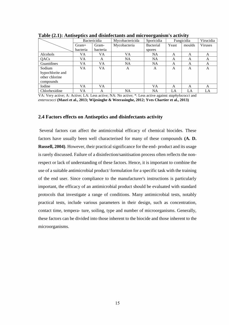
Table (2.1): Antiseptics and disinfectants and microorganism's activity Bactericidia Mycobactericida Sporicidia Fungicidia Virucidia
Gram+
bacteria
Gram-
bacteria
Mycobacteria Bacterial
spores
Yeast moulds Viruses
Alcohols VA VA VA NA A A A
QACs VA A NA NA A A A
Guanidines VA VA NA NA A A A
Sodium
hypochlorite and
other chlorine
compounds
VA VA A A A A A
Iodine VA VA VA A A A
Chlorhexidine VA A NA NA LA LA LA
VA: Very active; A: Active; LA: Less active; NA: No active; *: Less active against staphylococci and
enterococci (Masri et al., 2013; Wijesinghe & Weerasinghe, 2012; Yves Chartier et al., 2013)
2.4 Factors effects on Antiseptics and disinfectants activity
Several factors can affect the antimicrobial efficacy of chemical biocides. These
factors have usually been well characterised for many of these compounds (A. D.
Russell, 2004). However, their practical significance for the end- product and its usage
is rarely discussed. Failure of a disinfection/sanitisation process often reflects the non-
respect or lack of understanding of these factors. Hence, it is important to combine the
use of a suitable antimicrobial product/ formulation for a specific task with the training
of the end user. Since compliance to the manufacturer's instructions is particularly
important, the efficacy of an antimicrobial product should be evaluated with standard
protocols that investigate a range of conditions. Many antimicrobial tests, notably
practical tests, include various parameters in their design, such as concentration,
contact time, tempera- ture, soiling, type and number of microorganisms. Generally,
these factors can be divided into those inherent to the biocide and those inherent to the
microorganisms.
16
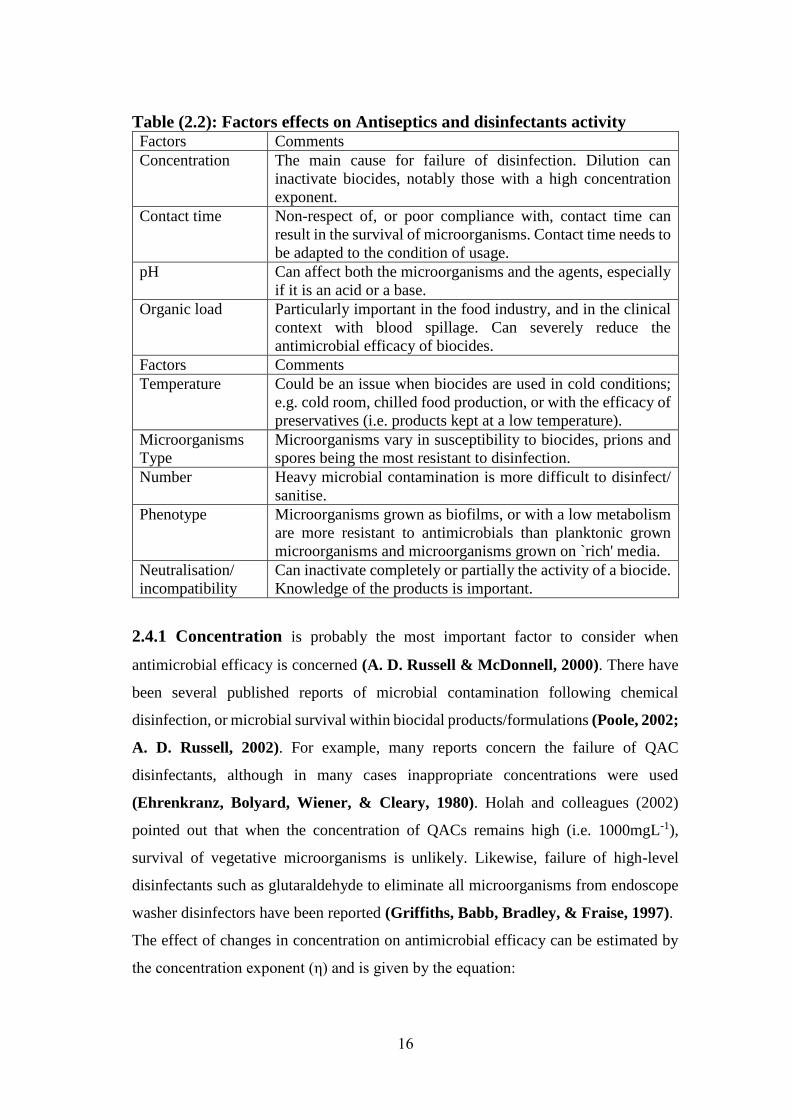
Table (2.2): Factors effects on Antiseptics and disinfectants activity Factors Comments
Concentration The main cause for failure of disinfection. Dilution can
inactivate biocides, notably those with a high concentration
exponent.
Contact time Non-respect of, or poor compliance with, contact time can
result in the survival of microorganisms. Contact time needs to
be adapted to the condition of usage.
pH Can affect both the microorganisms and the agents, especially
if it is an acid or a base.
Organic load Particularly important in the food industry, and in the clinical
context with blood spillage. Can severely reduce the
antimicrobial efficacy of biocides.
Factors Comments
Temperature Could be an issue when biocides are used in cold conditions;
e.g. cold room, chilled food production, or with the efficacy of
preservatives (i.e. products kept at a low temperature).
Microorganisms
Type
Microorganisms vary in susceptibility to biocides, prions and
spores being the most resistant to disinfection.
Number Heavy microbial contamination is more difficult to disinfect/
sanitise.
Phenotype Microorganisms grown as biofilms, or with a low metabolism
are more resistant to antimicrobials than planktonic grown
microorganisms and microorganisms grown on `rich' media.
Neutralisation/
incompatibility
Can inactivate completely or partially the activity of a biocide.
Knowledge of the products is important.
2.4.1 Concentration is probably the most important factor to consider when
antimicrobial efficacy is concerned (A. D. Russell & McDonnell, 2000). There have
been several published reports of microbial contamination following chemical
disinfection, or microbial survival within biocidal products/formulations (Poole, 2002;
A. D. Russell, 2002). For example, many reports concern the failure of QAC
disinfectants, although in many cases inappropriate concentrations were used
(Ehrenkranz, Bolyard, Wiener, & Cleary, 1980). Holah and colleagues (2002)
pointed out that when the concentration of QACs remains high (i.e. 1000mgL-1),
survival of vegetative microorganisms is unlikely. Likewise, failure of high-level
disinfectants such as glutaraldehyde to eliminate all microorganisms from endoscope
washer disinfectors have been reported (Griffiths, Babb, Bradley, & Fraise, 1997).
The effect of changes in concentration on antimicrobial efficacy can be estimated by
the concentration exponent (η) and is given by the equation:
17
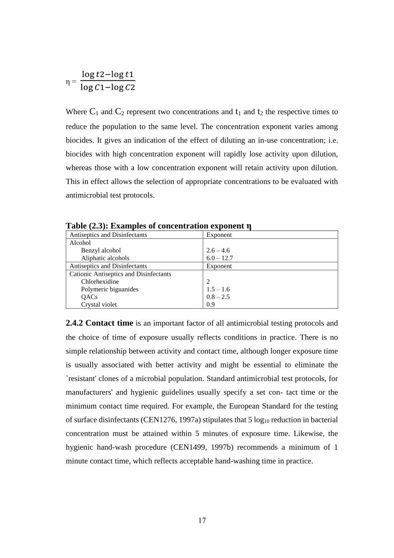
η = log 𝑡2−log 𝑡1
log 𝐶1−log 𝐶2
Where C1 and C2 represent two concentrations and t1 and t2 the respective times to
reduce the population to the same level. The concentration exponent varies among
biocides. It gives an indication of the effect of diluting an in-use concentration; i.e.
biocides with high concentration exponent will rapidly lose activity upon dilution,
whereas those with a low concentration exponent will retain activity upon dilution.
This in effect allows the selection of appropriate concentrations to be evaluated with
antimicrobial test protocols.
Table (2.3): Examples of concentration exponent η Antiseptics and Disinfectants Exponent
Alcohol
Benzyl alcohol
Aliphatic alcohols
2.6 – 4.6
6.0 – 12.7
Antiseptics and Disinfectants Exponent
Cationic Antiseptics and Disinfectants
Chlorhexidine
Polymeric biguanides
QACs
Crystal violet
2
1.5 – 1.6
0.8 – 2.5
0.9
2.4.2 Contact time is an important factor of all antimicrobial testing protocols and
the choice of time of exposure usually reflects conditions in practice. There is no
simple relationship between activity and contact time, although longer exposure time
is usually associated with better activity and might be essential to eliminate the
`resistant' clones of a microbial population. Standard antimicrobial test protocols, for
manufacturers' and hygienic guidelines usually specify a set con- tact time or the
minimum contact time required. For example, the European Standard for the testing
of surface disinfectants (CEN1276, 1997a) stipulates that 5 log10 reduction in bacterial
concentration must be attained within 5 minutes of exposure time. Likewise, the
hygienic hand-wash procedure (CEN1499, 1997b) recommends a minimum of 1
minute contact time, which reflects acceptable hand-washing time in practice.
18
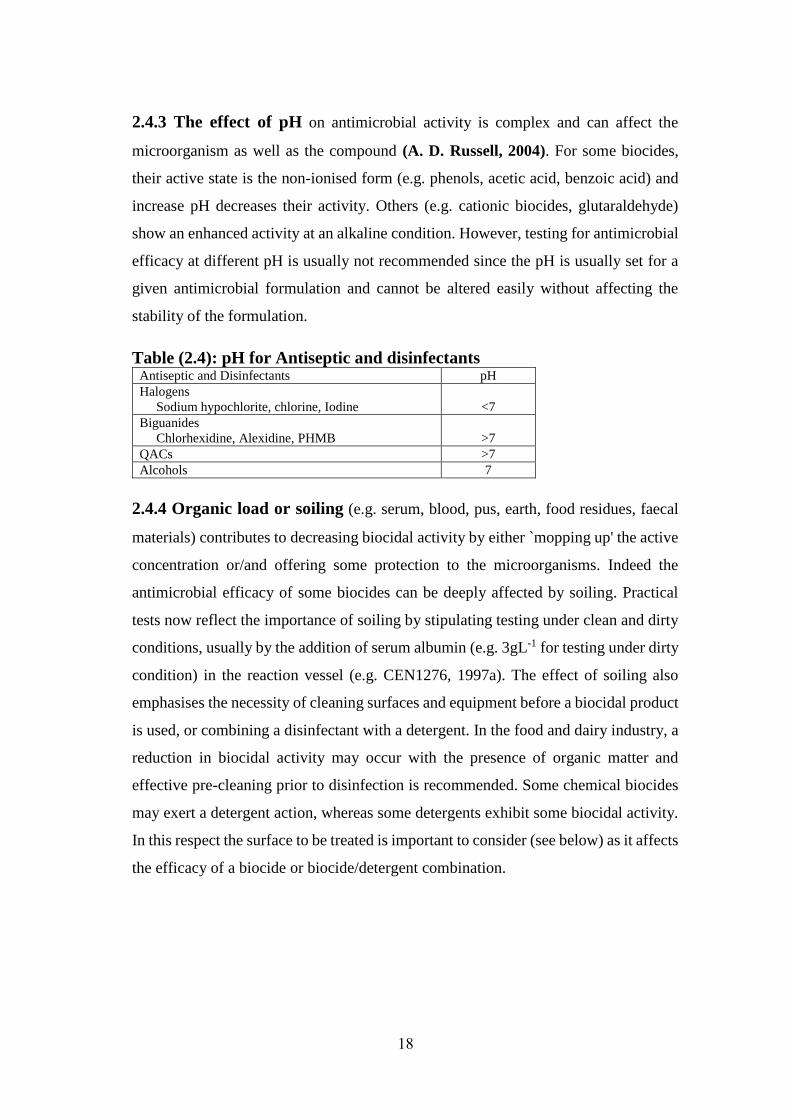
2.4.3 The effect of pH on antimicrobial activity is complex and can affect the
microorganism as well as the compound (A. D. Russell, 2004). For some biocides,
their active state is the non-ionised form (e.g. phenols, acetic acid, benzoic acid) and
increase pH decreases their activity. Others (e.g. cationic biocides, glutaraldehyde)
show an enhanced activity at an alkaline condition. However, testing for antimicrobial
efficacy at different pH is usually not recommended since the pH is usually set for a
given antimicrobial formulation and cannot be altered easily without affecting the
stability of the formulation.
Table (2.4): pH for Antiseptic and disinfectants Antiseptic and Disinfectants pH
Halogens
Sodium hypochlorite, chlorine, Iodine
<7
Biguanides
Chlorhexidine, Alexidine, PHMB
>7
QACs >7
Alcohols 7
2.4.4 Organic load or soiling (e.g. serum, blood, pus, earth, food residues, faecal
materials) contributes to decreasing biocidal activity by either `mopping up' the active
concentration or/and offering some protection to the microorganisms. Indeed the
antimicrobial efficacy of some biocides can be deeply affected by soiling. Practical
tests now reflect the importance of soiling by stipulating testing under clean and dirty
conditions, usually by the addition of serum albumin (e.g. 3gL-1 for testing under dirty
condition) in the reaction vessel (e.g. CEN1276, 1997a). The effect of soiling also
emphasises the necessity of cleaning surfaces and equipment before a biocidal product
is used, or combining a disinfectant with a detergent. In the food and dairy industry, a
reduction in biocidal activity may occur with the presence of organic matter and
effective pre-cleaning prior to disinfection is recommended. Some chemical biocides
may exert a detergent action, whereas some detergents exhibit some biocidal activity.
In this respect the surface to be treated is important to consider (see below) as it affects
the efficacy of a biocide or biocide/detergent combination.
19
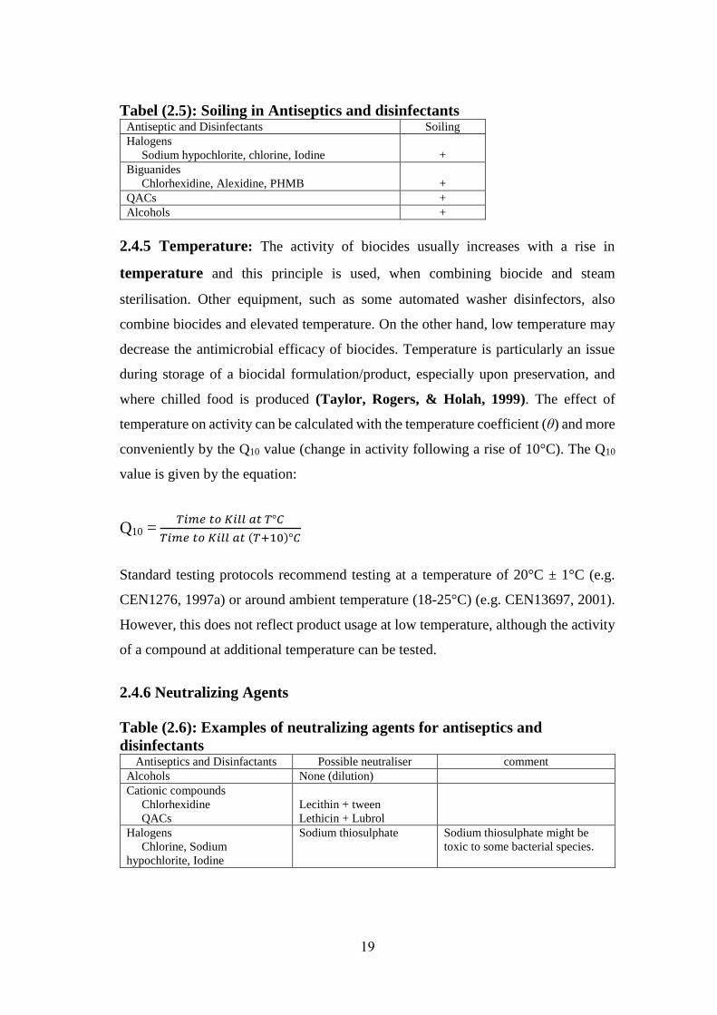
Tabel (2.5): Soiling in Antiseptics and disinfectants Antiseptic and Disinfectants Soiling
Halogens
Sodium hypochlorite, chlorine, Iodine
+
Biguanides
Chlorhexidine, Alexidine, PHMB
+
QACs +
Alcohols +
2.4.5 Temperature: The activity of biocides usually increases with a rise in
temperature and this principle is used, when combining biocide and steam
sterilisation. Other equipment, such as some automated washer disinfectors, also
combine biocides and elevated temperature. On the other hand, low temperature may
decrease the antimicrobial efficacy of biocides. Temperature is particularly an issue
during storage of a biocidal formulation/product, especially upon preservation, and
where chilled food is produced (Taylor, Rogers, & Holah, 1999). The effect of
temperature on activity can be calculated with the temperature coefficient (θ) and more
conveniently by the Q10 value (change in activity following a rise of 10°C). The Q10
value is given by the equation:
Q10 = 𝑇𝑖𝑚𝑒 𝑡𝑜 𝐾𝑖𝑙𝑙 𝑎𝑡 𝑇°𝐶
𝑇𝑖𝑚𝑒 𝑡𝑜 𝐾𝑖𝑙𝑙 𝑎𝑡 (𝑇+10)°𝐶
Standard testing protocols recommend testing at a temperature of 20°C ± 1°C (e.g.
CEN1276, 1997a) or around ambient temperature (18-25°C) (e.g. CEN13697, 2001).
However, this does not reflect product usage at low temperature, although the activity
of a compound at additional temperature can be tested.
2.4.6 Neutralizing Agents
Table (2.6): Examples of neutralizing agents for antiseptics and
disinfectants Antiseptics and Disinfactants Possible neutraliser comment
Alcohols None (dilution)
Cationic compounds
Chlorhexidine
QACs
Lecithin + tween
Lethicin + Lubrol
Halogens
Chlorine, Sodium
hypochlorite, Iodine
Sodium thiosulphate Sodium thiosulphate might be
toxic to some bacterial species.
20
2.4.7 The surface to be disinfected is not usually listed as a factor influencing the
activity of a biocide as such, but needs to be considered here. The antimicrobial
efficacy of disinfectants or sanitisers will depend to some extent on the surface upon
which they are used. Surfaces can vary greatly, particularly whether they are porous
or non-porous. Porous surfaces will have a tendency to entrap and protect microbial
contaminants, whereas non-porous surfaces can reduce bacterial adhesion and
facilitate a cleaning or a disinfection process.
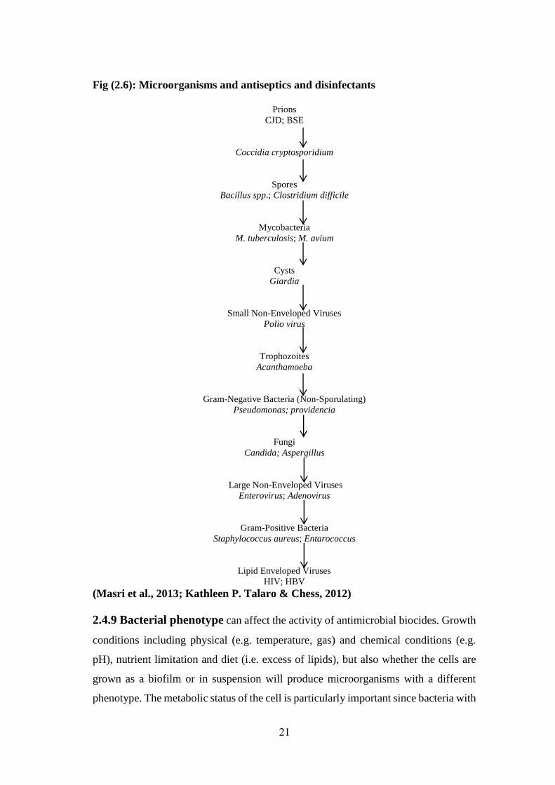
2.4.8 Different types of microorganisms present different levels of sensitivity to
a given antimicrobial biocide. Attempts have been made to classify microorganisms
according to their overall susceptibility to biocides. Usually, such classification relies
upon information on the intrinsic property of a microorganism but is not designed to
give a definite answer about the susceptibility of a type of microorganism, since
variation within species and even strains might occur. Practically, the type of
microorganisms expected on a given surface help in the selection of an appropriate
disinfectant or sanitiser. Antimicrobial test protocols usually include testing against a
range of bacteria and fungi, which are selected depending upon the expected usage of
the biocide, i.e. food industry, hospital environment, etc. However, the number of test
protocols available to evaluate virucidal and mycobactericidal activity is limited. In
addition, there is no standardisation and these protocols tend to vary greatly between
countries (Campos et al., 2012; Fraise et al., 2012; Sauerbrei et al., 2012) notably
with the test organisms. a similar protocol for the healthcare and veterinary
environment has not been published yet (J. Holah, 2003). The choice of the viral
indicator is particularly contentious (Jean‐Yves Maillard, 2004).as seen in figure
(2.6)
21
Fig (2.6): Microorganisms and antiseptics and disinfectants
Prions
CJD; BSE
Coccidia cryptosporidium
Spores
Bacillus spp.; Clostridium difficile
Mycobacteria
M. tuberculosis; M. avium
Cysts
Giardia
Small Non-Enveloped Viruses
Polio virus
Trophozoites
Acanthamoeba
Gram-Negative Bacteria (Non-Sporulating)
Pseudomonas; providencia
Fungi
Candida; Aspergillus
Large Non-Enveloped Viruses
Enterovirus; Adenovirus
Gram-Positive Bacteria
Staphylococcus aureus; Entarococcus
Lipid Enveloped Viruses
HIV; HBV (Masri et al., 2013; Kathleen P. Talaro & Chess, 2012)
2.4.9 Bacterial phenotype can affect the activity of antimicrobial biocides. Growth
conditions including physical (e.g. temperature, gas) and chemical conditions (e.g.
pH), nutrient limitation and diet (i.e. excess of lipids), but also whether the cells are
grown as a biofilm or in suspension will produce microorganisms with a different
phenotype. The metabolic status of the cell is particularly important since bacteria with

22
a `low metabolism' or quiescent bacteria are particularly resilient to the antimicrobial
effects of biocides (Gilbert, McBain, & Rickard, 2003; J. T. Holah, Taylor,
Dawson, & Hall, 2002).
2.4.10 The number of microorganisms that should be used in standard tests has
long been debated and differs between test protocols. It is generally accepted that the
higher the level of microbial contaminant, the more difficult the disinfection.
Predicting the level of contamination might be difficult and often the worst case
scenario is considered, i.e. a high-inoculum. Most tests work on the basis of reducing
the number of microorganisms to an acceptable level (e.g. a 5 log10 reduction on
surface), but not to the complete elimination (i.e. sterilisation) of the microorganisms.
If this is generally acceptable for most microorganisms, a problem can arise with
highly infectious or virulent microorganisms such as the hepatitis B virus, Escherichia
coli O157 for which a complete elimination would be recommendable (Masri et al.,
2013; Steinhauer, 2010; Wijesinghe & Weerasinghe, 2012).
2.5 Limitations in the use of Antiseptics and disinfectants
The limitations in using biocidal products usually refer to their toxicity, to the
alteration of the surface/equipment onto which they are used (e.g. corrosiveness,
colour formation), to their incompatibility with other components of a formulation, but
also to their overall efficacy against a given predicted microorganism. For example,
high-level disinfectants are needed for the disinfection of critical surfaces in the
hospital environment (W. A. Rutala & Weber, 1999). Toxicity is also important to
consider not only for the end user (e.g. with antisepsis and preservation) but also for
the environment. For example, the use of high concentrations might not be acceptable
because of the high toxicity for the environment. Within the food industry,
consideration must also be given to the potential for any biocide residues to taint or
otherwise change the organoleptic properties of the foodstuffs produced.
Limitations related with antiseptic and disinfectants activity (Lelieveld, Mostert, &
Holah, 2005):
Broad spectrum activity including activity against bacteria, fungi, viruses
Rapid antimicrobial activity
23
Retain stability (product) and antimicrobial efficacy over a wide range of pH
Retain stability (product) and antimicrobial efficacy over a wide range of
temperature
Retain activity in the presence of organic load and hard water
Retain activity upon dilution
Residual activity: The presence of remaining low concentration (below the
minimum inhibitory concentration, MIC) of a biocide on a surface is the subject of
much debate with current evidence on emerging microbial resistance to biocides.
Limitations related with safty:
No or low toxicity
Degradable in the environment
Limitations related with formulation and usage:
No or low corrosiveness
Non-staining
No odour
Good wetting and detergency
Easily combined with liquid or powder
Compatible with other chemicals (e.g. surfactants)
Cost-effective
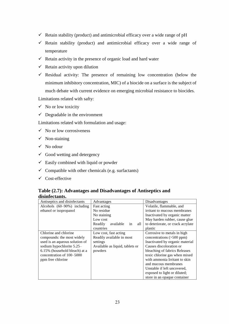
Table (2.7): Advantages and Disadvantages of Antiseptics and
disinfectants. Antiseptics and disinfectants Advantages Disadvantages
Alcohols (60–90%) including
ethanol or isopropanol
Fast acting
No residue
No staining
Low cost
Readily available in all
countries
Volatile, flammable, and
irritant to mucous membranes
Inactivated by organic matter
May harden rubber, cause glue
to deteriorate, or crack acrylate
plastic
Chlorine and chlorine
compounds: the most widely
used is an aqueous solution of
sodium hypochlorite 5.25–
6.15% (household bleach) at a
concentration of 100–5000
ppm free chlorine
Low cost, fast acting
Readily available in most
settings
Available as liquid, tablets or
powders
Corrosive to metals in high
concentrations (>500 ppm)
Inactivated by organic material
Causes discoloration or
bleaching of fabrics Releases
toxic chlorine gas when mixed
with ammonia Irritant to skin
and mucous membranes
Unstable if left uncovered,
exposed to light or diluted;
store in an opaque container
24
2.6 Mechanism of Action of Common Antiseptics and Disinfectants
The mechanisms of antimicrobial action of a range of chemical agents that are used as
antiseptics or disinfectants or both are discussed. Different types of microorganisms
are considered, and similarities or differences in the nature of the effect are
emphasized. The mechanisms of action of antiseptics and disinfectants on
microorganisms, especially bacteria (Denyer & Stewart, 1998). These include
examination of uptake (Ioannou, Hanlon, & Denyer, 2007; Yeaman & Yount,
2003), lysis and leakage of intracellular constituents (J‐Y Maillard, 2002),
perturbation of cell homeostasis (Dodd, Sharman, Bloomfield, Booth, & Stewart,
1997), effects on model membranes (Lambert, 2004), inhibition of enzymes, electron
transport, and oxidative phosphorylation (J‐Y Maillard, 2002), interaction with
macromolecules (Setlow, 2006), effects on macromolecular biosynthetic processes (J‐
Y Maillard, 2002), and microscopic examination of biocide-exposed cells. Additional
and useful information can be obtained by calculating concentration exponents (n
values (Denyer & Stewart, 1998)) and relating these to membrane activity (Denyer
& Stewart, 1998). Many of these procedures are valuable for detecting and evaluating
antiseptics or disinfectants used in combination (A. Russell, 2004). Similar techniques
have been used to study the activity of antiseptics and disinfectants against fungi, in
particular yeasts. Additionally, studies on cell wall porosity (De Nobel, Klis, Priem,
Munnik, & Van Den Ende, 1990) may provide useful information about intracellular
entry of disinfectants and antiseptics (S. Hiom, Furr, Russell, & Hann, 1995).
Mechanisms of antiprotozoal action have not been widely investigated. One reason for
this is the difficulty in culturing some protozoa (e.g., Cryptosporidium) under
laboratory conditions. However, the different life stages (trophozoites and cysts) do
provide a fascinating example of the problem of how changes in cytology and
physiology can modify responses to antiseptics and disinfectants. Khunkitti et al.
(Khunkitti, Avery, Lloyd, Furr, & Russell, 1997; Lloyd et al., 2001) have explored
this aspect by using indices of viability, leakage, uptake, and electron microscopy as
experimental tools. Some of these procedures can also be modified for studying effects
on viruses and phages (e.g., uptake to whole cells and viral or phage components,
effects on nucleic acids and proteins, and electron microscopy) (Rodgers, Hufton,
25
Kurzawska, Molloy, & Morgan, 1985). Viral targets are predominantly the viral
envelope (if present), derived from the host cell cytoplasmic or nuclear membrane; the
capsid, which is responsible for the shape of virus particles and for the protection of
viral nucleic acid; and the viral genome. Release of an intact viral nucleic acid into the
environment following capsid destruction is of potential concern since some nucleic
acids are infective when liberated from the capsid (Brul & Coote, 1999; A. Russell,
1991), an aspect that must be considered in viral disinfection. Important considerations
in viral inactivation are dealt with by Klein and Deforest and Prince et al. (Prince,
Prince, & Prince, 1991), while an earlier paper by Grossgebauer is highly
recommended (Grossgebauer, 1970).
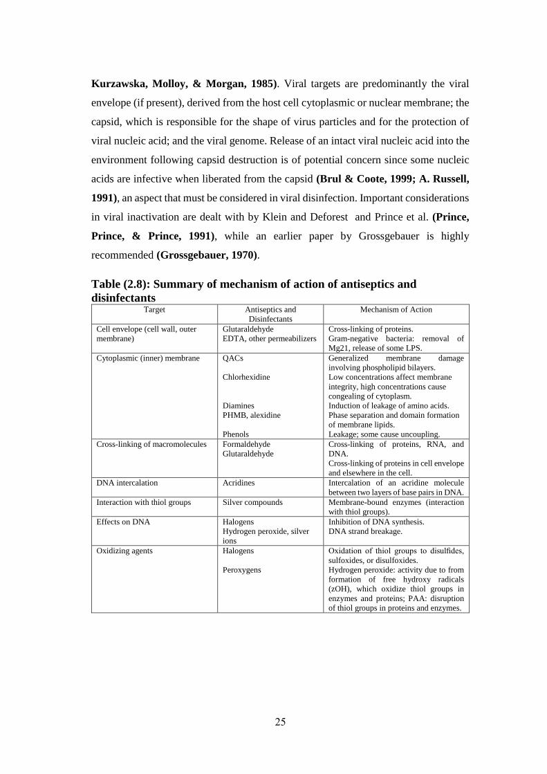
Table (2.8): Summary of mechanism of action of antiseptics and
disinfectants Target Antiseptics and
Disinfectants
Mechanism of Action
Cell envelope (cell wall, outer
membrane)
Glutaraldehyde
EDTA, other permeabilizers
Cross-linking of proteins.
Gram-negative bacteria: removal of
Mg21, release of some LPS.
Cytoplasmic (inner) membrane QACs
Chlorhexidine
Diamines
PHMB, alexidine
Phenols
Generalized membrane damage
involving phospholipid bilayers.
Low concentrations affect membrane
integrity, high concentrations cause
congealing of cytoplasm.
Induction of leakage of amino acids.
Phase separation and domain formation
of membrane lipids.
Leakage; some cause uncoupling.
Cross-linking of macromolecules Formaldehyde
Glutaraldehyde
Cross-linking of proteins, RNA, and
DNA.
Cross-linking of proteins in cell envelope
and elsewhere in the cell.
DNA intercalation Acridines Intercalation of an acridine molecule
between two layers of base pairs in DNA.
Interaction with thiol groups Silver compounds Membrane-bound enzymes (interaction
with thiol groups).
Effects on DNA Halogens
Hydrogen peroxide, silver
ions
Inhibition of DNA synthesis.
DNA strand breakage.
Oxidizing agents Halogens
Peroxygens
Oxidation of thiol groups to disulfides,
sulfoxides, or disulfoxides.
Hydrogen peroxide: activity due to from
formation of free hydroxy radicals
(zOH), which oxidize thiol groups in
enzymes and proteins; PAA: disruption
of thiol groups in proteins and enzymes.
26
2.6.1: Mechanism of Action of Alcohols
Little is known about the specific mode of action of alcohols, but based on the
increased efficacy in the presence of water, it is generally believed that they cause
membrane damage and rapid denaturation of proteins, with subsequent interference
with metabolism and cell lysis (E. Larson & Morton, 1991).
The mode of action of alcohol depends upon its concentration. Alcohol with a
concentration of 50% and higher dissolves membrane lipids, disrupts cell surface
tension and compromises membrane integrity. An alcohol that has entered the
protoplasm denatures protein through coagulation but only in alcohol-water solution
of 50-95%. Absolute alcohol (100%) dehydrates cells and inhibits their growth. Some
of its effectiveness as surface disinfectants can be attributed to its cleansing or
detergent action, which helps in the mechanical removal of microorganisms. Solutions
of 70-95% alcohol are used as skin degerming agents (McDonnell & Russell, 2001).
2.6.2 Mechanism of Action of Halogens
Chlorine compounds In solution these compounds combine with water and release
hypochlorus acid (HOCl), that oxidises the sulfhydryl (S-H) group on the amino acid
cysteine, that interferes with the disulfide (S-S) bridges of numerous enzymes. The
resulting denaturation of the enzymes is irreversible and suspends metabolic reactions
(Kathleen P. Talaro & Chess, 2012).
CRAs are highly active oxidizing agents and thereby destroy the cellular activity of
proteins (Chlorine, 1995); potentiation of oxidation may occur at low pH, where the
activity of CRAs is maximal, although increased penetration of outer cell layers may
be achieved with CRAs in the unionized state. Hypochlorous acid has long been
considered the active moiety responsible for bacterial inactivation by CRAs, the OCl2
ion having a minute effect compared to undissolved HOCl (Seymour Stanton Block,
2001a). This correlates with the observation that CRA activity is greatest when the
percentage of undissolved HOCl is highest. This concept applies to hypochlorites,
NaDCC, and chloramine-T.
Deleterious effects of CRAs on bacterial DNA that involve the formation of
chlorinated derivatives of nucleotide bases (Dukan & Touati, 1996). Hypochlorous
acid has also been found to disrupt oxidative phosphorylation (Barrette Jr, Hannum,
27
Wheeler, & Hurst, 1989) and other membrane-associated activity (Camper &
McFETERS, 1979). inhibition of bacterial growth by hypochlorous acid. At 50 mM
(2.6 ppm), HOCl completely inhibited the growth of E. coli within 5 min, and DNA
synthesis was inhibited by 96% but protein synthesis was inhibited by only 10 to 30%.
Because concentrations below 5mM (260ppm) did not induce bacterial membrane
disruption or extensive protein degradation, it was inferred that DNA synthesis was
the sensitive target. In contrast, chlorine dioxide inhibited bacterial protein synthesis
(McDonnell & Russell, 2001).
CRAs at higher concentrations are sporicidal (A. Russell & Day, 1995); this depends
on the pH and concentration of available chlorine (Allan Denver Russell, 1982).
During treatment, the spores lose refractivity, the spore coat separates from the cortex,
and lysis occurs (Kulikovsky, Pankratz, & Sadoff, 1975). In addition, a number of
studies have concluded that CRA-treated spores exhibit increased permeability of the
spore coat (Allan Denver Russell, 1982).
CRAs also possess virucidal activity (Seymour Stanton Block, 2001b). chlorine
inactivated naked f2 RNA at the same rate as RNA in intact phage, whereas f2 capsid
proteins could still adsorb to the host. the RNA of polio virus type 1 was degraded into
fragments by chlorine but that poliovirus inactivation preceded any severe
morphological changes. And in other studies found that the capsid of poliovirus type
1 was broken down (Sharp & Leong, 1980).
Iodine compounds Iodine rapidly penetrates the cells of microorganisms where it
apparently disturbs a variety of metabolic functions by interfering with disulfide bonds
of protein. It also iodinates cell proteins.
the exact mode of action is unknown. Iodine rapidly penetrates into microorganisms
and attacks key groups of proteins (in particular the free- sulfur amino acids cysteine
and methionine (Seymour Stanton Block, 2001a), nucleotides, and fatty acids
(Seymour Stanton Block, 2001a), which culminates in cell death . Less is known
about the antiviral action of iodine, but nonlipid viruses and parvoviruses are less
sensitive than lipid enveloped viruses (Prince et al., 1991). Similarly to bacteria, it is
likely that iodine attacks the surface proteins of enveloped viruses, but they may also
destabilize membrane fatty acids by reacting with unsaturated carbon bonds
(Springthorpe & Sattar, 1990).
28
2.6.3 Mechanism of Action of Quaternary Ammonium Compounds
QACs lower cellular surface tension. This can have several effects but chief among
them is the disruption of the cell membrane and the loss of its selective permeability.
QACs kill micro-organisms by causing a leakage of microbial protoplasm,
precipitating proteins and inhibiting metabolism (Kathleen P. Talaro & Chess,
2012).
2.6.4 Mechanism of Action of Biguanides
Chlorhexidine is a bactericidal agent (Denyer & Stewart, 1998). Its interaction and
uptake by bacteria, the uptake of chlorhexidine by E. coli and S. aureus was very rapid
and depended on the chlorhexidine concentration and pH. More recently, by using
[14C] chlorhexidine gluconate, the uptake by bacteria (Hageman & Havinga, 2006)
and yeasts (S. J. Hiom, Furr, Russell, & Dickinson, 1992b) was shown to be
extremely rapid, with a maximum effect occurring within 20 s. Damage to the outer
cell layers takes place (El Moug, Rogers, Furr, El-Falaha, & Russell, 1986) but is
insufficient to induce lysis or cell death. The agent then crosses the cell wall or outer
membrane, presumably by passive diffusion, and subsequently attacks the bacterial
cytoplasmic or inner membrane or the yeast plasma membrane. In yeasts,
chlorhexidine “partitions” into the cell wall, plasma membrane, and cytoplasm of cells
(S. J. Hiom, Furr, Russell, & Dickinson, 1992a). Damage to the delicate
semipermeable membrane is followed by leak- age of intracellular constituents, which
can be measured by appropriate techniques. Leakage is not per se responsible for
cellular inactivation but is a consequence of cell death (A. Russell & Hugo, 1988).
High concentrations of chlorhexidine cause coagulation of intracellular constituents.
As a result, the cytoplasm becomes congealed, with a consequent reduction in leakage
(Davies, 1973), so that there is a biphasic effect on membrane permeability. An initial
high rate of leakage rises as the concentration of chlorhexidine increases, but leakage
is reduced at higher biocide concentrations because of the coagulation of the cytosol.
an inhibitor of both membrane-bound and soluble ATPase as well as of net K+ uptake
in Enterococcus faecalis. However, only high biguanide concentrations inhibit
membrane-bound ATPase (Chopra, Linton, Hugo, & Russell, 1987), which suggests
29
that the enzyme is not a primary target for chlorhexidine action. Although
chlorhexidine collapses the membrane potential, it is membrane disruption rather than
ATPase inactivation that is associated with its lethal effects (McDonnell & Russell,
2001).
The effects of chlorhexidine on yeast cells are probably similar to those previously
described for bacteria (S. Hiom, Hann, Furr, & Russell, 1995). Chlorhexidine has a
biphasic effect on protoplast lysis, with reduced lysis at higher biguanide
concentrations. Furthermore, in whole cells, the yeast cell wall may have some effect
in limiting the uptake of the biguanide (S. Hiom, Furr, et al., 1995). an effect on the
fungal plasma membrane but with significant actions elsewhere in the cell (Bobichon
& Bouchet, 1987). Increasing concentrations of chlorhexidine (up to 25 mg/ml)
induce progressive lysis of Saccharomyces cerevisiae protoplasts, but higher
biguanide concentrations result in reduced lysis (S. J. Hiom et al., 1992a).
chlorhexidine has a similar effect on the trophozoites of Acanthameoba castellanii,
with the cysts being less sensitive (Khunkitti, Lloyd, Furr, & Russell, 1998). the
effects of chlorhexidine and other biocides on Acanthameoba and showed that
membrane damage in these protozoa is a significant factor in their inactivation.
Mycobacteria are generally highly resistant to chlorhexidine (Selvaraju, Khan, &
Yadav, 2005). Little is known about the uptake of chlorhexidine (and other antiseptics
and disinfectants) by mycobacteria and on the biochemical changes that occur in the
treated cells. Since the MICs for some mycobacteria are on the order of those for
chlorhexidine-sensitive, gram-positivecocci, the inhibitory effects of chlorhexidine on
mycobacteria may not be dissimilar to those on susceptible bacteria. Mycobacterium
avium-intracellulare is considerably more resistant than other mycobacteria (Bradley
& Fraise, 1996).
Chlorhexidine is not sporicidal (discussed in “Mechanisms of resistance”). Even high
concentrations of the bisbiguanide do not affect the viability of Bacillus spores at
ambient temperatures (Shaker, Dancer, Russell, & Furr, 1988), although a marked
sporicidal effect is achieved at elevated temperatures (Shaker, Russell, & Furr,
1986). Presumably, sufficient changes occur in the spore structure to permit an
increased uptake of the biguanide, although this has yet to be shown experimentally.
Little is known about the uptake of chlorhexidine by bacterial spores, although coatless
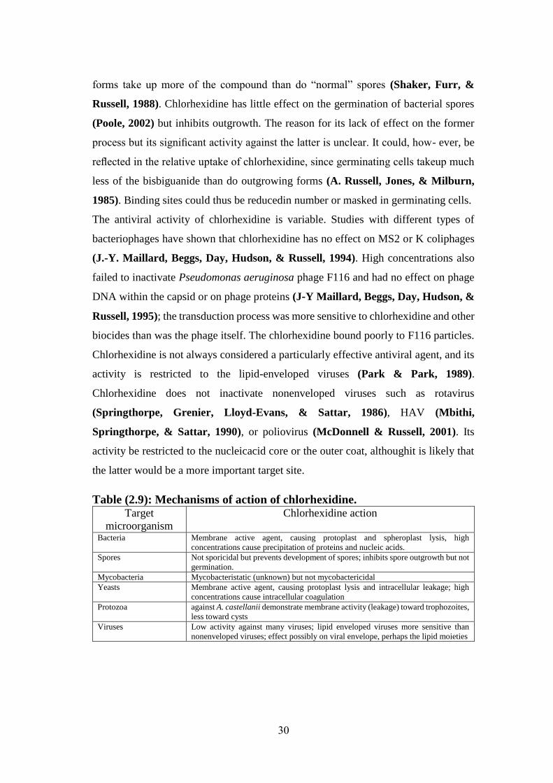
30
forms take up more of the compound than do “normal” spores (Shaker, Furr, &
Russell, 1988). Chlorhexidine has little effect on the germination of bacterial spores
(Poole, 2002) but inhibits outgrowth. The reason for its lack of effect on the former
process but its significant activity against the latter is unclear. It could, how- ever, be
reflected in the relative uptake of chlorhexidine, since germinating cells takeup much
less of the bisbiguanide than do outgrowing forms (A. Russell, Jones, & Milburn,
1985). Binding sites could thus be reducedin number or masked in germinating cells.
The antiviral activity of chlorhexidine is variable. Studies with different types of
bacteriophages have shown that chlorhexidine has no effect on MS2 or K coliphages
(J.-Y. Maillard, Beggs, Day, Hudson, & Russell, 1994). High concentrations also
failed to inactivate Pseudomonas aeruginosa phage F116 and had no effect on phage
DNA within the capsid or on phage proteins (J‐Y Maillard, Beggs, Day, Hudson, &
Russell, 1995); the transduction process was more sensitive to chlorhexidine and other
biocides than was the phage itself. The chlorhexidine bound poorly to F116 particles.
Chlorhexidine is not always considered a particularly effective antiviral agent, and its
activity is restricted to the lipid-enveloped viruses (Park & Park, 1989).
Chlorhexidine does not inactivate nonenveloped viruses such as rotavirus
(Springthorpe, Grenier, Lloyd-Evans, & Sattar, 1986), HAV (Mbithi,
Springthorpe, & Sattar, 1990), or poliovirus (McDonnell & Russell, 2001). Its
activity be restricted to the nucleicacid core or the outer coat, althoughit is likely that
the latter would be a more important target site.
Table (2.9): Mechanisms of action of chlorhexidine. Target
microorganism
Chlorhexidine action
Bacteria Membrane active agent, causing protoplast and spheroplast lysis, high
concentrations cause precipitation of proteins and nucleic acids.
Spores Not sporicidal but prevents development of spores; inhibits spore outgrowth but not
germination.
Mycobacteria Mycobacteristatic (unknown) but not mycobactericidal
Yeasts Membrane active agent, causing protoplast lysis and intracellular leakage; high
concentrations cause intracellular coagulation
Protozoa against A. castellanii demonstrate membrane activity (leakage) toward trophozoites,
less toward cysts
Viruses Low activity against many viruses; lipid enveloped viruses more sensitive than
nonenveloped viruses; effect possibly on viral envelope, perhaps the lipid moieties
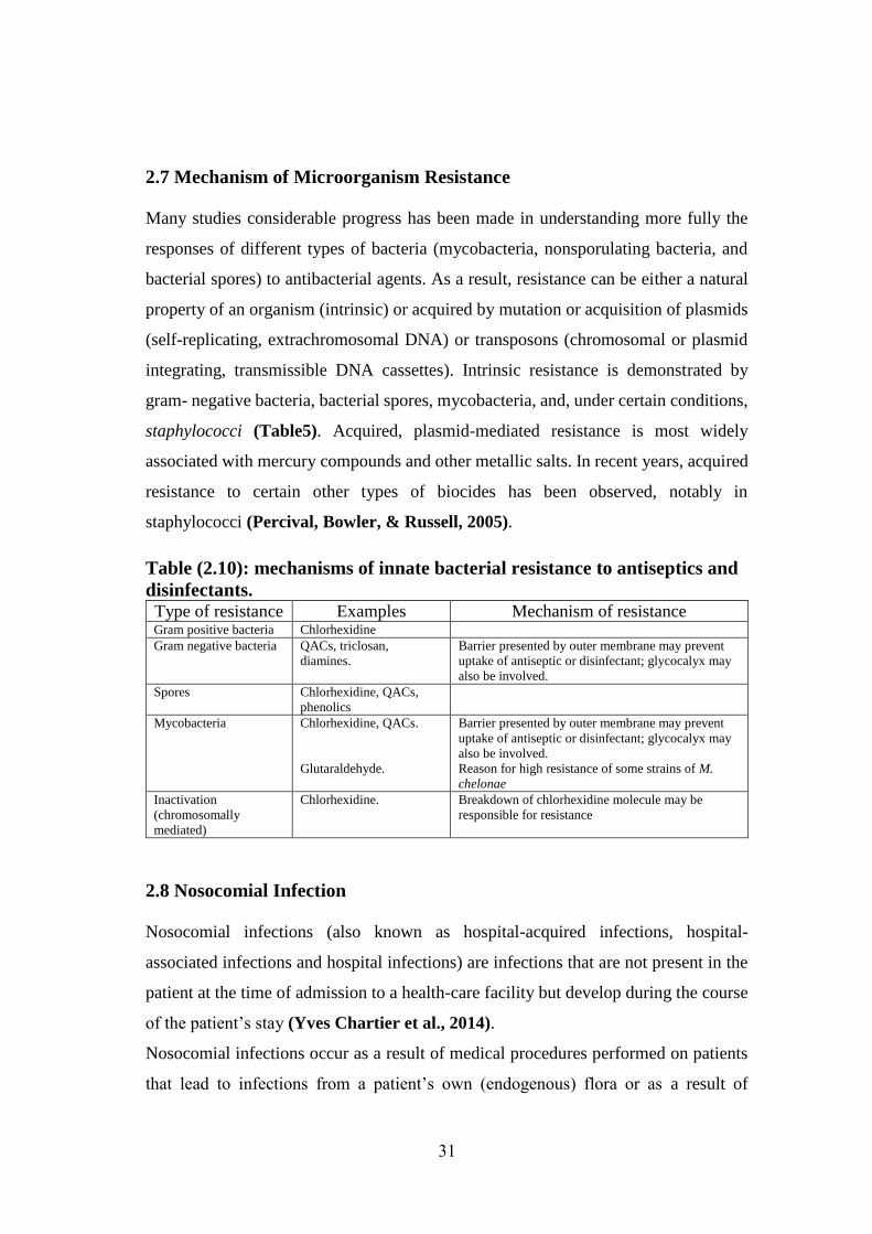
31
2.7 Mechanism of Microorganism Resistance
Many studies considerable progress has been made in understanding more fully the
responses of different types of bacteria (mycobacteria, nonsporulating bacteria, and
bacterial spores) to antibacterial agents. As a result, resistance can be either a natural
property of an organism (intrinsic) or acquired by mutation or acquisition of plasmids
(self-replicating, extrachromosomal DNA) or transposons (chromosomal or plasmid
integrating, transmissible DNA cassettes). Intrinsic resistance is demonstrated by
gram- negative bacteria, bacterial spores, mycobacteria, and, under certain conditions,
staphylococci (Table5). Acquired, plasmid-mediated resistance is most widely
associated with mercury compounds and other metallic salts. In recent years, acquired
resistance to certain other types of biocides has been observed, notably in
staphylococci (Percival, Bowler, & Russell, 2005).
Table (2.10): mechanisms of innate bacterial resistance to antiseptics and
disinfectants. Type of resistance Examples Mechanism of resistance Gram positive bacteria Chlorhexidine
Gram negative bacteria QACs, triclosan,
diamines.
Barrier presented by outer membrane may prevent
uptake of antiseptic or disinfectant; glycocalyx may
also be involved.
Spores Chlorhexidine, QACs,
phenolics
Mycobacteria Chlorhexidine, QACs.
Glutaraldehyde.
Barrier presented by outer membrane may prevent
uptake of antiseptic or disinfectant; glycocalyx may
also be involved.
Reason for high resistance of some strains of M.
chelonae
Inactivation
(chromosomally
mediated)
Chlorhexidine. Breakdown of chlorhexidine molecule may be
responsible for resistance
2.8 Nosocomial Infection
Nosocomial infections (also known as hospital-acquired infections, hospital-
associated infections and hospital infections) are infections that are not present in the
patient at the time of admission to a health-care facility but develop during the course
of the patient’s stay (Yves Chartier et al., 2014).
Nosocomial infections occur as a result of medical procedures performed on patients
that lead to infections from a patient’s own (endogenous) flora or as a result of
32
exposure to items contaminated with infectious agents. Additionally, the risk of
acquiring an infection increases for patients with altered or compromised immunity
(Bennett et al., 2007).
Human beings are reservoirs of numerous types of microorgansims. Faeces contain
approximately 1013 bacteria per gram, and the number of microorganisms on skin
varies between 102 and 104 per cm2. Many species of microorganisms live on mucous
membranes and are considered normal flora. When the integrity of these barriers is
challenged (e.g. microorganisms penetrate the skin or the mucous membrane), this
creates an opportunity for an infection to occur (Yves Chartier et al., 2014).
2.8.1 From Exposure to Infection
Whether an infection will develop after an exposure to microorganisms depends upon
the interaction between the microorganisms and the host. Healthy individuals have a
normal general resistance to infection. Patients with underlying disease, newborn
babies and the elderly have less resistance and are at greater risk to develop an infection
after exposure (Health Canada & Occupational, 2012).
Local resistance to infection also plays an important role: the skin and the mucous
membranes act as barriers in contact with the environment. Infection may occur when
these barriers are breached. Local resistance may also be overcome by the long-term
presence of an irritant, such as a cannula or catheter. The likelihood of infection
increases daily when a patient has a catheter attached (Filipe, 2010).
The most important determinants of infection are the nature and number of the
infectious agents. Microorganisms range from the completely innocuous to the
extremely pathogenic; the former will never cause an infection even in
immunocompromised individuals, while the latter will cause an infection in virtually
every case of exposure (Andersen, 2010).
When only a few organisms are present, an infection will not necessarily develop.
However, when a critical number is exceeded, it is very likely that an infection will
become established. For every type of microorganism, the minimal infective dose can
be determined. This is the lowest number of bacteria, viruses or fungi that cause the
first clinical signs of infection in a healthy individual. For most causative agents of
nosocomial infections, the minimal infective dose is relatively high. For example, for
33
Klebsiella and Serratia spp. and other Enterobacteriaceae, it is more than 105 colony-
forming units (CFUs)/gram, but for hepatitis B virus it is less than 10 plaque-forming
units (PFUs)/ gram (Yves Chartier et al., 2014).
2.8.2 Source of Infection
In a health-care facility, the sources of infectious agents may be the personnel, the
patients or the inanimate environment (Prüss, Giroult, & Rushbrook, 2014).
The hospital environment can be contaminated with pathogens. Salmonella or Shigella
spp., Escherichia coli O157:H7 or other pathogens may be present in the food and
cause an outbreak, just as they can in a community outside the hospital. Waterborne
infections may develop if the water-distribution system breaks down. In more
sophisticated facilities, the water-cooling system of air-conditioning equipment may
become contaminated with Legionella pneumophilia, causing Legionnaires’ disease in
susceptible patients. Pharmaceuticals may become contaminated during production or
preparation; an outbreak of infection by Pseudomonas aeruginosa, Burkholderia
cepacia or Serratia marcescens may occur as a consequence. In all these examples, it
may be possible to isolate the same causative agent in several patients, which would
suggest a common source. All possible measures should be taken to prevent the
recurrence of such incidents.
The source of a nosocomial infection may also be a health-care worker who is infected
or colonized (a carrier) with an infectious agent. The symptoms of infection will make
the potential transmission apparent to the health-care worker and/or to managerial
staff, and infected personnel are usually taken off patient care duties. Sometimes a
carrier may be symptomless (i.e. is colonized by potentially pathogenic organisms but
does not develop any infection). A typical example is methicillin-resistant
Staphylococcus aureus, which may be carried in the nasal passages of 30–60% of
health-care personnel. Faecal carriage of enteropathogens such as Salmonella spp. also
occurs frequently, but the prevalence varies according to the region. Other
conventional pathogens that can be found in symptomless carriers include
Streptococcus pyogenes, Corynebacterium diphtheriae, Neisseria meningitidis,
hepatitis B virus and cytomegalovirus. Exposure of patients to carriers can give rise to
an outbreak of disease. Careful investigation and isolation of the same organisms from
34
a cluster of patients as well as the carrier should reveal the cause of the outbreak (Prüss
et al., 2014).
The source of most hospital epidemics is infected patients; that is, patients infected
with pathogenic microorganisms. These microorganisms are often released into the
environment in very high numbers, depending on the disease, exceeding the minimal
infective dose, and exposing other patients, who subsequently develop hospital-
acquired infections. The recent case of severe acute respiratory syndrome and its
impact on health-care waste-generation rates (Chiang, Sung, Chang, & Tsai, 2006)
is a classic example of hospital-based epidemics relating to a respiratory disease.
2.8.3Nosocomial Infection Pathogens
2.8.3.1 Conventional pathogens
Cause disease in healthy individuals in the absence of specific immunity (Prüss et al.,
2014).
Examples: Methicillin-resistant Staphylococcus aureus, Streptococcus pyogenes (beta
strep group A), Salmonella spp., Shigella spp., vancomycin-resistant Enterococcus,
Corynebacterium 34iphtheria, Mycobacterium tuberculosis, Bordetella pertussis,
hepatitis A and B viruses, rubella virus, rotaviruses, human immunodeficiency virus
(HIV) (Prüss et al., 2014).
2.8.3.2Conditional pathogens
Cause disease, other than trivial local infections, only in persons with reduced
resistance to infection (including newborn infants) or when implanted directly into
tissue or a normally sterile body area (Prüss et al., 2014).
Examples: Streptococcus agalactiae, Enterococcus spp., Clostridium tetani,
Escherichia coli, Klebsiella spp., Serratia marcescens, Acinetobacter baumanii,
Pseudomonas aeruginosa, Candida spp (Prüss et al., 2014).
35
2.8.3.3Opportunistic pathogens
Cause generalised disease, but only in patients with profoundly diminished resistance
to infection (Prüss et al., 2014).
Examples: Atypical mycobacteria, Nocardia asteroides, Pneumocystis carinii
(Gerberding, 1998).
2.9 Reference Studies
2.9.1 Bacterial contamination in liquid soap for hospital and public use
In exploratory, cross-sectional study was developed at the hospitalization units of a
medium-sized hospital in Fortaleza, Ceará, Brazil. Data were collected between May
and July 2007. Fifty-nine liquid soap dispensers were analyzed, of which 33 contained
the following microorganisms: Burkholderia cepacia (14), Pseudomonas putidas (9),
Pseudomonas aeruginosa (3), Klebsiella pneumoniae (3), Enterobacter clocae (2),
and Pseudomonas luteola (2). The units with the largest number of contaminated
samples were the surgical (n=7) and the dermatological clinics (n=4). Contamination
was also found in an original flask of the same lot of liquid soap used to fill up the
dispensers. In conclusion, there is a need to regulate and control the quality of these
products in the production lines as well as during use in hospital services, mainly
because they are used to prevent hospital infection(J. A. Caetano et al., 2011).
Burkholderia species move through one single polar flagellum, or flagellum cluster.
The best-known species is Burkholderia cepacia, which is aerobic, gram-negative and
rod-shaped, capable of growing even in disinfecting solutions. This species has an
extraordinary nutritional spectrum and can degrade more than 100 different organic
molecules. This ability results from factors that facilitate equipment, product and drugs
contamination in hospitals (Tortora et al., 2013), it can colonize a range of humid
environmental surfaces and is commonly associated with hospital infections.
According to literature (Murray, Rosenthal, & Pfaller, 2006), infections caused by
this microorganism include respiratory tract infections in patients with cystic fibrosis
or chronic granulomatous disease; urinary tract infection; urinary tract infection in
patients using catheters; and sepsis, particularly in patients with contaminated
36
intravascular catheters. Except for pulmonary infection, in general, B. cepacia has a
relatively low virulence level, and infections with this microorganism generally do not
result in death(J. A. Caetano et al., 2011).
Pseudomonas spp. are straight or slightly curved gram- negative bacilli, which are
mobile through polar flagella; they are omnipresent organisms, easily found
throughout the hospital environment in humid reservoirs, including food, cut flowers,
sinks, toilets, floor cleaning mops, equipment, particularly for respiratory treatment,
and even in disinfectant solutions. The large-scale environmental distribution of
Pseudomonas is guaranteed by its simple requirement for growth. They also have
different structural factors and toxins that stimulate their virulence potential, making
them resistant to the most commonly used antibiotics. Pseudomonas aeruginosa is the
most common clinically significant species, causing various infections, as it is
typically resistant to most antibiotics. Another species found in the study was
Pseudomonas putida, little associated with infections in human beings (Tortora et al.,
2013).
Klebsiella pneumoniae can cause primary lobar pneumonia, which frequently involves
the necrotic destruction of alveolar spaces, formation of cavities and production of
bloody sputum. These bacteria also cause infections in wounds, soft tissues and the
urinary tract (Murray et al., 2006).Another gram- negative bacillus that was found,
from the Enterobacteriaceae family, was Enterobactercloacae. Infections caused by
microorganisms from the Enterobacter genre are rare in immunocompetent patients,
but common in neonates and immunocompromised patients. The main problem with
this bacteria group is resistance to multiple antibiotics (Murray et al., 2006)
In the same study on liquid soap dispensers in a hospital environment demonstrated,
out of 28 dispensers, 19 (68%) tested positively for one or more bacterial species. The
isolated bacteria were: A. baumannii, P. aeruginosa, Staphylococcus spp.,
Enterobacter cloacae, K. pneumoniae, Methicillin-resistant Staphylococcus aureus,
Candida albicans, and Bacillus species. Dispensers in that study were plastic,
rectangular and wall-mounted, with a button for soap dispensing. Moreover, they were
cleaned weekly. The same study observed that a significant num- ber of soap residues
remained close to the distribution hole and in the slits around the dispenser button (J.
A. Caetano et al., 2011).
37
In old study conducted in Japan in the early 1990s by Amemiya and Taguchi (1992)
revealed significant contamination in soap from public restrooms. They found that as
many as 4 x 10⁷ bacteria per mL could be recovered from liquid hand soaps and that
71% contained 1,000 or more bacteria per mL. after 19 years in United States study
the 541 Liquid soap samples were collected from public restrooms in five cities
(Boston; Atlanta; Columbus, Ohio; Los Angeles; and Dallas), consisting of 428 from
sink areas and 113 from showers. The percentage of samples that contained
Heterotrophic plate counts, numbers above 500 CFU/mL was 24.8%, averaging 3.0 x
106 CFU/mL and ranging from 590 to 1.3 x 10⁷ CFU/mL. Total coliform bacteria were
detected in 15.9% of the samples, averaging 3.9 x 10⁶ CFU/mL and ranging from <10
CFU/mL to 6.5 x 10⁷ CFU/mL. The different species of Gram-negative bacteria that
was isolated. Species of Klebsiella occurred most frequently, followed by
Enterobacter, Serratia, and Pseudomonas. No S. aureus was detected in any of the
liquid soap samples analyzed. Office restrooms had the highest percentage of
contamination with heterotrophic bacteria (47.5%) and coliform bacteria (35.0%) and
restrooms in retail stores had the least (15.3% for heterotrophic and 10.6% for coliform
bacteria). The rates of contamination of soap were similar among all five metropolitan
areas (M. Chattman et al., 2011).
2.9.2 Gram-negative bacteria
All of the organisms detected in the soap samples were Gram-negative bacteria. This
is most likely because of the presence of sodium lauryl sulfate (SLS) in the soap, which
inhibits Gram-positive bacterial growth. In fact, SLS is used to inhibit Gram-positive
growth in selective media such as mEndo agar (Laboratories, 1998). All of the
organisms that were identified in study were Gram-negative opportunistic pathogens.
The opportunistic pathogens most commonly found in liquid hand soap included
Pseudomonas spp., Serratia marcescens, and Klebsiella pneumoniae. These bacteria
are also among the most prevalent organisms that cause opportunistic infections. These
were also the same species of bacteria that Amemiya and Taguchi (1992) isolated most
often in liquid soap in Japan. While largely associated with infections in compromised
patients (immunocomprimised, burn patients, post-surgical) they are capable of
38
causing infections of wounds (cuts to the skin), folliculitis, and urinary tract infections
(Kallman, Lundberg, Wretlind, & Ortqvist, 2006). It has also been reported that
bacteria present in the liquid soap remain on the hands after use (Sartor et al., 2000).
Found that after hands washing with an S. marcesens contaminated liquid hand soap
pump, the hands of health care workers were 54 times more likely to be contaminated
with S. marcescens. Infections and outbreaks resulting from the use of contaminated
soap or disinfection products are not uncommon in health care settings. Several
different types of soaps contaminated with S. marcescens have caused a variety of
infections including bacteremia, conjunctivitis, meningitis, and joint infections.
Species of Pseudomonas and Burkholderia found in soap have been linked to various
outbreaks in hospitals and infections including skin ulcers, bacteremia, and urinary
tract infections (Dolan, 2006). Hand lotion contaminated with P. aeruginosa was
implicated as the vector resulting in hand transfer of the organism to infected infants
(Becks & Lorenzoni, 1995).
In same year the University of Arizona published a study about using the contaminated
Bulk-Soap-Refillable dispensers after several outbreaks linked to the use of
contaminated soap in health care settings have been reported (Buffet-Bataillon, 2009;
D. J. Weber, W. A. Rutala, & E. E. Sickbert-Bennett, 2007), and study conducted
in Japan, examined bacterial contamination of hand washing soaps obtained from
restrooms of various public use facilities. The authors found 17 different species of
bacteria, many of which were opportunistic pathogens, including Klebsiella
pneumoniae, Serratia marcescens, Enterobacter species, and Pseudomonas species
(Amemiya & Taguchi, 1992), and other studies conducted in the United States
demonstrated that 25% of bulk-soap-refillable dispensers in public restrooms were
excessively contaminated (M. Chattman et al., 2011). Bacterial loads averaged more
than 106 CFU/ml of soap, and 16% of the samples contained coliform bacteria.
Interestingly, of the 15 different species isolated in this study, 7 were identical to those
found in the Japanese study, including both K.pneumoniae and S.marcescens.Both S.
marcescens and K. pneumoniae are opportunistic pathogens known to transmit via the
hands (G. Reybrouck, 1983)
39
University of Arizona study conducted in three experiments: the first soap
experimentally contaminated with either K. pneumoniae (5.85 log10 CFU/ml) or S.
marcescens (3.72 log10 CFU/ml) followed by a 30-s rinse. Neither test organism was
recovered from the hands of subjects prior to washing hands or from the subjects that
washed with uncontaminated control soap. In contrast, for K. pneumoniae, a mean of
2.74 log10 CFU/hand was recovered from subjects after washing with K. pneumonia
contaminated soap, and for S. marcescens, a mean of 3.60 log10 CFU/hand was
recovered from subjects after washing with S. marcescens contaminated soap.
Interestingly, more bacteria were recovered from hands washed with S. marcescens
contaminated soap than from those washed with K. pneumonia contaminated soap (P
< 0.0001), even though the level of K. pneumoniae contamination was 100-fold higher.
In a second experiment, subjects performed a 10-s hand wash with 1.5 ml of liquid
soap experimentally contaminated with either a high level of S. marcescens (7.51log10
CFU/ml) or with a low level of S. marcescens (4.51 log10 CFU/ml) followed by a 10-
s rinse. It is known that when soap that is not contaminated is used for hand washing,
it is more effective at removing transient bacteria when greater volumes of soap and
longer wash times are used (Fuls et al., 2008). Therefore, the second controlled study
was conducted under conditions chosen to be more representative of the hand washing
behaviors typically observed (Garbutt, Simmons, Patrick, & Miller, 2007). The
mean numbers of S. marcescens cells recovered after washing with high-and low-
level-contaminated soap were 5.28 log10 CFU and 1.70 log10 CFU per hand,
respectively (P < 0.0001). The number of bacteria transferred to an agar surface after
washing were 2.23 log10 CFU and 0.30 log10 CFU per hand for the high- and low-
level- contaminated soap, respectively (P = 0.001). The third experiment study was
conducted with students and staff to assess the levels of Gram-negative bacteria
remaining on or transferred from hands after washing with contaminated soap from
these dispensers or with uncontaminated control soaps. Prior to washing with
contaminated bulk soap, uncontaminated bulk soap, and uncontaminated soap from
sealed refills, the mean numbers of bacteria recovered from hands of subjects were
1.17, 0.99, and 1.67 log10 CFU per hand, respectively. The mean number of bacteria
recovered from the hands after hand washing with the contaminated soap (2.59 log10
CFU per hand) was significantly higher than the pre-hand- washing value (P < 0.0001).
40
Gram-negative bacteria were de- tected in 97% (60/62) of hands tested after washing
with bulk soap compared to 52% (32/62) before washing. Incontrast, the mean number
of bacteria recovered from hands after washing with uncontaminated bulk soap (0.82
log10 CFU per hand) was reduced compared to the prewashing numbers. When hands
were washed with uncontaminated soap from the new replacement sealed-system
dispensers, the mean numbers of bacteria recovered from hands after washing (1.37
log10 CFU perhand) were also reduced compared to the prewashing numbers and were
statistically lower than those recovered from hands washed with contaminated soap (P
< 0.0001). The mean number of Gram-negative bacteria recovered from the hands after
washing with contaminated soap was significantly higher for students (2.82 log10 CFU
per hand) than that for staff (2.22 log10 CFU per hand; P = 0.008) (Zapka et al., 2011).
The most common transient microorganisms include gram negative coliforms and
Staphylococcus aureus. Hand washing with plain soap is effective in removing most
transient microorganisms (Katz, 2004). The mechanical action of washing and rinsing
removes most of the transient microorganism present (Noskin, Stosor, Cooper, &
Peterson, 1995). Health care workers wash their hands in two ways: (a) the social
hand wash, which is the cleaning of hands with plain, non-medicated bar or liquid soap
and water for removal of dirt, soil, and various organic substances; (b) the hygienic or
antiseptic hand wash, which is the cleaning of hands with antimicrobial or medicated
soap and water. Most antimicrobial soaps contain a single active agent and are usually
available as liquid preparations. Appropriate hand washing results in a reduced
incidence of both nosocomial and community infections (Kampf & Kramer, 2004).
Much studies have been written and debated regarding the use of bar versus liquid skin
cleansers in relation to infection control (Boyce & Pittet, 2002). And in the study by
university of Baghdad – Iraq - show that among 50 swabs of bar soaps, 30 (60%) swabs
were found colonized. A total of 44 microorganisms were isolated. Pseudomonas
aeruginosa (41%) was the most frequent isolated bacteria followed by Escherichia
coli (13.6%) and Acinetobacter baumanii (11.4%).From liquid soaps, 6
microorganisms were detected at only 7 tips (15.9%) of the total 44 containers. This
includes 4 (66.6%) P. aeruginosa, one (16.6%) Proteus penneri and one (16.6%)
Flavimonas oryzihabitans. Comparison of the rates of bacterial colonization between
41
bar soaps and liquid soaps: Bar soaps were found more colonized than the liquid soaps
significantly (p<0.05). P. aeruginosa was the most frequent isolate in both two group
whereas isolation rate was significantly higher (p<0.05) in bar soaps but not in the
liquid soaps (p>0.05) as statistically.
2.9.3 Bar or liquid Soap
The most common hand-cleaning agents are bar soap and liquid soap in disposable
plastic containers. When in use, bar soaps are frequently misused because they are
typically stored in contact with moisture and remain moist for long periods of time. It
is usually kept in a container, on or next to a wash basin. More often than not, it resides
in surface water. The resulting jelly mass is unsightly, difficult to use effectively. This
supplies an environment which provides the perfect opportunity for bacteria and
organisms to grow. Most bars of soap in communal areas are used by a number of
different people. This means that one bar of soap can be in direct contact with skin
bacteria from more than one person, and may harbour live pathogenic bacteria (E. L.
Larson, Eke, Wilder, & Laughon, 1987). Cross infection can and does occur under
these circumstances (McBride, 1984). When using a bar of soap, the CDC (Centre for
Disease Control) recommends placement on a drainable rack between uses (Boyce &
Pittet, 2002). Soap racks that promote drainage of all water from the bar should be
installed. In addition, there should be easy access to replacements when soap is lost,
dropped, melted, or consumed. Small soap bars were also recommended that can be
changed and used in preference to larger bars that are more likely to melt or become
colonized with bacteria (Nix, 2000). Liquid soap on the other hand is much better to
use. Liquid soap is dispensed straight from a plastic container. It has not been exposed
to skin bacteria or other contaminants. As a result, cross contamination is not likely to
occur, providing a more cleaning and more hygienic alternative (McBride, 1984).
McBride et al reported that bar soaps were found to have higher bacterial cultures after
use than liquid soaps (McBride, 1984). In another study, Kabara and Brady obtained
samples from bar and liquid soaps from 26 public bathrooms which were investigated.
Liquid soaps were found to be negative for bacteria, while 100% of the 84 samples
obtained from bar soaps yielded positive cultures (Kabara & Brady, 1984). In an
epidemiological study, the researchers isolated several strains of Pseudomonas from
42
45 of 353 environmental samples used by multiple providers (13%) and found that the
5 most common strains were frequently found on patients. They also affirmed that the
hands are a major vehicle for the transfer of Pseudomonas bacteria and implicated bar
soap in its spread (Bruun, McGarrity, Blakemore, & Coriell, 1976). Other groups
of researchers have found that bacteria survive on soap bars in continuous use in public
lavatories, even when cultured 48 hours following their last use (E. L. Larson et al.,
1987). The role of the soap dishes in infection control has also been studied by Sarmad
M.H. Zeiny from Iraq he found that dishes were found wet, and surfaces of soaps were
generally covered by squashy mass and bars were found heavily contaminated (%88).
This study revealed quite lower contamination rate in liquid soaps compared with bar
soaps, although they didn’t include suggested antibacterial agents for hand antisepsis
such as triclorasan or chlorhexidine (S. M. H. Zeiny, 2009). However, liquid soaps
would be expected to be sterile. So, there should be problems with the handling.
Honestly, in this study any strict procedures had not been followed in the wards for
the how often liquid dispensers should be cleaned, disinfected or exchanged. After the
results were obtained, procedures were described for handling and usage of liquid
soaps and dispensers immediately (S. M. H. Zeiny, 2009).
2.9.4 Strain of bacteria capable of metabolizing anionic detergent
Sodium dodecyl sulfate (SDS) is an anionic detergent widely used in the
manufacturing of a number of household and industrially useful products (Karsa,
1999). Like any other chemical, SDS is discharged in water bodies like ponds and
rivers (Singer & Tjeerdema, 1993). Studies have revealed that SDS is toxic to aquatic
animals such as fish, microbes like yeasts and bacteria (Venkatesh Chaturvedi &
Kumar, 2010b), and also to mammals (Venkatesh Chaturvedi & Kumar, 2010a)
So, bioremediation of this detergent was realized to be an effective method to reduce
its toxicity in environments (Zeng et al., 2007) There have been a number of reports
of isolation of SDS-degrading bacteria from different parts of the world (Shukor,
Husin, Rahman, Shamaan, & Syed, 2009) It has been reported that members of the
family Pseudomonas are capable of degrading SDS and utilizing it as a carbon source
(Ellis, Hales, Ur-Rehman, & White, 2002) though bacterial strains other than
Pseudomonas have also been reported from different parts of the word (Shukor et al.,
43
2009). The pathway of SDS degradation is also well documented (Thomas & White,
1989). The pathway is initiated with the enzyme alkyl sulfates which cleaves sulfate
group of SDS forming 1-dodecanol (C12 alcohol), which is subsequently oxidized to
1-dodecanoic acid. Finally, 1-dodecanoic acid enters into β- oxidation pathway and is
utilized as carbon source (Thomas & White, 1989). Most if not all commercially
available soaps contain some type of preservative to inhibit microbial growth. It would
thus appear that degradation of the preservatives over time is the likely explanation for
the occurrence of the bacteria. Future research will be aimed at assessing the public
health risk associated with this problem, to determine the factors that result in
contamination, and to determine the best methods to reduce the problem. Possible
solutions might include better maintenance of the dispensers (cleaning, replacement of
soap), use of preservatives more resistant to degradation, or the use of disposable
sealed soap refills (M. Chattman et al., 2011).
44
Chapter 3
Methodology
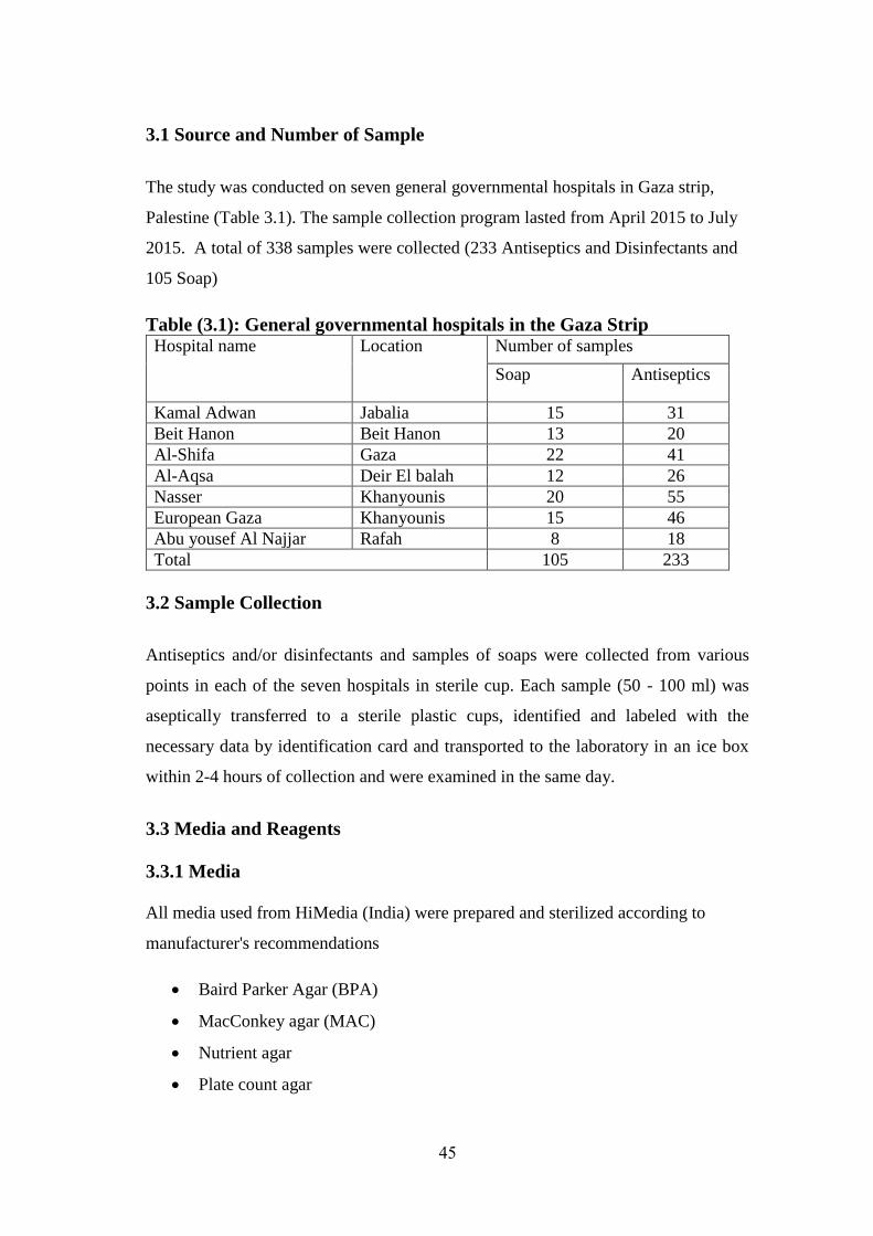
45
3.1 Source and Number of Sample
The study was conducted on seven general governmental hospitals in Gaza strip,
Palestine (Table 3.1). The sample collection program lasted from April 2015 to July
2015. A total of 338 samples were collected (233 Antiseptics and Disinfectants and
105 Soap)
Table (3.1): General governmental hospitals in the Gaza Strip Hospital name Location Number of samples
Soap Antiseptics
Kamal Adwan Jabalia 15 31
Beit Hanon Beit Hanon 13 20
Al-Shifa Gaza 22 41
Al-Aqsa Deir El balah 12 26
Nasser Khanyounis 20 55
European Gaza Khanyounis 15 46
Abu yousef Al Najjar Rafah 8 18
Total 105 233
3.2 Sample Collection
Antiseptics and/or disinfectants and samples of soaps were collected from various
points in each of the seven hospitals in sterile cup. Each sample (50 - 100 ml) was
aseptically transferred to a sterile plastic cups, identified and labeled with the
necessary data by identification card and transported to the laboratory in an ice box
within 2-4 hours of collection and were examined in the same day.
3.3 Media and Reagents
3.3.1 Media
All media used from HiMedia (India) were prepared and sterilized according to
manufacturer's recommendations
Baird Parker Agar (BPA)
MacConkey agar (MAC)
Nutrient agar
Plate count agar
46
Violet Red Bile Agar (VRBA)
Mueller Hinton Agar (MHA)
Rose Bengal Agar (RBA)
Cetrimide Agar (CA)
Mannitol Egg Yolk Polymyxin Agar (MYP) (Oxoid)
3.3.2 Reagents and Identification Systems
Analytical profile index (API) 20E (BioMerieux, France)
Catalase reagent (3% H2O2) (HiMedia, India)
Coagulase test (HiMedia, India)
Gram stain kit (HiMedia, India)
Oxidase test (Oxoid, UK)
Potassium iodide
Sodium thiosulfate
Starch solution
3.4 Equipment, Glassware and Disposables
Autoclave (Tuttnauer, USA)
Automatic pipettors and associated sterile pipette tips capable of delivering
up to 10 ml and 1 ml volumes
Balance (weights; 2000g capacity, sensitivity of 0.1 g) (Sartorius, Germany)
Colony counter (Anderman, England)
Incubators (Memert, Germany)
Light Microscope (Zeiss, Germany)
Refrigerator (Sanyo, USA)
Vortex mixer
Top pan balance capable of weighing to 0.1 g Cups (sterile, 100 ml)
(Firatmed, Turkey)
Flasks (sterile)
47
Inoculating loops
L-shaped glass rods (sterile)
Needles and Syringes (1.5 and 10 ml)
Petri dishes (90ˣ15) (Firatmed, Turkey)
Pipettes (sterile total delivery) 10 ml and 1 ml graduated in 0.1 ml volumes
(optional)
Spreaders (sterile, disposable)
3.5 Quality of Soap and Efficacy of Antiseptics and Disinfectants
Soap samples were tested for their microbiological quality while antiseptics and/or
disinfectants samples were tested for their microbiological quality and for their
chemical content.
3.5.1 Microbiological Quality of Soap
Using separate sterile pipettes, decimal dilutions of 10-2, 10-3, and 10-4 were prepared,
and more as appropriate, of soap by transferring 10 ml of previously diluted sample to
90 ml of diluents. All dilutions were shaken 25 times in 3 cm arc. One ml of each
dilution was pipetted into separate, duplicate, and appropriately marked Petri dishes
containing different types of media as shown in table 3.2. Dilution bottle was re-shaken
25 times in 3 cm arc if it stands more than 3 min before it is pipette into Petri dish. 0.1
ml of each dilution was transferred aseptically into the nutrient agar surface. The
diluted samples are distributed onto the surface of the specified plate by a sterile L-
shaped glass rod (spreader). The plates were incubated at 35oC for 24-48 h. Colonies
were counted and the total aerobic microorganisms was calculated per gram
(Andrews, 2001).
48
Table (3.2): Plated media and their purpose Media Microorganism growth
Plate count agar For total aerobic bacterial count
MacConkey agar selective isolation and differentiation
of Gram negative bacteria
Nutrient agar General media
Baird Parker Agar Staphylococci
Violet Red Bile Agar E. aerogenes, E. coli, Salmonella spp.
Dichloran Medium Base with Rose
Bengal
Fungi-(yeasts and molds count)
Cetrimide Agar Selective isolation of Pseudomonas
aeruginosa
Mannitol Egg Yolk Polymyxin Agar Bacillus cereus
3.5.2 Microbiological Efficacy of Antiseptics and Disinfectants
3.5.2.1 Microorganisms used
All antiseptics and disinfectant samples were tested for their microbiological efficacy
against three types of microorganisms those were E. coli, P. aeruginosa and S. aureus
(Clinical isolates from Al-Shifa hospital) were plated on Mueller Hinton Agar and
incubated in incubator for 24 hours of 37 oC.
3.5.2.2 Stainless steel cylinder method
It depends upon the diffusion of the Antiseptics and/or disinfectants from vertical steel
cylinders placed on the surface of inoculated agar medium. This procedure produces
zones of inhibition around the cylinder containing the tested Antiseptics and/or
disinfectants solution depending upon their types and concentration (Figure 3.1). This
method is commonly employed in the assay of pharmaceutical substance. For assay,
use of petri plates with 20 X 100 mm dimension and stainless steel cylinders with the
outside diameter 8 mm, inside diameter 6 mm and length 10 mm is recommended.
Were one cylinder was used per plate. The cylinder was placed on inoculated plates.
(Souza-Filho et al., 2008) The incubation the plates was done for 24 hours of 37 oC.
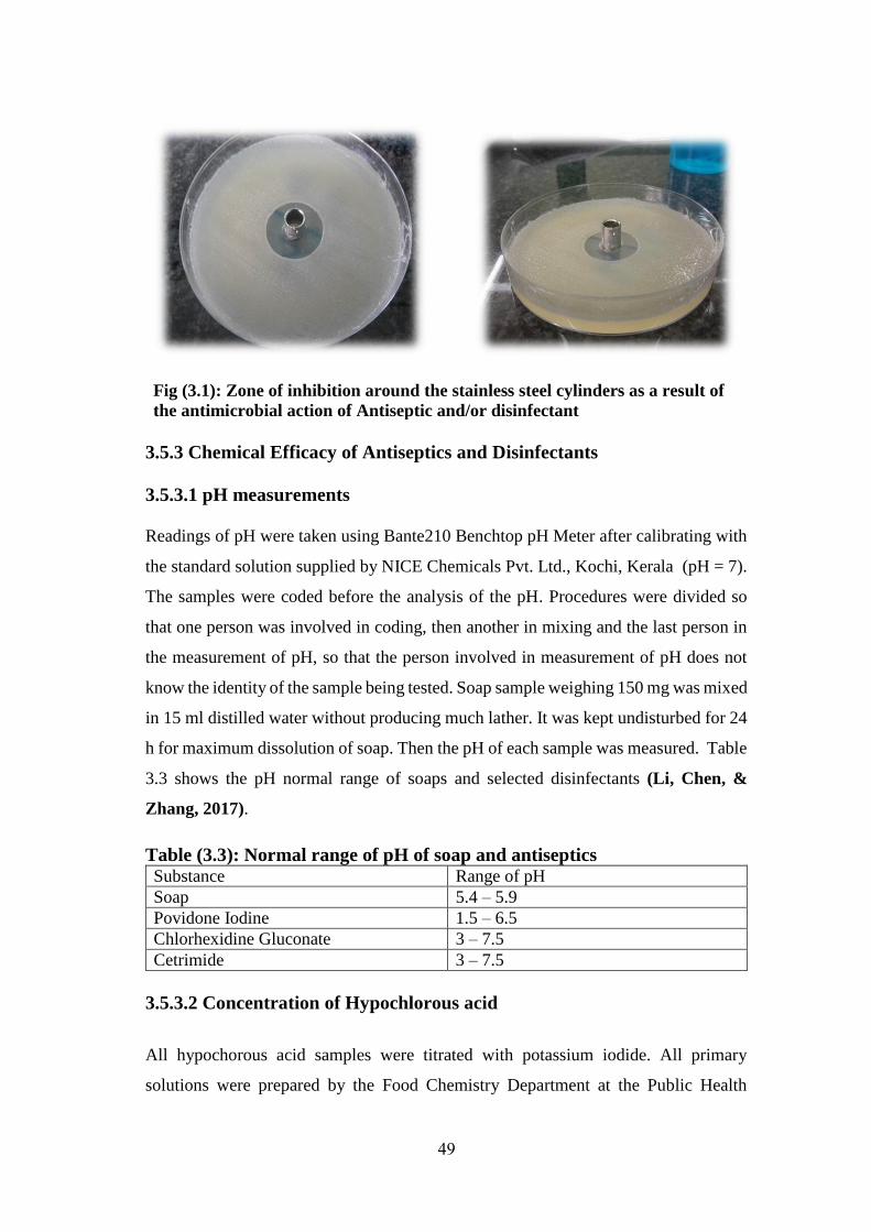
49
Fig (3.1): Zone of inhibition around the stainless steel cylinders as a result of
the antimicrobial action of Antiseptic and/or disinfectant
3.5.3 Chemical Efficacy of Antiseptics and Disinfectants
3.5.3.1 pH measurements
Readings of pH were taken using Bante210 Benchtop pH Meter after calibrating with
the standard solution supplied by NICE Chemicals Pvt. Ltd., Kochi, Kerala (pH = 7).
The samples were coded before the analysis of the pH. Procedures were divided so
that one person was involved in coding, then another in mixing and the last person in
the measurement of pH, so that the person involved in measurement of pH does not
know the identity of the sample being tested. Soap sample weighing 150 mg was mixed
in 15 ml distilled water without producing much lather. It was kept undisturbed for 24
h for maximum dissolution of soap. Then the pH of each sample was measured. Table
3.3 shows the pH normal range of soaps and selected disinfectants (Li, Chen, &
Zhang, 2017).
Table (3.3): Normal range of pH of soap and antiseptics Substance Range of pH
Soap 5.4 – 5.9
Povidone Iodine 1.5 – 6.5
Chlorhexidine Gluconate 3 – 7.5
Cetrimide 3 – 7.5
3.5.3.2 Concentration of Hypochlorous acid
All hypochorous acid samples were titrated with potassium iodide. All primary
solutions were prepared by the Food Chemistry Department at the Public Health
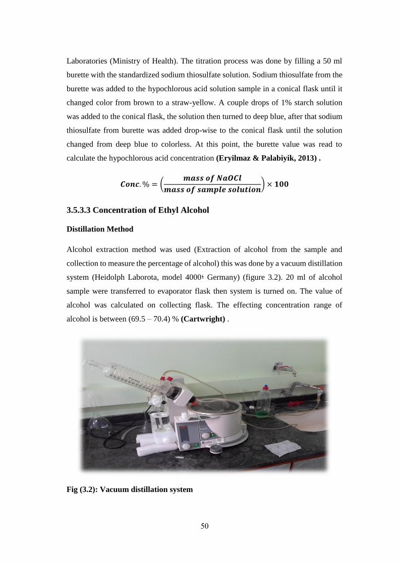
50
Laboratories (Ministry of Health). The titration process was done by filling a 50 ml
burette with the standardized sodium thiosulfate solution. Sodium thiosulfate from the
burette was added to the hypochlorous acid solution sample in a conical flask until it
changed color from brown to a straw-yellow. A couple drops of 1% starch solution
was added to the conical flask, the solution then turned to deep blue, after that sodium
thiosulfate from burette was added drop-wise to the conical flask until the solution
changed from deep blue to colorless. At this point, the burette value was read to
calculate the hypochlorous acid concentration (Eryilmaz & Palabiyik, 2013) .
𝑪𝒐𝒏𝒄. % = (𝒎𝒂𝒔𝒔 𝒐𝒇 𝑵𝒂𝑶𝑪𝒍
𝒎𝒂𝒔𝒔 𝒐𝒇 𝒔𝒂𝒎𝒑𝒍𝒆 𝒔𝒐𝒍𝒖𝒕𝒊𝒐𝒏) × 𝟏𝟎𝟎
3.5.3.3 Concentration of Ethyl Alcohol
Distillation Method
Alcohol extraction method was used (Extraction of alcohol from the sample and
collection to measure the percentage of alcohol) this was done by a vacuum distillation
system (Heidolph Laborota, model 4000; Germany) (figure 3.2). 20 ml of alcohol
sample were transferred to evaporator flask then system is turned on. The value of
alcohol was calculated on collecting flask. The effecting concentration range of
alcohol is between (69.5 – 70.4) % (Cartwright) .
Fig (3.2): Vacuum distillation system

51
3.5.3.4 Concentration of Povidone Iodine
Calculation the concentration of povidone iodine were done by titration with sodium
thiosulphite and all the primary solution were prepared by Drug Chemistry Department
at the Public Health Lab. The effective concentration range of Povidone Iodine is
between (85 - 120) % (Cartwright). 10 ml of sample was transferred to 250 ml flask
and 10 ml of 0.1 M HCl and sufficient distilled water were added to produce 150-ml
to fill 50 ml burette with 0.02 M sodium thiosulfate and titrated with sample. The end
point was determined by removing the color of povidone iodine (Cartwright) .
3.5.3.5 Concentration of Chlorhexidine Gluconate
This test was done by spectrophotometer at 254 nm in the Water Chemistry
Department at the Public Health Lab. The effective concentration range of
Chlorhexidine Glucanate is between (90-110) %. (Cartwright)
3.5.3.6 Concentration of Cetrimide: The effective concentration range of
Cetrimide is between (90 - 110) % (Cartwright). This test was not done because the
required reagents for the assay were not available in public health lab.
3.6 Data Analysis: All data obtained from the sample analysis were tabulated using
Microsoft Excel and then uploaded to SPSS v. 13 (Statistical Package for Social
Sciences). Chi square test was used for assessing the statistical significance of the data,
and p-values of 0.05 were considered significant.
52
Chapter 4
Results
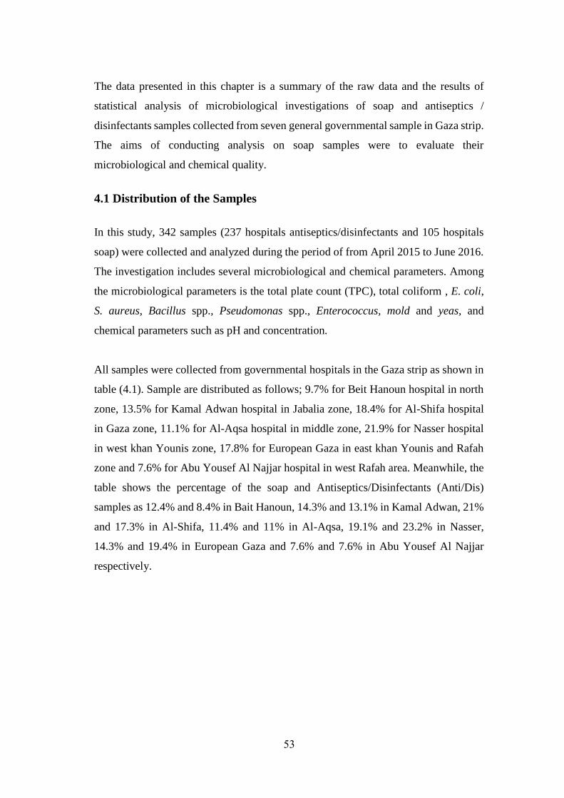
53
The data presented in this chapter is a summary of the raw data and the results of
statistical analysis of microbiological investigations of soap and antiseptics /
disinfectants samples collected from seven general governmental sample in Gaza strip.
The aims of conducting analysis on soap samples were to evaluate their
microbiological and chemical quality.
4.1 Distribution of the Samples
In this study, 342 samples (237 hospitals antiseptics/disinfectants and 105 hospitals
soap) were collected and analyzed during the period of from April 2015 to June 2016.
The investigation includes several microbiological and chemical parameters. Among
the microbiological parameters is the total plate count (TPC), total coliform , E. coli,
S. aureus, Bacillus spp., Pseudomonas spp., Enterococcus, mold and yeas, and
chemical parameters such as pH and concentration.
All samples were collected from governmental hospitals in the Gaza strip as shown in
table (4.1). Sample are distributed as follows; 9.7% for Beit Hanoun hospital in north
zone, 13.5% for Kamal Adwan hospital in Jabalia zone, 18.4% for Al-Shifa hospital
in Gaza zone, 11.1% for Al-Aqsa hospital in middle zone, 21.9% for Nasser hospital
in west khan Younis zone, 17.8% for European Gaza in east khan Younis and Rafah
zone and 7.6% for Abu Yousef Al Najjar hospital in west Rafah area. Meanwhile, the
table shows the percentage of the soap and Antiseptics/Disinfectants (Anti/Dis)
samples as 12.4% and 8.4% in Bait Hanoun, 14.3% and 13.1% in Kamal Adwan, 21%
and 17.3% in Al-Shifa, 11.4% and 11% in Al-Aqsa, 19.1% and 23.2% in Nasser,
14.3% and 19.4% in European Gaza and 7.6% and 7.6% in Abu Yousef Al Najjar
respectively.
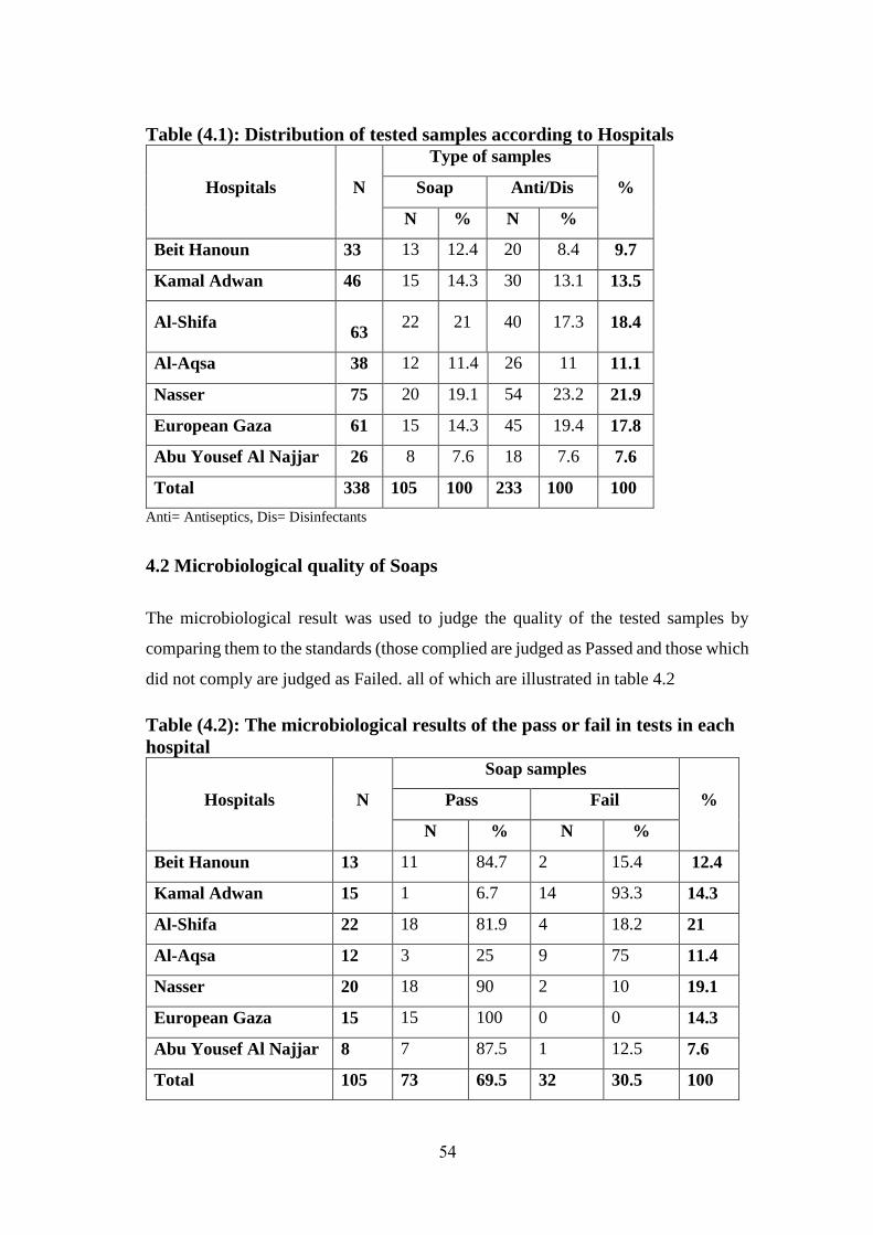
54
Table (4.1): Distribution of tested samples according to Hospitals
Hospitals N
Type of samples
% Soap Anti/Dis
N % N %
Beit Hanoun 33 13 12.4 20 8.4 9.7
Kamal Adwan 46 15 14.3 30 13.1 13.5
Al-Shifa
63 22 21 40 17.3 18.4
Al-Aqsa 38 12 11.4 26 11 11.1
Nasser 75 20 19.1 54 23.2 21.9
European Gaza 61 15 14.3 45 19.4 17.8
Abu Yousef Al Najjar 26 8 7.6 18 7.6 7.6
Total 338 105 100 233 100 100
Anti= Antiseptics, Dis= Disinfectants
4.2 Microbiological quality of Soaps
The microbiological result was used to judge the quality of the tested samples by
comparing them to the standards (those complied are judged as Passed and those which
did not comply are judged as Failed. all of which are illustrated in table 4.2
Table (4.2): The microbiological results of the pass or fail in tests in each
hospital
Hospitals N
Soap samples
% Pass Fail
N % N %
Beit Hanoun 13 11 84.7 2 15.4 12.4
Kamal Adwan 15 1 6.7 14 93.3 14.3
Al-Shifa 22 18 81.9 4 18.2 21
Al-Aqsa 12 3 25 9 75 11.4
Nasser 20 18 90 2 10 19.1
European Gaza 15 15 100 0 0 14.3
Abu Yousef Al Najjar 8 7 87.5 1 12.5 7.6
Total 105 73 69.5 32 30.5 100
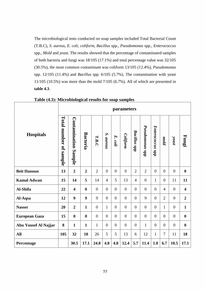
55
The microbiological tests conducted on soap samples included Total Bacterial Count
(T.B.C), S. aureus, E. coli, coliform, Bacillus spp., Pseudomonas spp., Enterococcus
spp., Mold and yeast. The results showed that the percentage of contaminated samples
of both bacteria and fungi was 18/105 (17.1%) and total percentage value was 32/105
(30.5%), the most common contaminant was coliform 13/105 (12.4%), Pseudomonas
spp. 12/105 (11.4%) and Bacillus spp. 6/105 (5.7%). The contamination with yeast
11/105 (10.5%) was more than the mold 7/105 (6.7%). All of which are presented in
table 4.3.
Table )4.3(: Microbiological results for soap samples
Hospitals
parameters
To
tal n
um
ber o
f sam
ple
Con
tam
inatio
n S
am
ple
Ba
cteria
T.B
.C
S. a
ureu
s
E. co
li
Colifo
rm
Bacillu
s spp
Pseu
dom
on
as sp
p
En
teroco
ccus sp
p
mold
yeast
Fu
ng
i
Beit Hanoun 13 2 2 2 0 0 0 2 2 0 0 0 0
Kamal Adwan 15 14 5 14 4 5 13 4 0 1 0 11 11
Al-Shifa 22 4 0 0 0 0 0 0 0 0 4 0 4
Al-Aqsa 12 9 9 9 0 0 0 0 9 0 2 0 2
Nasser 20 2 1 0 1 0 0 0 0 0 1 0 1
European Gaza 15 0 0 0 0 0 0 0 0 0 0 0 0
Abu Yousef Al Najjar 8 1 1 1 0 0 0 0 1 0 0 0 0
All 105 32 18 26 5 5 13 6 12 1 7 11 18
Percentage
30.5 17.1 24.8 4.8 4.8 12.4 5.7 11.4 1.0 6.7 10.5 17.1
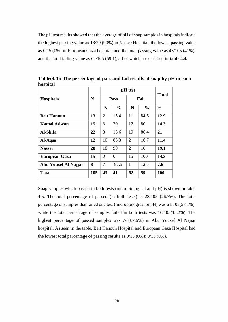
56
The pH test results showed that the average of pH of soap samples in hospitals indicate
the highest passing value as 18/20 (90%) in Nasser Hospital, the lowest passing value
as 0/15 (0%) in European Gaza hospital, and the total passing value as 43/105 (41%),
and the total failing value as 62/105 (59.1), all of which are clarified in table 4.4.
Table)4.4(: The percentage of pass and fail results of soap by pH in each
hospital
Hospitals N
pH test Total
Pass Fail
N % N % %
Beit Hanoun 13 2 15.4 11 84.6 12.9
Kamal Adwan 15 3 20 12 80 14.3
Al-Shifa 22 3 13.6 19 86.4 21
Al-Aqsa 12 10 83.3 2 16.7 11.4
Nasser 20 18 90 2 10 19.1
European Gaza 15 0 0 15 100 14.3
Abu Yousef Al Najjar 8 7 87.5 1 12.5 7.6
Total 105 43 41 62 59 100
Soap samples which passed in both tests (microbiological and pH) is shown in table
4.5. The total percentage of passed (in both tests) is 28/105 (26.7%). The total
percentage of samples that failed one test (microbiological or pH) was 61/105(58.1%),
while the total percentage of samples failed in both tests was 16/105(15.2%). The
highest percentage of passed samples was 7/8(87.5%) in Abu Yousef Al Najjar
hospital. As seen in the table, Beit Hanoun Hospital and European Gaza Hospital had
the lowest total percentage of passing results as 0/13 (0%); 0/15 (0%).

57
Table )4.5(: The percentage of pass results in microbiological and pH tests
in each hospital
Hospitals N
Total
Pass Fail one Failed
both
N % N % N %
Beit Hanoun 13 0 0 13 100 0 0
Kamal Adwan 15 1 6.7 2 13.3 12 80
Al-Shifa 22 2 9.1 17 77.3 3 13.7
Al-Aqsa 12 1 8.3 11 91.7 0 0
Nasser 20 17 85 2 10 1 5
European Gaza 15 0 0 15 100 0 0
Abu Yousef Al Najjar 8 7 87.5 2 25 0 0
Total 105 28 26.7 61 58.1 16 15.2
4.3 Antiseptics/Disinfectants Tests
The five most common chemical compound used as Antiseptics/Disinfectants in
Palestinian authority hospitals in Gaza-Palestine is Povidone Iodine (with different
brand names; BETADINE 10%, BETAVIDINE 10%, BETODINE 10%, IODOCARE
10%, POTEDENE 10%, POVIOFIX 10%, SOMEDINE 10%, IODIFLOR 7.5% and
POVDINE 7.5%), Alcohol compound (Alcohol and Ethyl Alcohol), Chlorhexidine
(CHX) with different brand names (Cetrimide BP and SEPTAL, SAVIOR, SUNDEN
and CAR FIRST AID) and Hypochlorous acid locally manufactured known as
(Chlorine). (Table 4.6)
Table )4.6(: Number and percentage of Antiseptics/Disinfectants collected
from all hospitals
Antiseptics/Disinfectants Frequency Percentage
Povidone Iodine 85 35. 9
Alcohol 60 25. 3
Chlorhexidine 59 24.9
Chlorine 29 12.2
Total 233 100
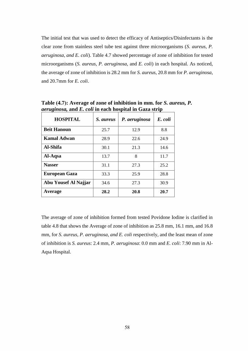
58
The initial test that was used to detect the efficacy of Antiseptics/Disinfectants is the
clear zone from stainless steel tube test against three microorganisms (S. aureus, P.
aeruginosa, and E. coli). Table 4.7 showed percentage of zone of inhibition for tested
microorganisms (S. aureus, P. aeruginosa, and E. coli) in each hospital. As noticed,
the average of zone of inhibition is 28.2 mm for S. aureus, 20.8 mm for P. aeruginosa,
and 20.7mm for E. coli.
Table )4.7(: Average of zone of inhibition in mm. for S. aureus, P.
aeruginosa, and E. coli in each hospital in Gaza strip
HOSPITAL S. aureus P. aeruginosa E. coli
Beit Hanoun 25.7 12.9 8.8
Kamal Adwan 28.9 22.6 24.9
Al-Shifa 30.1 21.3 14.6
Al-Aqsa 13.7 8 11.7
Nasser 31.1 27.3 25.2
European Gaza 33.3 25.9 28.8
Abu Yousef Al Najjar 34.6 27.3 30.9
Average 28.2 20.8 20.7
The average of zone of inhibition formed from tested Povidone Iodine is clarified in
table 4.8 that shows the Average of zone of inhibition as 25.8 mm, 16.1 mm, and 16.8
mm, for S. aureus, P. aeruginosa, and E. coli respectively, and the least mean of zone
of inhibition is S. aureus: 2.4 mm, P. aeruginosa: 0.0 mm and E. coli: 7.90 mm in Al-
Aqsa Hospital.
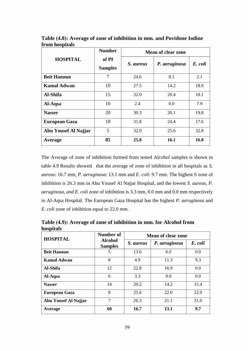
59
Table )4.8(: Average of zone of inhibition in mm. and Povidone Iodine
from hospitals
HOSPITAL
Number
of PI
Samples
Mean of clear zone
S. aureus P. aeruginosa E. coli
Beit Hanoun 7 24.6 8.1 2.1
Kamal Adwan 10 27.5 14.2 18.9
Al-Shifa 15 32.0 20.4 18.1
Al-Aqsa 10 2.4 0.0 7.9
Nasser 20 30.3 20.1 19.8
European Gaza 18 31.8 24.4 17.6
Abu Yousef Al Najjar 5 32.0 25.6 32.8
Average 85 25.8 16.1 16.8
The Average of zone of inhibition formed from tested Alcohol samples is shown in
table 4.9 Results showed that the average of zone of inhibition in all hospitals as S.
aureus: 16.7 mm, P. aeruginosa: 13.1 mm and E. coli: 9.7 mm. The highest S zone of
inhibition is 26.3 mm in Abu Yousef Al Najjar Hospital, and the lowest S. aureus, P.
aeruginosa, and E. coli zone of inhibition is 3.3 mm, 0.0 mm and 0.0 mm respectively
in Al-Aqsa Hospital. The European Gaza Hospital has the highest P. aeruginosa and
E. coli zone of inhibition equal to 22.0 mm.
Table )4.9(: Average of zone of inhibition in mm. for Alcohol from
hospitals
HOSPITAL Number of
Alcohol
Samples
Mean of clear zone
S. aureus P. aeruginosa E. coli
Beit Hanoun 5 13.6 6.0 0.0
Kamal Adwan 8 4.9 11.3 9.3
Al-Shifa 12 22.8 16.9 0.0
Al-Aqsa 6 3.3 0.0 0.0
Nasser 14 20.2 14.2 15.4
European Gaza 8 25.6 22.0 22.0
Abu Yousef Al Najjar 7 26.3 21.1 21.0
Average 60 16.7 13.1 9.7
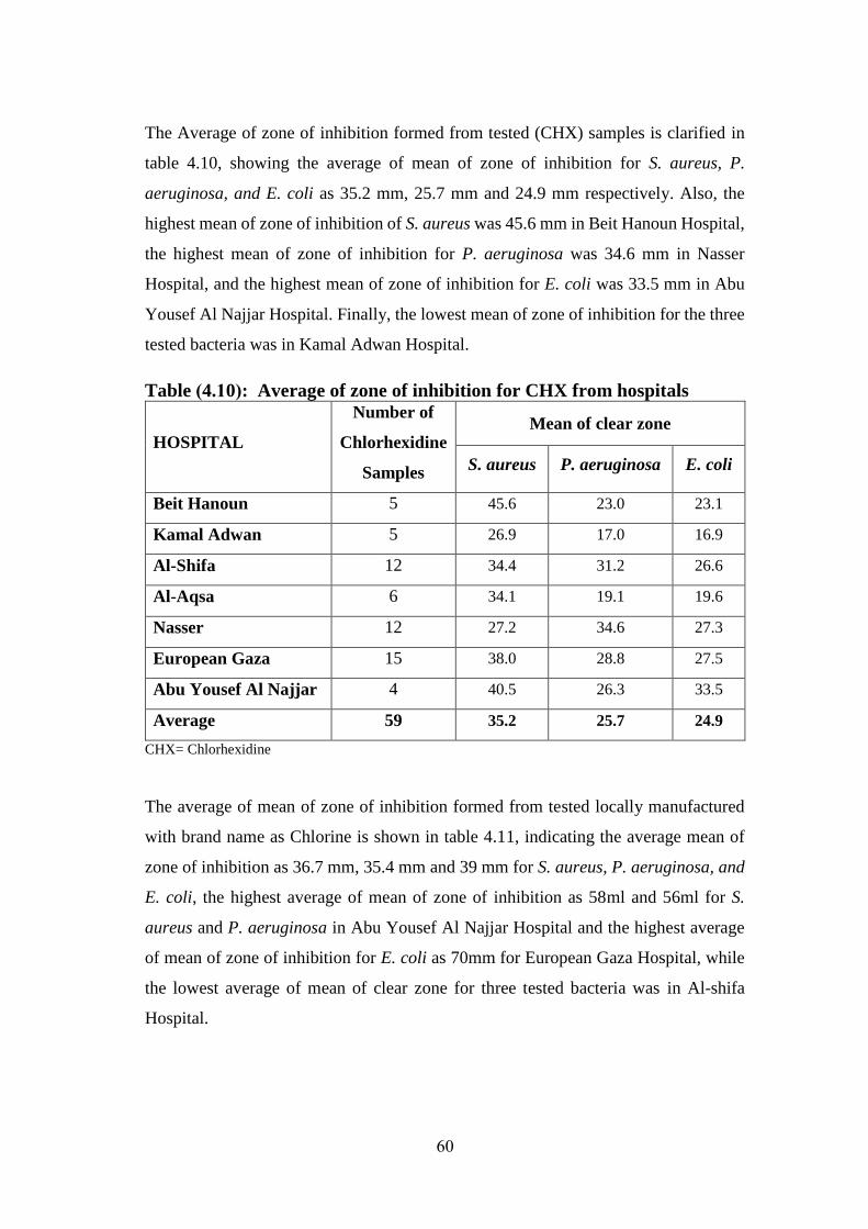
60
The Average of zone of inhibition formed from tested (CHX) samples is clarified in
table 4.10, showing the average of mean of zone of inhibition for S. aureus, P.
aeruginosa, and E. coli as 35.2 mm, 25.7 mm and 24.9 mm respectively. Also, the
highest mean of zone of inhibition of S. aureus was 45.6 mm in Beit Hanoun Hospital,
the highest mean of zone of inhibition for P. aeruginosa was 34.6 mm in Nasser
Hospital, and the highest mean of zone of inhibition for E. coli was 33.5 mm in Abu
Yousef Al Najjar Hospital. Finally, the lowest mean of zone of inhibition for the three
tested bacteria was in Kamal Adwan Hospital.
Table )4.10(: Average of zone of inhibition for CHX from hospitals
HOSPITAL
Number of
Chlorhexidine
Samples
Mean of clear zone
S. aureus P. aeruginosa E. coli
Beit Hanoun 5 45.6 23.0 23.1
Kamal Adwan 5 26.9 17.0 16.9
Al-Shifa 12 34.4 31.2 26.6
Al-Aqsa 6 34.1 19.1 19.6
Nasser 12 27.2 34.6 27.3
European Gaza 15 38.0 28.8 27.5
Abu Yousef Al Najjar 4 40.5 26.3 33.5
Average 59 35.2 25.7 24.9
CHX= Chlorhexidine
The average of mean of zone of inhibition formed from tested locally manufactured
with brand name as Chlorine is shown in table 4.11, indicating the average mean of
zone of inhibition as 36.7 mm, 35.4 mm and 39 mm for S. aureus, P. aeruginosa, and
E. coli, the highest average of mean of zone of inhibition as 58ml and 56ml for S.
aureus and P. aeruginosa in Abu Yousef Al Najjar Hospital and the highest average
of mean of zone of inhibition for E. coli as 70mm for European Gaza Hospital, while
the lowest average of mean of clear zone for three tested bacteria was in Al-shifa
Hospital.
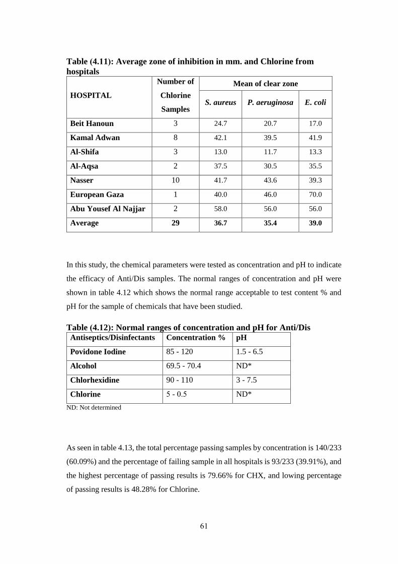
61
Table )4.11(: Average zone of inhibition in mm. and Chlorine from
hospitals
HOSPITAL
Number of
Chlorine
Samples
Mean of clear zone
S. aureus P. aeruginosa E. coli
Beit Hanoun 3 24.7 20.7 17.0
Kamal Adwan 8 42.1 39.5 41.9
Al-Shifa 3 13.0 11.7 13.3
Al-Aqsa 2 37.5 30.5 35.5
Nasser 10 41.7 43.6 39.3
European Gaza 1 40.0 46.0 70.0
Abu Yousef Al Najjar 2 58.0 56.0 56.0
Average 29 36.7 35.4 39.0
In this study, the chemical parameters were tested as concentration and pH to indicate
the efficacy of Anti/Dis samples. The normal ranges of concentration and pH were
shown in table 4.12 which shows the normal range acceptable to test content % and
pH for the sample of chemicals that have been studied.
Table )4.12(: Normal ranges of concentration and pH for Anti/Dis
Antiseptics/Disinfectants Concentration % pH
Povidone Iodine 85 - 120 1.5 - 6.5
Alcohol 69.5 - 70.4 ND*
Chlorhexidine 90 - 110 3 - 7.5
Chlorine 0.5 - 5 ND*
ND: Not determined
As seen in table 4.13, the total percentage passing samples by concentration is 140/233
(60.09%) and the percentage of failing sample in all hospitals is 93/233 (39.91%), and
the highest percentage of passing results is 79.66% for CHX, and lowing percentage
of passing results is 48.28% for Chlorine.
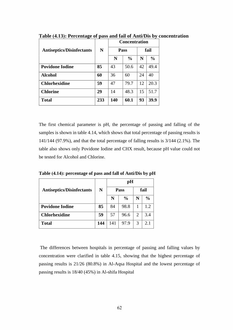
62
Table )4.13(: Percentage of pass and fail of Anti/Dis by concentration
Antiseptics/Disinfectants N
Concentration
Pass fail
N % N %
Povidone Iodine 85 43 50.6 42 49.4
Alcohol 60 36 60 24 40
Chlorhexidine 59 47 79.7 12 20.3
Chlorine 29 14 48.3 15 51.7
Total 233 140 60.1 93 39.9
The first chemical parameter is pH, the percentage of passing and falling of the
samples is shown in table 4.14, which shows that total percentage of passing results is
141/144 (97.9%), and that the total percentage of falling results is 3/144 (2.1%). The
table also shows only Povidone Iodine and CHX result, because pH value could not
be tested for Alcohol and Chlorine.
Table )4.14(: percentage of pass and fall of Anti/Dis by pH
Antiseptics/Disinfectants N
pH
Pass fail
N % N %
Povidone Iodine 85 84 98.8 1 1.2
Chlorhexidine 59 57 96.6 2 3.4
Total 144 141 97.9 3 2.1
The differences between hospitals in percentage of passing and falling values by
concentration were clarified in table 4.15, showing that the highest percentage of
passing results is 21/26 (80.8%) in Al-Aqsa Hospital and the lowest percentage of
passing results is 18/40 (45%) in Al-shifa Hospital
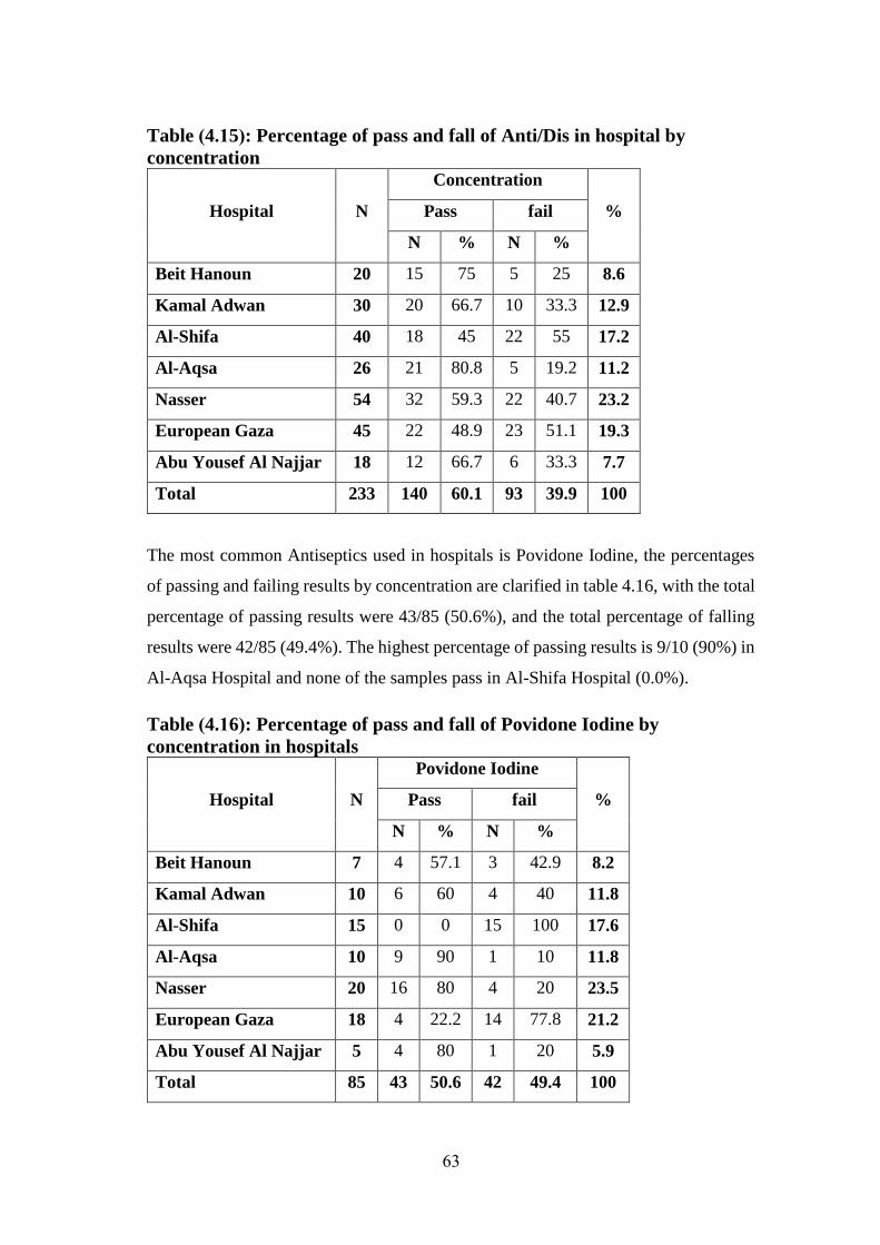
63
Table )4.15(: Percentage of pass and fall of Anti/Dis in hospital by
concentration
Hospital N
Concentration
% Pass fail
N % N %
Beit Hanoun 20 15 75 5 25 8.6
Kamal Adwan 30 20 66.7 10 33.3 12.9
Al-Shifa 40 18 45 22 55 17.2
Al-Aqsa 26 21 80.8 5 19.2 11.2
Nasser 54 32 59.3 22 40.7 23.2
European Gaza 45 22 48.9 23 51.1 19.3
Abu Yousef Al Najjar 18 12 66.7 6 33.3 7.7
Total 233 140 60.1 93 39.9 100
The most common Antiseptics used in hospitals is Povidone Iodine, the percentages
of passing and failing results by concentration are clarified in table 4.16, with the total
percentage of passing results were 43/85 (50.6%), and the total percentage of falling
results were 42/85 (49.4%). The highest percentage of passing results is 9/10 (90%) in
Al-Aqsa Hospital and none of the samples pass in Al-Shifa Hospital (0.0%).
Table )4.16(: Percentage of pass and fall of Povidone Iodine by
concentration in hospitals
Hospital N
Povidone Iodine
% Pass fail
N % N %
Beit Hanoun 7 4 57.1 3 42.9 8.2
Kamal Adwan 10 6 60 4 40 11.8
Al-Shifa 15 0 0 15 100 17.6
Al-Aqsa 10 9 90 1 10 11.8
Nasser 20 16 80 4 20 23.5
European Gaza 18 4 22.2 14 77.8 21.2
Abu Yousef Al Najjar 5 4 80 1 20 5.9
Total 85 43 50.6 42 49.4 100
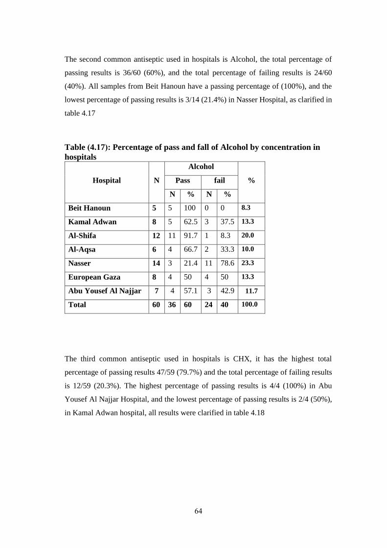
64
The second common antiseptic used in hospitals is Alcohol, the total percentage of
passing results is 36/60 (60%), and the total percentage of failing results is 24/60
(40%). All samples from Beit Hanoun have a passing percentage of (100%), and the
lowest percentage of passing results is 3/14 (21.4%) in Nasser Hospital, as clarified in
table 4.17
Table )4.17(: Percentage of pass and fall of Alcohol by concentration in
hospitals
Hospital N
Alcohol
% Pass fail
N % N %
Beit Hanoun 5 5 100 0 0 8.3
Kamal Adwan 8 5 62.5 3 37.5 13.3
Al-Shifa 12 11 91.7 1 8.3 20.0
Al-Aqsa 6 4 66.7 2 33.3 10.0
Nasser 14 3 21.4 11 78.6 23.3
European Gaza 8 4 50 4 50 13.3
Abu Yousef Al Najjar 7 4 57.1 3 42.9 11.7
Total 60 36 60 24 40 100.0
The third common antiseptic used in hospitals is CHX, it has the highest total
percentage of passing results 47/59 (79.7%) and the total percentage of failing results
is 12/59 (20.3%). The highest percentage of passing results is 4/4 (100%) in Abu
Yousef Al Najjar Hospital, and the lowest percentage of passing results is 2/4 (50%),
in Kamal Adwan hospital, all results were clarified in table 4.18
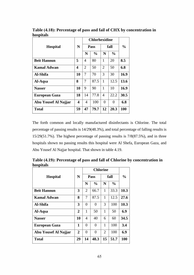
65
Table )4.18(: Percentage of pass and fail of CHX by concentration in
hospitals
Hospital N
Chlorhexidine
% Pass fall
N % N %
Beit Hanoun 5 4 80 1 20 8.5
Kamal Adwan 4 2 50 2 50 6.8
Al-Shifa 10 7 70 3 30 16.9
Al-Aqsa 8 7 87.5 1 12.5 13.6
Nasser 10 9 90 1 10 16.9
European Gaza 18 14 77.8 4 22.2 30.5
Abu Yousef Al Najjar 4 4 100 0 0 6.8
Total 59 47 79.7 12 20.3 100
The forth common and locally manufactured disinfectants is Chlorine. The total
percentage of passing results is 14/29(48.3%), and total percentage of falling results is
15/29(51.7%). The highest percentage of passing results is 7/8(87.5%), and in three
hospitals shown no passing results this hospital were Al Shefa, European Gaza, and
Abu Yousef Al Najjar hospital. That shown in table 4.19.
Table )4.19(: Percentage of pass and fall of Chlorine by concentration in
hospitals
Hospital N
Chlorine
% Pass fall
N % N %
Beit Hanoun 3 2 66.7 1 33.3 10.3
Kamal Adwan 8 7 87.5 1 12.5 27.6
Al-Shifa 3 0 0 3 100 10.3
Al-Aqsa 2 1 50 1 50 6.9
Nasser 10 4 40 6 60 34.5
European Gaza 1 0 0 1 100 3.4
Abu Yousef Al Najjar 2 0 0 2 100 6.9
Total 29 14 48.3 15 51.7 100
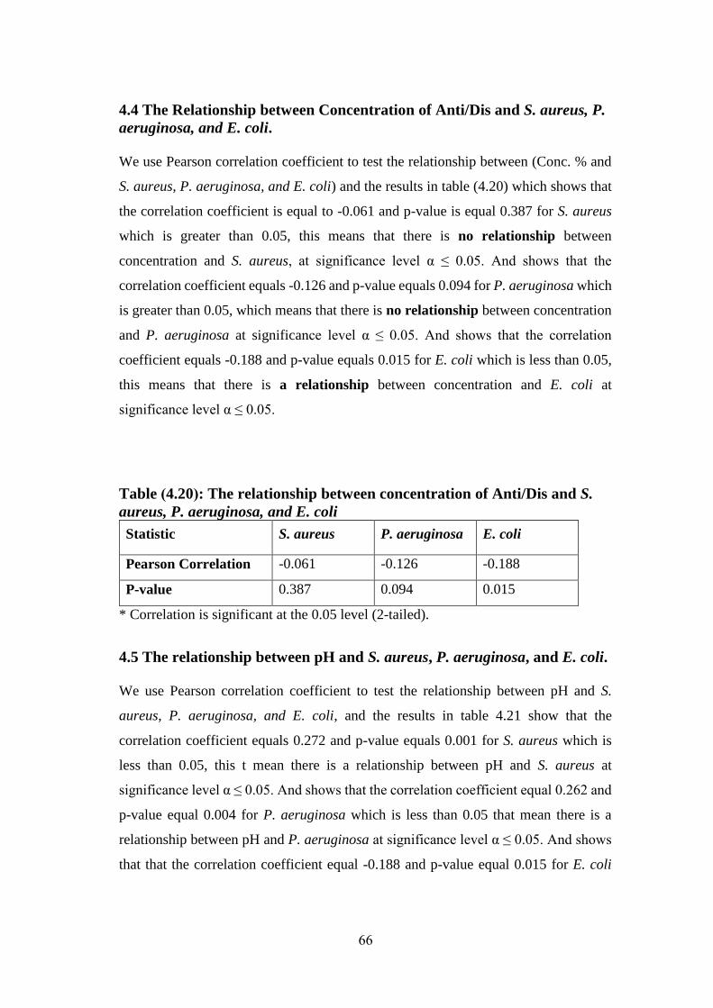
66
4.4 The Relationship between Concentration of Anti/Dis and S. aureus, P.
aeruginosa, and E. coli.
We use Pearson correlation coefficient to test the relationship between (Conc. % and
S. aureus, P. aeruginosa, and E. coli) and the results in table (4.20) which shows that
the correlation coefficient is equal to -0.061 and p-value is equal 0.387 for S. aureus
which is greater than 0.05, this means that there is no relationship between
concentration and S. aureus, at significance level α ≤ 0.05. And shows that the
correlation coefficient equals -0.126 and p-value equals 0.094 for P. aeruginosa which
is greater than 0.05, which means that there is no relationship between concentration
and P. aeruginosa at significance level α ≤ 0.05. And shows that the correlation
coefficient equals -0.188 and p-value equals 0.015 for E. coli which is less than 0.05,
this means that there is a relationship between concentration and E. coli at
significance level α ≤ 0.05.
Table )4.20(: The relationship between concentration of Anti/Dis and S.
aureus, P. aeruginosa, and E. coli
Statistic S. aureus P. aeruginosa E. coli
Pearson Correlation -0.061 -0.126 -0.188
P-value 0.387 0.094 0.015
* Correlation is significant at the 0.05 level (2-tailed).
4.5 The relationship between pH and S. aureus, P. aeruginosa, and E. coli.
We use Pearson correlation coefficient to test the relationship between pH and S.
aureus, P. aeruginosa, and E. coli, and the results in table 4.21 show that the
correlation coefficient equals 0.272 and p-value equals 0.001 for S. aureus which is
less than 0.05, this t mean there is a relationship between pH and S. aureus at
significance level α ≤ 0.05. And shows that the correlation coefficient equal 0.262 and
p-value equal 0.004 for P. aeruginosa which is less than 0.05 that mean there is a
relationship between pH and P. aeruginosa at significance level α ≤ 0.05. And shows
that that the correlation coefficient equal -0.188 and p-value equal 0.015 for E. coli
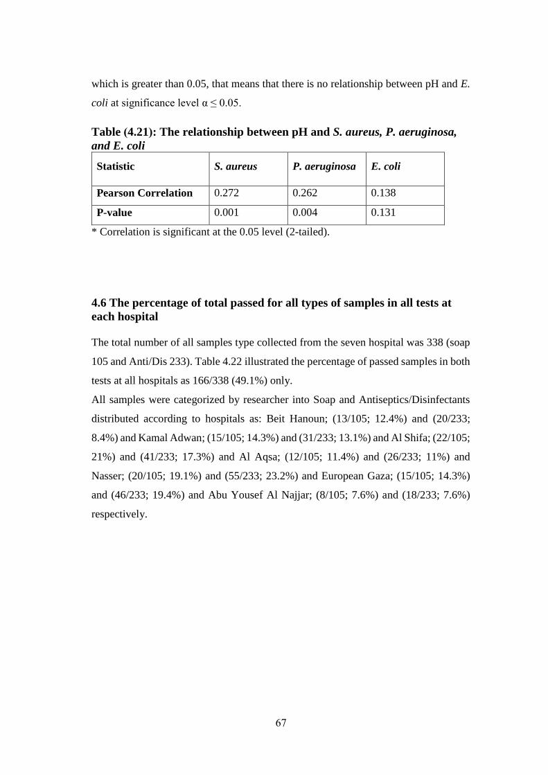
67
which is greater than 0.05, that means that there is no relationship between pH and E.
coli at significance level α ≤ 0.05.
Table )4.21(: The relationship between pH and S. aureus, P. aeruginosa,
and E. coli
Statistic S. aureus P. aeruginosa E. coli
Pearson Correlation 0.272 0.262 0.138
P-value 0.001 0.004 0.131
* Correlation is significant at the 0.05 level (2-tailed).
4.6 The percentage of total passed for all types of samples in all tests at
each hospital
The total number of all samples type collected from the seven hospital was 338 (soap
105 and Anti/Dis 233). Table 4.22 illustrated the percentage of passed samples in both
tests at all hospitals as 166/338 (49.1%) only.
All samples were categorized by researcher into Soap and Antiseptics/Disinfectants
distributed according to hospitals as: Beit Hanoun; (13/105; 12.4%) and (20/233;
8.4%) and Kamal Adwan; (15/105; 14.3%) and (31/233; 13.1%) and Al Shifa; (22/105;
21%) and (41/233; 17.3%) and Al Aqsa; (12/105; 11.4%) and (26/233; 11%) and
Nasser; (20/105; 19.1%) and (55/233; 23.2%) and European Gaza; (15/105; 14.3%)
and (46/233; 19.4%) and Abu Yousef Al Najjar; (8/105; 7.6%) and (18/233; 7.6%)
respectively.
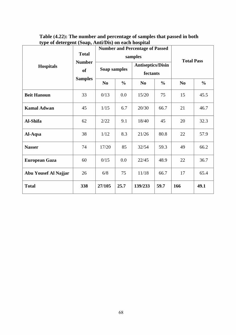
68
Table )4.22(: The number and percentage of samples that passed in both
type of detergent (Soap, Anti/Dis) on each hospital
Hospitals
Total
Number
of
Samples
Number and Percentage of Passed
samples Total Pass
Soap samples Antiseptics/Disin
fectants
No % No % No %
Beit Hanoun 33 0/13 0.0 15/20 75 15 45.5
Kamal Adwan 45 1/15 6.7 20/30 66.7 21 46.7
Al-Shifa 62 2/22 9.1 18/40 45 20 32.3
Al-Aqsa 38 1/12 8.3 21/26 80.8 22 57.9
Nasser 74 17/20 85 32/54 59.3 49 66.2
European Gaza 60 0/15 0.0 22/45 48.9 22 36.7
Abu Yousef Al Najjar 26 6/8 75 11/18 66.7 17 65.4
Total 338 27/105 25.7 139/233 59.7 166 49.1
69
Chapter 5
DISCUSSION
70
In this study we evaluated the microbiological (bacteria and fungi) quality of
antiseptics and soaps samples, measured the efficacy of antiseptics/disinfectants, and
determined the chemical concentration of antiseptics, in seven general hospitals in
Gaza-Palestine, There is no published data on this issue. Therefore, results obtained in
the present study could not be compared with any previous local data.
Samples (338 samples divided to 105 Soap samples and 233 Antiseptics/Disinfectants
samples) were collected from seven general governmental hospitals in Gaza strip,
distributed over the five governorates in Gaza strip: North (Beit Hanoun and Kamal
Adwan hospital), Gaza (Al Shifa hospital), Midzone (Al Aqsa hospital), Khan Younis
(Nasser and European Gaza hospital), Rafah zone (Abu Yousef Al Najjar). All of them,
are operated by the ministry of health of the Palestinian Authority. Only liquid soap
were examined in this study (which is the available form) and
Antiseptics/Disinfectants including Alcohol, Povidone Iodine, Chlorhexdine and
Chlorine.
The microbiological parameters which were examined for soap samples in this study
are Total Bacterial Count (T.B.C), Staphylococcus aureus, Escherichia coli, coliform,
Bacillus spp, Pseudomonas spp, Enterococcus and Mold and yeast. While Anti/Dis
samples were evaluated for their antibacterial activities against S. aureus,
Pseudomonas spp., and E. coli. In addition, the pH of soap and Anti/Dis was also
determined. The chemical concentration of Ani/Dis was determined chemically.
5.1 The percentage of soap and Anti/Dis based on various microbiological
and chemical tests in seven hospitals
The axioms anyone think that any material used in hospitals must be completely sterile,
because it directly affect human’s life and public health, espically the material used
for that purpose as soap, antiseptics and disinfectants. This study showed different
results. The total soap percentage that complied with the standards in the largest and
most popular seven governmental general hospitals in Gaza strip – Palestine was only
27/105 (25.7%). And this is lower than those found in many studies as in Iraq (41%)
71
(S. M. Zeiny, 2009), in Brazil (44.7%) (Joselany Afio Caetano, Lima, Miranda,
Serufo, & Ponte, 2011), and in USA (75.2%) (M. Chattman, S. Gerba, & C.
Maxwell, 2011). The most common cause of contamination was Pseudomonas spp as
shown in an Iraqi study (S. M. Zeiny, 2009) and in Brazilian study (Joselany Afio
Caetano et al., 2011) but USA study show the Klebsiella spp was the most frequent
organisms instead of Pseudomonas spp (Marisa Chattman et al., 2011). The total
Anti/Dis percentage of passed results in those hospitals was 100% with regard to the
microbiological quality. This is unlike the study in Republic of Trinidad and Tobago
hospitals shown 169/180 (93.9%) the 11 contaminated samples contamination by
Pseudomonas spp (Gajadhar, Lara, Sealy, & Adesiyun, 2003) and unlike another
study in Gondar university hospital in Ethiopia which showed 83/86 (96.5%) and the
3 contaminated samples with Klebsiella spp (Deress, Girma, Birhan, Biadgo, &
Alemu, 2014).
The average of zone of inhibition for S. aureus, P. aeruginosa, and E. coli were 29
mm, 22.1 mm, 21.7 mm respectively. This is in accordance with many studies that
showed the gram-negative bacteria tend to be more resistant than gram-positive
organisms as staphylococci (Billeter, Levy, Chomel, & Breitschwerdt, 2008). An in
Indian study showed the E. coli as the most resistant organisms followed by P.
aeroginosa. While, S. aureus was the least resistant organisms (Bhat, Prajna,
Menezez, & Shetty, 2011).
5.2 Distribution of tested samples according to hospital
Highest percentage of collected samples was from Nasser 21.9%, followed by the
largest hospital "Al-Shifa hospital" with percentage of 18.4% because some
departments in Al-Shifa did not possess some types of Antiseptics/Disinfectants.

72
5.3 The percentage of pass and fail results of soap in hospitals
There are two parameter (microbiological test, pH test) used to judge sample
compliance. For any sample to pass it should pass in both tests, and the results by
hospital were:
5.3.1 Beit Hanoun: total number of soap sample was 13, no one pass in both tests, and
all sample fail on one of test (2 in micro, 11 in pH). That mean the percentage of fail
was 100%. The most common microbes found in the two contaminated sample were
Bacillus spp and Pseudomonas spp. It is of a significance finding that all 11 sample
that pass in the microbiological test failed in pH test which means that the pH has
extreme pH has prevented bacteria from growing or surviving. It is also worth noting
that all tested soap samples are locally manufactured under no supervision.
5.3.2 Kamal Adwan: Out of the 15 soap samples, only one sample passed both test.
Two sampled failed in the microbiological test. Coliform, yeast, E. coli, S. aureus,
Enterococcus spp were recovered from contaminated samples. The total percentage of
failure was 93.3% that was highest value among all hospitals. All tested soap samples
are locally manufactured under no supervision using refillable bottles.
5.3.3 Al-Shifa: 22 soap samples were collected. Only two from them passed both test.
17 samples failed in one test (1 in microbiology test, 16 in pH test), and three samples
failed in both tests. Molds contaminated four of the failed samples. The total
percentage of failure was 91% (the third highest value between hospitals). The same
explanation as in the above section applies.
5.3.4 Al-Aqsa: 12 soap samples were collected, only one sample passed both tests,
and other 11 samples failed in one test (9 in microbiological test, 2 in pH test). Sample
passing in pH test failed in microbiological test. Total percentage of pass was 91.7%
(the second highest failure value among hospitals).
5.3.5 Nasser: from the 20 soap samples collected, 17 samples passed both tests that
is highest pass value, tow sample fail in one test ( 1 in microbiological test, 1 in pH
73
test), and only one sample was failed in both test, two sample fail in microbiological
test contaminated with mold and S. aureus.
5.3.6 European Gaza: all 15 collected samples failed in one test (pH test).
Microbiologically, all samples did not show any growth for the obvious reason of
having extreme pH values which interfere with microbial growth.
5.3.7 Abu Yousef Al Najjar: 6 out 8 of the collected sample passed both test and other
two sample failed in one test. One of two failed in microbiological test by
contamination with Pseudomonas spp and other one failed in pH test. Unsupervised
production and storage of local soap may be the reason for this failure.
5.4 Soap
The total failing value as 32/105 (30.5%), these results are lower than the Brazilian
study (55.9%) (Joselany Afio Caetano et al., 2011), and higher than the Iraqi study
(15.9%) (S. Zeiny, 2009),and USA study (24.8%) (Chattman & Maxwell, 2011). And
the most microorganisms cause contamination are coliform (12.4%), then
Pseudomonas spp (11.4%), and yeast (10.5%). Unlike Brazilian study the most
microorganisms cause contamination for fifty-nine liquid soap are Burkholderia
cepacia (4.6%), Pseudomonas spp (4%) and Klebsiella (1%)(Joselany Afio Caetano
et al., 2011). But in the Iraqi study are (66.6%) Pseudomonas, (16.6%) Proteus, and
(16.6%) Flavimonas(S. Zeiny, 2009). And in USA study the most frequency of
detection are Klebsiella oxytoca (28.6%), Klebsiella pneumonia (27.6%), and
Enterobacter aerogenes (12.4%)(Chattman & Maxwell, 2011). And the total failed
value by pH is (59.1%). I did not found any study test pH of soap in hospitals that’s
May because in Gaza hospitals used local manufacture soap, without any scientific
basis and used highly amounts of high concentration acid or high concentration base
to bass in microbiological test. That was very clear from form of some sample of liquid
soap.
74
5.5 Antiseptics and Disinfectants
5.5.1 Microbiological test
All antiseptics and disinfectants (Anti/Dis) samples (233) collected has passed the
microbiological test, unlike that reportedly in USA study particularly in North Carolina
which reported many type of bacteria contaminating several types of Anti/Dis.
Bacillus cereus and Burkholderia cepacia were found to contaminate Alcohols.
Pseudomonas spp., Flavobacterium sp., Ralstonia pickettii, Serratia marcescens and
Achromobacter xylosoxidans were detected in Chlorhexidine, Burkholderia cepacia
and Pseudomonas aeruginosa contaminated Povidone iodine (David J Weber,
William A Rutala, & Emily E Sickbert-Bennett, 2007).
The average of zone of inhibition for S. aureus, P. aeruginosa, and E. coli were 29
mm, 22.1 mm, 21.7 mm respectively this is as in many studies show the gram-negative
bacteria tend to be more resistant than gram-positive organisms as staphylococci
(Billeter et al., 2008), and as in Indian studies shown the E.coli was most resistant
organisms then P.aeroginosa. And S.aureus was less resistant organisms (Bhat et al.,
2011).
5.5.2 Concentration and pH tests
The total percentage failure by concentration is 39.9%. The highest failure was in
Chlorine (51.7%). Iit is locally manufactured with primitive means and the dilutions
is usually made without scientific basis. No previous studies testing the concentration
of Anti/Dis because all Anti/Dis used in original bottles comes with mostly effective
dilution. unlike the situation where, many Anti/Dis were diluted and are not in their
original bottles.
The percentage failure by concentration in Povidone Iodine is (49.4%), it is second to
locally manufactured Chlorine. The main cause is believed to be the dilution, and in
Alcohol is (40%) this is may be because alcohol is highly volatile substance and poor
handling and storage for long time, and maybe because of poor dilution. The lowest
percentage failed value is CHX (20.3%). The only possible explanation for this is that
CHX comes in small bottles, thus, lowering risks of poor handling.
75
The percentage of failing by pH for Povidone Iodine is (1.2%) and for CHX is (3.4%).
No previous studies about pH of Anti/Dis in hospital were found in the literature. The
low results compared with the failure due to concentration test maybe because the
dilution by distilled water had no effect in pH.
5.6 Relationship between the Concentration and pH of Anti/Dis and zone
of inhibition of S. aureus, P. aeruginosa, and E. coli.
There is asignificant relationship between the concentration of Anti/Dis and zone of
inhibition of E. coli when P-value is lower 0.05. The concentration effect is clearly
demonstrated on E. coli than S. aureus and P. aeruginosa. On the contrary, there is
significantly relationship between pH of Anti/Dis and zone of inhibition of S. aureus
and P. aeruginosa with P-value is lower 0.05. The pH effect is obvious on S. aureus
and P. aeruginosa than E. coli. Both concentration and pH test is important to control
the infection elements in hospital.
76
Chapter 6
CONCLUSIONS AND
RECOMMENDATIONS

77
6.1 Conclusion
This study is the first in the Gaza strip to evaluate the microbiological (bacteria and
fungi) quality of soap and antiseptics/disinfectants and measure microbiologically and
chemically the efficacy antiseptic/disinfectants. Therefore, results obtained in the
current study could not be compared with any local data. The followings could be
concluded from the results of this study:
1) More than half sample collected from biggest central hospital in Gaza stripe
was failed in tests, the results shows 172 sample from 338 was failed with
percentage 50.9%.
2) The percentage of fail in Antiseptics/Disinfectants samples were 40.3%, and in
soap samples were 74.3%.
3) The results shows by hospitals:
Beit Hanoun: total failing percentage = 54.5%, and failed in
Antiseptics/Disinfectants = 25%, and all soap samples were failed.
Kamal Adwan: total failing percentage = 53.3%, and failed in
Antiseptics/Disinfectants = 33.3%, and failed in soap = 93.3%.
Al-Shifa: total failing percentage = 67.7%, and failed in
Antiseptics/Disinfectants = 55%, and failed in soap = 90.9%.
Al-Aqsa: total failing percentage = 42.1%, and failed in
Antiseptics/Disinfectants = 19.2%, and failed in soap = 91.7%.
Nasser: total failing percentage = 33.8%, and failed in
Antiseptics/Disinfectants = 40.7%, and failed in soap = 15%.
European Gaza: total failing percentage = 63.3%, and failed in
Antiseptics/Disinfectants = 14.4%, and all soap samples were failed.
Abu Yousef Al Najjar: total failing percentage = 34.6%, and failed in
Antiseptics/Disinfectants = 33.3%, and failed in soap = 25%.
4) The most three microbiological parameter cause contamination of soaps were
coliform (12.4%), Pseudomonas spp. (11.4) and yeast (10.5%).
5) Total fail percentage in both tests (microbiological and pH, concentration) was
73.3%.
78
6) Highest failed percentage of Antiseptics/Disinfectant concentration test was
55% in Al-Shifa, then 51.7% in European Gaza, and 40.7% in Nasser. Which
biggest three hospital in Gaza strip.
7) The total failed percentage by Antiseptics/Disinfectants:
Povidone Iodine: all Povidone Iodine samples were collected from Al-
Shifa hospital (biggest hospital in Gaza strip) was failed, and 77.8%
from European Gaza was failed, and 42.9%from Beit Hanoun was
failed. And 49.4 % as total failed result.
Alcohol: 78.6% of sample that collected from Nasser hospital was
failed, 50% was failed in European Gaza, and 42.9% was failed in Abu
Yousef Al Najjar with total failed result 40%.
Chlorhexidine: 50% of Kamal Adwan samples was failed, 30% of Al-
Shifa, and 22.2% of European Gaza was failed. With total failed 20.3%.
Chlorine: All sample of Al-Shifa, European Gaza, and Abu Yousef Al
Najjar was completely failed, and 60% of Nasser hospital, and 50% of
Al Aqsa hospital Sample was failed with total percentage 51.7%.
8) There was strong relationship between zone of inhibition and concentration for
E. coli and no relationship for S. aureus, and P. aeruginosa.
9) There was strong relationship between zone of inhibition and pH for S.
aureus, and P. aeruginosa and no relationship for E. coli.
6.2 Recommendations
In light of the results and the above-mentioned conclusions, the following
recommendations may be valuable in reducing the health risks of using soap and
Antiseptics/Disinfectants in hospitals:
1) The continuous monitoring of all types of soaps and antiseptics/disinfectants
that are used in hospitals is required as an urgent need.
2) Locally manufactured soaps and disinfectants should be regulated and
monitored especially small-scale manufacturers.
3) The dilution and storage of antiseptics/disinfectants policy should be
established and monitored in hospitals.
79
4) Make use of the services of the public health laboratory, which is capable of
performing the required tests.
5) Utilize both microbiological and chemical tests for detecting the
quality/efficacy of soaps and antiseptics/disinfectants.
6) Collect samples from all departments in hospitals and not from stores or
suppliers only.
7) Use the originals antiseptics/disinfectants bottles of antiseptics and never dilute
the material unless indicated by the manufacturer.
8) Review the infection control policies to ensure that any materials used in the
process is of a suitable quality.
9) It is recommended that there should be more studies on this subject in Gaza.
80
Referencs
Akinsanya, J. A. (1993). Introduction to Sterilization, Disinfection and Infection
Control. Journal of Advanced Nursing, 18(2), 334-334.
Alberts, B., Wilson, J. H., & Hunt, T. (2008). Molecular biology of the cell (5th ed.).
New York: Garland Science.
Amemiya, K., & Taguchi, F. (1992). Survey of bacterial contamination of hand
washing liquids. journal Antibacterial Antifungal Agents, 20, 459-463.
Andersen, B. M. (2010). European Centre for Disease Prevention and Control swine
flu guidelines: 'cough hygienically' into your sleeve? J Hosp Infect, 75(1), 73-
74. doi: 10.1016/j.jhin.2010.01.010
Andrews, W., Hammack, T., Maturin, L., Peeler, J., Hitchins, A., Feng, P., Watkins,
W., Rippey, S., Chandler, L. and Hammack, T. (2001). Bacteriological
analytical manual. Center for Food Safety and Applied Nutrition.
Ascenzi, J. M. (1995). Handbook of Disinfectants and Antiseptics: Taylor & Francis.
Barrette Jr, W. C., Hannum, D. M., Wheeler, W. D., & Hurst, J. K. (1989). General
mechanism for the bacterial toxicity of hypochlorous acid: abolition of ATP
production. Biochemistry, 28(23), 9172-9178.
Becks, V. E., & Lorenzoni, N. M. (1995). Pseudomonas aeruginosa outbreak in a
neonatal intensive care unit: a possible link to contaminated hand lotion. Am J
Infect Control, 23(6), 396-398.
Bennett, J. V., Jarvis, W. R., & Brachman, P. S. (2007). Bennett & Brachman's
hospital infections (5th ed.). Philadelphia: Wolters Kluwer Health/Lippincott
Williams & Wilkins.
Bhat, R., Prajna, P., Menezez, V. P., & Shetty, P. (2011). Antimicrobial activities of
soap and detergents. Advances in Bioresearch, 2(2), 52-62.
Billeter, S. A., Levy, M. G., Chomel, B. B., & Breitschwerdt, E. B. (2008). Vector
transmission of Bartonella species with emphasis on the potential for tick
transmission. Med Vet Entomol, 22(1), 1-15. doi: 10.1111/j.1365-
2915.2008.00713.x
Block, S. S. (1991). Disinfection, Sterilization, and Preservation (January 1991 ed.,
pp. 1162): Lippincott Williams & Wilkins.
Block, S. S. (2001a). Disinfection, sterilization, and preservation (5th ed.).
Philadelphia, PA: Lippincott Williams & Wilkins.
Block, S. S. (2001b). Disinfection, sterilization, and preservation: Lippincott
Williams & Wilkins.
Bloomfield, S. F. (2002). Significance of biocide usage and antimicrobial resistance
in domiciliary environments. J Appl Microbiol, 92(31), 144S-157S.
Bobichon, H., & Bouchet, P. (1987). Action of chlorhexidine on buddingCandida
albicans: Scanning and transmission electron microscopic study.
Mycopathologia, 100(1), 27-35.
Bohnet, M. (2003). Ullmann's encyclopedia of industrial chemistry (6th, completely
rev. ed.). Weinheim: Wiley-VCH.
Boyce, J. M., & Pittet, D. (2002). Guideline for hand hygiene in health-care settings:
recommendations of the Healthcare Infection Control Practices Advisory
Committee and the HICPAC/SHEA/APIC/IDSA Hand Hygiene Task Force.
Infect Control Hosp Epidemiol, 23(S12), S3-S40.
Bradley, C., & Fraise, A. (1996). Heat and chemical resistance of enterococci.
Journal of Hospital Infection, 34(3), 191-196.
81
Brul, S., & Coote, P. (1999). Preservative agents in foods: mode of action and
microbial resistance mechanisms. International journal of food microbiology,
50(1), 1-17.
Bruun, F. N., McGarrity, G. J., Blakemore, W. S., & Coriell, L. L. (1976).
Epidemiology of Pseudomonas aeruginosa infections: determination by
pyocin typing. J Clin Microbiol, 3(3), 264-271.
Buffet-Bataillon, S. R., V. Betremieux, P. Beuchee, A. Bauer, M. Pladys, P. Le Gall,
E. Cormier, M. Jolivet-Gougeon, A. (2009). Outbreak of Serratia marcescens
in a neonatal intensive care unit: contaminated unmedicated liquid soap and
risk factors. Hospital Infection, 72(1), 17-22. doi: 10.1016/j.jhin.2009.01.010
Bush, L. W., Benson, L. M., & White, J. H. (1986). Pig skin as test substrate for
evaluating topical antimicrobial activity. J Clin Microbiol, 24(3), 343-348.
Caetano, J. A., Lima, M. A., Di Ciero Miranda, M., Serufo, J. C., & Ponte, P. R.
(2011). Identification of bacterial contamination in liquid soap for hospital
use. Rev Esc Enferm USP, 45(1), 153-160.
Caetano, J. A., Lima, M. A., Miranda, M. D. C., Serufo, J. C., & Ponte, P. R. L.
(2011). Identification of bacterial contamination in liquid soap for hospital
use. Revista da Escola de Enfermagem da USP, 45(1), 153-160.
Camper, A. K., & McFETERS, G. A. (1979). Chlorine injury and the enumeration of
waterborne coliform bacteria. Applied and Environmental Microbiology,
37(3), 633-641.
Campos, R. K., Andrade, K. R., Ferreira, P. C., Bonjardim, C. A., La Scola, B.,
Kroon, E. G., & Abrahao, J. S. (2012). Virucidal activity of chemical
biocides against mimivirus, a putative pneumonia agent. J Clin Virol, 55(4),
323-328. doi: 10.1016/j.jcv.2012.08.009
Cartwright, A. C. The British pharmacopoeia, 1864 to 2014 : medicines,
international standards, and the state.
Cavitch, S. M. (1995). The natural soap book : making herbal and vegetable-based
soaps. Pownal, Vt.: Storey Communcations.
Chattman, M., Gerba, S., & Maxwell, C. (2011). Occurrence of heterotrophic and
coliform bacteria in liquid hand soaps from bulk refillable dispensers in
public facilities. J Environ Health, 73(7), 26-29.
Chattman, M., Gerba, S. L., & Maxwell, C. P. (2011). Occurrence of heterotrophic
and coliform bacteria in liquid hand soaps from bulk refillable dispensers in
public facilities. J Environ Health, 73(7), 26-29.
Chattman, M., & Maxwell, S. L. (2011). Occurrence of heterotrophic and coliform
bacteria in liquid hand soaps from bulk refillable dispensers in public
facilities. J Environ Health, 73(7), 26.
Chaturvedi, V., & Kumar, A. (2010a). Bacterial utilization of sodium dodecyl
sulfate. Int J Appl Biol Pharmaceut Tech, 3, 1126-1131.
Chaturvedi, V., & Kumar, A. (2010b). TOXICITY OF SODIUM DODECYL
SULFATE IN FISHES AND ANIMALS. A REVIEW. Int J Appl Biol
Pharmaceut Tech, 2, 630-633.
Chaturvedi, V., & Kumar, A. (2011). Isolation of a strain of Pseudomonas putida
capable of metabolizing anionic detergent sodium dodecyl sulfate (SDS).
Iran J Microbiol, 3(1), 47-53.
82
Chiang, C. F., Sung, F. C., Chang, F. H., & Tsai, C. T. (2006). Hospital waste
generation during an outbreak of severe acute respiratory syndrome in
Taiwan. Hospital, 27(5), 519-000522.
Chlorine, I. (1995). Chlorine and Iodine Formulations. Handbook of Disinfectants
and Antiseptics, 133.
Chopra, I., Linton, A., Hugo, W., & Russell, A. (1987). Microbial resistance to
veterinary disinfectants and antiseptics. Disinfection in veterinary and farm
animal practice. Oxford: Blackwell Scientific Publications Ltd, 43-65.
Collins, C. H., Allwood, M., Bloomfield, S. F., & Fox, A. (1981). Disinfectants, their
use and evaluation of effectiveness: Academic Press.
Control, A. f. P. i. I., & Epidemiology. (1996). APIC Infection Control and Applied
Epidemiology: Principles and Practice: Mosby.
Cordier, J.-L. (2004). Microbiological criteria: Purpose and limitations. Mitteilungen
aus Lebensmitteluntersuchung und Hygiene, 95(1), 28-31.
Davies, A. (1973). The mode of action of chlorhexidine. Journal of Periodontal
research, 8(s12), 68-75.
De Nobel, J. G., Klis, F. M., Priem, J., Munnik, T., & Van Den Ende, H. (1990). The
glucanase‐soluble mannoproteins limit cell wall porosity in Saccharomyces
cerevisiae. Yeast, 6(6), 491-499.
Denyer, S. P., & Stewart, G. (1998). Mechanisms of action of disinfectants.
International Biodeterioration & Biodegradation, 41(3), 261-268.
Deress, T., Girma, M., Birhan, W., Biadgo, B., & Alemu, A. (2014). Isolation of
Bacteria from Commonly Used Antiseptic and Disinfectant Solutions in
Gondar University Hospital. North West Ethiopia. American Journal of
Nursing Research, 2(3), 44-49.
Dodd, C. E., Sharman, R. L., Bloomfield, S. F., Booth, I. R., & Stewart, G. S. (1997).
Inimical processes: bacterial self-destruction and sub-lethal injury. Trends in
Food Science & Technology, 8(7), 238-241.
Dolan, S. A. E., T. James, J. F. (2006). Bubbles to Wubbles: an investigation
involving the contamination of soap bubble products at a pediatric hospital. J
Spec Pediatr Nurs, 11(3), 189-195. doi: 10.1111/j.1744-6155.2006.00065
Dukan, S., & Touati, D. (1996). Hypochlorous acid stress in Escherichia coli:
resistance, DNA damage, and comparison with hydrogen peroxide stress. J
Bacteriol, 178(21), 6145-6150.
Ehrenkranz, N. J., Bolyard, E. A., Wiener, M., & Cleary, T. J. (1980). Antibiotic-
sensitive Serratia marcescens infections complicating cardiopulmonary
operations: contaminated disinfectant as a reservoir. Lancet, 2(8207), 1289-
1292.
El Moug, T., Rogers, D., Furr, J., El-Falaha, B., & Russell, A. (1986). Antiseptic-
induced changes in the cell surface of a chlorhexidine-sensitive and a
chlorhexidine-resistant strain of Providencia stuartii. Journal of Antimicrobial
Chemotherapy, 16(6), 685-689.
Ellis, A. J., Hales, S. G., Ur-Rehman, N. G., & White, G. F. (2002). Novel
alkylsulfatases required for biodegradation of the branched primary alkyl
sulfate surfactant 2-butyloctyl sulfate. Appl Environ Microbiol, 68(1), 31-36.
Eryilmaz, M., & Palabiyik, I. M. (2013). Hypochlorous acid-analytical methods and
antimicrobial activity. Tropical Journal of Pharmaceutical Research, 12(1),
123-126.
83
Favero, M. S. (2002). Products containing biocides: perceptions and realities. J Appl
Microbiol, 92 Suppl(31), 72S-77S.
Filipe, R. (2010). The European Centre for Disease Prevention and Control launches
call for external experts. Euro Surveill, 15(45).
Fraise, A., Maillard, J.-Y., & Sattar, S. (2012). Russell, Hugo and Ayliffe's Principles
and Practice of Disinfection, Preservation and Sterilization: John Wiley &
Sons.
Fuls, J. L., Rodgers, N. D., Fischler, G. E., Howard, J. M., Patel, M., Weidner, P. L.,
& Duran, M. H. (2008). Alternative hand contamination technique to
compare the activities of antimicrobial and nonantimicrobial soaps under
different test conditions. Appl Environ Microbiol, 74(12), 3739-3744. doi:
10.1128/AEM.02405-07
Gajadhar, T., Lara, A., Sealy, P., & Adesiyun, A. A. (2003). Microbial contamination
of disinfectants and antiseptics in four major hospitals in Trinidad. Revista
Panamericana de Salud Pública, 14(3), 193-199.
Garbutt, C., Simmons, G., Patrick, D., & Miller, T. (2007). The public hand hygiene
practices of New Zealanders: a national survey. N Z Med J, 120(1265),
U2810.
Gerberding, J. L. (1998). Nosocomial transmission of opportunistic infections. Infect
Control Hosp Epidemiol, 19(8), 574-577.
Gilbert, P., McBain, A., & Rickard, A. (2003). Formation of microbial biofilm in
hygienic situations: a problem of control. International Biodeterioration &
Biodegradation, 51(4), 245-248.
Griffiths, P. A., Babb, J. R., Bradley, C. R., & Fraise, A. P. (1997). Glutaraldehyde-
resistant Mycobacterium chelonae from endoscope washer disinfectors. J
Appl Microbiol, 82(4), 519-526.
Grossgebauer, K. (1970). Virus disinfection. Disinfection. Marcel Dekker, Inc., New
York, NY, 103-148.
Guralnik, D. B. (1980). Webster's New world dictionary of the American language
(2d college ed.). Cleveland, Ohio: W. Collins.
Hageman, H., & Havinga, E. (2006). uptake of 14c-chlorhexidine diacetate to
escherichia coli and pseudomonas aeruginosa and its release by azolectin.
FEMS Microbiology Letters.
Health Canada, L. C. f. D. C. D. o. N., & Occupational, I. (2012). Routine practices
and additional precautions: preventing the transmission of infection in health
care. Can Commun Dis Rep, 25 Suppl 4, 1-126.
Hiom, S., Furr, J., Russell, A., & Hann, A. (1995). The possible role of yeast cell
walls in modifying cellular response to chlorhexidine diacetate. Cytobios,
86(345), 123-135.
Hiom, S., Hann, A., Furr, J., & Russell, A. (1995). X‐ray microanalysis of
chlorhexidine‐treated cells of Saccharomyces cerevisiae. Letters in applied
microbiology, 20(6), 353-356.
Hiom, S. J., Furr, J., Russell, A., & Dickinson, J. (1992a). Effects of chlorhexidine
diacetate and cetylpyridinium chloride on whole cells and protoplasts of
Saccharomyces cerevisiae. Microbios, 74(299), 111-120.
Hiom, S. J., Furr, J., Russell, A., & Dickinson, J. (1992b). Effects of chlorhexidine
diacetate on Candida albicans, C. glabrata and Saccharomyces cerevisiae.
Journal of Applied Bacteriology, 72(4), 335-340.
84
Holah, J. (2003). CEN/TC 216: its role in producing current and future European
disinfectant testing standards. International Biodeterioration &
Biodegradation, 51(4), 239-243.
Holah, J. T., Taylor, J. H., Dawson, D. J., & Hall, K. E. (2002). Biocide use in the
food industry and the disinfectant resistance of persistent strains of Listeria
monocytogenes and Escherichia coli. Symp Ser Soc Appl Microbiol(31),
111S-120S.
Hugo, W. B. (1971). Inhibition and destruction of the microbial cell: Academic
Press.
Hume, E. B., Flanagan, J., Masoudi, S., Zhu, H., Cole, N., & Willcox, M. D. (2009).
Soft contact lens disinfection solution efficacy: clinical Fusarium isolates vs.
ATCC 36031. Optom Vis Sci, 86(5), 415-419. doi:
10.1097/OPX.0b013e31819fa239
Ioannou, C. J., Hanlon, G. W., & Denyer, S. P. (2007). Action of disinfectant
quaternary ammonium compounds against Staphylococcus aureus.
Antimicrobial agents and chemotherapy, 51(1), 296-306.
Johnston, M., Lambert, R., Hanlon, G., & Denyer, S. (2002). A rapid method for
assessing the suitability of quenching agents for individual biocides as well as
combinations. Journal of applied microbiology, 92(4), 784-789.
Kabara, J. J., & Brady, M. B. (1984). Contamination of bar soaps under "in-use"
conditions. J Environ Pathol Toxicol Oncol, 5(4-5), 1-14.
Kallman, O., Lundberg, C., Wretlind, B., & Ortqvist, A. (2006). Gram-negative
bacteria from patients seeking medical advice in Stockholm after the tsunami
catastrophe. Scand J Infect Dis, 38(6-7), 448-450. doi:
10.1080/00365540500452499
Kampf, G., & Kramer, A. (2004). Epidemiologic background of hand hygiene and
evaluation of the most important agents for scrubs and rubs. Clin Microbiol
Rev, 17(4), 863-893, table of contents. doi: 10.1128/CMR.17.4.863-893.2004
Karsa, D. R. (1999). Industrial applications of surfactants IV: Elsevier.
Katz, J. D. (2004). Hand washing and hand disinfection: more than your mother
taught you. Anesthesiol Clin North America, 22(3), 457-471, vi. doi:
10.1016/j.atc.2004.04.002
Khunkitti, W., Avery, S., Lloyd, D., Furr, J., & Russell, A. (1997). Effects of
biocides on Acanthamoeba castellanii as measured by flow cytometry and
plaque assay. Journal of Antimicrobial Chemotherapy, 40(2), 227-233.
Khunkitti, W., Lloyd, D., Furr, J., & Russell, A. (1998). Acanthamoeba castellanii:
growth, encystment, excystment and biocide susceptibility. Journal of
Infection, 36(1), 43-48.
Kulikovsky, A., Pankratz, H., & Sadoff, H. (1975). Ultrastructural and chemical
changes in spores of Bacillus cereus after action of disinfectants. Journal of
Applied Bacteriology, 38(1), 39-46.
Laboratories, D. (1998). Difco Manual (2nd ed.): Difco Laboratories.
Lambert, P. A. (2004). Mechanisms of action of biocides. Russell, Hugo and Ayliffe's
principles and practice of disinfection, preservation and sterilization, 139-
153.
Larson, E., & Morton, H. (1991). Alcohols. Disinfection, sterilization and
preservation. 4th ed. Philadelphia: Lea & Febiger, 191-203.
85
Larson, E. L., Eke, P. I., Wilder, M. P., & Laughon, B. E. (1987). Quantity of soap as
a variable in handwashing. Infect Control, 8(9), 371-375.
Lelieveld, H., Mostert, M., & Holah, J. (2005). Handbook of hygiene control in the
food industry: Elsevier.
Levy, S. B. (2001). Antibacterial household products: cause for concern. Emerg
Infect Dis, 7(3 Suppl), 512-515. doi: 10.3201/eid0707.010705
Li, F., Chen, M., & Zhang, W. (2017). Effect of Binary/Ternary Fatty Acids Ratio
and Glycerin on the Phase Behaviors of Soap Solutions. Journal of
Surfactants and Detergents, 20(2), 425-434.
Lloyd, D., Turner, N., Khunkitti, W., Hann, A., Furr, J., & Russell, A. (2001).
Encystation in Acanthamoeba castellanii: Development of Biocide
Resistance1. Journal of Eukaryotic Microbiology, 48(1), 11-16.
Madigan, M. T., Martinko, J. M., Bender, K. S., Buckley, D. H., & Stahl, D. A.
(2012). Brock biology of microorganisms (13th ed.). San Francisco:
Benjamin Cummings.
Maillard, J.-Y., Beggs, T., Day, M., Hudson, R., & Russell, A. (1994). Effect of
biocides on MS2 and K coliphages. Applied and Environmental
Microbiology, 60(6), 2205-2206.
Maillard, J. Y. (2002). Bacterial target sites for biocide action. Journal of applied
microbiology, 92(s1), 16S-27S.
Maillard, J. Y. (2004). Viricidal activity of biocides. Principles and practice of
disinfection, preservation and sterilization, 4, 272-323.
Maillard, J. Y., Beggs, T., Day, M., Hudson, R., & Russell, A. (1995). Effects of
biocides on the transduction of Pseudomonas aeruginosa PAO by F116
bacteriophage. Letters in applied microbiology, 21(4), 215-218.
Masri, N. M., Hanbali, L. B., Kamar, A. H., Kanafani, L. M. S., Hanbali, M. B., &
Haddad, J. J. (2013). The Immunomodulatory, Antimicrobial and Bactericidal
Efficacy of Commonly Used Commercial Household Disinfectants,
Sterilizers and Antiseptics: Putative Anti-Inflammatory Infection Control
Mechanisms and Comparative Biochemical Analysis of the Microbial
Growth of Gram-Positive Bacteria. American Journal of Medical and
Biological Research, 1(4), 103-133.
Mbithi, J. N., Springthorpe, V. S., & Sattar, S. A. (1990). Chemical disinfection of
hepatitis A virus on environmental surfaces. Applied and Environmental
Microbiology, 56(11), 3601-3604.
McBride, M. E. (1984). Microbial flora of in-use soap products. Appl Environ
Microbiol, 48(2), 338-341.
McDonnell, G., & Russell, A. D. (1999). Antiseptics and disinfectants: activity,
action, and resistance. Clinical microbiology reviews, 12(1), 147-179.
McDonnell, G., & Russell, A. D. (2001). Antiseptics and disinfectants: activity,
action, and resistance. Clinical microbiology reviews, 14(1), 227.
McNaught, A. D., Wilkinson, A., & International Union of Pure and Applied
Chemistry. (1997). Compendium of chemical terminology : IUPAC
recommendations (2nd ed.). Oxford England ; Malden, MA, USA: Blackwell
Science.
Murray, P. R., Rosenthal, K. S., & Pfaller, M. A. (2006). Microbiología médica:
Elsevier.
86
Nix, D. H. (2000). Factors to consider when selecting skin cleansing products. J
Wound Ostomy Continence Nurs, 27(5), 260-268. doi:
10.1067/mjw.2000.107876
Noskin, G. A., Stosor, V., Cooper, I., & Peterson, L. R. (1995). Recovery of
vancomycin-resistant enterococci on fingertips and environmental surfaces.
Infect Control Hosp Epidemiol, 16(10), 577-581.
Oxford University Press. (AskOxford.com Oxford dictionaries Retrieved from
http://www.askoxford.com/?view=uk
Park, J. B., & Park, N.-H. (1989). Effect of chlorhexidine on the in vitro and in vivo
herpes simplex virus infection. Oral surgery, oral medicine, oral pathology,
67(2), 149-153.
Percival, S., Bowler, P., & Russell, D. (2005). Bacterial resistance to silver in wound
care. Journal of Hospital Infection, 60(1), 1-7.
Poole, K. (2002). Mechanisms of bacterial biocide and antibiotic resistance. Journal
of applied microbiology, 92(s1), 55S-64S.
Prescott, L. M., Harley, J. P., & Klein, D. A. (2005). Microbiology: McGraw-Hill
Higher Education.
Price, P., B. (1939). Ethyl alcohol as a germicide. Archives of Surgery, 38(3), 528-
542.
Prince, H. N., Prince, D. L., & Prince, R. N. (1991). Principles of viral control and
transmission. Disinfection, sterilization, and preservation, 4th ed.
Philadelphia: Lea & Febiger, 411-444.
Prüss, A., Giroult, E., & Rushbrook, P. (2014). Safe management of wastes from
health-care activities: World Health Organization.
Reybrouck, G. (1983). Role of the hands in the spread of nosocomial infections. 1. J
Hosp Infect, 4(2), 103-110.
Reybrouck, G. (1998). The testing of disinfectants. International Biodeterioration &
Biodegradation, 41(3), 269-272.
Rodgers, F., Hufton, P., Kurzawska, E., Molloy, C., & Morgan, S. (1985).
Morphological response of human rotavirus to ultra-violet radiation, heat and
disinfectants. Journal of medical microbiology, 20(1), 123-130.
Russell, A. (1991). Mechanisms of bacterial resistance to non‐antibiotics: food
additives and food and pharmaceutical preservatives. Journal of Applied
Bacteriology, 71(3), 191-201.
Russell, A. (2004). Bacterial adaptation and resistance to antiseptics, disinfectants
and preservatives is not a new phenomenon. Journal of Hospital Infection,
57(2), 97-104.
Russell, A., & Day, M. (1995). Antibiotic and biocide resistance in bacteria.
Microbios, 85(342), 45-65.
Russell, A., & Hugo, W. (1988). Perturbation of homeostatic mechanisms in bacteria
by pharmaceuticals. Homeostatic mechanisms in microorganisms. Bath
University Press, Bath, England, 206-219.
Russell, A., Jones, B. D., & Milburn, P. (1985). Reversal of the inhibition of
bacterial spore germination and outgrowth by antibacterial agents.
International journal of pharmaceutics, 25(1), 105-112.
Russell, A. D. (1982). The destruction of bacterial spores.
Russell, A. D. (2002). Introduction of biocides into clinical practice and the impact
on antibiotic-resistant bacteria. J Appl Microbiol, 92 Suppl, 121S-135S.
87
Russell, A. D. (2004). Factors influencing the efficacy of antimicrobial agents.
Russell, Hugo and Ayliffe's principles and practice of disinfection,
preservation and sterilization, 98-127.
Russell, A. D., & Day, M. J. (1993). Antibacterial activity of chlorhexidine. J Hosp
Infect, 25(4), 229-238.
Russell, A. D., & McDonnell, G. (2000). Concentration: a major factor in studying
biocidal action. J Hosp Infect, 44(1), 1-3. doi: 10.1053/jhin.1999.0654
Rutala, W. A. (1996). APIC guideline for selection and use of disinfectants.
American journal of infection control, 24(4), 313-342.
Rutala, W. A., & Weber, D. J. (1999). Infection control: the role of disinfection and
sterilization. J Hosp Infect, 43 Suppl, S43-55.
Sartor, C., Jacomo, V., Duvivier, C., Tissot-Dupont, H., Sambuc, R., & Drancourt,
M. (2000). Nosocomial Serratia marcescens infections associated with
extrinsic contamination of a liquid nonmedicated soap. Infect Control Hosp
Epidemiol, 21(3), 196-199. doi: 10.1086/501743
Sauerbrei, A., Schacke, M., Gluck, B., Bust, U., Rabenau, H. F., & Wutzler, P.
(2012). Does limited virucidal activity of biocides include duck hepatitis B
virucidal action? BMC Infect Dis, 12, 276. doi: 10.1186/1471-2334-12-276
Selvaraju, S. B., Khan, I. U., & Yadav, J. S. (2005). Biocidal activity of
formaldehyde and nonformaldehyde biocides toward Mycobacterium
immunogenum and Pseudomonas fluorescens in pure and mixed suspensions
in synthetic metalworking fluid and saline. Applied and Environmental
Microbiology, 71(1), 542-546.
Setlow, P. (2006). Spores of Bacillus subtilis: their resistance to and killing by
radiation, heat and chemicals. Journal of applied microbiology, 101(3), 514-
525.
Shaker, L., Dancer, B., Russell, A., & Furr, J. (1988). Emergence and development
of chlorhexidine resistance during sporulation of Bacillus subtilis 168. FEMS
Microbiology Letters, 51(1), 73-76.
Shaker, L., Furr, J., & Russell, A. (1988). Mechanism of resistance of Bacillus
subtilis spores to chlorhexidine. Journal of Applied Bacteriology, 64(6), 531-
539.
Shaker, L., Russell, A., & Furr, J. (1986). Aspects of the action of chlorhexidine on
bacterial spores. International journal of pharmaceutics, 34(1), 51-56.
Sharp, D., & Leong, J. (1980). Inactivation of poliovirus I (Brunhilde) single
particles by chlorine in water. Applied and Environmental Microbiology,
40(2), 381-385.
Shukor, M. Y., Husin, W. S., Rahman, M. F., Shamaan, N. A., & Syed, M. A.
(2009). Isolation and characterization of an SDS-degrading Klebsiella
oxytoca. J Environ Biol, 30(1), 129-134.
Singer, M. M., & Tjeerdema, R. S. (1993). Fate and effects of the surfactant sodium
dodecyl sulfate. Rev Environ Contam Toxicol, 133, 95-149.
Solveig Langsrud, Maan Singh Sidhu, Even Heir, & Holck, A. L. (2003). Bacterial
disinfectant resistance-a challenge for the food industry. International
Biodeterioration & Biodegradation, 51(4), 283–290. doi: 10.1016/S0964-
8305(03)00039-8
Souza-Filho, F. J. d., Soares, A. d. J., Vianna, M. E., Zaia, A. A., Ferraz, C. C. R., &
Gomes, B. P. F. d. A. (2008). Antimicrobial effect and pH of chlorhexidine
88
gel and calcium hydroxide alone and associated with other materials.
Brazilian dental journal, 19(1), 28-33.
Springthorpe, V. S., Grenier, J. L., Lloyd-Evans, N., & Sattar, S. A. (1986).
Chemical disinfection of human rotaviruses: efficacy of commercially-
available products in suspension tests. Epidemiology and Infection, 97(1),
139-161.
Springthorpe, V. S., & Sattar, S. A. (1990). Chemical disinfection of virus‐contaminated surfaces. Critical Reviews in Environmental Science and
Technology, 20(3), 169-229.
Steinhauer, K. (2010). Antimicrobial efficacy and systematic use of disinfectants.
Current Research, Technology Ans Education Topics In Applied
Microbiology Ans Microbial Biotecnology, 369-376.
Stevenson, A. (2010). Oxford dictionary of English: Oxford University Press.
Talaro, K. P., & Chess, B. (2012). Foundations in microbiology (8th ed.). New York:
McGraw-Hill.
Talaro, K. P., & Rhoads, T. L. (2012). Foundations in Microbiology (8th ed.).
Taylor, J. H., Rogers, S. J., & Holah, J. T. (1999). A comparison of the bactericidal
efficacy of 18 disinfectants used in the food industry against Escherichia coli
O157:H7 and Pseudomonas aeruginosa at 10 and 20 degrees C. J Appl
Microbiol, 87(5), 718-725.
Thomas, O. R., & White, G. F. (1989). Metabolic pathway for the biodegradation of
sodium dodecyl sulfate by Pseudomonas sp. C12B. Biotechnol Appl Biochem,
11(3), 318-327.
Tortora, G. J., Funke, B. R., & Case, C. L. (2013). Microbiology : an introduction
(11th ed.). Boston: Pearson.
Trujillo, R., & Laible, N. (1970). Reversible inhibition of spore germination by
alcohols. Appl Microbiol, 20(4), 620-623.
Wadhwa, V., Kabra, S., Khaki, P., Gur, R., Bhalla, P., Rai, S., . . . Gautam, V. K.
(2007). Outbreak of burn wound infections by Salmonella enterica serovar
Menston and the role of disinfectant testing in finding the cause of spread. J
Hosp Infect, 65(2), 180-181. doi: 10.1016/j.jhin.2006.10.001
Wallen, R. D. (2002). Disinfection, Sterilization, and Preservation. Biomedical
Instrumentation & Technology, 36(2), 141-141.
Weber, D. J., Rutala, W. A., & Sickbert-Bennett, E. E. (2007). Outbreaks associated
with contaminated antiseptics and disinfectants. Antimicrob Agents
Chemother, 51(12), 4217-4224. doi: 10.1128/AAC.00138-07
Weber, D. J., Rutala, W. A., & Sickbert-Bennett, E. E. (2007). Outbreaks associated
with contaminated antiseptics and disinfectants. Antimicrobial agents and
chemotherapy, 51(12), 4217-4224.
Wijesinghe, L., & Weerasinghe, T. (2012). A Study on the Bactericidal Efficiency of
Selected Chemical Disinfectants and Antiseptics. OUSL Journal, 6, 44-58.
Yasuda-Yasaki, Y., Namiki-Kanie, S., & Hachisuka, Y. (1978). Inhibition of
Bacillus subtilis spore germination by various hydrophobic compounds:
demonstration of hydrophobic character of the L-alanine receptor site. J
Bacteriol, 136(2), 484-490.
Yeaman, M. R., & Yount, N. Y. (2003). Mechanisms of antimicrobial peptide action
and resistance. Pharmacological reviews, 55(1), 27-55.
89
Yves Chartier, Jorge Emmanuel, Ute Pieper, Annette Prüss, Philip Rushbrook, Ruth
Stringer, . . . Zghondi., R. (2013). Safe management of wastes from health-
care activities (2nd ed.): World Health Organization.
Yves Chartier, Jorge Emmanuel, Ute Pieper, Annette Prüss, Philip Rushbrook, Ruth
Stringer, . . . Zghondi., R. (2014). Safe management of wastes from health-
care activities (2nd ed.): World Health Organization.
Zapka, C. A., Campbell, E. J., Maxwell, S. L., Gerba, C. P., Dolan, M. J., Arbogast,
J. W., & Macinga, D. R. (2011). Bacterial hand contamination and transfer
after use of contaminated bulk-soap-refillable dispensers. Appl Environ
Microbiol, 77(9), 2898-2904. doi: 10.1128/AEM.02632-10
Zeiny, S. (2009). Isolation of some microorganisms from bar soaps and liquid soaps
in hospital environments. Iraqi J Pharm Sci, 18(1), 28-32.
Zeiny, S. M. (2009). Isolation of Some Microorganisms from Bar Soaps and Liquid
Soaps in Hospital Environments. Iraqi Journal of Pharmaceutical Sciences,
18(1), 28-32.
Zeiny, S. M. H. (2009). Isolation of Some Microorganisms from Bar Soaps and
Liquid Soaps in Hospital Environments. Iraqi Journal of Pharmaceutical
Sciences, 18(1), 28-32.
Zeng, G., Fu, H., Zhong, H., Yuan, X., Fu, M., Wang, W., & Huang, G. (2007). Co-
degradation with glucose of four surfactants, CTAB, Triton X-100, SDS and
Rhamnolipid, in liquid culture media and compost matrix. Biodegradation,
18(3), 303-310. doi: 10.1007/s10532-006-9064-8