Mechanism by which the lectin actinohivin blocks … · Mechanism by which the lectin actinohivin...
6
Mechanism by which the lectin actinohivin blocks HIV infection of target cells Haruo Tanaka a,1 , Harumi Chiba b , Junji Inokoshi b , Atsushi Kuno c , Takahiro Sugai b , Atsushi Takahashi a , Yukishige Ito d , Masaru Tsunoda a , Kaoru Suzuki a , Akio Take ´ naka a,2 , Takeshi Sekiguchi a , Hideaki Umeyama b , Jun Hirabayashi c , and Satoshi Omura e,1 a Faculty of Pharmacy and College of Science and Engineering, Iwaki Meisei University, Iwaki, Fukushima 970-8551, Japan; b School of Pharmacy and e Kitasato Institute for Life Sciences, Kitasato University, Minato-ku, Tokyo 108-8641, Japan; c Research Center for Glycoscience, National Institute of Advanced Industrial Science and Technology, Tsukuba, Ibaraki 305-8568, Japan; and d Synthetic Cellular Chemistry Laboratory, RIKEN Advanced Science Institute, Wako, Saitama 351-0198, Japan Contributed by Satoshi Omura, July 27, 2009 (sent for review May 1, 2009) Various lectins have attracted attention as potential microbicides to prevent HIV transmission. Their capacity to bind glycoproteins has been suggested as a means to block HIV binding and entry into susceptible cells. The previously undescribed lectin actinohivin (AH), isolated by us from an actinomycete, exhibits potent in vitro anti-HIV activity by binding to high-mannose (Man) type glycans (HMTGs) of gp120, an envelope glycoprotein of HIV. AH contains 114 aa and consists of three segments, all of which need to show high affinity to gp120 for the anti-HIV characteristic. To generate the needed mechanistic understanding of AH binding to HIV in anticipation of seeking approval for human testing as a microbi- cide, we have used multiple molecular tools to characterize it. AH showed a weak affinity to Man(1–2)Man, Man(1–2)Man(1– 2)Man, of HMTG (Man8 or Man9) or RNase B (which has a single HMTG), but exhibited a strong and highly specific affinity (K d 3.4 10 8 M) to gp120 of HIV, which contains multiple Man8 and/or Man9 units. We have compared AH to an alternative lectin, cyanovirin-N, which did not display similar levels of discrimination between high- and low-density HMTGs. X-ray crystal analysis of AH revealed a 3D structure containing three sugar-binding pockets. Thus, the strong specific affinity of AH to gp120 is considered to be due to multivalent interaction of the three sugar-binding pockets with three HMTGs of gp120 via the ‘‘cluster effect’’ of lectin. Thus, AH is a good candidate for investigation as a safe microbicide to help prevent HIV transmission. action mechanism anti-HIV gp120 high-mannose type glycan cyanovirin-N T he global public health community has become acutely aware of the difficulties and obstacles that still remain in the path of HIV vaccine development (1). To find alternative means of blocking HIV infection, multiple groups (such as the Alliance for Microbicide Development) have promoted research on com- pounds that can be formulated for vaginal or rectal delivery, and which would bind to the infectious virus to block its association with cellular receptors. These compounds, termed microbicides, offer the tantalizing promise of a new strategy for global public health intervention to prevent AIDS. Unfortunately, human clinical trials have so far failed to demonstrate efficacy of broad-spectrum microbicides against vaginal HIV-1 transmis- sion (2). The failure in human trials has been stated to be, at least in part, a lack of underpinning scientific understanding of how candidate microbicides work (2). The current study was con- ducted to provide a detailed basis for the mode of action of actinohivin (AH) before its entry into human clinical trials. The HIV-1 envelope, which is responsible for both receptor binding and membrane fusion that permit viral entry into susceptible cells, consists of a gp120 surface subunit that is noncovalently associated with a gp41 membrane-spanning sub- unit. In the first step of viral entry, the surface subunit of gp120 binds to the target cell and determines viral tropism through interaction with the cell surface receptor CD4, and is one of several coreceptors that are members of the chemokine receptor family (3). The HIV-1 gp120 is highly glycosylated, and half of its molecular weight is due to its carbohydrate side chain (glycan). As an example, the recombinant gp120 produced in CHO cells was found to contain 13 complex-type glycans and 11 high- mannose (Man) or hybrid-type glycans (4). Such a high-density, clustering oligomannose structure as that present on HIV-1 gp120 has not been found in mammalian glycoproteins or envelope proteins of different virus families. Therefore, a spe- cific oligomannose-binding agent that binds only to glycopro- teins with a clustering oligomannose structure may be a specific microbicide (virucide) for prevention of HIV transmission and infection by blocking viral entry into the cell. We previously sought HIV entry inhibitors using a syncytium formation assay that uses T cell line-tropic and macrophage- tropic HIV env expressed cell lines (5), and found a previously undescribed anti-HIV lectin, AH, in a cultured actinomycete, Longispora albida K97-0003 T (6–8). AH exhibits potent anti- HIV activity against various strains of T-tropic and M-tropic HIV-1 and HIV-2 (IC 50 2–110 nM) and inhibits viral entry to cells by binding to the high-Man type glycans (HMTGs) of gp120 (9). AH has a unique sequence consisting of 114 aa and a highly conserved internal sequence triplication (comprising amino acids 1–38, 39 –77, and 78 –114; segments 1, 2, and 3, respectively) (7). These three segments of AH are necessary for potent anti-HIV activity (10). In recent years, carbohydrate-binding agents have received specific attention as microbicides, because their binding to HIV could prevent the initial step in viral infection by blocking association with cell receptors. Several plant lectins with spec- ificity for Man and/or N-acetylglucosamine have been reported to possess favorable properties to qualify as potential microbi- cidal drugs (11, 12). Also, several carbohydrate-binding agents of prokaryotic or- igin or from invertebrates have been isolated and characterized. Among them, cyanovirin (CV)-N, an 11-kDa protein derived Author contributions: H.T. and S. O. designed research; H.C., J.I., A.K., T. Sugai, A. Takahashi, Y.I., K.S., and J.H. performed research; M.T., A. Take ´ naka, T. Sekiguchi, and H.U. analyzed data; and H.T. and A. Take ´ naka wrote the paper. The authors declare no conflict of interest. Freely available online through the PNAS open access option. Data deposition: The atomic coordinates have been deposited in the Protein Data Bank, www.pdb.org (PDB ID code 3A07). 1 To whom correspondence may be addressed. E-mail: [email protected] or [email protected]. 2 Present address: Graduate School of Bioscience and Biotechnology, Tokyo Institute of Technology, Midori-ku, Yokohama 226-8501, Japan. This article contains supporting information online at www.pnas.org/cgi/content/full/ 0907572106/DCSupplemental. www.pnas.orgcgidoi10.1073pnas.0907572106 PNAS September 15, 2009 vol. 106 no. 37 15633–15638 BIOCHEMISTRY
-
Upload
duongxuyen -
Category
Documents
-
view
215 -
download
0
Transcript of Mechanism by which the lectin actinohivin blocks … · Mechanism by which the lectin actinohivin...
Mechanism by which the lectin actinohivin blocks HIVinfection of target cellsHaruo Tanakaa,1, Harumi Chibab, Junji Inokoshib, Atsushi Kunoc, Takahiro Sugaib, Atsushi Takahashia, Yukishige Itod,Masaru Tsunodaa, Kaoru Suzukia, Akio Takenakaa,2, Takeshi Sekiguchia, Hideaki Umeyamab, Jun Hirabayashic,and Satoshi �Omurae,1
aFaculty of Pharmacy and College of Science and Engineering, Iwaki Meisei University, Iwaki, Fukushima 970-8551, Japan; bSchool of Pharmacy and eKitasatoInstitute for Life Sciences, Kitasato University, Minato-ku, Tokyo 108-8641, Japan; cResearch Center for Glycoscience, National Institute of AdvancedIndustrial Science and Technology, Tsukuba, Ibaraki 305-8568, Japan; and dSynthetic Cellular Chemistry Laboratory, RIKEN Advanced Science Institute,Wako, Saitama 351-0198, Japan
Contributed by Satoshi �Omura, July 27, 2009 (sent for review May 1, 2009)
Various lectins have attracted attention as potential microbicidesto prevent HIV transmission. Their capacity to bind glycoproteinshas been suggested as a means to block HIV binding and entry intosusceptible cells. The previously undescribed lectin actinohivin(AH), isolated by us from an actinomycete, exhibits potent in vitroanti-HIV activity by binding to high-mannose (Man) type glycans(HMTGs) of gp120, an envelope glycoprotein of HIV. AH contains114 aa and consists of three segments, all of which need to showhigh affinity to gp120 for the anti-HIV characteristic. To generatethe needed mechanistic understanding of AH binding to HIV inanticipation of seeking approval for human testing as a microbi-cide, we have used multiple molecular tools to characterize it. AHshowed a weak affinity to Man�(1–2)Man, Man�(1–2)Man�(1–2)Man, of HMTG (Man8 or Man9) or RNase B (which has a singleHMTG), but exhibited a strong and highly specific affinity (Kd �
3.4 � 10�8 M) to gp120 of HIV, which contains multiple Man8and/or Man9 units. We have compared AH to an alternative lectin,cyanovirin-N, which did not display similar levels of discriminationbetween high- and low-density HMTGs. X-ray crystal analysis ofAH revealed a 3D structure containing three sugar-binding pockets.Thus, the strong specific affinity of AH to gp120 is considered to bedue to multivalent interaction of the three sugar-binding pocketswith three HMTGs of gp120 via the ‘‘cluster effect’’ of lectin. Thus,AH is a good candidate for investigation as a safe microbicide tohelp prevent HIV transmission.
action mechanism � anti-HIV � gp120 � high-mannose type glycan �cyanovirin-N
The global public health community has become acutely awareof the difficulties and obstacles that still remain in the path
of HIV vaccine development (1). To find alternative means ofblocking HIV infection, multiple groups (such as the Alliance forMicrobicide Development) have promoted research on com-pounds that can be formulated for vaginal or rectal delivery, andwhich would bind to the infectious virus to block its associationwith cellular receptors. These compounds, termed microbicides,offer the tantalizing promise of a new strategy for global publichealth intervention to prevent AIDS. Unfortunately, humanclinical trials have so far failed to demonstrate efficacy ofbroad-spectrum microbicides against vaginal HIV-1 transmis-sion (2). The failure in human trials has been stated to be, at leastin part, a lack of underpinning scientific understanding of howcandidate microbicides work (2). The current study was con-ducted to provide a detailed basis for the mode of action ofactinohivin (AH) before its entry into human clinical trials.
The HIV-1 envelope, which is responsible for both receptorbinding and membrane fusion that permit viral entry intosusceptible cells, consists of a gp120 surface subunit that isnoncovalently associated with a gp41 membrane-spanning sub-unit. In the first step of viral entry, the surface subunit of gp120binds to the target cell and determines viral tropism through
interaction with the cell surface receptor CD4, and is one ofseveral coreceptors that are members of the chemokine receptorfamily (3).
The HIV-1 gp120 is highly glycosylated, and half of itsmolecular weight is due to its carbohydrate side chain (glycan).As an example, the recombinant gp120 produced in CHO cellswas found to contain 13 complex-type glycans and 11 high-mannose (Man) or hybrid-type glycans (4). Such a high-density,clustering oligomannose structure as that present on HIV-1gp120 has not been found in mammalian glycoproteins orenvelope proteins of different virus families. Therefore, a spe-cific oligomannose-binding agent that binds only to glycopro-teins with a clustering oligomannose structure may be a specificmicrobicide (virucide) for prevention of HIV transmission andinfection by blocking viral entry into the cell.
We previously sought HIV entry inhibitors using a syncytiumformation assay that uses T cell line-tropic and macrophage-tropic HIV env expressed cell lines (5), and found a previouslyundescribed anti-HIV lectin, AH, in a cultured actinomycete,Longispora albida K97-0003T (6–8). AH exhibits potent anti-HIV activity against various strains of T-tropic and M-tropicHIV-1 and HIV-2 (IC50 � 2–110 nM) and inhibits viral entry tocells by binding to the high-Man type glycans (HMTGs) of gp120(9). AH has a unique sequence consisting of 114 aa and a highlyconserved internal sequence triplication (comprising aminoacids 1–38, 39–77, and 78–114; segments 1, 2, and 3, respectively)(7). These three segments of AH are necessary for potentanti-HIV activity (10).
In recent years, carbohydrate-binding agents have receivedspecific attention as microbicides, because their binding to HIVcould prevent the initial step in viral infection by blockingassociation with cell receptors. Several plant lectins with spec-ificity for Man and/or N-acetylglucosamine have been reportedto possess favorable properties to qualify as potential microbi-cidal drugs (11, 12).
Also, several carbohydrate-binding agents of prokaryotic or-igin or from invertebrates have been isolated and characterized.Among them, cyanovirin (CV)-N, an 11-kDa protein derived
Author contributions: H.T. and S. �O. designed research; H.C., J.I., A.K., T. Sugai, A. Takahashi,Y.I., K.S., and J.H. performed research; M.T., A. Takenaka, T. Sekiguchi, and H.U. analyzeddata; and H.T. and A. Takenaka wrote the paper.
The authors declare no conflict of interest.
Freely available online through the PNAS open access option.
Data deposition: The atomic coordinates have been deposited in the Protein Data Bank,www.pdb.org (PDB ID code 3A07).
1To whom correspondence may be addressed. E-mail: [email protected] [email protected].
2Present address: Graduate School of Bioscience and Biotechnology, Tokyo Institute ofTechnology, Midori-ku, Yokohama 226-8501, Japan.
This article contains supporting information online at www.pnas.org/cgi/content/full/0907572106/DCSupplemental.
www.pnas.org�cgi�doi�10.1073�pnas.0907572106 PNAS � September 15, 2009 � vol. 106 � no. 37 � 15633–15638
BIO
CHEM
ISTR
Y
from the cyanobacterium Nostoc ellipsosporum, has received themost attention and has already been thoroughly investigated todetermine its structural properties, carbohydrate-binding poten-tial, and antiviral activity (13–19). CV-N was recently shown toinhibit SHIV infection in a vaginal transmission model (femaleMacaca fascicularis infected with a pathogenic recombinantchimerical SIV 89.6P), which suggests that CV-N is a goodcandidate for testing in humans as an anti-HIV topical micro-bicide (20). Therefore, we aimed to clarify which structures ofHMTGs of gp120 are necessary for AH binding, and comparedthe specificities of AH and CV-N. The investigation was per-formed by ELISA and analysis with a resonance mirror biosensorand a frontal affinity chromatography (FAC) system, whichenabled us to analyze lectin–oligosaccharide interactions in ahigh-throughput manner (21, 22). Consequently, we found thattarget units of AH are a HMTG with Man�(1–2)Man units, thatAH exhibits a very specific, high affinity solely to glycoproteinshaving many HMTGs (unlike CV-N), and that AH is much morespecific to HIV than CV-N.
Also, X-ray crystal analysis revealed the 3D structure of AH,which contains three similar sugar-binding pockets.
ResultsAH Recognizes �(1–2)Man Residues of N-Linked Glycan on the Surfaceof gp120. As shown in a previous article (9), AH–gp120 bindingis strongly inhibited by yeast mannan (IC50 � 3.0 �g/mL), an�-type mannan, but not by coffee mannan, a �-type mannan, ormany other saccharides tested, including monosaccharides anddisaccharides. AH binds to glycoproteins having N-linkedHMTGs, such as thyroglobulin, but not to other glycoproteinshaving hybrid or complex type N-linked glycans alone. Toinvestigate the saccharide structure recognized by AH, weexamined the effects of mannosaccharides, which are commonlycontained in the structures of yeast mannan, and a HMTG onAH–gp120 binding by ELISA.
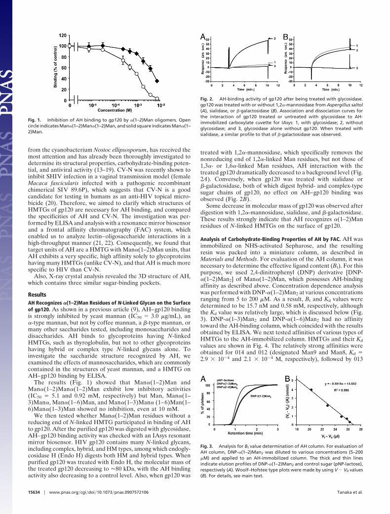
The results (Fig. 1) showed that Man�(1–2)Man andMan�(1–2)Man�(1–2)Man exhibit low inhibitory activities(IC50 � 5.1 and 0.92 mM, respectively) but Man, Man�(1–3)Man�, Man�(1–6)Man, and Man�(1–3)Man� (1–6)Man(1–6)Man�(1–3)Man showed no inhibition, even at 10 mM.
We then tested whether Man�(1–2)Man residues without areducing end of N-linked HMTG participated in binding of AHto gp120. After the purified gp120 was digested with glycosidase,AH–gp120 binding activity was checked with an IAsys resonantmirror biosensor. HIV gp120 contains many N-linked glycans,including complex, hybrid, and HM types, among which endogly-cosidase H (Endo H) digests both HM and hybrid types. Whenpurified gp120 was treated with Endo H, the molecular mass ofthe treated gp120 decreasing to �80 kDa, with the AH bindingactivity also decreasing to a control level. Also, when gp120 was
treated with 1,2�-mannosidase, which specifically removes thenonreducing end of 1,2�-linked Man residues, but not those of1,3�- or 1,6�-linked Man residues, AH interaction with thetreated gp120 dramatically decreased to a background level (Fig.2A). Conversely, when gp120 was treated with sialidase or�-galactosidase, both of which digest hybrid- and complex-typesugar chains of gp120, no effect on AH–gp120 binding wasobserved (Fig. 2B).
Some decrease in molecular mass of gp120 was observed afterdigestion with 1,2�-mannosidase, sialidase, and �-galactosidase.These results strongly indicate that AH recognizes �(1–2)Manresidues of N-linked HMTGs on the surface of gp120.
Analysis of Carbohydrate-Binding Properties of AH by FAC. AH wasimmobilized on NHS-activated Sepharose, and the resultingresin was packed into a miniature column, as described inMaterials and Methods. For evaluation of the AH column, it wasnecessary to determine the effective ligand content (Bt). For thispurpose, we used 2,4-dinitrophenyl (DNP) derivative [DNP-�(1–2)Man2] of Man�(1–2)Man, which possesses AH-bindingaffinity as described above. Concentration dependence analysiswas performed with DNP-�(1–2)Man2 at various concentrationsranging from 5 to 200 �M. As a result, Bt and Kd values weredetermined to be 15.7 nM and 0.58 mM, respectively, althoughthe Kd value was relatively large, which is discussed below (Fig.3). DNP-�(1–3)Man2 and DNP-�(1–6)Man2 had no affinitytoward the AH-binding column, which coincided with the resultsobtained by ELISA. We next tested affinities of various types ofHMTGs to the AH-immobilized column. HMTGs and their Kdvalues are shown in Fig. 4. The relatively strong affinities wereobtained for 014 and 012 (designated Man9 and Man8, Kd �2.9 � 10�4 and 2.1 � 10�4 M, respectively), followed by 013
Fig. 1. Inhibition of AH binding to gp120 by �(1–2)Man oligomers. Opencircle indicates Man�(1–2)Man�(1–2)Man, and solid square indicates Man�(1–2)Man.
Fig. 2. AH-binding activity of gp120 after being treated with glycosidase.gp120 was treated with or without 1,2�-mannosidase from Aspergillus saltol(A), sialidase, or �-galactosidase (B). Association and dissociation curves forthe interaction of gp120 treated or untreated with glycosidase to AH-immobilized carboxylate cuvette for IAsys: 1, with glycosidase; 2, withoutglycosidase; and 3, glycosidase alone without gp120. When treated withsialidase, a similar profile to that of �-galactosidase was observed.
Fig. 3. Analysis for Bt value determination of AH column. For evaluation ofAH column, DNP-�(1–2)Man2 was diluted to various concentrations (5–200�M) and applied to an AH-immobilized column. The thick and thin linesindicate elution profiles of DNP-�(1–2)Man2 and control sugar (pNP-lactose),respectively (A). Woolf–Hofstee type plots were made by using V � V0 values(B). For details, see main text.
15634 � www.pnas.org�cgi�doi�10.1073�pnas.0907572106 Tanaka et al.
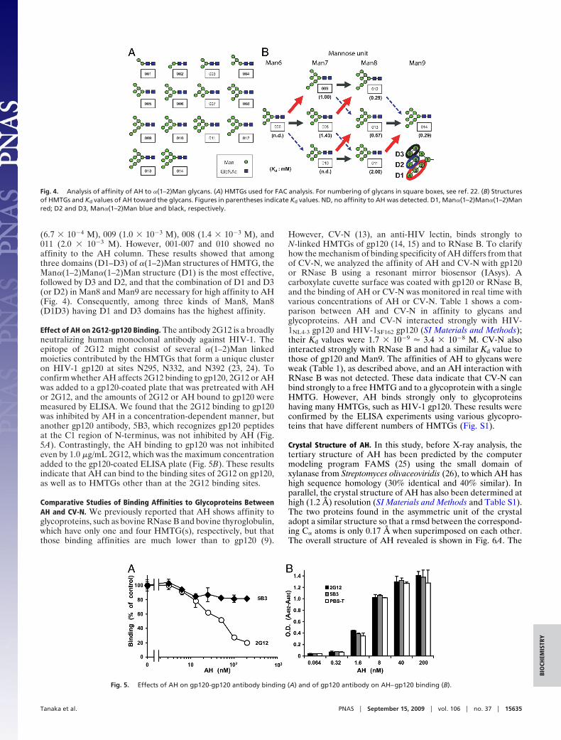
(6.7 � 10�4 M), 009 (1.0 � 10�3 M), 008 (1.4 � 10�3 M), and011 (2.0 � 10�3 M). However, 001-007 and 010 showed noaffinity to the AH column. These results showed that amongthree domains (D1–D3) of �(1–2)Man structures of HMTG, the�an�(1–2)Man�(1–2)Man structure (D1) is the most effective,followed by D3 and D2, and that the combination of D1 and D3(or D2) in Man8 and Man9 are necessary for high affinity to AH(Fig. 4). Consequently, among three kinds of Man8, Man8(D1D3) having D1 and D3 domains has the highest affinity.
Effect of AH on 2G12-gp120 Binding. The antibody 2G12 is a broadlyneutralizing human monoclonal antibody against HIV-1. Theepitope of 2G12 might consist of several �(1–2)Man linkedmoieties contributed by the HMTGs that form a unique clusteron HIV-1 gp120 at sites N295, N332, and N392 (23, 24). Toconfirm whether AH affects 2G12 binding to gp120, 2G12 or AHwas added to a gp120-coated plate that was pretreated with AHor 2G12, and the amounts of 2G12 or AH bound to gp120 weremeasured by ELISA. We found that the 2G12 binding to gp120was inhibited by AH in a concentration-dependent manner, butanother gp120 antibody, 5B3, which recognizes gp120 peptidesat the C1 region of N-terminus, was not inhibited by AH (Fig.5A). Contrastingly, the AH binding to gp120 was not inhibitedeven by 1.0 �g/mL 2G12, which was the maximum concentrationadded to the gp120-coated ELISA plate (Fig. 5B). These resultsindicate that AH can bind to the binding sites of 2G12 on gp120,as well as to HMTGs other than at the 2G12 binding sites.
Comparative Studies of Binding Affinities to Glycoproteins BetweenAH and CV-N. We previously reported that AH shows affinity toglycoproteins, such as bovine RNase B and bovine thyroglobulin,which have only one and four HMTG(s), respectively, but thatthose binding affinities are much lower than to gp120 (9).
However, CV-N (13), an anti-HIV lectin, binds strongly toN-linked HMTGs of gp120 (14, 15) and to RNase B. To clarifyhow the mechanism of binding specificity of AH differs from thatof CV-N, we analyzed the affinity of AH and CV-N with gp120or RNase B using a resonant mirror biosensor (IAsys). Acarboxylate cuvette surface was coated with gp120 or RNase B,and the binding of AH or CV-N was monitored in real time withvarious concentrations of AH or CV-N. Table 1 shows a com-parison between AH and CV-N in affinity to glycans andglycoproteins. AH and CV-N interacted strongly with HIV-1NL4-3 gp120 and HIV-1SF162 gp120 (SI Materials and Methods);their Kd values were 1.7 � 10�9 � 3.4 � 10�8 M. CV-N alsointeracted strongly with RNase B and had a similar Kd value tothose of gp120 and Man9. The affinities of AH to glycans wereweak (Table 1), as described above, and an AH interaction withRNase B was not detected. These data indicate that CV-N canbind strongly to a free HMTG and to a glycoprotein with a singleHMTG. However, AH binds strongly only to glycoproteinshaving many HMTGs, such as HIV-1 gp120. These results wereconfirmed by the ELISA experiments using various glycopro-teins that have different numbers of HMTGs (Fig. S1).
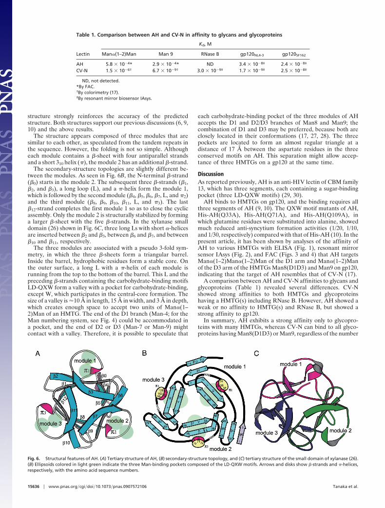
Crystal Structure of AH. In this study, before X-ray analysis, thetertiary structure of AH has been predicted by the computermodeling program FAMS (25) using the small domain ofxylanase from Streptomyces olivaceoviridis (26), to which AH hashigh sequence homology (30% identical and 40% similar). Inparallel, the crystal structure of AH has also been determined athigh (1.2 Å) resolution (SI Materials and Methods and Table S1).The two proteins found in the asymmetric unit of the crystaladopt a similar structure so that a rmsd between the correspond-ing C� atoms is only 0.17 Å when superimposed on each other.The overall structure of AH revealed is shown in Fig. 6A. The
Fig. 4. Analysis of affinity of AH to �(1–2)Man glycans. (A) HMTGs used for FAC analysis. For numbering of glycans in square boxes, see ref. 22. (B) Structuresof HMTGs and Kd values of AH toward the glycans. Figures in parentheses indicate Kd values. ND, no affinity to AH was detected. D1, Man�(1–2)Man�(1–2)Manred; D2 and D3, Man�(1–2)Man blue and black, respectively.
Fig. 5. Effects of AH on gp120-gp120 antibody binding (A) and of gp120 antibody on AH–gp120 binding (B).
Tanaka et al. PNAS � September 15, 2009 � vol. 106 � no. 37 � 15635
BIO
CHEM
ISTR
Y
structure strongly reinforces the accuracy of the predictedstructure. Both structures support our previous discussions (6, 9,10) and the above results.
The structure appears composed of three modules that aresimilar to each other, as speculated from the tandem repeats inthe sequence. However, the folding is not so simple. Althougheach module contains a �-sheet with four antiparallel strandsand a short 310 helix (�), the module 2 has an additional �-strand.
The secondary-structure topologies are slightly different be-tween the modules. As seen in Fig. 6B, the N-terminal �-strand(�0) starts in the module 2. The subsequent three �-strands (�1,�2, and �3), a long loop (L), and a �-helix form the module 1,which is followed by the second module (�4, �5, �6, �7, L, and �2)and the third module (�8, �9, �10, �11, L, and �3). The last�12-strand completes the first module 1 so as to close the cyclicassembly. Only the module 2 is structurally stabilized by forminga larger �-sheet with the five �-strands. In the xylanase smalldomain (26) shown in Fig. 6C, three long Ls with short �-helicesare inserted between �2 and �3, between �6 and �7, and between�10 and �11, respectively.
The three modules are associated with a pseudo 3-fold sym-metry, in which the three �-sheets form a triangular barrel.Inside the barrel, hydrophobic residues form a stable core. Onthe outer surface, a long L with a �-helix of each module isrunning from the top to the bottom of the barrel. This L and thepreceding �-strands containing the carbohydrate-binding motifsLD-QXW form a valley with a pocket for carbohydrate-binding,except W, which participates in the central-core formation. Thesize of a valley is �10 Å in length, 15 Å in width, and 3 Å in depth,which creates enough space to accept two units of Man�(1–2)Man of an HMTG. The end of the D1 branch (Man-4; for theMan numbering system, see Fig. 4) could be accommodated ina pocket, and the end of D2 or D3 (Man-7 or Man-9) mightcontact with a valley. Therefore, it is possible to speculate that
each carbohydrate-binding pocket of the three modules of AHaccepts the D1 and D2/D3 branches of Man8 and Man9; thecombination of D1 and D3 may be preferred, because both areclosely located in their conformations (17, 27, 28). The threepockets are located to form an almost regular triangle at adistance of 17 Å between the aspartate residues in the threeconserved motifs on AH. This separation might allow accep-tance of three HMTGs on a gp120 at the same time.
DiscussionAs reported previously, AH is an anti-HIV lectin of CBM family13, which has three segments, each containing a sugar-bindingpocket (three LD-QXW motifs) (29, 30).
AH binds to HMTGs on gp120, and the binding requires allthree segments of AH (9, 10). The QXW motif mutants of AH,His-AH(Q33A), His-AH(Q71A), and His-AH(Q109A), inwhich glutamine residues were substituted into alanine, showedmuch reduced anti-syncytium formation activities (1/20, 1/10,and 1/30, respectively) compared with that of His-AH (10). In thepresent article, it has been shown by analyses of the affinity ofAH to various HMTGs with ELISA (Fig. 1), resonant mirrorsensor IAsys (Fig. 2), and FAC (Figs. 3 and 4) that AH targetsMan�(1–2)Man�(1–2)Man of the D1 arm and Man�(1–2)Manof the D3 arm of the HMTGs Man8(D1D3) and Man9 on gp120,indicating that the target of AH resembles that of CV-N (17).
A comparison between AH and CV-N affinities to glycans andglycoproteins (Table 1) revealed several differences. CV-Nshowed strong affinities to both HMTGs and glycoproteinshaving a HMTG(s) including RNase B. However, AH showed aweak or no affinity to HMTG(s) and RNase B, but showed astrong affinity to gp120.
In summary, AH exhibits a strong affinity only to glycopro-teins with many HMTGs, whereas CV-N can bind to all glyco-proteins having Man8(D1D3) or Man9, regardless of the number
Fig. 6. Structural features of AH. (A) Tertiary structure of AH, (B) secondary-structure topology, and (C) tertiary structure of the small domain of xylanase (26).(B) Ellipsoids colored in light green indicate the three Man-binding pockets composed of the LD-QXW motifs. Arrows and disks show �-strands and �-helices,respectively, with the amino acid sequence numbers.
Table 1. Comparison between AH and CV-N in affinity to glycans and glycoproteins
Lectin
Kd, M
Man�(1–2)Man Man 9 RNase B gp120NL4-3 gp120SF162
AH 5.8 � 10�4* 2.9 � 10�4* ND 3.4 � 10�8‡ 2.4 � 10�8‡
CV-N 1.5 � 10�6† 6.7 � 10�9† 3.0 � 10�9‡ 1.7 � 10�9‡ 2.5 � 10�8‡
ND, not detected.*By FAC.†By colorimetry (17).‡By resonant mirror biosensor IAsys.
15636 � www.pnas.org�cgi�doi�10.1073�pnas.0907572106 Tanaka et al.
of glycans on a glycoprotein. CV-N contains two carbohydratebinding sites that binds to Man8(D1D3) and Man9 with nano-molar affinity (17), and a possible cross-linking binding modelfor CV-N and trimeric gp120 has been proposed (31). Conse-quently, AH has a much more specific affinity to glycoproteinswith many HMTGs than CV-N.
Also, although CV-N proved effective in preventing SHIVtransmission in macaques in the absence of toxic side-effects(20), CV-N can also cause a broad variety of side effects such asa cytopathic effect and mitogenic activity (32, 33). AH showedno cytotoxicity, even at 100 �M in MT-4 cells (6). It is consideredthat the basis of the side effects of CV-N may be due to its strongbinding to a single HMTG in contrast with AH, or by protein–protein interaction of CV-N with cellular factions.
As described above, the crystal structure (Fig. 6A) of AHshowed that AH has three sugar-binding pockets that areconstructed by the LD-QXW motifs. The ends of D1 and D3branches of Man8(D1D3) and Man9 could be bound in thepocket and its nearby valley. It appears that the amino acidsequence of the motif determines the kind of glycans that lectinbinds to. Interestingly, amino acid homology of three LD-QXWmotifs of AH is extremely high among known lectins of CBM 13.Seventeen of 22 aa (77%) in the LD-QXW motif are conserved.Practically, the structures of the three pockets are very similar toeach other with high symmetry. In other words, it suggests thatthese pockets have an almost equal binding affinity to Mans inparticular. However, in the small domain of xylanase of Strep-tomyces olivaceoviridis E-86 (26) and the ricin B chain of Ricinuscommunis (34), which belong to CBM 13, only 41 and 3% haveconserved between the three LD-QXW motifs, respectively.Therefore, the three pockets of the small domain are deformed,suggesting that they are not equivalent to each other, as shownin Fig. 6C. Consistently with the structural differences, thespecificities of these lectins to sugars are not very high; forexample, the xylan-binding domain accepts glucose, galactose,and lactose in addition to xylose and xylooligosaccharides (26).The extremely high specificity of AH to HMTGs is likely due tothe high homology of the three-pocket structures. Consequently,the binding of D1 and D3 domains of three HMTGs on onemolecule of gp120 to the three equivalent sugar-binding pocketsof AH might underlie the highly specific, strong affinity via theso-called cluster effect of lectin (35).
Human monoclonal antibody 2G12 against HIV-1 recognizesa unique oligomannose cluster that is mainly contributed by theHMTG at glycosylation sites N295, N332, and N392 (23, 24). Theresults depicted in Fig. 5 indicate that AH can also bind toHMTGs other than the epitope of 2G12. Incidentally, theinhibitory activity of AH against gp120-soluble CD4 binding isvery weak (IC50 � 1.2 mM by ELISA) (6). It seems to show thatHMTGs are not involved in gp120 binding to CD4 on susceptiblecells, and that AH interferes with the entry steps after gp120-CD4 binding occurs. The Man specific lectins from Galanthusnivalis and Hippeastrum hybrid also do not interfere with HIVbinding to CD4 (36).
A typical HIV-1 gp120 is highly glycosylated and has manyHMTGs. Although a Man oligosaccharide moiety exists in somehuman glycoproteins, a high-density, clustering oligomannosestructure similar to the one present on gp120 has not yet beenfound in any other human glycoproteins. Therefore, it seems thatAH does not bind to such human glycoproteins. The aboveresults support AH being developed as a potential topicalmicrobicide to prevent HIV transmission and infection.
Materials and MethodsMaterials. AH was prepared from a cultured broth of L. albida K97-0003T asdescribed previously (4). Man�(1–2)Man, Man�(1–3)Man, Man�(1–6)Man,Man�(1–3)Man�(1–6)Man�(1–6)Man�(1–3)Man, bovine thyroglobulin, andbovine RNase B were purchased from Sigma. Man�(1–2)Man�(1–2)Man was
donated by Y. Ito and I. Matsuo (RIKEN Advanced Science Institute). CV-N andits polyclonal antibody were a gift from M. R. Boyd (University of SouthCarolina, Columbia, SC). Pyridylamine (PA)-labeled Man type N-glycans (001-014) were purchased from Takara and Seikagaku Kogyo. DNP oligosaccha-rides, DNP-�-d-Manp-� (1–2)d-Manp (DNP-�(1–2)Man2), DNP-�-d-Manp-� (1–3)d-Manp (DNP-�(1–3)Man2), and DNP-�-d-Manp-� (1–6)d-Manp (DNP-�(1–6)Man2) were generous gifts from T. Nakamura (Obihiro University ofAgriculture and Veterinary Medicine, Obihiro, Japan). Anti-AH polyclonalantibody was prepared as previously described (9). Human anti-HIV-1 gp120mAb 2G12 was provided by Hermann Katinger through the National Institutesof Health (NIH) AIDS Research and Reference Program, Division of AIDS,National Institute of Allergy and Infectious Diseases, NIH: HIV-1 gp120 mAb2G12. Anti-HIV gp120 antibody 5B3 and HIV����gp120 were generous giftsfrom Genentech.
Effects of Man Oligomers on AH–gp120 Interaction. We used the ELISA methodas follows. Each well of a 96-well protein-adsorbing plate (Maxisorp Immu-noplate; Nalge NINC) was treated with 50 �L of AH solution (3.0 �g/mL) for18 h at 4 °C. The plate was blocked with 3.0% nonfat dry milk (50 �L) for 1 h,washed with PBS (10 mM phosphate buffer, 2.7 mM potassium chloride and137 mM sodium chloride, pH 7.4) containing 0.05% Tween 20 (PBS-T), andincubated for 2 h with a solution containing 0.18 �g/mL sgp120IIIB and variousconcentrations of Man oligomers [Man�(1–2)Man,Man�(1–3)Man, Man�(1–6)Man, Man�(1–2)Man�(1–2)Man, and Man�(1–3)Man�(1– 6)Man�(1–6)Man�(1–3)Man]. Each well was rinsed four times with PBS-T.
A solution (50 �L) of anti-gp120 monoclonal antibody 5B3 (0.3 �g/mL) wasadded to the wells and incubated at room temperature for 30 min, followedby washing with PBS-T three times.
A solution (50 �L) of HRP-conjugated anti-mouse IgG (1/2,000 dilution) wasadded to each well, and the plate was incubated at room temperature for 30min. The plate was washed four times with PBS-T, followed by incubation with100 �L of o-phenylenediamine dihydrochloride (OPD) solution (0.45 mg/mLOPD/16 mM citrate/50 mM disodium hydrogenphosphate, pH 5.0) for 8 min atroom temperature. The color reaction was stopped by adding 2.0 M sulfuricacid, and the optical density was measured at 405 nm.
Glycosidase Digestion. Endo H, 1,2�-mannosidase (from Aspergillus saitoi),and �-galactosidase were purchased from Seikagaku, and sialidase was pur-chased from Roche Diagnostics. The gp120 from HeLa/T-env/Tat cells wasdigested with a glycosidase in a glycosidase digestion buffer as specified by themanufacturer.
Assay of Protein–Protein Interaction Activity Using a Resonant Mirror Biosensor.Analysis of protein–protein interaction was performed using an IAsys reso-nant mirror biosensor (Affinity Sensors). For preparation of an AH-coatedcuvette, we used dual-well IAsys amino cuvette. Polymerized glutaraldehyde(PG) was prepared by taking 5.0 mL of 5% (vol/vol) glutaraldehyde, adding 500�L of 0.1 M NaOH, and leaving it for 30 min to polymerize before neutralizingwith 500 �L of 0.1 M HCl [final 4.2% (vol/vol) PG]. A dual-well IAsys aminocuvette was washed with 10 mM sodium phosphate (PB, pH 7.7) and activatedwith PG for 30 min. One well was blocked with BSA as a control surface. Theother one was treated with 10 �L of 80 �M solution of AH in PB (0.24 ng of AHbound/mm2). A gp120 or RNase B-coated cuvette was prepared using adual-well IAsys carboxylate cuvette. The IAsys carboxylate cuvette was acti-vated with a mixture of 0.2 M N-ethyl-N�-(3-dimethylaminopropyl)carbodiim-ide hydrochloride and 0.05 M NHS as described in IAsys protocols. The purifiedgp120 or RNase B was immobilized on a well of the cuvette (4.13 ng/mm2 ofHIV-1NL4-3 gp120, 4.88 ng/mm2 of HIV-1SF162 gp120, or 4.4 ng/mm2 of RNase B).The other well of each cuvette was blocked with �-casein as a control surface.Remaining activated groups were blocked with 1.0 M 2-aminoethanol (pH8.5). Binding reaction was carried out in PBS (total volume of 50 �L) containing0.02% Tween20 at 25 °C.
Functional Analysis by FAC. Kd of AH for PA-labeled HMTGs (001-014; Fig. 4)were determined by FAC (21, 22). All FAC data were obtained in 10 mM Tris-Clbuffer, pH 7.4, containing 0.7% NaCl (TBS) at a flow rate of 0.125 mL/min withthe FAC-1 machine, which had been specially designed and manufactured byShimadzu. Purified AH (1.0 mg) was immobilized on 100 �L of NHS-activatedSepharose resin (Amersham Bioscience). The slurry of the AH-immobilizedSepharose was packed into a capsule-type miniature column (inner diameter,2 mm; length, 10 mm; bed volume, 31.4 �L). The resulting column was slottedinto a stainless steel holder, and connected to the FAC-1 machine. For thedetermination of effective ligand content, Bt of the AH-immobilized column,concentration-dependence analysis and subsequent Woolf–Hofstee typeplots were performed. Here, various concentrations ([A]0) of DNP-�(1,2)Man2
Tanaka et al. PNAS � September 15, 2009 � vol. 106 � no. 37 � 15637
BIO
CHEM
ISTR
Y
dissolved in TBS were applied to the miniature column, and the elution wasmonitored by absorbance at 304 nm. Woolf–Hofstee type plots, i.e., (Vf – V0)vs. [A]0 � (Vf � V0), were made to determine Bt and Kd values from the interceptand the slope, respectively, of the fitted curve (Fig. 3). Vf is the elution volumeof A and V0 is the elution volume of pNP-�-lactose, which has no affinitytoward AH.
Next, PA-labeled HMTGs were applied to the column, and the elution wasmonitored by fluorescence (excitation and emission wavelength 320 and 400nm, respectively). As in the assays used in this study, V0 is the elution volumeof PA-lactose, which has no affinity toward AH and [A]0, the initial concen-tration of the PA-labeled HMTGs of interest (A), was 2.5 nM, which wasnegligible compared with Kd. Thus, Vf approached its maximum value, whichwas independent of [A]0, and we used the following equation to obtain thevalues of Kd of AH toward HM type N-glycan derivatives.
Kd � Bt/(Vf � V0).
Effects of AH on Human mAb 2G12-gp120 Binding and of 2G12 on AH–gp120Binding. Each well of a 96-well protein-adsorbing plate was coated withpurified HIV-1NL4-3 gp120 for 18 h at 4 °C and then washed three times withPBS-T. Various concentrations of AH were added to the gp120-coated wellsand held for 1 h at room temperature. Each well was then washed four timeswith PBS-T.
Human anti-HIV-1 gp120 mAb 2G12 (1.0 �g/mL) or anti-HIV-1 gp120 anti-
body 5B3 (as a control) was added to the appropriate wells and incubated for1 h at room temperature. The plate was washed four times with PBS-T, andthen antibody bound to gp120 was detected, as described above. To deter-mine whether AH–gp120 binding is inhibited by 2G12 antibody, the gp120-coated plate was next incubated with 1.0 �g/mL 2G12 or 5B3 antibody for 1 h.The plate was washed four times with PBS-T and various concentrations of AHwere added to the wells. After incubation for 1 h, the plate was washed withPBS-T four times and AH bound to gp120 was detected by anti-AH polyclonalantibody (9).
ACKNOWLEDGMENTS. We thank Dr. M. R. Boyd (University of South Carolina,Columbia, SC) for generous gifts of samples of CV-N and its polyclonal anti-body; Genentech Inc. for generous gifts of HIV-1IIIB gp120 and anti-gp120mouse mAb 5B3; Ms. E. Kanda and Mr. T. Sagara for their helpful assistance;and Drs Y. Yamada, N. Matsugaki, and N. Igarashi (Photon Factory, Tsukuba,Japan) for their assistance in data collection with the synchrotron facility.HIV-1 gp120 mAb 2G12 was obtained through the National Institutes ofHealth AIDS Research and Reference Program, Division of AIDS, NationalInstitute of Allergy and Infectious Diseases, NIH, courtesy of Dr. HermannKatinger. This work was supported by Japan Ministry of Education, Culture,Sports, Science, and Technology grants from the 21st Century Program (to H.T.and S. �O.) and the Grant-in-Aid for Young Scientists B, 14771313, 2004 (to H.C.);the Japan Ministry of Health, Labor, and Welfare Research on Japan HealthScience Foundation on Drug Innovation (H.T.); and the Iwaki City, JapanStrategic Industry Producing Project (H.T.).
1. Cohen J (2007) Did Merck’s failed HIV vaccine cause harm? Science 318:1048–1049.2. Grant RM, et al. (2008) Whither or wither microbicides? Science 321:532–534.3. Clapham PR, McKnight A (2002) Cell surface receptors, virus entry and tropism of
primate lentiviruses. J Gen Virol 83:1809–1829.4. Leonard CK, et al. (1990) Assignment of intrachain disulfide bonds and characteriza-
tion of potential glycosylation sites of the type-1 recombinant human immunodefi-ciency virus envelope glycoprotein (gp120) expressed in Chinese hamster ovary cells.J Biol Chem 265:10373–10382.
5. Chiba H, et al. (2001) A simple screening system for anti-HIV drugs: Syncytium forma-tion assay using T-cell line tropic and macrophage tropic HIV env expressing celllines—establishment and validation. J Antibiot 54:818–826.
6. Chiba H, et al. (2001) Actinohivin, a novel anti-HIV protein from an actinomycete thatinhibits syncytium formation: Isolation, characterization, and biological activities.Biochem Biophys Res Commun 282:595–601.
7. Inokoshi J, et al. (2001) Molecular cloning of actinohivin, a novel anti-HIV protein froman actinomycete, and its expression in Escherichia coli. Biochem Biophys Res Commun281:1261–1265.
8. Matsumoto A, et al. (2003) Longispora albida gen nov, sp nov, a novel genus of thefamily Micromonosporaceae. Int J Syst Evol Microbiol 53:1553–1559.
9. Chiba H, et al. (2004) Actinohivin, a novel anti-human immunodeficiency virus proteinfrom an actinomycete, inhibits viral entry to cells by binding high-mannose type sugarchains of gp120. Biochem Biophys Res Commun 316:203–210.
10. Takahashi A, et al. (2005) Essential regions for antiviral activities of actinohivin, asugar-binding anti-human immunodeficiency virus protein from an actinomycete.Arch Biochem Biophys 437:233–240.
11. Balzarini J (2007) Targeting the glycans of glycoproteins: A novel paradigm for antiviraltherapy. Nat Rev Microbiol 5:583–597.
12. Balzarini J (2005) Targeting the glycans of gp120: A novel approach at the Achilles heelof HIV. Lancet 5:726–731.
13. Boyd MR, et al. (1997) Discovery of cyanovirin-N, a novel human immunodeficiencyvirus-inactivating protein that binds viral surface envelope glycoprotein gp120: Po-tential applications to microbicide development. Antimicrob Agents Chemother41:1521–1530.
14. Shenoy SR, et al. (2001) Selective interactions of the human immunodeficiencyvirus-inactivating protein cyanovirin-N with high-mannose oligosaccharides on gp120 andother glycoproteins. J Pharmacol Exp Ther 297:704–710.
15. Bolmstedt AJ, et al. (2001) Cyanovirin-N defines a new class of antiviral agent targetingN-linked, high-mannose glycans in an oligosaccharide-specific manner. Mol Pharmacol59:949–954.
16. Bewley CA (2001) Solution structure of a cyanovirin-N:Man alpha 1–2Man alphacomplex: Structural basis for high-affinity carbohydrate-mediated binding to gp120.Structure 9:931–940.
17. Bewley CA, Otero-Quintero S (2001) The potent anti-HIV protein cyanovirin-N containstwo novel carbohydrate binding sites that selectively bind to Man(8) D1D3 and Man(9)with nanomolar affinity: Implications for binding to the HIV envelope protein gp120.J Am Chem Soc 123:3892–3902.
18. Bewley CA (2001) Rapid validation of the overall structure of an internal domain-swapped mutant of the anti-HIV protein cyanovirin-N using residual dipolar couplings.J Am Chem Soc 123:1014–1015.
19. Bewley CA, Kiyonaka S, Hamachi I (2002) Site-specific discrimination by cyanovirin-Nfor alpha-linked trisaccharides comprising the three arms of Man(8) and Man(9). J MolBiol 322:881–889.
20. Tsai CC, et al. (2004) Cyanovirin-N inhibits AIDS virus infections in vaginal transmissionmodels. AIDS Res Hum Retroviruses 20:11–18.
21. Hirabayashi J, Arata Y, Kasai K (2003) Frontal affinity chromatography as a tool forelucidation of sugar recognition properties of lectins. Methods Enzymol 362:353–368.
22. Nakamura-Tsuruta S, Uchiyama N, Hirabayashi J (2006) High-thoughtput analysis oflectin-oligosaccharide interactions by automated frontal affinity chromatography.Methods Enzymol 415:311–325.
23. Sanders RW, et al. (2002) The mannose-dependent epitope for neutralizing antibody 2g12on human immunodeficiency virus type 1 glycoprotein gp120. J Virol 76:7293–7305.
24. Scanlan CN, et al. (2002) The broadly neutralizing anti-human immunodeficiency virustype 1 antibody 2g12 recognizes a cluster of �1-2 mannose residues on the outer faceof gp120. J Virol 76:7306–7321.
25. Umeyama H, Iwadate M (2004) Current Protocols in Bioinformatics, ed Baxevanis AD(Wiley, Hoboken, NJ), Chap 6, Unit 5.2.
26. Fujimoto Z, et al. (2000) Crystal structure of. Streptomyces olivaceoviridis E-86 beta-xylanase containing xylan-binding domain. J Mol Biol 300:575–585.
27. Shenoy SR, et al. (2002) Multisite and multivalent binding between cyanovirin-N andbranched oligomannosides: Calorimetric and NMR characterization. Chem Biol10:1109–1118.
28. Woods RJ, Pathiaseril A, Wormald MR, Edge CJ, Dwek RA (1998) The high degree ofinternal flexibility observed for an oligomannose oligosaccharide does not alter theoverall topology of the molecule. Eur J Biochem 258:372–386.
29. Hazes B (1996) The (QXW)3 domain: A flexible lectin scaffold. Protein Sci 5:1490–1501.30. Gilkes NR, et al. (1991) Domains in microbial �-1,4-glycanases: Sequence conservation,
function, and enzyme families. Microbiol Rev 55:303–315.31. Bewley CA (2001) Solution structure of a cyanovirin-N:Man�1–2Man� complex: Structural
basis for high-affinity carbohydrate-mediated binding to gp120. Structure 9:931–940.32. Balzarini J, et al. (2006) Mutational pathways, resistance profile, and side effects of
cyanovirin relative to human immunodeficiency virus type 1 strains with N-glycandeletions in their gp120 envelopes. J Virol 80:8411–8421.
33. Huskens D, et al. (2008) Safety concerns for the potential use of cyanovirin-N as amicrobicidal anti-HIV agent. Int J Biochem Cell Biol 40:2802–2814.
34. Rutenber E, Robertus JD (1991) Structure of ricin B-chain at 2.5 resolution. Proteins10:260–269.
35. Lee YC, Lee RT (1995) Carbohydrate–protein interactions: Basis of glycobiology. AccChem Res 28:321–327.
36. Balzarini J, et al. (2004) Mannose-specific plant lectins from the Amaryllidaceae familyqualify as efficient microbicides for prevention of human immunodeficiency virusinfection. Antimicrob Agents Chemother 48:3858–3870.
15638 � www.pnas.org�cgi�doi�10.1073�pnas.0907572106 Tanaka et al.