Marine Natural Products from New Caledonia—A Review
61
Marine Natural Products from New Caledonia-A Review Sofia-El´ ena Motuhi, Mohamed Mehiri, Claude Elisabeth Payri, St´ ephane La Barre, St´ ephane Bach To cite this version: Sofia-El´ ena Motuhi, Mohamed Mehiri, Claude Elisabeth Payri, St´ ephane La Barre, St´ ephane Bach. Marine Natural Products from New Caledonia-A Review. Marine drugs, MDPI, 2016, 14 (3), pp.58. <10.3390/md14030058>. <hal-01292225> HAL Id: hal-01292225 http://hal.upmc.fr/hal-01292225 Submitted on 22 Mar 2016 HAL is a multi-disciplinary open access archive for the deposit and dissemination of sci- entific research documents, whether they are pub- lished or not. The documents may come from teaching and research institutions in France or abroad, or from public or private research centers. L’archive ouverte pluridisciplinaire HAL, est destin´ ee au d´ epˆ ot et ` a la diffusion de documents scientifiques de niveau recherche, publi´ es ou non, ´ emanant des ´ etablissements d’enseignement et de recherche fran¸cais ou ´ etrangers, des laboratoires publics ou priv´ es. Distributed under a Creative Commons Attribution 4.0 International License
Transcript of Marine Natural Products from New Caledonia—A Review
Marine Natural Products from New Caledonia-A Review
Sofia-Elena Motuhi, Mohamed Mehiri, Claude Elisabeth Payri, Stephane La
Barre, Stephane Bach
To cite this version:
Sofia-Elena Motuhi, Mohamed Mehiri, Claude Elisabeth Payri, Stephane La Barre, StephaneBach. Marine Natural Products from New Caledonia-A Review. Marine drugs, MDPI, 2016,14 (3), pp.58. <10.3390/md14030058>. <hal-01292225>
HAL Id: hal-01292225
http://hal.upmc.fr/hal-01292225
Submitted on 22 Mar 2016
HAL is a multi-disciplinary open accessarchive for the deposit and dissemination of sci-entific research documents, whether they are pub-lished or not. The documents may come fromteaching and research institutions in France orabroad, or from public or private research centers.
L’archive ouverte pluridisciplinaire HAL, estdestinee au depot et a la diffusion de documentsscientifiques de niveau recherche, publies ou non,emanant des etablissements d’enseignement et derecherche francais ou etrangers, des laboratoirespublics ou prives.
Distributed under a Creative Commons Attribution 4.0 International License
marine drugs
Review
Marine Natural Products from NewCaledonia—A ReviewSofia-Eléna Motuhi 1,2, Mohamed Mehiri 3, Claude Elisabeth Payri 1, Stéphane La Barre 4,*and Stéphane Bach 2,*
1 Laboratoire d’Excellence Labex-CORAIL, Institut de Recherche pour le Développement (IRD),UMR ENTROPIE (IRD—Université de La Réunion—CNRS), BP A5, Nouméa Cedex 98848,Nouvelle-Calédonie; [email protected] (S.-E.M.); [email protected] (C.E.P.)
2 USR 3151 CNRS, Protein Phosphorylation & Human Diseases, Station Biologique de Roscoff,Sorbonne Universities, UPMC Univ Paris 06, CS 90074, Roscoff Cedex F-29688, France
3 UMR 7272 CNRS, Marine Natural Products Team, Nice Institute of Chemistry (ICN),University Nice Sophia Antipolis, Parc Valrose, Nice Cedex 02 F-06108, France; [email protected]
4 UMR 8227 CNRS, Integrative Biology of Marine Models, Station Biologique de Roscoff, CS 90074,Roscoff Cedex F-29688, France
* Correspondence: [email protected] (S.L.B.); [email protected] (S.B.); Tel.: +33-964-294-827 (S.L.B.);+33-298-292-391 (S.B.); Fax: +33-298-292-526 (S.L.B. & S.B.)
Academic Editor: Paul LongReceived: 20 July 2015; Accepted: 25 February 2016; Published: 16 March 2016
Abstract: Marine micro- and macroorganisms are well known to produce metabolites with highbiotechnological potential. Nearly 40 years of systematic prospecting all around the New Caledoniaarchipelago and several successive research programs have uncovered new chemical leads frombenthic and planktonic organisms. After species identification, biological and/or pharmaceuticalanalyses are performed on marine organisms to assess their bioactivities. A total of 3582 genera,1107 families and 9372 species have been surveyed and more than 350 novel molecular structureshave been identified. Along with their bioactivities that hold promise for therapeutic applications,most of these molecules are also potentially useful for cosmetics and food biotechnology. This reviewhighlights the tremendous marine diversity in New Caledonia, and offers an outline of the vastpossibilities for natural products, especially in the interest of pursuing collaborative fundamentalresearch programs and developing local biotechnology programs.
Keywords: New Caledonia; marine biodiversity; bioactive molecules pipeline; chemodiversity;coral reefs
1. Introduction
Located approximately at 165˝ E and 21˝301 S, the New Caledonia archipelago enjoys a privilegedposition east of the city of Rockhampton, central province of the Great Barrier Reef, 1500 km awayfrom the Australian Queensland coast, with surface seawater temperatures mild enough to avoidrecurrent coral bleaching episodes.
This tropical subregion of Melanesia is a relic of a continental landmass that drifted away fromthe super-continent Gondwana at the end of the Cretaceous period (see Pelletier 2007) [1]. Successivegeological events have resulted in an archipelago that comprises a main island called Grande Terre, Iledes Pins in the south, the Entrecasteaux reefs in the north, the Loyalty Islands in the east and, finally,the Chesterfield-Bellona plateau in the west, located at mid-distance to Australian coast (Figure 1).
New Caledonian waters represent an exclusive economic zone (EEZ) spanning more than1,700,000 km2. The EEZ includes a vast and complex reef system of 4537 km2 of reef area and
Mar. Drugs 2016, 14, 58; doi:10.3390/md14030058 www.mdpi.com/journal/marinedrugs

Mar. Drugs 2016, 14, 58 2 of 60
31,336 km2 of non-reef area [2] and represents the second most important coral reef complex and thelongest continuous barrier reef in the world, stretching over 1600 km.
The reef systems together make up a unique marine ecosystem [3] with more than 161 differentreef units [4] supporting high marine species diversity and abundance [5], including so-called “livingfossils” such as a stalked crinoid and a shelled cephalopod.
Mar. Drugs 2016, 14, x 2 of 62
km2 of non‐reef area [2] and represents the second most important coral reef complex and the longest
continuous barrier reef in the world, stretching over 1600 km.
The reef systems together make up a unique marine ecosystem [3] with more than 161 different
reef units [4] supporting high marine species diversity and abundance [5], including so‐called
“living fossils” such as a stalked crinoid and a shelled cephalopod.
Figure 1. The archipelago of New Caledonia and its reef complexes (adapted from Andréfouët et al.
2009 [2], with kind permission from the author).
Only since the 19th century has attention been paid to diversity of the New Caledonian reef
systems. Many expeditions have been conducted to study the oceanic life of these hitherto
unexplored oceanic zones. The first oceanographic cruise, the Challenger expedition (1872–1876), led
to the collection of thousands of previously unknown marine species [6]. Since the 19th century,
scientific interest has shifted from the study of “exotic” specimens stored in museum collections and
classified according to their morphological features, to the study and understanding of marine
ecosystems. Charles Darwin’s landmark expeditions in the Eastern Pacific islands and his
remarkable observations on the formation of atolls from subsiding volcanic islands sparked
continued interest in ecosystems biology in oceans across the world. Research vessels are now
equipped with sophisticated equipment to carry out on‐site sample analysis (e.g., molecular biology)
and data transmission via satellite systems. For example, the schooner Tara, specially fit out with
on‐board facilities, recently sailed around the world to study the impact of global warming on
plankton and coral reef systems across the Pacific [7].
Pioneering scientific investigations in New Caledonian waters were carried out locally after
World War II. The naturalist René Catala initiated ecological surveys and species census around Ile
aux Canards [8], an island off Nouméa now directly exposed to human activities, with recent
Figure 1. The archipelago of New Caledonia and its reef complexes (adapted from Andréfouët et al.2009 [2], with kind permission from the author).
Only since the 19th century has attention been paid to diversity of the New Caledonian reefsystems. Many expeditions have been conducted to study the oceanic life of these hitherto unexploredoceanic zones. The first oceanographic cruise, the Challenger expedition (1872–1876), led to the collectionof thousands of previously unknown marine species [6]. Since the 19th century, scientific interest hasshifted from the study of “exotic” specimens stored in museum collections and classified according totheir morphological features, to the study and understanding of marine ecosystems. Charles Darwin’slandmark expeditions in the Eastern Pacific islands and his remarkable observations on the formationof atolls from subsiding volcanic islands sparked continued interest in ecosystems biology in oceansacross the world. Research vessels are now equipped with sophisticated equipment to carry out on-sitesample analysis (e.g., molecular biology) and data transmission via satellite systems. For example, theschooner Tara, specially fit out with on-board facilities, recently sailed around the world to study theimpact of global warming on plankton and coral reef systems across the Pacific [7].
Pioneering scientific investigations in New Caledonian waters were carried out locally afterWorld War II. The naturalist René Catala initiated ecological surveys and species census around Ile auxCanards [8], an island off Nouméa now directly exposed to human activities, with recent changes in reefzonation and species composition. In addition, in 1956, Catala founded the Aquarium of Nouméa (nowthe Aquarium des Lagons), a first-of-its-kind structure specifically designed to observe rare and fragilemarine species in their environment, including homegrown corals. Catala incidentally discovered the

Mar. Drugs 2016, 14, 58 3 of 60
fluorescence of living corals [9] and other invertebrates (fluorescence appears to be a photoprotectiveand possibly temperature-regulating mechanism). A few years later, the Singer-Polignac expedition(1960–1963) explored St. Vincent Bay and the east coast of Grande Terre [10–12]. Similarly, the Americanecologist, Arthur Lyon Dahl, a regional adviser at the South Pacific Commission from 1974 to 1982,demonstrated the importance of surface area in ecological analysis and described several methods forconducting surveys of coral reefs [13,14].
Bioprospecting studies in New Caledonia were initiated in 1976 by Pierre Potier from the ICSN(Institute of Natural Substances Chemistry, Gif-sur-Yvette, mainland France) as part of the nationalresearch program SNOM (Substances Naturelles d’Origine Marine) and benefited from collaborationwith scientific divers from IRD (Institut de Recherche pour le Développement, ex. ORSTOM, Office de laRecherche Scientifique et Technique d’Outre-Mer) in Nouméa (Figure 2). SNOM set out to explore marinebiodiversity extensively with the taxonomic expertise from the National Museum of Natural History(MNHN) of Paris, to identify novel molecules and to assess their biological activities primarily forpharmaceutical purposes. Collecting efforts focused mainly on invertebrates, including octocorals,porifers, echinoderms, mollusks and ascidians. Little attention has been given to macroalgae due tothe lack of taxonomic support.
Mar. Drugs 2016, 14, x 3 of 62
changes in reef zonation and species composition. In addition, in 1956, Catala founded the
Aquarium of Nouméa (now the Aquarium des Lagons), a first‐of‐its‐kind structure specifically
designed to observe rare and fragile marine species in their environment, including homegrown
corals. Catala incidentally discovered the fluorescence of living corals [9] and other invertebrates
(fluorescence appears to be a photoprotective and possibly temperature‐regulating mechanism). A few
years later, the Singer‐Polignac expedition (1960–1963) explored St. Vincent Bay and the east coast of
Grande Terre [10–12]. Similarly, the American ecologist, Arthur Lyon Dahl, a regional adviser at the
South Pacific Commission from 1974 to 1982, demonstrated the importance of surface area in
ecological analysis and described several methods for conducting surveys of coral reefs [13,14].
Bioprospecting studies in New Caledonia were initiated in 1976 by Pierre Potier from the ICSN
(Institute of Natural Substances Chemistry, Gif‐sur‐Yvette, mainland France) as part of the national
research program SNOM (Substances Naturelles d’Origine Marine) and benefited from collaboration
with scientific divers from IRD (Institut de Recherche pour le Développement, ex. ORSTOM, Office de la
Recherche Scientifique et Technique d’Outre‐Mer) in Nouméa (Figure 2). SNOM set out to explore
marine biodiversity extensively with the taxonomic expertise from the National Museum of Natural
History (MNHN) of Paris, to identify novel molecules and to assess their biological activities
primarily for pharmaceutical purposes. Collecting efforts focused mainly on invertebrates, including
octocorals, porifers, echinoderms, mollusks and ascidians. Little attention has been given to
macroalgae due to the lack of taxonomic support.
Figure 2. Publications in scientific journals on organisms collected within various research programs.
Cumulative data representing the total number of publications generated since 1976 to present (red
line) and the number of publications produced per year since 1976 (blue line). The timeline on the
x‐axis shows the various regional marine biodiversity programs spanning the 40‐year period. These
statistics do not include reports, reviews and book chapters. The insert shows the relative
percentages of major taxon investigated up to 2007 [5]. SNOM: Substances Naturelles d’Origine Marine;
SMIB: Substances Marines d’Intérêt Biologique; LAGON; MUSORSTOM (now Tropical Deep‐Sea
Benthos): acronym for the joint expeditions of the National Museum of Natural History (MNHN)
and the Office de la Recherche Scientifique et Technique d’Outre‐Mer (ORSTOM, now IRD); CRISP: Coral
Reef InitiativeS for the Pacific; in progress: various research programs including chemical and
pharmacological investigations on micro‐ and macroalgae. Fl/Ma: Flora and Marine Angiosperms;
Pro: Protozoa; Alg: Algae; Por: Porifera; Cni: Cnidaria; Loph: Lophophorates; Mol: Mollusks; Wor:
Worms; Art: Arthropoda; Ech: Echinodermata; Tun: Tunicata; Ver: Vertebrata.
Figure 2. Publications in scientific journals on organisms collected within various research programs.Cumulative data representing the total number of publications generated since 1976 to present (red line)and the number of publications produced per year since 1976 (blue line). The timeline on the x-axisshows the various regional marine biodiversity programs spanning the 40-year period. These statisticsdo not include reports, reviews and book chapters. The insert shows the relative percentages of majortaxon investigated up to 2007 [5]. SNOM: Substances Naturelles d’Origine Marine; SMIB: SubstancesMarines d’Intérêt Biologique; LAGON; MUSORSTOM (now Tropical Deep-Sea Benthos): acronymfor the joint expeditions of the National Museum of Natural History (MNHN) and the Office de laRecherche Scientifique et Technique d’Outre-Mer (ORSTOM, now IRD); CRISP: Coral Reef InitiativeS for thePacific; in progress: various research programs including chemical and pharmacological investigationson micro- and macroalgae. Fl/Ma: Flora and Marine Angiosperms; Pro: Protozoa; Alg: Algae;Por: Porifera; Cni: Cnidaria; Loph: Lophophorates; Mol: Mollusks; Wor: Worms; Art: Arthropoda;Ech: Echinodermata; Tun: Tunicata; Ver: Vertebrata.

Mar. Drugs 2016, 14, 58 4 of 60
In 1985, the SNOM program was followed by the SMIB program (Substances Marines d’IntérêtBiologique), which was conducted jointly by ORSTOM and the Centre National de la Recherche Scientifique(CNRS), with a large collaborative network of public research institutions, e.g., the Centre d’Etudes Nucléairesde Saclay, the Institut National de la Santé et de la Recherche Médicale (INSERM), the MNHN, French andforeign universities, as well as several private companies (in particular, Pierre Fabre and Rhône-Poulenc).
The raw compounds and their fractions were extracted at the ORSTOM center in Nouméa andwere originally sent for purification and structural determination to the ICSN (mainland France), and thebiological screening was assigned to various mainland French laboratories. Gradually, separation andpurification, as well as preliminary testing and benchtop assays, were carried out locally in New Caledoniaat the ORSTOM (and later IRD) center, to avoid unnecessary duplication and to better target specificrequests from collaborating partners. During this time, a number of candidate molecules were identifiedfor their anticancer, cardiovascular or neurological interest (reviewed in [15]), prompting an extension ofthe research program and redefinition of the terms of scientific collaboration, now open to internationalexperts. The purchase of a larger research vessel, the R/V Alis, in 1992, provided the opportunity to extendthe bioprospecting range by dredging to 600 m deep and to access new biological resources.
The SNOM and SMIB programs led to an extensive survey of the marine biodiversity of theNew Caledonian archipelago, providing many in situ observations and records of new taxon, whichhave been published in several papers [5] and illustrated in field guides, especially on sponges [16],echinoderms [17], gorgonians [18], ascidians [19], marine snakes [20], and fishes [21]. Among the 300organisms studied, only 50 have been the object of chemical and therapeutic research and severaloriginal molecules have been described and tested on tumor development [22].
After the SNOM and SMIB programs, pharmacological bioprospecting in New Caledonia wasintegrated in the LAGON and MUSORSTOM biodiversity projects until 2000. Thereafter, researchactivities were extended to other countries in the Pacific region as part of the Coral Reef InitiativeS forthe Pacific (CRISP), with emphasis on legal agreements and economic benefits to host countries.
In the meantime, some research activity focused on ciguatera and cyanobacteria toxicity afterseveral severe poisoning events occurred in New Caledonia. In addition to pharmaceutical activities,natural marine compounds inspired new scientific approaches including chemotaxonomy andchemical ecology of benthic macroalgae to understand how opportunistic algae colonize livingcoral. These activities are conducted by the CoReUs/ENTROPIE research team at IRD (Figure 2).These projects are mentioned in Moretti et al., (1993) [22] and detailed in a comprehensive review ofnovel chemical structures and associated pharmacological activities described by Laurent and Pietrapublished in 2004 [15]. Another review emphasizes the developmental aspects of marine moleculesfrom South Pacific zone including New Caledonia [23].
Here, we provide an updated review of 40 years of exploration of the marine micro-/macrophyteand invertebrate chemodiversity of this species-rich zone of the Southwest Pacific, with itspharmacological potential and its ecological significance. After a description of the basic operationalaspects of discovering marine natural products, an overview of the work on each major taxon ispresented, illustrated by case studies that have been the highlights of the abovementioned programsfor the last 40 years, and carried out locally by experts in full compliance with existing regulations ofbiodiversity protection and the sustainability of valuable natural resources.
2. Taxonomy
SCUBA diving allows visual exploration of shallow-water marine biota, making it possible notonly to collect material at depths down to 60 m, but also to take photographs and record ecologicalinformation, e.g., interactions between organisms that can be useful for selecting organisms to collectand subsequent biological tests. Upon the development of blind transect dredging with limitedbiomass sampling on soft bottoms, collection efforts were extended to deeper zones (down to 600 m),thus sampling entirely different organisms.

Mar. Drugs 2016, 14, 58 5 of 60
2.1. Sample Collection Sites
Collection sites were selected to cover the large diversity of habitats ranging from shallow lagoonsto deep parts of the outer slopes of the barrier reefs. They include hard and sandy bottoms, shelteredand exposed areas of the mainland (Grande Terre), Loyalty Islands, as well as remote reefs and atolls(Entrecasteaux, Chesterfield) and seamounts (Figure 1).
Each site was georeferenced and described using geomorphological and biological descriptors.Historical data have been sorted, standardized and stored along with recent information in thededicated database LagPlon [24].
2.2. Biological Material Sampling
For all the groups collected for chemical purposes, specimens from each taxon were sampledfor taxonomical identification and preserved in ethanol as vouchers after labelling. Collections wereduplicated, one sample was kept for the IRD reference collection to facilitate new sampling effortsand the duplicate was sent to specialists for taxonomy work. In situ macrophotographs were taken aswell as additional ex situ laboratory photographs when necessary. Specimens used to describe newspecies (holotypes) have been deposited in various museum collections in Australia (Northern TerritoryMuseum, Darwin and Queensland Museum of South Brisbane), New Zealand, mainland France (NationalMuseum of Natural History, Paris), Belgium (Université Libre de Bruxelles). Paratypes were systematicallyfiled in the IRD reference collection in Nouméa, along with photographs and field records.
3. Chemistry
3.1. Biological Material
Chemical characterization and biological assays were initially carried on samples with wetweights ranging from 300 to 3000 g. However, recent progress in analytical techniques allows molecularinventory and pharmacological tests from smaller amounts of material in compliance with internationalregulations on the use of biological material for research purposes.
Below, we describe the basic protocols used locally at the IRD laboratories in Nouméa. Each of themolecules that have been investigated locally or with national and international partners have beentreated separately (Figure 3). It is beyond the scope of this review to detail each protocol individually;they can be found in the original research papers, or in a natural products database.
Mar. Drugs 2016, 14, x 5 of 62
2.1. Sample Collection Sites
Collection sites were selected to cover the large diversity of habitats ranging from shallow
lagoons to deep parts of the outer slopes of the barrier reefs. They include hard and sandy bottoms,
sheltered and exposed areas of the mainland (Grande Terre), Loyalty Islands, as well as remote reefs
and atolls (Entrecasteaux, Chesterfield) and seamounts (Figure 1).
Each site was georeferenced and described using geomorphological and biological descriptors.
Historical data have been sorted, standardized and stored along with recent information in the
dedicated database LagPlon [24].
2.2. Biological Material Sampling
For all the groups collected for chemical purposes, specimens from each taxon were sampled
for taxonomical identification and preserved in ethanol as vouchers after labelling. Collections were
duplicated, one sample was kept for the IRD reference collection to facilitate new sampling efforts
and the duplicate was sent to specialists for taxonomy work. In situ macrophotographs were taken as
well as additional ex situ laboratory photographs when necessary. Specimens used to describe new
species (holotypes) have been deposited in various museum collections in Australia (Northern
Territory Museum, Darwin and Queensland Museum of South Brisbane), New Zealand, mainland
France (National Museum of Natural History, Paris), Belgium (Université Libre de Bruxelles).
Paratypes were systematically filed in the IRD reference collection in Nouméa, along with
photographs and field records.
3. Chemistry
3.1. Biological Material
Chemical characterization and biological assays were initially carried on samples with wet
weights ranging from 300 to 3000 g. However, recent progress in analytical techniques allows
molecular inventory and pharmacological tests from smaller amounts of material in compliance
with international regulations on the use of biological material for research purposes.
Below, we describe the basic protocols used locally at the IRD laboratories in Nouméa. Each of
the molecules that have been investigated locally or with national and international partners have
been treated separately (Figure 3). It is beyond the scope of this review to detail each protocol
individually; they can be found in the original research papers, or in a natural products database.
Figure 3. Collaborative network of chemists and pharmacologists from 1976 to 2013 as compiled
individually from journal publications. Figure 3. Collaborative network of chemists and pharmacologists from 1976 to 2013 as compiledindividually from journal publications.
3.2. Conditioning Samples for Chemistry
Freshly collected material is sorted by taxon and frozen on board at ´20 ˝C, or at least placedin 70% ethanol in distilled water (slightly acidified to avoid oxidation of polar compounds) until it

Mar. Drugs 2016, 14, 58 6 of 60
can be deep frozen and freeze-dried for subsequent use. Material used for enzymology or genomestudies is snap-frozen on board (liquid nitrogen or dry ice in pure alcohol). For short collecting sessions(less than one full day), the material may be kept alive if each item is appropriately handled.
Back at the laboratory, the material must be ground/chopped/crushed then freeze-dried orethanol-preserved and stowed away for later use, or sent abroad to partner laboratories.
3.3. Extraction
3.3.1. Routine Procedure
Unless otherwise specified, a standard protocol is used for extracting compounds and separatingthem into crude non-polar (organic) and polar (aqueous) fractions (Figure 4).
Mar. Drugs 2016, 14, x 6 of 62
3.2. Conditioning Samples for Chemistry
Freshly collected material is sorted by taxon and frozen on board at −20 °C, or at least placed in
70% ethanol in distilled water (slightly acidified to avoid oxidation of polar compounds) until it can
be deep frozen and freeze‐dried for subsequent use. Material used for enzymology or genome
studies is snap‐frozen on board (liquid nitrogen or dry ice in pure alcohol). For short collecting
sessions (less than one full day), the material may be kept alive if each item is appropriately handled.
Back at the laboratory, the material must be ground/chopped/crushed then freeze‐dried or
ethanol‐preserved and stowed away for later use, or sent abroad to partner laboratories.
3.3. Extraction
3.3.1. Routine Procedure
Unless otherwise specified, a standard protocol is used for extracting compounds and
separating them into crude non‐polar (organic) and polar (aqueous) fractions (Figure 4).
Figure 4. Routine extraction protocol (IRD Nouméa).
Organic fractions are generally evaporated under vacuum (Rotovap) and kept in the dark at
−20 °C. Water solubles may need to be desalted using size‐exclusion gel chromatography (SEC) or by
membrane filtration under nitrogen (Amicon®, Alsace, France) prior to separation, but for long‐term
storage, desalting is not necessary. Aliquoting is necessary for multiple chemical characterizations
and for bioassays.
3.3.2. Peptide Protease Inhibitors
Live tissues are processed (minced or fragmented) on board and placed in an acidic solution
(methanol/2 N acetic acid) to optimize the extraction of polar substances and block any potential
hydrolysis by proteolytic enzymes. The cold (−20 °C) slurry is filtered twice and filtrates are reduced
by evaporation and neutralized to pH 5.5 prior to freeze‐drying. The total protein content of the
crude extract is assayed by measuring optical densities with a UV spectrophotometer.
Figure 4. Routine extraction protocol (IRD Nouméa).
Organic fractions are generally evaporated under vacuum (Rotovap) and kept in the dark at´20 ˝C. Water solubles may need to be desalted using size-exclusion gel chromatography (SEC) or bymembrane filtration under nitrogen (Amicon®, Alsace, France) prior to separation, but for long-termstorage, desalting is not necessary. Aliquoting is necessary for multiple chemical characterizations andfor bioassays.
3.3.2. Peptide Protease Inhibitors
Live tissues are processed (minced or fragmented) on board and placed in an acidic solution(methanol/2 N acetic acid) to optimize the extraction of polar substances and block any potentialhydrolysis by proteolytic enzymes. The cold (´20 ˝C) slurry is filtered twice and filtrates are reducedby evaporation and neutralized to pH 5.5 prior to freeze-drying. The total protein content of the crudeextract is assayed by measuring optical densities with a UV spectrophotometer.
3.4. Separation, Purification
Classical separation and purification techniques (TLC, HPLC, SEC) were not routinely used unlessrequested for products presenting an interesting bioactivity profile. This data is mentioned in theoriginal publications for each novel structure.

Mar. Drugs 2016, 14, 58 7 of 60
3.5. Structural Analysis
Basic spectroscopic (NMR, UV, IR) and spectrometric (MS) methods are used when available onsite, mostly to avoid replication.
3.6. Chemical Databases
The IRD proprietary information system Cantharella [25] compiles pharmacochemical data ofall organisms collected in New Caledonia and cross-Pacific oceanographic cruises for the study oftheir natural substances, with restricted access via Internet. The information system provides access tobiological data, accounts for all chemical processes from extraction to purification, and presents thebioactivity profile detected using the complete range of tests.
4. Biological Activities
4.1. Preliminary Testing
Vacuum-dried crude extracts or non-purified fractions thereof can be used for describing generalcharacteristics using toxicity assays on various invertebrate, vertebrate and plant models, and culturesof microbial reference strains (Table 1).
Preliminary tests performed on site are useful for making appropriate decisions as to whether agiven sample from a newly collected organism is worth investigating further. Along with taxonomiccriteria and dereplication issues, such decisions are now taken after consulting the scientific literatureand specialized databases, especially the IRD databases LagPlon [24] and Cantharella [25], which, inaddition to the more traditional MarinLit and other databases, considerably aid local researchers.
Table 1. Field observations and crude biological tests for preliminary screening.
Biological Model Species Type of Activity Tested Reference
Bacteria
Escherichia coli, Pseudomonas aeruginosa,Staphylococcus aureus, Streptococcusfaecalis (now Enterococcus faecalis),
Vibrio anguillarum
Antibacterial [26]
Fungi
Candida albicans, Candida tropicalis,Helminthosporium graminearum,
Helminthosporium turcicum, Penicilliumitalicum, Phytophthora parasitica,
Pyricularia oryzae
Antifungal [26]
Brine shrimp larvae Artemia salina Cytotoxicity [27]
Fish Gambusia affinis Neuro/Cytotoxicity [28,29]
Urchin eggs Echinometra mathaei Cytotoxicity [30]
Insect Hypothenemus hampei Insecticide [31]
Mite Rhipicephalus microplus (formerly,Boophilus microplus) Acaricide [32]
Algae Ceramium codii Anti-fouling [33]
Coral Algae Allelopathy [34]
Plant Amaranthus caudatus Anti-germinating [35]
4.1.1. Brine Shrimp Toxicity Assay
The brine shrimp (Artemia salina) toxicity bioassay is one of the most basic and widely used teststo detect cytotoxicity. Newly hatched brine shrimp nauplii are exposed to various concentrations ofsoluble or solubilized test substances and mortality is recorded, leading to a median lethal dose (LD50)estimate. A typical protocol is detailed in [27].
4.1.2. Mosquito Fish Toxicity Assay
The mosquito fish Gambusia affinis is often used to evaluate the toxicity of substances to bescreened for antimitotic activities, and it has been adapted from [28], a study that used this species for

Mar. Drugs 2016, 14, 58 8 of 60
comparative ecotoxicological studies of soft corals. The mosquito fish toxicity test has been extendedto include multiparametric behavioral observations and can provide valuable neurophysiologicalinformation as well as ecotoxicological data on crude extracts that are water soluble or solubilized [29].
4.1.3. Fertilization of Sea Urchin Eggs
The aim of this test is to observe the division pattern of eggs of the test sea urchin Echinometra mathaeifertilized in the laboratory, by adapting the method Kobayashi [30] originally designed for testing thetoxicity of heavy metals on early embryonic stages. This test is highly sensitive and works at extremely lowconcentrations of the test substances. Cell division arrest at specific early and embryonic stages provides apreliminary indication of specific enzymatic inhibition at determined steps of the cell cycle.
4.1.4. Anti-Serpin Activity
Serine protease inhibition assays are conducted locally using bovine trypsin (protocol inspiredfrom Green et al., 1953) [36] and porcine elastase [37], with respectively, benzoyl-arginine ethyl ester(BAEE) and succinyl (Alanyl)3 para-nitro anilide (Suc [Ala]3 pNa) as substrates. End-point titrationusing pH-stat benchtop equipment is used to evaluate the inhibition potential of the crude extracts onbovine trypsin. The colorimetric method is used to evaluate the anti-elastase activity of the crude extracts.
4.2. Further Biological Testing
For the identification of antiproliferative natural products, cancerous and non-cancerousmammalian cell lines are tested. The comprehensive list of cell lines that are used to characterize activemolecules from sponges is given in the footnote of Table 2.
Following the preliminary anti-serpin tests (see Section 4.1.4.), other serine proteases(chymotrypsin, subtilisin), cysteine proteases (papain, viral proteinases), aspartic proteases (pepsin,renin) and metalloproteases (thermolysin, carboxypeptidase A) have been carried out by M.A.Coletti-Previero, Montpellier (mainland France).
5. Natural Products by Taxon
5.1. Porifera (Sponges)
5.1.1. General Comments
Sponges are multicellular, filter-feeding diploblastic invertebrates, i.e., without organized tissuesor organs, but with a special inner layer of ciliated cells called choanocytes that generate constant waterflow through numerous feeding/excretory channels. There are two major subphyla of Porifera based ontheir biomineralization patterns: the Calcarea or calcareous sponges and the Silicispongia or siliceoussponges, the latter group being further subdivided into Hexactinellida (glass sponges), Demospongiae,which contain a proteinaceous matrix (spongin), and Homoscleromorpha, now recognized as distinctfrom the latter class. Calcareous sponges are primarily found in shallow waters, and particularlyabundant and diverse in tropical coral reefs. Siliceous sponges, which represent most of the livingsponge species today, are found at all bathymetric levels and also in freshwater, with large populationsin some rivers and lakes. Most glass sponges live in deep waters, and are difficult to access [16].Since the 1980s, deep-water dredging on the outer reef slope of the barrier reef down to 800 m has ledto the discovery of a number of non-described species. As of 2007, 149 species of Porifera have beenrecorded in New Caledonian waters [5].
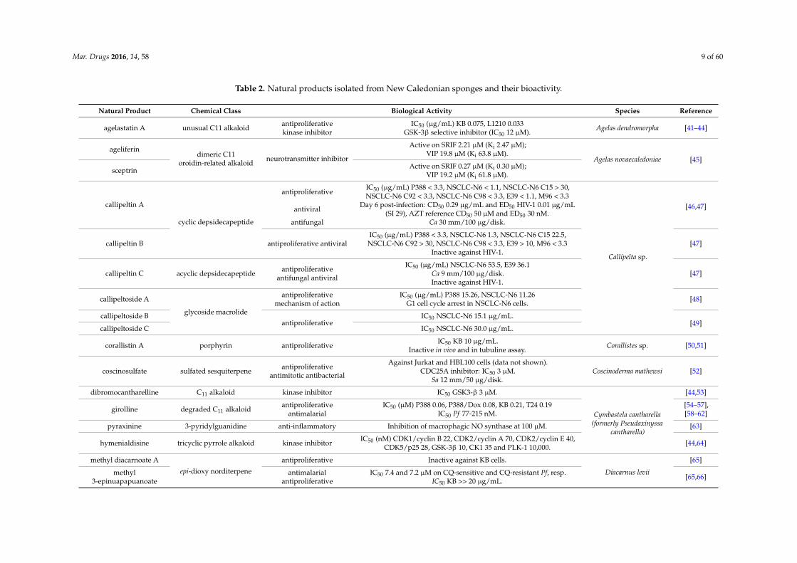
Most shallow-water sponge species live in symbiosis with Archaea, eubacteria and cyanobacteria,forming holobiont systems that represent the main source of bioactive compounds [38,39] primarilyisolated from host tissues, but occasionally isolated from cultures of associated microbiota. Spongescontain a wide range of so-called secondary metabolites, some of which afford interesting biologicalactivities [40] (Table 2).
Mar. Drugs 2016, 14, 58 9 of 60
Table 2. Natural products isolated from New Caledonian sponges and their bioactivity.
Natural Product Chemical Class Biological Activity Species Reference
agelastatin A unusual C11 alkaloid antiproliferativekinase inhibitor
IC50 (µg/mL) KB 0.075, L1210 0.033GSK-3β selective inhibitor (IC50 12 µM). Agelas dendromorpha [41–44]
ageliferindimeric C11
oroidin-related alkaloidneurotransmitter inhibitor
Active on SRIF 2.21 µM (Ki 2.47 µM);VIP 19.8 µM (Ki 63.8 µM).
Agelas novaecaledoniae [45]sceptrin Active on SRIF 0.27 µM (Ki 0.30 µM);
VIP 19.2 µM (Ki 61.8 µM).
callipeltin A
cyclic depsidecapeptide
antiproliferativeIC50 (µg/mL) P388 < 3.3, NSCLC-N6 < 1.1, NSCLC-N6 C15 > 30,NSCLC-N6 C92 < 3.3, NSCLC-N6 C98 < 3.3, E39 < 1.1, M96 < 3.3
Callipelta sp.
[46,47]antiviralDay 6 post-infection: CD50 0.29 µg/mL and ED50 HIV-1 0.01 µg/mL
(SI 29), AZT reference CD50 50 µM and ED50 30 nM.antifungal Ca 30 mm/100 µg/disk.
callipeltin B antiproliferative antiviralIC50 (µg/mL) P388 < 3.3, NSCLC-N6 1.3, NSCLC-N6 C15 22.5,
NSCLC-N6 C92 > 30, NSCLC-N6 C98 < 3.3, E39 > 10, M96 < 3.3Inactive against HIV-1.
[47]
callipeltin C acyclic depsidecapeptide antiproliferativeantifungal antiviral
IC50 (µg/mL) NSCLC-N6 53.5, E39 36.1Ca 9 mm/100 µg/disk.Inactive against HIV-1.
[47]
callipeltoside A
glycoside macrolide
antiproliferativemechanism of action
IC50 (µg/mL) P388 15.26, NSCLC-N6 11.26G1 cell cycle arrest in NSCLC-N6 cells. [48]
callipeltoside Bantiproliferative
IC50 NSCLC-N6 15.1 µg/mL.[49]
callipeltoside C IC50 NSCLC-N6 30.0 µg/mL.
corallistin A porphyrin antiproliferative IC50 KB 10 µg/mL.Inactive in vivo and in tubuline assay. Corallistes sp. [50,51]
coscinosulfate sulfated sesquiterpene antiproliferativeantimitotic antibacterial
Against Jurkat and HBL100 cells (data not shown).CDC25A inhibitor: IC50 3 µM.
Sa 12 mm/50 µg/disk.Coscinoderma mathewsi [52]
dibromocantharelline C11 alkaloid kinase inhibitor IC50 GSK3-β 3 µM.
Cymbastela cantharella(formerly Pseudaxinyssa
cantharella)
[44,53]
girolline degraded C11 alkaloid antiproliferativeantimalarial
IC50 (µM) P388 0.06, P388/Dox 0.08, KB 0.21, T24 0.19IC50 Pf 77-215 nM.
[54–57],[58–62]
pyraxinine 3-pyridylguanidine anti-inflammatory Inhibition of macrophagic NO synthase at 100 µM. [63]
hymenialdisine tricyclic pyrrole alkaloid kinase inhibitor IC50 (nM) CDK1/cyclin B 22, CDK2/cyclin A 70, CDK2/cyclin E 40,CDK5/p25 28, GSK-3β 10, CK1 35 and PLK-1 10,000. [44,64]
methyl diacarnoate A
epi-dioxy norditerpene
antiproliferative Inactive against KB cells.
Diacarnus levii
[65]
methyl3-epinuapapuanoate
antimalarialantiproliferative
IC50 7.4 and 7.2 µM on CQ-sensitive and CQ-resistant Pf, resp.IC50 KB >> 20 µg/mL. [65,66]

Mar. Drugs 2016, 14, 58 10 of 60
Table 2. Cont.
Natural Product Chemical Class Biological Activity Species Reference
2-epimukubilin benzyl ester
epi-dioxy norsesterterpene antiproliferative
IC50 KB 1.0 µg/mL.
[65]methyl prenyldiacarnoate A IC50 KB 3.3 µg/mL.
methyl2-epiprenyldiacarnoate A IC50 KB 0.9 µg/mL.
nortopsentin D bis-indole alkaloid antiproliferative EC50 KB 0.014 µg/mL (permethylated derivative). Dragmacidon sp. [67]
arsenicin A polyarsenic antibacterial antifungal 10 µg/disk, Sa/Ec/Ca: 24/28/26 (mm) for arsenicin A, and 22/30/22(mm) for gentamicin. Echinochalina bargibanti [68]
euryspongiol A1 polyhydroxylated9,11-secosterol antihistaminic
Reduction of histamine release by 26% (control 35%). Euryspongia sp. [69]euryspongiol A2 Reduction of histamine release by 15% (control 35%).
homophymine A
cyclodepsipeptide
antiviral antiproliferative
IC50 HIV-1 75 nM. IC50 (nM) KB 7.3, MCF-7 23.6, MCF-7R 22.9,HCT116 6.0, HCT15 22.5, HT29 70.0, OVCAR8 5.4, OV3 7.5, PC3 4.2,Vero 5.0, MRC5 11.0, HL60 24.1, HL60R 22.4, K562 24.0, PaCa 31.4,
SF268 9.9, A549 8.3, MDA231 10.9, MDA435 39.0, HepG2 68.6, EPC 5.0
Homophymia sp.
[70,71]
homophymine B antiproliferativemechanism of action
IC50 (nM) KB 18.0, MCF-7 16.8, MCF-7R 26.3, HCT116 13.8, HCT1522.9, HT29 101.9, OVCAR8 8.0, OV3 9.9, PC3 6.2, Vero 8.6, MRC5 17.1,
HL60 43.1, HL60R 36.7, K562 26.7, PaCa 62.0, SF268 17.2, A549 19.8,MDA231 17.0, MDA435 40.1, HepG2 99.0, EPC 8.0.Caspase-independent cell death pathway (HL60).
[71]
homophymine C
antiproliferative
IC50 (nM) KB 8.5, MCF-7 8.8, MCF-7R 10.8, HCT116 4.9, HCT15 19.2,HT29 62.8, OVCAR8 4.3, OV3 3.7, PC3 3.0, Vero 4.2, MRC5 16.8, HL6023.0, HL60R 23.5, K562 22.5, PaCa 25.9, SF268 13.6, A549 8.3, MDA231
16.2, MDA435 35.0, HepG2 72.1, EPC 9.3
homophymine D
IC50 (nM) KB 12.7, MCF-7 19.6, MCF-7R 37.7, HCT116 19.8, HCT1543.2, HT29 81.3, OVCAR8 8.1, OV3 10.6, PC3 6.3, Vero 10.9, MRC5 16.9,
HL60 29.6, HL60R 24.9, K562 35.3, PaCa 37.4, SF268 17.9, A549 13.8,MDA231 18.9, MDA435 49.9, HepG2 78.7, EPC 11.1
homophymine E
IC50 (nM) KB 6.0, MCF-7 14.2, MCF-7R 15.6, HCT116 5.5, HCT15 27.2,HT29 35.1, OVCAR8 4.6, OV3 4.2, PC3 3.9, Vero 7.0, MRC5 9.5, HL6023.3, HL60R 21.4, K562 22.2, PaCa 18.1, SF268 8.1, A549 9.6, MDA231
13.3, MDA435 38.3, HepG2 60.5, EPC 9.5
homophymine A1
IC50 (nM) KB 7.1, MCF-7 12.4, MCF-7R 13.5, HCT116 6.1, HCT15 13.5,HT29 30.9, OVCAR8 5.1, OV3 5.5, PC3 3.7, Vero 6.1, MRC5 7.8, HL6017.3, HL60R 11.1, K562 12.8, PaCa 19.2, SF268 6.3, A549 6.0, MDA231
8.4, MDA435 27.0, HepG2 91.4, EPC 7.8

Mar. Drugs 2016, 14, 58 11 of 60
Table 2. Cont.
Natural Product Chemical Class Biological Activity Species Reference
homophymine B1
cyclodepsipeptide
antiproliferative
IC50 (nM) KB 16.4, MCF-7 14.2, MCF-7R 12.3, HCT116 11.4, HCT1514.1, HT29 93.8, OVCAR8 6.5, OV3 8.0, PC3 4.7, Vero 6.1, MRC5 10.2,
HL60 18.7, HL60R 25.8, K562 16.6, PaCa 22.2, SF268 11.7, A549 8.6,MDA231 18.2, MDA435 29.5, HepG2 100.3, EPC 6.6
Homophymia sp. [71]
homophymine C1 antiproliferativemechanism of action
IC50 (nM) KB 6.8, MCF-7 6.3, MCF-7R 5.4, HCT116 2.7, HCT15 17.2,HT29 38.2, OVCAR8 2.6, OV3 2.4, PC3 2.6, Vero 3.1, MRC5 8.0, HL6014.6, HL60R 17.1, K562 11.9, PaCa 14.4, SF268 7.1, A549 6.2, MDA23115.8, MDA435 20.3, HepG2 58.6, EPC 12.2 Caspase-independent cell
death pathway (HL60).
homophymine D1 antiproliferativemechanism of action
IC50 (nM) KB 10.6, MCF-7 3.5, MCF-7R 3.5, HCT116 1.8, HCT15 11.4,HT29 32.2, OVCAR8 1.6, OV3 1.4, PC3 1.4, Vero 1.8, MRC5 10.5, HL6013.1, HL60R 21.9, K562 12.9, PaCa 17.6, SF268 7.9, A549 5.0, MDA23111.1, MDA435 23.4, HepG2 80.4, EPC 7.7 Caspase-independent cell
death pathway (HL60).
homophymine E1 antiproliferative
IC50 (nM) KB 12.5, MCF-7 3.9, MCF-7R 7.1, HCT116 2.3, HCT15 10.1,HT29 31.8, OVCAR8 4.0, OV3 2.7, PC3 3.5, Vero 4.4, MRC5 12.3, HL6020.5, HL60R 23.2, K562 17.8, PaCa 10.6, SF268 10.1, A549 11.4, MDA231
20.0, MDA435 37.0, HepG2 62.8, EPC 29.0
12-epi-heteroneminscalarane sesterterpene farnesyltransferase
inhibitor
Inactive on farnesyl transferase. Hyrtios erecta [72,73]
heteronemin IC50 3 µM. Hyrtios reticulata [73]
thorectolideterpene
antiproliferative antiviral IC50 KB 5.3 µg/mL.HIV-1 nucleocapside and integrase inhibitor 10 and 20 µg/mL, resp.
Hyrtios sp.
[74]thorectolide monoacetate antiproliferative
anti-inflammatoryIC50 KB 0.3 µg/mL.
Cobra venom PLA2 inhibitor 2 µM, bee venom PLA2 inactive.
puupehenone
merosesquiterpene
antiproliferativeantifungal antibacterial
antimalarial
IC50 KB 0.8 µg/mL.Ct 12 mm/50 µg/disk.Sa 12 mm/50 µg/disk.
IC50 (µg/mL) Pf F32 0.6, FcB1 2.1 and PFB 1.5
[75,76]
dipuupehedione antiproliferativeantifungal antibacterial
IC50 KB 3 µg/mL.Inactive against Ct.Inactive against Sa.
[75]
15α-methoxypuupehenolantiproliferative
antibacterial antifungalantimalarial
IC50 KB 6 µg/mL.Sa 7 mm/1 µg/disk.
Ct 9 mm/50 µg/disk.IC50 (µg/mL) Pf F32 0.4, FcB1 1.4 and PFB 1.2
[76]
Mar. Drugs 2016, 14, 58 12 of 60
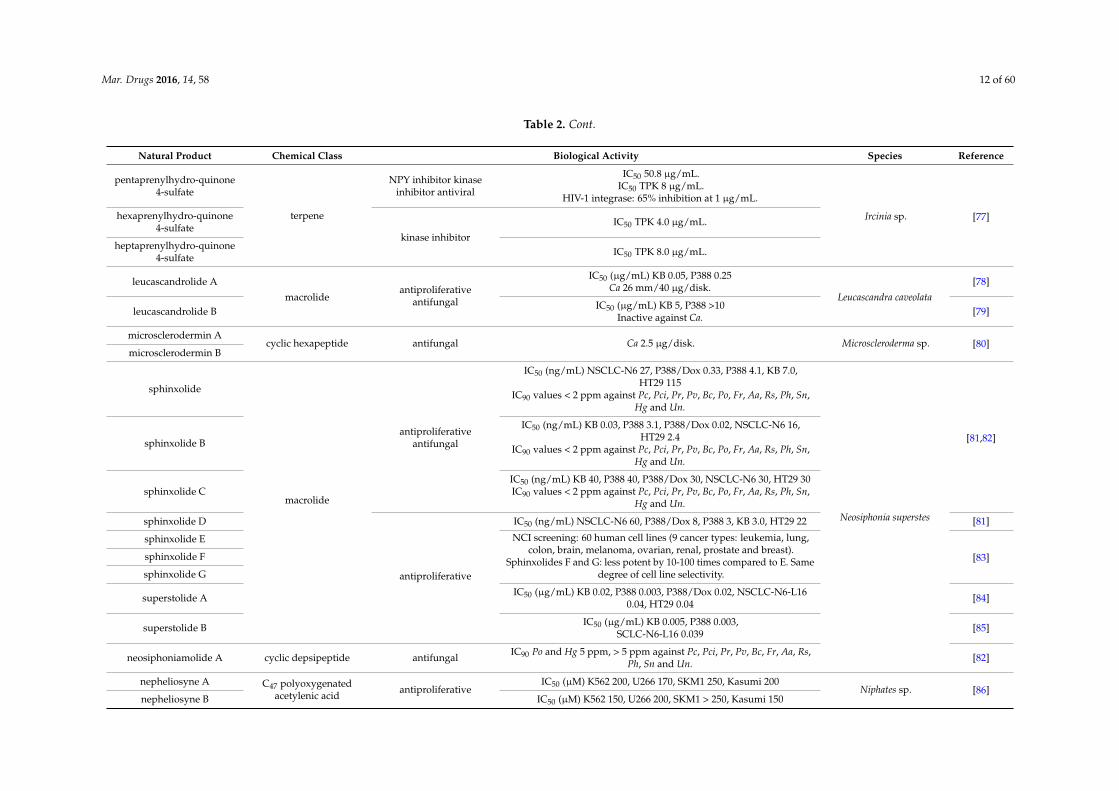
Table 2. Cont.
Natural Product Chemical Class Biological Activity Species Reference
pentaprenylhydro-quinone4-sulfate
terpene
NPY inhibitor kinaseinhibitor antiviral
IC50 50.8 µg/mL.IC50 TPK 8 µg/mL.
HIV-1 integrase: 65% inhibition at 1 µg/mL.
Ircinia sp. [77]hexaprenylhydro-quinone4-sulfate
kinase inhibitor
IC50 TPK 4.0 µg/mL.
heptaprenylhydro-quinone4-sulfate IC50 TPK 8.0 µg/mL.
leucascandrolide A
macrolideantiproliferative
antifungal
IC50 (µg/mL) KB 0.05, P388 0.25Ca 26 mm/40 µg/disk.
Leucascandra caveolata[78]
leucascandrolide B IC50 (µg/mL) KB 5, P388 >10Inactive against Ca. [79]
microsclerodermin Acyclic hexapeptide antifungal Ca 2.5 µg/disk. Microscleroderma sp. [80]
microsclerodermin B
sphinxolide
macrolide
antiproliferativeantifungal
IC50 (ng/mL) NSCLC-N6 27, P388/Dox 0.33, P388 4.1, KB 7.0,HT29 115
IC90 values < 2 ppm against Pc, Pci, Pr, Pv, Bc, Po, Fr, Aa, Rs, Ph, Sn,Hg and Un.
Neosiphonia superstes
[81,82]sphinxolide B
IC50 (ng/mL) KB 0.03, P388 3.1, P388/Dox 0.02, NSCLC-N6 16,HT29 2.4
IC90 values < 2 ppm against Pc, Pci, Pr, Pv, Bc, Po, Fr, Aa, Rs, Ph, Sn,Hg and Un.
sphinxolide CIC50 (ng/mL) KB 40, P388 40, P388/Dox 30, NSCLC-N6 30, HT29 30IC90 values < 2 ppm against Pc, Pci, Pr, Pv, Bc, Po, Fr, Aa, Rs, Ph, Sn,
Hg and Un.
sphinxolide D
antiproliferative
IC50 (ng/mL) NSCLC-N6 60, P388/Dox 8, P388 3, KB 3.0, HT29 22 [81]
sphinxolide E NCI screening: 60 human cell lines (9 cancer types: leukemia, lung,colon, brain, melanoma, ovarian, renal, prostate and breast).
Sphinxolides F and G: less potent by 10-100 times compared to E. Samedegree of cell line selectivity.
[83]sphinxolide F
sphinxolide G
superstolide A IC50 (µg/mL) KB 0.02, P388 0.003, P388/Dox 0.02, NSCLC-N6-L160.04, HT29 0.04 [84]
superstolide B IC50 (µg/mL) KB 0.005, P388 0.003,SCLC-N6-L16 0.039 [85]
neosiphoniamolide A cyclic depsipeptide antifungal IC90 Po and Hg 5 ppm, > 5 ppm against Pc, Pci, Pr, Pv, Bc, Fr, Aa, Rs,Ph, Sn and Un. [82]
nepheliosyne A C47 polyoxygenatedacetylenic acid
antiproliferativeIC50 (µM) K562 200, U266 170, SKM1 250, Kasumi 200
Niphates sp. [86]nepheliosyne B IC50 (µM) K562 150, U266 200, SKM1 > 250, Kasumi 150
Mar. Drugs 2016, 14, 58 13 of 60
Table 2. Cont.
Natural Product Chemical Class Biological Activity Species Reference
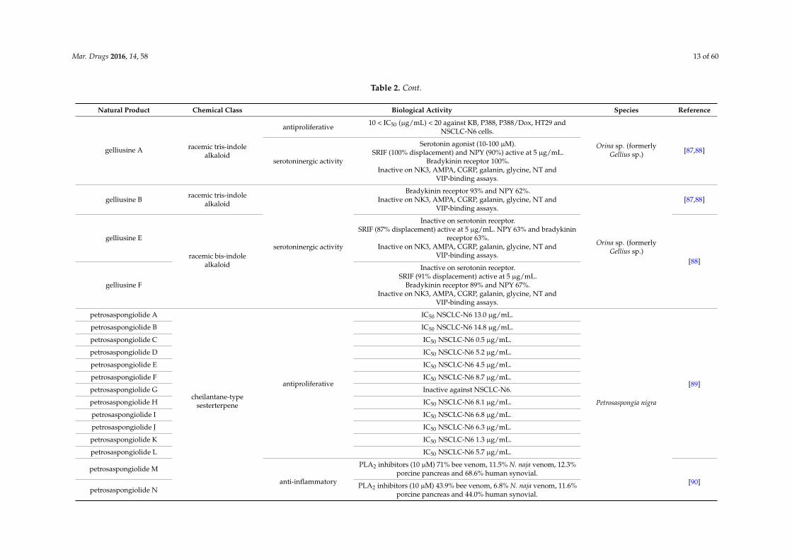
gelliusine A racemic tris-indolealkaloid
antiproliferative 10 < IC50 (µg/mL) < 20 against KB, P388, P388/Dox, HT29 andNSCLC-N6 cells.
Orina sp. (formerlyGellius sp.) [87,88]
serotoninergic activity
Serotonin agonist (10-100 µM).SRIF (100% displacement) and NPY (90%) active at 5 µg/mL.
Bradykinin receptor 100%.Inactive on NK3, AMPA, CGRP, galanin, glycine, NT and
VIP-binding assays.
gelliusine B racemic tris-indolealkaloid
serotoninergic activity
Bradykinin receptor 93% and NPY 62%.Inactive on NK3, AMPA, CGRP, galanin, glycine, NT and
VIP-binding assays.
Orina sp. (formerlyGellius sp.)
[87,88]
gelliusine E
racemic bis-indolealkaloid
Inactive on serotonin receptor.SRIF (87% displacement) active at 5 µg/mL. NPY 63% and bradykinin
receptor 63%.Inactive on NK3, AMPA, CGRP, galanin, glycine, NT and
VIP-binding assays.[88]
gelliusine F
Inactive on serotonin receptor.SRIF (91% displacement) active at 5 µg/mL.
Bradykinin receptor 89% and NPY 67%.Inactive on NK3, AMPA, CGRP, galanin, glycine, NT and
VIP-binding assays.
petrosaspongiolide A
cheilantane-typesesterterpene
antiproliferative
IC50 NSCLC-N6 13.0 µg/mL.
Petrosaspongia nigra
[89]
petrosaspongiolide B IC50 NSCLC-N6 14.8 µg/mL.
petrosaspongiolide C IC50 NSCLC-N6 0.5 µg/mL.
petrosaspongiolide D IC50 NSCLC-N6 5.2 µg/mL.
petrosaspongiolide E IC50 NSCLC-N6 4.5 µg/mL.
petrosaspongiolide F IC50 NSCLC-N6 8.7 µg/mL.
petrosaspongiolide G Inactive against NSCLC-N6.
petrosaspongiolide H IC50 NSCLC-N6 8.1 µg/mL.
petrosaspongiolide I IC50 NSCLC-N6 6.8 µg/mL.
petrosaspongiolide J IC50 NSCLC-N6 6.3 µg/mL.
petrosaspongiolide K IC50 NSCLC-N6 1.3 µg/mL.
petrosaspongiolide L IC50 NSCLC-N6 5.7 µg/mL.
petrosaspongiolide M
anti-inflammatory
PLA2 inhibitors (10 µM) 71% bee venom, 11.5% N. naja venom, 12.3%porcine pancreas and 68.6% human synovial.
[90]petrosaspongiolide N PLA2 inhibitors (10 µM) 43.9% bee venom, 6.8% N. naja venom, 11.6%
porcine pancreas and 44.0% human synovial.

Mar. Drugs 2016, 14, 58 14 of 60
Table 2. Cont.
Natural Product Chemical Class Biological Activity Species Reference
petrosaspongiolide P
cheilantane-typesesterterpene
anti-inflammatory
PLA2 inhibitors (10 µM) 37.9% bee venom, 3.0% N. naja venom, 0.0%porcine pancreas and 60.9% human synovial.
Petrosaspongia nigra [90]petrosaspongiolide Q PLA2 inhibitors (10 µM) 12.5% bee venom, 4.2% N. naja venom, 0.0%porcine pancreas and 30.1% human synovial.
petrosaspongiolide R PLA2 inhibitors (10 µM) 18.8% bee venom, 1.0% N. naja venom, 0.8%porcine pancreas and 7.1% human synovial.
phloeodictine A
guanidine alkaloid antiproliferativeantibacterial
IC50 KB 1.5 µg/mL.MIC (µg/mL) Sf 5, Sa 1, Ec 1, Pa 10
Phloeodictyon sp.
[91]phloeodictine B IC50 KB 11.2 µg/mL.
MIC (µg/mL) Sf > 15, Sa 3, Ec 30, Pa > 30
phloeodictine A1 IC50 KB 2.2 µg/mL.2.6:1 mixture of phloeodictine A1 and A2. MIC (µg/mL) Sa 3, Ec 3, Pa
30, Cp 30, Bf 10 and Pas 10
[92]
phloeodictine A2
phloeodictine A3IC50 KB 3.5 µg/mL.
2.6:0.7:0.3 mixture of phloeodictine A3, A4 and A5.MIC (µg/mL) Sa 30, Ec 30, Pa > 30, Cp > 30, Bf > 30 and Pas > 30
phloeodictine A4
phloeodictine A5
phloeodictine A6 IC50 KB 0.6 µg/mL.1:1.4 mixture of phloeodictine A6 and A7.
MIC (µg/mL) Sa 1, Ec 3, Pa 30, Cp 1, Bf 3 and Pas 3phloeodictine A7
phloeodictine C1 IC50 KB 1.8 µg/mL. 1:1 mixture of phloeodictine C1 and C2. MIC(µg/mL) Sa 3, Ec > 30, Pa > 30, Cp > 100, Bf > 100 and Pas > 100phloeodictine C2
chondropsin A
macrolide lactam
antiproliferative
IC50 (nM) KB 1.5, HCT116 1.2, T47D 0.45, HBL100 1.7 and Changliver 2.4
G2/M cell cycle arrest in HL60 and KB cell lines (Ñ apoptosis).
Psammoclemma sp.
[93]
73-deoxychondropsin AIC50 (nM) KB 0.28, HCT116 0.22, T47D 0.18, HBL100 0.60, Chang
liver 0.24G2/M cell cycle arrest in HL60 and KB cell lines (Ñ apoptosis).
echinosulfonic acid D alkaloid IC50 KB 2 µg/mL. [94]
psammaplysene Cbromotyrosine alkaloid IC50 THP-1 7 µM. [95]
psammaplysene D
reidispongiolide A
sphinxolide-typemacrolide
antiproliferative
IC50 (µg/mL) KB 0.10, P388 0.16, P388/Dox 0.01,NSCLC-N6 0.07, HT29 0.04
Reidispongia coerulea
[83,96]
reidispongiolide B IC50 (µg/mL) KB 0.06, P388 0.06, P388/Dox 0.02,NSCLC-N6 0.05, HT29 0.04 [96]
reidispongiolide CNCI screening: 60 human cell lines (9 cancer types: leukemia, lung,colon, brain, melanoma, ovarian, renal, prostate and breast). Same
degree of cell line selectivity.[83]
Mar. Drugs 2016, 14, 58 15 of 60
Table 2. Cont.
Natural Product Chemical Class Biological Activity Species Reference
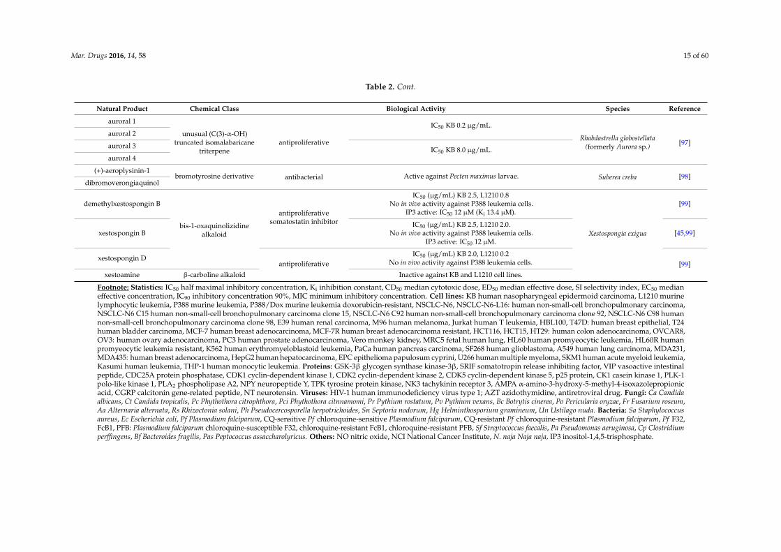
auroral 1
unusual (C(3)-α-OH)truncated isomalabaricane
triterpeneantiproliferative
IC50 KB 0.2 µg/mL.
Rhabdastrella globostellata(formerly Aurora sp.) [97]
auroral 2
auroral 3 IC50 KB 8.0 µg/mL.auroral 4
(+)-aeroplysinin-1bromotyrosine derivative antibacterial Active against Pecten maximus larvae. Suberea creba [98]
dibromoverongiaquinol
demethylxestospongin B
bis-1-oxaquinolizidinealkaloid
antiproliferativesomatostatin inhibitor
IC50 (µg/mL) KB 2.5, L1210 0.8No in vivo activity against P388 leukemia cells.
IP3 active: IC50 12 µM (Ki 13.4 µM).
Xestospongia exigua
[99]
xestospongin BIC50 (µg/mL) KB 2.5, L1210 2.0.
No in vivo activity against P388 leukemia cells.IP3 active: IC50 12 µM.
[45,99]
xestospongin Dantiproliferative
IC50 (µg/mL) KB 2.0, L1210 0.2No in vivo activity against P388 leukemia cells. [99]
xestoamine β-carboline alkaloid Inactive against KB and L1210 cell lines.
Footnote: Statistics: IC50 half maximal inhibitory concentration, Ki inhibition constant, CD50 median cytotoxic dose, ED50 median effective dose, SI selectivity index, EC50 medianeffective concentration, IC90 inhibitory concentration 90%, MIC minimum inhibitory concentration. Cell lines: KB human nasopharyngeal epidermoid carcinoma, L1210 murinelymphocytic leukemia, P388 murine leukemia, P388/Dox murine leukemia doxorubicin-resistant, NSCLC-N6, NSCLC-N6-L16: human non-small-cell bronchopulmonary carcinoma,NSCLC-N6 C15 human non-small-cell bronchopulmonary carcinoma clone 15, NSCLC-N6 C92 human non-small-cell bronchopulmonary carcinoma clone 92, NSCLC-N6 C98 humannon-small-cell bronchopulmonary carcinoma clone 98, E39 human renal carcinoma, M96 human melanoma, Jurkat human T leukemia, HBL100, T47D: human breast epithelial, T24human bladder carcinoma, MCF-7 human breast adenocarcinoma, MCF-7R human breast adenocarcinoma resistant, HCT116, HCT15, HT29: human colon adenocarcinoma, OVCAR8,OV3: human ovary adenocarcinoma, PC3 human prostate adenocarcinoma, Vero monkey kidney, MRC5 fetal human lung, HL60 human promyeocytic leukemia, HL60R humanpromyeocytic leukemia resistant, K562 human erythromyeloblastoid leukemia, PaCa human pancreas carcinoma, SF268 human glioblastoma, A549 human lung carcinoma, MDA231,MDA435: human breast adenocarcinoma, HepG2 human hepatocarcinoma, EPC epithelioma papulosum cyprini, U266 human multiple myeloma, SKM1 human acute myeloid leukemia,Kasumi human leukemia, THP-1 human monocytic leukemia. Proteins: GSK-3β glycogen synthase kinase-3β, SRIF somatotropin release inhibiting factor, VIP vasoactive intestinalpeptide, CDC25A protein phosphatase, CDK1 cyclin-dependent kinase 1, CDK2 cyclin-dependent kinase 2, CDK5 cyclin-dependent kinase 5, p25 protein, CK1 casein kinase 1, PLK-1polo-like kinase 1, PLA2 phospholipase A2, NPY neuropeptide Y, TPK tyrosine protein kinase, NK3 tachykinin receptor 3, AMPA α-amino-3-hydroxy-5-methyl-4-isoxazolepropionicacid, CGRP calcitonin gene-related peptide, NT neurotensin. Viruses: HIV-1 human immunodeficiency virus type 1; AZT azidothymidine, antiretroviral drug. Fungi: Ca Candidaalbicans, Ct Candida tropicalis, Pc Phythothora citrophthora, Pci Phythothora citnnamomi, Pr Pythium rostatum, Pv Pythium vexans, Bc Botrytis cinerea, Po Pericularia oryzae, Fr Fusarium roseum,Aa Alternaria alternata, Rs Rhizoctonia solani, Ph Pseudocercosporella herpotrichoides, Sn Septoria nodorum, Hg Helminthosporium gramineum, Un Ustilago nuda. Bacteria: Sa Staphylococcusaureus, Ec Escherichia coli, Pf Plasmodium falciparum, CQ-sensitive Pf chloroquine-sensitive Plasmodium falciparum, CQ-resistant Pf chloroquine-resistant Plasmodium falciparum, Pf F32,FcB1, PFB: Plasmodium falciparum chloroquine-susceptible F32, chloroquine-resistant FcB1, chloroquine-resistant PFB, Sf Streptococcus faecalis, Pa Pseudomonas aeruginosa, Cp Clostridiumperffingens, Bf Bacteroides fragilis, Pas Peptococcus assaccharolyricus. Others: NO nitric oxide, NCI National Cancer Institute, N. naja Naja naja, IP3 inositol-1,4,5-trisphosphate.

Mar. Drugs 2016, 14, 58 16 of 60
5.1.2. Porifera Success Stories
New Caledonian sponges have revealed more exciting chemicals than any other studied taxon,in terms of carbon skeleton, degree and patterns of unsaturation, halogenation, functional grouporiginality, presence of unusual heteroatoms etc. Some of these features are responsible for the ratherexceptional bioactivity profiles encountered. Some outstanding examples are described below:
Cymbastela Cantharella
Cymbastela cantharella (Family Axinellidae, Class Demospongiae, formerly Pseudaxinyssacantharella, Figure 5A) was collected on the outer south reef of New Caledonia at depths rangingfrom 10 to 40 m (voucher #R1279, IRD Nouméa).
Chemical analyses led to the isolation of sterols that contain a 3β-hydroxymethyl-A-norcholestaneskeleton, a typical feature of the Axinellidae (1–3, Figure 5B) [100], along with alkaloids such as odiline(4, Figure 5B), dibromocantharelline (5, Figure 5B) and dibromophakellin (6, Figure 5B) [53]. Studiesperformed on the crude ethanol extracts of C. cantharella led to the isolation of girolline (7, Figure 5B). Itsabsolute configuration has been established by X-ray diffraction [56] and total synthesis was achievedfew years later [53,57,58,63,100].
Girolline (7, Figure 5B) (or girodazole) has demonstrated potent antiproliferative activity in vitroon several cell lines such as P388 murine leukemia and P388/dox, a sub-line resistant to doxorubicin, ananthracycline, or KB naso-pharyngeal and T24 bladder carcinoma human cell lines. The half maximalinhibitory concentration (IC50) values are 0.06 µM, 0.08 µM, 0.21 µM and 0.19 µM, respectively [55].Subsequent in vivo assays have been performed on several grafted murine tumors used for preclinicalevaluation including P388 and L1210 leukemia, and solid tumors such as M5076 histiocytosarcomaand MA/16C mammary adenocarcinoma cells [54,59,63]. Moreover, girodazole (7, Figure 5B) also hasantitumor activity in vivo on the P388/dox cell line. This compound may inhibit the termination stepof protein synthesis in vivo [60,61]. Although toxicological studies in mice and dogs do not show anymajor toxic effects, girodazole (7, Figure 5B) clinical development was interrupted in 1991 in phase IIdue to severe side effects, in particular cardiovascular toxicity [59].
Girolline (7, Figure 5B) has also shown promising results for the treatment of malaria.This compound has demonstrated potent antiplasmodial activities against four Plasmodium falciparumstrains with IC50 values ranging from 77 to 215 nM, and may act synergistically with chloroquinein vitro by affecting protein synthesis [62].
Other studies have led to the characterization of pyraxinine (8, Figure 5B), a novel nitrogenouscompound, along with previously identified allantoin (9, Figure 5B), homarine (10, Figure 5B) andtrigonelline nitrogen compounds (11, Figure 5B) [63], hymenialdisine (previously identified; 12,Figure 5B) and new pyrrole-2-aminoimidazole alkaloids, which are dihydrohymenialdisine derivatives(13, Figure 5B) [64]. Further investigations revealed that hymenialdisine (12, Figure 5B) is a powerfulinhibitor of cyclin-dependent kinases such as CDK1/cyclin B, CDK2/cyclin A and CDK2/cyclin E(IC50 values of 22 nM, 70 nM, and 40 nM, respectively), CDK5/p25 (IC50 of 28 nM), as well as againstglycogen synthase kinase-3β (GSK-3β) with an IC50 of 10 nM and casein kinase 1 (CK1) with an IC50
of 35 nM [44].

Mar. Drugs 2016, 14, 58 17 of 60
Mar. Drugs 2016, 14, x 19 of 62
(A)
(9) (10) (11)
(12) (13)
HN
NH
O
O
NH
O
NH2O-
ON+
O-
O
N+
NH
NH
O
N
OHN
H2N
BrNH
NH
HN
N
O
OH2N
HH
Br
(B)
Figure 5. (A) Cymbastela cantharella. © IRD; (B) Structure of
hydroxymethyl‐3β‐methyl‐24S‐nor‐A‐cholest‐5α‐ene‐22‐Z (1),
hydroxymethyl‐3β‐methyl‐24R‐nor‐A‐cholest‐5α‐ene‐22‐Z (2),
hydroxymethyl‐3β‐ethyl‐24ξ‐methyl‐26ξ‐nor‐A‐cholest‐5α‐ene‐22‐E (3), odiline (4),
Figure 5. (A) Cymbastela cantharella. © IRD; (B) Structure of hydroxymethyl-3β-methyl-24S-nor-A-cholest-5α-ene-22-Z (1), hydroxymethyl-3β-methyl-24R-nor-A-cholest-5α-ene-22-Z (2),hydroxymethyl-3β-ethyl-24ξ-methyl-26ξ-nor-A-cholest-5α-ene-22-E (3), odiline (4), dibromocantharelline(5), dibromophakellin (6), girolline (7), pyraxinine (8), allantoin (9), homarine (10), trigonelline (11),hymenialdisine (12) and dihydrohymenialdisine (13).

Mar. Drugs 2016, 14, 58 18 of 60
Echinochalina Bargibanti
The demosponge Echinochalina bargibanti (voucher #R1858, IRD Nouméa, Figure 6A) was collectedalong the northeastern coast of Grande Terre between 18 and 25 m depth during the SMIB program.A bioassay-guided fractionation of its organic extracts led to the isolation of arsenicin A (14, Figure 6B),the first polyarsenic compound ever found in nature.
Arsenicin A (14, Figure 6B) demonstrates potent bactericidal and fungicidal activities againsthuman pathogenic strains such as Staphylococcus aureus, Escherichia coli and Candida albicans withinhibition circles of 24, 28 and 26 mm respectively at 10 µg per disk. In contrast, gentamicin, which isthe reference antibiotic, induces inhibition circles of 22, 30 and 22 mm [68]. Arsenicin A (14, Figure 6B)has been synthesized and its crystal structure determined [101]. An improvement in this synthesis hasled to (˘)-arsenicin A which shows potent antiproliferative activity on acute promyelocytic leukemiacell lines. This compound is more potent than arsenic trioxide (Trisenox) which is used for treatingacute promyelocytic leukemia. (˘)-Arsenicin A has also demonstrated antiproliferative activity againstpancreatic adenocarcinomas and glioblastomas [102].
Mar. Drugs 2016, 14, x 20 of 62
dibromocantharelline (5), dibromophakellin (6), girolline (7), pyraxinine (8), allantoin (9), homarine
(10), trigonelline (11), hymenialdisine (12) and dihydrohymenialdisine (13).
Echinochalina Bargibanti
The demosponge Echinochalina bargibanti (voucher #R1858, IRD Nouméa, Figure 6A) was
collected along the northeastern coast of Grande Terre between 18 and 25 m depth during the SMIB
program. A bioassay‐guided fractionation of its organic extracts led to the isolation of arsenicin A
(14, Figure 6B), the first polyarsenic compound ever found in nature.
Arsenicin A (14, Figure 6B) demonstrates potent bactericidal and fungicidal activities against
human pathogenic strains such as Staphylococcus aureus, Escherichia coli and Candida albicans with
inhibition circles of 24, 28 and 26 mm respectively at 10 μg per disk. In contrast, gentamicin, which is
the reference antibiotic, induces inhibition circles of 22, 30 and 22 mm [68]. Arsenicin A (14, Figure 6B)
has been synthesized and its crystal structure determined [101]. An improvement in this synthesis
has led to (±)‐arsenicin A which shows potent antiproliferative activity on acute promyelocytic
leukemia cell lines. This compound is more potent than arsenic trioxide (Trisenox) which is used for
treating acute promyelocytic leukemia. (±)‐Arsenicin A has also demonstrated antiproliferative
activity against pancreatic adenocarcinomas and glioblastomas [102].
(14)
AsAs O
AsO
OAs
(A) (B)
Figure 6. (A) Echinochalina bargibanti. © IRD; (B) Structure of arsenicin A (14).
Dendrilla sp.
Dendrilla sp. (voucher #R171, IRD Nouméa) is a common shallow‐water (lagoon, at
approximately 20 m depth) dendroceratid sponge belonging to the Demospongiae. It features an
unusual “scouring pad” appearance with its spongin fibers protruding from an elastic tissue mass
that can vary in color from dark green to reddish.
R171 is cytotoxic in brine shrimp larvae bioassays [103], and its crude aqueous extract is toxic to
the mosquito fish Gambusia affinis, causing erratic swimming patterns followed by 100% mortality
within 12 h [29]. Reported biological activities of the crude organic extracts of Dendrilla nigra (Figure
7) include antitumor, anti‐inflammatory and antimicrobial [103] properties with potential interest
for shrimp aquaculture for treating Vibrio pathogens [104].
Chemically, dendrillid sponges are known to contain lamellarins A–B (15–18, Figure 8),
aromatic alkaloids of probable symbiotic origin. These pyrrole derivatives are antitumoral (HIF‐1
inhibitors) [105]. Dendrilla cactos contains cyclic bastadins, which are bromotyrosine‐derived peptides
endowed with Gram+ antibacterial and anti‐inflammatory properties; in addition they inhibit
topoisomerase II, dehydrofolate reductase and the endothelin A receptor [106]. Cold‐water Dendrilla
membranosa contains membranolides used as natural antifeedants and other microbe‐derived
bioactive compounds [107]. The suspicion that indwelling bacteria may be responsible for much of
the reported bioactivities in sponges in the Dendrilla genus has prompted the taxonomic
characterization of culturable strains in Dendrilla nigra [108].
Figure 6. (A) Echinochalina bargibanti. © IRD; (B) Structure of arsenicin A (14).
Dendrilla sp.
Dendrilla sp. (voucher #R171, IRD Nouméa) is a common shallow-water (lagoon, at approximately20 m depth) dendroceratid sponge belonging to the Demospongiae. It features an unusual “scouringpad” appearance with its spongin fibers protruding from an elastic tissue mass that can vary in colorfrom dark green to reddish.
R171 is cytotoxic in brine shrimp larvae bioassays [103], and its crude aqueous extract is toxicto the mosquito fish Gambusia affinis, causing erratic swimming patterns followed by 100% mortalitywithin 12 h [29]. Reported biological activities of the crude organic extracts of Dendrilla nigra (Figure 7)include antitumor, anti-inflammatory and antimicrobial [103] properties with potential interest forshrimp aquaculture for treating Vibrio pathogens [104].
Chemically, dendrillid sponges are known to contain lamellarins A–B (15–18, Figure 8),aromatic alkaloids of probable symbiotic origin. These pyrrole derivatives are antitumoral (HIF-1inhibitors) [105]. Dendrilla cactos contains cyclic bastadins, which are bromotyrosine-derivedpeptides endowed with Gram+ antibacterial and anti-inflammatory properties; in addition theyinhibit topoisomerase II, dehydrofolate reductase and the endothelin A receptor [106]. Cold-waterDendrilla membranosa contains membranolides used as natural antifeedants and other microbe-derivedbioactive compounds [107]. The suspicion that indwelling bacteria may be responsible for much of thereported bioactivities in sponges in the Dendrilla genus has prompted the taxonomic characterizationof culturable strains in Dendrilla nigra [108].

Mar. Drugs 2016, 14, 58 19 of 60Mar. Drugs 2016, 14, x 21 of 62
Figure 7. Dendrilla nigra. Photo: Philippe Plailly (CNRS).
Figure 8. Structure of neolamellarin A (15), 7‐hydroxylamellarin A (16), neolamellarin B (17) and
5‐hydroxylamellarin B (18).
Another interest in Dendrilla R171 stems from bioassays as serine protease inhibitors (§ 4.1.4)
with remarkable anti‐trypsin and anti‐elastase activities in preliminary benchtop assays on crude
extracts. With up to 100% inhibition on both bovine trypsin and porcine elastase activities, this
sponge displays the most potent anti‐serpin activity of the 133 invertebrate species tested in total. In
1988, collaborators in Montpellier isolated the active compound (a peptide) from the water‐ethanol
extract, bioguided by an anti‐elastase assay (porcine and human) and they sequenced a 43‐residue
amino acid, but the structure was never published. This was long before aeruginosins, peptide
anti‐serine proteases first isolated from cyanobacteria in 1994. A whole family of more than 20
aeruginosins has been described since then, some from sponges of the genus Dysidea [109]. The
presence of such compounds may explain the anti‐trypsin activity if aeruginosins are also present in
Dendrilla R171 (which has not been verified), but anti‐elastase activity has never been associated with
these molecules.
Thus, the identity of the trypsin and elastase inhibitor(s) from Dendrilla R171 remains unknown
(or at least unpublished) to date. The fact that another tested Dendrilla (voucher #1225) does not
display any inhibitory activity suggests that microbial symbionts—possibly cyanobacteria—are
involved in the reported R171 activities.
Niphates sp.
A new C47 polyoxygenated acetylenic acid, nepheliosyne B (20, Figure 9B), along with the
previously described nepheliosyne A (19, Figure 9B), have been isolated from Niphates sp. (Figure 9A),
collected in 2008 in a southwest lagoon at 22 m depth (voucher #MHNM 1646, Natural History
Museum of Marseille, mainland France). Both nepheliosyne A and B have shown cytotoxicity
against K562 chronic myelogenous leukemia, U266 myeloma, SKM1 myelodysplastic syndrom and
Kasumi acute myeloid leukemia human cell lines with IC50 values ranging from 150 to 200 μM [86].
Several polyhydroxylated acetylenic metabolites of marine sponges with a diacetylenic carbinol and
Figure 7. Dendrilla nigra. Photo: Philippe Plailly (CNRS).
Mar. Drugs 2016, 14, x 21 of 62
Figure 7. Dendrilla nigra. Photo: Philippe Plailly (CNRS).
Figure 8. Structure of neolamellarin A (15), 7‐hydroxylamellarin A (16), neolamellarin B (17) and
5‐hydroxylamellarin B (18).
Another interest in Dendrilla R171 stems from bioassays as serine protease inhibitors (§ 4.1.4)
with remarkable anti‐trypsin and anti‐elastase activities in preliminary benchtop assays on crude
extracts. With up to 100% inhibition on both bovine trypsin and porcine elastase activities, this
sponge displays the most potent anti‐serpin activity of the 133 invertebrate species tested in total. In
1988, collaborators in Montpellier isolated the active compound (a peptide) from the water‐ethanol
extract, bioguided by an anti‐elastase assay (porcine and human) and they sequenced a 43‐residue
amino acid, but the structure was never published. This was long before aeruginosins, peptide
anti‐serine proteases first isolated from cyanobacteria in 1994. A whole family of more than 20
aeruginosins has been described since then, some from sponges of the genus Dysidea [109]. The
presence of such compounds may explain the anti‐trypsin activity if aeruginosins are also present in
Dendrilla R171 (which has not been verified), but anti‐elastase activity has never been associated with
these molecules.
Thus, the identity of the trypsin and elastase inhibitor(s) from Dendrilla R171 remains unknown
(or at least unpublished) to date. The fact that another tested Dendrilla (voucher #1225) does not
display any inhibitory activity suggests that microbial symbionts—possibly cyanobacteria—are
involved in the reported R171 activities.
Niphates sp.
A new C47 polyoxygenated acetylenic acid, nepheliosyne B (20, Figure 9B), along with the
previously described nepheliosyne A (19, Figure 9B), have been isolated from Niphates sp. (Figure 9A),
collected in 2008 in a southwest lagoon at 22 m depth (voucher #MHNM 1646, Natural History
Museum of Marseille, mainland France). Both nepheliosyne A and B have shown cytotoxicity
against K562 chronic myelogenous leukemia, U266 myeloma, SKM1 myelodysplastic syndrom and
Kasumi acute myeloid leukemia human cell lines with IC50 values ranging from 150 to 200 μM [86].
Several polyhydroxylated acetylenic metabolites of marine sponges with a diacetylenic carbinol and
Figure 8. Structure of neolamellarin A (15), 7-hydroxylamellarin A (16), neolamellarin B (17) and5-hydroxylamellarin B (18).
Another interest in Dendrilla R171 stems from bioassays as serine protease inhibitors (§ 4.1.4) withremarkable anti-trypsin and anti-elastase activities in preliminary benchtop assays on crude extracts.With up to 100% inhibition on both bovine trypsin and porcine elastase activities, this sponge displaysthe most potent anti-serpin activity of the 133 invertebrate species tested in total. In 1988, collaboratorsin Montpellier isolated the active compound (a peptide) from the water-ethanol extract, bioguidedby an anti-elastase assay (porcine and human) and they sequenced a 43-residue amino acid, but thestructure was never published. This was long before aeruginosins, peptide anti-serine proteases firstisolated from cyanobacteria in 1994. A whole family of more than 20 aeruginosins has been describedsince then, some from sponges of the genus Dysidea [109]. The presence of such compounds mayexplain the anti-trypsin activity if aeruginosins are also present in Dendrilla R171 (which has not beenverified), but anti-elastase activity has never been associated with these molecules.
Thus, the identity of the trypsin and elastase inhibitor(s) from Dendrilla R171 remains unknown(or at least unpublished) to date. The fact that another tested Dendrilla (voucher #1225) does not displayany inhibitory activity suggests that microbial symbionts—possibly cyanobacteria—are involved inthe reported R171 activities.
Niphates sp.
A new C47 polyoxygenated acetylenic acid, nepheliosyne B (20, Figure 9B), along with thepreviously described nepheliosyne A (19, Figure 9B), have been isolated from Niphates sp. (Figure 9A),collected in 2008 in a southwest lagoon at 22 m depth (voucher #MHNM 1646, Natural HistoryMuseum of Marseille, mainland France). Both nepheliosyne A and B have shown cytotoxicity againstK562 chronic myelogenous leukemia, U266 myeloma, SKM1 myelodysplastic syndrom and Kasumiacute myeloid leukemia human cell lines with IC50 values ranging from 150 to 200 µM [86]. Severalpolyhydroxylated acetylenic metabolites of marine sponges with a diacetylenic carbinol and a α-yne
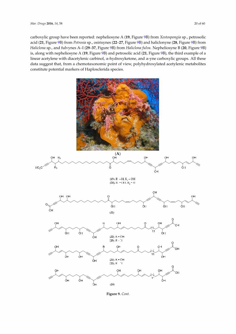
Mar. Drugs 2016, 14, 58 20 of 60
carboxylic group have been reported: nepheliosyne A (19, Figure 9B) from Xestospongia sp., petrosolicacid (21, Figure 9B) from Petrosia sp., osirisynes (22–27, Figure 9B) and haliclonyne (28, Figure 9B) fromHaliclona sp., and fulvynes A–I (29–37, Figure 9B) from Haliclona fulva. Nepheliosyne B (20, Figure 9B)is, along with nepheliosyne A (19, Figure 9B) and petrosolic acid (21, Figure 9B), the third example of alinear acetylene with diacetylenic carbinol, α-hydroxyketone, and α-yne carboxylic groups. All thesedata suggest that, from a chemotaxonomic point of view, polyhydroxylated acetylenic metabolitesconstitute potential markers of Haplosclerida species.
Mar. Drugs 2016, 14, x 22 of 62
a α‐yne carboxylic group have been reported: nepheliosyne A (19, Figure 9B) from Xestospongia sp.,
petrosolic acid (21, Figure 9B) from Petrosia sp., osirisynes (22–27, Figure 9B) and haliclonyne (28,
Figure 9B) from Haliclona sp., and fulvynes A–I (29–37, Figure 9B) from Haliclona fulva. Nepheliosyne
B (20, Figure 9B) is, along with nepheliosyne A (19, Figure 9B) and petrosolic acid (21, Figure 9B), the
third example of a linear acetylene with diacetylenic carbinol, α‐hydroxyketone, and α‐yne
carboxylic groups. All these data suggest that, from a chemotaxonomic point of view,
polyhydroxylated acetylenic metabolites constitute potential markers of Haplosclerida species.
(A)
Figure 9. Cont.

Mar. Drugs 2016, 14, 58 21 of 60
Mar. Drugs 2016, 14, x 23 of 62
(B)
Figure 9. (A) Niphates sp. Photo: Philippe Amade (INSERM); (B) Structure of nepheliosyne A (19),
nepheliosyne B (20), petrosolic acid (21), osirisynes A–F (22–27), haliclonyne (28) and fulvynes A–I
(29–37).
Corallistes sp.
This deep‐sea sponge was collected by beam trawl at a depth of 350 m in the Coral Sea. The
crude dichloromethanol extract shows cytotoxic activities against the KB naso‐pharyngeal
carcinoma cell line with an IC50 of 10 μg/mL. Subsequent chemical analyses led to the isolation of a
free porphyrin called corallistin A (38, Figure 10) [50]. Its total synthesis followed a few years later
[110].
Further studies conducted on Corallistes sp. led to the isolation of corallistins B (39, Figure 10), C
(40, Figure 10), D (41, Figure 10) and E (42, Figure 10). These porphyrins may be used as new
photosensitizers in phototherapy, particularly by generating singlet oxygen toxic to cancer cells [51].
Figure 9. (A) Niphates sp. Photo: Philippe Amade (INSERM); (B) Structure of nepheliosyne A(19), nepheliosyne B (20), petrosolic acid (21), osirisynes A–F (22–27), haliclonyne (28) and fulvynesA–I (29–37).
Corallistes sp.
This deep-sea sponge was collected by beam trawl at a depth of 350 m in the Coral Sea. The crudedichloromethanol extract shows cytotoxic activities against the KB naso-pharyngeal carcinoma cellline with an IC50 of 10 µg/mL. Subsequent chemical analyses led to the isolation of a free porphyrincalled corallistin A (38, Figure 10) [50]. Its total synthesis followed a few years later [110].
Further studies conducted on Corallistes sp. led to the isolation of corallistins B (39, Figure 10),C (40, Figure 10), D (41, Figure 10) and E (42, Figure 10). These porphyrins may be used as newphotosensitizers in phototherapy, particularly by generating singlet oxygen toxic to cancer cells [51].

Mar. Drugs 2016, 14, 58 22 of 60
Mar. Drugs 2016, 14, x 24 of 62
Figure 10. Structure of corallistin methyl ester A (38), B (39), C (40), D (41), and E (42).
5.2. Ascidians
5.2.1. General Comments
Ascidians are invertebrate filter feeders that belong to phylum Chordata. They represent the
most evolved invertebrates with a heart and a respiratory system. Living in both solitary and
colonial sessile modes, ascidians can be found in all the world’s seas and at all depths. The external
tunic of sea‐squirts (tunicates) is made of tunicin, a cellulosic substance that is extremely rare in the
animal kingdom [19]. Didemnids are soft‐bodied, bag‐like organisms. Like sponges, didemnids can
harbor cyanobacterial photosymbionts, e.g., the photosynthetic genus Prochloron which lives in
symbiosis with its host [111].
This class has a special “defense” feature: they can concentrate some toxic elements including
heavy metals (notably vanadium), hydrocarbons and elementary sulfur, which may be found in
Polycarpa aurata, or even sulfuric acid concentrated inside tiny vesicles of the outer tunic. Studies
have also revealed high amounts of nitrogenous compounds (cyclic peptides and alkaloids) with
powerful biological activities [112] (Table 3). As of 2007, 290 tunicates had been identified in New
Caledonian waters [5].
Figure 10. Structure of corallistin methyl ester A (38), B (39), C (40), D (41), and E (42).
5.2. Ascidians
5.2.1. General Comments
Ascidians are invertebrate filter feeders that belong to phylum Chordata. They represent the mostevolved invertebrates with a heart and a respiratory system. Living in both solitary and colonial sessilemodes, ascidians can be found in all the world’s seas and at all depths. The external tunic of sea-squirts(tunicates) is made of tunicin, a cellulosic substance that is extremely rare in the animal kingdom [19].Didemnids are soft-bodied, bag-like organisms. Like sponges, didemnids can harbor cyanobacterialphotosymbionts, e.g., the photosynthetic genus Prochloron which lives in symbiosis with its host [111].
This class has a special “defense” feature: they can concentrate some toxic elements includingheavy metals (notably vanadium), hydrocarbons and elementary sulfur, which may be found inPolycarpa aurata, or even sulfuric acid concentrated inside tiny vesicles of the outer tunic. Studies havealso revealed high amounts of nitrogenous compounds (cyclic peptides and alkaloids) with powerfulbiological activities [112] (Table 3). As of 2007, 290 tunicates had been identified in New Caledonianwaters [5].

Mar. Drugs 2016, 14, 58 23 of 60
Table 3. Natural products isolated from New Caledonian ascidians and their bioactivity.
Natural Product Chemical Class Biological Activity Species Reference
eudistalbin A
β-carboline alkaloid antiproliferative
ED50 KB 3.2 µg/mL.
Eudistoma album [113]eudistalbin B Inactive on KB cell line.
eudistomin E ED50 KB < 5 ng/mL.
(´)-woodininealkaloid antibacterial
Sa 16 mm/100 µg/disk and 18 mm/200 µg/disk.Ec 8/100 µg/disk and 11 mm/200 µg/ disk. Eudistoma fragum [114]
5-bromo-N,N-dimethylamino-ethyltryptamine
Sa 12 mm/100 µg/disk and 17 mm/200 µg/disk.Ec 17 mm/100µg/disk and 22 mm/200 µg/disk.
bistramide A
tetrahydropyranderivative
antiproliferative
IC50 (nM) KB 45, P388 20 and normal human endothelial cells 22IC50 NSCLCN6-L16 0.49 µM at 67 h.
IC50 (µg/mL) KB 0.53, P388 0.20, P388/Dox 0.05, B16 0.10, HT29 0.32,NSCLC-N6 0.03
Lissoclinum bistratum
[115–120]antibacterial Inactive agaisnt Ec, Kp, Mm, Pm, Pv, Pa, Sm, Sa and Streptococcus
group D (500 µg/mL).
mechanism of action G1 cell cycle arrest in NSCLCN6-L16 cells; polyploidy-inaptitudefor cytokinesis.
Na+ channels inhibitor At rest and in the inactivated state, occupied a site which was notlocated on the inactivation gate.
Ca2+ sensitivityimmunomodulator
Binding to contractile proteins for which it competes with Ca2+.Inhibition of T cell proliferation and activation of B cell proliferation.
bistramide B
antiproliferativemechanism of action
IC50 (µg/mL) KB 2.10, P388 0.20, P388/Dox 1.16, B16 1.20, HT29 0.71,NSCLC-N6 0.32. Significant decreases of S phase in NSCLC-N6 cells.
[117]bistramide C IC50 (µg/mL) KB 0.65, P388 0.02, P388/Dox 0.05, B16 0.06, HT29 0.50,
NSCLC-N6 0.05. Significant decreases of S phase in NSCLC-N6 cells.
bistramide D
IC50 (µg/mL) KB 10.00, P388 0.36, P388/Dox 5.82, B16 0.10, HT292.76, NSCLC-N6 3.43; In vivo (IV and IP) antitumor activity in nudemice engrafted SC with NSCLC-N6, T/C 53% at day 30. Significantdecreases of S phase; partial G1 cell cycle arrest in NSCLC-N6 cells.
bistramide K tetrahydropyranderivative
antiproliferativemechanism of action
IC50 (µg/mL) KB > 10.00, P388 0.57, P388/Dox > 10.00, B16 1.90,HT29 5.60, NSCLC-N6 3.23; In vivo (IV and IP) antitumor activity innude mice engrafted SC with NSCLC-N6, T/C 49% at day 30. G1 cell
cycle arrest in NSCLC-N6 cells.
Lissoclinum bistratum [117]
dichlorolissoclimidenitrogenous labdane
diterpene
antiproliferativemechanism of action
IC50 (ng/mL) KB 14, P388 1, P388/Dox 300 and NSCLC-N6 9; G1 cellcycle arrest in NSCLC-N6 cells (irreversible, total, dose-and
time-dependent). Lissoclinum voeltzkowi
[121,122]
chlorolissoclimide antiproliferativemechanism of action
IC50 (ng/mL) KB 52, P388 1.7, P388/Dox 200 and NSCLC-N6 10; G1cell cycle arrest in NSCLC-N6 cells. [121–123]
arborescidine D indole alkaloid antiproliferative IC50 KB 3 µg/mL. Pseudodistoma arborescens [124]
Footnote: Statistics: ED50 median effective dose, IC50 half maximal inhibitory concentration, T/C tumor growth inhibition ratio. Cell lines: KB human nasopharyngeal epidermoidcarcinoma, P388 murine leukemia, P388/Dox murine leukemia doxorubicin-resistant, NSCLCN6-L16, NSCLC-N6: human non-small-cell bronchopulmonary carcinoma, B16 murinemelanoma, HT29 human colon adenocarcinoma. Bacteria: Sa Staphylococcus aureus, Ec Escherichia coli, Kp Klehsiella pneumoniae, Mm Morganella morganii, Pm Proteus mirabilis, Pv Proteusvulgaris, Pa Pseudomonas aeruginosa, Sm Serratia mareescens. Ions: Na+ sodium, Ca2+ calcium. Others: IV intravenous, IP intraperitoneal, SC subcutaneous.

Mar. Drugs 2016, 14, 58 24 of 60
5.2.2. Lissoclinum bistratum
The didemnid Lissoclinum bistratum (voucher # UA264, IRD Nouméa, Figure 11A) wascollected near the Ua islet in the southwest lagoon. Chemical studies followed when two casesof human intoxication were observed during manipulation of the lyophilized powder. The crudedichloromethanol extract of L. bistratum demonstrates acute toxicity on rats, mice and rabbits. Moreover,it has revealed cytotoxic activities with IC50 values of 45 nM, 20 nM and 22 nM on KB, P388 and normalhuman endothelial cells, respectively [115].
Separation and purification led to the isolation of a tetrahydropyran derivative called bistramideA (43, Figure 11B) (or bistratene). Its partial structure has been characterized using two dimensionalNMR techniques [125].
Bistramide A (43, Figure 11B) appears to block the division of NSCLC-N6 (L16) cells at theG1 phase after a 24 h incubation period at a concentration of 1.42 µM. The IC50 value is 0.49 µM.Bistramide A (43, Figure 11B) may also induce polyploidy, causing unsuccessful cytokinesis [116].It has revealed immunomodulator activities (suppressor/stimulator) on the proliferation of T and Bcells [120]. Moreover, demonstrated using the voltage-clamp technique, bistramide A (43, Figure 11B)has proven to be responsible for a resting block by inhibiting sodium channels [118] and can bind tocontractile proteins in competition with calcium in frog skeletal muscle fibers [119].
Later investigations performed on L. bistratum led to the isolation of new tetrahydropyranderivatives called bistramides B (44, Figure 11B), C (45, Figure 11B), D (46, Figure 11B) and K (47,Figure 11B). Bistramides A (43, Figure 11B), B (44, Figure 11B) and C (45, Figure 11B) show potentcytotoxic activities with IC50 values of less than 1 µg/mL on several cancer cell lines including KB,P388, P388 doxorubicin-resistant, B16, HT29, NSCLC-N6, MRC5CV1 fibroblasts and T24 bladdercarcinoma cells. Bistramide K (47, Figure 11B), which is the least toxic compound, induces a completeblockage of NSCLC-N6 cells in the G1 phase after a 48 h incubation period with an IC50 value of3.23 µg/mL [117,126].
Mar. Drugs 2016, 14, x
5.2.2. Lissoclinum Bistratum
The didemnid Lissoclinum bistratum (voucher # UA264, IRD Nouméa, Figure 11A) was collected
near the Ua islet in the southwest lagoon. Chemical studies followed when two cases of human
intoxication were observed during manipulation of the lyophilized powder. The crude
dichloromethanol extract of L. bistratum demonstrates acute toxicity on rats, mice and rabbits.
Moreover, it has revealed cytotoxic activities with IC50 values of 45 nM, 20 nM and 22 nM on KB,
P388 and normal human endothelial cells, respectively [115].
Separation and purification led to the isolation of a tetrahydropyran derivative called
bistramide A (43, Figure 11B) (or bistratene). Its partial structure has been characterized using two
dimensional NMR techniques [125].
Bistramide A (43, Figure 11B) appears to block the division of NSCLC‐N6 (L16) cells at the G1
phase after a 24 h incubation period at a concentration of 1.42 μM. The IC50 value is 0.49 μM.
Bistramide A (43, Figure 11B) may also induce polyploidy, causing unsuccessful cytokinesis [116].
It has revealed immunomodulator activities (suppressor/stimulator) on the proliferation of T and B
cells [120]. Moreover, demonstrated using the voltage‐clamp technique, bistramide A (43, Figure 11B)
has proven to be responsible for a resting block by inhibiting sodium channels [118] and can bind to
contractile proteins in competition with calcium in frog skeletal muscle fibers [119].
Later investigations performed on L. bistratum led to the isolation of new tetrahydropyran
derivatives called bistramides B (44, Figure 11B), C (45, Figure 11B), D (46, Figure 11B) and K (47,
Figure 11B). Bistramides A (43, Figure 11B), B (44, Figure 11B) and C (45, Figure 11B) show potent
cytotoxic activities with IC50 values of less than 1 μg/mL on several cancer cell lines including KB,
P388, P388 doxorubicin‐resistant, B16, HT29, NSCLC‐N6, MRC5CV1 fibroblasts and T24 bladder
carcinoma cells. Bistramide K (47, Figure 11B), which is the least toxic compound, induces a
complete blockage of NSCLC‐N6 cells in the G1 phase after a 48 h incubation period with an IC50
value of 3.23 μg/mL [117,126].
(A)
Figure 11. Cont.

Mar. Drugs 2016, 14, 58 25 of 60Mar. Drugs 2016, 14, x 28 of 62
(B)
Figure 11. (A) Lissoclinum bistratum, © IRD; (B) Structure of bistramide A (43), B (44), C (45), D (46),
and K (47).
5.3. Cnidaria
This phylum is divided into two subclasses, Octocorallia, which includes alcyonarians,
gorgonians and pennatularians, and Hexacorallia, which includes scleratinarians. The chemical and
biological properties of Cnidaria collected in New Caledonia are reported in Table 4.
Figure 11. (A) Lissoclinum bistratum, © IRD; (B) Structure of bistramide A (43), B (44), C (45), D (46),and K (47).
5.3. Cnidaria
This phylum is divided into two subclasses, Octocorallia, which includes alcyonarians, gorgoniansand pennatularians, and Hexacorallia, which includes scleratinarians. The chemical and biologicalproperties of Cnidaria collected in New Caledonia are reported in Table 4.

Mar. Drugs 2016, 14, 58 26 of 60
Table 4. Natural products isolated from New Caledonian Cnidaria and their bioactivity.
Natural Product Chemical Class Biological Activity Species Reference
7-epi-11,19-desoxyhavannahine xenicane diterpene anti-foulingCeramium codii RGR after 2 days: 4% of
control at 50 ppm, 21% at 25 ppm, 42% at12.5 ppm.
Xenia garciae [33]
iela melst protein elastase inhibitor Inhibitor of amidolysis of Suc(Ala)3p-NA byporcine pancreatic elastase (Ki 1.5 nM). Melithea cf. stormii [37]
villogorgin A caffeine-xanthine typealkaloid anti-inflammatory Acetylcholine antagonist. Anti-aggregatory
(thrombin, A23187). Villogorgia rubra [127]
lituarine A
polyethermacrolide antiproliferative
IC50 KB 3.7–5 ng/mL.
Lituaria australasiae [128]lituarine B IC50 KB 1–2 ng/mL.
lituarine C IC50 KB 5–6 ng/mL.
pteroidinbriarane diterpene ichtyotoxic
LD100 50 µg/mL, t = 90 min (fish of thegenus Mugil).
Pteroides laboutei [129]O-deacetyl-12-O-benzoyl-12-pteroidin LD100 50 µg/mL, t = 150 min (fish of the
genus Mugil).
Statistics: RGR Relative Growth Rate, Ki inhibition constant, IC50 half maximal inhibitory concentration, LD100 lethal dose 100%. Cell line: KB human nasopharyngeal epidermoidcarcinoma. Other: A23187 calcium ionophore.
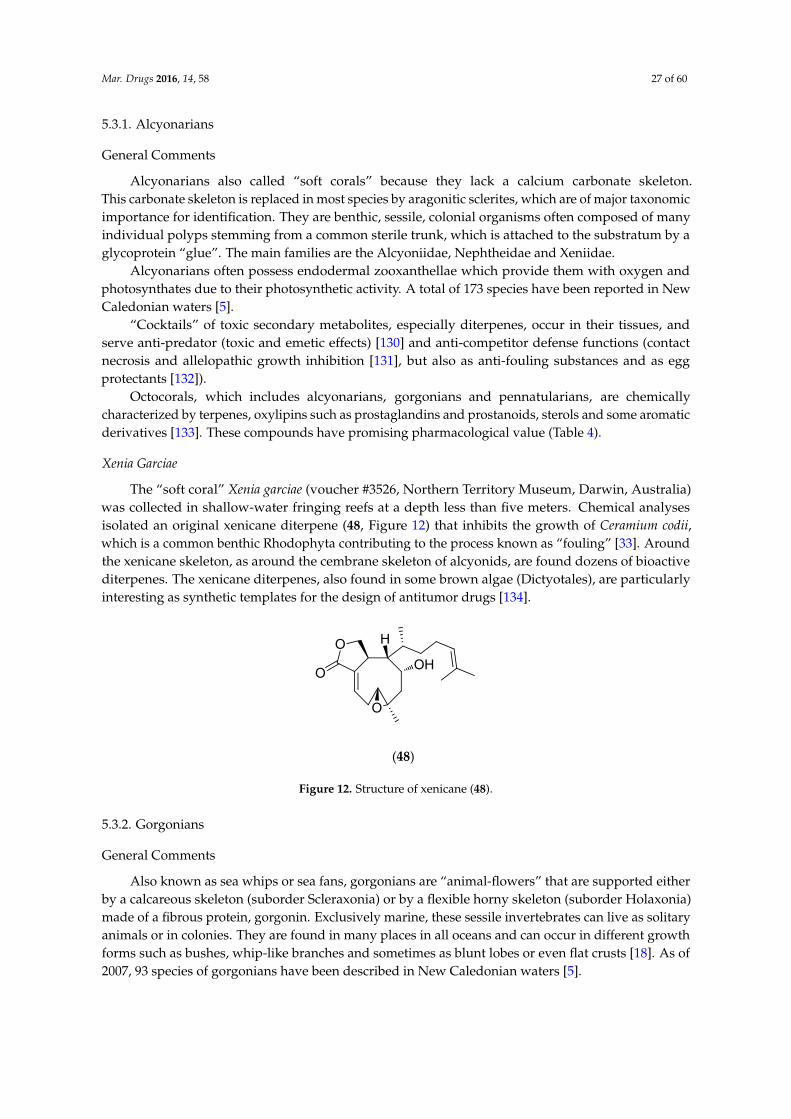
Mar. Drugs 2016, 14, 58 27 of 60
5.3.1. Alcyonarians
General Comments
Alcyonarians also called “soft corals” because they lack a calcium carbonate skeleton.This carbonate skeleton is replaced in most species by aragonitic sclerites, which are of major taxonomicimportance for identification. They are benthic, sessile, colonial organisms often composed of manyindividual polyps stemming from a common sterile trunk, which is attached to the substratum by aglycoprotein “glue”. The main families are the Alcyoniidae, Nephtheidae and Xeniidae.
Alcyonarians often possess endodermal zooxanthellae which provide them with oxygen andphotosynthates due to their photosynthetic activity. A total of 173 species have been reported in NewCaledonian waters [5].
“Cocktails” of toxic secondary metabolites, especially diterpenes, occur in their tissues, andserve anti-predator (toxic and emetic effects) [130] and anti-competitor defense functions (contactnecrosis and allelopathic growth inhibition [131], but also as anti-fouling substances and as eggprotectants [132]).
Octocorals, which includes alcyonarians, gorgonians and pennatularians, are chemicallycharacterized by terpenes, oxylipins such as prostaglandins and prostanoids, sterols and some aromaticderivatives [133]. These compounds have promising pharmacological value (Table 4).
Xenia Garciae
The “soft coral” Xenia garciae (voucher #3526, Northern Territory Museum, Darwin, Australia)was collected in shallow-water fringing reefs at a depth less than five meters. Chemical analysesisolated an original xenicane diterpene (48, Figure 12) that inhibits the growth of Ceramium codii,which is a common benthic Rhodophyta contributing to the process known as “fouling” [33]. Aroundthe xenicane skeleton, as around the cembrane skeleton of alcyonids, are found dozens of bioactivediterpenes. The xenicane diterpenes, also found in some brown algae (Dictyotales), are particularlyinteresting as synthetic templates for the design of antitumor drugs [134].
Mar. Drugs 2016, 14, x
5.3.1. Alcyonarians
General Comments
Alcyonarians also called “soft corals” because they lack a calcium carbonate skeleton.
This carbonate skeleton is replaced in most species by aragonitic sclerites, which are of major
taxonomic importance for identification. They are benthic, sessile, colonial organisms often composed of
many individual polyps stemming from a common sterile trunk, which is attached to the substratum
by a glycoprotein “glue”. The main families are the Alcyoniidae, Nephtheidae and Xeniidae.
Alcyonarians often possess endodermal zooxanthellae which provide them with oxygen and
photosynthates due to their photosynthetic activity. A total of 173 species have been reported in
New Caledonian waters [5].
“Cocktails” of toxic secondary metabolites, especially diterpenes, occur in their tissues, and
serve anti‐predator (toxic and emetic effects) [130] and anti‐competitor defense functions (contact
necrosis and allelopathic growth inhibition [131], but also as anti‐fouling substances and as egg
protectants [132]).
Octocorals, which includes alcyonarians, gorgonians and pennatularians, are chemically
characterized by terpenes, oxylipins such as prostaglandins and prostanoids, sterols and some
aromatic derivatives [133]. These compounds have promising pharmacological value (Table 4).
Xenia Garciae
The “soft coral” Xenia garciae (voucher #3526, Northern Territory Museum, Darwin, Australia)
was collected in shallow‐water fringing reefs at a depth less than five meters. Chemical analyses
isolated an original xenicane diterpene (48, Figure 12) that inhibits the growth of Ceramium codii,
which is a common benthic Rhodophyta contributing to the process known as “fouling” [33].
Around the xenicane skeleton, as around the cembrane skeleton of alcyonids, are found dozens of
bioactive diterpenes. The xenicane diterpenes, also found in some brown algae (Dictyotales), are
particularly interesting as synthetic templates for the design of antitumor drugs [134].
(48)
O
OH
O
O
H
Figure 12. Structure of xenicane (48).
5.3.2. Gorgonians
General Comments
Also known as sea whips or sea fans, gorgonians are “animal‐flowers” that are supported either
by a calcareous skeleton (suborder Scleraxonia) or by a flexible horny skeleton (suborder Holaxonia)
made of a fibrous protein, gorgonin. Exclusively marine, these sessile invertebrates can live as
solitary animals or in colonies. They are found in many places in all oceans and can occur in different
growth forms such as bushes, whip‐like branches and sometimes as blunt lobes or even flat crusts
[18]. As of 2007, 93 species of gorgonians have been described in New Caledonian waters [5].
Melithea cf. Stormii
Figure 12. Structure of xenicane (48).
5.3.2. Gorgonians
General Comments
Also known as sea whips or sea fans, gorgonians are “animal-flowers” that are supported eitherby a calcareous skeleton (suborder Scleraxonia) or by a flexible horny skeleton (suborder Holaxonia)made of a fibrous protein, gorgonin. Exclusively marine, these sessile invertebrates can live as solitaryanimals or in colonies. They are found in many places in all oceans and can occur in different growthforms such as bushes, whip-like branches and sometimes as blunt lobes or even flat crusts [18]. As of2007, 93 species of gorgonians have been described in New Caledonian waters [5].
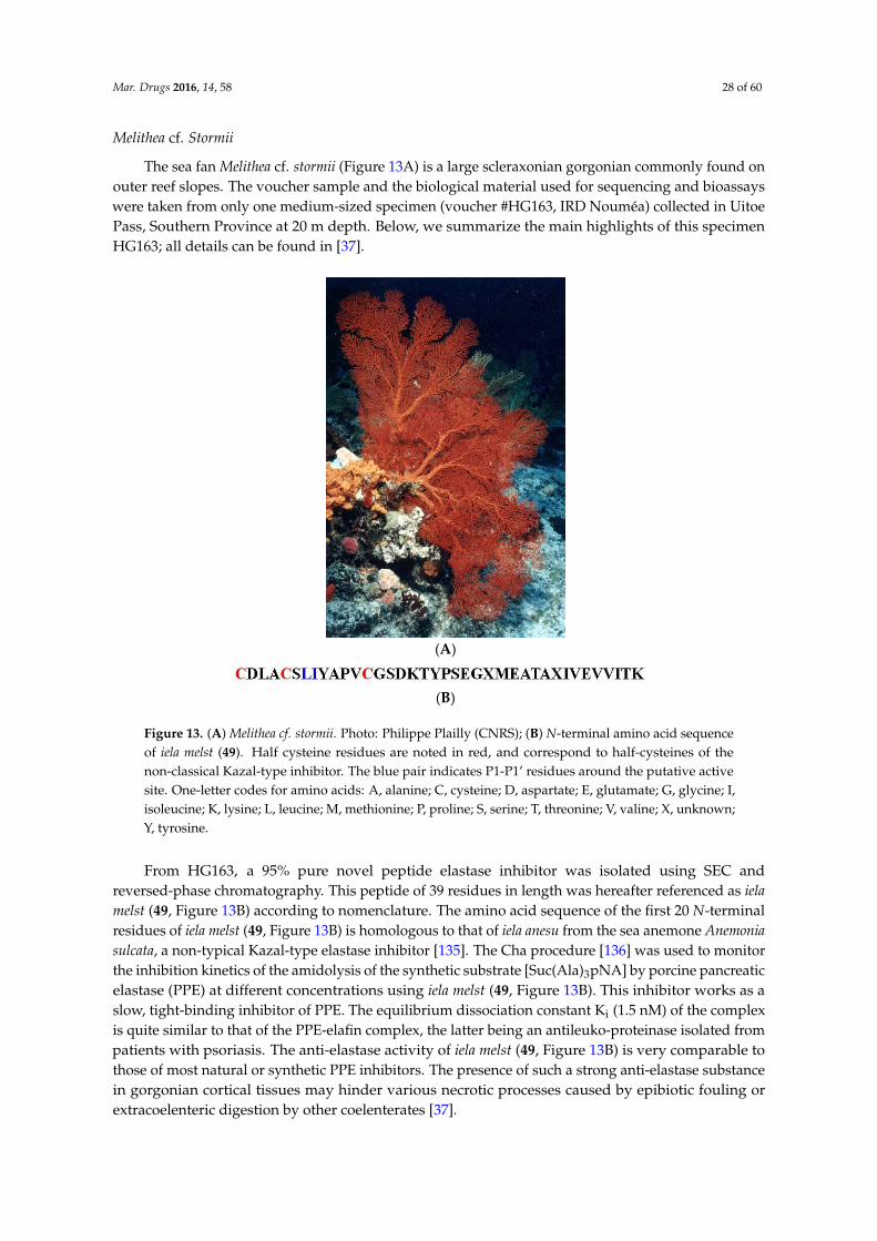
Mar. Drugs 2016, 14, 58 28 of 60
Melithea cf. Stormii
The sea fan Melithea cf. stormii (Figure 13A) is a large scleraxonian gorgonian commonly found onouter reef slopes. The voucher sample and the biological material used for sequencing and bioassayswere taken from only one medium-sized specimen (voucher #HG163, IRD Nouméa) collected in UitoePass, Southern Province at 20 m depth. Below, we summarize the main highlights of this specimenHG163; all details can be found in [37].
Mar. Drugs 2016, 14, x 31 of 62
The sea fan Melithea cf. stormii (Figure 13A) is a large scleraxonian gorgonian commonly found
on outer reef slopes. The voucher sample and the biological material used for sequencing and
bioassays were taken from only one medium‐sized specimen (voucher #HG163, IRD Nouméa)
collected in Uitoe Pass, Southern Province at 20 m depth. Below, we summarize the main highlights
of this specimen HG163; all details can be found in [37].
(A)
(B)
Figure 13. (A) Melithea cf. stormii. Photo: Philippe Plailly (CNRS); (B) N‐terminal amino acid sequence
of iela melst (49). Half cysteine residues are noted in red, and correspond to half‐cysteines of the
non‐classical Kazal‐type inhibitor. The blue pair indicates P1‐P1’ residues around the putative active
site. One‐letter codes for amino acids: A, alanine; C, cysteine; D, aspartate; E, glutamate; G, glycine; I,
isoleucine; K, lysine; L, leucine; M, methionine; P, proline; S, serine; T, threonine; V, valine; X,
unknown; Y, tyrosine.
From HG163, a 95% pure novel peptide elastase inhibitor was isolated using SEC and
reversed‐phase chromatography. This peptide of 39 residues in length was hereafter referenced as
iela melst (49, Figure 13B) according to nomenclature. The amino acid sequence of the first 20
N‐terminal residues of iela melst (49, Figure 13B) is homologous to that of iela anesu from the sea
anemone Anemonia sulcata, a non‐typical Kazal‐type elastase inhibitor [135]. The Cha procedure [136]
was used to monitor the inhibition kinetics of the amidolysis of the synthetic substrate
[Suc(Ala)3pNA] by porcine pancreatic elastase (PPE) at different concentrations using iela melst (49,
Figure 13B). This inhibitor works as a slow, tight‐binding inhibitor of PPE. The equilibrium
dissociation constant Ki (1.5 nM) of the complex is quite similar to that of the PPE‐elafin complex, the
latter being an antileuko‐proteinase isolated from patients with psoriasis. The anti‐elastase activity
of iela melst (49, Figure 13B) is very comparable to those of most natural or synthetic PPE inhibitors.
The presence of such a strong anti‐elastase substance in gorgonian cortical tissues may hinder
various necrotic processes caused by epibiotic fouling or extracoelenteric digestion by other
coelenterates [37].
Villogorgia Rubra
Studies performed on the gorgonian Villogorgia rubra, collected near the Chesterfield Islands,
led to the characterization of two new alkaloids called villogorgin A (50, Figure 14) and B (51, Figure
Figure 13. (A) Melithea cf. stormii. Photo: Philippe Plailly (CNRS); (B) N-terminal amino acid sequenceof iela melst (49). Half cysteine residues are noted in red, and correspond to half-cysteines of thenon-classical Kazal-type inhibitor. The blue pair indicates P1-P1’ residues around the putative activesite. One-letter codes for amino acids: A, alanine; C, cysteine; D, aspartate; E, glutamate; G, glycine; I,isoleucine; K, lysine; L, leucine; M, methionine; P, proline; S, serine; T, threonine; V, valine; X, unknown;Y, tyrosine.
From HG163, a 95% pure novel peptide elastase inhibitor was isolated using SEC andreversed-phase chromatography. This peptide of 39 residues in length was hereafter referenced as ielamelst (49, Figure 13B) according to nomenclature. The amino acid sequence of the first 20 N-terminalresidues of iela melst (49, Figure 13B) is homologous to that of iela anesu from the sea anemone Anemoniasulcata, a non-typical Kazal-type elastase inhibitor [135]. The Cha procedure [136] was used to monitorthe inhibition kinetics of the amidolysis of the synthetic substrate [Suc(Ala)3pNA] by porcine pancreaticelastase (PPE) at different concentrations using iela melst (49, Figure 13B). This inhibitor works as aslow, tight-binding inhibitor of PPE. The equilibrium dissociation constant Ki (1.5 nM) of the complexis quite similar to that of the PPE-elafin complex, the latter being an antileuko-proteinase isolated frompatients with psoriasis. The anti-elastase activity of iela melst (49, Figure 13B) is very comparable tothose of most natural or synthetic PPE inhibitors. The presence of such a strong anti-elastase substancein gorgonian cortical tissues may hinder various necrotic processes caused by epibiotic fouling orextracoelenteric digestion by other coelenterates [37].

Mar. Drugs 2016, 14, 58 29 of 60
Villogorgia Rubra
Studies performed on the gorgonian Villogorgia rubra, collected near the Chesterfield Islands, led tothe characterization of two new alkaloids called villogorgin A (50, Figure 14) and B (51, Figure 14), alongwith known compounds: caffeine (52, Figure 14), tryptamine (53, Figure 14), Nb-methyltryptamine (54,Figure 14) and 1,2,3,4-tetrahydro-β-carboline (55, Figure 14).
Villogorgin A (50, Figure 14) inhibits (i) the contraction of the guinea pig ileum induced byacetylcholine; (ii) the aggregation of human platelets induced by thrombin and calcium ionophoreA23187; this inhibition may be due to modulation of calcium/calmodulin-dependent enzymes.Villogorgin A (50, Figure 14) and B (51, Figure 14) are structurally related to the marine tunicateβ-carboline alkaloid eudistomidin-A (56, Figure 14), a strong calmodulin antagonist [127].
Mar. Drugs 2016, 14, x 32 of 62
14), along with known compounds: caffeine (52, Figure 14), tryptamine (53, Figure 14),
Nb‐methyltryptamine (54, Figure 14) and 1,2,3,4‐tetrahydro‐β‐carboline (55, Figure 14).
Villogorgin A (50, Figure 14) inhibits (i) the contraction of the guinea pig ileum induced by
acetylcholine; (ii) the aggregation of human platelets induced by thrombin and calcium ionophore
A23187; this inhibition may be due to modulation of calcium/calmodulin‐dependent enzymes.
Villogorgin A (50, Figure 14) and B (51, Figure 14) are structurally related to the marine tunicate
β‐carboline alkaloid eudistomidin‐A (56, Figure 14), a strong calmodulin antagonist [127].
Figure 14. Structure of villogorgin A (50), villogorgin B (51), caffeine (52), tryptamine (53),
Nb‐methyltryptamine (54), 1,2,3,4‐tetrahydro‐β‐carboline (55) and eudistomidin‐A (56).
5.3.3. Pennatularians
General Comments
Sea pens (sea feathers)—also called pennatularians—are a specialized and morphologically
distinct group of octocorallian cnidarians.
Pennatularians are made of a thin tissue called the coenenchyme. These sessile animals live in
colonies that are built from a single large primary polyp, the oozoid. They have been encountered in
all oceans down to 6100 m depth [137].
Lituaria Australasiae
Lituaria australasiae was collected at night, inside St. Vincent Bay (west coast, Southern Province).
From this octocoral, a novel class of macrocyclic lactones has been identified: lituarines A (57, Figure 15),
B (58, Figure 15) and C (59, Figure 15). Lituarines have demonstrated antifungal activities on
Fusarium oxysporum, Helminthosporium turscicum, Penicillium italicum and Phytophtora parasitica, as well as
cytotoxic activities on KB cells with IC50 values of 3.7–5.0 ng/mL for lituarine A (57, Figure 15), 1–2
ng/mL for lituarine B (58, Figure 15) and ranging from 5–6 ng/mL for lituarine C (59, Figure 15) [128].
Figure 14. Structure of villogorgin A (50), villogorgin B (51), caffeine (52), tryptamine (53),Nb-methyltryptamine (54), 1,2,3,4-tetrahydro-β-carboline (55) and eudistomidin-A (56).
5.3.3. Pennatularians
General Comments
Sea pens (sea feathers)—also called pennatularians—are a specialized and morphologicallydistinct group of octocorallian cnidarians.
Pennatularians are made of a thin tissue called the coenenchyme. These sessile animals live incolonies that are built from a single large primary polyp, the oozoid. They have been encountered inall oceans down to 6100 m depth [137].
Lituaria Australasiae
Lituaria australasiae was collected at night, inside St. Vincent Bay (west coast, Southern Province).From this octocoral, a novel class of macrocyclic lactones has been identified: lituarines A (57,Figure 15), B (58, Figure 15) and C (59, Figure 15). Lituarines have demonstrated antifungal activitieson Fusarium oxysporum, Helminthosporium turscicum, Penicillium italicum and Phytophtora parasitica,as well as cytotoxic activities on KB cells with IC50 values of 3.7–5.0 ng/mL for lituarine A (57,Figure 15), 1–2 ng/mL for lituarine B (58, Figure 15) and ranging from 5–6 ng/mL for lituarine C (59,Figure 15) [128].

Mar. Drugs 2016, 14, 58 30 of 60
Mar. Drugs 2016, 14, x 33 of 62
Figure 15. Structure of lituarine A (57), lituarine B (58) and lituarine C (59).
5.3.4. Scleractinians
Scleractinians are also called “stony corals” or “hard corals”. Scleractinian colonies act as
biological photosystems during sunlit hours and feed on plankton at night. Closely related to sea
anemones, they are also armed with cnidocytes that can be used for predation as well as for
territorial defense [138].
“Stony corals” have a large range of colonial growth forms, from crustose to massive, but
mostly branching, corymbose, tabular or lamellate, with a few solitary and free‐living forms, e.g.,
mushroom corals [14]. All corals originate from a minute swimming planula that settles and
metamorphoses into a single solitary polyp, which itself undergoes successive budding to form a
colony that encases itself in an aragonitic (calcium/magnesium carbonate) shell. Growth rates can
vary from a few centimeters per year for fast growing branching acroporids that are shorter‐lived
and rarely very large, to a few millimeters per year at most for large poritid corals that can grow up
to 10 m in diameter and live for many centuries.
Shallow‐water corals contain Symbiodinium (symbiotic unicellular microalgae known as
zooxanthellae) within their endodermal cells. These microalgae give the coral its color, which thus
can vary in hue depending on the symbiont species. Scleractinians are usually photosymbiotes.
As of 2007, 310 species of scleractinians have been reported in New Caledonian waters [5].
Furthermore, the anthozoan Hexacorallia class may produce toxins (e.g., palytoxin) and other
secondary metabolites such as venoms, similar to those of sea anemones, or photoprotection
pigments (e.g., mycosporins, zoanthoxanthins) [139].
5.4. Echinoderms
5.4.1. General Comments
Phylum Echinodermata is divided into two subphyla, the Pelmatozoa including the class of
Crinoidea (crinoids, sea lilies and feather stars), and the Eleutherozoa which comprises Asteroidea
(starfish, sea stars), Echinoidea (sea urchins), Holothuroidea (sea cucumbers) and Ophiuroidea
(brittle stars) [17].
Echinoderms are exclusively marine invertebrates that have an endoskeleton consisting of
magnesium calcite. In all, 257 species of echinoderms have been reported in New Caledonian waters
as of 2007 [5].
The therapeutic potential of these marine organisms is outlined in Table 5. Secondary metabolites
produced by Crinoidea are sulfated anthraquinonic pigments, whereas Eleutherozoa produce
quinonic pigments, naphthoquinones or sulfated saponins (e.g., asterosaponins, holothurins) [140].
Figure 15. Structure of lituarine A (57), lituarine B (58) and lituarine C (59).
5.3.4. Scleractinians
Scleractinians are also called “stony corals” or “hard corals”. Scleractinian colonies act as biologicalphotosystems during sunlit hours and feed on plankton at night. Closely related to sea anemones, theyare also armed with cnidocytes that can be used for predation as well as for territorial defense [138].
“Stony corals” have a large range of colonial growth forms, from crustose to massive, but mostlybranching, corymbose, tabular or lamellate, with a few solitary and free-living forms, e.g., mushroomcorals [14]. All corals originate from a minute swimming planula that settles and metamorphoses intoa single solitary polyp, which itself undergoes successive budding to form a colony that encases itselfin an aragonitic (calcium/magnesium carbonate) shell. Growth rates can vary from a few centimetersper year for fast growing branching acroporids that are shorter-lived and rarely very large, to a fewmillimeters per year at most for large poritid corals that can grow up to 10 m in diameter and live formany centuries.
Shallow-water corals contain Symbiodinium (symbiotic unicellular microalgae known aszooxanthellae) within their endodermal cells. These microalgae give the coral its color, which thus canvary in hue depending on the symbiont species. Scleractinians are usually photosymbiotes.
As of 2007, 310 species of scleractinians have been reported in New Caledonian waters [5].Furthermore, the anthozoan Hexacorallia class may produce toxins (e.g., palytoxin) and othersecondary metabolites such as venoms, similar to those of sea anemones, or photoprotection pigments(e.g., mycosporins, zoanthoxanthins) [139].
5.4. Echinoderms
5.4.1. General Comments
Phylum Echinodermata is divided into two subphyla, the Pelmatozoa including the class ofCrinoidea (crinoids, sea lilies and feather stars), and the Eleutherozoa which comprises Asteroidea(starfish, sea stars), Echinoidea (sea urchins), Holothuroidea (sea cucumbers) and Ophiuroidea (brittlestars) [17].
Echinoderms are exclusively marine invertebrates that have an endoskeleton consisting ofmagnesium calcite. In all, 257 species of echinoderms have been reported in New Caledonian watersas of 2007 [5].
The therapeutic potential of these marine organisms is outlined in Table 5. Secondary metabolitesproduced by Crinoidea are sulfated anthraquinonic pigments, whereas Eleutherozoa produce quinonicpigments, naphthoquinones or sulfated saponins (e.g., asterosaponins, holothurins) [140].
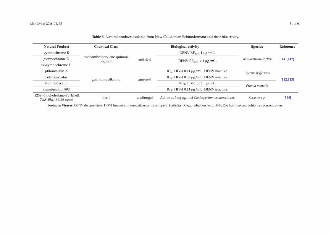
Mar. Drugs 2016, 14, 58 31 of 60
Table 5. Natural products isolated from New Caledonian Echinodermata and their bioactivity.
Natural Product Chemical Class Biological activity Species Reference
gymnochrome Bphenanthroperylene-quinone
pigment antiviral
DENV RF50% 1 µg/mL.
Gymnochrinus richeri [141,142]gymnochrome D DENV RF50% < 1 µg/mL.isogymnochrome D
ptilomycalin A
guanidine alkaloid antiviral
IC50 HIV-1 0.11 µg/mL. DENV inactive. Celerina heffernani
[142,143]celeromycalin IC50 HIV-1 0.32 µg/mL. DENV inactive.
fromiamycalin IC50 HIV-1 0.11 µg/mL.Fromia monolis
crambescidin 800 IC50 HIV-1 0.11 µg/mL. DENV inactive.
(25S)-5α-cholestane-3β,4β,6β,7α,8,15α,16β,26-octol sterol antifungal Active at 5 µg against Clodosporium cucumerinum. Rosaster sp. [144]
Footnote: Viruses: DENV dengue virus, HIV-1 human immunodeficiency virus type 1. Statistics: RF50% reduction factor 50%, IC50 half maximal inhibitory concentration.
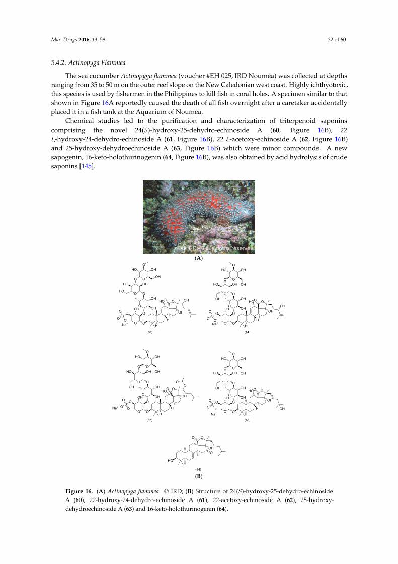
Mar. Drugs 2016, 14, 58 32 of 60
5.4.2. Actinopyga Flammea
The sea cucumber Actinopyga flammea (voucher #EH 025, IRD Nouméa) was collected at depthsranging from 35 to 50 m on the outer reef slope on the New Caledonian west coast. Highly ichthyotoxic,this species is used by fishermen in the Philippines to kill fish in coral holes. A specimen similar to thatshown in Figure 16A reportedly caused the death of all fish overnight after a caretaker accidentallyplaced it in a fish tank at the Aquarium of Nouméa.
Chemical studies led to the purification and characterization of triterpenoid saponinscomprising the novel 24(S)-hydroxy-25-dehydro-echinoside A (60, Figure 16B), 22ξ-hydroxy-24-dehydro-echinoside A (61, Figure 16B), 22 ξ-acetoxy-echinoside A (62, Figure 16B)and 25-hydroxy-dehydroechinoside A (63, Figure 16B) which were minor compounds. A newsapogenin, 16-keto-holothurinogenin (64, Figure 16B), was also obtained by acid hydrolysis of crudesaponins [145].
Mar. Drugs 2016, 14, x
5.4.2. Actinopyga Flammea
The sea cucumber Actinopyga flammea (voucher #EH 025, IRD Nouméa) was collected at depths
ranging from 35 to 50 m on the outer reef slope on the New Caledonian west coast. Highly
ichthyotoxic, this species is used by fishermen in the Philippines to kill fish in coral holes. A
specimen similar to that shown in Figure 16A reportedly caused the death of all fish overnight after a
caretaker accidentally placed it in a fish tank at the Aquarium of Nouméa.
(A)
OH
OHOO OH
H
OH
O O
OSO-
O
O
O
OH
OH
OO
OHO O
HOO
OH
HO
O
HO
OH
HNa+
OH
OHOO
OH
H
OH
O O
OSO-
O
O
O
OH
OH
OO
OHO O
HOO
OH
HO
O
OH
OH
HNa+
OH
OHOO
O
H
OH
O O
OS-O
O
O
O
OH
OH
OO
OH
O O
HO
O
OH
HO
O
O
OH
OH
H
Na+
OH
OHOO
OHH
OH
O O
OSO-
O
O
O
OH
OH
OO
OHO O
HOO
OH
HO
O
OH
OH
HNa+
OH
O
O
O
HOH
(60) (61)
(62) (63)
(64) (B)
Figure 16. (A) Actinopyga flammea. © IRD; (B) Structure of 24(S)‐hydroxy‐25‐dehydro‐echinoside A
(60), 22‐hydroxy‐24‐dehydro‐echinoside A (61), 22‐acetoxy‐echinoside A (62),
25‐hydroxy‐dehydroechinoside A (63) and 16‐keto‐holothurinogenin (64).
Figure 16. (A) Actinopyga flammea. © IRD; (B) Structure of 24(S)-hydroxy-25-dehydro-echinosideA (60), 22-hydroxy-24-dehydro-echinoside A (61), 22-acetoxy-echinoside A (62), 25-hydroxy-dehydroechinoside A (63) and 16-keto-holothurinogenin (64).
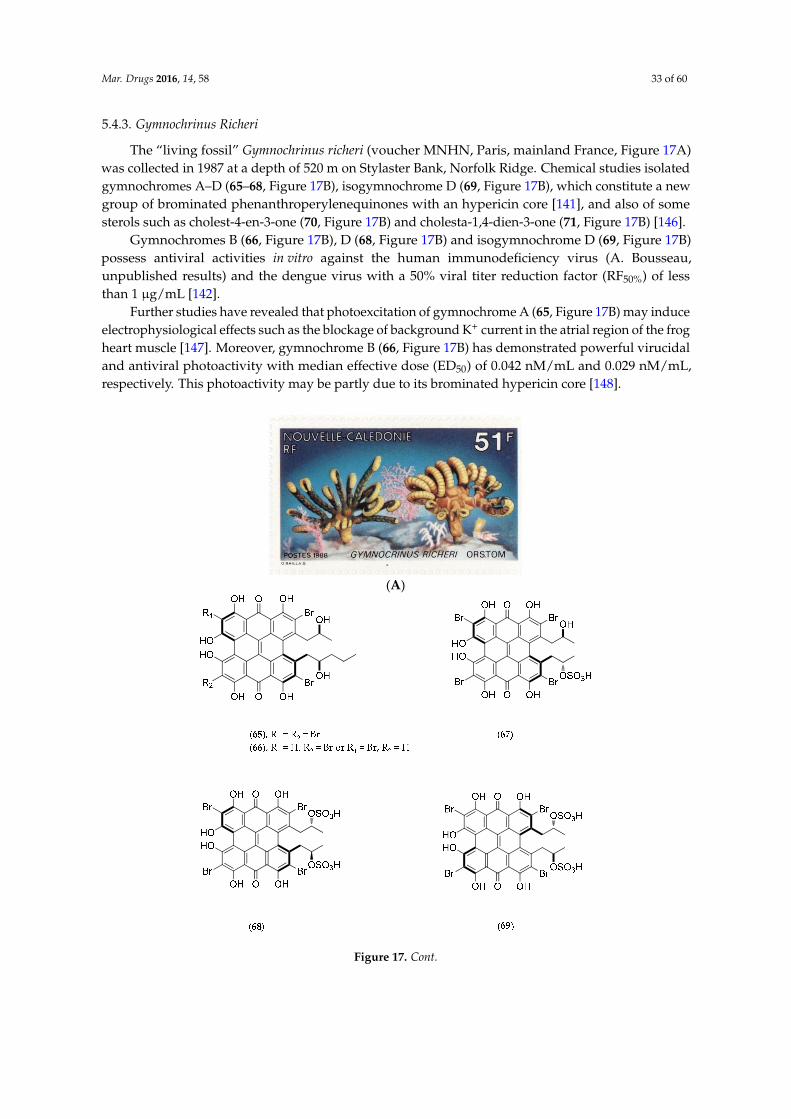
Mar. Drugs 2016, 14, 58 33 of 60
5.4.3. Gymnochrinus Richeri
The “living fossil” Gymnochrinus richeri (voucher MNHN, Paris, mainland France, Figure 17A)was collected in 1987 at a depth of 520 m on Stylaster Bank, Norfolk Ridge. Chemical studies isolatedgymnochromes A–D (65–68, Figure 17B), isogymnochrome D (69, Figure 17B), which constitute a newgroup of brominated phenanthroperylenequinones with an hypericin core [141], and also of somesterols such as cholest-4-en-3-one (70, Figure 17B) and cholesta-1,4-dien-3-one (71, Figure 17B) [146].
Gymnochromes B (66, Figure 17B), D (68, Figure 17B) and isogymnochrome D (69, Figure 17B)possess antiviral activities in vitro against the human immunodeficiency virus (A. Bousseau,unpublished results) and the dengue virus with a 50% viral titer reduction factor (RF50%) of lessthan 1 µg/mL [142].
Further studies have revealed that photoexcitation of gymnochrome A (65, Figure 17B) may induceelectrophysiological effects such as the blockage of background K+ current in the atrial region of the frogheart muscle [147]. Moreover, gymnochrome B (66, Figure 17B) has demonstrated powerful virucidaland antiviral photoactivity with median effective dose (ED50) of 0.042 nM/mL and 0.029 nM/mL,respectively. This photoactivity may be partly due to its brominated hypericin core [148].
Mar. Drugs 2016, 14, x 36 of 62
(B)
Figure 16. (A) Actinopyga flammea. © IRD; (B) Structure of 24(S)‐hydroxy‐25‐dehydro‐echinoside A
(60), 22‐hydroxy‐24‐dehydro‐echinoside A (61), 22‐acetoxy‐echinoside A (62),
25‐hydroxy‐dehydroechinoside A (63) and 16‐keto‐holothurinogenin (64).
5.4.3. Gymnochrinus Richeri
The “living fossil” Gymnochrinus richeri (voucher MNHN, Paris, mainland France, Figure 17A)
was collected in 1987 at a depth of 520 m on Stylaster Bank, Norfolk Ridge. Chemical studies isolated
gymnochromes A–D (65–68, Figure 17B), isogymnochrome D (69, Figure 17B), which constitute a
new group of brominated phenanthroperylenequinones with an hypericin core [141], and also of
some sterols such as cholest‐4‐en‐3‐one (70, Figure 17B) and cholesta‐1,4‐dien‐3‐one (71, Figure 17B)
[146].
Gymnochromes B (66, Figure 17B), D (68, Figure 17B) and isogymnochrome D (69, Figure 17B)
possess antiviral activities in vitro against the human immunodeficiency virus (A. Bousseau,
unpublished results) and the dengue virus with a 50% viral titer reduction factor (RF50%) of less than
1 μg/mL [142].
Further studies have revealed that photoexcitation of gymnochrome A (65, Figure 17B) may
induce electrophysiological effects such as the blockage of background K+ current in the atrial region
of the frog heart muscle [147]. Moreover, gymnochrome B (66, Figure 17B) has demonstrated
powerful virucidal and antiviral photoactivity with median effective dose (ED50) of 0.042 nM/mL
and 0.029 nM/mL, respectively. This photoactivity may be partly due to its brominated hypericin
core [148].
(A)
Figure 17. Cont.
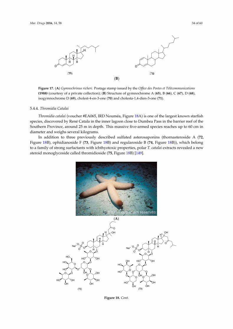
Mar. Drugs 2016, 14, 58 34 of 60
Mar. Drugs 2016, 14, x 37 of 62
(B)
Figure 17. (A) Gymnochrinus richeri. Postage stamp issued by the Office des Postes et Télécommunications
(1988) (courtesy of a private collection); (B) Structure of gymnochrome A (65), B (66), C (67), D (68),
isogymnochrome D (69), cholest‐4‐en‐3‐one (70) and cholesta‐1,4‐dien‐3‐one (71).
5.4.4. Thromidia Catalai
Thromidia catalai (voucher #EA065, IRD Nouméa, Figure 18A) is one of the largest known
starfish species, discovered by René Catala in the inner lagoon close to Dumbea Pass in the barrier
reef of the Southern Province, around 25 m in depth. This massive five‐armed species reaches up to
60 cm in diameter and weighs several kilograms.
In addition to three previously described sulfated asterosaponins (thornasteroside A (72, Figure
18B), ophidianoside F (73, Figure 18B) and regularoside B (74, Figure 18B)), which belong to a family
of strong surfactants with ichthyotoxic properties, polar T. catalai extracts revealed a new steroid
monoglycoside called thromidioside (75, Figure 18B) [149].
(A)
O
H
O
OH
OS
-O
O
O
O
OH
O
HO
OHO
OO
HO
HO
HO
OO
OH
OH
HO
O
OH
OH
OH
O
H
H
HNa+
(72)
OS
HO
O
OH
O
OH
O
OH
O
OH
O
OH
OHO
O
HO
HOOO
OH
HO
-O
O
O
O
O
OHH
H
H
H
Na+
(73)
Figure 17. (A) Gymnochrinus richeri. Postage stamp issued by the Office des Postes et Télécommunications(1988) (courtesy of a private collection); (B) Structure of gymnochrome A (65), B (66), C (67), D (68),isogymnochrome D (69), cholest-4-en-3-one (70) and cholesta-1,4-dien-3-one (71).
5.4.4. Thromidia Catalai
Thromidia catalai (voucher #EA065, IRD Nouméa, Figure 18A) is one of the largest known starfishspecies, discovered by René Catala in the inner lagoon close to Dumbea Pass in the barrier reef of theSouthern Province, around 25 m in depth. This massive five-armed species reaches up to 60 cm indiameter and weighs several kilograms.
In addition to three previously described sulfated asterosaponins (thornasteroside A (72,Figure 18B), ophidianoside F (73, Figure 18B) and regularoside B (74, Figure 18B)), which belongto a family of strong surfactants with ichthyotoxic properties, polar T. catalai extracts revealed a newsteroid monoglycoside called thromidioside (75, Figure 18B) [149].
Mar. Drugs 2016, 14, x 37 of 62
(B)
Figure 17. (A) Gymnochrinus richeri. Postage stamp issued by the Office des Postes et Télécommunications
(1988) (courtesy of a private collection); (B) Structure of gymnochrome A (65), B (66), C (67), D (68),
isogymnochrome D (69), cholest‐4‐en‐3‐one (70) and cholesta‐1,4‐dien‐3‐one (71).
5.4.4. Thromidia Catalai
Thromidia catalai (voucher #EA065, IRD Nouméa, Figure 18A) is one of the largest known
starfish species, discovered by René Catala in the inner lagoon close to Dumbea Pass in the barrier
reef of the Southern Province, around 25 m in depth. This massive five‐armed species reaches up to
60 cm in diameter and weighs several kilograms.
In addition to three previously described sulfated asterosaponins (thornasteroside A (72, Figure
18B), ophidianoside F (73, Figure 18B) and regularoside B (74, Figure 18B)), which belong to a family
of strong surfactants with ichthyotoxic properties, polar T. catalai extracts revealed a new steroid
monoglycoside called thromidioside (75, Figure 18B) [149].
(A)
O
H
O
OH
OS
-O
O
O
O
OH
O
HO
OHO
OO
HO
HO
HO
OO
OH
OH
HO
O
OH
OH
OH
O
H
H
HNa+
(72)
OS
HO
O
OH
O
OH
O
OH
O
OH
O
OH
OHO
O
HO
HOOO
OH
HO
-O
O
O
O
O
OHH
H
H
H
Na+
(73)
Figure 18. Cont.

Mar. Drugs 2016, 14, 58 35 of 60
Mar. Drugs 2016, 14, x 38 of 62
(B)
Figure 18. (A) Thromidia catalai. © IRD; (B) Structure of thornasteroside A (72), ophidianoside F (73),
regularoside B (74) and thromidioside (75).
5.5. Macroalgae
Macroalgae belong to three different divisions: red algae (Rhodophyta), brown algae
(Heterokontophyta, also known as the Ochrophyta, class Phaeophyceae) and green algae
(Chlorophyta). All three evolutionary lineages diverged early on according to their respective
plastid configurations, reflecting their endosymbiotic history [150].
Algae have a relatively unspecialized vegetative apparatus called a thallus. Important
differences are noted in many ultrastructural and biochemical features including photosynthetic
pigments, storage compounds, composition of cell walls, presence/absence of flagella, ultrastructure
of mitosis, connections between adjacent cells, and the fine structure of chloroplasts. They vary from
small, single‐celled forms to complex multicellular forms, such as the giant kelp Macrocystis pyrifera
that can grow up to 49 m in length in New Zealand and holds the record of the fastest growing
organism (0.6 m per day).
Marine macroalgae generally live attached to rocks or other hard substrata in coastal areas but
can be also found in sandy areas such as the Udoteaceae in tropical areas. They occur in all of the
world’s oceans where they occupy the seabed (phytobenthos), although a few species float freely on
the surface (e.g., the pelagic algae of the Sargasso Sea). In New Caledonia [151], the large brown
algae are mainly Fucales (Sargassum, Turbinaria) and Dictyotales and form large stands on rocky
lagoon bottoms, while several Chlorophyta species from the genera Halimeda and Caulerpa colonize
sandy bottoms [152–155]. The large fleshy Rhodophyta are mainly restricted to outer slopes or the
cooler waters of the southern lagoon. As of 2007, 454 species of algae and marine angiosperms had
been reported in New Caledonian waters [5].
Despite their capacity to produce large amounts of natural products, macroalgae from New
Caledonia have been little documented in this respect. Caulerpa species were investigated for
caulerpicin (76–80, Figure 19) activity in the 1980s for cosmetic purposes, but the highly publicized
invasion of Caulerpa taxifolia in Mediterranean Sea brought the project to an immediate halt [156].
The genus Lobophora (Phaeophyta) has been investigated in biological interaction studies between
macroalgae and several corals as part of a PhD thesis. Three new compounds have been isolated and
described [34], and their bioactivity experimentally tested on corals results in severe coral bleaching.
Figure 18. (A) Thromidia catalai. © IRD; (B) Structure of thornasteroside A (72), ophidianoside F (73),regularoside B (74) and thromidioside (75).
5.5. Macroalgae
Macroalgae belong to three different divisions: red algae (Rhodophyta), brown algae(Heterokontophyta, also known as the Ochrophyta, class Phaeophyceae) and green algae (Chlorophyta).All three evolutionary lineages diverged early on according to their respective plastid configurations,reflecting their endosymbiotic history [150].
Algae have a relatively unspecialized vegetative apparatus called a thallus. Important differencesare noted in many ultrastructural and biochemical features including photosynthetic pigments,storage compounds, composition of cell walls, presence/absence of flagella, ultrastructure of mitosis,connections between adjacent cells, and the fine structure of chloroplasts. They vary from small,single-celled forms to complex multicellular forms, such as the giant kelp Macrocystis pyrifera that cangrow up to 49 m in length in New Zealand and holds the record of the fastest growing organism (0.6 mper day).
Marine macroalgae generally live attached to rocks or other hard substrata in coastal areas butcan be also found in sandy areas such as the Udoteaceae in tropical areas. They occur in all of theworld’s oceans where they occupy the seabed (phytobenthos), although a few species float freely onthe surface (e.g., the pelagic algae of the Sargasso Sea). In New Caledonia [151], the large brown algaeare mainly Fucales (Sargassum, Turbinaria) and Dictyotales and form large stands on rocky lagoonbottoms, while several Chlorophyta species from the genera Halimeda and Caulerpa colonize sandybottoms [152–155]. The large fleshy Rhodophyta are mainly restricted to outer slopes or the coolerwaters of the southern lagoon. As of 2007, 454 species of algae and marine angiosperms had beenreported in New Caledonian waters [5].
Despite their capacity to produce large amounts of natural products, macroalgae from NewCaledonia have been little documented in this respect. Caulerpa species were investigated forcaulerpicin (76–80, Figure 19) activity in the 1980s for cosmetic purposes, but the highly publicizedinvasion of Caulerpa taxifolia in Mediterranean Sea brought the project to an immediate halt [156].The genus Lobophora (Phaeophyta) has been investigated in biological interaction studies betweenmacroalgae and several corals as part of a PhD thesis. Three new compounds have been isolated anddescribed [34], and their bioactivity experimentally tested on corals results in severe coral bleaching.

Mar. Drugs 2016, 14, 58 36 of 60
Mar. Drugs 2016, 14, x 39 of 62
Figure 19. Structure of caulerpicin C18 (76), C20 (77), C22 (78), C24 (79) and C26 (80).
5.6. Microalgae and Cyanobacteria
Less than 1% of described marine phytoplanktonic species are known to produce potent toxins
[157]. Microalgal benthic dinoflagellates species are involved, including Gambierdiscus toxicus that
produces maitotoxin (81, Figure 20). In vivo toxicity in mice indicates that intraperitoneal injection of
0.13 μg/kg is lethal [158]. A culture of ca. 4000 L of dinoflagellate cells is needed for the purification
of 20 mg of MTX, enough to kill about four million mice. MTX activates both voltage‐sensitive and
receptor‐operated calcium channels in the plasma membrane, thus inducing calpain protease that
rapidly leads to cell death. MTX has since been considered as a useful tool for investigating the
physiological and pathophysiological roles of calpain in neuronal cells [159].
Figure 20. Structure of maitotoxin (81).
Figure 19. Structure of caulerpicin C18 (76), C20 (77), C22 (78), C24 (79) and C26 (80).
5.6. Microalgae and Cyanobacteria
Less than 1% of described marine phytoplanktonic species are known to produce potenttoxins [157]. Microalgal benthic dinoflagellates species are involved, including Gambierdiscus toxicusthat produces maitotoxin (81, Figure 20). In vivo toxicity in mice indicates that intraperitoneal injectionof 0.13 µg/kg is lethal [158]. A culture of ca. 4000 L of dinoflagellate cells is needed for the purificationof 20 mg of MTX, enough to kill about four million mice. MTX activates both voltage-sensitive andreceptor-operated calcium channels in the plasma membrane, thus inducing calpain protease thatrapidly leads to cell death. MTX has since been considered as a useful tool for investigating thephysiological and pathophysiological roles of calpain in neuronal cells [159].
Mar. Drugs 2016, 14, x 39 of 62
Figure 19. Structure of caulerpicin C18 (76), C20 (77), C22 (78), C24 (79) and C26 (80).
5.6. Microalgae and Cyanobacteria
Less than 1% of described marine phytoplanktonic species are known to produce potent toxins
[157]. Microalgal benthic dinoflagellates species are involved, including Gambierdiscus toxicus that
produces maitotoxin (81, Figure 20). In vivo toxicity in mice indicates that intraperitoneal injection of
0.13 μg/kg is lethal [158]. A culture of ca. 4000 L of dinoflagellate cells is needed for the purification
of 20 mg of MTX, enough to kill about four million mice. MTX activates both voltage‐sensitive and
receptor‐operated calcium channels in the plasma membrane, thus inducing calpain protease that
rapidly leads to cell death. MTX has since been considered as a useful tool for investigating the
physiological and pathophysiological roles of calpain in neuronal cells [159].
Figure 20. Structure of maitotoxin (81). Figure 20. Structure of maitotoxin (81).
Species belonging to the genus Gambierdiscus have also been shown to produce ciguatoxinanalogues (CTX analogs) causing ciguatera fish poisoning (CFP). New Caledonia is a CFP-endemicarea. CFP constitutes a global health problem with more than 50,000 people worldwide affected by
Mar. Drugs 2016, 14, 58 37 of 60
this disease annually. CTXs arise from biotransformation/metabolization of CTX analogs throughthe food chain: from herbivorous fish that accumulate dinoflagellate toxins by eating dead coral andmarine algae to carnivorous fish [157]. In Pacific areas, the principal and most potent CTX is Pacificciguatoxin-1 (82, Figure 21). CTXs are potent activators of voltage-sensitive sodium channels and causean increase in neuronal excitability and neurotransmitter release [160]. In New Caledonia, traditionalremedies are commonly employed in the treatment of CFP: 90 plant species have been cataloguedas useful CFP remedies, including the leaves of Heliotropium foertherianum used to prepare the mostpopular herbal remedy [161].
A study conducted by D. Laurent et al. following the observation of many cases of seafoodpoisoning on Lifou Island between 2001 and 2005 (Loyalty Islands, New Caledonia, Figure 1), indicatesthat bloom-forming cyanobacteria, such as the filamentous Trichodesmium erythraeum, can also produceCTX-like compounds [160].
Mar. Drugs 2016, 14, x 40 of 62
Species belonging to the genus Gambierdiscus have also been shown to produce ciguatoxin
analogues (CTX analogs) causing ciguatera fish poisoning (CFP). New Caledonia is a CFP‐endemic
area. CFP constitutes a global health problem with more than 50,000 people worldwide affected by
this disease annually. CTXs arise from biotransformation/metabolization of CTX analogs through
the food chain: from herbivorous fish that accumulate dinoflagellate toxins by eating dead coral and
marine algae to carnivorous fish [157]. In Pacific areas, the principal and most potent CTX is Pacific
ciguatoxin‐1 (82, Figure 21). CTXs are potent activators of voltage‐sensitive sodium channels and
cause an increase in neuronal excitability and neurotransmitter release [160]. In New Caledonia,
traditional remedies are commonly employed in the treatment of CFP: 90 plant species have been
catalogued as useful CFP remedies, including the leaves of Heliotropium foertherianum used to
prepare the most popular herbal remedy [161].
A study conducted by D. Laurent et al. following the observation of many cases of seafood
poisoning on Lifou Island between 2001 and 2005 (Loyalty Islands, New Caledonia, Figure 1),
indicates that bloom‐forming cyanobacteria, such as the filamentous Trichodesmium erythraeum, can
also produce CTX‐like compounds [160].
Figure 21. Structure of Pacific ciguatoxin‐1 (P‐CTX‐1) (82).
In line with programs exploring the biodiversity of planktonic species such as OCEANOMICs
(see [162] for details), the project AMICAL (Aquaculture of MIcroalgae in New CALedonia, [163])
instigated by IFREMER‐New Caledonia, the Physiology and Biotechnology of Algae (PBA)
laboratory (Nantes, mainland France) and ADECAL (Caledonian Economic Development
Agency‐Nouméa, [164]), aims to develop industrial microalgal production based on indigenous
microalgae species isolated along the mainland coasts (Figure 1). Partners of this program will
transfer the cultivation and extraction techniques to selected algal species for industrial programs in
New Caledonia targeting the animal nutrition market and high added‐value compounds (cosmetics,
health food, etc.).
5.7. Other Biological Sources
Statistically “minor” phyla in terms of the number of studies on local species include
prokaryotes, several invertebrate phyla, and snakes as the sole vertebrate representatives.
5.7.1. Prokaryotes and Fungi
Micrococcus Luteus
Micrococcus luteus is a Gram+ bacterium that was isolated from Xestospongia exigua sponges
collected off Nouméa in the Southwest Pacific. Chemical studies reveal that this bacterium can
produce lutoside (83, Figure 22), an acyl‐1‐(acyl‐6′‐mannobiosyl)‐3‐glycerol, and also triclosan (84,
Figure 22) (2,4,4′‐trichloro‐2′‐hydroxydiphenylether), with both molecules presenting antibacterial
activities [165,166].
Figure 21. Structure of Pacific ciguatoxin-1 (P-CTX-1) (82).
In line with programs exploring the biodiversity of planktonic species such as OCEANOMICs(see [162] for details), the project AMICAL (Aquaculture of MIcroalgae in New CALedonia, [163])instigated by IFREMER-New Caledonia, the Physiology and Biotechnology of Algae (PBA) laboratory(Nantes, mainland France) and ADECAL (Caledonian Economic Development Agency-Nouméa, [164]),aims to develop industrial microalgal production based on indigenous microalgae species isolatedalong the mainland coasts (Figure 1). Partners of this program will transfer the cultivation andextraction techniques to selected algal species for industrial programs in New Caledonia targeting theanimal nutrition market and high added-value compounds (cosmetics, health food, etc.).
5.7. Other Biological Sources
Statistically “minor” phyla in terms of the number of studies on local species include prokaryotes,several invertebrate phyla, and snakes as the sole vertebrate representatives.
5.7.1. Prokaryotes and Fungi
Micrococcus Luteus
Micrococcus luteus is a Gram+ bacterium that was isolated from Xestospongia exigua spongescollected off Nouméa in the Southwest Pacific. Chemical studies reveal that this bacterium canproduce lutoside (83, Figure 22), an acyl-1-(acyl-61-mannobiosyl)-3-glycerol, and also triclosan (84,Figure 22) (2,4,41-trichloro-21-hydroxydiphenylether), with both molecules presenting antibacterialactivities [165,166].
Bacteria from Pseudoalteromonas and Vibrio Genus
Along the west coast of New Caledonia, 205 environmental samples were collected on a varietyof surfaces (sediments, intertidal rocks, invertebrates, plants, fish and biofilms found on organicsubstrates). This sampling led to the isolation of 493 marine bacteria [167,168]. Studies were at firstperformed to assess their ability to produce exopolysaccharides (EPSs). Among them, the new strain
Mar. Drugs 2016, 14, 58 38 of 60
Vibrio neocaledonicus sp. nov. (NC470), isolated from a biofilm found on Holothuroidea in St. VincentBay, has demonstrated the production of EPSs that have a high N-acetyl-hexosamine and uronic acidcontent with a low amount of neutral sugars. Preliminary experiments conducted on these EPSshave shown high metal-binding capacity [167]. Further studies have shown that four other strains(NC282, NC412, NC272 and NC120), which belong to the genus Pseudoalteromonas, have antibacterialpotential against reference and multidrug-resistant pathogen strains such as Staphylococcus aureus,Pseudomonas aeruginosa, Escherichia coli and Enterococcus faecalis [168].Mar. Drugs 2016, 14, x 41 of 62
Figure 22. Structure of lutoside (83) and triclosan (84).
Bacteria from Pseudoalteromonas and Vibrio Genus
Along the west coast of New Caledonia, 205 environmental samples were collected on a variety
of surfaces (sediments, intertidal rocks, invertebrates, plants, fish and biofilms found on organic
substrates). This sampling led to the isolation of 493 marine bacteria [167,168]. Studies were at first
performed to assess their ability to produce exopolysaccharides (EPSs). Among them, the new strain
Vibrio neocaledonicus sp. nov. (NC470), isolated from a biofilm found on Holothuroidea in St. Vincent
Bay, has demonstrated the production of EPSs that have a high N‐acetyl‐hexosamine and uronic
acid content with a low amount of neutral sugars. Preliminary experiments conducted on these EPSs
have shown high metal‐binding capacity [167]. Further studies have shown that four other strains
(NC282, NC412, NC272 and NC120), which belong to the genus Pseudoalteromonas, have antibacterial
potential against reference and multidrug‐resistant pathogen strains such as Staphylococcus aureus,
Pseudomonas aeruginosa, Escherichia coli and Enterococcus faecalis [168].
Acremonium Neocaledoniae
The culture of the marine fungus Acremonium neocaledoniae (moniliaceae), collected on driftwood in
the southwestern lagoon of New Caledonia, led to the production of verrol 4‐acetate (85, Figure 23) [169],
a new cytotoxic metabolite sesquiterpene trichothecene, along with known trichothecenic
mycotoxins (e.g., verrucarine A (86, Figure 23), isororidine A (87, Figure 23) and some previously
described styrylpyrones, such as kawain (88, Figure 23), 7,8‐dihydrokawain (89, Figure 23) and
5,6‐dehydrokawain (90, Figure 23) [170].
Figure 22. Structure of lutoside (83) and triclosan (84).
Acremonium Neocaledoniae
The culture of the marine fungus Acremonium neocaledoniae (moniliaceae), collected on driftwoodin the southwestern lagoon of New Caledonia, led to the production of verrol 4-acetate (85,Figure 23) [169], a new cytotoxic metabolite sesquiterpene trichothecene, along with knowntrichothecenic mycotoxins (e.g., verrucarine A (86, Figure 23), isororidine A (87, Figure 23) andsome previously described styrylpyrones, such as kawain (88, Figure 23), 7,8-dihydrokawain (89,Figure 23) and 5,6-dehydrokawain (90, Figure 23) [170].
Mar. Drugs 2016, 14, x 42 of 62
Figure 23. Structure of verrol 4‐acetate (85), verrucarine A (86), isororidine A (87), kawain (88),
7,8‐dihydrokawain (89) and 5,6‐dehydrokawain (90).
5.7.2. Venomous Cone Snails
Venomous marine cone snails (Conoidea) are neogastropod mollusks that predate on marine
worms and small benthic invertebrates (thick‐shelled snails, e.g., Conus textile) or that actively
“hunt” fish (thin‐shelled snails, e.g., Conus geographus) primarily by inflicting cocktails of peptide
neurotoxins produced by venom glands via their modified harpoon‐like radula (see review in Kaas
and Craik, 2014 [171]). Fish‐hunting cone snails may switch from prey‐stimulated to defensive
envenomation strategies as shown by the use of different and more potent “high threat” neurotoxins
if threatened, the latter being regarded as a specialization in response to predation pressure from
fish and cephalopods [172]. In addition, C. geographus (common in New Caledonia) diffuses a
“cloud” of fish‐like insulin that literally ʺtetanizesʺ the fish by eliciting hypoglycemic shock prior to
envenomation [173], a possible metabolic cost‐saving feature. Pharmacological applications of cone
snail venoms have stemmed from the work of Olivera’s group on Conus magus, which inspired the
biosynthesis of the most potent pain‐killer on the market (ziconotide, Prialt®, Dublin, Ireland),
claimed to be 1000 times more potent than morphine for the sedation of terminally ill cancer patients.
The CONCO cone snail genome project for health funded by the European Commission was
launched in 2005 to study the toxins composing the venom of the cone snail Conus consors (Figure 24)
from the Chesterfield Islands using genomic, transcriptomic and proteomic approaches. The
mitochondrial genome of C. consors has now been sequenced and gene annotated. The authors report
the presence of a novel 700 bp control region absent from the hitherto known mitochondrial genomes of
cone shells [174]. The species richness of venomous cone mollusks in New Caledonia is exceptional
and prompts further investigations in the molecular biology, pharmacology and ecology of this
fascinating group.
Figure 24. Conus consors. Photo Jan Delsing [175].
Figure 23. Structure of verrol 4-acetate (85), verrucarine A (86), isororidine A (87), kawain (88),7,8-dihydrokawain (89) and 5,6-dehydrokawain (90).
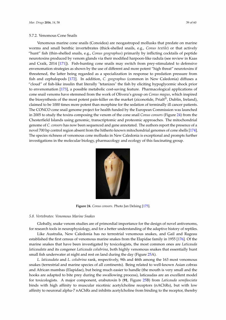
Mar. Drugs 2016, 14, 58 39 of 60
5.7.2. Venomous Cone Snails
Venomous marine cone snails (Conoidea) are neogastropod mollusks that predate on marineworms and small benthic invertebrates (thick-shelled snails, e.g., Conus textile) or that actively“hunt” fish (thin-shelled snails, e.g., Conus geographus) primarily by inflicting cocktails of peptideneurotoxins produced by venom glands via their modified harpoon-like radula (see review in Kaasand Craik, 2014 [171]). Fish-hunting cone snails may switch from prey-stimulated to defensiveenvenomation strategies as shown by the use of different and more potent “high threat” neurotoxins ifthreatened, the latter being regarded as a specialization in response to predation pressure fromfish and cephalopods [172]. In addition, C. geographus (common in New Caledonia) diffuses a“cloud” of fish-like insulin that literally "tetanizes" the fish by eliciting hypoglycemic shock priorto envenomation [173], a possible metabolic cost-saving feature. Pharmacological applications ofcone snail venoms have stemmed from the work of Olivera’s group on Conus magus, which inspiredthe biosynthesis of the most potent pain-killer on the market (ziconotide, Prialt®, Dublin, Ireland),claimed to be 1000 times more potent than morphine for the sedation of terminally ill cancer patients.The CONCO cone snail genome project for health funded by the European Commission was launchedin 2005 to study the toxins composing the venom of the cone snail Conus consors (Figure 24) from theChesterfield Islands using genomic, transcriptomic and proteomic approaches. The mitochondrialgenome of C. consors has now been sequenced and gene annotated. The authors report the presence of anovel 700 bp control region absent from the hitherto known mitochondrial genomes of cone shells [174].The species richness of venomous cone mollusks in New Caledonia is exceptional and prompts furtherinvestigations in the molecular biology, pharmacology and ecology of this fascinating group.
Mar. Drugs 2016, 14, x 42 of 62
Figure 23. Structure of verrol 4‐acetate (85), verrucarine A (86), isororidine A (87), kawain (88),
7,8‐dihydrokawain (89) and 5,6‐dehydrokawain (90).
5.7.2. Venomous Cone Snails
Venomous marine cone snails (Conoidea) are neogastropod mollusks that predate on marine
worms and small benthic invertebrates (thick‐shelled snails, e.g., Conus textile) or that actively
“hunt” fish (thin‐shelled snails, e.g., Conus geographus) primarily by inflicting cocktails of peptide
neurotoxins produced by venom glands via their modified harpoon‐like radula (see review in Kaas
and Craik, 2014 [171]). Fish‐hunting cone snails may switch from prey‐stimulated to defensive
envenomation strategies as shown by the use of different and more potent “high threat” neurotoxins
if threatened, the latter being regarded as a specialization in response to predation pressure from
fish and cephalopods [172]. In addition, C. geographus (common in New Caledonia) diffuses a
“cloud” of fish‐like insulin that literally ʺtetanizesʺ the fish by eliciting hypoglycemic shock prior to
envenomation [173], a possible metabolic cost‐saving feature. Pharmacological applications of cone
snail venoms have stemmed from the work of Olivera’s group on Conus magus, which inspired the
biosynthesis of the most potent pain‐killer on the market (ziconotide, Prialt®, Dublin, Ireland),
claimed to be 1000 times more potent than morphine for the sedation of terminally ill cancer patients.
The CONCO cone snail genome project for health funded by the European Commission was
launched in 2005 to study the toxins composing the venom of the cone snail Conus consors (Figure 24)
from the Chesterfield Islands using genomic, transcriptomic and proteomic approaches. The
mitochondrial genome of C. consors has now been sequenced and gene annotated. The authors report
the presence of a novel 700 bp control region absent from the hitherto known mitochondrial genomes of
cone shells [174]. The species richness of venomous cone mollusks in New Caledonia is exceptional
and prompts further investigations in the molecular biology, pharmacology and ecology of this
fascinating group.
Figure 24. Conus consors. Photo Jan Delsing [175]. Figure 24. Conus consors. Photo Jan Delsing [175].
5.8. Vertebrates: Venomous Marine Snakes
Globally, snake venom studies are of primordial importance for the design of novel antivenoms,for research tools in neurophysiology, and for a better understanding of the adaptive history of reptiles.
Like Australia, New Caledonia has no terrestrial venomous snakes, and Gail and Rageauestablished the first census of venomous marine snakes from the Elapidae family in 1955 [176]. Of themarine snakes that have been investigated by toxicologists, the most common ones are Laticaudalaticaudata and its congener Laticauda colubrina, both highly venomous snakes that essentially huntsmall fish underwater at night and rest on land during the day (Figure 25A).
L. laticaudata and L. colubrina rank, respectively, 9th and 46th among the 163 most venomoussnakes (terrestrial and marine species of all continents). Being related to well-known Asian cobrasand African mambas (Elapidae), but being much easier to handle (the mouth is very small and thehooks are adapted to bite prey during the swallowing process), laticaudas are an excellent modelfor toxicologists. A major component, erabutoxin b (91, Figure 25B) from Laticauda semifasciatabinds with high affinity to muscular nicotinic acetylcholine receptors (nAChRs), but with lowaffinity to neuronal alpha-7 nAChRs and inhibits acetylcholine from binding to the receptor, thereby
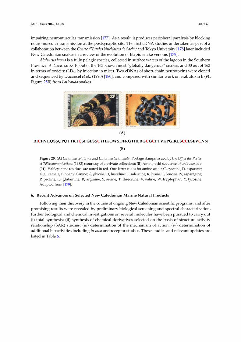
Mar. Drugs 2016, 14, 58 40 of 60
impairing neuromuscular transmission [177]. As a result, it produces peripheral paralysis by blockingneuromuscular transmission at the postsynaptic site. The first cDNA studies undertaken as part of acollaboration between the Centre d'Etudes Nucléaires de Saclay and Tokyo University [178] later includedNew Caledonian snakes in a review of the evolution of Elapid snake venoms [179].
Aipisurus laevis is a fully pelagic species, collected in surface waters of the lagoon in the SouthernProvince. A. laevis ranks 10 out of the 163 known most “globally dangerous” snakes, and 30 out of 163in terms of toxicity (LD50 by injection in mice). Two cDNAs of short-chain neurotoxins were clonedand sequenced by Ducancel et al., (1990) [180], and compared with similar work on erabutoxin b (91,Figure 25B) from Laticauda snakes.
Mar. Drugs 2016, 14, x 43 of 62
5.8. Vertebrates: Venomous Marine Snakes
Globally, snake venom studies are of primordial importance for the design of novel antivenoms,
for research tools in neurophysiology, and for a better understanding of the adaptive history of reptiles.
Like Australia, New Caledonia has no terrestrial venomous snakes, and Gail and Rageau
established the first census of venomous marine snakes from the Elapidae family in 1955 [176]. Of
the marine snakes that have been investigated by toxicologists, the most common ones are Laticauda
laticaudata and its congener Laticauda colubrina, both highly venomous snakes that essentially hunt
small fish underwater at night and rest on land during the day (Figure 25A).
L. laticaudata and L. colubrina rank, respectively, 9th and 46th among the 163 most venomous
snakes (terrestrial and marine species of all continents). Being related to well‐known Asian cobras
and African mambas (Elapidae), but being much easier to handle (the mouth is very small and the
hooks are adapted to bite prey during the swallowing process), laticaudas are an excellent model for
toxicologists. A major component, erabutoxin b (91, Figure 25B) from Laticauda semifasciata binds
with high affinity to muscular nicotinic acetylcholine receptors (nAChRs), but with low affinity to
neuronal alpha‐7 nAChRs and inhibits acetylcholine from binding to the receptor, thereby impairing
neuromuscular transmission [177]. As a result, it produces peripheral paralysis by blocking
neuromuscular transmission at the postsynaptic site. The first cDNA studies undertaken as part of a
collaboration between the Centre dʹEtudes Nucléaires de Saclay and Tokyo University [178] later
included New Caledonian snakes in a review of the evolution of Elapid snake venoms [179].
Aipisurus laevis is a fully pelagic species, collected in surface waters of the lagoon in the
Southern Province. A. laevis ranks 10 out of the 163 known most “globally dangerous” snakes, and 30
out of 163 in terms of toxicity (LD50 by injection in mice). Two cDNAs of short‐chain neurotoxins
were cloned and sequenced by Ducancel et al., (1990) [180], and compared with similar work on
erabutoxin b (91, Figure 25B) from Laticauda snakes.
(A)
(B)
Figure 25. (A) Laticauda colubrina and Laticauda laticaudata. Postage stamps issued by the Office des
Postes et Télécommunications (1983) (courtesy of a private collection); (B) Amino‐acid sequence of
erabutoxin b (91). Half cysteine residues are noted in red. One‐letter codes for amino acids: C,
cysteine; D, aspartate; E, glutamate; F, phenylalanine; G, glycine; H, histidine; I, isoleucine; K, lysine;
L, leucine; N, asparagine; P, proline; Q, glutamine; R, arginine; S, serine; T, threonine; V, valine; W,
tryptophan; Y, tyrosine. Adapted from [179].
6. Recent Advances on Selected New Caledonian Marine Natural Products
Following their discovery in the course of ongoing New Caledonian scientific programs, and
after promising results were revealed by preliminary biological screening and spectral
characterization, further biological and chemical investigations on several molecules have been
pursued to carry out (i) total synthesis; (ii) synthesis of chemical derivatives selected on the basis of
structure‐activity relationship (SAR) studies; (iii) determination of the mechanism of action; (iv)
determination of additional bioactivities including in vivo and receptor studies. These studies and
relevant updates are listed in Table 6.
RICFNHQSSQPQTTKTCSPGESSCYHKQWSDFRGTIIERGCGCPTVKPGIKLSCCESEVCNN
Figure 25. (A) Laticauda colubrina and Laticauda laticaudata. Postage stamps issued by the Office des Posteset Télécommunications (1983) (courtesy of a private collection); (B) Amino-acid sequence of erabutoxin b(91). Half cysteine residues are noted in red. One-letter codes for amino acids: C, cysteine; D, aspartate;E, glutamate; F, phenylalanine; G, glycine; H, histidine; I, isoleucine; K, lysine; L, leucine; N, asparagine;P, proline; Q, glutamine; R, arginine; S, serine; T, threonine; V, valine; W, tryptophan; Y, tyrosine.Adapted from [179].
6. Recent Advances on Selected New Caledonian Marine Natural Products
Following their discovery in the course of ongoing New Caledonian scientific programs, and afterpromising results were revealed by preliminary biological screening and spectral characterization,further biological and chemical investigations on several molecules have been pursued to carry out(i) total synthesis; (ii) synthesis of chemical derivatives selected on the basis of structure-activityrelationship (SAR) studies; (iii) determination of the mechanism of action; (iv) determination ofadditional bioactivities including in vivo and receptor studies. These studies and relevant updates arelisted in Table 6.
Mar. Drugs 2016, 14, 58 41 of 60
Table 6. Subsequent and ongoing development of selected natural products isolated from New Caledonian marine organisms.
Natural Product and/or Analogs Chemical Synthesis and Biological Activity Reference
(´)-agelastatin A–F
-Enantioselective total synthesis involving late-stage C-ring formation.-(´)-agelastatin A: antiproliferative activity highly potent on blood cancer cell lines (CEM EC50 20 nM; Jurkat EC50
74 nM; Daudi EC50 20 nM; HL-60 EC50 138 nM; CA46 EC50 187 nM) vs. normal red blood cells (EC50 > 333 µM);dose-dependent induction of apoptosis (G2/M phase arrest), no effect on tubulin dynamics.
[181]
agelastatin A/13-debromo-13-trifluoromethyl agelastatin A
-In vitro and in vivo antiproliferative activity: CLL patient (CLL1 and CLL2) and JVM-2 cell line (EC50 0.064 µM and0.16 µM respectively).
-Chemical modifications outside the pyrrole ring result in significant loss in activity.-Importance of an electronegative functional group at position C-13 for CLL activity.
[182]
suvanine
-Anti-inflammatory activity, Hsp 60 inhibitor. [183]
-Antagonist of farnesoid-X-receptor (FXR).-Identification of conformational changes responsible for agonist/antagonist form on FXR using suvanine
as template.[184]
-Inhibitor of hepatitis C virus NS3 helicase (IC50 of 3 µM).-Inhibition of ATPase, RNA binding, and serine protease activities of NS3 helicase with IC50 7, 3, and
34 µM, respectively.-Interaction with an allosteric site in NS3 rather than binding to the catalytic core.
[185]
isohymenialdisine andhymenialdisine
-Inhibition of CLK kinases (hymenialdisine only). [186]
-Stimulation of translation: isohymenialdisine and hymenialdisine act on PKR (RNA-dependent protein kinase) byinhibiting its autophosphorylation and pertub the PKR-eIF2α phosphorylation axis; models indicate that it fits in
the PKR ATP binding site.[187]
-Short (6 steps), concise, and high yielding (44%) total synthesis of hymenialdisine will enable the synthesis ofnovel libraries for subsequent SAR studies. [188]
arsenicin A-Synthesis of sulfur-derivatives.
-Antiproliferative activity of monosulfide (˘)-arsenicin A (more potent on APL cells than (˘)-arsenicin A andarsenic (III) oxide); induction of apoptosis.
[189]
heteronemin
-Anti-intravasative properties. [190]
-Targets TDP-43, binds to specific sequences on DNA and RNA. [191]
-Antiproliferative (A498 EC50 1.57 µM).-Induction of both apoptosis and autophagy (heteronemin inhibits the phosphorylation of ERK and AKT signaling
pathways and increases the phosphorylation of p38 and JNK).[192]
Mar. Drugs 2016, 14, 58 42 of 60
Table 6. Cont.
Natural Product and/or Analogs Chemical Synthesis and Biological Activity Reference
bengamides and analogs
-Synthesis of novel caprolactam-ring-opened bengamide analogs with antitumor activity on MDA-MB-435(compounds 3a (EC50 4 nM) and 2i (EC50 9 nM) more potent activity than LAF389, analog of bengamide (EC50 40
nM) and the original caprolactam analog 10’ (EC50 17 nM).-Improved water solubility.
[193]
Methionine aminopeptidases inhibitors (HsMetAP1 and HsMetAP2). [194]
-Synthesis of a series of inhibitors of methionine aminopeptidases of Mycobacterium tuberculosis.-New X-ray structures of MtMetAP1c in complex with inhibitors in Mn(II) and Ni(II) forms; all amide moieties bind
to the unique shallow cavity and interact with flat surface created by His-212 of MtMetAP1c in the Mn(II) form.Influence of active site metal on binding mode (amide takes on a different conformation in the Ni(II) form).
[195]
-Analogs of bengamide E with antiproliferative properties (modified at the terminal olefinic position).-More potent activity: compound 56 (a cyclopentyl group replaced the isopropyl group at the terminal
olefinic position).[196]
-Synthesis of stereoisomers of bengamide E (2,3-bis-epi- and the 2-epianalogues), a collection of C2-modifiedanalogues, and various epoxy bengamides.
-Stereochemistry at C2 and C3 positions and methoxyl group at C2: essential for retaining the cytotoxic potency.[197]
naamidine A -Antitumor agent; induction of apoptosis (caspase-dependent). [198,199]
microsclerodermin A-Inhibition of NFκB transcriptional activity, reduced levels of phosphorylated (active) NFκB in the AsPC-1 cell line.
-Antiproliferative activities: AsPC-1, BxPC-3, MIA PaCa-2 and PANC-1 pancreatic cancer cell lines.-Induction of apoptosis in the AsPC-1 mediated by GSK-3β pathway.
[200]
petrosaspongiolide M and analogs
-Synthetic derivative of benzo[b]thiophen-2-yl-3-bromo-5-hydroxy-5H-furan-2-one.-In vitro and in vivo potent anti-inflammatory activity (inhibition of NF-κB signaling pathway and
STAT3 phosphorylation).[201]
-Proteasome inhibitor.-Inhibitory activity by binding the active sites in the inner core of the immunoproteasome and/or covalently
linking a Lys residue at the proteasome core/11S activator particle interface.-Modulation of intracellular proteolysis through a dual inhibition of the immunoproteasome and autophagy.
[202]
aeroplysinin-1
-Induction of apoptosis in endothelial cells (caspase dependent). [203]
-Antiproliferative effect on acute myeloid leukemia (AML) cells (dose-dependent EC50 10–20 µM); inductionof apoptosis.
-Agent-specific pleiotropic effects.[204]
fistularin-3 -Antiproliferative activity dose- and time-dependent (EC50 7.39 and 8.10 µM for Jurkat E6.1 and U937 resp.);Pro-apoptotic. [205]

Mar. Drugs 2016, 14, 58 43 of 60
Table 6. Cont.
Natural Product and/or Analogs Chemical Synthesis and Biological Activity Reference
Analogs of bistramide A
-Design and synthesis of analog of bistramide A targeting cytoskeletal organization of cancer cells in vivo(combination of reversible G-actin binding and effective F-actin severing).
-Potent and reversible binding of monomeric actin (Kd 9.0 nM), in vitro depolymerization of actin; in vitro andin vivo inhibition of A549 cells.
[206]
-Computational analyses of non-covalent actin-inhibitor interactions (AutoDock and DrugScore scoring function).-Design of a novel, highly modular class of hybrid analogs with a rationale to address both the bistramide and
rhizopodin binding sites.-Antiproliferative activity is conserved in analogs resembling the original side chain of rhizopodin.
[207]
luzonicoside A -Potent immunomodulatory agent: more effective in stimulating lysosomal activity, intracellular ROS levelelevation, and NO synthesis up-regulation in RAW 264.7 murine macrophages cells (0.01–0.1 µM). [208]
Cell lines: CEM human acute lymphoblastic leukemia, Jurkat human T leukemia, Daudi human B lymphoma (Burkitt’s lymphoma), HL-60 human acute promyelocytic leukemia,CA46 human lymphoma (Burkitt’s lymphoma), CLL chronic lymphocytic leukemia, JVM-2 human lymphoma (Mantle Cell Lymphoma), APL acute promyelocytic leukemia, A498human kidney carcinoma, MDA-MB-435 human breast adenocarcinoma, AML acute myeloid leukemia, AsPC-1, BxPC-3, MIA Paca-2, PANC-1 pancreatic cancer cell lines. Proteins:Hsp-60 Heat shock protein 60, FXR farnesoid X receptor, NS3 nonstructural protein 3, CLK protein kinases, PKR protein kinase R, TDP-43 TAR (trans-activator regulatory) DNA-bindingProtein 43, ERK extracellular signal-regulated kinase, AKT protein kinase B, JNK c-Jun N-terminal kinase, GSK-3β glycogen synthase kinase-3β, STAT3 signal transducer and activatorof transcription. Virus: HCV hepatitis C virus. Others: SAR structure-activity relationship, NO nitric oxide.

Mar. Drugs 2016, 14, 58 44 of 60
Despite recent advances in developing therapeutic strategies, cancer is still one of the leadingcauses of death worldwide. Cancer is a group of diseases characterized by the uncontrolled growthand spread of abnormal cells. Drug-induced programmed cell death holds promise for cancertherapeutic approaches. Apoptosis is one of the well-known pathways used by cells to die. It isa morphological event characterized by chromosomal DNA fragmentation, nuclear disintegration,cell shrinkage, translocation of phosphatidyl serine moieties to the outer membrane leaflet, andmembrane blebbing [209]. Numerous anticancer drugs, both on the market and in development,have apoptosis-modulating properties (e.g., Yondelis®, Madrid, Spain, has been recently approvedby the American Food and Drug Administration (FDA) to treat patients with advanced soft tissuesarcoma [210]). Apoptogens are agents that can induce rapid death by modulating the apoptosispathway. Numerous marine natural products reported in Table 6 are apoptogenic compoundsassociated with antitumor activity.
Agelastatins, members of the chemically diverse pyrrole-imidazole alkaloids (PIA), are amongthe best examples of this class of molecules. Since the discovery of agelastatin A (92, Figure 26) in1993 by Pietra et al., in the New Caledonian coral sea sponge Agelas dendromorpha, more than tendifferent research groups have reported innovative solutions to synthesize it. As reported in Han et al.,2013, recent development of a concise, stereo-controlled, and biosynthetically inspired strategy tosynthesize agelastatin alkaloids and a new synthetic methodology for azaheterocycle synthesis hasgiven rise to many agelastatin derivatives. These developments also facilitate the first side-by-sidetesting of all known agelastatin alkaloids for their ability to induce cell death in various cancercells [181]: U-937 (lymphoma), HeLa (cervical carcinoma), A549 (non-small-cell lung carcinoma),BT549 (breast carcinoma), and IMR90 (immortalized lung fibroblasts) human cell lines. AgelastatinsA (92, Figure 26) and D (95, Figure 26) both induce dose-dependent apoptosis (in particular cellmembrane permeabilization, activation of procaspase-3 to active caspase-3 and proteolytic cleavageof poly(ADP-ribose) polymerase (PARP)) and exhibit dose-dependent G2/M cell cycle arrest insynchronized U-937 cells without affecting tubulin dynamics within cells [181]. In addition, thepotency of all agelastatins (92–97, Figure 26) has been evaluated in five human blood cancer cell lines.Interestingly, agelastatin C (94, Figure 26) is only weakly active and agelastatin F (97, Figure 26) isinactive against the tested cell lines. Agelastatin A (92, Figure 26) is remarkably active, particularlyagainst CEM from acute lymphoblastic leukemia and against Daudi from Burkitt’s lymphoma (valuesreported in Table 6) and specifically target these white blood cell lines over normal red blood cells: it is16,650 times more active on cancer cells than on normal cells! These results are in line with a previousstudy showing the inhibition of osteopontin-mediated neoplastic transformation (notably by β-catenininhibition) and metastasis by agelastatin A (92, Figure 26) in MDA-MB-231 and MDA-MB-435 humanbreast cancer cell lines [211]. Therefore, agelastatins exhibit considerable potential as antitumor drugsthat can simultaneously inhibit cancer cell growth as well as act as a potent anti-metastatic drug [211].However, the mechanism through which agelastatin A (92, Figure 26) causes this cellular effect,particularly cycle arrest, still needs to be elucidated. Identification of potential intracellular targets ofagelastatins can be performed by affinity chromatography on immobilized agelastatin. This approachhas previously provided valuable insights into the cellular targets of various protein kinases inhibitorsincluding hymenialdisine (12, Figure 5) [212].
Mar. Drugs 2016, 14, x
Despite recent advances in developing therapeutic strategies, cancer is still one of the leading
causes of death worldwide. Cancer is a group of diseases characterized by the uncontrolled growth
and spread of abnormal cells. Drug‐induced programmed cell death holds promise for cancer
therapeutic approaches. Apoptosis is one of the well‐known pathways used by cells to die. It is a
morphological event characterized by chromosomal DNA fragmentation, nuclear disintegration, cell
shrinkage, translocation of phosphatidyl serine moieties to the outer membrane leaflet, and
membrane blebbing [209]. Numerous anticancer drugs, both on the market and in development,
have apoptosis‐modulating properties (e.g., Yondelis®, Madrid, Spain, has been recently approved
by the American Food and Drug Administration (FDA) to treat patients with advanced soft tissue
sarcoma [210]). Apoptogens are agents that can induce rapid death by modulating the apoptosis
pathway. Numerous marine natural products reported in Table 6 are apoptogenic compounds
associated with antitumor activity.
Agelastatins, members of the chemically diverse pyrrole‐imidazole alkaloids (PIA), are among
the best examples of this class of molecules. Since the discovery of agelastatin A (92, Figure 26) in
1993 by Pietra et al., in the New Caledonian coral sea sponge Agelas dendromorpha, more than ten
different research groups have reported innovative solutions to synthesize it. As reported in
Han et al., 2013, recent development of a concise, stereo‐controlled, and biosynthetically inspired
strategy to synthesize agelastatin alkaloids and a new synthetic methodology for azaheterocycle
synthesis has given rise to many agelastatin derivatives. These developments also facilitate the first
side‐by‐side testing of all known agelastatin alkaloids for their ability to induce cell death in various
cancer cells [181]: U‐937 (lymphoma), HeLa (cervical carcinoma), A549 (non‐small‐cell lung
carcinoma), BT549 (breast carcinoma), and IMR90 (immortalized lung fibroblasts) human cell lines.
Agelastatins A (92, Figure 26) and D (95, Figure 26) both induce dose‐dependent apoptosis (in
particular cell membrane permeabilization, activation of procaspase‐3 to active caspase‐3 and
proteolytic cleavage of poly(ADP‐ribose) polymerase (PARP)) and exhibit dose‐dependent G2/M
cell cycle arrest in synchronized U‐937 cells without affecting tubulin dynamics within cells [181]. In
addition, the potency of all agelastatins (92–97, Figure 26) has been evaluated in five human blood
cancer cell lines. Interestingly, agelastatin C (94, Figure 26) is only weakly active and agelastatin F
(97, Figure 26) is inactive against the tested cell lines. Agelastatin A (92, Figure 26) is remarkably
active, particularly against CEM from acute lymphoblastic leukemia and against Daudi from
Burkitt’s lymphoma (values reported in Table 6) and specifically target these white blood cell lines
over normal red blood cells: it is 16,650 times more active on cancer cells than on normal cells! These
results are in line with a previous study showing the inhibition of osteopontin‐mediated neoplastic
transformation (notably by β‐catenin inhibition) and metastasis by agelastatin A (92, Figure 26) in
MDA‐MB‐231 and MDA‐MB‐435 human breast cancer cell lines [211]. Therefore, agelastatins exhibit
considerable potential as antitumor drugs that can simultaneously inhibit cancer cell growth as well
as act as a potent anti‐metastatic drug [211]. However, the mechanism through which agelastatin A
(92, Figure 26) causes this cellular effect, particularly cycle arrest, still needs to be elucidated.
Identification of potential intracellular targets of agelastatins can be performed by affinity
chromatography on immobilized agelastatin. This approach has previously provided valuable
insights into the cellular targets of various protein kinases inhibitors including hymenialdisine (12,
Figure 5) [212].
Figure 26. Cont.
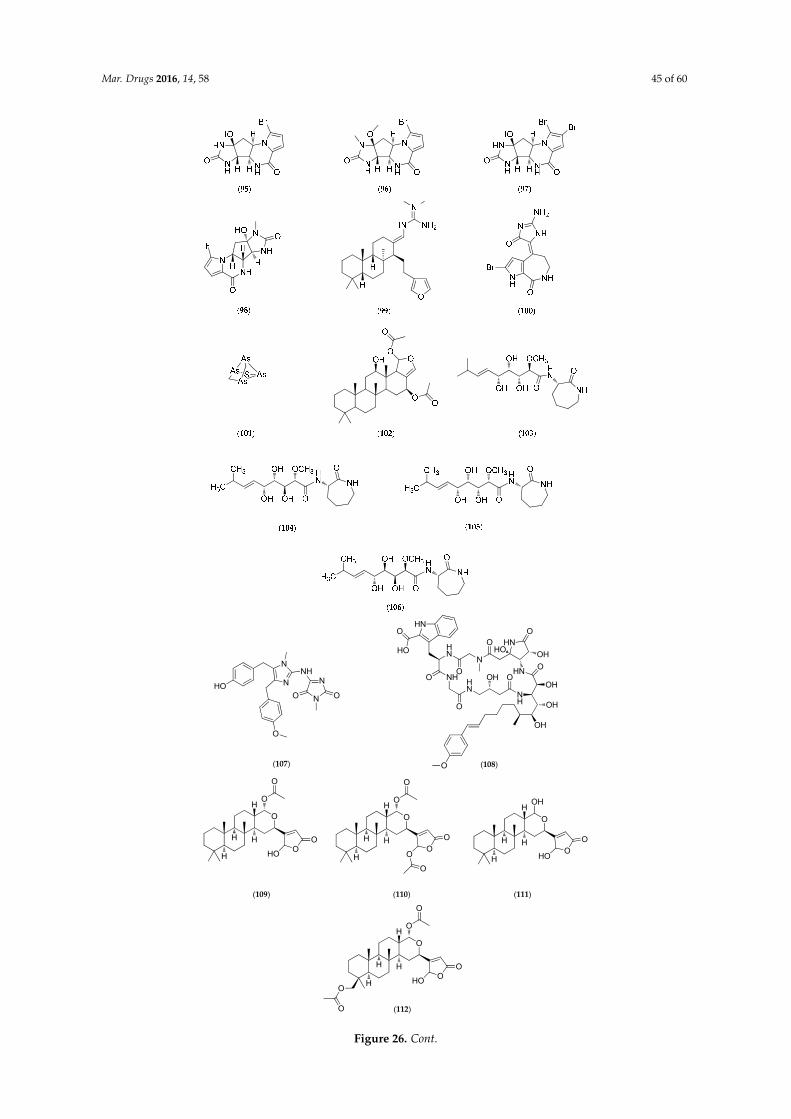
Mar. Drugs 2016, 14, 58 45 of 60Mar. Drugs 2016, 14, x 48 of 62
(107)
N
NHO
O
NHN
NO O
HN
HN
O NHO
N
O HN
O
HN
NH
O
HO
OHHN
O
OH
O
OH
OH
O
OH
O
HO
(108)
O
O
H
H
O
O
O
HO
H
H
(109)
O
O
H
H
O
O
O
O
H
HO
(110)
O
O
H
H
OH
O
HO
H
H
(111)
O
O
H
H
O
O
O
HO
H
HO
O (112)
Figure 26. Cont.
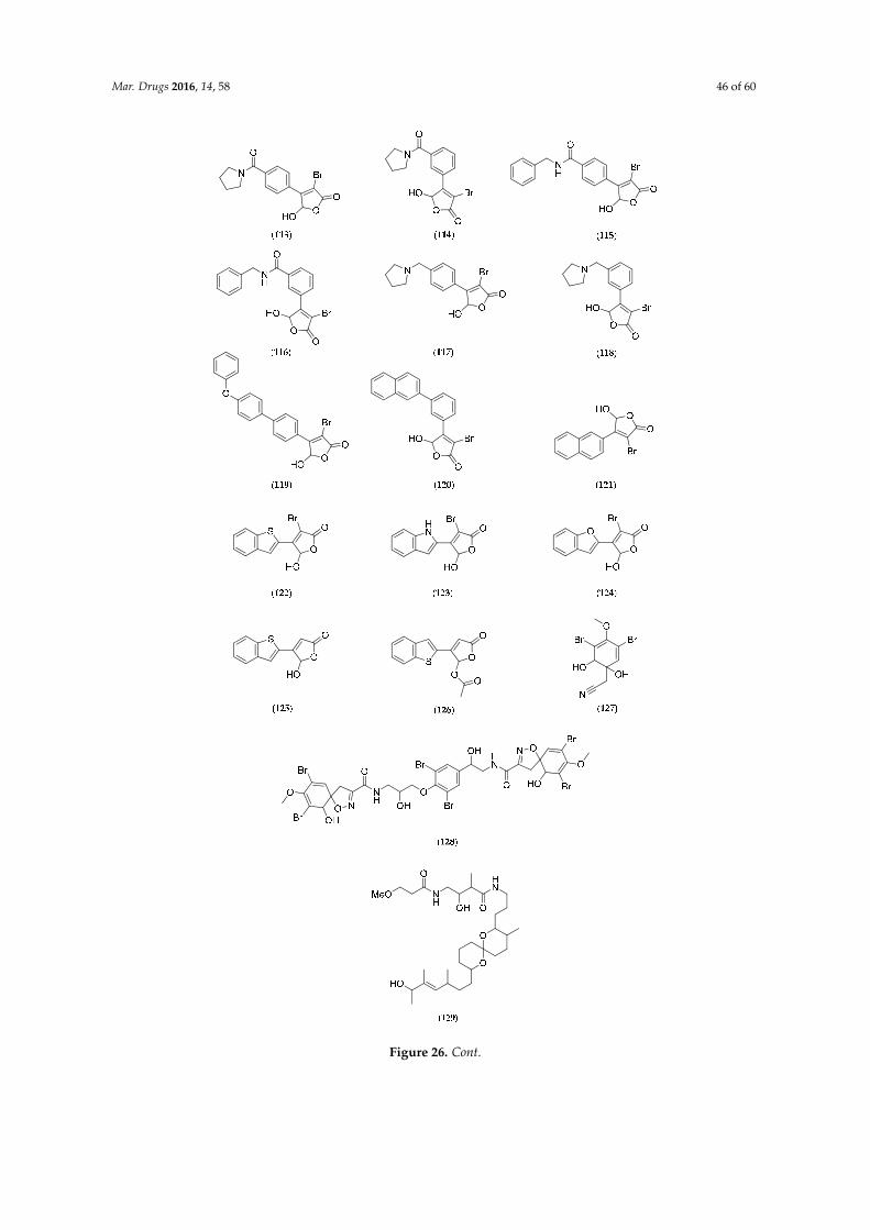
Mar. Drugs 2016, 14, 58 46 of 60Mar. Drugs 2016, 14, x 49 of 62
Figure 26. Cont.

Mar. Drugs 2016, 14, 58 47 of 60Mar. Drugs 2016, 14, x 50 of 62
Figure 26. Structure of agelastatin A–F (92–97), 13‐debromo‐13‐trifluoromethyl agelastatin A (98),
suvanine (99), isohymenialdisine (100), monosulfide arsenicin A (101), heteronemin (102),
bengamide E (103) and analogs: 2,3‐bis‐epi‐bengamide E (104), 2‐epi‐bengamide E (105) and
3,4‐bis‐epi‐bengamide E (106), naamidine A (107), microsclerodermin A (108), petrosaspongiolide M
(109), natural analogs of petrosaspongiolide M (110–112), first generation of synthetic
petrosaspongiolide M analogs (113–120), second generation of synthetic petrosaspongiolide M
analogs (121–126), aeroplysinin‐1 (127), fistularin‐3 (128), analogs of bistramide A (129), adapted
from [206] and luzonicoside A (130).
Among the molecules reported in Table 6, heteronemin (102, Figure 26), a marine sesterterpene,
has been shown to induce apoptosis in A‐498 human renal carcinoma cells by downregulating
apoptosis inhibitors Bcl‐2 and Bcl‐xL and upregulating the death agonist Bax, leading to the
disruption of the mitochondrial membrane potential and the release of cytochrome c from
mitochondria [192]. As also reported for agelastatins, these effects are associated with the activation
of caspases (3/8 and 9), followed by PARP cleavage. Interestingly, the same study showed that
heteronemin (102, Figure 26) also induces autophagy in A‐498 cells, but the inhibition of autophagy
enhances the anticancer effect of heteronemin (102, Figure 26) in A‐498 cells. This result suggests that
the combination of heteronemin (102, Figure 26) with autophagy inhibitors further enhances its
therapeutic effects for cancer treatment [192]. Therefore, marine molecules can represent chemical
tools for the exploration of novel combined therapeutic strategies. Petrosaspongiolide M (109, Figure 26)
may also represent an equally valuable chemical tool for its capacity to modulate intracellular
proteolysis through dual inhibition of the immunoproteasome and autophagy [202].
Synergistic effects (e.g., treatment with heteronemin (102, Figure 26) and inhibitors of
autophagy such as chloroquine) of marine products and therapeutic drugs on disease‐related
phenotypes highlight the concept of combination therapy. The cytotoxicity of new marine molecules
should be tested alone or in combination with known anticancer compounds to overcome drug
resistance, as reported in Elmallah and Micheau (2015) for the resistance of cancer cells to
TNF‐related apoptosis inducing ligand (TRAIL)‐induced cell death [209]. TRAIL is a molecule that
selectively kills—via apoptosis—transformed and cancer cells, but not most normal cells [213].
Manzamine A, an alkaloid originally isolated from an Okinawan sponge Haliclona sp., may restore
TRAIL‐induced apoptotic cell death in the TRAIL‐resistance pancreatic AsPC‐1 cell line via
inhibition of glycogen synthase kinase‐3β (GSK‐3β) and subsequent inhibition of the survival factor
NF‐κB [209]. It will thus be very interesting to evaluate the TRAIL‐sensitizing activity of agelastatin
A (92, Figure 26); which is known to inhibit GSK‐3β with an IC50 of 12 μM [44] (manzamine A has an
IC50 for GSK‐3β inhibition of 10 μM [214]). All new marine natural products, including those from
biodiversity hotspots such as the New Caledonia archipelago, represent putative new hope for
cancer treatments, in particular for overcoming defects in apoptosis signaling.
As highlighted above, targeting apoptotic pathways may have a direct role in inducing tumor
cell death, as confirmed by recent advances on marine molecules found in New Caledonia. In
addition to agelastatins and heteronemin (102, Figure 26), naamidine A (107, Figure 26),
microsclerodermin A (108, Figure 26), aeroplysinin‐1 (127, Figure 26), fistularin‐3 (128, Figure 26)
have been shown to induce apoptosis (Table 6). Cancer is the principal target of the marine
molecules released on the market today. Of the seven marine compounds currently on the market,
only three (Prialt®, Dublin, Ireland; Yondelis®, Madrid, Spain and Lovaza®, London, UK) were not
chemically modified to become drugs [215,216]. The four other molecules underwent lead
Figure 26. Structure of agelastatin A–F (92–97), 13-debromo-13-trifluoromethyl agelastatin A (98),suvanine (99), isohymenialdisine (100), monosulfide arsenicin A (101), heteronemin (102), bengamideE (103) and analogs: 2,3-bis-epi-bengamide E (104), 2-epi-bengamide E (105) and 3,4-bis-epi-bengamideE (106), naamidine A (107), microsclerodermin A (108), petrosaspongiolide M (109), natural analogsof petrosaspongiolide M (110–112), first generation of synthetic petrosaspongiolide M analogs(113–120), second generation of synthetic petrosaspongiolide M analogs (121–126), aeroplysinin-1(127), fistularin-3 (128), analogs of bistramide A (129), adapted from [206] and luzonicoside A (130).
Among the molecules reported in Table 6, heteronemin (102, Figure 26), a marine sesterterpene,has been shown to induce apoptosis in A-498 human renal carcinoma cells by downregulating apoptosisinhibitors Bcl-2 and Bcl-xL and upregulating the death agonist Bax, leading to the disruption of themitochondrial membrane potential and the release of cytochrome c from mitochondria [192]. As alsoreported for agelastatins, these effects are associated with the activation of caspases (3/8 and 9),followed by PARP cleavage. Interestingly, the same study showed that heteronemin (102, Figure 26)also induces autophagy in A-498 cells, but the inhibition of autophagy enhances the anticancereffect of heteronemin (102, Figure 26) in A-498 cells. This result suggests that the combination ofheteronemin (102, Figure 26) with autophagy inhibitors further enhances its therapeutic effects forcancer treatment [192]. Therefore, marine molecules can represent chemical tools for the explorationof novel combined therapeutic strategies. Petrosaspongiolide M (109, Figure 26) may also representan equally valuable chemical tool for its capacity to modulate intracellular proteolysis through dualinhibition of the immunoproteasome and autophagy [202].
Synergistic effects (e.g., treatment with heteronemin (102, Figure 26) and inhibitors of autophagysuch as chloroquine) of marine products and therapeutic drugs on disease-related phenotypeshighlight the concept of combination therapy. The cytotoxicity of new marine molecules shouldbe tested alone or in combination with known anticancer compounds to overcome drug resistance, asreported in Elmallah and Micheau (2015) for the resistance of cancer cells to TNF-related apoptosisinducing ligand (TRAIL)-induced cell death [209]. TRAIL is a molecule that selectively kills—viaapoptosis—transformed and cancer cells, but not most normal cells [213]. Manzamine A, an alkaloidoriginally isolated from an Okinawan sponge Haliclona sp., may restore TRAIL-induced apoptoticcell death in the TRAIL-resistance pancreatic AsPC-1 cell line via inhibition of glycogen synthasekinase-3β (GSK-3β) and subsequent inhibition of the survival factor NF-κB [209]. It will thus be veryinteresting to evaluate the TRAIL-sensitizing activity of agelastatin A (92, Figure 26); which is knownto inhibit GSK-3β with an IC50 of 12 µM [44] (manzamine A has an IC50 for GSK-3β inhibition of10 µM [214]). All new marine natural products, including those from biodiversity hotspots such asthe New Caledonia archipelago, represent putative new hope for cancer treatments, in particular forovercoming defects in apoptosis signaling.
As highlighted above, targeting apoptotic pathways may have a direct role in inducing tumor celldeath, as confirmed by recent advances on marine molecules found in New Caledonia. In additionto agelastatins and heteronemin (102, Figure 26), naamidine A (107, Figure 26), microscleroderminA (108, Figure 26), aeroplysinin-1 (127, Figure 26), fistularin-3 (128, Figure 26) have been shownto induce apoptosis (Table 6). Cancer is the principal target of the marine molecules released onthe market today. Of the seven marine compounds currently on the market, only three (Prialt®,Dublin, Ireland; Yondelis®, Madrid, Spain and Lovaza®, London, UK) were not chemically modified
Mar. Drugs 2016, 14, 58 48 of 60
to become drugs [215,216]. The four other molecules underwent lead optimization during the variousdifferent stages of their development. Developments in structural chemistry must be followed by thecharacterization of biological targets to optimize therapeutic applications.
7. Conclusions
Though far from being exhaustive, this review outlines 40 years of exciting research on thechemodiversity of marine organisms, ranging from microbes and invertebrates to vertebrates, frommicroalgae to macroalgae and halophytes, belonging to very different biota in association with thecomplex coral reef systems of New Caledonia.
Traditionally “interesting” lead groups such as sponges, cnidarians and ascidians have beenintensely investigated because they not only provide the most interesting array of original chemicalstructures, but they also show the most potent anticancer, anti-inflammatory and antibiotic properties.Other groups have occasionally led to original and stimulating research: echinoderms, mollusks,etc. This review focused on novel products although several studies have been performed on NewCaledonian marine species leading to the rediscovery of some secondary metabolites (e.g., Acremoniumneocaledoniae, p. 41).
The fact that “minor” groups have been largely left aside does not reflect their lack of intrinsicinterest for chemical exploration, but rather the technical difficulties to collect or cultivate them inNew Caledonia. Only about 2% of marine bacteria can be cultivated using classical growth media.Some small invertebrates require time-consuming field and laboratory work to get enough molecularmaterial to work on (e.g., bryozoans), are haphazardly encountered (e.g., many “naked” nudibranchand opistobranch mollusks) or are on an endangered species list, etc. Noteworthy are independentinvestigations on different models: e.g., biomaterials from scleractinian corals for maxillofacialreplacement of bone tissue [217] or from oyster nacre as an osteoinductor in odontology [218].Another example is that of the endemic Nautilus macromphallus cephalopod excretory metabolism. Thismetabolism relies on a unique and seemingly very stable symbiosis with specific bacterial symbiontsto produce molecular nitrogen in the chambered shell and regulate the buoyancy [219].
Figure 2 indicates that the “golden age” of traditional screening for novel molecules and bioactivityhas ceased after several periods of intense investigation. This reflects the successive explorations ofnew “territories”, e.g., sea mounts, outer reef slopes and deep benthos in general. Based on what hasbeen achieved thus far, taking into account the exceptional richness of the local biota and the growingneed to develop local economy in a balanced and sustainable way, it appears necessary to develop apermanent, reliable and collaborative research and development pipeline linking field exploration to:
-drug development, including (i) isolation of active principles; (ii) high-throughput bioactivityscreening; (iii) structure-activity investigations and structural elucidation; (iv) cultivationtechnologies or bioinspired synthesis; (v) clinical trials and beyond;-aquaculture focusing on the treatment of locally grown species of prawns and oyster varieties thatare sensitive to seasonal blooms of toxigenic bacteria and microalgae.
Molecular approaches have now come of age, making new biological models more attractive andpromising. New Caledonia represents a living laboratory with its unique source of marine organismsthat have not been investigated to date. There is now a need to better understand the relationshipsbetween host organisms and their microbial flora. This requires on-site operations and specializedequipment (aquaria, cultivation facilities for microorganisms).
Finally, in the general context of “blue growth”, New Caledonia is ideally located for investigatingthe production of third-generation biofuels and of high value-added products (e.g., cosmetics,nutraceuticals) via the biomass of microalgal primary producers.
New Caledonia is a treasure chest for scientists to explore, but is also fragile. With the perspectiveof climate change due to global warming and emerging anthropogenic forcings, the sustainabilityof the sources of molecules, including resident bacteria, must take precedence. New Caledonian

Mar. Drugs 2016, 14, 58 49 of 60
coral reefs—of which some portions were added to UNESCO’s World Heritage list in 2008—must beprotected for the livelihood of resident populations, as well as for biological inspiration for the sciencesand the arts. Extensive studies of their chemodiversity should contribute to their protection.
Acknowledgments: This review is dedicated in memoriam to the late Pierre Potier (CNRS) and to the late AndréMénez (MNHN) who made the important decisions and provided essential support at crucial times. Majorchemistry/pharmacology contributors historically involved in program management include (in alphabeticalorder): Ali Al-Mourabit, Philippe Amade, Dominique Bourret, Cécile Debitus, Stéphane La Barre, DominiqueLaurent, Jacques Pusset and Thierry Sévenet. Major contributors with respect to the biology, ecology andhistory of New Caledonia include (in alphabetical order): Georges Bargibant, Pierre Laboute, Claude Lévy,Jean-Louis Menou, Claude Payri, Bertrand Richer de Forges and Philippe Tirard. Chemists and biologistsfrom Italian Universities (Trente and Napoli) who have significantly contributed are gratefully acknowledged.Georges de Noni is warmly acknowledged for his continuing support. Serge Andréfouët is acknowledgedfor providing maps. The authors thank the Managing Editor for the publication fee waiver kindly offeredto S. La Barre. S.-E. Motuhi is the recipient of a thesis grant from the Government of New Caledonia (Gov.NC). Authors warmly thank the Gov. NC for continuing support for research on marine natural products.S. Bach is supported by Biogenouest, Cancéropôle Grand-Ouest (axis: Natural sea products in cancer treatment),ANR/Investissements d’Avenir program via the OCEANOMICs project (grant #ANR-11-BTBR-0008) and INCa(“NECROTRAIL” Program).
Conflicts of Interest: The authors declare no conflict of interest.
References
1. Pelletier, B. Geology of the New Caledonia Region and its Implications for the Study of the New CaledonianBiodiversity. In Compendium of Marine Species from New Caledonia, 2nd ed.; Doc. Sci. Tech. II7; Payri, C.E.,de Forges, B.R., Eds.; IRD Nouméa: Nouvelle-Calédonie, France, 2007; pp. 19–32.
2. Andréfouët, S.; Cabioch, G.; Flamand, B.; Pelletier, B. A reappraisal of the diversity of geomorphologicaland genetic processes of New Caledonian coral reefs: A synthesis from optical remote sensing, coring andacoustic multibeam observations. Coral Reefs 2009, 28, 691–707. [CrossRef]
3. Leal, M.C.; Puga, J.; Serôdio, J.; Gomes, N.C.; Calado, R. Trends in the discovery of new marine naturalproducts from invertebrates over the last two decades—Where and what are we bioprospecting. PLoS ONE2012, 7. [CrossRef] [PubMed]
4. Andréfouët, S.; Cabioch, G.; Flamand, B.; Pelletier, B. The diversity of New Caledonia Coral ReefGeomorphology and Genetic Processes: A Synthesis from Optical Remote Sensing, Coring and AcousticMulti-beam Observations. In Compendium of Marine Species from New Caledonia, 2nd ed.; Payri, C.E.,de Forges, B.R., Eds.; IRD Editions: Nouméa, Nouvelle-Calédonie, 2007; pp. 33–49.
5. Payri, C.; de Forges Richer, B. Compendium of Marine Species of New Caledonia, 2nd ed.; IRD Editions: Nouméa,Nouvelle-Calédonie, 2007.
6. Natural History Museum. Available online: http://www.nhm.ac.uk/nature-online/science-of-natural-history/expeditions-collecting/hms-challenger-expedition/ (accessed on 21 May 2015).
7. Armbrust, E.V.; Palumbi, S.R. Uncovering hidden worlds of ocean biodiversity. Science 2015, 348, 865–867.[CrossRef] [PubMed]
8. Catala, R. Contribution à l’étude écologique des îlots coralliens du Pacifique Sud. Premiers élémentsd’écologie terrestre et marine des îlots voisins du littoral de la Nouvelle-Calédonie. Bull. Biol. Fr. Belg. 1950,84, 234–310. (In French) [PubMed]
9. Catala-Stucki, R. Fluorescence effects from corals irradiated with ultra-violet rays. Nature 1959, 183, 949.[CrossRef]
10. Salvat, B. Prospections faunistiques en Nouvelle-Calédonie dans le cadre de la mission d’Etudes des RécifsCoralliens. Cahtay. Pac. 1964, 6, 77–119. (In French)
11. Plessis, Y. Un voyage en Nouvelle-Calédonie dans le cadre de la mission Singer-Polignac. Cahtay. Pac. 1962,4, 81–83.
12. Foundation Singer-Polignac. Expédition Française sur les récifs Coralliens de la Nouvelle-Calédonie, OrganiséeSous l'égide de la Fondation Singer-Polignac, 1960–1962; Editions de la Fondation Singer-Polignac: Paris, France,1961. (In French)
13. Dahl, A.L. Surface area in ecological analysis: Quantification of benthic coral reef algae. Mar. Biol. 1973, 23,239–249. [CrossRef]

Mar. Drugs 2016, 14, 58 50 of 60
14. Dahl, A.L. Coral Reef Monitoring Handbook; South Pacific Commission: Noumea, New Caledonia, 1981.15. Laurent, D.; Pietra, F. Natural-Product Diversity of the New Caledonian Marine Ecosystem Compared
to Other Ecosystems: A Pharmacologically Oriented View. Chem. Biodivers. 2004, 1, 539–594. [CrossRef][PubMed]
16. Lévi, C.; Laboute, P.; Bargibant, G.; Menou, J.L. Sponges of the New Caledonian Lagoon; ORSTOM Editions(IRD): Nouméa, Nouvelle-Calédonie, 1998.
17. Guille, A.; Laboute, P.; Menou, J.L. Guide des étoiles de mer, Oursins et Autres échinodermes du lagon deNouvelle-Calédonie; ORSTOM Editions (IRD): Nouméa, Nouvelle-Calédonie, 1986. (In French)
18. Grasshoff, M.; Bargibant, G. Coral Reef Gorgonians of New Caledonia; IRD Editions: Nouméa,Nouvelle-Calédonie, 2001.
19. Monniot, C.; Monniot, F.; Laboute, P. Coral Reef Ascidians of New Caledonia; IRD Editions: Nouméa,Nouvelle-Calédonie, 1991.
20. Ineich, Y.; Laboute, P. Sea Snakes of New Caledonia; IRD Editions/Muséum National d’Histoire Naturelle:Paris, France, 2002.
21. Laboute, P.; Grandperrin, R. Poissons de Nouvelle-Calédonie; Editions Catherine Ledru: Nouméa,Nouvelle-Calédonie, 2000. (In French)
22. Moretti, C.; Debitus, C.; Fournet, A.; Sauvain, M.; Bourdy, G.; Laurent, D. Diversité biologique tropicaleet innovation thérapeutique. Les recherches menées par l’ORSTOM. Ann. Soc. Belg. Med. Trop. 1993, 73,169–178. (In French) [PubMed]
23. Debitus, C.; Guézennec, J. Pharmacologie des organismes marins des milieux récifaux. In Valorisation etéconomie des Ressources Marines; Monaco, A., Prouzet, P., Eds.; John Wiley & Sons: Hoboken, NJ, USA, 2014;pp. 113–148. (In French)
24. LagPlon Database. Available online: http://lagplon.ird.nc/index.xhtml (accessed on 18 June 2015).25. Cantharella Database. Available online: http://cantharella.ird.nc (accessed on 19 June 2015).26. Kerr, T.J.; McHale, B.B. Applications in General Microbiology: A Laboratory Manual, 6th ed.; Hunter Textbooks:
Winston-Salem, NC, USA, 2001; pp. 202–203.27. Solis, P.N.; Wright, C.W.; Anderson, M.M.; Gupta, M.P.; Phillipson, J.D. A microwell cytotoxicity assay using.
Artemia Salina Plant Med. 1993, 59, 250–252. [CrossRef] [PubMed]28. Coll, J.C.; La Barre, S.; Sammarco, P.W.; Williams, W.T.; Bakus, G.J. Chemical defenses in soft corals
(Coelenterata: Octocorallia) of the Great Barrier Reef. Part I: A study of comparative toxicities. Mar. Ecol.Prog. Ser. 1982, 8, 271–278. [CrossRef]
29. La Barre, S.; Laurent, D.; Sammarco, P.; Williams, W.T.; Coll, J.C. Comparative ichthyotoxicity of shallow anddeep-water sponges of New Caledonia. In Proceedings of the 6th International Symposium on Coral Reefs,Townsville, Australia, 8–12 August 1988; Volume 3, pp. 55–59.
30. Kobayashi, N. Comparative sensitivity of various developmental stages of sea urchins to some chemicals.Mar. Biol. 1980, 58, 163–171. [CrossRef]
31. Brun, L.-O.; Urbain, R.; Wacapo, E.; Debitus, C. A Method for the Evaluation of Marine extracts Toxicityfor the Coffee Berry Borer: Hypothenemus Hampei. In Proceeding of the Third Pacific-Asia Symposium onBiologically Active Natural Products, Noumea, New Caledonia, 26–30 August 1991; p. 31.
32. Stone, B.F.; Haydock, K.P. Method derived from Stone and Haydock. A method for measuring theacaricide-susceptibility of the cattle tick Boophilus microplus (Can.). Bull. Entomol. Res. 1962, 53, 563–578.[CrossRef]
33. König, G.M.; Coll, J.C.; Bowden, B.F.; Gulbis, J.M.; MacKay, M.F.; La Barre, S.C.; Laurent, D. The structuredetermination of a Xenicane diterpene from Xenia garciae. J. Nat. Prod. 1989, 52, 294–299. [CrossRef]
34. Vieira, C.; Thomas, O.P.; Culioli, G.; Genta-Jouve, G.; Houlbreque, F.; Gaubert, J.; De Clerk, O.; Payri, C.E.Allelopathic interactions between the brown algal genus Lobophora (Dictyotales, Phaeophyceae) andscleractinian corals. Sci. Rep. 2016, 6. [CrossRef] [PubMed]
35. La Barre, S.; (CNRS, Nouméa, New Caledonia). Personal communication, 1989.36. Green, N.M.; Work, E. Pancreatic trypsin inhibitor. 2. Reaction with trypsin. Biochem. J. 1953, 54, 347–352.
[CrossRef] [PubMed]37. La Barre, S.; Longeon, A.; Barthélémy, M.; Guyot, M.; Le Caer, J.P.; Bargibant, G. Characterization of a novel
elastase inhibitor from a fan coral. C. R. Acad. Sci. III 1996, 319, 365–370. [PubMed]
Mar. Drugs 2016, 14, 58 51 of 60
38. Faulkner, D.J.; Unson, M.D.; Bewley, C.A. The chemistry of some sponges and their symbionts.Pure Appl. Chem. 1994, 66, 1983–1990. [CrossRef]
39. Kobayashi, J.; Ishibashi, M. Bioactive metabolites of symbiotic marine microorganisms. Chem. Rev. 1993, 93,1753–1769. [CrossRef]
40. Kornprobst, J.M. Porifera (Sponges). In Encyclopedia of Marine Natural Products, Greatly Enlarged Edition,2nd ed.; Wiley-Blackwell: Hoboken, NJ, USA, 2014; Volume 2, pp. 607–1086.
41. D’Ambrosio, M.; Guerriero, A.; Debitus, C.; Ribes, O.; Pusset, J.; Leroy, S.; Pietra, F. Agelastatin A, a newskeleton cytotoxic alkaloid of the oroidin family. Isolation from the axinellid sponge Agelas dendromorpha ofthe Coral Sea. J. Chem. Soc. Chem. Commun. 1993, 16, 1305–1306. [CrossRef]
42. D’Ambrosio, M.; Guerriero, A.; Chiasera, G.; Pietra, F. Conformational preferences and absolute configurationof agelastatin A, a cytotoxic alkaloid of the axinellid sponge Agelas dendromorpha from the Coral Sea, viacombined molecular modelling, NMR, and exciton splitting for diamide and hydroxyamide derivatives.Helv. Chim. Acta 1994, 77, 1895–1902.
43. D’Ambrosio, M.; Guerriero, A.; Ripamonti, M.; Debitus, C.; Waikedre, J.; Pietra, F. The active centresof agelastatin A, a strongly cytotoxic alkaloid of the Coral Sea axinellid sponge Agelas dendromorpha, asdetermined by comparative bioassays with semisynthetic derivatives. Helv. Chim. Acta 1996, 79, 727–735.[CrossRef]
44. Meijer, L.; Thunnissen, A.M.; White, A.W.; Garnier, M.; Nikolic, M.; Tsai, L.H.; Walter, J.; Cleverley, K.E.;Salinas, P.C.; Wu, Y.Z.; et al. Inhibition of cyclin-dependent kinases, GSK-3β and CK1 by hymenialdisine,a marine sponge constituent. Chem. Biol. 2000, 7, 51–63. [CrossRef]
45. Vassas, A.; Bourdy, G.; Paillard, J.J.; Lavayre, J.; Pais, M.; Quirion, J.C.; Debitus, C. Naturally occurringsomatostatin and vasoactive intestinal peptide inhibitors. Isolation of alkaloids from two marine sponges.Plant. Med. 1996, 62, 28–30. [CrossRef] [PubMed]
46. Zampella, A.; D’Auria, M.V.; Paloma, L.G.; Casapullo, A.; Minale, L.; Debitus, C.; Henin, Y. Callipeltin A. ananti-HIV cyclic depsipeptide from the New Caledonian Lithistida sponge Callipelta sp. J. Am. Chem. Soc.1996, 118, 6202–6209. [CrossRef]
47. D’Auria, M.V.; Zampella, A.; Paloma, L.G.; Minale, L.; Debitus, C.; Roussakis, C.; Le Bert, V. Callipeltins Band C; bioactive peptides from a marine Lithistida sponge Callipelta sp. Tetrahedron 1996, 52, 9589–9596.
48. Zampella, A.; D’Auria, M.V.; Minale, L.; Debitus, C.; Roussakis, C. Callipeltoside A: A cytotoxic aminodeoxysugar-containing macrolide of a new type from the marine Lithistida sponge Callipelta sp. J. Am. Chem. Soc.1996, 118, 11085–11088. [CrossRef]
49. Zampella, A.; D’Auria, M.V.; Minale, L.; Debitus, C. Callipeltosides B and C, two novel cytotoxic glycosidemacrolides from a marine lithistida sponge Callipelta sp. Tetrahedron 1997, 53, 3243–3248. [CrossRef]
50. D’Ambrosio, M.; Guerriero, A.; Debitus, C.; Ribes, O.; Richer de Forges, B.; Pietra, F. Corallistin A, a secondexample of a free porphyrin from a living organism. Isolation from the demosponge Corallistes sp. of theCoral Sea and inhibition of abnormal cells. Helv. Chim. Acta 1989, 72, 1451–1454. [CrossRef]
51. D’Ambrosio, M.; Guerriero, A.; Debitus, C.; Ribes, O.; Pietra, F. On the Novel Free Porphyrins Corallistin B,C, D, and E: Isolation from the demosponge Corallistes sp. of the Coral Sea and Reactivity of Their Nickel 55(II) Complexes toward Formylating Reagents. Helv. Chim. Acta 1993, 76, 1489–1496. [CrossRef]
52. Loukaci, A.; Le Saout, I.; Samadi, M.; Leclerc, S.; Damiens, E.; Meijer, L.; Debitus, C.; Guyot, M. Coscinosulfate,a CDC25 phosphatase inhibitor from the sponge Coscinoderma mathewsi. Bioorg. Med. Chem. 2001, 9,3049–3054. [CrossRef]
53. De Nanteuil, G.; Ahond, A.; Guilhem, J.; Poupat, C.; Tran Huu Dau, E.; Potier, P.; Pusset, M.; Pusset, J.;Laboute, P. Invertébrés marins du lagon néo-calédonien-V. Isolement et identification des métabolitesd’une nouvelle espèce de spongiaire, Pseudaxinyssa cantharella. Tetrahedron 1985, 41, 6019–6033. (In French)[CrossRef]
54. Ahond, A.; Laboute, P.; Laurent, D.; Potier, P.; Poupat, C.; Pusset, J.; Pusset, M.; Thoison, O. New BiologicallyActive Substance Called Girolline, Extracted from the Sponge Pseudaxinyssa cantharella Process for itsPreparation and Pharmaceutical Compositions Containing It. U.S. Patent 4,801,602, 31 January 1989.
55. Ahond, A.; Bedoya Zurita, M.; Colin, M.; Fizames, C.; Laboute, P.; Lavelle, F.; Laurent, D.; Poupat, C.;Pusset, J.; Pusset, M.; et al. La girolline, nouvelle substance antitumorale extraite de l’éponge, Pseudaxinyssacantharella n. sp. (Axinellidae). C. R. Acad. Sci. Ser. 1988, 307, 145–148. (In French)

Mar. Drugs 2016, 14, 58 52 of 60
56. Chiaroni, A.; Riche, C.; Ahond, A.; Poupat, C.; Pusset, M.; Potier, P. Structure cristalline et configurationabsolue de la girolline. C. R. Acad. Sci. Ser. 1991, 312, 49–53.
57. Bedoya Zurita, M.; Ahond, A.; Poupat, C.; Potier, P. Première synthèse totale de la girolline. Tetrahedron 1989,45, 6713–6720. (In French) [CrossRef]
58. Marchais, S.; Al Mourabit, A.; Ahond, A.; Poupat, C.; Potier, P. A short synthesis of the marine bioactivemetabolite (+/´) girolline. Tetrahedron Lett. 1998, 39, 8085–8088. [CrossRef]
59. Colson, G.; Rabault, B.; Lavelle, F.; Zerial, A. Mode of action of the antitumor compound girodazole (RP49532A, NSC 627434). Biochem. Pharmacol. 1992, 43, 1717–1723. [CrossRef]
60. Catimel, G.; Coquard, R.; Guastalla, J.P.; Merrouche, Y.; Le Bail, N.; Alakl, M.K.; Dumortier, A.; Foy, M.;Clavel, M. Phase I study of RP 49532A, a new protein-synthesis inhibitor, in patients with advanced refractorysolid tumors. Cancer Chemother. Pharm. 1995, 35, 246–248. [CrossRef] [PubMed]
61. Diop, D.; Chauvin, C.; Salhi, S.; Poupat, C.; Ahond, A.; Jean-Jean, O. Girolline interferes with cell-cycleprogression, but not with translation. C. R. Biol. 2007, 330, 855–860. [CrossRef] [PubMed]
62. Benoit-Vical, F.; Saléry, M.; Soh, P.N.; Ahond, A.; Poupat, C. Girolline: A potential lead structure forantiplasmodial drug research. PlantMed 2008, 74, 438–444. [CrossRef] [PubMed]
63. Al Mourabit, A.; Pusset, M.; Chtourou, M.; Gaigne, C.; Ahond, A.; Poupat, C.; Potier, P. Pyraxinine, a novelnitrogenous compound from the marine sponge Cymbastela cantharella. J. Nat. Prod. 1997, 60, 290–291.[CrossRef]
64. Sauleau, P.; Retailleau, P.; Nogues, S.; Carletti, I.; Marcourt, L.; Raux, R.; Al Mourabit, A.; Debitus, C.Dihydrohymenialdisines, new pyrrole-2-aminoimidazole alkaloids from the marine sponge Cymbastelacantharella. Tetrahedron Lett. 2011, 52, 2676–2678. [CrossRef]
65. D’Ambrosio, M.; Guerriero, A.; Debitus, C.; Waikedre, J.; Pietra, F. Relative contributions to antitumoralactivity of lipophilic vs. polar reactive moieties in marine terpenoids. Tetrahedron Lett. 1997, 38, 6285–6288.[CrossRef]
66. D’Ambrosio, M.; Guerriero, A.; Deharo, E.; Debitus, C.; Munoz, V.; Pietra, F. New Types of PotentiallyAntimalarial Agents: Epidioxy-substituted norditerpene and norsesterpenes from the marine spongeDiacarnus Levii. Helv. Chim. Acta 1998, 81, 1285–1292. [CrossRef]
67. Mancini, I.; Guella, G.; Debitus, C.; Waikedre, J.; Pietra, F. From inactive nortopsentin D, a novel bis (indole)alkaloid isolated from the axinellid sponge Dragmacidon sp. from deep waters south of New Caledonia, to astrongly cytotoxic derivative. Helv. Chim. Acta 1996, 79, 2075–2082. [CrossRef]
68. Mancini, I.; Guella, G.; Frostin, M.; Hnawia, E.; Laurent, D.; Debitus, C.; Pietra, F. On the first polyarsenicorganic compound from nature: Arsenicin A from the New Caledonian marine sponge Echinochalinabargibanti. Chem. Eur. J. 2006, 12, 8989–8994. [CrossRef] [PubMed]
69. Dopeso, J.; Quiñoá, E.; Riguera, R.; Debitus, C.; Bergquist, P.R. Euryspongiols: Ten new highly hydroxylated 9,11-secosteroids with antihistaminic activity from the sponge Euryspongia sp. Stereochemistry and reduction.Tetrahedron 1994, 50, 3813–3828. [CrossRef]
70. Zampella, A.; Sepe, V.; Luciano, P.; Bellotta, F.; Monti, M.C.; D’Auria, M.V.; Jepsen, T.; Petek, S.; Adeline, M.T.;Laprévôte, O.; et al. Homophymine A, an anti-HIV cyclodepsipeptide from the sponge Homophymia sp.J. Org. Chem. 2008, 73, 5319–5327. [CrossRef] [PubMed]
71. Zampella, A.; Sepe, V.; Bellotta, F.; Luciano, P.; D’Auria, M.V.; Cresteil, T.; Debitus, C.; Petek, S.; Poupat, C.;Ahond, A. Homophymines B–E and A1–E1, a family of bioactive cyclodepsipeptides from the spongeHomophymia sp. Org. Biomol. Chem. 2009, 7, 4037–4044. [CrossRef] [PubMed]
72. Bourguet-Kondracki, M.L.; Martin, M.T.; Debitus, C.; Guyot, M. 12-epi-heteronemin: New sesterterpene fromthe marine sponge Hyrtios erecta. Tetrahedron Lett. 1994, 35, 109–110. [CrossRef]
73. Ledroit, V.; Debitus, C.; Ausseil, F.; Raux, R.; Menou, J.L.; Hill, B. Heteronemin as a protein farnesyltransferase inhibitor. Pharm. Biol. 2004, 42, 454–456. [CrossRef]
74. Bourguet-Kondracki, M.L.; Debitus, C.; Guyot, M. Biologically active sesterterpenes from a New Caledonianmarine sponge Hyrtios sp. J. Chem. Res. 1996, 192–193.
75. Bourguet-Kondracki, M.L.; Debitus, C.; Guyot, M. Dipuupehedione, a cytotoxic new red dimer from a NewCaledonian marine sponge Hyrtios sp. Tetrahedron Lett. 1996, 37, 3861–3864. [CrossRef]
76. Bourguet-Kondracki, M.L.; Lacombe, F.; Guyot, M. Methanol adduct of puupehenone, a biologically activederivative from the marine sponge Hyrtios species. J. Nat. Prod. 1999, 62, 1304–1305. [CrossRef] [PubMed]

Mar. Drugs 2016, 14, 58 53 of 60
77. Bifulco, G.; Bruno, I.; Minale, L.; Riccio, R.; Debitus, C.; Bourdy, G.; Vassas, A.; Lavayre, J. Bioactiveprenylhydroquinone sulfates and a novel C31 furanoterpene alcohol sulfate from the marine sponge, Irciniasp. J. Nat. Prod. 1995, 58, 1444–1449. [CrossRef]
78. D’Ambrosio, M.; Guerriero, A.; Debitus, C.; Pietra, F. Leucascandrolide A, a new type of macrolide: The firstpowerfully bioactive metabolite of calcareous sponges (Leucascandra caveolata, a new genus from the CoralSea). Helv. Chim. Acta 1996, 79, 51–60. [CrossRef]
79. D’Ambrosio, M.; Tatò, M.; Pocsfalvi, G.; Debitus, C.; Pietra, F. Leucascandrolide B, a New 16-Membered,Extensively Methyl-Branched Polyoxygenated Macrolide from the Calcareous Sponge Leucascandra caveolatafrom Northeastern Waters of New Caledonia. Helv. Chim. Acta 1999, 82, 347–353. [CrossRef]
80. Bewley, C.A.; Debitus, C.; Faulkner, D.J. Microsclerodermins A and B. Antifungal cyclic peptides from theLithistid sponge Microscleroderma sp. J. Am. Chem. Soc. 1994, 116, 7631–7636. [CrossRef]
81. D’Auria, M.V.; Paloma, L.G.; Minale, L.; Zampella, A.; Verbist, J.F.; Roussakis, C.; Debitus, C. Three newpotent cytotoxic macrolides closely related to sphinxolide from the New Caledonian sponge Neosiphoniasuperstes. Tetrahedron 1993, 49, 8657–8664. [CrossRef]
82. D’Auria, M.V.; Paloma, L.G.; Minale, L.; Zampella, A.; Debitus, C.; Perez, J. Neosiphoniamolide A, a novelcyclodepsipeptide, with antifungal activity from the marine sponge Neosiphonia superstes. J. Nat. Prod. 1995,58, 121–123. [CrossRef] [PubMed]
83. Carbonelli, S.; Zampella, A.; Randazzo, A.; Debitus, C.; Gomez-Paloma, L. Sphinxolides E–G andreidispongiolide C: Four new cytotoxic macrolides from the New Caledonian lithistida sponges N. superstesand R. coerulea. Tetrahedron 1999, 55, 14665–14674. [CrossRef]
84. D’Auria, M.V.; Debitus, C.; Paloma, L.G.; Minale, L.; Zampella, A. Superstolide A: A potent cytotoxicmacrolide of a new type from the New Caledonian deep water marine sponge Neosiphonia superstes. J. Am.Chem. Soc. 1994, 116, 6658–6663. [CrossRef]
85. D’Auria, M.V.; Paloma, L.G.; Minale, L.; Zampella, A.; Debitus, C. A novel cytotoxic macrolide, superstolideB, related to superstolide A, from the New Caledonian marine sponge Neosiphonia superstes. J. Nat. Prod.1994, 57, 1595–1597. [CrossRef] [PubMed]
86. Legrave, N.; Hamrouni-Buonomo, S.; Dufies, M.; Guérineau, V.; Vacelet, J.; Auberger, P.; Amade, P.; Mehiri, M.Nepheliosyne B, a new polyacetylenic acid from the New Caledonian marine sponge Niphates sp. Mar. Drugs2013, 11, 2282–2292. [CrossRef] [PubMed]
87. Bifulco, G.; Bruno, I.; Minale, L.; Riccio, R.; Calignano, A.; Debitus, C. (˘)-Gelliusines A and B, twodiastereomeric brominated tris-indole alkaloids from a deep water New Caledonian marine sponge (Gelliusor Orina sp.). J. Nat. Prod. 1994, 57, 1294–1299. [CrossRef] [PubMed]
88. Bifulco, G.; Bruno, I.; Riccio, R.; Lavayre, J.; Bourdy, G. Further brominated bis- and tris-indole alkaloidsfrom the deep-water New Caledonian marine sponge Orina sp. J. Nat. Prod. 1995, 58, 1254–1260. [CrossRef][PubMed]
89. Paloma, L.G.; Randazzo, A.; Minale, L.; Debitus, C.; Roussakis, C. New cytotoxic sesterterpenes fromthe New Caledonian marine sponge Petrosaspongia nigra (Bergquist). Tetrahedron 1997, 53, 10451–10458.[CrossRef]
90. Randazzo, A.; Debitus, C.; Minale, L.; Garcia Pastor, P.; Alcaraz, M.J.; Payá, M.; Gomez-Paloma, L.Petrosaspongiolides M–R: New Potent and Selective Phospholipase A2 Inhibitors from the New CaledonianMarine Sponge Petrosaspongia nigra. J. Nat. Prod. 1998, 61, 571–575. [CrossRef] [PubMed]
91. Kourany-Lefoll, E.; Pais, M.; Sévenet, T.; Guittet, E.; Montagnac, A.; Fontaine, C.; Guénard, D.; Adeline, M.T.;Debitus, C. Phloeodictines A and B: New antibacterial and cytotoxic bicyclic amidinium salts from the NewCaledonian sponge, Phloeodictyon sp. J. Org. Chem. 1992, 57, 3832–3835. [CrossRef]
92. Kourany-Lefoll, E.; Laprévote, O.; Sévenet, T.; Montagnac, A.; Pais, M.; Debitus, C. Phloeodictines A1–A7and C1–C2, antibiotic and cytotoxic guanidine alkaloids from the New Caledonian sponge, Phloeodictyon sp.Tetrahedron 1994, 50, 3415–3426. [CrossRef]
93. Chevallier, C.; Laprévote, O.; Bignon, J.; Debitus, C.; Guénard, D.; Sévenet, T. Isolation of cytotoxicchondropsins, macrolide lactams from the New Caledonian marine sponge Psammoclemma sp. andelectrospray ion trap multiple stage MS study of these macrolides. Nat. Prod. Res. 2004, 18, 479–484.[CrossRef] [PubMed]

Mar. Drugs 2016, 14, 58 54 of 60
94. Rubnov, S.; Chevallier, C.; Thoison, O.; Debitus, C.; Laprevote, O.; Guénard, D.; Sévenet, T. Echinosulfonicacid D: An ESI MSn evaluation of a new cytotoxic alkaloid from the New Caledonian spongePsammoclemma sp. Nat. Prod. Res. 2005, 19, 75–79. [CrossRef] [PubMed]
95. Buchanan, M.S.; Carroll, A.R.; Addepalli, R.; Avery, V.M.; Hooper, J.N.; Quinn, R.J. Psammaplysenes C andD, cytotoxic alkaloids from Psammoclemma sp. J. Nat. Prod. 2007, 70, 1827–1829. [CrossRef] [PubMed]
96. D’Auria, M.V.; Paloma, L.G.; Minale, L.; Zampella, A.; Verbist, J.F.; Roussakis, C.; Debitus, C.; Patissou, J.Reidispongiolide A and B, two new potent cytotoxic macrolides from the New Caledonian spongeReidispongia coerulea. Tetrahedron 1994, 50, 4829–4834. [CrossRef]
97. Bourguet-Kondracki, M.L.; Longeon, A.; Debitus, C.; Guyot, M. New cytotoxic isomalabaricane-typesesterterpenes from the New Caledonian marine sponge Rhabdastrella globostellata. Tetrahedron Lett. 2000, 41,3087–3090. [CrossRef]
98. Debitus, C.; Guella, G.; Mancini, I.; Waikedre, J.; Guemas, J.P.; Nicolas, J.L.; Pietra, F. Quinolones from abacterium and tyrosine metabolites from its host sponge, Suberea creba from the Coral Sea. J. Mar. Biotechnol.1998, 6, 136–141. [PubMed]
99. Quirion, J.C.; Sévenet, T.; Husson, H.P.; Weniger, B.; Debitus, C. Two new alkaloids from Xestospongia sp.,a New Caledonian sponge. J. Nat. Prod. 1992, 55, 1505–1508. [CrossRef] [PubMed]
100. De Nanteuil, G.; Ahond, A.; Poupat, C.; Potier, P.; Pusset, M.; Pusset, J.; Laboute, P. Invertebres marins dulagon neo-caledonien—VI : Isolement et identification de onze sterols de type hydroxymethyl-3β nor-Acholestane du spongiaire, Pseudaxinyssa cantharella. Tetrahedron 1985, 41, 6035–6039. [CrossRef]
101. Lu, D.; Rae, A.D.; Salem, G.; Weir, M.L.; Willis, A.C.; Wild, S.B. Arsenicin A, a natural polyarsenical: Synthesisand crystal structure. Organometallics 2010, 29, 32–33. [CrossRef]
102. Lu, D.; Coote, M.L.; Ho, J.; Kilah, N.L.; Lin, C.Y.; Salem, G.; Weir, M.L.; Willis, A.C.; Wild, S.B. Resolutionand Improved Synthesis of (˘)-Arsenicin A: A Natural Adamantane-Type Tetraarsenical Possessing StrongAnti-Acute Promelocytic Leukemia Cell Line Activity. Organometallics 2012, 31, 1808–1816. [CrossRef]
103. Valentin, B.B.; Vinod, V.; Beulah, M.C. Biopotential of secondary metabolites isolated from marine spongeDendrilla nigra. Asian Pac. J. Trop. Dis. 2011, 1, 299–303. [CrossRef]
104. Selvin, J.; Lipton, A.P. Dendrilla nigra, a marine sponge, as potential source of antibacterial substances formanaging shrimp diseases. Aquaculture 2004, 236, 277–283. [CrossRef]
105. Liu, R.; Liu, Y.; Zhou, Y.D.; Nagle, D.G. Molecular-targeted antitumor agents. 15. Neolamellarins from themarine sponge Dendrilla nigra inhibit hypoxia-inducible factor-1 activation and secreted vascular endothelialgrowth factor production in breast tumor cells. J. Nat. Prod. 2007, 70, 1741–1745. [CrossRef] [PubMed]
106. Reddy, A.V.; Ravinder, K.; Narasimhulu, M.; Sridevi, A.; Satyanarayana, N.; Kondapi, A.K.; Venkateswarlu, Y.New anticancer bastadin alkaloids from the sponge Dendrilla cactos. Bioorg. Med. Chem. 2006, 14, 4452–4457.[CrossRef] [PubMed]
107. Witowski, C.G. Investigation of Bioactive Metabolites from the Antarctic Sponge Dendrilla Membranosa andMarine Microorganisms. Ph.D. thesis, University of South Florida, Tampa, FL, USA, 10 April 2015.
108. Selvin, J.; Gandhimathi, R.; Kiran, G.S.; Priya, S.S.; Ravji, T.R.; Hema, T.A. Culturable heterotrophicbacteria from the marine sponge Dendrilla nigra: Isolation and phylogenetic diversity of actinobacteria.Helgol. Mar. Res. 2009, 63, 239–247. [CrossRef]
109. Ersmark, K.; Del Valle, J.R.; Hanessian, S. Chemistry and biology of the aeruginosin family of serine proteaseinhibitors. Angew. Chem. Int. Ed. 2008, 47, 1202–1223. [CrossRef] [PubMed]
110. Yon-Hin, P.; Scott, A.I. Total synthesis of corallistin A. Tetrahedron Lett. 1991, 32, 4231–4234. [CrossRef]111. Donia, M.S.; Fricke, W.F.; Partensky, F.; Cox, J.; Elshahawi, S.I.; White, J.R.; Phillippy, A.M.; Schatz, M.C.;
Piel, J.; Haygoood, M.G.; et al. Complex microbiome underlying secondary and primary metabolism in thetunicate-Prochloron symbiosis. Proc. Natl. Acad. Sci. USA 2011, 108, 1423–1432. [CrossRef] [PubMed]
112. Kornprobst, J.M. Ascidians (Tunicates). In Encyclopedia of Marine Natural Products, Greatly Enlarged Edition,2nd ed.; Wiley-Blackwell: Hoboken, NJ, USA, 2014; Volume 3, pp. 1609–1731.
113. Adesanya, S.A.; Chbani, M.; Pais, M.; Debitus, C. Brominated β-carbolines from the marine tunicateEudistoma album. J. Nat. Prod. 1992, 55, 525–527. [CrossRef] [PubMed]
114. Debitus, C.; Laurent, D.; Pais, M. Alcaloides d’une ascidie néocalédonienne, Eudistoma fragum. J. Nat. Prod.1988, 51, 799–801. (In French) [CrossRef]

Mar. Drugs 2016, 14, 58 55 of 60
115. Gouiffes, D.; Juge, M.; Grimaud, N.; Welin, L.; Sauviat, M.P.; Barbin, Y.; Laurent, D.; Roussakis, C.;Hénichart, J.P.; Verbist, J.F. Bistramide A, a new toxin from the Urochordata Lissoclinum bistratum Sluiter:Isolation and preliminary characterization. Toxicon 1988, 26, 1129–1136. [CrossRef]
116. Roussakis, C.; Robillard, N.; Riou, D.; Biard, J.F.; Pradal, G.; Piloquet, P.; Debitus, C.; Verbist, J.F. Effects ofbistramide A on a non-small-cell bronchial carcinoma line. Cancer Chemother. Pharmacol. 1991, 28, 283–292.[PubMed]
117. Biard, J.F.; Roussakis, C.; Kornprobst, J.M.; Gouiffes-Barbin, D.; Verbist, J.F.; Cotelle, P.; Foster, M.P.;Ireland, C.M.; Debitus, C. Bistramides A, B, C, D, and K: A new class of bioactive cyclic polyethers fromLissoclinum bistratum. J. Nat. Prod. 1994, 57, 1336–1345. [CrossRef] [PubMed]
118. Sauviat, M.P.; Gouiffes-Barbin, D.; Ecault, E.; Verbist, J.F. Blockade of sodium channels by bistramide A involtage-clamped frog skeletal muscle fibres. Biochim. BBA-Biomembr. 1992, 1103, 109–114. [CrossRef]
119. Sauviat, M.P.; Chesnais, J.M.; Choukri, N.; Diacono, J.; Biard, J.F.; Verbist, J.F. The polyether bistramide Aaffects the calcium sensitivity of the contractile proteins in frog atrial heart muscle. Cell Calcium 1993, 14,301–309. [CrossRef]
120. Pusset, J.; Maillere, B.; Debitus, C. Evidence that bistramide A, from the ascidian Lissoclinum bistratum Sluiter,has immunomodulating properties in vitro. J. Nat. Toxins 1996, 5, 1–6.
121. Malochet-Grivois, C.; Cotelle, P.; Biard, J.F.; Hénichart, J.P.; Debitus, C.; Roussakis, C.; Verbist, J.F.Dichlorolissoclimide, a new cytotoxic labdane derivative from Lissoclinum voeltzkowi Michaelson(Urochordata). Tetrahedron Lett. 1991, 32, 6701–6702. [CrossRef]
122. Malochet-Grivois, C.; Roussakis, C.; Robillard, N.; Biard, J.F.; Riou, D.; Debitus, C.; Verbist, J.F. Effectsin vitro of two marine substances, chlorolissoclimide and dichlorolissoclimide, on a non-small-cellbronchopulmonary carcinoma line (NSCLC-N6). Anticancer Drug Des. 1992, 7, 493–502. [PubMed]
123. Biard, J.F.; Malochet-Grivois, C.; Roussakis, C.; Cotelle, P.; Hénichart, J.P.; Debitus, C.; Verbist, J.F.Lissoclimides, cytotoxic diterpenes from Lissoclinum voeltzkowi Michaelsen. Nat. Prod. Lett. 1994, 4, 43–50.[CrossRef]
124. Chbani, M.; Pais, M.; Delauneux, J.M.; Debitus, C. Brominated indole alkaloids from the marine tunicatePseudodistoma arborescens. J. Nat. Prod. 1993, 56, 99–104. [CrossRef] [PubMed]
125. Gouiffes, D.; Moreau, S.; Helbecque, N.; Bernier, J.L.; Hénichart, J.P.; Barbin, Y.; Laurent, D.; Verbist, J.F.Proton nuclear magnetic study of bistramide A, a new cytotoxic drug isolated from Lissoclinum bistratumSluiter. Tetrahedron 1988, 44, 451–459. [CrossRef]
126. Foster, M.P.; Mayne, C.L.; Dunkel, R.; Pugmire, R.J.; Grant, D.M.; Kornprobst, J.M.; Verbist, J.F.; Biard, J.F.;Ireland, C.M. Revised structure of bistramide A (bistratene A): Application of a new program for theautomated analysis of 2D INADEQUATE spectra. J. Am. Chem. Soc. 1992, 114, 1110–1111. [CrossRef]
127. Espada, A.; Jiménez, C.; Debitus, C.; Riguera, R. Villagorgin A and B. New type of indole alkaloids withacetylcholine antagonist activity from the gorgonian Villagorgia rubra. Tetrahedron Lett. 1993, 34, 7773–7776.[CrossRef]
128. Vidal, J.P.; Escale, R.; Girard, J.P.; Rossi, J.C.; Chantraine, J.M.; Aumelas, A. Lituarines A, B, and C: A newclass of macrocyclic lactones from the New Caledonian sea pen Lituaria australasiae. J. Org. Chem. 1992, 57,5857–5860. [CrossRef]
129. Clastres, A.; Ahond, A.; Poupat, C.; Potier, P.; Kan, S.K. Invertébrés Marins Du Lagon Néo-Calédonien, II.Étude Structurale De Trois Nouveaux Diterpènes Isolés Du Pennatulaire Pteroides laboutei. J. Nat. Prod. 1984,47, 155–161. (In French) [CrossRef]
130. La Barre, S.; Coll, J.C.; Sammarco, P.W. Defensive strategies of soft corals (Coelenterata: Octocorallia) ofthe Great Barrier Reef. II—The relationship between toxicity and feeding deterrence. Biol. Bull. 1986, 171,565–576. [CrossRef]
131. La Barre, S.; Coll, J.C.; Sammarco, P.W. Competitive strategies of soft corals (Coelenterata: Octocorallia): III.Spacing and aggressive interactions between alcyonaceans. Mar. Ecol. Prog. Ser. 1986, 28, 147–156. [CrossRef]
132. Coll, J.C.; Bowden, B.F.; Heaton, A.; Scheuer, P.J.; Li, M.K.W.; Clardy, J.; Schulte, G.K.; Finer-Moore, J.S.Structures and possible functions of epoxypu-kalide and pukalide: Diterpenes associated with eggs ofsinularian soft corals (Cnidaria, Anthozoa, Octocorallia, Alcyonacea, Alcyoniidae). J. Chem. Ecol. 1989, 15,1177–1191. [CrossRef] [PubMed]
133. Kornprobst, J.M. Cnidaria and Ctenophora. In Encyclopedia of Marine Natural Products, Greatly EnlargedEdition, 2nd ed.; Wiley-Blackwell: Hoboken, NJ, USA, 2014; Volume 3, pp. 1087–1286.

Mar. Drugs 2016, 14, 58 56 of 60
134. Kornprobst, J.M. Phaeophyceae (Brown Algae). In Encyclopedia of Marine Natural Products, Greatly EnlargedEdition, 2nd ed.; Wiley-Blackwell: Hoboken, NJ, USA, 2014; Volume 1, pp. 435–436.
135. Tschesche, H; Kolkenbrock, H.; Bode, W. The covalent structure of the elastase inhibitor from Anemoniasulcata—A “non classical” Kazal-type protein. Biol. Chem. Hoppe-Seyler 1989, 368, 1297–1304.
136. Cha, S. Tight-binding inhibitors-kinetic behaviour. Biochem. Pharmacol. 1975, 24, 2177–2185. [CrossRef]137. Williams, G.C. Living genera of sea pens (Coelenterata: Octocorallia: Pennatulacea): Illustrated key and
synopses. Zool. J. Linn. Soc. 1995, 113, 93–140. [CrossRef]138. Lang, J.C. Interspecific aggression by scleractinian corals. II. Why the race is not only to the swift.
Bull. Mar. Sci. 1973, 23, 261–279.139. Salih, A.; Larkum, A.; Cox, G.; Kühl, M.; Hoegh-Guldberg, O. Fluorescent pigments in corals are
photoprotective. Nature 2000, 408, 850–853. [CrossRef] [PubMed]140. Kornprobst, J.M. Echinodermata. In Encyclopedia of Marine Natural Products, Greatly Enlarged Edition, 2nd ed.;
Wiley-Blackwell: Hoboken, NJ, USA, 2014; Volume 3, pp. 1499–1599.141. De Riccardis, F.; Iorizzi, M.; Minale, L.; Riccio, R.; de Forges, B. R.; Debitus, C. The gymnochromes: Novel
marine brominated phenanthroperylenequinone pigments from the stalked crinoid Gymnocrinus richeri.J. Org. Chem. 1991, 56, 6781–6787. [CrossRef]
142. Laille, M.; Gerald, F.; Debitus, C. In vitro antiviral activity on dengue virus of marine natural products.Cell. Mol. Life Sci. 1998, 54, 167–170. [CrossRef] [PubMed]
143. Palagiano, E.; De Marino, S.; Minale, L.; Riccio, R.; Zollo, F.; Iorizzi, M.; Carré, J.B.; Debitus, C.; Lucarain, L.;Provost, J. Ptilomycalin A, crambescidin 800 and related new highly cytotoxic guanidine alkaloids from thestarfishes Fromia monilis and Celerina heffernani. Tetrahedron 1995, 51, 3675–3682. [CrossRef]
144. Bruno, I.; Minale, L.; Riccio, R.; La Barre, S.; Laurent, D. Isolation and structure of new polyhydroxylatedsterols from a deep-water starfish of the genus Rosaster. Gazz. Chim. Ital. 1990, 120, 449–451.
145. Bhatnagar, S.; Dudouet, B.; Ahond, A.; Poupat, C.; Thoison, O.; Clastres, A.; Laurent, D.; Potier, P. Invertébrésmarins du lagon néocalédonien. IV: Saponines et sapogénines d’une holothurie, Actinopyga flammea. Bull. Soc.Chim. Fr. 1985, 1, 124–129. (In French)
146. De Riccardis, F.; Giovannitti, B.; Iorizzi, M.; Minale, L.; Riccio, R.; Debitus, C.; Richer de Forges, B. Sterolcomposition of the “living fossil” crinoid Gymnocrinus richeri. Comp. Biochem. Physiol. B Comp. Biochem. 1991,100, 647–651. [CrossRef]
147. Sauviat, M.P.; Benoit, A.G.; Debitus, C.; Pouny, I.; Laurent, D. Alterations of transmembrane currents in frogatrial heart muscle induced by photoexcited gymnochrome A purified from the crinoid, Gymnochrinus richeri.Photochem. Photobiol. 2001, 74, 115–119. [CrossRef]
148. Laurent, D.; Baumann, F.; Benoit, A.G.; Mortelecqe, A.; Nitatpattana, N.; Desvignes, I.; Debitus, C.;Laille, M.; Gonzalez, J.P.; Chungue, E. Structure-activity relationships of dengue antiviral polycyclic quinones.South. Asian J. Trop. Med. Public Health 2005, 36, 901–905.
149. Riccio, R.; Greco, O.S.; Minale, L.; La Barre, S.; Laurent, D. Starfish Saponins, Part 36. SteroidalOligoglycosides from the Pacific Starfish Thromidia catalai. J. Nat. Prod. 1988, 51, 1003–1005. [CrossRef][PubMed]
150. Kornprobst, J.M. Photosynthetic eukaryotes. In Encyclopedia of Marine Natural Products, Greatly EnlargedEdition, 2nd ed.; Wiley-Blackwell: Hoboken, NJ, USA, 2014; Volume 1, pp. 285–504.
151. Garrigue, C.; Tsuda, R.T. Catalog of marine benthic algae from New Caledonia. Micronesica 1988, 21, 53–70.152. Garrigue, C. Biomass and production of two Halimeda species in the Southwest New Caledonian lagoon.
Oceano. Acta 1991, 14, 581–588.153. Laurent, D.; Garrigue, C.; Bargibant, G.; Menou, J.L.; Tirard, P. Répartition bathymétrique des Caulerpes
(Chlorophyces) et corrélation avec la présence de caulerpine. In Proceedings of the 5th International CoralReef Congress, Tahiti, French Polynesia, 27 May–1 June 1985.
154. Mattio, L.; Dirberg, G.; Payri, C.E.; Andréfouët, S. Diversity, biomass and distribution pattern of Sargassumbeds in the South West lagoon of New Caledonia (South Pacific). In Proceedings Nineteenth InternationalSeaweed Symposium, Kobe, Japan, 26–31 March 2007; Springer: Dordrecht, The Netherlands, 2007;pp. 361–373.
155. Mattio, L.; Payri, C.E.; Verlaque, M. Taxonomic revision and geographic distribution of the subgenusSargassum (Fucales, Phaeophyceae) in the Western and central pacific islands based on morphological andmolecular analyses. J. Phycol. 2009, 45, 1213–1227. [CrossRef]
Mar. Drugs 2016, 14, 58 57 of 60
156. Vidal, J.P.; Laurent, D.; Kabore, S.A.; Rechencq, E.; Boucard, M.; Girard, J.P.; Escale, R.; Rossi, J.C. Caulerpin,caulerpicin, Caulerpa scalpelliformis: Comparative acute toxicity study. Bot. Mar. 1984, 27, 533–538. [CrossRef]
157. Amade, P.; Mehiri, M.; Lewis, R.J. Outstanding Marine Biotoxins: Stx, Ttx, and Ctx; Wiley-Blackwell: Hoboken,NJ, USA, 2014.
158. Yokoyama, A.; Murata, M.; Oshima, Y.; Iwashita, T.; Yasumoto, T. Some chemical properties of maitotoxin,a putative calcium channel agonist isolated from a marine dinoflagellate. J. Biochem. 1988, 104, 184–187.[PubMed]
159. Wang, K.K.; Nath, R.; Raser, K.J.; Hajimohammadreza, I. Maitotoxin induces calpain activation in sh-sy5yneuroblastoma cells and cerebrocortical cultures. Arch. Biochem. Biophys. 1996, 331, 208–214. [CrossRef][PubMed]
160. Roué, M.; Gugger, M.; Golubic, S.; Amzil, Z.; Aráoz, R.; Turquet, J.; Chinain, M.; Laurent, D. Marinecyanotoxins potentially harmul to human health. Chapter 1. In Outstanding Marine Molecules—Chemistry,Biology, Analysis, 1st ed.; La Barre, S., Kornprobst, J.M., Eds.; Wiley-VCH Verlag GmbH & Co KgaA: Weinheim,Germany, 2014; pp. 3–22.
161. Kumar-Roine, S.; Taiana Darius, H.; Matsui, M.; Fabre, N.; Haddad, M.; Chinain, M.; Pauillac, S.; Laurent, D.A review of traditional remedies of ciguatera fish poisoning in the pacific. Phytother. Res. 2011, 25, 947–958.[CrossRef] [PubMed]
162. Abida, H.; Ruchaud, S.; Rios, L.; Humeau, A.; Probert, I.; De Vargas, C.; Bach, S.; Bowler, C. Bioprospectingmarine plankton. Mar. Drugs 2013, 11, 4594–4611. [CrossRef] [PubMed]
163. Physiology and Biotechnology of Algae laboratory. AMICAL. Available online: http://wwz.ifremer.fr/pba_eng/Projects/AMICAL (accessed on 26 May 2015).
164. ADECAL. Available online: http://www.adecal.nc/en/2013-10-25-03-02-53/adecal (accessed on 26May 2015).
165. Bultel-Poncé, V.; Debitus, C.; Blond, A.; Cerceau, C.; Guyot, M. Lutoside: An acyl-1-(acyl-61-mannobiosyl)-3-glycerol isolated from the sponge-associated bacterium Micrococcus luteus. Tetrahedron Lett. 1997, 38,5805–5808. [CrossRef]
166. Bultel-Poncé, V.; Berge, J.P.; Debitus, C.; Nicolas, J.L.; Guyot, M. Metabolites from the sponge-associatedbacterium Pseudomonas species. Mar. Biotechnol. 1999, 1, 384–390. [CrossRef] [PubMed]
167. Chalkiadakis, E.; Dufourcq, R.; Schmitt, S.; Brandily, C.; Kervarec, N.; Coatanea, D.; Amir, H.; Loubersac, L.;Chanteau, S.; Guezennec, J.; et al. Partial characterization of an exopolysaccharide secreted by a marinebacterium, Vibrio neocaledonicus sp. nov, from New Caledonia. J. Appl. Microbiol. 2013, 114, 1702–1712.[CrossRef] [PubMed]
168. Dufourcq, R.; Chalkiadakis, E.; Fauchon, M.; Deslandes, E.; Kerjean, V.; Chanteau, S.; Petit, E.; Guezennec, J.;Dupont-Rouzeyrol, M. Isolation and partial characterization of bacteria (Pseudoalteromonas sp.) with potentialantibacterial activity from a marine costal environment from New Caledonia. Lett. Appl. Microbiol. 2014, 58,102–108. [CrossRef] [PubMed]
169. Dupont, J.; Bettucci, L.; Pietra, F.; Laurent, D.; Roquebert, M.F. Acremonium neocaledoniae, a new species fromwood in the Southwestern Lagoon of New Caledonia. Mycotaxon 2000, 75, 349–356.
170. Laurent, D.; Guella, G.; Roquebert, M.F.; Farinole, F.; Mancini, I.; Pietra, F. Cytotoxins, mycotoxins and drugsfrom a new deuteromycete, Acremonium neocaledoniae, from the southwestern lagoon of New Caledonia.Plant. Med. 2000, 66, 63–66. [CrossRef] [PubMed]
171. Kaas, Q.; Craik, J. Conotoxins and Other Conopeptides. Chapter 14. In Outstanding MarineMolecules—Chemistry, Biology, Analysis, 1st ed.; La Barre, S., Kornprobst, J.M., Eds.; Wiley-VCH VerlagGmbH & Co KgaA: Weinheim, Germany, 2014; pp. 319–332.
172. Dutertre, S.; Jin, A.H.; Vetter, I.; Hamilton, B.; Sunagar, K.; Lavergne, V.; Dutertre, V.; Fry, B.G.; Antunes, A.;Venter, D.J.; et al. Evolution of separate predation- and defence-evoked venoms in carnivorous cone snails.Nat. Commun. 2014, 5, 3521. [CrossRef] [PubMed]
173. Safavi-Hemami, H.; Gajewiak, J.; Karanth, S.; Robinson, S.D.; Ueberheide, B.; Douglass, A.D.; Schlegel, A.;Imperial, J.S.; Watkins, M.; Bandyopadhyay, P.K.; et al. Specialized insulin is used for chemical warfare byfish-hunting cone snails. Proc. Natl. Acad. Sci. USA 2015, 112, 1743–1748. [CrossRef] [PubMed]
174. Brauer, A.; Kurz, A.; Sockwell, T.; Baden-Tillson, H.; Heidler, J.; Wittig, I.; Kauferstein, S.; Mebs, D.;Stöcklin, R.; Remm, M. The mitochondrial genome of the venomous cone snail Conus consors. PLoS ONE2012, 7. [CrossRef] [PubMed]

Mar. Drugs 2016, 14, 58 58 of 60
175. Conus consors, Available online: http://www.biolib.cz/en/taxonimages/id582835/?type=1 (accessed on28 March 2015).
176. Gail, R.; Rageau, J. First findings on a venomous marine fish from New Caledonia, the stonefish Synanceiaverrucos. Bull. Soc. Path. Exot. Film. 1956, 49, 846–854.
177. Servent, D.; Winckler-Dietrich, V.; Hu, H.-Y.; Kessler, P.; Drevet, P.; Bertrand, D.; Ménez, A. Only snakecuraremimetic toxins with a fifth disulfide bond have high affinity for the neuronal α7 nicotinic receptor.J. Biol. Chem. 1997, 272, 24279–24286. [CrossRef] [PubMed]
178. Obara, K.; Fuse, N.; Tsuchiya, T.; Nonomura, Y.; Ménez, A.; Tamiya, T. Sequence analysis of a cDNA encodingerabutoxin b from the sea-snake Laticauda semifasciata. Nucl. Acids Res. 1989, 17, 10490. [CrossRef][PubMed]
179. Tamiya, N.; Yagi, T. Studies on sea snake venom. Proc Jpn. Acad. Ser. B. 2011, 87, 41–52. [CrossRef]180. Ducancel, F.; Guignery-Frelat, G.; Boulain, J.C.; Ménez, A. Nucleotide sequencing and structure analysis of
cDNAs encoding short-chain neurotoxins from venom glands of a sea snake (Aipisurus laevis). Toxicon 1990,28, 119–123. [CrossRef]
181. Han, S.; Siegel, D.S.; Morrison, K.C.; Hergenrother, P.J.; Movassaghi, M. Synthesis and anticancer activity ofall known (´)-Agelastatin alkaloids. J. Org. Chem. 2013, 78, 11970–11984. [CrossRef] [PubMed]
182. Stout, E.P.; Choi, M.Y.; Castro, J.E.; Molinski, T.F. Potent Fluorinated Agelastatin Analogues for ChronicLymphocytic Leukemia: Design, Synthesis, and Pharmacokinetic Studies. J. Med. Chem. 2014, 57, 5085–5093.[CrossRef] [PubMed]
183. Cassiano, C.; Monti, M.C.; Festa, C.; Zampella, A.; Riccio, R.; Casapullo, A. Chemical proteomics reveals heatshock protein 60 to be the main cellular target of the marine bioactive sesterterpene suvanine. ChemBioChem2012, 13, 1953–1958. [CrossRef] [PubMed]
184. Di Leva, F.S.; Festa, C.; D’Amore, C.; De Marino, S.; Renga, B.; D’Auria, M.V.; Novellino, E.; Limongelli, V.;Zampella, A.; Fiorucci, S. Binding mechanism of the farnesoid X receptor marine antagonist suvanine revealsa strategy to forestall drug modulation on nuclear receptors. Design, synthesis, and biological evaluation ofnovel ligands. J. Med. Chem. 2013, 56, 4701–4717. [CrossRef] [PubMed]
185. Furuta, A.; Salam, K.A.; Hermawan, I.; Akimitsu, N.; Tanaka, J.; Tani, H.; Yamashita, A.; Moriishi, K.;Nakakoshi, M.; Tsubuki, M.; et al. Identification and biochemical characterization of halisulfate 3 andsuvanine as novel inhibitors of hepatitis C virus NS3 helicase from a marine sponge. Mar. Drugs 2014, 12,462–476. [CrossRef] [PubMed]
186. Jain, P.; Karthikeyan, C.; Hari Narayana Moorthy, N.S.; Kumar Waiker, D.; Kumar Jain, A.; Trivedi, P. HumanCDC2-like kinase 1 (CLK1): A novel target for Alzheimer’s disease. Curr. Drug Targets 2014, 15, 539–550.[CrossRef] [PubMed]
187. Shin, U.; Williams, D.E.; Kozakov, D.; Hall, D.R.; Beglov, D.; Vajda, S.; Andersen, R.J.; Pelletier, J. Stimulatorsof translation identified during a small molecule screening campaign. Anal. Biochem. 2014, 447, 6–14.[CrossRef] [PubMed]
188. Saleem, R.S.Z.; Tepe, J.J. A concise total synthesis of hymenialdisine. Tetrahedron Lett. 2015, 56, 3011–3013.[CrossRef]
189. Lu, D.; Arulmozhiraja, S.; Coote, M.L.; Rae, A.D.; Salem, G.; Willis, A.C.; Wild, S.B.; Benhenda, S.; LallemandBreitenbach, V.; de Thé, H.; Zhai, X.; et al. Sulfur Derivatives of the Natural Polyarsenical Arsenicin A:Biologically Active, Organometallic Arsenic–Sulfur Cages Related to the Minerals Realgar and Uzonite.Organometallics 2015, 34, 829–840. [CrossRef]
190. Kopf, S.; Viola, K.; Atanasov, A.G.; Jarukamjorn, K.; Rarova, L.; Kretschy, N.; Teichmann, M.; Vonach, C.;Saiko, P.; Giessrigl, B.; et al. In vitro characterisation of the anti-intravasative properties of the marine productheteronemin. Arch. Toxicol. 2013, 87, 1851–1861. [CrossRef] [PubMed]
191. Cassiano, C.; Esposito, R.; Tosco, A.; Zampella, A.; D’Auria, M.V.; Riccio, R.; Casapullo, A.; Monti, M.C.Heteronemin, a marine sponge terpenoid, targets TDP-43, a key factor in several neurodegenerative disorders.Chem. Commun. 2014, 50, 406–408. [CrossRef] [PubMed]
192. Wu, S.Y.; Sung, P.J.; Chang, Y.L.; Pan, S.L.; Teng, C.M. Heteronemin, a Spongean Sesterterpene, InducesCell Apoptosis and Autophagy in Human Renal Carcinoma Cells. BioMed Res. Int. 2015, 2015. [CrossRef][PubMed]
193. Tai, W.Y.; Zhang, R.T.; Ma, Y.M.; Gu, M.; Liu, G.; Li, J.; Nan, F.J. Design, Synthesis, and Biological Evaluationof Ring-Opened Bengamide Analogues. ChemMedChem 2011, 6, 1555–1558. [CrossRef] [PubMed]

Mar. Drugs 2016, 14, 58 59 of 60
194. Xu, W.; Lu, J.P.; Ye, Q.Z. Structural analysis of bengamide derivatives as inhibitors of methionineaminopeptidases. J. Med. Chem. 2012, 55, 8021–8027. [CrossRef] [PubMed]
195. Lu, J.P.; Yuan, X.H.; Ye, Q.Z. Structural analysis of inhibition of Mycobacterium tuberculosis methionineaminopeptidase by bengamide derivatives. Eur. J. Med. Chem. 2012, 47, 479–484. [CrossRef] [PubMed]
196. Martín-Gálvez, F.; García-Ruiz, C.; Sánchez-Ruiz, A.; Valeriote, F.A.; Sarabia, F. An array of bengamide Eanalogues modified at the terminal olefinic position: Synthesis and antitumor properties. ChemMedChem2013, 8, 819–831. [CrossRef] [PubMed]
197. Sarabia, F.; Martin-Galvez, F.; García-Ruiz, C.; Sánchez-Ruiz, A.; Vivar-García, C. Epi-, epoxy-, andC2-modified bengamides: Synthesis and biological evaluation. J. Org. Chem. 2013, 78, 5239–5253. [CrossRef][PubMed]
198. LaBarbera, D.V.; Modzelewska, K.; Glazar, A.I.; Gray, P.D.; Kaur, M.; Liu, T.; Grossman, D.; Harper, M.K.;Kuwada, S.K.; Moghal, N.; et al. The marine alkaloid naamidine A promotes caspase-dependent apoptosis intumor cells. Anticancer Drugs 2009, 20, 425–436. [CrossRef] [PubMed]
199. Copp, B.R.; Fairchild, C.R.; Cornell, L.; Casazza, A.M.; Robinson, S.; Ireland, C.M. Naamidine A is anantagonist of the epidermal growth factor receptor and an in vivo active antitumor agent. J. Med. Chem. 1998,41, 3909–3911. [CrossRef] [PubMed]
200. Guzmán, E.A.; Maers, K.; Roberts, J.; Kemami-Wangun, H.V.; Harmody, D.; Wright, A.E. The marine naturalproduct microsclerodermin A is a novel inhibitor of the nuclear factor kappa B and induces apoptosis inpancreatic cancer cells. Investig. New Drugs 2015, 33, 86–94. [CrossRef] [PubMed]
201. Andrés, R.M.; Montesinos, M.C.; Navalón, P.; Payá, M.; Terencio, M.C. NF-κB and STAT3 inhibition as atherapeutic strategy in psoriasis: In vitro and in vivo effects of BTH. J. Investig. Dermatol. 2013, 133, 2362–2371.[CrossRef] [PubMed]
202. Monti, M.C.; Margarucci, L.; Riccio, R.; Bonfili, L.; Mozzicafreddo, M.; Eleuteri, A.M.; Casapullo, A.Mechanistic insights on petrosaspongiolide M inhibitory effects on immunoproteasome and autophagy.Biochim. Biophys. Acta (BBA)-Proteins Proteom. 2014, 1844, 713–721. [CrossRef] [PubMed]
203. Martínez-Poveda, B.; Rodríguez-Nieto, S.; García-Caballero, M.; Medina, M.Á.; Quesada, A.R.The antiangiogenic compound aeroplysinin-1 induces apoptosis in endothelial cells by activating themitochondrial pathway. Mar. Drugs 2012, 10, 2033–2046. [CrossRef] [PubMed]
204. Stuhldreier, F.; Kassel, S.; Schumacher, L.; Wesselborg, S.; Proksch, P.; Fritz, G. Pleiotropic effects of spongeanalkaloids on mechanisms of cell death, cell cycle progression and DNA damage response (DDR) of acutemyeloid leukemia (AML) cells. Cancer Lett. 2015, 361, 39–48. [CrossRef] [PubMed]
205. Mijares, M.R.; Ochoa, M.; Barroeta, A.; Martinez, G.P.; Suarez, A.I.; Compagnone, R.S.; Chirinos, P.; Avila, R.;De Sanctis, J.B. Cytotoxic effects of Fisturalin-3 and 11-Deoxyfisturalin-3 on Jurkat and U937 cell lines.Biomed. Pap. 2013, 157, 222–226. [CrossRef] [PubMed]
206. Rizvi, S.A.; Liu, S.; Chen, Z.; Skau, C.; Pytynia, M.; Kovar, D.R.; Chmura, S.J.; Kozmin, S.A. Rationallysimplified bistramide analog reversibly targets actin polymerization and inhibits cancer progression in vitroand in vivo. J. Am. Chem. Soc. 2010, 132, 7288–7290. [CrossRef] [PubMed]
207. Herkommer, D.; Dreisigacker, S.; Sergeev, G.; Sasse, F.; Gohlke, H.; Menche, D. Design, Synthesis, andBiological Evaluation of Simplified Side Chain Hybrids of the Potent Actin Binding Polyketides Rhizopodinand Bistramide. ChemMedChem 2015, 10, 470–489. [CrossRef] [PubMed]
208. Kicha, A.A.; Kalinovsky, A.I.; Malyarenko, T.V.; Ivanchina, N.V.; Dmitrenok, P.S.; Menchinskaya, E.S.;Yurchenko, E.A.; Pislyagin, E.A.; Aminin, D.L.; Huong, T.T.T.; et al. Cyclic Steroid Glycosides from theStarfish Echinaster luzonicus: Structures and Immunomodulatory Activities. J. Nat. Prod. 2015, 78, 1397–1405.[CrossRef] [PubMed]
209. Elmallah, M.I.; Micheau, O. Marine drugs regulating apoptosis induced by tumor necrosis factor-relatedapoptosis-inducing ligand (trail). Mar. Drugs 2015, 13, 6884–6909. [CrossRef] [PubMed]
210. FDA Approves New Therapy for Certain Types of Advanced Soft Tissue Sarcoma. Available online:http://www.fda.gov/NewsEvents/Newsroom/PressAnnouncements/ucm468832.htm (accessed on23 October 2015).
211. Mason, C.K.; McFarlane, S.; Johnston, P.G.; Crowe, P.; Erwin, P.J.; Domostoj, M.M.; Campbell, F.C.;Manaviazar, S.; Hale, K.J.; El-Tanani, M. Agelastatin A: A novel inhibitor of osteopontin-mediated adhesion,invasion, and colony formation. Mol. Cancer Ther. 2008, 7, 548–558. [CrossRef] [PubMed]

Mar. Drugs 2016, 14, 58 60 of 60
212. Wan, Y.; Hur, W.; Cho, C.Y.; Liu, Y.; Adrian, F.J.; Lozach, O.; Bach, S.; Mayer, T.; Fabbro, D.; Meijer, L. Synthesisand target identification of hymenialdisine analogs. Chem. Biol. 2004, 11, 247–259. [CrossRef] [PubMed]
213. Ashkenazi, A.; Dixit, V.M. Death receptors: Signaling and modulation. Science 1998, 281, 1305–1308.[CrossRef] [PubMed]
214. Hamann, M.; Alonso, D.; Martin-Aparicio, E.; Fuertes, A.; Perez-Puerto, M.J.; Castro, A.; Morales, S.;Navarro, M.L.; del Monte-Millan, M.; Medina, M. Glycogen synthase kinase-3 (GSK-3) inhibitory activityand structure-activity relationship (sar) studies of the manzamine alkaloids. Potential for alzheimer’s disease.J. Nat. Prod. 2007, 70, 1397–1405. [CrossRef] [PubMed]
215. Marine Pharmaceuticals: The Clinical Pipeline. Available online: http://marinepharmacology.midwestern.edu/clinPipeline.htm (accessed on 21 February 2016).
216. Martins, A.; Vieira, H.; Gaspar, H.; Santos, S. Marketed marine natural products in the pharmaceutical andcosmeceutical industries: Tips for success. Mar. Drugs 2014, 12, 1066–1101. [CrossRef] [PubMed]
217. Green, D.W.; Kwon, H.J.; Jung, H.S. Osteogenic potency of nacre on human mesenchymal stem cells.Mol. Cells 2015, 38, 267–272. [CrossRef] [PubMed]
218. Lopez, E.; Vidal, B.; Berland, S.; Camprasse, S.; Camprasse, G.; Silve, C. Demonstration of the capacity ofnacre to induce bone formation by human osteoblasts maintained in vitro. Tissue Cell 1992, 24, 667–679.[CrossRef]
219. Pernice, M; Wetzel, S.; Gros, O.; Boucher-Rodoni, N.; Dubilier, N. Enigmatic dual symbiosis in the excretoryorgan of Nautilus macromphalus (Cephalopoda: Nautiloidea). Proc. R. Soc. B 2007, 274, 1143–1152.
© 2016 by the authors; licensee MDPI, Basel, Switzerland. This article is an open accessarticle distributed under the terms and conditions of the Creative Commons by Attribution(CC-BY) license (http://creativecommons.org/licenses/by/4.0/).