Manipulation of Th1/Th2 balance in vivo by adoptive transfer of antigen-specific Th1 or Th2 cells
8
Ž . Journal of Immunological Methods 209 1997 85–92 Manipulation of Th1rTh2 balance in vivo by adoptive transfer of antigen-specific Th1 or Th2 cells Akio Ohta a , Naoko Sato a , Takashi Yahata a , Yasushi Ohmi a , Kazuki Santa a , Takehito Sato a , Hiroyuki Tashiro b , Sonoko Habu a , Takashi Nishimura a, ) a Department of Immunology, Tokai UniÕersity School of Medicine, Bohseidai, Isehara 259-11, Japan b BioScience Research Laboratory, Fujiya Co., Ltd., Sone, Hadano 257, Japan Received 21 February 1997; revised 23 June 1997; accepted 1 September 1997 Abstract We have investigated the possibility that the Th1rTh2 balance in vivo may be modulated by adoptive transfer of Th1 or Th2 cells induced in vitro. Th1 cells were induced from I-A d -binding OVA323–339-specific T-cell receptor-transgenic Ž . Ž . TCR-Tg mouse spleen cells by culturing with OVA323–339 peptide and antigen presenting cells APC in the presence of IL-2, IL-12 and anti-IL-4 mAb. Th2 cells were induced from TCR-Tg mouse spleen cells by culturing with IL-2, IL-4 and anti-IL-12 mAb in addition to OVA323–339 plus APC. Immunomodulating activities of both Th1 and Th2 cells were Ž . determined by their effect on delayed type hypersensitivity DTH responses or cytokine production. No significant DTH Ž . responses footpad swelling were observed in untreated BALBrc mice following a single injection of OVA323–339-pulsed syngeneic spleen cells. However, adoptive transfer of Th1 cells into BALBrc mice induced strong dose dependent DTH responses in response to I-A d -bound OVA323–339 but not unrelated peptide. In contrast, only slight DTH responses were detected in BALBrc mice transferred with Th2 cells. In parallel with the DTH responses, increased levels of serum IFN-g were demonstrated in mice adoptively transferred with Th1, while no significant increase was observed in Th2-transferred mice. In vitro analysis also demonstrated that both spleen cells and popliteal lymph node cells prepared from Th1-transferred mice showed Th1-type cytokine production, while cells obtained from Th2-transferred mice revealed Th2-dominant cytokine production. Such immune deviation induced by antigen-specific Th1 cells was demonstrated up to three months after cell transfer. Therefore, it may be possible to manipulate the Th1rTh2 balance in vivo by adoptive transfer of antigen-specific Th1 or Th2 cells. q 1997 Elsevier Science B.V. Keywords: Th1; Th2; DTH; LFA-1; IFN-g ; IL-4 Abbreviations: DTH, delayed type hypersensitivity; mAb, monoclonal antibody; TCR-Tg, OVA323–339-specific T cell re- ceptor transgenic ) Corresponding author. Tel.: q81-463-931121 ext. 2597; fax: q81-463-942976. 1. Introduction T-helper cell populations have been divided into two subpopulations according to their different cy- 0022-1759r97r$17.00 q 1997 Elsevier Science B.V. All rights reserved. Ž . PII S0022-1759 97 00152-X
Transcript of Manipulation of Th1/Th2 balance in vivo by adoptive transfer of antigen-specific Th1 or Th2 cells
Ž .Journal of Immunological Methods 209 1997 85–92
Manipulation of Th1rTh2 balance in vivo by adoptive transfer ofantigen-specific Th1 or Th2 cells
Akio Ohta a, Naoko Sato a, Takashi Yahata a, Yasushi Ohmi a, Kazuki Santa a,Takehito Sato a, Hiroyuki Tashiro b, Sonoko Habu a, Takashi Nishimura a,)
a Department of Immunology, Tokai UniÕersity School of Medicine, Bohseidai, Isehara 259-11, Japanb BioScience Research Laboratory, Fujiya Co., Ltd., Sone, Hadano 257, Japan
Received 21 February 1997; revised 23 June 1997; accepted 1 September 1997
Abstract
We have investigated the possibility that the Th1rTh2 balance in vivo may be modulated by adoptive transfer of Th1 orTh2 cells induced in vitro. Th1 cells were induced from I-Ad-binding OVA323–339-specific T-cell receptor-transgenicŽ . Ž .TCR-Tg mouse spleen cells by culturing with OVA323–339 peptide and antigen presenting cells APC in the presence ofIL-2, IL-12 and anti-IL-4 mAb. Th2 cells were induced from TCR-Tg mouse spleen cells by culturing with IL-2, IL-4 andanti-IL-12 mAb in addition to OVA323–339 plus APC. Immunomodulating activities of both Th1 and Th2 cells were
Ž .determined by their effect on delayed type hypersensitivity DTH responses or cytokine production. No significant DTHŽ .responses footpad swelling were observed in untreated BALBrc mice following a single injection of OVA323–339-pulsed
syngeneic spleen cells. However, adoptive transfer of Th1 cells into BALBrc mice induced strong dose dependent DTHresponses in response to I-Ad-bound OVA323–339 but not unrelated peptide. In contrast, only slight DTH responses weredetected in BALBrc mice transferred with Th2 cells. In parallel with the DTH responses, increased levels of serum IFN-gwere demonstrated in mice adoptively transferred with Th1, while no significant increase was observed in Th2-transferredmice. In vitro analysis also demonstrated that both spleen cells and popliteal lymph node cells prepared from Th1-transferredmice showed Th1-type cytokine production, while cells obtained from Th2-transferred mice revealed Th2-dominant cytokineproduction. Such immune deviation induced by antigen-specific Th1 cells was demonstrated up to three months after celltransfer. Therefore, it may be possible to manipulate the Th1rTh2 balance in vivo by adoptive transfer of antigen-specificTh1 or Th2 cells. q 1997 Elsevier Science B.V.
Keywords: Th1; Th2; DTH; LFA-1; IFN-g ; IL-4
Abbreviations: DTH, delayed type hypersensitivity; mAb,monoclonal antibody; TCR-Tg, OVA323–339-specific T cell re-ceptor transgenic
) Corresponding author. Tel.: q81-463-931121 ext. 2597; fax:q81-463-942976.
1. Introduction
T-helper cell populations have been divided intotwo subpopulations according to their different cy-
0022-1759r97r$17.00 q 1997 Elsevier Science B.V. All rights reserved.Ž .PII S0022-1759 97 00152-X

( )A. Ohta et al.rJournal of Immunological Methods 209 1997 85–9286
Žtokine production patterns Mosmann and Coffman,.1989; Scott and Kaufmann, 1991 . Cells producing
Ž .interferon-g IFN-g and IL-2 but no IL-4 are desig-nated as Th1 cells and are believed to be responsible
Žfor cellular immunity Heinzel et al., 1989; Sypek et.al., 1993 . In contrast, Th2 cells can produce IL-4,
IL-5, IL-10 and IL-13 and mediate humoral immu-Ž .nity Vercelli et al., 1990; Hsieh et al., 1992 . It has
been suggested that a Th1rTh2 imbalance underliesŽvarious immune diseases Liblau et al., 1995;.O’Garra and Murphy, 1995 . Examples of Th1-
dominant reactions include delayed type hypersensi-Ž . Žtivity DTH responses Cher and Mosmann, 1987;
.Magram et al., 1996 , contact hypersensitivityŽ .Gautam et al., 1992 , experimental autoimmune en-
Žcephalomyelitis Kennedy et al., 1992; Martin et al.,.1992; Baron et al., 1993 and rheumatoid arthritis
Ž .Simon et al., 1994 . In contrast, Th2 cells areresponsible for atopic diseases and immuno-
Žglobulin-mediated autoimmunity Biancone et al.,.1996; Corry et al., 1996 .
It has also been demonstrated that there is agenetically controlled difference between mouse
Ž .strains especially BALBrc and C57BLr6 mice intheir susceptibilities to Leishmaniasis and Th1-de-
Žpendent liver injury Chatelain et al., 1992; Heinzelet al., 1993; Shankar and Titus, 1995; Tanaka et al.,
.1996 . BALBrc mice, which exhibit Th2 dominantimmunity, are susceptible to Leishmania major in-fection whereas C57BLr6 mice manifesting Th1-dominant immunity are resistant. Conversely,C57BLr6 mice are sensitive to Propionibacteriumacnes and LPS-induced Th1-dependent liver injurywhereas BALBrc mice are resistant. Thus, the sus-ceptibility of mice to immune disease appears to beaffected by the genetically controlled differentiationof T-helper cells into Th1 or Th2 cells.
It is of interest to investigate whether it is possibleto manipulate the Th1rTh2 balance in vivo by anappropriate method. In this study, we demonstratethat it is possible to manipulate the Th1rTh2 bal-ance in vivo by cell transfer with antigen-specific
Ž .Th1- or Th2 cells induced from ovalbumin OVA -specific I-Ad-restricted T-cell receptor-transgenicŽ .TCR-Tg mice in vitro. We also demonstrate thatour established DTH model is suitable for investiga-tion of the role of the LFA-1 molecule in the DTHeffector phase.
2. Materials and methods
2.1. Animals
Female BALBrc mice were obtained from Char-Ž .les River Japan Yokohama, Japan and used at 5–6
weeks of age. OVA323–339-specific I-Ad-restrictedT-cell receptor-transgenic mice, OVA23-3, were es-
Ž .tablished in this laboratory Sato et al., 1994 .
2.2. Cell transfer
Th1 and Th2 cells were induced from OVA23-3mouse spleen cells. Cells were cultured in RPMI
Ž .1640 medium Gibco BRL, NY containing 10%Ž .fetal calf serum Filtron Pty., Brooklyn, Australia
Žwith 10 mgrml of OVA323–339 peptide Fujiya. ŽCo., Hadano, Japan in the presence of IL-12 20
Urml; kindly donated from Genetics Institute, Cam-. Žbridge, MA plus anti-IL-4 mAb 50 mgrml; 11B11,
.purchased from ATCC, Rockville, MD for Th1 cellsŽor IL-4 1 ngrml; PM-19231W, Pharmingen, San. ŽDiego, CA plus anti-IL-12 mAb 50 mgrml; C15.1
and C15.6, a kind gift from Dr. G. Trinchieri, Wistar.Institute of Anatomy and Biology, Philadelphia, PA
for Th2 cells. Functional differentiation of these cellswas confirmed by the development of specific cy-tokine production patterns using ELISA kitsŽAmersham International, Buckinghamshire, Eng-
.land .
2.3. Induction of DTH response
Spleen cells from normal BALBrc mice wereŽ .incubated with OVA323–339 peptide 100 mgrml
Table 1Cytokine production profiles of Th1 cells and Th2 cells inducedfrom OVA23-3 mouse spleen cells
Ž . Ž .Cells Stimulation IFN-g pgrml IL-4 pgrml
Th1 — 201 11Ž .OVA 323–339 36852 28
Th2 — 84 36Ž .OVA 323–339 5541 3474
Th1 cells and Th2 cells were induced as described in Section 2.Cells were restimulated with 10 mgrml OVA323–339 for 24 hand culture supernatants were collected. IFN-g levels and IL-4levels were determined by ELISA.

( )A. Ohta et al.rJournal of Immunological Methods 209 1997 85–92 87
Fig. 1. Induction of primary DTH responses in mice following adoptive transfer of Th1 cells. BALBrc mice, which received cell transfersŽ . Ž . Ž . Ž .of Th1 cells closed circles , Th2 cells open circles or neither open triangles , were primed with 20 mg of OVA323–339. A
Ž .Time-dependent changes in footpad swelling. Footpad swelling was calculated by the following formula; Foodpad swelling mm santigen-Ž .injected left hind footpad swellingyuntreated right hind footpad swelling. B Cell number-dependent DTH induction by Th1 cell transfer.
Ž .The indicated numbers of cells were transferred into BALBrc mice. Footpad swelling was measured 18 h after antigen challenge. CSecondary DTH response in mice transferred with Th1 or Th2 cells. 8 days after the primary challenge, OVA323–339 pulsed syngeneicspleen cells were injected into the left hind foodpad without cell transfer. Foodpad swelling was measured at various times after antigenchallenge. The data represent mean"SE of 5 mice.
Ž 9.at 378C for 1 h. After washing twice, the cells 10were suspended in 1 ml of PBS including 2 mgrmlof OVA323–339 peptide. As a control peptide, I-
d ŽA -binding PT6 peptide obtained from Fujiya Co.,.Hadano, Japan which was derived from L. major
Ž .cell surface glycoprotein gp63 Jardim et al., 1990was used instead of OVA323–339. Then, the cell
Ž .suspension 10 ml was injected into the left hindfootpad of the mice, which had received cell trans-fers 4 h previously. Footpad swelling was measuredwith a digital caliper. Secondary challenge ofOVA323–339 peptide was performed by the sameprotocol 8 days after the primary challenge withoutcell transfer.
2.4. Histochemistry
Cell migration at the DTH test site was examinedafter the secondary challenge of antigen. Mice weretreated as described above with one modification.Antigen-pulsed spleen cells were replaced by peptide
Žemulsified in Freund’s incomplete adjuvant DIFCO.Labs., Detroit, MI for the secondary challenge, be-
cause antigen-pulsed cells may disturb the observa-tion of cell migration. Mice were killed by cervicaldislocation and footpads amputated 6 h after thesecondary injection were stored in 10% formalin–
PBS. Fixed footpads were embedded in paraffinfollowed by staining with hematoxylin and eosin.
2.5. Cytokine production
Splenic and popliteal lymph node lymphocytesŽwere collected 48 h after the secondary challenge 10
Fig. 2. DTH responses were induced specifically by Th1 cells inresponse to OVA323–339 peptide. BALBrc mice, which re-ceived TCR-Tg mouse-derived Th1 cells or freshly isolated syn-
Ž 7 .geneic spleen cells 2=10 cellsrmouse , were injected with 20mg of OVA323–339 or L. major PT6 peptide. As a negative
Ž .control, PBS was injected into footpads y . Footpad swellingwas measured 24 h after the challenge. The data represent mean"
SE of 4 mice.
( )A. Ohta et al.rJournal of Immunological Methods 209 1997 85–9288
.days after the cell transfer , and were restimulated inŽ .vitro with OVA323–339 10 mgrml . After 48 h of
culture, culture supernatants were tested for IL-4 andIFN-g levels using ELISA kits from Amersham
Ž .International Buckinghamshire, England .
2.6. Blocking of DTH responses by the administra-tion of mAbs
The blocking experiment was performed using ananti-LFA-1 mAb, which was produced in this labora-
Ž .tory Nishimura et al., 1985 . BALBrc mice wereŽ .injected intraperitoneally i.p. with 500 mgrmouse
of anti-LFA-1 mAb or, as a control, the same amountŽof purified rat IgG Organon Teknika Corp., Durham,
.NC 24 and 1 h before the cell transfer.
3. Results
The culture of TCR-Tg mouse spleen cells underTh1-skewing conditions resulted in the generation of
an antigen-specific Th1-dominant cell population,producing high levels of IFN-g but not IL-4 inresponse to OVA323–339. In contrast, cells culturedusing Th2 skewing conditions revealed high levels ofIL-4 production though they also produced IFN-gŽ .Table 1 . We used these Th1 or Th2-dominant cellpopulations for the manipulation of the Th1rTh2balance in vivo.
The effects of Th1 or Th2 cell transfer on DTHresponses were first determined. DTH responses wereinduced by local administration of OVA323–339-pulsed normal syngeneic BALBrc spleen cells intothe footpads of mice pretreated with or without2=107 Th1 or Th2 cells. As shown in Fig. 1A, asingle injection of antigen caused no significant DTHinduction in mice without cell transfer. However, astriking footpad swelling was observed in mice re-ceiving Th1 cells. Footpad swelling was more promi-nent in Th1-recipient mice than in Th2-recipientmice which showed only slight swelling. Footpadswelling increased with time after the antigen chal-lenge, reaching a maximum at 18 h and then decreas-
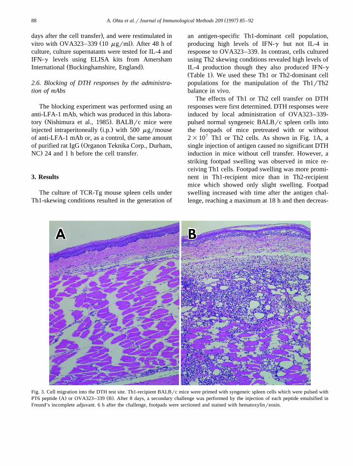
Fig. 3. Cell migration into the DTH test site. Th1-recipient BALBrc mice were primed with syngeneic spleen cells which were pulsed withŽ . Ž .PT6 peptide A or OVA323–339 B . After 8 days, a secondary challenge was performed by the injection of each peptide emulsified in
Freund’s incomplete adjuvant. 6 h after the challenge, footpads were sectioned and stained with hematoxylinreosin.

( )A. Ohta et al.rJournal of Immunological Methods 209 1997 85–92 89
ing. Furthermore, induction of DTH responses wasdependent on the numbers of Th1 cells transferred.As few as 1=107 cellsrmouse was sufficient to
Ž .induce maximum swelling at 18 h Fig. 1B . 8 daysafter the first immunization, when the footpadswelling had completely vanished, the mice weretreated again with the same dose of OVA323–339peptide antigen without cell transfer. This secondarychallenge caused a faster DTH response, which
Ž .reached a peak at 6 h Fig. 1C . It was also demon-strated that this DTH response was totally dependent
Ž .on antigen-specific Th1 cells Fig. 2 . Immunizationwith unrelated PT6 peptide did not cause significantfootpad swelling in mice transferred with TCR-Tgmouse-derived Th1 cells and transfer of syngeneicwhole spleen cells failed to induce footpad swellingin response to OVA323–339. Typical DTH re-sponses were also confirmed by histological exami-
Ž .nation of footpads Fig. 3 . Massive cell migrationwas observed only in OVA323–339-treated mouse
Ž .footpads Fig. 3B , whereas no significant cell mi-gration was observed in mouse footpads challenged
Ž .with PT6 peptide Fig. 3A . This suggested thatstrong antigen-specific DTH responses were inducedby Th1 cell transfer, but not by Th2 cells.
We next investigated cytokine production in micereceiving either Th1 or Th2 cells. Consistent with thelevels of the DTH responses, the serum IFN-g levelsof mice receiving Th1 cells were markedly elevated
Ž .20 h after challenge Fig. 4 . Such significant IFN-g
Fig. 4. Elevation of serum IFN-g levels following antigen chal-lenge in Th1 cell transferred mice. DTH responses were inducedby the same method described in the legend to Fig. 1. 24 h after
Ž .the transfer of Th1 cells, Th2 cells or neither y into the mice,serum IFN-g levels were determined by ELISA. The data repre-sent mean"SE of 5 mice.
Table 2Th1- or Th2-dominant cytokine production from popliteal lymphnode and spleen cells of BALBrc mice following Th1 or Th2 celltransfer
Ž . Ž .Cell transfer IFN-g pgrml IL-4 pgrml
Ž . Ž .— OVA 323–339 — OVA 323–339
Ž .a Popliteal lymph node— 800 800 142 145Th1 1200 170100 25 47Th2 300 23450 22 6499
Ž .b Spleen— 100 150 44 33Th1 500 131600 150 43Th2 100 31100 61 1371
Ž .Following cell transfer or not , mice were injected in the hindfootpad with OVA323–339-pulsed cells at 4 h and 8 days. Afteran additional 2 days, spleen cells and popliteal lymph node cellswere cultured with 10 mgrml OVA323–339. Supernatants werecollected after 48 h of culture for the determination of levels of
Ž . Ž .IFN-g and IL-4. a Popliteal lymph node cells; b spleen cells.
production in vivo was not detected in mice receiv-ing Th2 cells. Cytokine production patterns in vitroof the spleen and lymph nodes obtained from Th1-or Th2-recipient mice were also examined. After thesecondary challenge, spleen cells and popliteal lymph
Žnode cells were restimulated with OVA323–339 10. Žmgrml for 48 h and the cytokine levels IL-4 and.IFN-g in the culture supernatants were determined
using ELISA kits. As shown in Table 2, both spleencells and popliteal lymph node cells from Th1-recipi-ent mice produced a great amount of IFN-g inresponse to OVA323–339 but no IL-4 was detected.Conversely, cells from Th2-recipient mice producedhigh levels of IL-4 with some IFN-g . Both spleencells and popliteal lymph node cells showed similar
Table 3Cytokine production upon restimulation of spleen cells 3 monthsafter Th1 cell transfer
Ž . Ž .Stimulation IFN-g pgrml IL-4 pgrml
— 63"4 20"5Ž .OVA 323–339 12744"2366 37"11
BALBrc mice received 2=107 Th1 cells and were then immu-nized twice with OVA323–339 into the hind footpad as describedin the legend to Fig. 1. After 3 months, spleen cells prepared fromthe mice were restimulated with 10 mgrml OVA323–339 andsupernatants after 48 h of culture were tested for levels of IFN-gand IL-4 using ELISA.
( )A. Ohta et al.rJournal of Immunological Methods 209 1997 85–9290
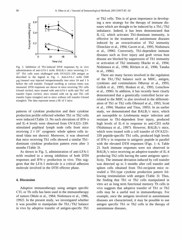
Fig. 5. Inhibition of Th1-induced DTH responses by in vivoadministration of anti-LFA-1 mAb. BALBrc mice receiving 5=
106 Th1 cells were challenged with OVA323–339 antigen asŽdescribed in the legend to Fig. 1. Anti-LFA-1 mAb 500
.mgrmouse was injected intraperitoneally into mice 24 and 1 hbefore the cell transfer. Footpad swelling at various times wasmeasured. DTH responses are shown in mice receiving Th1 cellsŽ .closed circles , mice treated with anti-LFA-1 mAb and Th1 cell
Ž .transfer open circles , mice treated with rat Ig and Th1 cellŽ . Žtransfer open triangles and in mice without cell transfer closed.triangles . The data represent mean"SE of 5 mice.
patterns of cytokine production and their cytokineproduction profile reflected whether Th1 or Th2 cells
Ž .were induced Table 1 . No such elevations of IFN-gand IL-4 levels were observed from OVA323–339-stimulated popliteal lymph node cells from micereceiving 2=107 syngeneic whole spleen cells in-
Ž .stead data not shown . Moreover, it was observedthat mice receiving Th1 cells showed a similar Th1-dominant cytokine production pattern even after 3
Ž .months Table 3 .As shown in Fig. 5, administration of anti-LFA-1
mAb resulted in a strong inhibition of both DTHresponses and IFN-g production in vivo. This sug-gests that the LFA-1 molecule is a critical adhesionmolecule involved in the DTH effector phase.
4. Discussion
Adoptive immunotherapy using antigen specificCTL or Th cells has been used in the immunotherapy
Žof tumors Mule et al., 1984; Nishimura et al., 1986,.1992 . In the present study, we investigated whether
it was possible to manipulate the Th1rTh2 balancein vivo by adoptive transfer of antigen-specific Th1
or Th2 cells. This is of great importance in develop-ing a new strategy for the therapy of immune dis-eases which are thought to be induced by a Th1rTh2imbalance. Indeed, it has been demonstrated thatIL-12, which activates Th1-dominant immunity, iseffective in the treatment of autoimmune diseasesinduced by an overactivation of Th2 immunityŽDonckier et al., 1994; Gavett et al., 1995; Nishimura
.et al., 1996 . Conversely, Th1-dependent immunediseases such as liver injury and graft versus hostdisease are blocked by suppression of Th1 immunity
Žor activation of Th2 immunity Racke et al., 1994;Nishimura et al., 1996; Rocken et al., 1996; Tanaka¨
.et al., 1996 .There are many factors involved in the regulation
of the Th1rTh2 balance such as MHC, antigen,Žcytokines and costimulators Murray et al., 1993;
Gollob et al., 1995; Hosken et al., 1995; Lenschow.et al., 1996 . In addition, it has recently been clearly
demonstrated that a genetically controlled factor un-related to the MHC is also involved in the differenti-
Žation of Th1 or Th2 cells Heinzel et al., 1993; Scott.et al., 1994; Shankar and Titus, 1995 . In an earlier
study, we demonstrated that BALBrc mice, whichare susceptible to Leishmania major infection andresistant to Th1-dependent liver injury, producedhigh levels of IL-4 in response to anti-CD3 mAbŽ .Nishimura et al., 1997 . However, BALBrc mice,which were treated with a cell transfer of OVA323–339 peptide-specific Th1 cells, produced high levelsof IFN-g in response to antigenic peptide in parallel
Žwith the elevated DTH responses Figs. 1–4, Table.2 . Such immune responses were not observed in
BALBrc mice receiving an adoptive transfer of IL-4producing Th2 cells having the same antigenic speci-ficity. The immune deviation induced by cell transferwas detected up to 3 months after cell transfer andspleen cells obtained from Th1-recipient mice re-vealed a Th1-type cytokine production pattern fol-
Ž .lowing restimulation with antigen Table 3 . Thus,the finding that Th1 or Th2 cells manipulated invitro act as long term functional memory Th cells invivo suggests that adoptive transfer of Th1 or Th2cells may be a useful tool in immunotherapy. Forexample, once the antigens involved in autoimmunediseases are characterized, it may be possible to useantigen specific Th1 or Th2 cells in the therapy ofautoimmune diseases.
( )A. Ohta et al.rJournal of Immunological Methods 209 1997 85–92 91
Our established DTH model also makes it possi-ble to investigate the mechanisms of the DTH effec-
Žtor phase. In confirmation of previous results Cher.and Mosmann, 1987 , systemically transferred Th1
cells, but not Th2 cells, were found to be the majoreffector cells inducing DTH responses. Moreover,the LFA-1 molecule was shown to be one of the keyadhesion molecules involved in the DTH effector
Ž .phase Fig. 5 .We are now investigating various models of im-
mune disease and immune therapy using adoptivetransfer of Th1 or Th2 cells.
Acknowledgements
We would like to thank Dr. S.H. Herrmann andŽDr. M. Kobayashi Genetics Institute, Cambridge,
.MA for their kind gift of IL-12 and Dr. G. TrinchieriŽWistar Institute of Anatomy and Biology, Philadel-
.phia, PA for his kind donation of anti-IL-12 mAb.This work was supported in part by a Grant-in-Aidfor Scientific Research on Priority Areas and Scien-
Ž .tific Research C from the Ministry of Education,Science and Culture, Japan, by a Grant-in-Aid forthe IL-12 project of Tokai University School of
ŽMedicine, and by CREST Core Research for Envi-. Žronmental Science and Technology of JST Japan.Science and Technology Corporation .
References
Baron, J.L., Madri, J.A., Ruddle, N.H., Hashim, G., Janeway,C.A. Jr., 1993. Surface expression of a4 integrin by CD4T-cells is required for their entry into brain parenchyma. J.Exp. Med. 177, 57.
Biancone, L., Andres, G., Ahn, H., Lim, A., Dai, C., Noelle, R.,Yagita, H., De Martino, C., Stamenkovic, I., 1996. Distinctregulatory roles of lymphocyte costimulatory pathways onT-helper type 2-mediated autoimmune disease. J. Exp. Med.183, 1473.
Chatelain, R., Varkila, K., Coffman, R.L., 1992. IL-4 induces aTh2 response in Leishmania major-infected mice. J. Immunol.148, 1182.
Cher, D.J., Mosmann, T.R., 1987. Two types of murine helperT-cell clone. II. Delayed-type hypersensitivity is mediated byT 1 clones. J. Immunol. 138, 3688.H
Corry, D.B., Folkesson, H.G., Warnock, M.L., Erle, D.J., Matthay,M.A., Wiener-Kronish, J.P., Locksley, R.M., 1996. Interleukin4, but not interleukin 5 or eosinophils, is required in a murinemodel of acute airway hyperreactivity. J. Exp. Med. 183, 109.
Donckier, V., Abramowicz, D., Bruyns, C., Florquin, S., Vander-haeghen, M.-L., Amraoui, Z., Dubois, C., Vandenabeele, P.,Goldman, M., 1994. IFN-g prevents Th2 cell-mediatedpathology after neonatal injection of semiallogenic spleen cellsin mice. J. Immunol. 153, 2361.
Gautam, S.C., Chikkala, N.F., Hamilton, T.A., 1992. Anti-in-flammatory action of IL-4. Negative regulation of contactsensitivity to trinitrochlorobenzene. J. Immunol. 148, 1411.
Gavett, S.H., O’Hearn, D.J., Li, X., Huang, S.-K., Finkelman,F.D., Wills-Karp, M., 1995. Interleukin 12 inhibits antigen-in-duced airway hyperresponsiveness, inflammation and Th2 cy-tokine expression in mice. J. Exp. Med. 182, 1527.
Gollob, J.A., Li, J., Reinherz, E.L., Ritz, J., 1995. CD2 regulatesresponsiveness of activated T-cells to interleukin 12. J. Exp.Med. 182, 721.
Heinzel, F.P., Sadick, M.D., Holaday, B.J., Coffman, R.L., Lock-sley, R.M., 1989. Reciprocal expression of interferon g orinterleukin 4 during the resolution or progression of murineleishmaniasis. Evidence for expansion of distinct helper T-cellsubsets. J. Exp. Med. 169, 59.
Heinzel, F.P., Schoenhaut, D.S., Rerko, R.M., Rosser, L.E.,Gately, M.K., 1993. Recombinant interleukin 12 cures miceinfected with Leishmania major. J. Exp. Med. 177, 1505.
Hosken, N.A., Shibuya, K., Heath, A.W., Murphy, K.M., O’Garra,A., 1995. The effect of antigen dose on CD4q T-helper cellphenotype development in a T-cell receptor-ab-transgenicmodel. J. Exp. Med. 182, 1579.
Hsieh, C.-S., Heimberger, A.B., Gold, J.S., O’Garra, A., Murphy,K.M., 1992. Differential regulation of T-helper phenotypedevelopment by interleukins 4 and 10 in an ab T-cell-recep-tor transgenic system. Proc. Natl. Acad. Sci. USA 89, 6065.
Jardim, A., Alexander, J., Teh, H.S., Ou, D., Olafson, R.W., 1990.Immunoprotective Leishmania major synthetic T-cell epi-topes. J. Exp. Med. 172, 645.
Kennedy, M.K., Torrance, D.S., Picha, K.S., Hohler, K.M., 1992.Analysis of cytokine mRNA expression in the central nervoussystem of mice with experimental autoimmune en-cephalomyelitis reveals that IL-10 mRNA expression corre-lates with recovery. J. Immunol. 149, 2496.
Lenschow, D.J., Herold, K.C., Rhee, L., Patel, B., Koons, A., Qin,H.-Y., Fuchs, E., Singh, B., Thompson, C.B., Bluestone, J.A.,1996. CD28rB7 regulation of Th1 and Th2 subsets in thedevelopment of autoimmune diabetes. Immunity 5, 285.
Liblau, R.S., Singer, S.M., McDevitt, H.O., 1995. Th1 and Th2CD4q T-cells in the pathogenesis of organ-specific autoim-mune diseases. Immunol. Today 16, 34.
Magram, J., Connaughton, S.E., Warrier, R.R., Daisy, M.C., Wu,C.-Y., Ferrante, J., Stewart, C., Sarmiento, U., Faherty, D.A.,Gately, M.K., 1996. IL-12-deficient mice are defective inIFNg production and type 1 cytokine responses. Immunity 4,471.
Martin, R., McFarland, H.F., McFarlin, D.E., 1992. Immunologi-cal aspects of demyelinating diseases. Annu. Rev. Immunol.10, 153.
Mosmann, T.R., Coffman, R.L., 1989. Th1 and Th2 cells: Differ-ent patterns of lymphokine secretion lead to different func-tional properties. Annu. Rev. Immunol. 7, 145.
Mule, J.J., Shu, S., Schwarz, S.L., Rosenberg, S.A., 1984. Adop-
( )A. Ohta et al.rJournal of Immunological Methods 209 1997 85–9292
tive immunotherapy of established pulmonary metastases withLAK cells and recombinant interleukin-2. Science 225, 1487.
Murray, J.S., Madri, J., Pasqualini, T., Bottomly, K., 1993. Func-tional CD4 T-cell subset interplay in an intact immune system.J. Immunol. 150, 4270.
Nishimura, T., Yagi, H., Yagita, H., Uchiyama, Y., Hashimoto,Y., 1985. Lymphokine-activated killer cell-mediated cytotoxic-ity. Cell. Immunol. 94, 122.
Nishimura, T., Togashi, Y., Goto, M., Yagi, H., Uchiyama, Y.,Hashimoto, Y., 1986. Augmentation of the therapeutic effi-cacy of adoptive tumor immunotherapy by in vivo administra-tion of slowly released recombinant interleukin 2. CancerImmunol. Immunother. 21, 12.
Nishimura, T., Nakamura, Y., Takeuchi, Y., Tokuda, Y., Iwa-sawa, M., Kawasaki, A., Okumura, K., Habu, S., 1992. Gener-ation, propagation, and targeting of human CD4q helperrkillerT-cells induced by anti-CD3 monoclonal antibody plus recom-binant IL-2. An efficient strategy for adoptive tumor im-munotherapy. J. Immunol. 148, 285.
Nishimura, T., Sadata, A., Yahagi, C., Santa, K., Otsuki, K.,Watanabe, K., Yahata, T., Habu, S., 1996. The therapeuticeffect of interleukin-12 or its antagonist in transplantationimmunity. Ann. N. Y. Acad. Sci. 795, 371.
Nishimura, T., Santa, K., Yahata, T., Sato, N., Ohta, A., Ohmi,Y., Sato, T., Hozumi, K., Habu, S., 1997. Involvement of IL-4producing Vb 8.2q CD4q CD62Ly CD45RBy T-cells in non-MHC gene controlled predisposition toward skewing into T-helper type-2 immunity in BALBrc mice. J. Immunol. 158,5698.
O’Garra, A., Murphy, K.M., 1995. T-cell subsets in autoimmu-nity. Curr. Opin. Immunol. 5, 880.
Racke, M.K., Bonomo, A., Scott, D.E., Cannella, B., Levine, A.,Raine, C.S., Shevach, E.M., Rocken, M., 1994. Cytokine-in-¨duced immune deviation as a therapy for inflammatory dis-ease. J. Exp. Med. 180, 1961.
Rocken, M., Racke, M., Shevach, E.M., 1996. IL-4-induced im-¨mune deviation as antigen-specific therapy for inflammatoryautoimmune disease. Immunol. Today 17, 225.
Sato, T., Sasahara, T., Nakamura, Y., Osaki, T., Hasegawa, T.,Tadakuma, T., Arata, Y., Kumagai, Y., Katsuki, M., Habu, S.,1994. Naive T-cells can mediate delayed-type hypersensitivityresponse in T-cell receptor transgenic mice. Eur. J. Immunol.24, 1512.
Scott, B., Liblau, R., Degermann, S., Marconi, L.A., Ogata, L.,Caton, A.J., McDevitt, H.O., Lo, D., 1994. A role for non-MHC genetic polymorphism in susceptibility to spontaneousautoimmunity. Immunity 1, 73.
Scott, P., Kaufmann, S.H.E., 1991. The role of T-cell subsets andcytokines in the regulation of infection. Immunol. Today 12,346.
Shankar, A.H., Titus, R.G., 1995. T-cell and non-T-cell compart-ments can independently determine resistance to Leishmaniamajor. J. Exp. Med. 181, 845.
Simon, A.K., Seipelt, E., Sieper, J., 1994. Divergent T-cell cy-tokine patterns in inflammatory arthritis. Proc. Natl. Acad. Sci.USA 91, 8562.
Sypek, J.P., Chung, C.L., Mayor, S.E.H., Subramanyam, J.M.,Goldman, S.J., Sieburth, D.S., Wolf, S.F., Schaub, R.G., 1993.Resolution of cutaneous Leishmaniasis: Interleukin 12 initiatesa protective T-helper type 1 immune response. J. Exp. Med.177, 1797.
Tanaka, Y., Takahashi, A., Watanabe, K., Takayama, K., Yahata,T., Habu, S., Nishimura, T., 1996. A pivotal role of IL-12 inT 1-dependent mouse liver injury. Int. Immunol. 8, 569.h
Vercelli, D., Jabara, H.H., Lauener, R.P., Geha, R.S., 1990. IL-4inhibits the synthesis of IFN-g and induces the synthesis ofIgE in human mixed lymphocyte cultures. J. Immunol. 144,570.