malignancies - University of Queensland301778/UQ301778_OA.pdf · Mt SinaiJMed. 1981;48:61 ......
2
out that invasion and metastasis in typical Bowen's disease is not common, and the absence of a fully documented case with metastatic disease cannot be used to eliminate the possibility that malignant transformation may rarely occur. Another possible factor in the development of these lesions could be human papilloma virus. Recently, human papilloma virus has been found in some seborrheic kératoses with histo¬ logie changes consistent with a viral effect.8 Human papilloma virus has been associated with various tumors and is felt to be a cofactor in the development of at least some malignancies.9 The most striking feature of the lesion we report is that it is a mucin-positive adenocarcinoma arising in the center of a lesion clinically and histologically seborrheic keratosis. In this case, there are three possible explanations for the occurrence of adenocarcinoma in seborrheic keratosis. These include me¬ tastasis from a distant primary tumor to the seborrheic kera¬ tosis, incidental origin of an adnexal carcinoma at the site of the seborrheic keratosis, and origin of an adenocarcinoma from the seborrheic keratosis. Since no other primary tumor was found, and since there was no evidence of any other cutaneous or malignant tumor after 10 years, the adenocarci¬ noma was probably not a metastasis. While incidental origin at the site of a seborrheic keratosis cannot be absolutely disproven, an epidermal origin of seborrheic keratosis is sup¬ ported by direct dermal invasion of the tumor from the epider¬ mis rather than from an adnexal structure, the central loca¬ tion of the adenocarcinoma, and the clinical history. As with some cases of extramammary Paget's disease, in which rau- cin-producing epithelial cells are present within the epidermis with no primary tumor and with no evidence of adnexal involvement, only theories exist to explain this phenomenon. These theories could include malignant transformation of a pluripotential germinative cell within the epidermis or trans¬ formation of a cell from the distal portion of an adnexal structure located within the epidermis. Cells from these structures would be at least partially committed toward ad¬ nexal differentiation. LTC Kathleen J. Smith, MC, USA Department of Dermatology Walter Reed Army Medical Center Washington, DC 20307 CDR Henry G. Skelton III, MC, USA COL George P. Lupton, MC, USA Department of Dermatopathology Armed Forces Institute of Pathology Washington, DC 20306 NolaJ. Palomino, MD Department of Pathology Miami Valley Hospital Dayton, OH 45409 1. Baer RL, Garcia RL, Partsalidou V, Ackermann AB. Papillated squa- mous cell carcinoma in situ arising in a seborrheic keratosis. J Am Acad Dermatol. 1981;5:561-565. 2. Goette DK. Basal cell carcinoma arising in seborrheic keratosis. J Derma- tol Surg Oncol. 1985;1:1014-1016. 3. Christeler A, Delacretaz J. Verrues s\l=e'\borrh\l=e'\iques et transformation maligne. Dermtologica. 1966;133:33-39. 4. Kwittken J. Squamous cell carcinoma arising in seborrheic keratosis. Mt Sinai J Med. 1981;48:61-62. 5. Bloch PH. Transformation of seborrheic keratosis in Bowen's disease. J Cutan Pathol. 1978;5:361-367. 6. Monteagudo JC, Jorda E, Terencio C, Llombort-Bosch A. Squamous cell carcinoma in situ (Bowen's disease) arising in seborrheic keratosis: three lesions in two patients. J Cutan Pathol. 1989;16:348-352. 7. Clemmensen OJ, Sjolin KE. Malignancy in seborrheic keratoses. Acta Derm Venereol (Stockh). 1986;66:158-161. 8. Zhao Y, Lin Y, Luo R, et al. Human papillomavirus (HPV) infection in seborrheic keratosis. Am J Dermatopathol.1989;11:209-212. 9. Arends MJ, Wyllie AH, Bird CC. Papillomaviruses and human cancer. Hum Pathol. 1990;21:686-698. Widespread Cutaneous Necrosis in a Patient With Rheumatoid Arthritis Associated With Anticardiolipin Antibodies To the Editor. \p=m-\ Antiphospholipid antibodies, detected by the presence of lupus anticoagulant and/or abnormally high levels of anticardiolipin antibodies, have been shown to be associ- ated with an increased risk of arterial or venous thrombosis. The association between antiphospholipid antibodies and thrombotic events was first noted in patients with systemic lupus erythematosus but were later also observed in a variety of other disorders and as an isolated finding. The existence of a separate entity, the anticardiolipin or antiphospholipid syn- drome, was suggested in several studies.1 Cutaneous symp- toms linked to the antiphospholipid syndrome include throm- bophlebitis, leg ulcers, livedo reticularis, livedo vasculitis, unfading acral microlivedo, peripheral gangrene, hemor- rhage (ecchymosis and hematoma), and necrotizing purpu- ra. 1,2 We observed widespread cutaneous necrosis3 as a rare manifestation of the antiphospholipid syndrome in a patient with rheumatoid arthritis. Report of a Case.\p=m-\A 73-year-old woman with long-standing de- forming arthritis, fulfilling the criteria of the American College of Rheumatism, Atlanta, Ga (formerly the Arthritis and Rheumatism Association), for "classic" rheumatoid arthritis, was admitted to our department in July 1988 because of painful widespread skin lesions of 4 days' duration. On clinical examination, several sharply demar¬ cated, hemorrhagic patches with a bizarre configuration were ob¬ served on her arms (Fig 1), breasts, and legs. A skin biopsy specimen of a hemorrhagic area on the left thigh (Fig 2) revealed thrombi within capillaries, venules, and small- and medi¬ um-sized vessels throughout the dermis and subcutaneous fat with¬ out any evidence of fibrin in the vessel walls. No nuclear "dust" was present. In addition, a sparse, perivascular mixed-cell infiltrate and extravasation of erythrocytes was observed. Results or findings from laboratory studies were as follows: eryth- rocyte sedimentation rate, 108 mm/h; erythrocytes, 3.97x10 L; hemoglobin, 87 g/L; leukocytes, 6.9 x 10⁹/L, with 0.69 neutrophils, 0.1 band forms, 0.1 eosinophils, and 0.29 lymphocytes; platelets, 336 x 109/L; serum electrolyte, serum urea nitrogen level, serum glucose level, and urinalysis, normal; liver function test results, Fig 1.—Widespread cutaneous necrosis. Sharply demarcated, hem¬ orrhagic patches with bizarre configuration on the left arm. DownloadedFrom:http://archderm.jamanetwork.com/byaUQLibraryUseron11/29/2015
-
Upload
truongkhanh -
Category
Documents
-
view
215 -
download
3
Transcript of malignancies - University of Queensland301778/UQ301778_OA.pdf · Mt SinaiJMed. 1981;48:61 ......
out that invasion and metastasis in typical Bowen's disease isnot common, and the absence of a fully documented case withmetastatic disease cannot be used to eliminate the possibilitythatmalignant transformation may rarely occur.Another possible factor in the development of these lesions
could be human papilloma virus. Recently, human papillomavirus has been found in some seborrheic kératoses with histo¬logie changes consistent with a viral effect.8 Human papillomavirus has been associated with various tumors and is felt to bea cofactor in the development of at least some malignancies.9The most striking feature of the lesion we report is that it is
a mucin-positive adenocarcinoma arising in the center of alesion clinically and histologically seborrheic keratosis. In thiscase, there are three possible explanations for the occurrenceof adenocarcinoma in seborrheic keratosis. These include me¬tastasis from a distant primary tumor to the seborrheic kera¬tosis, incidental origin of an adnexal carcinoma at the site ofthe seborrheic keratosis, and origin of an adenocarcinomafrom the seborrheic keratosis. Since no other primary tumorwas found, and since there was no evidence of any othercutaneous ormalignant tumor after 10 years, the adenocarci¬noma was probably not a metastasis. While incidental originat the site of a seborrheic keratosis cannot be absolutelydisproven, an epidermal origin of seborrheic keratosis is sup¬ported by direct dermal invasion of the tumor from the epider¬mis rather than from an adnexal structure, the central loca¬tion of the adenocarcinoma, and the clinical history. As withsome cases of extramammary Paget's disease, in which rau-cin-producing epithelial cells are presentwithin the epidermiswith no primary tumor and with no evidence of adnexalinvolvement, only theories exist to explain this phenomenon.These theories could include malignant transformation of apluripotential germinative cell within the epidermis or trans¬formation of a cell from the distal portion of an adnexalstructure located within the epidermis. Cells from thesestructures would be at least partially committed toward ad¬nexal differentiation.
LTC Kathleen J. Smith, MC, USADepartment ofDermatologyWalter Reed Army Medical CenterWashington, DC 20307CDR Henry G. Skelton III, MC, USACOL George P. Lupton, MC, USADepartment ofDermatopathologyArmed Forces Institute ofPathologyWashington, DC 20306NolaJ. Palomino, MDDepartment ofPathologyMiami Valley HospitalDayton, OH 45409
1. Baer RL, Garcia RL, Partsalidou V, Ackermann AB. Papillated squa-mous cell carcinoma in situ arising in a seborrheic keratosis. J Am AcadDermatol. 1981;5:561-565.2. Goette DK. Basal cell carcinoma arising in seborrheic keratosis. JDerma-
tol Surg Oncol. 1985;1:1014-1016.3. Christeler A, Delacretaz J. Verrues s\l=e'\borrh\l=e'\iqueset transformation
maligne. Dermtologica. 1966;133:33-39.4. Kwittken J. Squamous cell carcinoma arising in seborrheic keratosis. Mt
Sinai JMed. 1981;48:61-62.5. Bloch PH. Transformation of seborrheic keratosis in Bowen's disease. J
Cutan Pathol. 1978;5:361-367.6. Monteagudo JC, Jorda E, Terencio C, Llombort-Bosch A. Squamous cell
carcinoma in situ (Bowen's disease) arising in seborrheic keratosis: threelesions in two patients. J Cutan Pathol. 1989;16:348-352.7. Clemmensen OJ, Sjolin KE. Malignancy in seborrheic keratoses. Acta
Derm Venereol (Stockh). 1986;66:158-161.8. Zhao Y, Lin Y, Luo R, et al. Human papillomavirus (HPV) infection in
seborrheic keratosis. Am J Dermatopathol.1989;11:209-212.9. Arends MJ, Wyllie AH, Bird CC. Papillomaviruses and human cancer.
Hum Pathol. 1990;21:686-698.
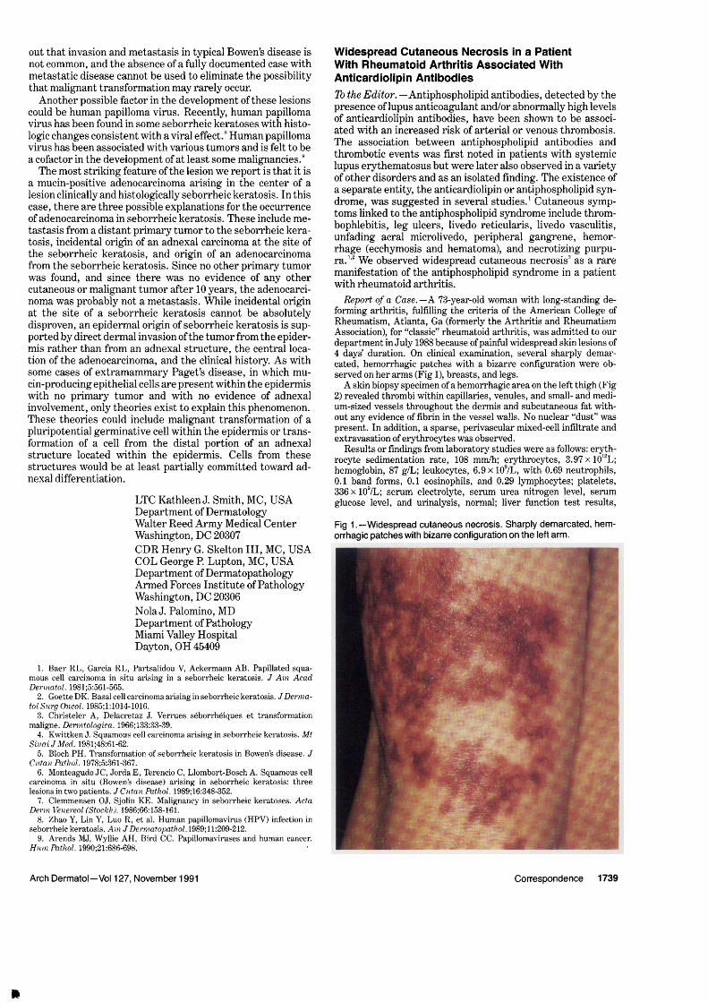
Widespread Cutaneous Necrosis in a PatientWith Rheumatoid Arthritis Associated WithAnticardiolipin AntibodiesTo the Editor. \p=m-\Antiphospholipid antibodies, detected by thepresence of lupus anticoagulant and/or abnormally high levelsof anticardiolipin antibodies, have been shown to be associ-ated with an increased risk of arterial or venous thrombosis.The association between antiphospholipid antibodies andthrombotic events was first noted in patients with systemiclupus erythematosus but were later also observed in a varietyof other disorders and as an isolated finding. The existence ofa separate entity, the anticardiolipin or antiphospholipid syn-drome, was suggested in several studies.1 Cutaneous symp-toms linked to the antiphospholipid syndrome include throm-bophlebitis, leg ulcers, livedo reticularis, livedo vasculitis,unfading acral microlivedo, peripheral gangrene, hemor-rhage (ecchymosis and hematoma), and necrotizing purpu-ra. 1,2We observed widespread cutaneous necrosis3 as a raremanifestation of the antiphospholipid syndrome in a patientwith rheumatoid arthritis.Report of a Case.\p=m-\A 73-year-old woman with long-standing de-
forming arthritis, fulfilling the criteria of the American College ofRheumatism, Atlanta, Ga (formerly the Arthritis and RheumatismAssociation), for "classic" rheumatoid arthritis, was admitted to ourdepartment in July 1988 because ofpainful widespread skin lesions of4 days' duration. On clinical examination, several sharply demar¬cated, hemorrhagic patches with a bizarre configuration were ob¬served on her arms (Fig 1), breasts, and legs.A skin biopsy specimen of a hemorrhagic area on the left thigh (Fig
2) revealed thrombi within capillaries, venules, and small- and medi¬um-sized vessels throughout the dermis and subcutaneous fat with¬out any evidence of fibrin in the vessel walls. No nuclear "dust" waspresent. In addition, a sparse, perivascular mixed-cell infiltrate andextravasation oferythrocytes was observed.Results or findings from laboratory studies were as follows: eryth-
rocyte sedimentation rate, 108 mm/h; erythrocytes, 3.97x10 L;hemoglobin, 87 g/L; leukocytes, 6.9 x 10⁹/L, with 0.69 neutrophils,0.1 band forms, 0.1 eosinophils, and 0.29 lymphocytes; platelets,336 x 109/L; serum electrolyte, serum urea nitrogen level, serumglucose level, and urinalysis, normal; liver function test results,
Fig 1.—Widespread cutaneous necrosis. Sharply demarcated, hem¬orrhagic patches with bizarre configuration on the left arm.
Downloaded From: http://archderm.jamanetwork.com/ by a UQ Library User on 11/29/2015

normal, except for the alkaline phosphatase level (216 U/L) and thecholinesterase level (3395 U/L); direct Coomb's test, VDRL, and thetest for cryoglobulins, negative; immunoelectrophoresis, increase ofpolyclonal immunoglobulins; latex rheumatoid factor, 3390 U/mL;and latex C-reactive protein, 32 mg/L (both positive); antinuclearantibodies, 1:1280 (speckled type, positive titer); antibodies againstdouble-stranded DNA, Sjögren syndrome associated antigens A andB, SM-SCL-70, and nuclear ribonucleoprotein, not detectable; par¬tial thromboplastin time, Russell's viper venom test, and prothrom-bin time, normal; lupus anticoagulant, none detected; tests for clot¬ting factors, fibrinogen, 4.8 g/L; factor II, 0.82; factor V, 0.74; factorX, 0.74; factor III, 0.66; protein C, 0.75; enzyme-linked immunosor-bent assay for anticardiolipin antibodies, IgG, 27.7 U/mL (normal,<12.0 U/mL); and IgM, 3.5 U/mL (normal, <6 U/mL) (Elias, Frei¬burg, Germany).Initial therapy was prednisolone (50 mg/d). Within 7 weeks, skin
lesions healed with moderate scarring. Six months later, anticardioli¬pin antibodies were still present but the level was clearly reduced(IgG, 15.7 U/mL).Comment.—Widespread cutaneous necrosis, which is
characterized by painful purpuric and necrotic areas withunderlying dermal thrombosis, was first reported by Dodd etal3 in 1985 in a patient with lupus anticoagulant. Subsequent¬ly, further cases with identical clinical and histologie features,but different serological profile of antiphospholipid antibod¬ies, were described in systemic lupus erythematosus and inpatients with no demonstrable underlying disease (Table).uCutaneous phenomena, including purpura resulting from
disseminated intravascular coagulation, coumarin necrosis,coumarin-induced skin lesions in protein C deficiency, hepa-rin necrosis, and purpura cryoglobulinemia, may present aswidespread cutaneous necrosis, and all of these conditionsshould be considered in the clinical differential diagnosis. Thesignificance of anticardiolipin antibodies in the development
of skin lesions in our patient is suspected because of the lack ofother possible causes and because the titer of these antibodiesdecreased after therapy. The histologie findings of thrombo¬sis in dermal and subcutaneous vessels without any evidenceof leukocytoclastic vasculitis is also characteristic for skinlesions of the antiphospholipid syndrome.1,2Antiphospholipid antibodies in rheumatoid arthritis have
been investigated in a number of studies, but estimates of itsprevalence are conflicting and the clinical implications ofthese antibodies in rheumatoid arthritis remain controversialat present.M Our case, however, shows that anticardiolipinantibodies may be involved at least occasionally in the patho¬genesis of skin lesions in rheumatoid arthritis.
Fig 2.—Widespread cutaneous necrosis. Thrombi within dermal ves¬sels (arrows). Extravasated erythrocytes in the papillary dermis. Nosigns of vasculitis.
Reports of Widespread Cutaneous Necrosis AssociatedWith Antiphospholipid Antibodies *
No. of UnderlyingSource, y Cases Disease ACA LAC VDRL ds-DNA
Dodd et al,31985 1 No ND + ND
-
Frances et al,"1989 3f SLE - +
O'Neill et al,51990 1 No
Present case 1 RA
*ACA indicates anticardiolipin antibodies; LAC, lupus anticoagulant;ds-DNA double-stranded DNA antibodies; RA, rheumatoid arthritis; ND,not done; plus sign, condition present; and minus sign, condition absent.fAII three reported cases revealed the same sérologie profile.
PeterWolf, MDH. Peter Soyer, MDPiet Auer-Grumbach, MDHelmut Kerl, MDDepartment ofDermatologyUniversity ofGrazAuenbruggerplatz 88036 Graz, Austria
1. Sontheimer RD. The anticardiolipin syndrome: a new way to slice an oldpie, or a new pie to slice? Arch Dermatol. 1987;123:590-595.2. Alegre VA, Gastineau DA, Winkelmann RK. Skin lesions associated with
circulating lupus anticoagulant. Br J Dermatol. 1989;120:419-429.3. Dodd HJ, Sarkany I, O'Shaughnessy D. Widespread cutaneous necrosis
associated with the lupus anticoagulant. Clin Exp Dermatol. 1985;10:581-586.4. Frances C, Tribout B, Boisnic S, et al. Cutaneous necrosis associated with
the lupus anticoagulant. Dermatologica. 1989;178:194-201.5. O'Neill A, Gatenby PA, McGaw B, Painter DM, McKenzie PR. Wide-
spread cutaneous necrosis associated with cardiolipin antibodies. J Am AcadDermatol. 1990;22:356-359.6. Keane A, Woods R, Dowding V, Roden D, Barry C. Anticardiolipin
antibodies in rheumatoid arthritis. Br J Rheumatol. 1987;26:346-350.7. Love PE, Santoro SA. Antiphospholipid antibodies: anticardiolipin and
the lupus anticoagulant in systemic lupus erythematosus (SLE) and in non-SLE disorders: prevalence and clinical significance (review). Ann Intern Med.1990;112:682-698.
Friction Dermatitis of the ThumbsCaused by PantyhoseTo the Editor. \p=m-\Dermatitisof the volar aspect of the thumbpads can be a difficult problem in clinical diagnosis. The thickskin here is rather resistant to many contactants, but frictionmay produce irritation.Report of a Case.\p=m-\A 57-year-old woman was seen because of
persistent fissured dermatitis limited to the volar aspect of the thumbpads. When asked if some activities had become awkward or difficultbecause of the dermatitis, she answered that none had, but thenadded, "Except for putting on my pantyhose."Millions of women carry out this daily maneuver that puts quite a
lot of friction on the volar aspect of the thumb pads. Consider thefollowing: the garment is gathered up, one leg at a time, thumbs onthe inside. The toes are put in and the user, maintaining lateralpressure with the thumbs, pulls the garment up into place. It isduring this latter action that the thumb pads run quickly up a longlength of nylon. Repeated daily, this may produce enough of a fric-tional stimulus to cause skin irritation."It was the pantyhose that caused the problem," the patient wrote
in a follow-up letter. "It has only come back (the dermatitis) when Iuse a certain type hose and/or don't protect the area from the abrasivepressure caused by pulling on the hose."Comment.—Here is yet another example of the value of
careful history taking when faced with a puzzling clinicalproblem.
William M. Gould, MDDepartment ofDermatologyStanford UniversityStanford, CA 94305
Downloaded From: http://archderm.jamanetwork.com/ by a UQ Library User on 11/29/2015