
Machined and sandblasted human dental implants retrieved ... · rylate resin (Technovit 7200 VLC,...
6
VOLUME 43 • NUMBER 4 • APRIL 2012 287 QUINTESSENCE INTERNATIONAL bone-implant interface. 1 Rarely is it possible to obtain implants, retrieved for various rea- sons, in which the interface with the mineral- ized bone is maintained. Implants retrieved from humans with intact bone-implant inter- faces play a pivotal role in validating data obtained from in vitro studies and animal experiments. 8 Moreover, the bone response to different implant surfaces may be evalu- ated over a longer period in vivo. 8,9 It is also important to analyze whether modifica- tions of the implant surface characteristics improve the phenomena at the interface, 10 with an increase in the rate and extent of mineralized bone formation. 10 Sandblasted surfaces have been produced by blasting the metal with different types of blasting or gritting agents. 11–13 This process was Dental implants fail for many reasons. 1–7 In some of these cases, it is not easy to obtain meaningful information about the 1 Researcher, Dental School, University of Chieti-Pescara, Chieti, Italy. 2 Private Practice, Lecce, Italy. 3 Head, Oral Implantology Clinic, and Associate Professor, Department of Periodontology, Dental Research Division, Guarulhos University (UnG), Guarulhos, SP, Brazil. 4 Research Fellow, Dental School, University of Chieti-Pescara, Chieti, Italy. 5 Professor, Oral Pathology and Medicine, Dental School, University of Chieti-Pescara, Chieti, Italy. 6 Research Fellow, Dental School, University of Chieti-Pescara, Chieti, Italy. Correspondence: Dr Vittoria Perrotti, Via Dei Vestini 31, 66100 Chieti, Italy. Email: [email protected] Machined and sandblasted human dental implants retrieved after 5 years: A histologic and histomorphometric analysis of three cases Giovanna Iezzi, DDS, PhD 1 /Giovanni Vantaggiato, DDS 2 / Jamil A. Shibli, DDS, MS, PhD 3 / Elisabetta Fiera, DDS 2 / Antonello Falco, DDS 4 /Adriano Piattelli, MD, DDS 5 / Vittoria Perrotti, DDS, PhD 6 Objective: Human retrieved implants with an intact bone-implant interface play a pivotal role in validating data obtained from in vitro studies and animal experiments. This study presents a histologic and histomorphometric analysis of peri-implant tissue reactions and of the bone-titanium interface in three machined and sandblasted dental implants retrieved after a 5-year loading period. Method and Materials: Three implants, with an intact bone-implant interface, were found in the Archives of the Implant Retrieval Center of the Dental School of the University of Chieti-Pescara, Chieti, Italy. The three implants had been used in a two-stage submerged procedure and loaded as part of a small prosthetic restoration. One implant had been retrieved because of an abutment fracture, while there was a fracture of the connecting screw in the other two. One implant was in the maxilla (sandblasted surface), and two were in the mandible (one with a machined surface and the other with a sandblasted surface). All implants had been processed for histology. Results: All three implants presented mature, compact, lamellar bone at the interface. Many remodeling areas were present in the peri-implant bone, especially inside the implant threads. The bone was always in close contact with the implant surface. The bone-implant contact percentage of the machined implant was 92.7%, while the two sandblasted implants showed bone-implant contact percentages of 85.9% and 76.6%. Conclusion: The present histologic results confirmed that these implants with different surfaces maintained a good level of osseointegration over a 5-year loading period, with continuous remodeling at the interface, and showed high bone-implant contact percentages. (Quintessence Int 2012;43:287–292) Key words: bone remodeling, human histology, implant surfaces, retrieved dental implants 5
Transcript of Machined and sandblasted human dental implants retrieved ... · rylate resin (Technovit 7200 VLC,...
VOLUME 43 • NUMBER 4 • APRIL 2012 287
QUINTESSENCE INTERNATIONAL
bone-implant interface.1 Rarely is it possible
to obtain implants, retrieved for various rea-
sons, in which the interface with the mineral-
ized bone is maintained. Implants retrieved
from humans with intact bone-implant inter-
faces play a pivotal role in validating data
obtained from in vitro studies and animal
experiments.8 Moreover, the bone response
to different implant surfaces may be evalu-
ated over a longer period in vivo.8,9 It is
also important to analyze whether modifica-
tions of the implant surface characteristics
improve the phenomena at the interface,10
with an increase in the rate and extent of
mineralized bone formation.10 Sandblasted
surfaces have been produced by blasting
the metal with different types of blasting
or gritting agents.11–13 This process was
Dental implants fail for many reasons.1–7
In some of these cases, it is not easy to
obtain meaningful information about the
1 Researcher, Dental School, University of Chieti-Pescara, Chieti,
Italy.
2 Private Practice, Lecce, Italy.
3 Head, Oral Implantology Clinic, and Associate Professor,
Department of Periodontology, Dental Research Division,
Guarulhos University (UnG), Guarulhos, SP, Brazil.
4 Research Fellow, Dental School, University of Chieti-Pescara,
Chieti, Italy.
5 Professor, Oral Pathology and Medicine, Dental School,
University of Chieti-Pescara, Chieti, Italy.
6 Research Fellow, Dental School, University of Chieti-Pescara,
Chieti, Italy.
Correspondence: Dr Vittoria Perrotti, Via Dei Vestini 31, 66100
Chieti, Italy. Email: [email protected]
Machined and sandblasted human dental implants retrieved after 5 years: A histologic and histomorphometric analysis of three casesGiovanna Iezzi, DDS, PhD1/Giovanni Vantaggiato, DDS2/
Jamil A. Shibli, DDS, MS, PhD3/ Elisabetta Fiera, DDS2/
Antonello Falco, DDS4/Adriano Piattelli, MD, DDS5/
Vittoria Perrotti, DDS, PhD6
Objective: Human retrieved implants with an intact bone-implant interface play a pivotal role in validating data obtained from in vitro studies and animal experiments. This study presents a histologic and histomorphometric analysis of peri-implant tissue reactions and of the bone-titanium interface in three machined and sandblasted dental implants retrieved after a 5-year loading period. Method and Materials: Three implants, with an intact bone-implant interface, were found in the Archives of the Implant Retrieval Center of the Dental School of the University of Chieti-Pescara, Chieti, Italy. The three implants had been used in a two-stage submerged procedure and loaded as part of a small prosthetic restoration. One implant had been retrieved because of an abutment fracture, while there was a fracture of the connecting screw in the other two. One implant was in the maxilla (sandblasted surface), and two were in the mandible (one with a machined surface and the other with a sandblasted surface). All implants had been processed for histology. Results: All three implants presented mature, compact, lamellar bone at the interface. Many remodeling areas were present in the peri-implant bone, especially inside the implant threads. The bone was always in close contact with the implant surface. The bone-implant contact percentage of the machined implant was 92.7%, while the two sandblasted implants showed bone-implant contact percentages of 85.9% and 76.6%. Conclusion: The present histologic results confirmed that these implants with different surfaces maintained a good level of osseointegration over a 5-year loading period, with continuous remodeling at the interface, and showed high bone-implant contact percentages. (Quintessence Int 2012;43:287–292)
Key words: bone remodeling, human histology, implant surfaces, retrieved dental implants
5
VOLUME 43 • NUMBER 4 • APRIL 2012 289
QUINTESSENCE INTERNATIONAL
288 VOLUME 43 • NUMBER 4 • APRIL 2012
QUINTESSENCE INTERNATIONAL
Iezzi et alIezzi et al
influenced by the number and size of par-
ticles used. The blasting procedure served
to increase the irregularities of the implant
surface by using agents such as alumi-
num oxide (Al2O3) or titanium oxide (TiO2).
The large variability in surface appearance
under scanning electron microscopy (SEM)
of different implant surfaces is due to the dif-
ferent techniques employed in the blasting
procedure. In in vitro studies, the sandblast-
ed surfaces have shown higher adhesion,
proliferation, and differentiation of cells.14
In histologic studies that compared blasted
and turned surfaces, higher bone-implant
values were found in blasted surfaces.11,13
Blasting procedures leave residual particles
on the surface of the implant, however,
and this could alter the healing process
of the bone. Some researchers think that
aluminum ions could impair bone forma-
tion by a possible competitive action to
calcium, while others suggested that histo-
logic data did not provide evidence to sup-
port the hypothesis that residual aluminum
oxide particles on the implant surface could
affect the osseointegration of titanium dental
implants.15 The bone growth pattern around
blasted, rough surfaces has been said to be
characterized by contact osteogenesis—
ie, the osteoblasts start depositing osteoid
matrix directly on the implant surface.11,13
Around machined surfaces, the bone
growth pattern has been termed distance
osteogenesis, with bone growing from the
host bone bed toward the metal surface.11,13
The type of bone growth around blasted
surfaces could produce an earlier and
a higher quantity of bone at the implant
interface.11,13 Other types of surfaces have
been reported in the literature. Sandblasted
and acid-etched surfaces were obtained
with a combined procedure of blasting (to
produce a macrotexture) followed by acid-
etching (to produce a final microtexture).
The blasting is used to achieve a rough-
ness optimal for mechanical fixation, while
the etching serves to smooth some sharp
peaks.16 Sandblasted and acid-etched
implants promoted higher bone-implant
values at earlier time points compared with
plasma-sprayed implants.16 Sandblasted
and acid-etched surfaces showed high
osteoconductive properties and capabili-
ties to induce cell proliferation.16
The aim of this study was to analyze,
histologically and histomorphometrically
the peri-implant tissue reactions and the
bone-titanium interface in machined and
sandblasted titanium dental implants with
an intact bone-implant interface retrieved
after a 5-year loading period.
METHOD AND MATERIALS
In the Archives of the Implant Retrieval
Center of the Dental School of the University
of Chieti-Pescara, Chieti, Italy, three
retrieved implants (Implacil, De Bortoli) with
an intact bone-implant interface and a load-
ing history of 5 years were found. The proto-
col of the study was approved by the ethics
committee of the University of Guarulhos
(UnG), São Paulo, Brazil. The three implants
had been retrieved from three different
patients (two women and one man 53, 54,
and 59 years of age, respectively). The
medical history of all the patients was non-
contributory. All implants had been used
in a two-stage submerged procedure, and
all implants had been loaded as part of a
small prosthetic restoration. One implant
had been retrieved because of a fracture of
the abutment, while there was a fracture of
the connecting screw in the other two. One
implant was in the maxilla (sandblasted sur-
face), and the other two were in the mandi-
ble (one with a machined and the other with
a sandblasted surface). One implant had a
machined surface, while the other two had
a sandblasted surface. All these implants
were stable before retrieval, and each was
retrieved with a 5-mm trephine bur.
Specimen processingAll the specimens were washed in saline
solution and immediately fixed in 4% para-
formaldehyde and 0.1% glutaraldehyde in
0.15 M cacodylate buffer at 4°C and pH 7.4
to be processed for histology. The speci-
mens were processed to obtain thin ground
sections with the Precise 1 Automated
System (Assing).17 The specimens were
dehydrated in an ascending series of alco-
hol rinses and embedded in glycolmethac-
rylate resin (Technovit 7200 VLC, Kulzer).
After polymerization, the specimens were
sectioned, along their longitudinal axis, with
a high-precision diamond disk at about 150
μm and ground down to about 30 μm with
a specially designed grinding machine.
Two slides were obtained for each speci-
men. The slides were stained with basic
fuchsin and toluidine blue. The slides were
observed in normal transmitted light under
a Laborlux microscope (Leitz) and polar-
ized-light microscopy (Leitz).
Histomorphometry Histomorphometry of the bone-implant con-
tact percentages was carried out using a
light microscope (Laborlux) connected to a
high-resolution video camera (3CCD, JVC
KY-F55B, JVC) and interfaced to a monitor
and PC (Intel Pentium III 1200 MMX, Intel).
This optical system was associated with a
digitizing pad (Matrix Vision) and histometry
software with image-capturing capabilities
(Image ProPlus 4.5, Media Cybernetics).
RESULTS
All three implants presented mature, com-
pact, lamellar bone at the interface (Figs 1
to 3). No differences were found in the pat-
tern of bone growth between the machined
and sandblasted surfaces. The structure
of this bone was lamellar, and the lamellae
were distributed in several directions. Many
remodeling areas were present in the peri-
implant bone, especially inside the implant
Fig 1 Compact and mature lamellar bone, with few marrow spaces in the apical portion, was present around the machined implant (toluidine blue and basic fuchsin, original magnification ×12).
Fig 2 Lamellar bone with several marrow spaces was observed around the sandblasted implant (toluidine blue and basic fuchsin, original magnification ×12).
Fig 3 Compact, mature bone with small marrow spaces was present in the intermediate and apical portion of the sandblasted implant (toluidine blue and basic fuchsin, original magnification ×12).
6

VOLUME 43 • NUMBER 4 • APRIL 2012 289
QUINTESSENCE INTERNATIONAL
288 VOLUME 43 • NUMBER 4 • APRIL 2012
QUINT ESSENCE INTERNATIONAL
Iezzi et alIezzi et al
influenced by the number and size of par-
ticles used. The blasting procedure served
to increase the irregularities of the implant
surface by using agents such as alumi-
num oxide (Al2O3) or titanium oxide (TiO2).
The large variability in surface appearance
under scanning electron microscopy (SEM)
of different implant surfaces is due to the dif-
ferent techniques employed in the blasting
procedure. In in vitro studies, the sandblast-
ed surfaces have shown higher adhesion,
proliferation, and differentiation of cells.14
In histologic studies that compared blasted
and turned surfaces, higher bone-implant
values were found in blasted surfaces.11,13
Blasting procedures leave residual particles
on the surface of the implant, however,
and this could alter the healing process
of the bone. Some researchers think that
aluminum ions could impair bone forma-
tion by a possible competitive action to
calcium, while others suggested that histo-
logic data did not provide evidence to sup-
port the hypothesis that residual aluminum
oxide particles on the implant surface could
affect the osseointegration of titanium dental
implants.15 The bone growth pattern around
blasted, rough surfaces has been said to be
characterized by contact osteogenesis—
ie, the osteoblasts start depositing osteoid
matrix directly on the implant surface.11,13
Around machined surfaces, the bone
growth pattern has been termed distance
osteogenesis, with bone growing from the
host bone bed toward the metal surface.11,13
The type of bone growth around blasted
surfaces could produce an earlier and
a higher quantity of bone at the implant
interface.11,13 Other types of surfaces have
been reported in the literature. Sandblasted
and acid-etched surfaces were obtained
with a combined procedure of blasting (to
produce a macrotexture) followed by acid-
etching (to produce a final microtexture).
The blasting is used to achieve a rough-
ness optimal for mechanical fixation, while
the etching serves to smooth some sharp
peaks.16 Sandblasted and acid-etched
implants promoted higher bone-implant
values at earlier time points compared with
plasma-sprayed implants.16 Sandblasted
and acid-etched surfaces showed high
osteoconductive properties and capabili-
ties to induce cell proliferation.16
The aim of this study was to analyze,
histologically and histomorphometrically
the peri-implant tissue reactions and the
bone-titanium interface in machined and
sandblasted titanium dental implants with
an intact bone-implant interface retrieved
after a 5-year loading period.
METHOD AND MATERIALS
In the Archives of the Implant Retrieval
Center of the Dental School of the University
of Chieti-Pescara, Chieti, Italy, three
retrieved implants (Implacil, De Bortoli) with
an intact bone-implant interface and a load-
ing history of 5 years were found. The proto-
col of the study was approved by the ethics
committee of the University of Guarulhos
(UnG), São Paulo, Brazil. The three implants
had been retrieved from three different
patients (two women and one man 53, 54,
and 59 years of age, respectively). The
medical history of all the patients was non-
contributory. All implants had been used
in a two-stage submerged procedure, and
all implants had been loaded as part of a
small prosthetic restoration. One implant
had been retrieved because of a fracture of
the abutment, while there was a fracture of
the connecting screw in the other two. One
implant was in the maxilla (sandblasted sur-
face), and the other two were in the mandi-
ble (one with a machined and the other with
a sandblasted surface). One implant had a
machined surface, while the other two had
a sandblasted surface. All these implants
were stable before retrieval, and each was
retrieved with a 5-mm trephine bur.
Specimen processingAll the specimens were washed in saline
solution and immediately fixed in 4% para-
formaldehyde and 0.1% glutaraldehyde in
0.15 M cacodylate buffer at 4°C and pH 7.4
to be processed for histology. The speci-
mens were processed to obtain thin ground
sections with the Precise 1 Automated
System (Assing).17 The specimens were
dehydrated in an ascending series of alco-
hol rinses and embedded in glycolmethac-
rylate resin (Technovit 7200 VLC, Kulzer).
After polymerization, the specimens were
sectioned, along their longitudinal axis, with
a high-precision diamond disk at about 150
μm and ground down to about 30 μm with
a specially designed grinding machine.
Two slides were obtained for each speci-
men. The slides were stained with basic
fuchsin and toluidine blue. The slides were
observed in normal transmitted light under
a Laborlux microscope (Leitz) and polar-
ized-light microscopy (Leitz).
Histomorphometry Histomorphometry of the bone-implant con-
tact percentages was carried out using a
light microscope (Laborlux) connected to a
high-resolution video camera (3CCD, JVC
KY-F55B, JVC) and interfaced to a monitor
and PC (Intel Pentium III 1200 MMX, Intel).
This optical system was associated with a
digitizing pad (Matrix Vision) and histometry
software with image-capturing capabilities
(Image ProPlus 4.5, Media Cybernetics).
RESULTS
All three implants presented mature, com-
pact, lamellar bone at the interface (Figs 1
to 3). No differences were found in the pat-
tern of bone growth between the machined
and sandblasted surfaces. The structure
of this bone was lamellar, and the lamellae
were distributed in several directions. Many
remodeling areas were present in the peri-
implant bone, especially inside the implant
Fig 1 Compact and mature lamellar bone, with few marrow spaces in the apical portion, was present around the machined implant (toluidine blue and basic fuchsin, original magnification ×12).
Fig 2 Lamellar bone with several marrow spaces was observed around the sandblasted implant (toluidine blue and basic fuchsin, original magnification ×12).
Fig 3 Compact, mature bone with small marrow spaces was present in the intermediate and apical portion of the sandblasted implant (toluidine blue and basic fuchsin, original magnification ×12).
7
VOLUME 43 • NUMBER 4 • APRIL 2012 291
QUINTESSENCE INTERNATIONAL
290 VOLUME 43 • NUMBER 4 • APRIL 2012
QUINTESSENCE INTERNATIONAL
Iezzi et alIezzi et al
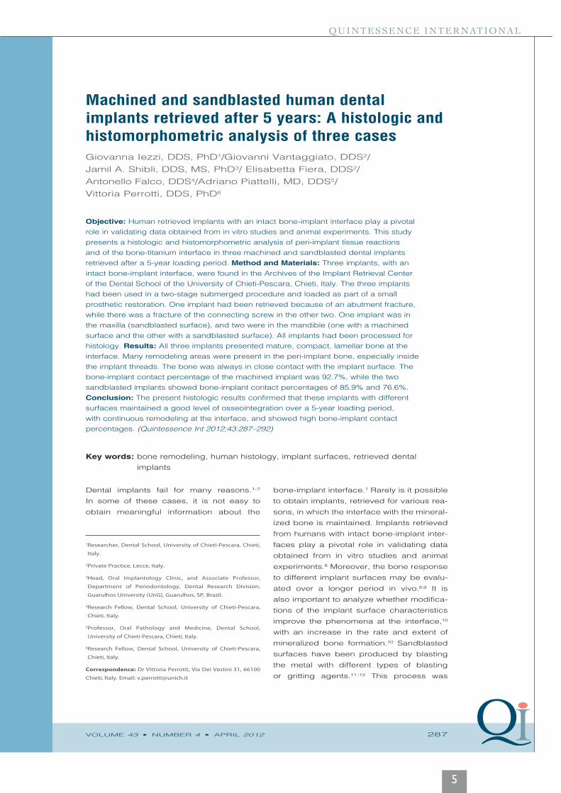
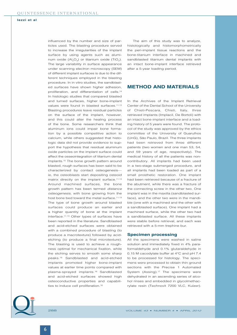
threads (Fig 4). The bone was always in
close contact with the implant surface (Fig 5).
At higher magnifications, no gaps or fibrous
connective tissue were present at the bone-
implant interface (Fig 6). Within some of the
threads, near the implant surface, it was
possible to see small, secondary osteons
with Haversian canals. Other osteonic struc-
tures, which had a direction perpendicular
to the implant long axis, were visible. Many
reversal lines were present in the areas of
bone remodeling. Both surfaces appeared
to be highly osteoconductive. Large marrow
spaces were present near the implant sur-
face (Fig 7). Osteocytes could be seen near
the implant surface. No inflammatory cell
infiltrates were present. No calculus, bac-
teria, or epithelial downgrowth was present.
The bone-implant contact percentage of
the machined implant was 92.7%, while the
two sandblasted implants showed a bone-
implant contact percentage of 85.9% and
76.6%.
DISCUSSION
Retrieval analysis of human dental implants
is a valuable tool for the evaluation of both
implant success and failure.1,8 The bone-
implant contact percentage has been used
as an indicator of the success of a dental
implant at different implantation times, and
it is also possible to observe the behavior of
the implant in the bone over time. Bone was
found to be more mature and well organized
around loaded implants, and many areas of
remodeling and well-defined osteons were
present. The implant loading also had an
effect on the distribution of the collagen
fibers of the bone tissue.18,19 A Haversian-
like structure running perpendicular to and
along with the implants’ long axis has been
reported around root-form implants func-
tionally loaded for long periods of time.8 In
the implants removed for implant or abut-
ment fracture, a high bone-implant contact
percentage (about 70%) was present, and
the fractures usually occurred after some
years (3 to 4).1–3 The specimens retrieved
after longer periods may contain important
information about the host biologic reaction
and the effects of the implant presence in
bone-remodeling processes.8 The capa-
bility of the bone to remain attached to
implant surfaces, placed in function, may
also help to demonstrate the performance
of the micro- and macrostructure of the
dental implants over time. Osteogenesis
at the bone-implant interface seems to
be influenced by several mechanisms.10,16
The different implant surfaces and designs
may affect a series of coordinated events,
including protein adsorption, proliferation,
and bone-tissue deposition.10,16
Due to ethical constraints, a limited
sample of human retrieved implants is avail-
able, so each implant must be evaluated
and reported on to improve knowledge
of the healing and remodeling process-
es at the interface, especially after lon-
ger loading periods. Only in such a way
can a larger quantity of human retrieved
implants be assembled, corroborating the
results from in vitro investigations, ani-
mal experimental studies, and clinical
investigations in humans. The retrieved
implants from the present study were
removed for reasons other than a failure
at the interface, so an intact mineralized
tissue-titanium interface could be evalu-
ated. No differences were found in the bone
growth pattern (distance osteogenesis vs
contact osteogenesis) around the implant
with the machined surface and the implants
with the blasted surface, even if the lim-
ited number of evaluated samples can-
not, clearly, allow any definitive conclusion.
Bone was found close to the surface of all
three implants, and at higher magnification,
no gaps or connective fibrous tissue was
found at the interface.
Even if previous studies reported in
the literature found that the blasted sur-
faces presented a higher bone-implant
contact than machined surfaces, in the
present specimens, it has to be noted that
the bone-implant contact of the machined
implants was higher than that of both blast-
ed implants. This is just a single anecdotal
observation, and certainly, no conclusions
can be drawn from a single case. The bone-
implant contact around all three implants
was high, even after a loading period of 5
years. This fact could be explained by the
evidence, as reported in the literature,20–23
that higher bone-implant contact has been
usually found around loaded implants. It
can also be explained by the fact that all
three implants were splinted, because they
were part of small prosthetic reconstruc-
tions, so deleterious micromovements at the
interface were limited. Bone remodeling,
with areas of more recent, newly formed
bone and osteonic Haversian structures,
was found around all three implants. This
bone remodeling, with the elimination of
the areas of microfractures present in the
peri-implant bone, and produced by the
microstrains generated, is thought to be
essential for the high long-term success
rates of dental implants.
Fig 4 High-power view of the implant shown in Fig 1. Many remodel-ing areas were present in the peri-implant bone, especially inside the implant threads (toluidine blue and basic fuchsin, original magnification ×40).
Fig 5 High-power view of the implant shown in Fig 2. The bone was always in close contact with the implant surface, with no gaps at the interface (tolu-idine blue and basic fuchsin, original magnification ×40).
Fig 6 High-power view of the implant shown in Fig 1. No gaps or fibrous connective tissues were present at the bone-implant interface. Osteocytes can be observed inside the respective lacunae (toluidine blue and basic fuchsin, original magnification ×100).
Fig 7 High-power view of the implant shown in Fig 2. The sandblasted surface appeared to be highly osteoconductive as newly formed bone can be observed in tight contact with the implant surface (toluidine blue and basic fuchsin, original magnifica-tion ×40).
8
VOLUME 43 • NUMBER 4 • APRIL 2012 291
QUINTESSENCE INTERNATIONAL
290 VOLUME 43 • NUMBER 4 • APRIL 2012
QUINT ESSENCE INTERNATIONAL
Iezzi et alIezzi et al
threads (Fig 4). The bone was always in
close contact with the implant surface (Fig 5).
At higher magnifications, no gaps or fibrous
connective tissue were present at the bone-
implant interface (Fig 6). Within some of the
threads, near the implant surface, it was
possible to see small, secondary osteons
with Haversian canals. Other osteonic struc-
tures, which had a direction perpendicular
to the implant long axis, were visible. Many
reversal lines were present in the areas of
bone remodeling. Both surfaces appeared
to be highly osteoconductive. Large marrow
spaces were present near the implant sur-
face (Fig 7). Osteocytes could be seen near
the implant surface. No inflammatory cell
infiltrates were present. No calculus, bac-
teria, or epithelial downgrowth was present.
The bone-implant contact percentage of
the machined implant was 92.7%, while the
two sandblasted implants showed a bone-
implant contact percentage of 85.9% and
76.6%.
DISCUSSION
Retrieval analysis of human dental implants
is a valuable tool for the evaluation of both
implant success and failure.1,8 The bone-
implant contact percentage has been used
as an indicator of the success of a dental
implant at different implantation times, and
it is also possible to observe the behavior of
the implant in the bone over time. Bone was
found to be more mature and well organized
around loaded implants, and many areas of
remodeling and well-defined osteons were
present. The implant loading also had an
effect on the distribution of the collagen
fibers of the bone tissue.18,19 A Haversian-
like structure running perpendicular to and
along with the implants’ long axis has been
reported around root-form implants func-
tionally loaded for long periods of time.8 In
the implants removed for implant or abut-
ment fracture, a high bone-implant contact
percentage (about 70%) was present, and
the fractures usually occurred after some
years (3 to 4).1–3 The specimens retrieved
after longer periods may contain important
information about the host biologic reaction
and the effects of the implant presence in
bone-remodeling processes.8 The capa-
bility of the bone to remain attached to
implant surfaces, placed in function, may
also help to demonstrate the performance
of the micro- and macrostructure of the
dental implants over time. Osteogenesis
at the bone-implant interface seems to
be influenced by several mechanisms.10,16
The different implant surfaces and designs
may affect a series of coordinated events,
including protein adsorption, proliferation,
and bone-tissue deposition.10,16
Due to ethical constraints, a limited
sample of human retrieved implants is avail-
able, so each implant must be evaluated
and reported on to improve knowledge
of the healing and remodeling process-
es at the interface, especially after lon-
ger loading periods. Only in such a way
can a larger quantity of human retrieved
implants be assembled, corroborating the
results from in vitro investigations, ani-
mal experimental studies, and clinical
investigations in humans. The retrieved
implants from the present study were
removed for reasons other than a failure
at the interface, so an intact mineralized
tissue-titanium interface could be evalu-
ated. No differences were found in the bone
growth pattern (distance osteogenesis vs
contact osteogenesis) around the implant
with the machined surface and the implants
with the blasted surface, even if the lim-
ited number of evaluated samples can-
not, clearly, allow any definitive conclusion.
Bone was found close to the surface of all
three implants, and at higher magnification,
no gaps or connective fibrous tissue was
found at the interface.
Even if previous studies reported in
the literature found that the blasted sur-
faces presented a higher bone-implant
contact than machined surfaces, in the
present specimens, it has to be noted that
the bone-implant contact of the machined
implants was higher than that of both blast-
ed implants. This is just a single anecdotal
observation, and certainly, no conclusions
can be drawn from a single case. The bone-
implant contact around all three implants
was high, even after a loading period of 5
years. This fact could be explained by the
evidence, as reported in the literature,20–23
that higher bone-implant contact has been
usually found around loaded implants. It
can also be explained by the fact that all
three implants were splinted, because they
were part of small prosthetic reconstruc-
tions, so deleterious micromovements at the
interface were limited. Bone remodeling,
with areas of more recent, newly formed
bone and osteonic Haversian structures,
was found around all three implants. This
bone remodeling, with the elimination of
the areas of microfractures present in the
peri-implant bone, and produced by the
microstrains generated, is thought to be
essential for the high long-term success
rates of dental implants.
Fig 4 High-power view of the implant shown in Fig 1. Many remodel-ing areas were present in the peri-implant bone, especially inside the implant threads (toluidine blue and basic fuchsin, original magnification ×40).
Fig 5 High-power view of the implant shown in Fig 2. The bone was always in close contact with the implant surface, with no gaps at the interface (tolu-idine blue and basic fuchsin, original magnification ×40).
Fig 6 High-power view of the implant shown in Fig 1. No gaps or fibrous connective tissues were present at the bone-implant interface. Osteocytes can be observed inside the respective lacunae (toluidine blue and basic fuchsin, original magnification ×100).
Fig 7 High-power view of the implant shown in Fig 2. The sandblasted surface appeared to be highly osteoconductive as newly formed bone can be observed in tight contact with the implant surface (toluidine blue and basic fuchsin, original magnifica-tion ×40).
9
292 VOLUME 43 • NUMBER 4 • APRIL 2012
QUINTESSENCE INTERNATIONAL
Iezzi et al
CONCLUSION
The present histologic results confirmed
that these implants with different surfaces
maintained a good level of osseointegra-
tion over a 5-year loading period, with a
continuous remodeling at the interface,
and showed a high bone-implant contact
percentage.
ACKNOWLEDGMENTS
This study was partially supported by the Ministry of Education, University, and Research (M.I.U.R.), Rome, Italy.
REFERENCES
1. Piattelli A, Scarano A, Piattelli M. Histologic observa-
tions on 230 retrieved dental implants: An 8-year
experience (1989–1996). J Periodontol 1998;69:
178–184.
2. Piattelli A, Scarano A, Piattelli M, Vaia E, Matarasso
S. Hollow implants retrieved for fracture: A light and
scanning electron microscope analysis of 4 cases.
J Periodontol 1998;69:185–189.
3. Piattelli A, Piattelli M, Scarano A, Montesani L.
Light and scanning electron microscopic study of
4 fractured implants. Int J Oral Maxillofac Implants
1998;13:561–564.
4. Piattelli A, Scarano A, Dalla Nora A, De Bona G,
Favero GA. Microscopical features in retrieved
human Branemark implants: A report of 19 cases.
Biomaterials 1998;19:643–649.
5. Piattelli A, Piattelli M, Mangano C, Scarano A. A
histologic evaluation of eight cases of failed dental
implants: Is bone overheating the most probable
cause? Biomaterials 1998;19:683–690.
6. Piattelli A, Scarano A, Piattelli M, Vaia E, Matarasso
S. A microscopical evaluation of 24 retrieved failed
hollow implants. Biomaterials 1999;20:485-489.
7. Piattelli A, Scarano A, Favero L, Iezzi G, Petrone
G, Favero GA. Clinical and histological aspects of
dental implants removed because of mobility.
J Periodontol 2003;74:385–390.
8. Coelho PG, Marin C, Granato R, Suzuki M.
Histomorphologic analysis of 30 plateau root form
implants retrieved after 8 to 13 years in function. A
human retrieval study. J Biomed Mater Res B Appl
Biomater 2009;91:975–979.
9. Bolind PK, Johansson CB, Becker W, Langer L,
Sevetz EB Jr, Albrektsson TO. A descriptive study
of retrieved non-threaded and threaded implant
designs. Clin Oral Implant Res 2005;16:447–455.
10. Junker R, Dimakis A, Thoneick M, Jansen JA. Effects
of implant surface coatings and composition on
bone integration: A systematic review. Clin Oral
Implant Res 2009;20(suppl):185–206.
11. Piattelli A, Manzon L, Scarano A, Paolantonio M,
Piattelli M. Histologic and morphologic analysis of
the bone response to machined and sandblasted
titanium implants: An experimental study in rab-
bits. Int J Oral Maxillofac Implants 1998;13:805–810.
12. Orsini G, Assenza B, Scarano A, Piattelli M, Piattelli
A. Surface analysis of machined versus sandblast-
ed and acid-etched titanium implant. Int J Oral
Maxillofac Implants 2000;15:779–784.
13. Piattelli M, Scarano A, Paolantonio M, Iezzi G,
Petrone G, Piattelli A. Bone response to RBM sand-
blasted titanium implants: An experimental study in
rabbits. J Oral Implantol 2002;28:2–8.
14. Di Carmine M, Toto P, Feliciani C, et al. Spreading
of epithelial cells on machined and sandblasted
titanium surfaces: An in vitro study. J Periodontol
2003;74:289–295.
15. Piattelli A, Degidi M, Paolantonio M, Mangano C,
Scarano A. Residual aluminum oxide on the surface
of titanium implants has no effect on osseointegra-
tion. Biomaterials 2003;24:4081–4089.
16. Wennerberg A, Albrektsson T. Effects of titanium
surface topography on bone integration: A system-
atic review. Clin Oral Implants Res 2009;20(suppl):
172–184.
17. Piattelli A, Scarano A, Quaranta M. High-precision,
cost-effective cutting system for producing thin
sections of oral tissues containing dental implants.
Biomaterials 1997;18:577–579.
18. Traini T, De Paoli S, Caputi S, Iezzi G, Piattelli A.
Collagen fibers orientation near a fractured dental
implant after a 5 years loading period: A case report.
Implant Dent 2006;15:70–76.
19. Traini T, Iezzi G, Pecora G, Piattelli A. Preferred col-
lagen fiber orientation in the human peri-implant
bone after short- and long-term loading periods: A
case report. J Oral Implantol 2006;32:177–181.
20. Testori T, Szmukler-Moncler S, Francetti L, Del
Fabbro M, Trisi P, Weinstein RL. Healing of Osseotite
implants under submerged and immediate loading
conditions in a single patient: A case report and
interface analysis after 2 months. Int J Periodontics
Restorative Dent 2002;22:345–353.
21. Degidi M, Scarano A, Piattelli M, Perrotti V, Piattelli A.
Bone remodeling in immediately loaded and unload-
ed titanium implants: A histologic and histomorpho-
metric study in man. J Oral Implantol 2005;31:18–24.
22. Degidi M, Petrone G, Iezzi G, Piattelli A. Histologic
evaluation of 2 human immediately loaded and 1
titanium implants inserted in the posterior man-
dible and submerged retrieved after 6 months.
J Oral Implantol 2003;29:223–229.
23. Piattelli A, Corigliano M, Scarano A, Costigliola
G, Paolantonio M. Immediate loading of titanium
plasma-sprayed implants: A pilot study in monkeys.
J Periodontol 1998;69:321–327.
10


![2007 - Atsuko Hashimoto - GeographicalRepresentationsEmbeddedwithinSouvenirs[Retrieved-2015!03!07]](https://static.fdocument.pub/doc/165x107/563dba45550346aa9aa42dca/2007-atsuko-hashimoto-geographicalrepresentationsembeddedwithinsouvenirsretrieved-20150307.jpg)

![2010 - Ioan Iovitz Popescu - Zipfslawanotherview[Retrieved-2014!07!26]](https://static.fdocument.pub/doc/165x107/577cc0db1a28aba711915b49/2010-ioan-iovitz-popescu-zipfslawanotherviewretrieved-20140726.jpg)
![PCL 2 - Case 7: CASE OF MICHAEL PHILLIPS Michal Zacharzewski. (2011). Last Man Standing [Online image]. Retrieved from //.](https://static.fdocument.pub/doc/165x107/56649dc95503460f94abfcce/pcl-2-case-7-case-of-michael-phillips-michal-zacharzewski-2011-last.jpg)


