Long-Term Monitoring and Analysis of Age-Related...
23
Health, 2017, 9, 323-344 http://www.scirp.org/journal/health ISSN Online: 1949-5005 ISSN Print: 1949-4998 DOI: 10.4236/health.2017.92023 February 21, 2017 Long-Term Monitoring and Analysis of Age-Related Changes on Autonomic Nervous Function Kenichi Itao 1,2 , Makoto Komazawa 1 , Zhiwei Luo 3 , Hiroyuki Kobayashi 2 1 WINFrontier Co. Ltd., Tokyo, Japan 2 Juntendo University, Tokyo, Japan 3 Organization of Advanced Science and Technology, Kobe University, Kobe, Japan Abstract This study used a small wearable heart rate sensor to monitor the daily auto- nomic function of 600 subjects from across all age groups over a prolonged period of time. The results indicated that the LF/HF ratio (Heart Rate Varia- bility, LF: frequencies between 0.04 Hz - 0.15 Hz, HF: frequencies between 0.15 Hz - 0.4 Hz) an indicator of balance in the autonomic nervous system, tended to peak for subjects in their 40’s and decline thereafter. This conceiva- bly may be partially due to the causes for concern and stress changing and/or declining for the group aged 50-plus. A decline in diurnal variation of auto- nomic nervous activity was also exhibited in subjects aged 50 and up, showing a tendency for decline in the function of rising sympathetic nerve activity par- ticularly in the morning. It is conceivable that this stems from a decline in the responsiveness of the autonomic nervous system. Subjects in the 50-plus group furthermore exhibited a tendency for declining variation in autonomic nerv- ous activity between sleeping and waking hours. This phenomenon was con- sistent with the tendency for there to be a rise in wake after sleep onset coupled with a decline in slow-wave sleep in middle- to old-age. Keywords Heart Rate Variability, Autonomic Nervous System, Stress, Age, Circadian Rhythm, Sleep 1. Introduction Having breached the threshold of being a hyper-aging society, the prevalence of lifestyle-related diseases and soaring medical costs have emerged as issues of dire consequence in Japan. Modern-day society, in particular, is referred to as a How to cite this paper: Itao, K., Komaza- wa, M., Luo, Z.W. and Kobayashi, H. (2017) Long-Term Monitoring and Analysis of Age-Related Changes on Autonomic Ner- vous Function. Health, 9, 323-344. https://doi.org/10.4236/health.2017.92023 Received: December 21, 2016 Accepted: February 18, 2017 Published: February 21, 2017 Copyright © 2017 by authors and Scientific Research Publishing Inc. This work is licensed under the Creative Commons Attribution International License (CC BY 4.0). http://creativecommons.org/licenses/by/4.0/ Open Access
Transcript of Long-Term Monitoring and Analysis of Age-Related...
Health, 2017, 9, 323-344 http://www.scirp.org/journal/health
ISSN Online: 1949-5005 ISSN Print: 1949-4998
DOI: 10.4236/health.2017.92023 February 21, 2017
Long-Term Monitoring and Analysis of Age-Related Changes on Autonomic Nervous Function
Kenichi Itao1,2, Makoto Komazawa1, Zhiwei Luo3, Hiroyuki Kobayashi2
1WINFrontier Co. Ltd., Tokyo, Japan 2Juntendo University, Tokyo, Japan 3Organization of Advanced Science and Technology, Kobe University, Kobe, Japan
Abstract This study used a small wearable heart rate sensor to monitor the daily auto-nomic function of 600 subjects from across all age groups over a prolonged period of time. The results indicated that the LF/HF ratio (Heart Rate Varia-bility, LF: frequencies between 0.04 Hz - 0.15 Hz, HF: frequencies between 0.15 Hz - 0.4 Hz) an indicator of balance in the autonomic nervous system, tended to peak for subjects in their 40’s and decline thereafter. This conceiva-bly may be partially due to the causes for concern and stress changing and/or declining for the group aged 50-plus. A decline in diurnal variation of auto-nomic nervous activity was also exhibited in subjects aged 50 and up, showing a tendency for decline in the function of rising sympathetic nerve activity par-ticularly in the morning. It is conceivable that this stems from a decline in the responsiveness of the autonomic nervous system. Subjects in the 50-plus group furthermore exhibited a tendency for declining variation in autonomic nerv-ous activity between sleeping and waking hours. This phenomenon was con-sistent with the tendency for there to be a rise in wake after sleep onset coupled with a decline in slow-wave sleep in middle- to old-age.
Keywords Heart Rate Variability, Autonomic Nervous System, Stress, Age, Circadian Rhythm, Sleep
1. Introduction
Having breached the threshold of being a hyper-aging society, the prevalence of lifestyle-related diseases and soaring medical costs have emerged as issues of dire consequence in Japan. Modern-day society, in particular, is referred to as a
How to cite this paper: Itao, K., Komaza-wa, M., Luo, Z.W. and Kobayashi, H. (2017) Long-Term Monitoring and Analysis of Age-Related Changes on Autonomic Ner- vous Function. Health, 9, 323-344. https://doi.org/10.4236/health.2017.92023 Received: December 21, 2016 Accepted: February 18, 2017 Published: February 21, 2017 Copyright © 2017 by authors and Scientific Research Publishing Inc. This work is licensed under the Creative Commons Attribution International License (CC BY 4.0). http://creativecommons.org/licenses/by/4.0/
Open Access
K. Itao et al.
324
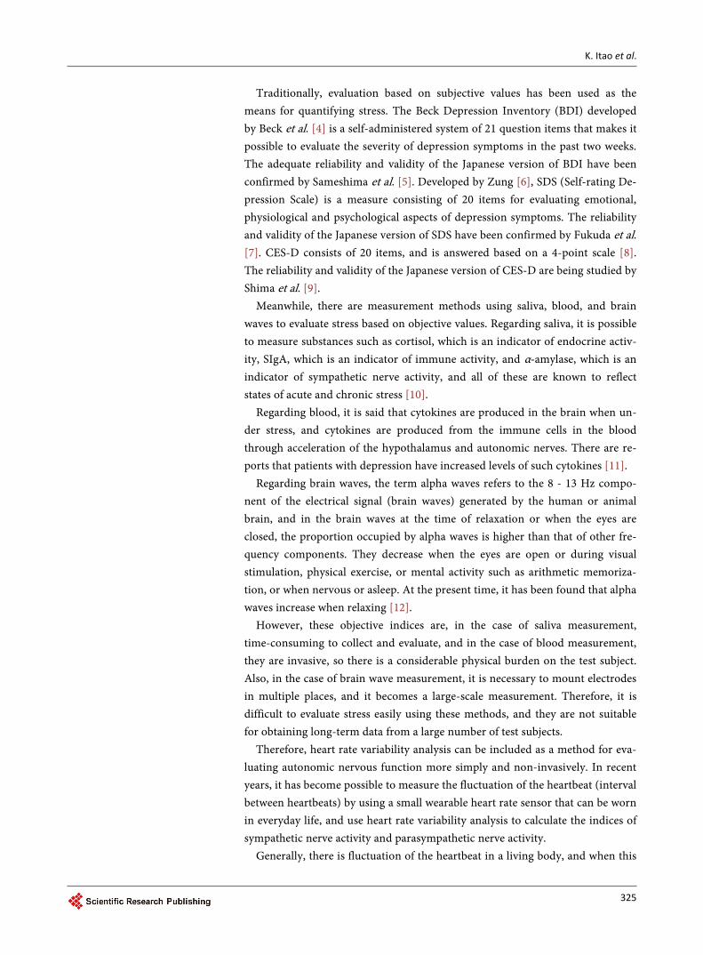
“stress society” and according to statistics from the Health, Labour, and Welfare Ministry [1], nearly half of all people are living with some kind of stress as 46.5% of people reported experiencing worries or stress in their daily lives (Figure 1).
It is said that this kind of stress suffered by many Japanese people is closely related to autonomic nervous function [2]. Seen structurally and functionally, the autonomic nerves are positioned to connect the internal environment and the external environment, harmonize the functions of the body, regulate ho-meostasis (cardiovascular/respiratory control, body temperature regulation, ga-strointestinal motility, urine/feces excretion function, reproductive function, metabolism/endocrine function), and perform adaptive response to stress (“fight or flight” reaction). Thus, the autonomic nervous system is doing what could be called indefatigable work in order to ensure the survival and proliferation of the species [3].
In 1921 Langley, a physiologist at the University of Cambridge, published “Autonomic Nervous System” and classified the autonomic nervous system into three systems: sympathetic nerves, parasympathetic nerves, and enteric nerves, establishing the current concept of autonomic nerves [3].
The sympathetic nerves together with parasympathetic nerves form the auto-nomic nervous system, and are the nerves that control the secretory glands, blood vessels, internal organs, and so on. During mental stimulation or exercise, they work to increase the activity of the whole body, such as secreting saliva, in-creasing blood pressure/blood sugar, constricting blood vessels in the skin and internal organs, and collecting blood in the muscles/brain [3].
On the other hand, the parasympathetic nerves emanate from the brain region and the sacral region and, mainly through the vagus nerve, secrete acetylcholine as a neurotransmitter. It acts to suppress the heart and excite the gastrointestinal tract. It also acts to promote bile secretion, promote secretion of tears and saliva, dilate the pupils, etc. These autonomic nervous functions are affected not only by physical stress but also by psychological stress [3].
Figure 1. Presence of worries or stress (age 12 and over).
K. Itao et al.
325
Traditionally, evaluation based on subjective values has been used as the means for quantifying stress. The Beck Depression Inventory (BDI) developed by Beck et al. [4] is a self-administered system of 21 question items that makes it possible to evaluate the severity of depression symptoms in the past two weeks. The adequate reliability and validity of the Japanese version of BDI have been confirmed by Sameshima et al. [5]. Developed by Zung [6], SDS (Self-rating De-pression Scale) is a measure consisting of 20 items for evaluating emotional, physiological and psychological aspects of depression symptoms. The reliability and validity of the Japanese version of SDS have been confirmed by Fukuda et al. [7]. CES-D consists of 20 items, and is answered based on a 4-point scale [8]. The reliability and validity of the Japanese version of CES-D are being studied by Shima et al. [9].
Meanwhile, there are measurement methods using saliva, blood, and brain waves to evaluate stress based on objective values. Regarding saliva, it is possible to measure substances such as cortisol, which is an indicator of endocrine activ-ity, SIgA, which is an indicator of immune activity, and α-amylase, which is an indicator of sympathetic nerve activity, and all of these are known to reflect states of acute and chronic stress [10].
Regarding blood, it is said that cytokines are produced in the brain when un-der stress, and cytokines are produced from the immune cells in the blood through acceleration of the hypothalamus and autonomic nerves. There are re-ports that patients with depression have increased levels of such cytokines [11].
Regarding brain waves, the term alpha waves refers to the 8 - 13 Hz compo-nent of the electrical signal (brain waves) generated by the human or animal brain, and in the brain waves at the time of relaxation or when the eyes are closed, the proportion occupied by alpha waves is higher than that of other fre-quency components. They decrease when the eyes are open or during visual stimulation, physical exercise, or mental activity such as arithmetic memoriza-tion, or when nervous or asleep. At the present time, it has been found that alpha waves increase when relaxing [12].
However, these objective indices are, in the case of saliva measurement, time-consuming to collect and evaluate, and in the case of blood measurement, they are invasive, so there is a considerable physical burden on the test subject. Also, in the case of brain wave measurement, it is necessary to mount electrodes in multiple places, and it becomes a large-scale measurement. Therefore, it is difficult to evaluate stress easily using these methods, and they are not suitable for obtaining long-term data from a large number of test subjects.
Therefore, heart rate variability analysis can be included as a method for eva-luating autonomic nervous function more simply and non-invasively. In recent years, it has become possible to measure the fluctuation of the heartbeat (interval between heartbeats) by using a small wearable heart rate sensor that can be worn in everyday life, and use heart rate variability analysis to calculate the indices of sympathetic nerve activity and parasympathetic nerve activity.
Generally, there is fluctuation of the heartbeat in a living body, and when this
K. Itao et al.
326
fluctuation is frequency analyzed, a peak can be seen at a certain frequency. In the case of a person, there appear a high frequency component (0.15 - 0.40 Hz: HF) reflecting the variation of the respiratory cycle and a low frequency compo-nent (0.05 - 0.15 Hz: LF) reflecting fluctuation in blood pressure, both of which reflect autonomic nerve activity. It is said that HF is regulated by the parasym-pathetic nerves, and LF is regulated by both sympathetic and parasympathetic nerves [3].
There are several prior studies of the relationship between autonomic nervous indices and physiological phenomena that used such heart rate variability analy-sis.
Previous research has explored the relationship between the severity of de-pression based upon the SDS (“Self-rating Depression Scale”) and autonomic ac-tivity obtained by analyzing heart rate variability from a Holter ECG monitor in 31 subjects (12 males and 19 females) diagnosed with mood disorders. In associ-ation with the severity of depression, the results demonstrated a significant rise in sympathetic nerve function along with significantly diminishing parasympa-thetic nerve function. Accordingly, in assessments of depression severity con-ducted with the use of Holter ECG monitors, these results support the onset of abnormalities in autonomic nerve function brought on by depression [13].
In another paper examining the effects of depression on the autonomic nerv-ous system in 16 male subjects diagnosed with mild heart attacks, a trend was observed of elevated sympathetic nerve function and suppressed parasympa-thetic nerve function as subjects’ scores rose on the SDS (denoting increasingly severe depression) [14]. Thus, abnormalities in autonomic nervous function wherein the sympathetic nerves are excited and the parasympathetic nerves are suppressed may conceivably have an effect on the pathogenesis and prognosis of heart attacks.
Also, in the Multiethnic Study of Atherosclerosis (MESA), a leading epidemi-ological study in the United States, as a result of investigating the relationship between depression symptoms, anger, anxiety, social support scores and para-sympathetic nerve functions, evaluated in questionnaire form, it was observed that there was a significant negative association with symptoms of depression. Therefore, it is said that continuing depression in particular affects autonomic nervous function, especially parasympathetic function [15].
In addition to those people who have specific diseases as described above, there are several reports that the balance of the autonomic nervous system also changes with age. This is because the internal environment of the human body is kept constant by the homeostasis mechanism, but with age the balance of the autonomic nervous system deteriorates, so it is said to become difficult for the body to respond appropriately to changes in the external environment. In pre-vious reports, a gradual decline in parasympathetic function due to aging in fe-males and predominance of sympathetic function due to aging in males have been recognized, but many reports are based on the results of short-term mea-surement [16].
K. Itao et al.
327
Consequently, in this study we used a small wearable heart rate sensor capable of long-term monitoring to test how age-related changes in levels of autonomic activity and diurnal variations in the autonomic nerves change as a function of age. We also attempt to reveal variations across age groups, diurnal variations, and variations between sleeping and waking states by monitoring a large pool of subjects ranging across all age groups as they went about their daily lives.
2. Methodology
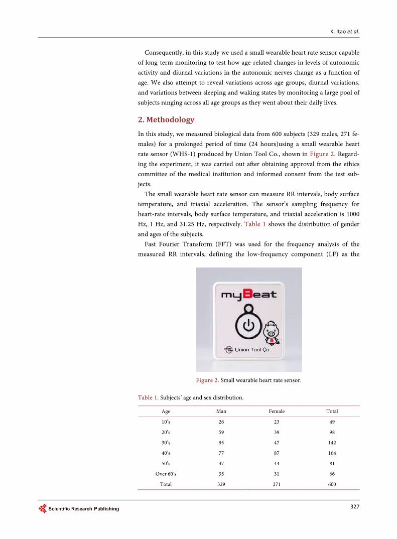
In this study, we measured biological data from 600 subjects (329 males, 271 fe-males) for a prolonged period of time (24 hours)using a small wearable heart rate sensor (WHS-1) produced by Union Tool Co., shown in Figure 2. Regard-ing the experiment, it was carried out after obtaining approval from the ethics committee of the medical institution and informed consent from the test sub-jects.
The small wearable heart rate sensor can measure RR intervals, body surface temperature, and triaxial acceleration. The sensor’s sampling frequency for heart-rate intervals, body surface temperature, and triaxial acceleration is 1000 Hz, 1 Hz, and 31.25 Hz, respectively. Table 1 shows the distribution of gender and ages of the subjects.
Fast Fourier Transform (FFT) was used for the frequency analysis of the measured RR intervals, defining the low-frequency component (LF) as the
Figure 2. Small wearable heart rate sensor.
Table 1. Subjects’ age and sex distribution.
Age Man Female Total
10’s 26 23 49
20’s 59 39 98
30’s 95 47 142
40’s 77 87 164
50’s 37 44 81
Over 60’s 35 31 66
Total 329 271 600
K. Itao et al.
328
frequencies between 0.04 Hz - 0.15 Hz and the high-frequency component (HF) as the frequencies between 0.15 Hz - 0.4 Hz. The sum of the LF and HF compo-nents is referred to as total power (TP), and is considered to be an indicator of overall autonomic nervous activity. Our method for computing indicators of autonomic nerve activity adhered to procedures from paper [17] in the refer-ences. These indicators of autonomic nerve activity are also said to correlate with fatigue [18].
In this experiment, we use solely the data where subjects exhibit little body movement from among the long-term measurement data. To analyze the data, body movement levels were determined on the basis of the acceleration sensors embedded in the small wearable heart rate sensor using the supplied software (produced by WINFrontier Co.). It has been reported that, in general, the com-posite value of triaxial acceleration correlates to body activity indicators and energy consumption, and the values change while walking, running, and resting [19] [20] [21].
The threshold for the composite value of acceleration was configured in alignment with each of the activities (walking, running, resting) on the sensor to distinguish the activity. Only the data determined as being recorded at rest when there is little body movement was used. IBM SPSS Statistics Version 22 was used for statistical processing in this study, with the level of statistical significance being 5%. We used the games-howell method for multiple comparison.
3. The Relationship between Autonomic Nerve Function and Aging
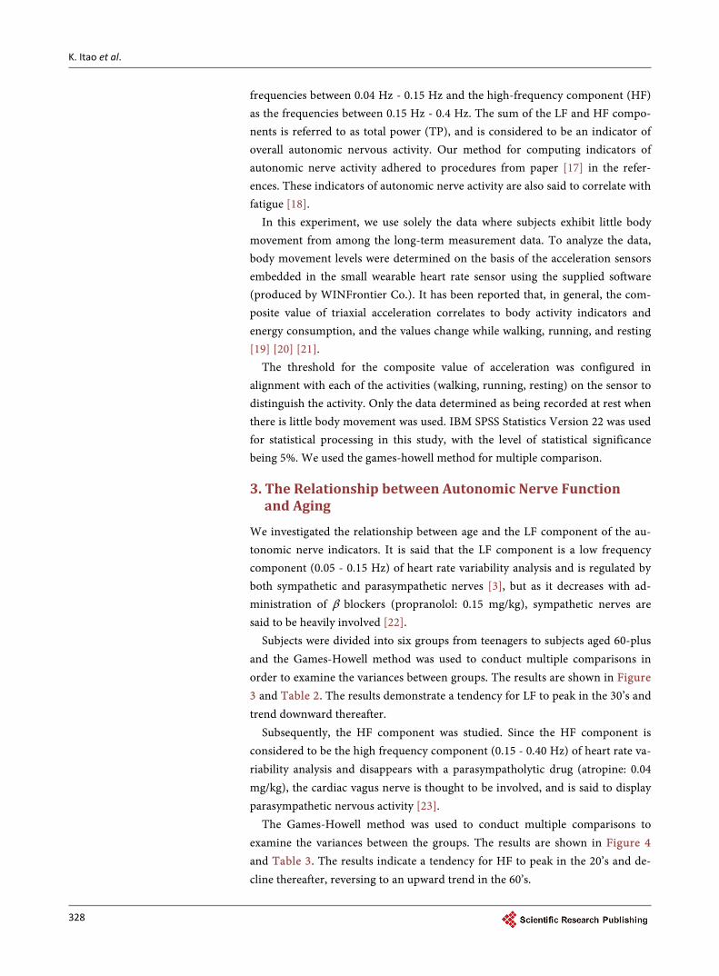
We investigated the relationship between age and the LF component of the au-tonomic nerve indicators. It is said that the LF component is a low frequency component (0.05 - 0.15 Hz) of heart rate variability analysis and is regulated by both sympathetic and parasympathetic nerves [3], but as it decreases with ad-ministration of β blockers (propranolol: 0.15 mg/kg), sympathetic nerves are said to be heavily involved [22].
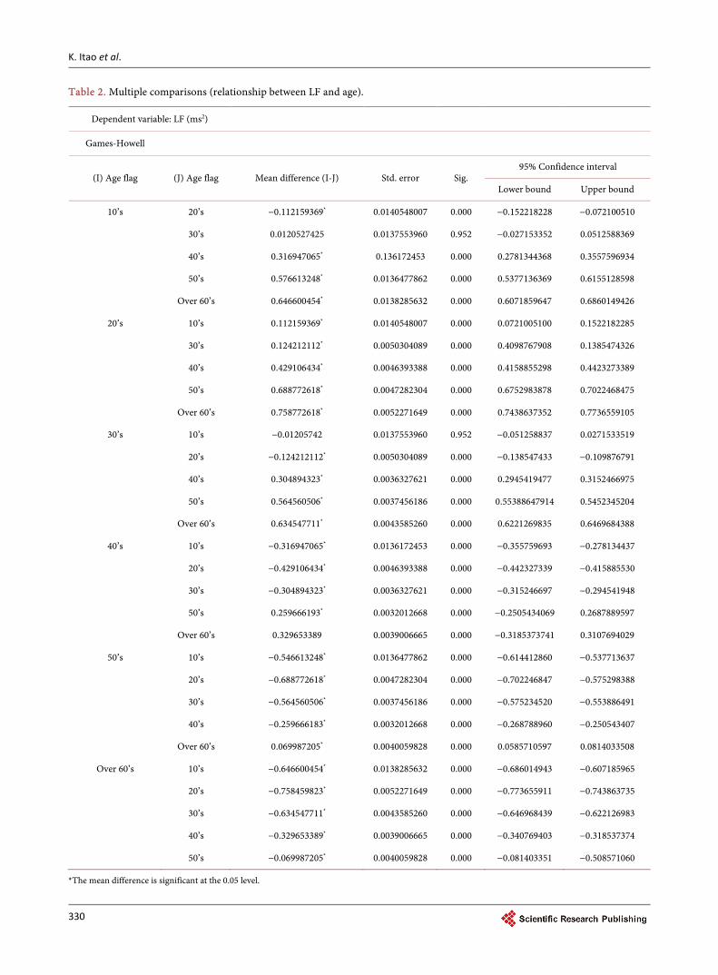
Subjects were divided into six groups from teenagers to subjects aged 60-plus and the Games-Howell method was used to conduct multiple comparisons in order to examine the variances between groups. The results are shown in Figure 3 and Table 2. The results demonstrate a tendency for LF to peak in the 30’s and trend downward thereafter.
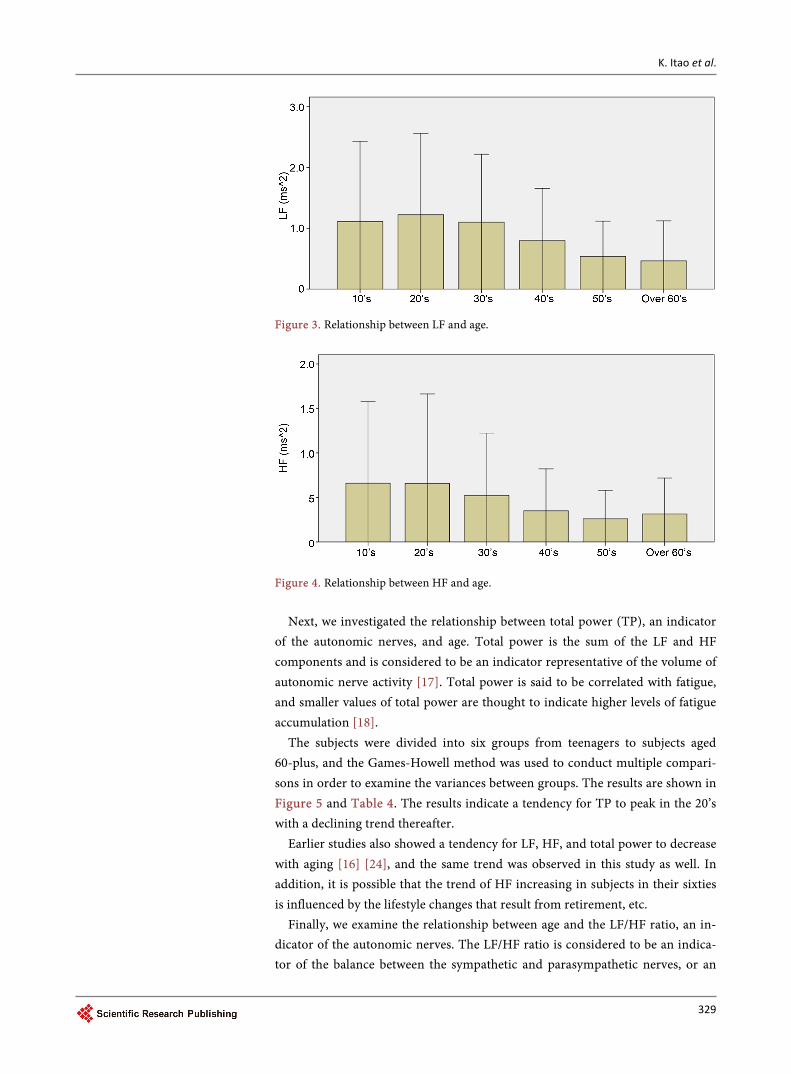
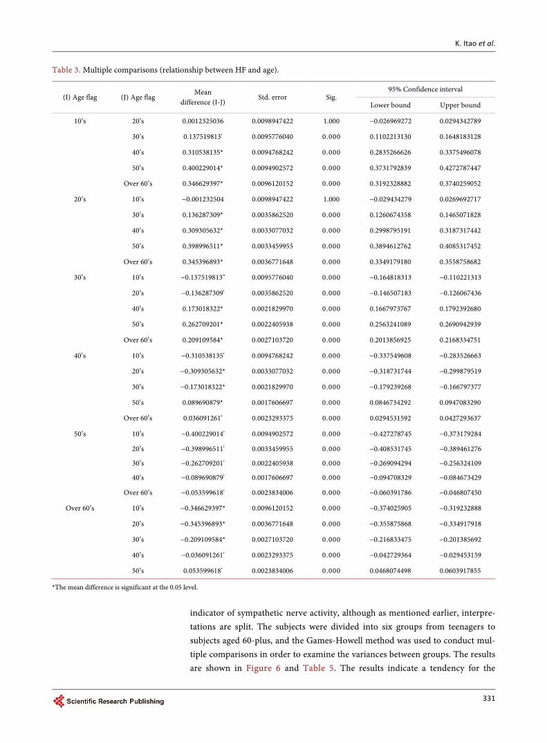
Subsequently, the HF component was studied. Since the HF component is considered to be the high frequency component (0.15 - 0.40 Hz) of heart rate va-riability analysis and disappears with a parasympatholytic drug (atropine: 0.04 mg/kg), the cardiac vagus nerve is thought to be involved, and is said to display parasympathetic nervous activity [23].
The Games-Howell method was used to conduct multiple comparisons to examine the variances between the groups. The results are shown in Figure 4 and Table 3. The results indicate a tendency for HF to peak in the 20’s and de-cline thereafter, reversing to an upward trend in the 60’s.
K. Itao et al.
329
Figure 3. Relationship between LF and age.
Figure 4. Relationship between HF and age.
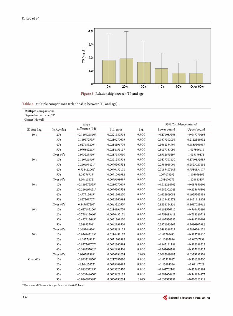
Next, we investigated the relationship between total power (TP), an indicator
of the autonomic nerves, and age. Total power is the sum of the LF and HF components and is considered to be an indicator representative of the volume of autonomic nerve activity [17]. Total power is said to be correlated with fatigue, and smaller values of total power are thought to indicate higher levels of fatigue accumulation [18].
The subjects were divided into six groups from teenagers to subjects aged 60-plus, and the Games-Howell method was used to conduct multiple compari-sons in order to examine the variances between groups. The results are shown in Figure 5 and Table 4. The results indicate a tendency for TP to peak in the 20’s with a declining trend thereafter.
Earlier studies also showed a tendency for LF, HF, and total power to decrease with aging [16] [24], and the same trend was observed in this study as well. In addition, it is possible that the trend of HF increasing in subjects in their sixties is influenced by the lifestyle changes that result from retirement, etc.
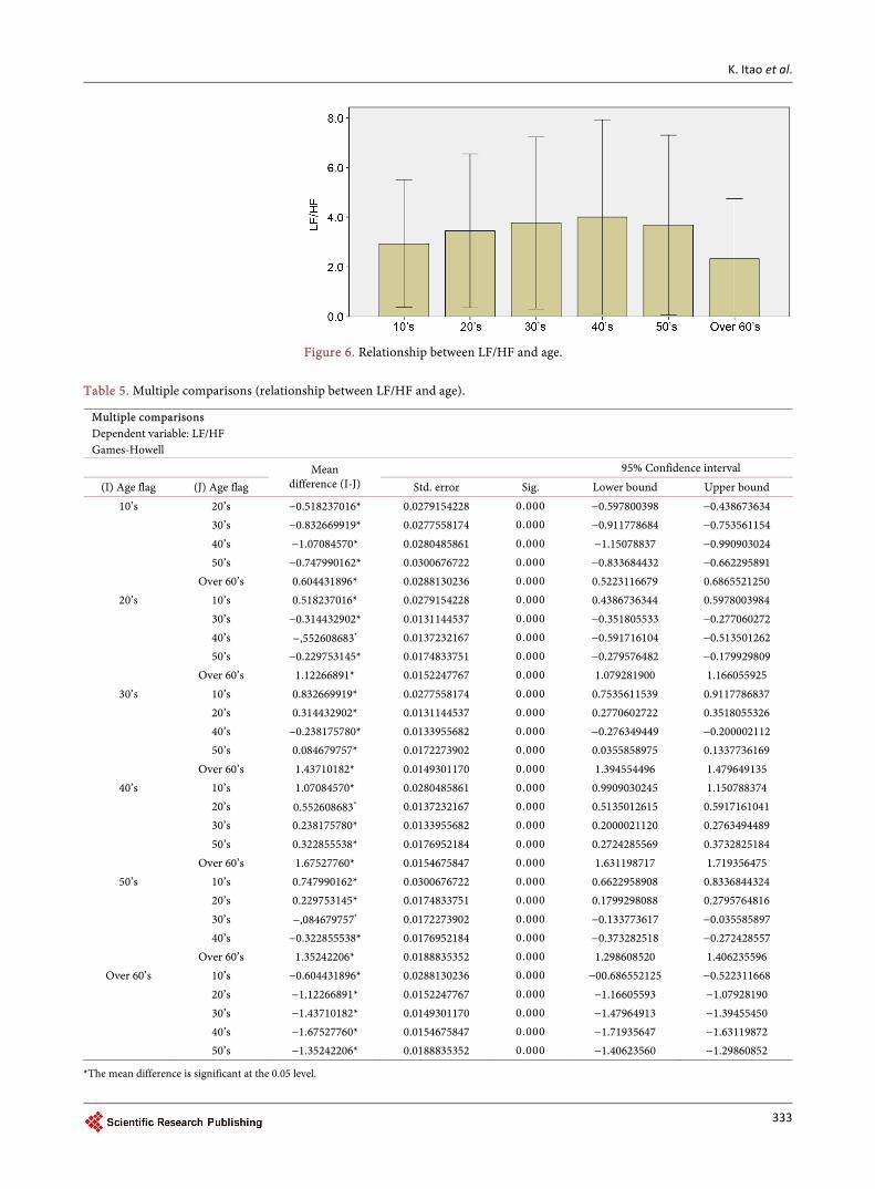
Finally, we examine the relationship between age and the LF/HF ratio, an in-dicator of the autonomic nerves. The LF/HF ratio is considered to be an indica-tor of the balance between the sympathetic and parasympathetic nerves, or an
K. Itao et al.
330
Table 2. Multiple comparisons (relationship between LF and age).
Dependent variable: LF (ms2)
Games-Howell
(I) Age flag (J) Age flag Mean difference (I-J) Std. error Sig. 95% Confidence interval
Lower bound Upper bound
10’s 20’s −0.112159369* 0.0140548007 0.000 −0.152218228 −0.072100510
30’s 0.0120527425 0.0137553960 0.952 −0.027153352 0.0512588369
40’s 0.316947065* 0.136172453 0.000 0.2781344368 0.3557596934
50’s 0.576613248* 0.0136477862 0.000 0.5377136369 0.6155128598
Over 60’s 0.646600454* 0.0138285632 0.000 0.6071859647 0.6860149426
20’s 10’s 0.112159369* 0.0140548007 0.000 0.0721005100 0.1522182285
30’s 0.124212112* 0.0050304089 0.000 0.4098767908 0.1385474326
40’s 0.429106434* 0.0046393388 0.000 0.4158855298 0.4423273389
50’s 0.688772618* 0.0047282304 0.000 0.6752983878 0.7022468475
Over 60’s 0.758772618* 0.0052271649 0.000 0.7438637352 0.7736559105
30’s 10’s −0.01205742 0.0137553960 0.952 −0.051258837 0.0271533519
20’s −0.124212112* 0.0050304089 0.000 −0.138547433 −0.109876791
40’s 0.304894323* 0.0036327621 0.000 0.2945419477 0.3152466975
50’s 0.564560506* 0.0037456186 0.000 0.55388647914 0.5452345204
Over 60’s 0.634547711* 0.0043585260 0.000 0.6221269835 0.6469684388
40’s 10’s −0.316947065* 0.0136172453 0.000 −0.355759693 −0.278134437
20’s −0.429106434* 0.0046393388 0.000 −0.442327339 −0.415885530
30’s −0.304894323* 0.0036327621 0.000 −0.315246697 −0.294541948
50’s 0.259666193* 0.0032012668 0.000 −0.2505434069 0.2687889597
Over 60’s 0.329653389 0.0039006665 0.000 −0.3185373741 0.3107694029
50’s 10’s −0.546613248* 0.0136477862 0.000 −0.614412860 −0.537713637
20’s −0.688772618* 0.0047282304 0.000 −0.702246847 −0.575298388
30’s −0.564560506* 0.0037456186 0.000 −0.575234520 −0.553886491
40’s −0.259666183* 0.0032012668 0.000 −0.268788960 −0.250543407
Over 60’s 0.069987205* 0.0040059828 0.000 0.0585710597 0.0814033508
Over 60’s 10’s −0.646600454* 0.0138285632 0.000 −0.686014943 −0.607185965
20’s −0.758459823* 0.0052271649 0.000 −0.773655911 −0.743863735
30’s −0.634547711* 0.0043585260 0.000 −0.646968439 −0.622126983
40’s −0.329653389* 0.0039006665 0.000 −0.340769403 −0.318537374
50’s −0.069987205* 0.0040059828 0.000 −0.081403351 −0.508571060
*The mean difference is significant at the 0.05 level.
K. Itao et al.
331
Table 3. Multiple comparisons (relationship between HF and age).
(I) Age flag (J) Age flag Mean
difference (I-J) Std. error Sig.
95% Confidence interval
Lower bound Upper bound
10’s 20’s 0.0012325036 0.0098947422 1.000 −0.026969272 0.0294342789
30’s 0.137519813' 0.0095776040 0.000 0.1102213130 0.1648183128
40’s 0.310538135* 0.0094768242 0.000 0.2835266626 0.3375496078
50’s 0.400229014* 0.0094902572 0.000 0.3731792839 0.4272787447
Over 60’s 0.346629397* 0.0096120152 0.000 0.3192328882 0.3740259052
20’s 10’s −0.001232504 0.0098947422 1.000 −0.029434279 0.0269692717
30’s 0.136287309* 0.0035862520 0.000 0.1260674358 0.1465071828
40’s 0.309305632* 0.0033077032 0.000 0.2998795191 0.3187317442
50’s 0.398996511* 0.0033459955 0.000 0.3894612762 0.4085317452
Over 60’s 0.345396893* 0.0036771648 0.000 0.3349179180 0.3558758682
30’s 10’s −0.137519813" 0.0095776040 0.000 −0.164818313 −0.110221313
20’s −0.136287309' 0.0035862520 0.000 −0.146507183 −0.126067436
40’s 0.173018322* 0.0021829970 0.000 0.1667973767 0.1792392680
50’s 0.262709201* 0.0022405938 0.000 0.2563241089 0.2690942939
Over 60’s 0.209109584* 0.0027103720 0.000 0.2013856925 0.2168334751
40’s 10’s −0.310538135' 0.0094768242 0.000 −0.337549608 −0.283526663
20’s −0.309305632* 0.0033077032 0.000 −0.318731744 −0.299879519
30’s −0.173018322* 0.0021829970 0.000 −0.179239268 −0.166797377
50’s 0.089690879* 0.0017606697 0.000 0.0846734292 0.0947083290
Over 60’s 0.036091261' 0.0023293375 0.000 0.0294531592 0.0427293637
50’s 10’s −0.400229014' 0.0094902572 0.000 −0.427278745 −0.373179284
20’s −0.398996511' 0.0033459955 0.000 −0.408531745 −0.389461276
30’s −0.262709201' 0.0022405938 0.000 −0.269094294 −0.256324109
40’s −0.089690879' 0.0017606697 0.000 −0.094708329 −0.084673429
Over 60’s −0.053599618' 0.0023834006 0.000 −0.060391786 −0.046807450
Over 60’s 10’s −0.346629397* 0.0096120152 0.000 −0.374025905 −0.319232888
20’s −0.345396893* 0.0036771648 0.000 −0.355875868 −0.334917918
30’s −0.209109584* 0.0027103720 0.000 −0.216833475 −0.201385692
40’s −0.036091261' 0.0023293375 0.000 −0.042729364 −0.029453159
50’s 0.053599618' 0.0023834006 0.000 0.0468074498 0.0603917855
*The mean difference is significant at the 0.05 level.
indicator of sympathetic nerve activity, although as mentioned earlier, interpre-tations are split. The subjects were divided into six groups from teenagers to subjects aged 60-plus, and the Games-Howell method was used to conduct mul-tiple comparisons in order to examine the variances between groups. The results are shown in Figure 6 and Table 5. The results indicate a tendency for the
K. Itao et al.
332
Figure 5. Relationship between TP and age.
Table 4. Multiple comparisons (relationship between TP and age).
Multiple comparisons Dependent variable: TP Games-Howell
Mean difference (I-J)
95% Confidence interval (I) Age flag (j) Age flag Std. error Sig. Lower bound Upper bound
10’s 20’s −0.110926866* 0.0221587508 0.000 −0.174083568 −0.047770163 30’s 0.149572555* 0.0216270603 0.000 0.0879302055 0.2112149052 40’s 0.627485200* 0.0214196776 0.000 0.5664334909 0.6885369097 50’s 0.976842263* 0.0214451137 0.000 0.9157181096 1.037966416 Over 60’s 0.993229850* 0.0217387010 0.000 0.9312695297 1.055190171
20’s 10’s 0.110926866* 0.0221587508 0.000 0.0477701630 0.1740835683 30’s 0.260499421* 0.0076507554 0.000 0.2386968006 0.2823020414 40’s 0.738412066' 0.0070432171 0.000 0.7183407143 0.7584834177 50’s 1.08776913* 0.0071201982 0.000 1.067478395 1.108059862 Over 60’s 1.10415672* 0.0079608695 0.000 1.081470275 1.126843157
30’s 10’s −0.149572555* 0.0216270603 0.000 −0.211214905 −0.087930206 20’s −0.260499421* 0.0076507554 0.000 −0.282302041 −0.238696801 40’s 0.477912645* 0.0051309270 0.000 0.4632909081 0.4925343818 50’s 0.827269707* 0.0052360984 0.000 0.8123482271 0.8421911876 Over 60’s 0.843657295' 0.0063320570 0.000 0.8256124836 0.8617021062
40’s 10’s −0.627485200* 0.0214196776 0.000 −0.688536910 −0.566433491 20’s −0.738412066* 0.0070432171 0.000 −0.758483418 −0.718340714 30’s −0.477912645* 0.0051309270 0.000 −0.492534382 −0.463290908 50’s 0.34935706* 0.0042999506 0.000 0.3371033265 0.3616107982 Over 60’s 0.365744650* 0.0055828125 0.000 0.3498348727 0.3816544272
50’s 10’s −0.976842263* 0.0214451137 0.000 −1.03796642 −0.915718110 20’s −1.08776913* 0.0071201982 0.000 −1.10805986 −1.06747839 30’s −0.827269707* 0.0052360984 0.000 −0.842191188 −0.812348227 40’s −0.349357062* 0.0042999506 0.000 −0.361610798 −0.337103327 Over 60’s 0.016387588* 0.0056796224 0.045 0.0002019182 0.0325732570
Over 60’s 10’s −0.993229850* 0.0217387010 0.000 −1.05519017 −0.931269530 20’s −1.10415672* 0.0079608695 0.000 −1.12684316 −1.08147028 30’s −0.843657295* 0.0063320570 0.000 −0.861702106 −0.825612484 40’s −0.365744650* 0.0055828125 0.000 −0.381654427 −0.349834873 50’s −0.016387588* 0.0056796224 0.045 −0.032573257 −0.000201918
*The mean difference is significant at the 0.05 level.
K. Itao et al.
333
Figure 6. Relationship between LF/HF and age.
Table 5. Multiple comparisons (relationship between LF/HF and age).
Multiple comparisons Dependent variable: LF/HF Games-Howell
Mean difference (I-J)
95% Confidence interval (I) Age flag (J) Age flag Std. error Sig. Lower bound Upper bound
10’s 20’s −0.518237016* 0.0279154228 0.000 −0.597800398 −0.438673634 30’s −0.832669919* 0.0277558174 0.000 −0.911778684 −0.753561154 40’s −1.07084570* 0.0280485861 0.000 −1.15078837 −0.990903024 50’s −0.747990162* 0.0300676722 0.000 −0.833684432 −0.662295891 Over 60’s 0.604431896* 0.0288130236 0.000 0.5223116679 0.6865521250
20’s 10’s 0.518237016* 0.0279154228 0.000 0.4386736344 0.5978003984 30’s −0.314432902* 0.0131144537 0.000 −0.351805533 −0.277060272 40’s −,552608683* 0.0137232167 0.000 −0.591716104 −0.513501262 50’s −0.229753145* 0.0174833751 0.000 −0.279576482 −0.179929809 Over 60’s 1.12266891* 0.0152247767 0.000 1.079281900 1.166055925
30’s 10’s 0.832669919* 0.0277558174 0.000 0.7535611539 0.9117786837 20’s 0.314432902* 0.0131144537 0.000 0.2770602722 0.3518055326 40’s −0.238175780* 0.0133955682 0.000 −0.276349449 −0.200002112 50’s 0.084679757* 0.0172273902 0.000 0.0355858975 0.1337736169 Over 60’s 1.43710182* 0.0149301170 0.000 1.394554496 1.479649135
40’s 10’s 1.07084570* 0.0280485861 0.000 0.9909030245 1.150788374 20’s 0.552608683* 0.0137232167 0.000 0.5135012615 0.5917161041 30’s 0.238175780* 0.0133955682 0.000 0.2000021120 0.2763494489 50’s 0.322855538* 0.0176952184 0.000 0.2724285569 0.3732825184 Over 60’s 1.67527760* 0.0154675847 0.000 1.631198717 1.719356475
50’s 10’s 0.747990162* 0.0300676722 0.000 0.6622958908 0.8336844324 20’s 0.229753145* 0.0174833751 0.000 0.1799298088 0.2795764816 30’s −,084679757* 0.0172273902 0.000 −0.133773617 −0.035585897 40’s −0.322855538* 0.0176952184 0.000 −0.373282518 −0.272428557 Over 60’s 1.35242206* 0.0188835352 0.000 1.298608520 1.406235596
Over 60’s 10’s −0.604431896* 0.0288130236 0.000 −00.686552125 −0.522311668 20’s −1.12266891* 0.0152247767 0.000 −1.16605593 −1.07928190 30’s −1.43710182* 0.0149301170 0.000 −1.47964913 −1.39455450 40’s −1.67527760* 0.0154675847 0.000 −1.71935647 −1.63119872 50’s −1.35242206* 0.0188835352 0.000 −1.40623560 −1.29860852
*The mean difference is significant at the 0.05 level.
K. Itao et al.
334
LF/HF ratio to peak in the 40s and trend downward thereafter. In previous studies by Zhang et al. [24], there was a tendency for this LF/HF
decrease from a peak in the subject’s 50’s, which is about 10 years different from this study, but the tendency for stress to decrease with is consistent.
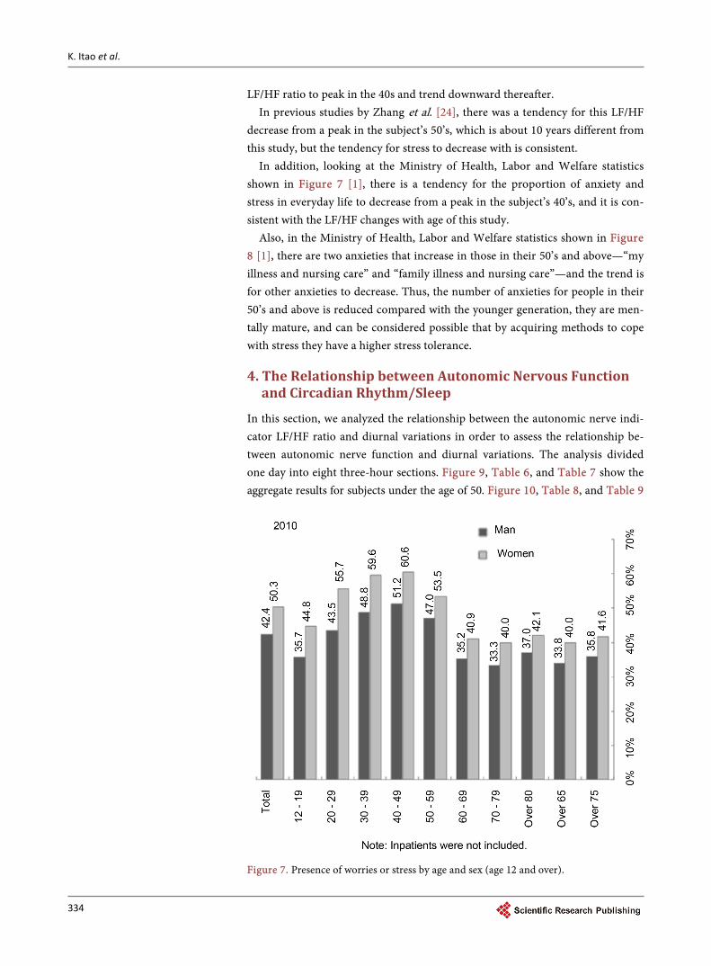
In addition, looking at the Ministry of Health, Labor and Welfare statistics shown in Figure 7 [1], there is a tendency for the proportion of anxiety and stress in everyday life to decrease from a peak in the subject’s 40’s, and it is con-sistent with the LF/HF changes with age of this study.
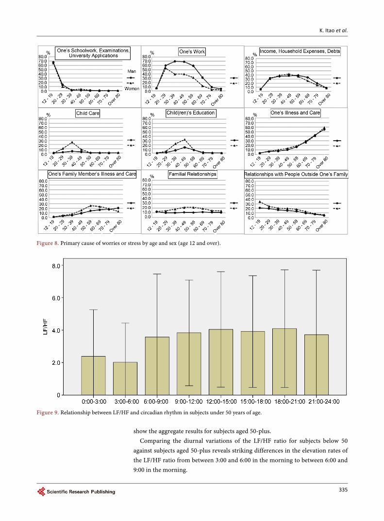
Also, in the Ministry of Health, Labor and Welfare statistics shown in Figure 8 [1], there are two anxieties that increase in those in their 50’s and above—“my illness and nursing care” and “family illness and nursing care”—and the trend is for other anxieties to decrease. Thus, the number of anxieties for people in their 50’s and above is reduced compared with the younger generation, they are men-tally mature, and can be considered possible that by acquiring methods to cope with stress they have a higher stress tolerance.
4. The Relationship between Autonomic Nervous Function and Circadian Rhythm/Sleep
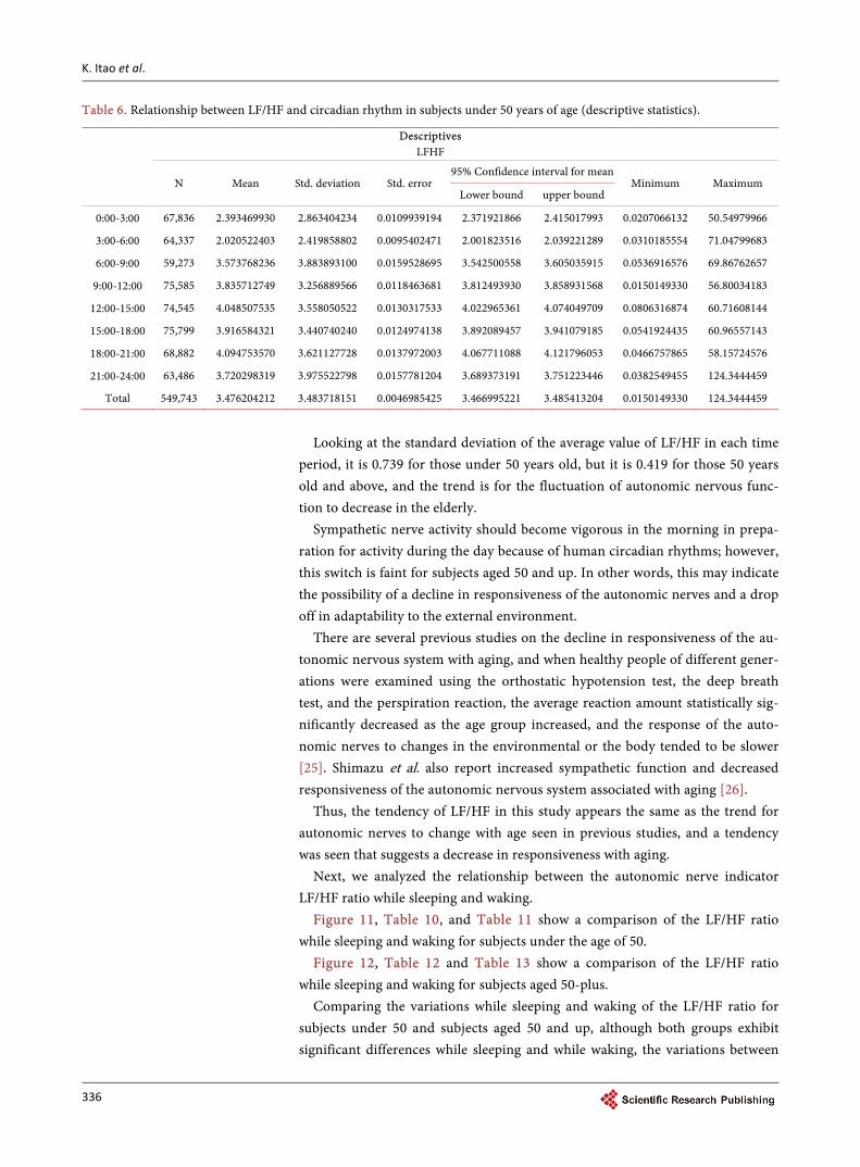
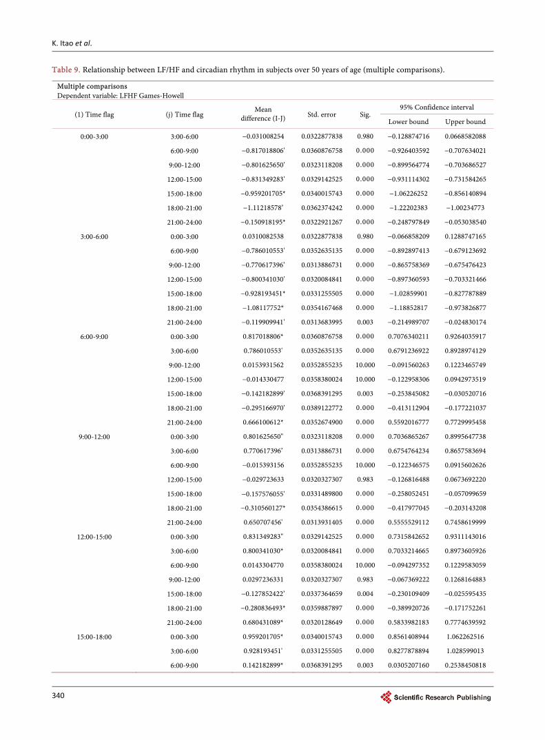
In this section, we analyzed the relationship between the autonomic nerve indi-cator LF/HF ratio and diurnal variations in order to assess the relationship be-tween autonomic nerve function and diurnal variations. The analysis divided one day into eight three-hour sections. Figure 9, Table 6, and Table 7 show the aggregate results for subjects under the age of 50. Figure 10, Table 8, and Table 9
Figure 7. Presence of worries or stress by age and sex (age 12 and over).
K. Itao et al.
335
Figure 8. Primary cause of worries or stress by age and sex (age 12 and over).
Figure 9. Relationship between LF/HF and circadian rhythm in subjects under 50 years of age.
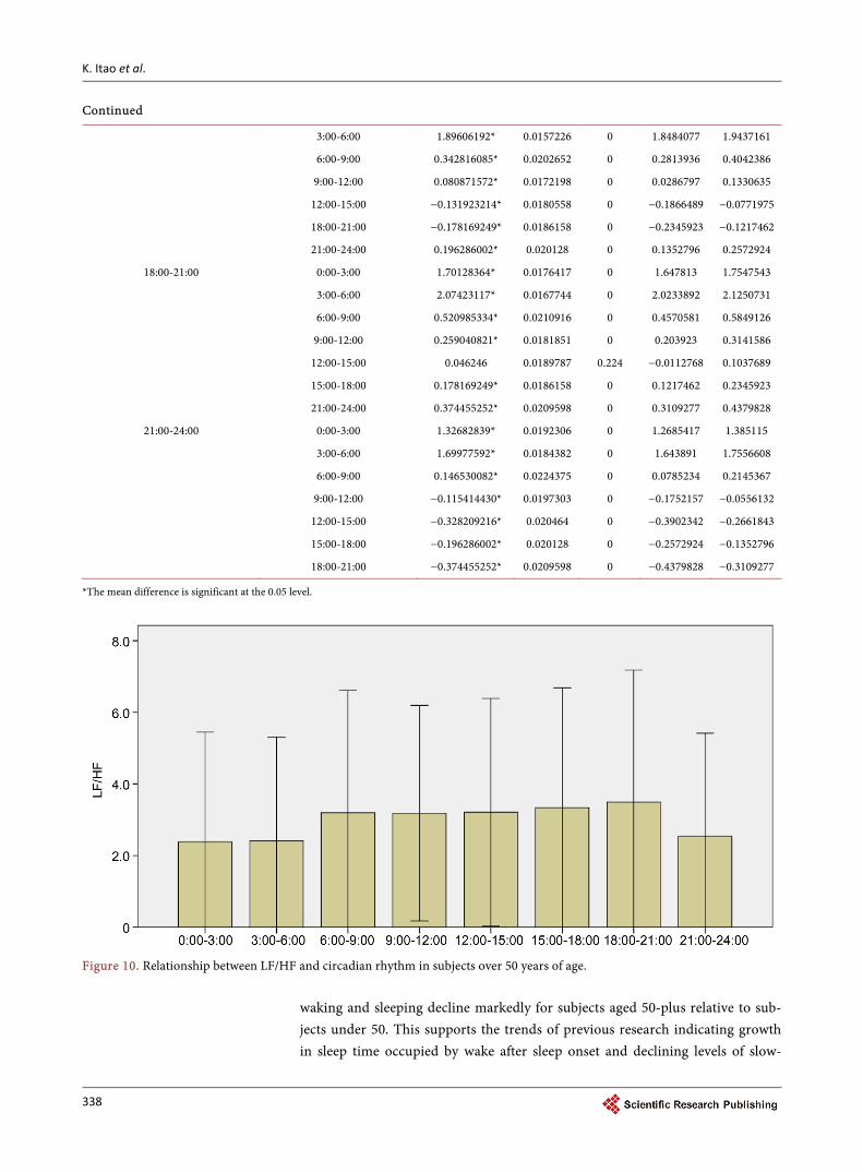
show the aggregate results for subjects aged 50-plus.
Comparing the diurnal variations of the LF/HF ratio for subjects below 50 against subjects aged 50-plus reveals striking differences in the elevation rates of the LF/HF ratio from between 3:00 and 6:00 in the morning to between 6:00 and 9:00 in the morning.
K. Itao et al.
336
Table 6. Relationship between LF/HF and circadian rhythm in subjects under 50 years of age (descriptive statistics).
Descriptives LFHF
N Mean Std. deviation Std. error 95% Confidence interval for mean
Minimum Maximum Lower bound upper bound
0:00-3:00 67,836 2.393469930 2.863404234 0.0109939194 2.371921866 2.415017993 0.0207066132 50.54979966
3:00-6:00 64,337 2.020522403 2.419858802 0.0095402471 2.001823516 2.039221289 0.0310185554 71.04799683
6:00-9:00 59,273 3.573768236 3.883893100 0.0159528695 3.542500558 3.605035915 0.0536916576 69.86762657
9:00-12:00 75,585 3.835712749 3.256889566 0.0118463681 3.812493930 3.858931568 0.0150149330 56.80034183
12:00-15:00 74,545 4.048507535 3.558050522 0.0130317533 4.022965361 4.074049709 0.0806316874 60.71608144
15:00-18:00 75,799 3.916584321 3.440740240 0.0124974138 3.892089457 3.941079185 0.0541924435 60.96557143
18:00-21:00 68,882 4.094753570 3.621127728 0.0137972003 4.067711088 4.121796053 0.0466757865 58.15724576
21:00-24:00 63,486 3.720298319 3.975522798 0.0157781204 3.689373191 3.751223446 0.0382549455 124.3444459
Total 549,743 3.476204212 3.483718151 0.0046985425 3.466995221 3.485413204 0.0150149330 124.3444459
Looking at the standard deviation of the average value of LF/HF in each time
period, it is 0.739 for those under 50 years old, but it is 0.419 for those 50 years old and above, and the trend is for the fluctuation of autonomic nervous func-tion to decrease in the elderly.
Sympathetic nerve activity should become vigorous in the morning in prepa-ration for activity during the day because of human circadian rhythms; however, this switch is faint for subjects aged 50 and up. In other words, this may indicate the possibility of a decline in responsiveness of the autonomic nerves and a drop off in adaptability to the external environment.
There are several previous studies on the decline in responsiveness of the au-tonomic nervous system with aging, and when healthy people of different gener-ations were examined using the orthostatic hypotension test, the deep breath test, and the perspiration reaction, the average reaction amount statistically sig-nificantly decreased as the age group increased, and the response of the auto-nomic nerves to changes in the environmental or the body tended to be slower [25]. Shimazu et al. also report increased sympathetic function and decreased responsiveness of the autonomic nervous system associated with aging [26].
Thus, the tendency of LF/HF in this study appears the same as the trend for autonomic nerves to change with age seen in previous studies, and a tendency was seen that suggests a decrease in responsiveness with aging.
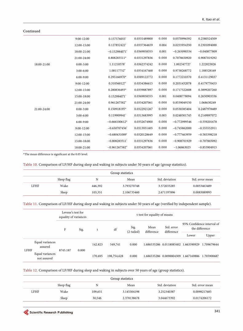
Next, we analyzed the relationship between the autonomic nerve indicator LF/HF ratio while sleeping and waking.
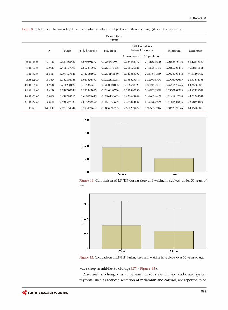
Figure 11, Table 10, and Table 11 show a comparison of the LF/HF ratio while sleeping and waking for subjects under the age of 50.
Figure 12, Table 12 and Table 13 show a comparison of the LF/HF ratio while sleeping and waking for subjects aged 50-plus.
Comparing the variations while sleeping and waking of the LF/HF ratio for subjects under 50 and subjects aged 50 and up, although both groups exhibit significant differences while sleeping and while waking, the variations between
K. Itao et al.
337
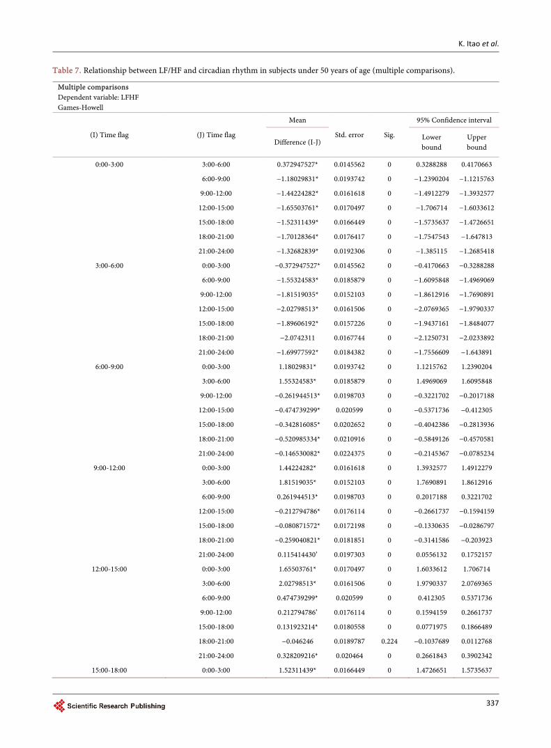
Table 7. Relationship between LF/HF and circadian rhythm in subjects under 50 years of age (multiple comparisons).
Multiple comparisons Dependent variable: LFHF Games-Howell
(I) Time flag (J) Time flag
Mean
Std. error Sig.
95% Confidence interval
Difference (I-J) Lower bound
Upper bound
0:00-3:00 3:00-6:00 0.372947527* 0.0145562 0 0.3288288 0.4170663
6:00-9:00 −1.18029831* 0.0193742 0 −1.2390204 −1.1215763
9:00-12:00 −1.44224282* 0.0161618 0 −1.4912279 −1.3932577
12:00-15:00 −1.65503761* 0.0170497 0 −1.706714 −1.6033612
15:00-18:00 −1.52311439* 0.0166449 0 −1.5735637 −1.4726651
18:00-21:00 −1.70128364* 0.0176417 0 −1.7547543 −1.647813
21:00-24:00 −1.32682839* 0.0192306 0 −1.385115 −1.2685418
3:00-6:00 0:00-3:00 −0.372947527* 0.0145562 0 −0.4170663 −0.3288288
6:00-9:00 −1.55324583* 0.0185879 0 −1.6095848 −1.4969069
9:00-12:00 −1.81519035* 0.0152103 0 −1.8612916 −1.7690891
12:00-15:00 −2.02798513* 0.0161506 0 −2.0769365 −1.9790337
15:00-18:00 −1.89606192* 0.0157226 0 −1.9437161 −1.8484077
18:00-21:00 −2.0742311 0.0167744 0 −2.1250731 −2.0233892
21:00-24:00 −1.69977592* 0.0184382 0 −1.7556609 −1.643891
6:00-9:00 0:00-3:00 1.18029831* 0.0193742 0 1.1215762 1.2390204
3:00-6:00 1.55324583* 0.0185879 0 1.4969069 1.6095848
9:00-12:00 −0.261944513* 0.0198703 0 −0.3221702 −0.2017188
12:00-15:00 −0.474739299* 0.020599 0 −0.5371736 −0.412305
15:00-18:00 −0.342816085* 0.0202652 0 −0.4042386 −0.2813936
18:00-21:00 −0.520985334* 0.0210916 0 −0.5849126 −0.4570581
21:00-24:00 −0.146530082* 0.0224375 0 −0.2145367 −0.0785234
9:00-12:00 0:00-3:00 1.44224282* 0.0161618 0 1.3932577 1.4912279
3:00-6:00 1.81519035* 0.0152103 0 1.7690891 1.8612916
6:00-9:00 0.261944513* 0.0198703 0 0.2017188 0.3221702
12:00-15:00 −0.212794786* 0.0176114 0 −0.2661737 −0.1594159
15:00-18:00 −0.080871572* 0.0172198 0 −0.1330635 −0.0286797
18:00-21:00 −0.259040821* 0.0181851 0 −0.3141586 −0.203923
21:00-24:00 0.115414430’ 0.0197303 0 0.0556132 0.1752157
12:00-15:00 0:00-3:00 1.65503761* 0.0170497 0 1.6033612 1.706714
3:00-6:00 2.02798513* 0.0161506 0 1.9790337 2.0769365
6:00-9:00 0.474739299* 0.020599 0 0.412305 0.5371736
9:00-12:00 0.212794786’ 0.0176114 0 0.1594159 0.2661737
15:00-18:00 0.131923214* 0.0180558 0 0.0771975 0.1866489
18:00-21:00 −0.046246 0.0189787 0.224 −0.1037689 0.0112768
21:00-24:00 0.328209216* 0.020464 0 0.2661843 0.3902342
15:00-18:00 0:00-3:00 1.52311439* 0.0166449 0 1.4726651 1.5735637
K. Itao et al.
338
Continued
3:00-6:00 1.89606192* 0.0157226 0 1.8484077 1.9437161
6:00-9:00 0.342816085* 0.0202652 0 0.2813936 0.4042386
9:00-12:00 0.080871572* 0.0172198 0 0.0286797 0.1330635
12:00-15:00 −0.131923214* 0.0180558 0 −0.1866489 −0.0771975
18:00-21:00 −0.178169249* 0.0186158 0 −0.2345923 −0.1217462
21:00-24:00 0.196286002* 0.020128 0 0.1352796 0.2572924
18:00-21:00 0:00-3:00 1.70128364* 0.0176417 0 1.647813 1.7547543
3:00-6:00 2.07423117* 0.0167744 0 2.0233892 2.1250731
6:00-9:00 0.520985334* 0.0210916 0 0.4570581 0.5849126
9:00-12:00 0.259040821* 0.0181851 0 0.203923 0.3141586
12:00-15:00 0.046246 0.0189787 0.224 −0.0112768 0.1037689
15:00-18:00 0.178169249* 0.0186158 0 0.1217462 0.2345923
21:00-24:00 0.374455252* 0.0209598 0 0.3109277 0.4379828
21:00-24:00 0:00-3:00 1.32682839* 0.0192306 0 1.2685417 1.385115
3:00-6:00 1.69977592* 0.0184382 0 1.643891 1.7556608
6:00-9:00 0.146530082* 0.0224375 0 0.0785234 0.2145367
9:00-12:00 −0.115414430* 0.0197303 0 −0.1752157 −0.0556132
12:00-15:00 −0.328209216* 0.020464 0 −0.3902342 −0.2661843
15:00-18:00 −0.196286002* 0.020128 0 −0.2572924 −0.1352796
18:00-21:00 −0.374455252* 0.0209598 0 −0.4379828 −0.3109277
*The mean difference is significant at the 0.05 level.
Figure 10. Relationship between LF/HF and circadian rhythm in subjects over 50 years of age.
waking and sleeping decline markedly for subjects aged 50-plus relative to sub-jects under 50. This supports the trends of previous research indicating growth in sleep time occupied by wake after sleep onset and declining levels of slow-
K. Itao et al.
339
Table 8. Relationship between LF/HF and circadian rhythm in subjects over 50 years of age (descriptive statistics).
Descriptives LFHF
N Mean Std. deviation Std. error 95% Confidence interval for mean Minimum Maximum
Lower bound Upper bound
0:00-3:00 17,108 2.380588839 3.069294877 0.0234659961 2.334593077 2.426584600 0.0052378176 51.12275387
3:00-6:00 17,066 2.411597093 2.897219037 0.0221776466 2.368126621 2.455067564 0.0085205484 40.38270518
6:00-9:00 15,535 3.197607645 3.417184907 0.0274165530 3.143868002 3.251347289 0.0078901472 49.81408403
9:00-12:00 18,385 3.182214489 3.011838897 0.0222126268 3.138675674 3.225753304 0.0316005655 31.97811159
12:00-15:00 18,928 3.211938122 3.175350653 0.0230801872 3.166698893 3.257177351 0.0651674696 64.45880871
15:00-18:00 18,440 3.339790544 3.341343045 0.0246059766 3.291560550 3.388020538 0.0520169243 44.92429550
18:00-21:00 17,843 3.492774616 3.688529619 0.0276133653 3.438649742 3.546899489 0.0141719790 44.61341598
21:00-24:00 16,892 2.531507033 2.883233297 0.0221839689 2.488024137 2.574989929 0.0100680885 43.78371076
Total 140,197 2.978154844 3.223821687 0.0086099703 2.961279472 2.995030216 0.0052378176 64.45880871
Figure 11. Comparison of LF /HF during sleep and waking in subjects under 50 years of age.
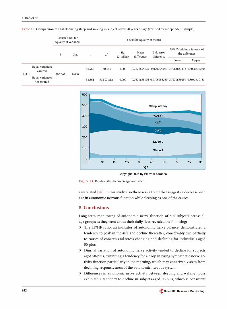
Figure 12. Comparison of LF/HF during sleep and waking in subjects over 50 years of age. wave sleep in middle- to old-age [27] (Figure 13).
Also, just as changes in autonomic nervous system and endocrine system rhythms, such as reduced secretion of melatonin and cortisol, are reported to be
K. Itao et al.
340
Table 9. Relationship between LF/HF and circadian rhythm in subjects over 50 years of age (multiple comparisons).
Multiple comparisons Dependent variable: LFHF Games-Howell
(1) Time flag (j) Time flag Mean
difference (I-J) Std. error Sig. 95% Confidence interval
Lower bound Upper bound
0:00-3:00 3:00-6:00 −0.031008254 0.0322877838 0.980 −0.128874716 0.0668582088
6:00-9:00 −0.817018806' 0.0360876758 0.000 −0.926403592 −0.707634021
9:00-12:00 −0.801625650' 0.0323118208 0.000 −0.899564774 −0.703686527
12:00-15:00 −0.831349283' 0.0329142525 0.000 −0.931114302 −0.731584265
15:00-18:00 −0.959201705* 0.0340015743 0.000 −1.06226252 −0.856140894
18:00-21:00 −1.11218578’ 0.0362374242 0.000 −1.22202383 −1.00234773
21:00-24:00 −0.150918195* 0.0322921267 0.000 −0.248797849 −0.053038540
3:00-6:00 0:00-3:00 0.0310082538 0.0322877838 0.980 −0.066858209 0.1288747165
6:00-9:00 −0.786010553' 0.0352635135 0.000 −0.892897413 −0.679123692
9:00-12:00 −0.770617396’ 0.0313886731 0.000 −0.865758369 −0.675476423
12:00-15:00 −0.800341030' 0.0320084841 0.000 −0.897360593 −0.703321466
15:00-18:00 −0.928193451* 0.0331255505 0.000 −1.02859901 −0.827787889
18:00-21:00 −1.08117752* 0.0354167468 0.000 −1.18852817 −0.973826877
21:00-24:00 −0.119909941' 0.0313683995 0.003 −0.214989707 −0.024830174
6:00-9:00 0:00-3:00 0.817018806* 0.0360876758 0.000 0.7076340211 0.9264035917
3:00-6:00 0.786010553' 0.0352635135 0.000 0.6791236922 0.8928974129
9:00-12:00 0.0153931562 0.0352855235 10.000 −0.091560263 0.1223465749
12:00-15:00 −0.014330477 0.0358380024 10.000 −0.122958306 0.0942973519
15:00-18:00 −0.142182899' 0.0368391295 0.003 −0.253845082 −0.030520716
18:00-21:00 −0.295166970' 0.0389122772 0.000 −0.413112904 −0.177221037
21:00-24:00 0.666100612* 0.0352674900 0.000 0.5592016777 0.7729995458
9:00-12:00 0:00-3:00 0.801625650" 0.0323118208 0.000 0.7036865267 0.8995647738
3:00-6:00 0.770617396’ 0.0313886731 0.000 0.6754764234 0.8657583694
6:00-9:00 −0.015393156 0.0352855235 10.000 −0.122346575 0.0915602626
12:00-15:00 −0.029723633 0.0320327307 0.983 −0.126816488 0.0673692220
15:00-18:00 −0.157576055' 0.0331489800 0.000 −0.258052451 −0.057099659
18:00-21:00 −0.310560127* 0.0354386615 0.000 −0.417977045 −0.203143208
21:00-24:00 0.650707456' 0.0313931405 0.000 0.5555529112 0.7458619999
12:00-15:00 0:00-3:00 0.831349283" 0.0329142525 0.000 0.7315842652 0.9311143016
3:00-6:00 0.800341030* 0.0320084841 0.000 0.7033214665 0.8973605926
6:00-9:00 0.0143304770 0.0358380024 10.000 −0.094297352 0.1229583059
9:00-12:00 0.0297236331 0.0320327307 0.983 −0.067369222 0.1268164883
15:00-18:00 −0.127852422’ 0.0337364659 0.004 −0.230109409 −0.025595435
18:00-21:00 −0.280836493* 0.0359887897 0.000 −0.389920726 −0.171752261
21:00-24:00 0.680431089* 0.0320128649 0.000 0.5833982183 0.7774639592
15:00-18:00 0:00-3:00 0.959201705* 0.0340015743 0.000 0.8561408944 1.062262516
3:00-6:00 0.928193451' 0.0331255505 0.000 0.8277878894 1.028599013
6:00-9:00 0.142182899* 0.0368391295 0.003 0.0305207160 0.2538450818
K. Itao et al.
341
Continued
9:00-12:00 0.157576055' 0.0331489800 0.000 0.0570996592 0.2580524509
12:00-15:00 0.127852422’ 0.0337364659 0.004 0.0255954350 0.2301094088
18:00-21:00 −0.152984071' 0.0369858355 0.001 −0.265090334 −0.040877809
21:00-24:00 0.808283511* 0.0331297836 0.000 0.7078650920 0.9087019292
18:00-21:00 0:00-3:00 1.11218578’ 0.0362374242 0.000 1.002347727 1.222023826
3:00-6:00 1.08117752" 0.0354167468 0.000 0.9738268772 1.188528169
6:00-9:00 0.295166970* 0.0389122772 0.000 0.1772210370 0.4131129037
9:00-12:00 0.310560127' 0.0354386615 0.000 0.2031432078 0.4179770453
12:00-15:00 0.280836493* 0.0359887897 0.000 0.1717522608 0.3899207260
15:00-18:00 0.152984071' 0.0369858355 0.001 0.0408778094 0.2650903336
21:00-24:00 0.961267582" 0.0354207061 0.000 0.8539049150 1.068630249
21:00-24:00 0:00-3:00 0.150918195* 0.0322921267 0.000 0.0530385404 0.2487978489
3:00-6:00 0.119909941' 0.0313683995 0.003 0.0248301745 0.2149897072
6:00-9:00 −0.666100612* 0.0352674900 0.000 −0.772999546 −0.559201678
9:00-12:00 −0.650707456' 0.0313931405 0.000 −0.745862000 −0.555552911
12:00-15:00 −0.680431089' 0.0320128649 0.000 −0.777463959 −0.583398218
15:00-18:00 −0.808283511' 0.0331297836 0.000 −0.908701929 −0.707865092
18:00-21:00 −0.961267582" 0.0354207061 0.000 −1.06863025 −0.853904915
*The mean difference is significant at the 0.05 level.
Table 10. Comparison of LF/HF during sleep and waking in subjects under 50 years of age (group statistics).
Group statistics
Sleep flag N Mean Std. deviation Std. error mean
LFHF Wake 446,392 3.793270748 3.572033285 0.0053463489
Sleep 103,351 2.106735460 2.671197896 0.0083089993
Table 11. Comparison of LF/HF during sleep and waking in subjects under 50 years of age (verified by independent sample).
Levene’s test for equality of variances
t-test for equality of means
F Sig. t df Sig.
(2-tailed) Mean
difference Std. error difference
95% Confidence interval of the difference
Lower Upper
LFHF
Equal variances assured
8745.187 0.000
142.823 549,741 0.000 1.686535286 0.0118085402 1.663390929 1.709679644
Equal variances not assured
170.695 198,754.628 0.000 1.686535286 0.0098804309 1.667169886 1.705900687
Table 12. Comparison of LF/HF during sleep and waking in subjects over 50 years of age (group statistics).
Group statistics
Sleep flag N Mean Std. deviation Std. error mean
LFHF Wake 109,651 3.145304198 3.252340387 0.0098217685
Sleep 30,546 2.378138678 3.044673392 0.0174206172
K. Itao et al.
342
Table 13. Comparison of LF/HF during sleep and waking in subjects over 50 years of age (verified by independent sample).
Levene’s test for equality of variances
t-test for equality of means
F Sig. t df Sig.
(2-tailed) Mean
difference Std. error difference
95% Confidence interval of the difference
Lower Upper
LFHF
Equal variances assured
380.367 0.000
36.960 140,195 0.000 0.7671655196 0.020756365 0.7264833132 0.8078477260
Equal variances not assured
38.361 51,597.812 0.000 0.7671655196 0.0199986260 0.7279680259 0.8063630133
Figure 13. Relationship between age and sleep. age-related [28], in this study also there was a trend that suggests a decrease with age in autonomic nervous function while sleeping as one of the causes.
5. Conclusions
Long-term monitoring of autonomic nerve function of 600 subjects across all age groups as they went about their daily lives revealed the following: The LF/HF ratio, an indicator of autonomic nerve balance, demonstrated a
tendency to peak in the 40’s and decline thereafter, conceivably due partially to causes of concern and stress changing and declining for individuals aged 50-plus.
Diurnal variation of autonomic nerve activity tended to decline for subjects aged 50-plus, exhibiting a tendency for a drop in rising sympathetic nerve ac-tivity function particularly in the morning, which may conceivably stem from declining responsiveness of the autonomic nervous system.
Differences in autonomic nerve activity between sleeping and waking hours exhibited a tendency to decline in subjects aged 50-plus, which is consistent
K. Itao et al.
343
with the tendency for rising levels of wake after sleep onset and declining le-vels of slow-wave sleep in middle- to old-age.
Moving forward, we hope to examine what kind of differences emerge in these trends as well as see how the conclusions reached herein change as the result of adding correlational analysis with other biological indicators such as saliva and brain waves.
References [1] Health, Labour, and Welfare Ministry (Japan) (2010) Review of National Livelihood
Survey: The State of Stress and Concerns.
[2] Onaka, T. (2005) Stress and Its Neural Mechanisms. Journal of Pharmacological Sciences, 126, 170-173.
[3] Robertson, D. (2007) Primer on the Autonomic Nervous System. Takahashi, A. and Mano, T., Trans., Elsevier, Japan.
[4] Beck, A.T., Ward, C.H., Mendelson, M., et al. (1961) An Inventory for Measuring Depression. Archives of General Psychiatry, 4, 561-571. https://doi.org/10.1001/archpsyc.1961.01710120031004
[5] Samejima, K., Matsushita, K. and Matsumoto, K. (1976) An Evaluation of Explicit Anxiety in Depressives and Healthy Subjects (MAS) and Clinical Study of the Beck Depression Inventory (BDI). Japanese Society of Psychosomatic Medicine, 311-319.
[6] Zung, W.W.K. (1965) A Self-Rating Depression Scale. Archives of General Psychia-try, 12, 63-70. https://doi.org/10.1001/archpsyc.1965.01720310065008
[7] Fukuda, K. and Kobayashi, S. (1973) An Evaluation of Self-Rating Depression Scales. Official Journal of the Japanese Society of Psychiatry and Neurology, 75, 673-679.
[8] Radloff, L.S. (1977) The CES-D Scale: A Self-Report Depression Scale for Research in the General Population. Applied Psychological Measurement, 1, 385-401. https://doi.org/10.1177/014662167700100306
[9] Shima, S. and Shiga, N., Kitamura, T., et al. (1985) New Self-Rating Depression Scales. Japanese Society of Psychosomatic Medicine, 27, 717-723.
[10] Yamaguchi, M. (2007) Measuring Stress with Salivary Markers. Journal of Pharma-cological Sciences, 129, 80-84.
[11] Schiepers, O.J., Wichers, M.C. and Maes, M. (2005) Cytokines and Major Depres-sion. Progress in Neuro-Psychopharmacology & Biological Psychiatry, 29, 201-217. https://doi.org/10.1016/j.pnpbp.2004.11.003
[12] Motonori, I. and Tomoyuki, Y. (2000) The Relationship between Brain Wave Fluc-tuation and Reaction Times with the Decrease in Arousal Level. Ergonomics, 36, 229-237.
[13] Association between Images of Mental State and Heart-Rate Variability (2005) As-sessment of Depression Severity by Power Spectral Analysis; Toshihiro Fujioka (Keiaikai Jozan Hospital, Department of Psychiatry), Yumiko Mori (0288-9250). Autonomic Nerves, 42, 191-192.
[14] Sugaya, J., Fukuma, N., Ushijima, A., Kato, Y., Aisu, T., Tsuchida, T., Takahashi, K., Kishida, H. and Mizuno, K. (2009) Relationship between Depression and Prognostic Factors in Cases of Mild Heart Attack. The Journal of the Japanese Coronary Asso-ciation, 15, 198-201.
[15] Ohira, T., Diez Roux, A.V., Prineas, R.J., et al. (2008) Associations of Psychosocial Factors with Heart Rate and Its Short-Term Variability: The Multi-Ethnic Study of
K. Itao et al.
344
Atherosclerosis. Psychosomatic Medicine, 70, 141-146. https://doi.org/10.1097/PSY.0b013e318160686a
[16] Yukishita, T., Lee, K., Kim, S., Yumoto, Y., Kobayashi, A., Shirasawa, T. and Ko-bayashi, H. (2010) Age and Sex-Dependent Alterations in Heart Rate Variability: Profiling the Characteristics of Men and Women in Their 30s. Anti-Aging Medi-cine, 7, 94-100. https://doi.org/10.3793/jaam.7.94
[17] Task Force of the European Society of Cardiology and the North American Society of Pacing and Electrophysiology (1996) Heart Rate Variability: Standards of Mea-surement, Physiological Interpretation, and Clinical Use. Circulation, 93, 1043- 1065. https://doi.org/10.1161/01.CIR.93.5.1043
[18] Kuratsune, H. (2011) Establishment of Objective Methods for Fatigue Diagnosis and Creation of Policies for the Diagnosis of Chronic Fatigue for Patients Com-plaining of Chronic Fatigue Associated with Autonomic Nerve Irregularities Health Labor Sciences Integrated Research Center for the Disabled (Mental Disability/ Nerve & Muscle Division) Integrated Research Reports from 2009-2011. 1-114.
[19] Oguma, Y., Yamamoto, S., Kinoshita, N., Katsukawa, F., Onishi, S. and Yamazaki, H. (1999) Fundamental Examination of Assessing 1 Body Activity and Intensity of Body Activity Using a Triaxial Accelerometer Simultaneously Recording Heart- Rate. Keio University Sports Medicine Research Center Bulletin, 25-31.
[20] Matsumura, Y., Yamamoto, M., Kitado, M., Nakamura, H., Hondera, K. and Fuji-moto, S. (2008) High-Precision Body Monitoring Using Triaxial Acceleration Sen-sor. Matsushita Denko Giho, 56, 60-66.
[21] (2007) Triaxial Acceleration Sensor Application Notes. Hokuriku Electric Works Co.
[22] Pomeranz, B., Macaulay, R.J., Caudill, M.A., Kutz, I., Adam, D., Gordon, D., Kil-born, K.M., Barger, A.C., Shannon, D.C. and Cohen, R.J. (1985) Assessment of Au-tonomic Function in Humans by Heart Rate Spectral Analysis. American Journal of Physiology, 248, H151-H153.
[23] Hayano, J., Sakakibara, Y., Yamada, M., Kamiya, T., Fujinami, T., Yokoyama, K., Watanabe, Y. and Takata, K. (1991) Accuracy of Assessment of Cardiac Vagal Tone by Heart Rate Variability in Normal Subjects. American Journal of Cardiology, 15, 199-204.
[24] Zhang, J. (2007) Effect of Age and Sex on Heart Rate Variability in Healthy Subjects. Journal of Manipulative and Physiological Therapeutics, 30, 374-379. https://doi.org/10.1016/j.jmpt.2007.04.001
[25] Parashar, R., Amir, M., Pakhare, A., Rathi, P. and Chaudhary, L. (2014) Age Related Changes in Autonomic Functions. Journal of Clinical and Diagnostic Research, 10, CC11.
[26] Shimazu, T., et al. (2005) Aging of the Autonomic Nervous System. Nippon Rinsho, 63, 973-977.
[27] (2005) Principles and Practice of Sleep Medicine. 4th Edition, Elsevier, Amsterdam.
[28] Van Coevorden, A., Mockel, J., Laurent, E., Kerkhofs, M., L’Hermite-Baleriaux, M., Decoster, C., et al. (1991) Neuroendocrine Rhythms and Sleep in Aging Men. Ame- rican Journal of Physiology, 260, E651-E661.

Submit or recommend next manuscript to SCIRP and we will provide best service for you:
Accepting pre-submission inquiries through Email, Facebook, LinkedIn, Twitter, etc. A wide selection of journals (inclusive of 9 subjects, more than 200 journals) Providing 24-hour high-quality service User-friendly online submission system Fair and swift peer-review system Efficient typesetting and proofreading procedure Display of the result of downloads and visits, as well as the number of cited articles Maximum dissemination of your research work
Submit your manuscript at: http://papersubmission.scirp.org/ Or contact [email protected]