LipopolysaccharidePrimestheNALP3Inflammasomeby...
11
Lipopolysaccharide Primes the NALP3 Inflammasome by Inhibiting Its Ubiquitination and Degradation Mediated by the SCF FBXL2 E3 Ligase * Received for publication, February 13, 2015, and in revised form, May 3, 2015 Published, JBC Papers in Press, June 2, 2015, DOI 10.1074/jbc.M115.645549 SeungHye Han ‡ , Travis B. Lear ‡ , Jacob A. Jerome ‡ , Shristi Rajbhandari ‡ , Courtney A. Snavely ‡ , Dexter L. Gulick ‡ , Kevin F. Gibson ‡ , Chunbin Zou ‡ , Bill B. Chen ‡1 , and Rama K. Mallampalli ‡§¶1,2 From the ‡ Department of Medicine, Acute Lung Injury Center of Excellence, and § Department of Cell Biology, Physiology, and Bioengineering, University of Pittsburgh, Pittsburgh, Pennsylvania 15213 and the ¶ Medical Specialty Service Line, Veterans Affairs Pittsburgh Healthcare System, Pittsburgh, Pennsylvania 15240 Background: LPS increases NALP3 levels, but the mechanisms remain unknown. Results: LPS prolongs the lifespan of NALP3 protein by reducing E3 ligase (SCF FBXL2 )-mediated ubiquitination. Conclusion: Proinflammatory cytokine release is reduced by a small molecule that restores cellular SCF FBXL2 levels. Significance: We identified a novel pathway of inflammasome priming that may serve as a springboard for future translational studies. The inflammasome is a multiprotein complex that augments the proinflammatory response by increasing the generation and cellular release of key cytokines. Specifically, the NALP3 inflam- masome requires two-step signaling, priming and activation, to be functional to release the proinflammatory cytokines IL-1 and IL-18. The priming process, through unknown mecha- nisms, increases the protein levels of NALP3 and pro-IL-1 in cells. Here we show that LPS increases the NALP3 protein life- span without significantly altering steady-state mRNA in human cells. LPS exposure reduces the ubiquitin-mediated pro- teasomal processing of NALP3 by inducing levels of an E3 ligase component, FBXO3, which targets FBXL2. The latter is an endogenous mediator of NALP3 degradation. FBXL2 recog- nizes Trp-73 within NALP3 for interaction and targets Lys-689 within NALP3 for ubiquitin ligation and degradation. A unique small molecule inhibitor of FBXO3 restores FBXL2 levels, resulting in decreased NALP3 protein levels in cells and, thereby, reducing the release of IL-1 and IL-18 in human inflammatory cells after NALP3 activation. Our findings uncover NALP3 as a molecular target for FBXL2 and suggest that therapeutic targeting of the inflammasome may serve as a platform for preclinical intervention. The inflammasome is a large multiprotein cytosolic complex that consists of three components: a sensor (NACHT, LRR, and PYD domain-containing protein (NALP)), 3 an adaptor (apopto- sis-associated speck-like protein containing a carboxy-terminal caspase-recruitment domain (ASC)), and an effector (pro- caspase 1). Inflammasomes have emerged as key complexes that, when assembled properly, result in the release of IL-1 and IL-18, which partake in illnesses characterized by severe inflammation. Some inflammasomes are activated by pore- forming toxins, ATP, or hypoxic cellular injury when these con- ditions are primed with microbial ligands or endogenous cyto- kines (1). When assembled, inflammasomes cleave and activate the potent proinflammatory cytokines IL-1 and IL-18, which are associated with a higher risk and worse outcomes of disor- ders such as acute respiratory distress syndrome (2, 3). NALP3, the most studied inflammasome so far, requires two-step sig- naling to engage in cytokine release: priming and activation. The Gram-negative bacterial endotoxin LPS is known to prime the NALP3 inflammasome by increasing NALP3 protein and pro-IL-1 levels in a dose- and time-dependent fashion in mice (4). However, the molecular mechanisms whereby LPS priming increases NALP3 protein abundance in cells remain elusive. This question is critical because the identification of molecular targets that transcriptionally or posttranslationally mediate increased NALP3 concentrations in cells could be important in pharmacotherapeutic designs to control innate immune responses. Protein ubiquitination is a highly conserved, irreversible, posttranslational modification among vertebrates that governs cellular protein concentrations. The ubiquitin proteasome sys- tem selectively targets proteins in humans by a series of enzy- matic steps requiring a ubiquitin-activating enzyme (E1), a * This work was performed on the basis of work supported, in part, by the Department of Veterans Affairs, Veterans Health Administration, Office of Research and Development, Biomedical Laboratory Research and Devel- opment. This work was supported, in whole or in part, by National Insti- tutes of Health R01 Grants HL096376, HL097376, HL098174, HL081784, 1UH2HL123502, P01HL114453 (to R. K. M.), and HL116472 (to B. B. C.). This work was also supported by a merit review award from the Department of Veterans Affairs, the Flight Attendant Medical Research Institute, and American Heart Association Award 12SDG12040330 (to C. Z.). The authors declare that they have no conflicts of interest with the contents of this article. 1 Both authors contributed equally to this work. 2 To whom correspondence should be addressed: University of Pittsburgh, Pulmonary, Allergy, and Critical Care Medicine, UPMC Montefiore, NW 628, Dept. of Medicine, Pittsburgh, PA 15213. Tel.: 412-624-8900; Fax: 412-692- 2260; E-mail: [email protected]. 3 The abbreviations used are: NALP, NACHT, LRR and PYD domain-containing protein; ASC, apoptosis-associated speck-like protein containing a car- boxy-terminal caspase recruitment domain; SCF, Skp-Cullin-F box; TRAF, TNF receptor-associated factor; CHX, cycloheximide; MLE, mouse lung epi- thelial; NLR, Nod-like receptor. THE JOURNAL OF BIOLOGICAL CHEMISTRY VOL. 290, NO. 29, pp. 18124 –18133, July 17, 2015 Published in the U.S.A. 18124 JOURNAL OF BIOLOGICAL CHEMISTRY VOLUME 290 • NUMBER 29 • JULY 17, 2015 by guest on April 11, 2020 http://www.jbc.org/ Downloaded from
Transcript of LipopolysaccharidePrimestheNALP3Inflammasomeby...
Lipopolysaccharide Primes the NALP3 Inflammasome byInhibiting Its Ubiquitination and Degradation Mediatedby the SCFFBXL2 E3 Ligase*
Received for publication, February 13, 2015, and in revised form, May 3, 2015 Published, JBC Papers in Press, June 2, 2015, DOI 10.1074/jbc.M115.645549
SeungHye Han‡, Travis B. Lear‡, Jacob A. Jerome‡, Shristi Rajbhandari‡, Courtney A. Snavely‡, Dexter L. Gulick‡,Kevin F. Gibson‡, Chunbin Zou‡, Bill B. Chen‡1, and Rama K. Mallampalli‡§¶1,2
From the ‡Department of Medicine, Acute Lung Injury Center of Excellence, and §Department of Cell Biology, Physiology, andBioengineering, University of Pittsburgh, Pittsburgh, Pennsylvania 15213 and the ¶Medical Specialty Service Line, Veterans AffairsPittsburgh Healthcare System, Pittsburgh, Pennsylvania 15240
Background: LPS increases NALP3 levels, but the mechanisms remain unknown.Results: LPS prolongs the lifespan of NALP3 protein by reducing E3 ligase (SCFFBXL2)-mediated ubiquitination.Conclusion: Proinflammatory cytokine release is reduced by a small molecule that restores cellular SCFFBXL2 levels.Significance: We identified a novel pathway of inflammasome priming that may serve as a springboard for future translationalstudies.
The inflammasome is a multiprotein complex that augmentsthe proinflammatory response by increasing the generation andcellular release of key cytokines. Specifically, the NALP3 inflam-masome requires two-step signaling, priming and activation, tobe functional to release the proinflammatory cytokines IL-1�
and IL-18. The priming process, through unknown mecha-nisms, increases the protein levels of NALP3 and pro-IL-1� incells. Here we show that LPS increases the NALP3 protein life-span without significantly altering steady-state mRNA inhuman cells. LPS exposure reduces the ubiquitin-mediated pro-teasomal processing of NALP3 by inducing levels of an E3 ligasecomponent, FBXO3, which targets FBXL2. The latter is anendogenous mediator of NALP3 degradation. FBXL2 recog-nizes Trp-73 within NALP3 for interaction and targets Lys-689within NALP3 for ubiquitin ligation and degradation. A uniquesmall molecule inhibitor of FBXO3 restores FBXL2 levels,resulting in decreased NALP3 protein levels in cells and,thereby, reducing the release of IL-1� and IL-18 in humaninflammatory cells after NALP3 activation. Our findingsuncover NALP3 as a molecular target for FBXL2 and suggestthat therapeutic targeting of the inflammasome may serve as aplatform for preclinical intervention.
The inflammasome is a large multiprotein cytosolic complexthat consists of three components: a sensor (NACHT, LRR, andPYD domain-containing protein (NALP)),3 an adaptor (apopto-sis-associated speck-like protein containing a carboxy-terminalcaspase-recruitment domain (ASC)), and an effector (pro-caspase 1). Inflammasomes have emerged as key complexesthat, when assembled properly, result in the release of IL-1�and IL-18, which partake in illnesses characterized by severeinflammation. Some inflammasomes are activated by pore-forming toxins, ATP, or hypoxic cellular injury when these con-ditions are primed with microbial ligands or endogenous cyto-kines (1). When assembled, inflammasomes cleave and activatethe potent proinflammatory cytokines IL-1� and IL-18, whichare associated with a higher risk and worse outcomes of disor-ders such as acute respiratory distress syndrome (2, 3). NALP3,the most studied inflammasome so far, requires two-step sig-naling to engage in cytokine release: priming and activation.The Gram-negative bacterial endotoxin LPS is known to primethe NALP3 inflammasome by increasing NALP3 protein andpro-IL-1� levels in a dose- and time-dependent fashion in mice(4). However, the molecular mechanisms whereby LPS primingincreases NALP3 protein abundance in cells remain elusive.This question is critical because the identification of moleculartargets that transcriptionally or posttranslationally mediateincreased NALP3 concentrations in cells could be importantin pharmacotherapeutic designs to control innate immuneresponses.
Protein ubiquitination is a highly conserved, irreversible,posttranslational modification among vertebrates that governscellular protein concentrations. The ubiquitin proteasome sys-tem selectively targets proteins in humans by a series of enzy-matic steps requiring a ubiquitin-activating enzyme (E1), a
* This work was performed on the basis of work supported, in part, by theDepartment of Veterans Affairs, Veterans Health Administration, Office ofResearch and Development, Biomedical Laboratory Research and Devel-opment. This work was supported, in whole or in part, by National Insti-tutes of Health R01 Grants HL096376, HL097376, HL098174, HL081784,1UH2HL123502, P01HL114453 (to R. K. M.), and HL116472 (to B. B. C.). Thiswork was also supported by a merit review award from the Department ofVeterans Affairs, the Flight Attendant Medical Research Institute, andAmerican Heart Association Award 12SDG12040330 (to C. Z.). The authorsdeclare that they have no conflicts of interest with the contents of thisarticle.
1 Both authors contributed equally to this work.2 To whom correspondence should be addressed: University of Pittsburgh,
Pulmonary, Allergy, and Critical Care Medicine, UPMC Montefiore, NW 628,Dept. of Medicine, Pittsburgh, PA 15213. Tel.: 412-624-8900; Fax: 412-692-2260; E-mail: [email protected].
3 The abbreviations used are: NALP, NACHT, LRR and PYD domain-containingprotein; ASC, apoptosis-associated speck-like protein containing a car-boxy-terminal caspase recruitment domain; SCF, Skp-Cullin-F box; TRAF,TNF receptor-associated factor; CHX, cycloheximide; MLE, mouse lung epi-thelial; NLR, Nod-like receptor.
THE JOURNAL OF BIOLOGICAL CHEMISTRY VOL. 290, NO. 29, pp. 18124 –18133, July 17, 2015Published in the U.S.A.
18124 JOURNAL OF BIOLOGICAL CHEMISTRY VOLUME 290 • NUMBER 29 • JULY 17, 2015
by guest on April 11, 2020
http://ww
w.jbc.org/
Dow
nloaded from
ubiquitin-conjugating enzyme (E2), and a ubiquitin E3 ligase.E3 ligases are multisubunit complexes that confer a relativelyhigh specificity to ubiquitination by bringing in close proximitya substrate to an E2 enzyme that facilitates the transfer of anactivated ubiquitin moiety from the E2 to the substrate (5).Hundreds of human genes encode RING domain-containingE3 ligases, and, among these, the Skp-Cullin-F box (SCF) familyis among the best studied (6). The SCF superfamily regulatesdiverse biological events, including cell growth, proliferation,DNA repair, and inflammation (7).
We have demonstrated previously that an SCF subunit, F boxprotein, F-box O3 (FBXO3), is a proinflammatory moleculethat is activated in leukocytes by endotoxin (8). FBXO3 ubiq-uitinates and mediates degradation of another F box protein,F-box L2 (FBXL2). FBXL2 is an anti-inflammatory moleculethat mediates the degradation of TNF receptor-associated fac-tors (TRAFs), resulting in a reduction of inflammatory cytokinerelease. The aim of our study is to investigate how LPS primingincreases NALP3 inflammasome protein levels. Here we showthat NALP3 is also a target for FBXL2. Moreover, we demon-strate a unique mechanism whereby LPS priming decreasesFBXL2-mediated NALP3 ubiquitination and degradation byactivating the FBXL2 inhibitor FBXO3 in cells. A small-mole-cule FBXO3 antagonist, BC-1215, potently reduces the severityof NALP3-mediated inflammation by increasing FBXL2 pro-tein levels, therefore antagonizing actions of endotoxin on theinflammasomes.
Experimental Procedures
Antibodies and Reagents—Antibodies against FBXL8, anti-mouse IgG, and anti-rabbit IgG were obtained from Santa CruzBiotechnology (Santa Cruz, CA). Antibodies against FBXL2were from Aviva Systems Biology (San Diego, CA). Antibodiesagainst F-box L7 (FBXL7) and NALP6 were obtained fromAbcam (Cambridge, MA). Ubiquitin and IL-1� antibodies werepurchased from Cell Signaling Technology (Danvers, MA).NALP3 antibodies were obtained from Adipogen (San Diego,CA). GAPDH and �-actin antibodies were obtained fromSigma. IL-18 human antibodies were purchased from MBL(Woburn, MA). QuikChange II site-directed mutagenesis kitswere purchased from Agilent Technologies (Santa Clara, CA).Pierce protein A/G-agarose beads were purchased fromThermo Scientific (Rockford, IL). Cycloheximide (CHX),MG132, and leupeptin were from Calbiochem (Darmstadt,Germany). Mouse V5 antibodies, pcDNA3.1D cloning kits, andthe pENTR directional TOPO cloning kits were purchasedfrom Invitrogen. ATP was obtained from Sigma. Miniprep spinkits were obtained from Qiagen (Valencia, CA) and Nucleo-bond Xtra Maxi prep kits were obtained from Macherey-Nagel(Düren, Germany). In vitro transcription and translation (TNT)kits were from Promega. BC-1215 was synthesized to �98%purity.
Cell Culture—Human monocyte U937 and THP1 cells werepurchased from Sigma-Aldrich (St. Louis, MO). Human eryth-roleukemia K562 cells, human bronchial epithelial A549 cells,Beas2B cells, mouse lung epithelial (MLE-12) cells, and humancervical epithelial HeLa cells were obtained from the ATCC.Human primary macrophages and their culture medium were
purchased from Celprogen (San Pedro, CA). A bronchoscopywas performed to collect bronchoalveolar lavage fluid afterobtaining informed consent. Human primary alveolar macro-phages from the fluid were cultured in culture medium (Cel-progen) as described previously (9). The study was approved bythe University of Pittsburgh Institutional Review Board.DMEM and RPMI 1640 medium were obtained from Gibco(Life Technologies) and supplemented with fetal bovine serum(FBS) from Gemini (Sacramento, CA). MLE cells were culturedin HITES (hydrocortisone, insulin, transferrin, estrogen, andselenium) medium supplemented with 10% FBS. Prior to CHXtreatment, cells were starved for 1 h with 0% FBS HITESmedium. U937, THP1, and K562 cells were cultured in RPMI 1640medium supplemented with 10% FBS. A549 and Beas2B cells werecultured in DMEM with 10% FBS. HeLa cells were cultured inEagle’s minimum essential medium (Gibco) supplemented with10% FBS. CHX treatment was carried out at a concentration of 40�g/ml at varying time points in 0% FBS medium with or withoutMG132 (20 �g/ml) or leupeptin (20 �g/ml).
Immunoprecipitation and Immunoblotting—Cells were lysedwith lysis buffer (0.05% Triton X-100 in PBS and 1:1000 prote-ase inhibitor mixture) and sonicated. After centrifugation at13,200 rpm for 10 min, cell lysates (containing 2 mg of protein)were incubated and rotated with 5 �g of anti-ubiquitin, anti-NALP3, or FBXL2 antibody at 4 °C for 3 h and then incu-bated with 30 �l of protein A/G-agarose beads overnight.The next day, the beads were spun down and washed withlysis buffer three times. The washed beads were mixed withSDS-PAGE loading dye prior to SDS-PAGE and immunoblotanalysis. Immunoblotting was performed as described previ-ously (10). We precipitated extracellular media with trichlo-roacetic acid and subsequently measured cytokine levels byimmunoblotting.
RT-PCR—RNA was isolated from cells using RNeasy minikits (Qiagen) according to the protocol provided. IsolatedRNAs were immediately converted to cDNA using high-capac-ity RNA-to-cDNA kits (Life Technologies) after their concen-trations were measured. Real-time PCR assays were performedusing SYBR� Select Master Mix for CFX (2�, Life Technolo-gies) per the protocol provided in C1000 Thermal Cycler(Bio-Rad).
Cloning and Mutagenesis—Human NALP3 cDNA wascloned into a pcDNA3.1D/V5-His vector. Site-directed mu-tagenesis of lysine to arginine residues was carried out via aPCR-based approach using the QuikChange II XL kit from Agi-lent Technologies and appropriate primers. Primers used forlysine to arginine mutagenesis were as follows: K689R for-ward (5�-CATAACATGCCCAGGGAGGAAGAGGAG-3�) andreverse (5�-CTCCTCTTCCTCCCTGGGCATGTTATG-3�);K696R forward (5�-GAAGAGGAGGAGGAAAGGGAAGGC-CGACACCTTG-3�) and reverse (5�-CAAGGTGTCGGCCT-TCCCTTTCCTCCTCCTCTTC-3�); K86R forward (5�-GAG-AGACCTTTATGAGAGAGCAAAAAGAGATGAG-3�) andreverse (5�-CTCATCTCTTTTTGCTCTCTCATAAAGGTCT-CTC-3�); and K742R forward (5�-GGTCCTCAGCAGCAACCA-GAGGCTGGTGGAGCTGGACCTGAG-3�) and reverse(5�-CTCATCTCTTTTTGCTCTCTCATAAAGGTCTCTC-3�).Generated mutants were sequence-confirmed (10).
Lipopolysaccharide Stabilizes the NALP3 Inflammasome
JULY 17, 2015 • VOLUME 290 • NUMBER 29 JOURNAL OF BIOLOGICAL CHEMISTRY 18125
by guest on April 11, 2020
http://ww
w.jbc.org/
Dow
nloaded from
Transfection—2.5 � 106 MLE cells were suspended in 100 �lof 20 mM HEPES and mixed with 4 �g of DNA in a cuvette. Cellswere nucleofected on the T-013 protocol using an AmaxaNucleofector II device (Basel, Switzerland). Immediately fol-lowing nucleofection, 0.5 ml of 10% HITES medium was addedto the cuvette, and the cell solution was plated in 1.5 ml of 10%HITES culture medium in a 6-well plate. Cells were allowed togrow until 50% confluency prior to half-life experiments(48 –72 h). For HeLa cells, the PolyJetTM DNA in vitro transfec-tion reagent was used following the protocols of SignaGen(Rockville, MD). Cells were then treated with 40 �g/ml of CHXat different time points for half-life studies and were harvestedin lysis buffer using a rubber policeman for subsequent immu-noblot analysis. siRNA to FBXL2, purchased from IntegratedDNA Technologies (Coralville, IA), was introduced into pri-mary human macrophages using GenMuteTM siRNA transfec-tion reagent based on protocols from SignaGen’s.
In Vitro Ubiquitination Conjugation Assay—The details ofthis assay have been described previously (11). Briefly, the ubiq-uitination of NALP3 was performed in a volume of 25 �l con-taining 50 mM Tris (pH 7.6); 5 mM MgCl2; 0.6 mM DTT; 2 mM
ATP; 1.5 ng/�l E1; 10 ng/�l Ubc5; 10 ng/�l Ubc7; 1 �g/�lubiquitin (Calbiochem); 1 �M ubiquitin aldehyde; 4 –16 �l ofpurified Cullin1; Skp1, Rbx1, and in vitro-synthesized FBXL2.Reaction products were processed for NALP3 immunoblotting.
In Vitro Protein Binding Assays—FBXL2 protein was immu-noprecipitated from 1 mg of cell lysate using FBXL2 antibodyand coupled to protein A/G-agarose resin. FBXL2 beads werethen incubated with in vitro-synthesized products (50 �l)expressing NALP3-V5 mutants. After washing, the proteinswere eluted and processed for NALP3-V5 immunoblotting.
ELISA—Quantikine ELISA kits were purchased from R&DSystems (Minneapolis, MN) to measure human IL-1� andhuman IL-18 levels. The quantitative assays were performedfollowing protocols provided by the manufacturer using cellculture supernatants after LPS/ATP exposure with or withoutBC-1215.
Statistical Analysis—Descriptive statistics were reported asmean � S.D. or S.E. as indicated. As our sample sizes of eachexperimental group were all less than 10, which limits the abil-ity to study a normal sample distribution, we employed appro-priate non-parametric methods such as a Mann-Whitney Utest and a Kruskal-Wallis equality of populations rank test tocompare multiple groups within and between experiments.Also, a Wilcoxon-type test for trend was used where appropri-ate to check trends in data significance. Using these methodsprovided a conservative analysis to determine statistical signif-icance. All analyses were performed using Stata Statistical Soft-ware, Release 10.1 (StataCorp 2007, College Station, TX).
Results
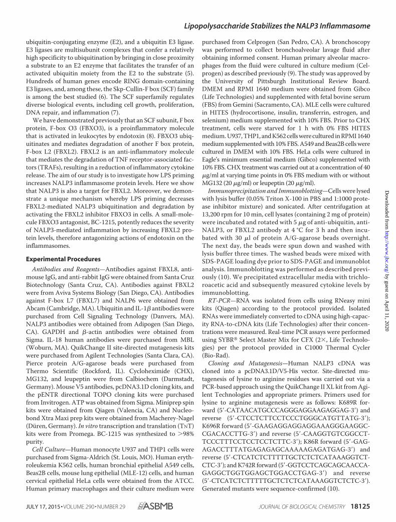
LPS Priming Up-regulates Steady-state NALP3 Protein Levelsby Prolonging Its Half-life—Human monocyte U937 cellsexpress NALP3, as evidenced by immunoblotting with anNALP3 antibody. NALP3 protein levels increased in a dose-and time-dependent fashion with LPS exposure, whereasNALP6, another NOD (nucleotide-binding oligomerizationdomain)-like receptor, did not (Fig. 1, A and B). There weremodest, if any, increases in mRNA expression of NALP3 withLPS exposure (Fig. 2, A and B), and the changes were not sta-tistically significant. These data suggest that LPS up-regulatesNALP3 protein mainly through posttranslational mechanisms.Therefore, we measured the half-life of NALP3 protein both atbaseline and after LPS exposure by blocking new protein syn-thesis with CHX. The half-life of NALP3 is �4 h in an unchal-lenged condition and prolonged to �6 h after LPS exposure(Fig. 2, C and D). NALP3 degradation was reduced with inclu-sion of MG132 in the medium but not with a lysosomal inhib-itor, leupeptin, indicating that the degradation of NALP3occurs via the ubiquitin proteasome system rather than the lys-osome (Fig. 2, E and F).
FBXL2 Mediates NALP3 Ubiquitination and Degradation,an Effect Inhibited by LPS—To confirm that NALP3 is degradedthrough the ubiquitin proteasome system, we transfected MLE
FIGURE 1. LPS priming increases NALP3 protein in human monocyte U937 cells. A and B, immunoblots for NALP3, NALP6 (negative control), and �-actin inlysates from human monocytes, U937 cells (total 3 � 106 cells), treated with LPS for 16 h as indicated (A) or treated with LPS (200 ng/ml) for the indicated periodsof time (B). The relative densitometries of NALP3 protein for each immunoblot are shown in the bottom panel. Data are mean � S.E. of three to five independentexperiments. The p values were determined by a nonparametric test for trend.
Lipopolysaccharide Stabilizes the NALP3 Inflammasome
18126 JOURNAL OF BIOLOGICAL CHEMISTRY VOLUME 290 • NUMBER 29 • JULY 17, 2015
by guest on April 11, 2020
http://ww
w.jbc.org/
Dow
nloaded from

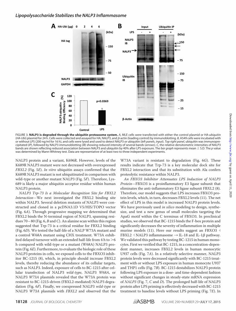
cells with either control plasmids or HA-ubiquitin plasmids.High-level ubiquitin cellular expression correlated with lowerlevels of NALP3 protein in cells (Fig. 3A). NALP3-ubiquitinbinding, measured by coimmunoprecipitation, was decreasedby 40% after LPS exposure when densitometrically controlledfor input loading (Fig. 3, B and C). Therefore, NALP3 is ubiq-uitinated, an effect reduced by LPS.
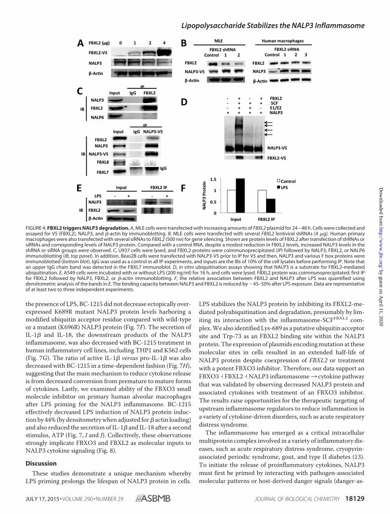
Because our prior studies have demonstrated that FBXO3targets FBXL2 for its disposal in cells and that an FBXO3 inhib-itor reduces IL-1� levels after LPS treatment (8), we tested thehypothesis that FBXL2 targets NALP3 to mediate its degrada-tion. Indeed, the ubiquitination of NALP3 is increased afterexpression of FBXL2. As shown in Fig. 4A, ectopic expressionwith the FBXL2 plasmid in MLE cells decreases levels ofNALP3. Furthermore, NALP3 protein levels in both MLE cellsand primary human macrophages were increased by using oneof the several lentivirally delivered shRNAs or siRNAs to FBXL2(Fig. 4B). To determine whether FBXL2 interacts with NALP3,we performed coimmunoprecipitation studies showing thatFBXL2 binds NALP3 selectively because it does not associatewith the NALP6 inflammasome (Fig. 4C). When V5-NALP3was overexpressed in cells, in coimmunoprecipitation studies itwas associated with FBXL2 but not other F-box proteins,FBXL7 or FBXL8. In vitro ubiquitin assays demonstrated thatFBXL2 robustly catalyzes NALP3 ubiquitination (Fig. 4D). Thebinding between NALP3 and FBXL2 was also decreased afterLPS exposure, as was NALP3 ubiquitination (Fig. 4, E and F).
These observations suggest that NALP3 is a molecular targetfor FBXL2-mediated degradation, a process impaired byendotoxin.
Lys-689 is a NALP3 Ubiquitination Acceptor Site—Proteinubiquitination involves the generation of an isopeptide bondbetween an acceptor lysine on a target protein (e.g. NALP3) andthe C-terminal glycine of the incoming ubiquitin moiety.Human NALP3 protein has 68 Lys residues. Using the UbPredsoftware (12), potential ubiquitination sites in NALP3 proteinwere predicted among the Lys residues: one with high proba-bility (Lys-689), five with medium probability (Lys-93, Lys-192,Lys-324, Lys-430, and Lys-696), and six residues with low prob-ability (Lys-86, Lys-194, Lys-437, Lys-510, Lys-742, and Lys-958) (Fig. 5A). We cloned the human NALP3 cDNA into apcDNA3.1D/V5-His vector and generated mutants substitut-ing arginine at each of the predicted Lys residues within theNALP3 protein. MLE cells were transfected with wild-type andmutant plasmids and were exposed to CHX to measure thehalf-life of ectopically expressed NALP3 (Fig. 5B). The half-lifeof wild-type ectopically expressed NALP3 is 4 –5 h, similar tothat of endogenous NALP3. The K689R NALP3 mutant has anextended half-life (�6 h) in cells compared with wild-typeNALP3, resembling the effects of LPS exposure on wild-typeNALP3 (Figs. 2C and 5, B and C). Other expressed mutants didnot exhibit a significantly prolonged lifespan in cells (Fig. 5D).Cotransfection of NALP3 mutant plasmids with or withoutFBXL2 plasmid decreased ectopically expressed wild-type
FIGURE 2. LPS priming increases NALP3 protein levels by prolonging its half-life. Box plots of messenger RNA expression –fold increase of NALP3 in LPSprimed human monocytes, U937 cells. A and B, cells were primed with LPS for 16 h as indicated (A) or with LPS (200 ng/ml) for the indicated periods of time (B).Data are from four independent experiments. The p values were determined by a Kruskal-Wallis test. C, U937 cells were incubated with CHX (40 �g/ml) forvarious times with or without LPS priming (200 ng/ml for 16 h). D, densitometric plot of NALP3 protein decay versus time of CHX exposure with a best fit line.The half-life was �4 h in an unchallenged condition and �6 h after LPS priming. E, U937 cells were incubated with CHX (40 �g/ml) and MG132 (20 �g/ml) orleupeptin (20 �g/ml), and immunoblotting for NALP3 was performed. F, decay curves over time in the presence of MG132 or leupeptin were best fitted. Dataare mean � S.E. of five to eight independent experiments. The p values were determined by a Mann-Whitney test comparing two groups at each time point.*, p � 0.05; **, p � 0.01.
Lipopolysaccharide Stabilizes the NALP3 Inflammasome
JULY 17, 2015 • VOLUME 290 • NUMBER 29 JOURNAL OF BIOLOGICAL CHEMISTRY 18127
by guest on April 11, 2020
http://ww
w.jbc.org/
Dow
nloaded from
NALP3 protein and a variant, K696R. However, levels of theK689R NALP3 mutant were not decreased with overexpressedFBXL2 (Fig. 5E). In vitro ubiquitin assays confirmed that theK689R NALP3 mutant is not ubiquitinated in comparison withwild-type or another mutant NALP3 (Fig. 5F). Therefore, Lys-689 is likely a major ubiquitin acceptor residue within humanNALP3 protein.
NALP3 Trp-73 Is a Molecular Recognition Site for FBXL2Interaction—We next investigated the FBXL2 binding sitewithin NALP3. Several deletion mutants of NALP3 were con-structed and cloned in a pcDNA3.1D V5/HIS/TOPO vector(Fig. 6A). Through progressive mapping we determined thatFBXL2 binds the N-terminal region of NALP3, spanning resi-dues 70 – 80 (Fig. 6, B and C). An alanine scan within this regionsuggested that Trp-73 is a critical residue for FBXL2 binding(Fig. 6D). We tested the half-life of a NALP W73A mutant anda control W68A mutant using CHX treatment. W73A exhib-ited delayed turnover with an extended half-life from 4 h to �6h compared with wild-type or a mutant (W68A) NALP3 pro-tein (Fig. 6E). Furthermore, to evaluate the biologic role of theseNALP3 proteins in cells, we exposed cells to the FBXO3 inhib-itor BC-1215 (8), which, in principle should increase FBXL2levels, thereby reducing the abundance of its cellular targets,such as NALP3. Indeed, exposure of cells to BC-1215 after cel-lular transfection of NALP3 wild-type, NALP3 W68A, orNALP3 W73A plasmids revealed that the W73A protein wasresistant to BC-1215-driven (FBXL2-mediated) NALP3 degra-dation (Fig. 6F). Finally, we coexpressed NALP3 wild-type orNALP3 W73A plasmids with FBXL2 and observed that the
W73A variant is resistant to degradation (Fig. 6G). Theseresults indicate that Trp-73 is a key molecular dock site forFBXL2 interaction and that its substitution with Ala confersproteolytic resistance within NALP3.
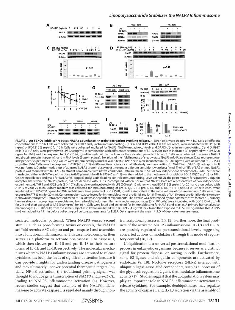
An FBXO3 Inhibitor Attenuates LPS Induction of NALP3Protein—FBXO3 is a proinflammatory E3 ligase subunit thateliminates the anti-inflammatory E3 ligase subunit FBXL2 (8).Therefore, our model suggests that LPS increases FBXO3 pro-tein levels, which, in turn, decreases FBXL2 levels (11). The neteffect of LPS in this model is increased NALP3 protein levels.We have previously used in silico modeling to design, synthe-size, and test a new genus of small molecules targeting theApaG motif within the C terminus of FBXO3. In preclinicalstudies, we observed that BC-1215 binds the F-box protein andsignificantly decreases the severity of inflammation in multiplemurine models (11). Here our results suggest an FBXO3 �
FBXL2 � NALP3 inflammasome3 IL-18 and IL-1� pathway.We validated this pathway by testing BC-1215 in human mono-cytes. First we verified that BC-1215, in a concentration-depen-dent manner, increases FBXL2 levels in human monocyticU937 cells (Fig. 7A). In a relatively selective manner, NALP3protein levels were decreased significantly with BC-1215 treat-ment with or without LPS exposure in human monocyte U937and THP1 cells (Fig. 7B). BC-1215 destabilizes NALP3 proteinfollowing LPS exposure in a dose- and time-dependent fashionwithout significant changes in steady-state mRNA expressionof NALP3 (Fig. 7, C and D). The prolonged half-life of NALP3protein after LPS priming is effectively decreased with BC-1215treatment to baseline levels without LPS priming (Fig. 7E). In
FIGURE 3. NALP3 is degraded through the ubiquitin proteasome system. A, MLE cells were transfected with either the control plasmid or HA-ubiquitin(HA-Ubi) plasmid for 24 h. Cells were collected and assayed for HA, NALP3, and �-actin (loading control) by immunoblotting. B, A549 cells were incubated withor without LPS (200 ng/ml for 16 h), and cells were lysed and used to detect NALP3 or ubiquitin (left panels, input). Top right panel, ubiquitin was immunopre-cipitated (IP), followed by NALP3 immunoblotting (IB) showing reduced intensity of several bands (arrows). C, the relative densitometric intensities of NALP3bands are shown reflecting reduced association between NALP3 and ubiquitin by 40% after LPS exposure. The bar graph represents mean � S.D. The p valuewas determined by Mann-Whitney test. Data are representative of at least two to three independent experiments.
Lipopolysaccharide Stabilizes the NALP3 Inflammasome
18128 JOURNAL OF BIOLOGICAL CHEMISTRY VOLUME 290 • NUMBER 29 • JULY 17, 2015
by guest on April 11, 2020
http://ww
w.jbc.org/
Dow
nloaded from
the presence of LPS, BC-1215 did not decrease ectopically over-expressed K689R mutant NALP3 protein levels harboring amodified ubiquitin acceptor residue compared with wild-typeor a mutant (K696R) NALP3 protein (Fig. 7F). The secretion ofIL-1� and IL-18, the downstream products of the NALP3inflammasome, was also decreased with BC-1215 treatment inhuman inflammatory cell lines, including THP1 and K562 cells(Fig. 7G). The ratio of active IL-1� versus pro-IL-1� was alsodecreased with BC-1215 in a time-dependent fashion (Fig. 7H),suggesting that the main mechanism to reduce cytokine releaseis from decreased conversion from premature to mature formsof cytokines. Lastly, we examined ability of the FBXO3 smallmolecule inhibitor on primary human alveolar macrophagesafter LPS priming for the NALP3 inflammasome. BC-1215effectively decreased LPS induction of NALP3 protein induc-tion by 44% (by densitometry when adjusted for � actin loading)and also reduced the secretion of IL-1� and IL-18 after a secondstimulus, ATP (Fig. 7, I and J). Collectively, these observationsstrongly implicate FBXO3 and FBXL2 as molecular inputs toNALP3 cytokine signaling (Fig. 8).
Discussion
These studies demonstrate a unique mechanism wherebyLPS priming prolongs the lifespan of NALP3 protein in cells.
LPS stabilizes the NALP3 protein by inhibiting its FBXL2-me-diated polyubiquitination and degradation, presumably by lim-iting its interaction with the inflammasome-SCFFBXL2 com-plex. We also identified Lys-689 as a putative ubiquitin acceptorsite and Trp-73 as an FBXL2 binding site within the NALP3protein. The expression of plasmids encoding mutation at thesemolecular sites in cells resulted in an extended half-life ofNALP3 protein despite coexpression of FBXL2 or treatmentwith a potent FBXO3 inhibitor. Therefore, our data support anFBXO3 � FBXL2 � NALP3 inflammasome3 cytokine pathwaythat was validated by observing decreased NALP3 protein andassociated cytokines with treatment of an FBXO3 inhibitor.The results raise opportunities for the therapeutic targeting ofupstream inflammasome regulators to reduce inflammation ina variety of cytokine-driven disorders, such as acute respiratorydistress syndrome.
The inflammasome has emerged as a critical intracellularmultiprotein complex involved in a variety of inflammatory dis-eases, such as acute respiratory distress syndrome, cryopyrin-associated periodic syndrome, gout, and type II diabetes (13).To initiate the release of proinflammatory cytokines, NALP3must first be primed by interacting with pathogen-associatedmolecular patterns or host-derived danger signals (danger-as-
FIGURE 4. FBXL2 triggers NALP3 degradation. A, MLE cells were transfected with increasing amounts of FBXL2 plasmid for 24 – 48 h. Cells were collected andassayed for V5 (FBXL2), NALP3, and �-actin by immunoblotting. B, MLE cells were transfected with several FBXL2 lentiviral shRNAs (4 �g). Human primarymacrophages were also transfected with several siRNAs to FBXL2 (500 nM) for gene silencing. Shown are protein levels of FBXL2 after transfection of shRNAs orsiRNAs and corresponding levels of NALP3 protein. Compared with a control RNA, despite a modest reduction in FBXL2 levels, increased NALP3 levels in theshRNA or siRNA groups were observed. C, U937 cells were lysed, and FBXL2 proteins were coimmunoprecipitated (IP) followed by NALP3, FBXL2, or NALP6immunoblotting (IB, top panel). In addition, Beas2B cells were transfected with NALP3-V5 prior to IP for V5 and then, NALP3 and various F box proteins wereimmunoblotted (bottom blot). IgG was used as a control in all IP experiments, and inputs are the IBs of 10% of the cell lysates before performing IP. Note thatan upper IgG chain band was detected in the FBXL7 immunoblot. D, in vitro ubiquitination assays showing that NALP3 is a substrate for FBXL2-mediatedubiquitination. E, A549 cells were incubated with or without LPS (200 ng/ml) for 16 h, and cells were lysed. FBXL2 protein was coimmunoprecipitated; first IPfor FBXL2 followed by NALP3, FBXL2, or �-actin immunoblotting. F, the relative association between FBXL2 and NALP3 after LPS was quantified usingdensitometric analysis of the bands in E. The binding capacity between NALP3 and FBXL2 is reduced by �45–50% after LPS exposure. Data are representativeof at least two to three independent experiments.
Lipopolysaccharide Stabilizes the NALP3 Inflammasome
JULY 17, 2015 • VOLUME 290 • NUMBER 29 JOURNAL OF BIOLOGICAL CHEMISTRY 18129
by guest on April 11, 2020
http://ww
w.jbc.org/
Dow
nloaded from
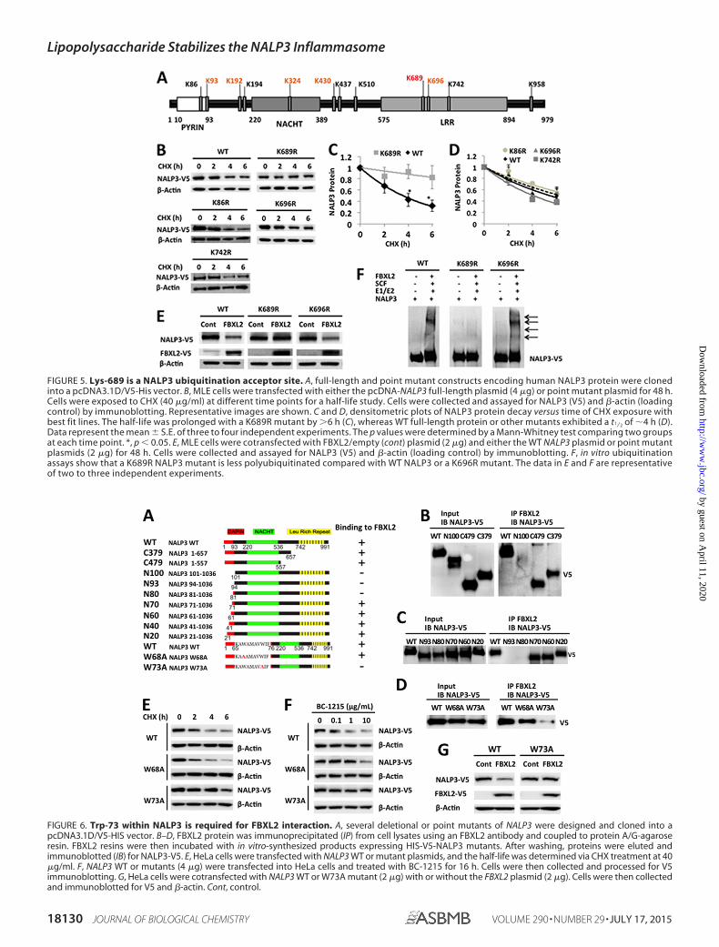
FIGURE 6. Trp-73 within NALP3 is required for FBXL2 interaction. A, several deletional or point mutants of NALP3 were designed and cloned into apcDNA3.1D/V5-HIS vector. B–D, FBXL2 protein was immunoprecipitated (IP) from cell lysates using an FBXL2 antibody and coupled to protein A/G-agaroseresin. FBXL2 resins were then incubated with in vitro-synthesized products expressing HIS-V5-NALP3 mutants. After washing, proteins were eluted andimmunoblotted (IB) for NALP3-V5. E, HeLa cells were transfected with NALP3 WT or mutant plasmids, and the half-life was determined via CHX treatment at 40�g/ml. F, NALP3 WT or mutants (4 �g) were transfected into HeLa cells and treated with BC-1215 for 16 h. Cells were then collected and processed for V5immunoblotting. G, HeLa cells were cotransfected with NALP3 WT or W73A mutant (2 �g) with or without the FBXL2 plasmid (2 �g). Cells were then collectedand immunoblotted for V5 and �-actin. Cont, control.
FIGURE 5. Lys-689 is a NALP3 ubiquitination acceptor site. A, full-length and point mutant constructs encoding human NALP3 protein were clonedinto a pcDNA3.1D/V5-His vector. B, MLE cells were transfected with either the pcDNA-NALP3 full-length plasmid (4 �g) or point mutant plasmid for 48 h.Cells were exposed to CHX (40 �g/ml) at different time points for a half-life study. Cells were collected and assayed for NALP3 (V5) and �-actin (loadingcontrol) by immunoblotting. Representative images are shown. C and D, densitometric plots of NALP3 protein decay versus time of CHX exposure withbest fit lines. The half-life was prolonged with a K689R mutant by �6 h (C), whereas WT full-length protein or other mutants exhibited a t1⁄2 of �4 h (D).Data represent the mean � S.E. of three to four independent experiments. The p values were determined by a Mann-Whitney test comparing two groupsat each time point. *, p � 0.05. E, MLE cells were cotransfected with FBXL2/empty (cont) plasmid (2 �g) and either the WT NALP3 plasmid or point mutantplasmids (2 �g) for 48 h. Cells were collected and assayed for NALP3 (V5) and �-actin (loading control) by immunoblotting. F, in vitro ubiquitinationassays show that a K689R NALP3 mutant is less polyubiquitinated compared with WT NALP3 or a K696R mutant. The data in E and F are representativeof two to three independent experiments.
Lipopolysaccharide Stabilizes the NALP3 Inflammasome
18130 JOURNAL OF BIOLOGICAL CHEMISTRY VOLUME 290 • NUMBER 29 • JULY 17, 2015
by guest on April 11, 2020
http://ww
w.jbc.org/
Dow
nloaded from
sociated molecular patterns). When NALP3 senses secondstimuli, such as pore-forming toxins or crystals, the NALP3scaffold recruits ASC adaptor and pro-caspase 1 and assemblesinto a functional inflammasome. This assembled complex thenserves as a platform to activate pro-caspase 1 to caspase 1,which then cleaves pro-IL-1� and pro-IL-18 to their matureforms of IL-1� and IL-18, respectively. The molecular mecha-nisms whereby NALP3 inflammasomes are activated to releasecytokines has been the focus of significant attention because itcan provide insights for understanding disease pathogenesisand may ultimately uncover potential therapeutic targets. Ini-tially, NF-�B activation, the traditional priming signal, wasthought to induce gene transcription of NALP3 and pro-IL-1�,leading to NALP3 inflammasome activation (4). However,recent studies suggest that assembly of the NALP3 inflam-masome to activate caspase 1 is regulated mainly through non-
transcriptional processes (14, 15). Furthermore, the final prod-ucts of the activated NALP3 inflammasome, IL-1� and IL-18,are possibly regulated at posttranslational levels, suggestingconcerted actions of modulators through this mode of regula-tory control (16, 17).
Ubiquitination is a universal posttranslational modificationprocess in eukaryotic organisms because it serves as a distinctsignal for protein disposal or sorting in cells. Furthermore,some E3 ligases and ubiquitin components are activated byendotoxin (8, 18). Nod-like receptors (NLRs) interact withubiquitin ligase-associated components, such as suppressor ofthe glycolysis regulation 2 gene, that modulate inflammasomeactivity (19). Studies suggest that the ubiquitination system mayplay an important role in NALP3 inflammasome activation torelease cytokines. For example, deubiquitinases may regulatethe activity of caspase 1 and IL-1� secretion via the assembly of
FIGURE 7. An FBXO3 inhibitor reduces NALP3 abundance, thereby decreasing cytokine release. A, U937 cells were treated with BC-1215 at differentconcentrations for 16 h. Cells were collected for FBXL2 and �-actin immunoblotting. B, U937 and THP1 cells (3 � 106 cells each) were incubated with LPS (200ng/ml) or BC-1215 (8 �g/ml) for 16 h. Cells were collected and lysed for NALP3, NALP6 (negative control), and GAPDH/�-actin immunoblotting. C and D, U937cells (3 � 106 cells) were primed with LPS (200 ng/ml) in combination with different concentrations of BC-1215 for 16 h as indicated (C) or primed with LPS (200ng/ml for 16 h) and then exposed to BC-1215 (4 �g/ml) in fresh culture medium for the indicated periods of time (D). Cells were collected to measure NALP3and �-actin protein (top panels) and mRNA levels (bottom panels). Box plots of the -fold increase of steady-state NALP3 mRNA are shown. Data represent fourindependent experiments. The p values were determined by a Kruskal-Wallis test. E, U937 cells were incubated in LPS (200 ng/ml) with or without BC-1215 (4�g/ml for 16 h). Cells were then exposed to CHX (40 �g/ml) at different time points for a half-life study. Immunoblotting for NALP3 and GAPDH (loading control)was performed. Densitometric plots of adjusted NALP3 protein decay over time under different conditions were best fitted. The half-life of LPS-primed NALP3protein was reduced with BC-1215 treatment comparable with native conditions. Data are mean � S.E. of two independent experiments. F, MLE cells weretransfected either with WT or point mutant NALP3 plasmids for 48 h. LPS (40 �g/ml) was then added to the medium with or without BC-1215 (20 �g/ml) for 18 h.Cells were collected and lysed for NALP3 (V5-tagged) and �-actin (loading control) immunoblotting. Levels of K689R, the point mutant for a putative ubiquitinacceptor site within the NALP3 protein, did not decrease with BC-1215 compared with WT or mutant NALP3. Data are representative of two independentexperiments. G, THP1 and K562 cells (3 � 106 cells each) were incubated with LPS (200 ng/ml) with or without BC-1215 (8 �g/ml) for 20 h and then pulsed withATP (5 mM for 20 min). Culture medium was collected for immunoblotting of pro-IL-1�, IL-1�, pro-IL-18, and IL-18. H, THP1 cells (3 � 106 cells each) wereincubated with LPS (200 ng/ml) for 20 h and different time periods of BC-1215 (8 �g/ml), as indicated, in the same volume of culture medium. Cells were thenexposed to ATP (5 mM for 20 min). Culture medium was collected for immunoblotting of pro-IL-1� and IL-1�. The ratio of IL-1� versus pro-IL-1� by densitometryis shown (bottom panel). Data represent mean � S.D. of two independent experiments. The p value was determined by nonparametric test for trend. I, primaryhuman alveolar macrophages were obtained from a healthy volunteer. Human alveolar macrophages (3 � 105 cells) were incubated with BC-1215 (4 �g/ml)for 2 h and then exposed to LPS (100 ng/ml) for 16 h. Cells were lysed and collected for immunoblotting for NALP3 and � actin. J, primary human alveolarmacrophages (3 � 105 cells) from the same subject as in I were incubated with BC-1215 (4 �g/ml) for 2 h and then exposed to LPS (100 ng/ml) for 16 h. ATP (5mM) was added for 15 min before collecting cell culture supernatants for ELISA. Data represent the mean � S.D. of duplicate measurements.
Lipopolysaccharide Stabilizes the NALP3 Inflammasome
JULY 17, 2015 • VOLUME 290 • NUMBER 29 JOURNAL OF BIOLOGICAL CHEMISTRY 18131
by guest on April 11, 2020
http://ww
w.jbc.org/
Dow
nloaded from
inflammasomes (20), although the level of BRCC3, one of thedeubiquitinases targeting NALP3, does not correlate with theabundance of NALP3 protein after LPS exposure (21). An E3ligase, tripartite motif 33, and the linear ubiquitin assemblycomplex also seem to be involved with NALP3 inflammasomeactivation (22, 23). While preparing this manuscript, dopaminewas reported to inhibit NALP3 inflammasome activation viathe dopamine D1 receptor by promoting actions of the E3 ubiq-uitin ligase, MARCH7 (24). However, to date, studies have onlyfocused on the link between ubiquitination and the terminalsteps of NALP3 inflammasome activation: assembly of theinflammasome complex and subsequent release of cytokines.Despite evidence that LPS priming increases NALP3 proteinlevels, the precise molecular mechanisms elucidating how LPSpriming up-regulates NALP3 protein levels and whether cyto-kine release can be altered by manipulating this priming pro-cess remain unclear. Here we describe a mechanism by whichLPS primes the NALP3 inflammasome by impairing its associ-ation with a previously unrecognized NALP3 inhibitor, FBXL2,which, in turn, extends the inflammasome lifespan in cells. Weidentified that FBXL2 interacts with Trp-73, facilitating ubiq-uitin ligation to Lys-689 within the NALP3 protein. Interest-ingly, a tryptophan within TRAFs 1– 6 also serves as a criticaldocking site for FBXL2 interaction (8). Therefore, tryptophan
may be a common recognition site, along with other motifs, forFBXL2 recruitment to its substrates.
Previously, the unique, selective, small-molecule FBXO3inhibitor BC-1215 has been shown to block IL-1� secretiontriggered by LPS (11). The inhibition of FBXO3 resulted in anincrease in FBXL2 cellular mass, promoting the degradation ofNALP3 protein. Here the inhibition of NALP3 protein byBC-1215 did not occur at the transcriptional level but, rather, atthe posttranslational level by decreasing NALP3 protein half-life. As the amount of NALP3 protein after LPS priming isreduced with BC-1215 treatment, the absolute amount of acti-vated NALP3 inflammasome complexes is decreased coordi-nately in cells, which led to a profound reduction of IL-1� andIL-18 release. One unexpected finding here is that prolongedexposure to the FBXO3 inhibitor also inhibited the LPS induc-tion of pro-IL-1� and, therefore, reduced the release of matureIL-1� because limited substrate is available for conversion tomature cytokine. This is probably due to the ability of BC-1215to inhibit TRAF adaptor proteins (8), leading to the interrup-tion of NF-�B activation, which normally induces the pro-IL-1� gene (25). Indeed, shorter exposure to BC-1215 effec-tively decreased mature IL-1� without significant changes inpro-IL-1�, indicating reduced conversion from premature tomature IL-1� by the FBXO3 inhibitor (Fig. 7H). On the otherhand, the reduction of IL-18 secretion by BC-1215 appears tobe mediated simply through the inhibited conversion to maturecytokines. Pro-IL-18 is known to be expressed constitutively incells in the absence of stimuli (17). The change in levels of pro-IL-18 protein after prolonged BC-1215 treatment was relativelysmall compared with that of pro-IL-1�. However, the reductionof mature IL-18 secretion was much more robust, similar tothat of IL-1�, indicating that the mechanism of decreased IL-18secretion by an FBXO3 inhibitor is mainly achieved by reducingthe conversion of pro-IL-18 to mature IL-18 (Fig. 7G). Theseresults are likely secondary to decreased levels of an activatedNALP3 inflammasome complex by BC-1215.
In conclusion, our study shows that LPS prolongs the lifespanof the NALP3 protein, likely via reduced ubiquitin-mediatedproteasomal processing. We found that SCFFBXL2 constitu-tively mediates the ubiquitination and degradation of NALP3,an effect disrupted by LPS. Through pharmacological inhibi-tion of FBXO3, which targets the disposal of FBXL2 in cells,NALP3 protein levels are reduced profoundly, subsequentlydecreasing the release of IL-1� and IL-18 in human inflamma-tory cell lines. Our findings provide valuable mechanisticinsights into inflammasome priming, and this may uncoverunique opportunities to develop therapeutic strategies to mod-ulate cytokine signaling during inflammatory illness.
Acknowledgment—We thank Sarah Dunn for assistance with theexperiments.
References1. Franchi, L., Muñoz-Planillo, R., and Núñez, G. (2012) Sensing and reacting
to microbes through the inflammasomes. Nat. Immunol. 13, 325–3322. Makabe, H., Kojika, M., Takahashi, G., Matsumoto, N., Shibata, S., Suzuki,
Y., Inoue, Y., and Endo, S. (2012) Interleukin-18 levels reflect the long-term prognosis of acute lung injury and acute respiratory distress syn-
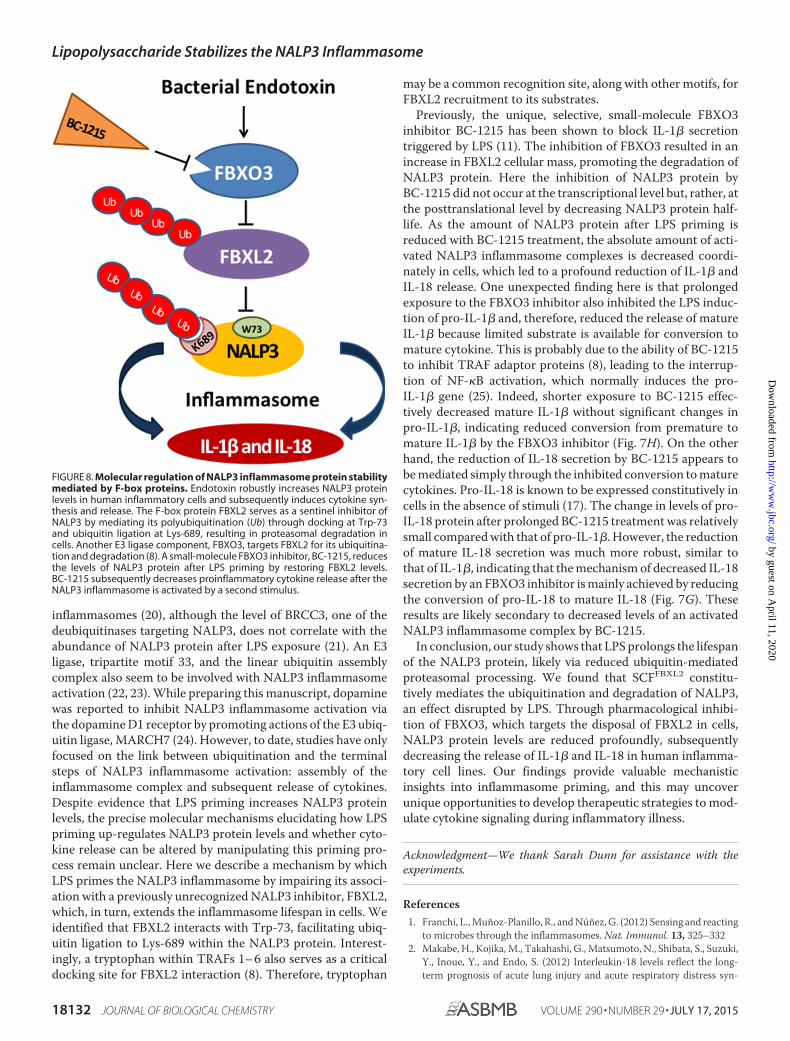
FIGURE 8. Molecular regulation of NALP3 inflammasome protein stabilitymediated by F-box proteins. Endotoxin robustly increases NALP3 proteinlevels in human inflammatory cells and subsequently induces cytokine syn-thesis and release. The F-box protein FBXL2 serves as a sentinel inhibitor ofNALP3 by mediating its polyubiquitination (Ub) through docking at Trp-73and ubiquitin ligation at Lys-689, resulting in proteasomal degradation incells. Another E3 ligase component, FBXO3, targets FBXL2 for its ubiquitina-tion and degradation (8). A small-molecule FBXO3 inhibitor, BC-1215, reducesthe levels of NALP3 protein after LPS priming by restoring FBXL2 levels.BC-1215 subsequently decreases proinflammatory cytokine release after theNALP3 inflammasome is activated by a second stimulus.
Lipopolysaccharide Stabilizes the NALP3 Inflammasome
18132 JOURNAL OF BIOLOGICAL CHEMISTRY VOLUME 290 • NUMBER 29 • JULY 17, 2015
by guest on April 11, 2020
http://ww
w.jbc.org/
Dow
nloaded from
drome. J. Anesth. 26, 658 – 6633. Dolinay, T., Kim, Y. S., Howrylak, J., Hunninghake, G. M., An, C. H.,
Fredenburgh, L., Massaro, A. F., Rogers, A., Gazourian, L., Nakahira, K.,Haspel, J. A., Landazury, R., Eppanapally, S., Christie, J. D., Meyer, N. J.,Ware, L. B., Christiani, D. C., Ryter, S. W., Baron, R. M., and Choi, A. M.(2012) Inflammasome-regulated cytokines are critical mediators of acutelung injury. Am. J. Respir. Crit. Care Med. 185, 1225–1234
4. Bauernfeind, F. G., Horvath, G., Stutz, A., Alnemri, E. S., MacDonald, K.,Speert, D., Fernandes-Alnemri, T., Wu, J., Monks, B. G., Fitzgerald, K. A.,Hornung, V., and Latz, E. (2009) Cutting edge: NF-�B activating patternrecognition and cytokine receptors license NLRP3 inflammasome activa-tion by regulating NLRP3 expression. J. Immunol. 183, 787–791
5. Hershko, A., and Ciechanover, A. (1998) The ubiquitin system. Annu. Rev.Biochem. 67, 425– 479
6. Deshaies, R. J., and Joazeiro, C. A. (2009) RING domain E3 ubiquitinligases. Annu. Rev. Biochem. 78, 399 – 434
7. Cardozo, T., and Pagano, M. (2004) The SCF ubiquitin ligase: insights intoa molecular machine. Nat. Rev. Mol. Cell Biol. 5, 739 –751
8. Chen, B. B., Coon, T. A., Glasser, J. R., McVerry, B. J., Zhao, J., Zhao, Y.,Zou, C., Ellis, B., Sciurba, F. C., Zhang, Y., and Mallampalli, R. K. (2013) Acombinatorial F box protein directed pathway controls TRAF adaptorstability to regulate inflammation. Nat. Immunol. 14, 470 – 479
9. Davies, J. Q., and Gordon, S. (2005) Isolation and culture of human macro-phages. Methods Mol. Biol. 290, 105–116
10. Chen, B. B., and Mallampalli, R. K. (2009) Masking of a nuclear signal motifby monoubiquitination leads to mislocalization and degradation of theregulatory enzyme cytidylyltransferase. Mol. Cell. Biol. 29, 3062–3075
11. Mallampalli, R. K., Coon, T. A., Glasser, J. R., Wang, C., Dunn, S. R.,Weathington, N. M., Zhao, J., Zou, C., Zhao, Y., and Chen, B. B. (2013)Targeting F box protein Fbxo3 to control cytokine-driven inflammation.J. Immunol. 191, 5247–5255
12. Radivojac, P., Vacic, V., Haynes, C., Cocklin, R. R., Mohan, A., Heyen,J. W., Goebl, M. G., and Iakoucheva, L. M. (2010) Identification, analysis,and prediction of protein ubiquitination sites. Proteins 78, 365–380
13. Schroder, K., and Tschopp, J. (2010) The inflammasomes. Cell 140,821– 832
14. Juliana, C., Fernandes-Alnemri, T., Kang, S., Farias, A., Qin, F., and Al-nemri, E. S. (2012) Non-transcriptional priming and deubiquitination reg-ulate NLRP3 inflammasome activation. J. Biol. Chem. 287, 36617–36622
15. Fernandes-Alnemri, T., Kang, S., Anderson, C., Sagara, J., Fitzgerald, K. A.,and Alnemri, E. S. (2013) Cutting edge: TLR signaling licenses IRAK1 forrapid activation of the NLRP3 inflammasome. J. Immunol. 191,
3995–399916. Niebler, M., Qian, X., Höfler, D., Kogosov, V., Kaewprag, J., Kaufmann,
A. M., Ly, R., Böhmer, G., Zawatzky, R., Rösl, F., and Rincon-Orozco, B.(2013) Post-translational control of IL-1� via the human papillomavirustype 16 E6 oncoprotein: a novel mechanism of innate immune escapemediated by the E3-ubiquitin ligase E6-AP and p53. PLoS Pathog. 9,e1003536
17. Ghonime, M. G., Shamaa, O. R., Das, S., Eldomany, R. A., Fernandes-Alnemri, T., Alnemri, E. S., Gavrilin, M. A., and Wewers, M. D. (2014)Inflammasome priming by lipopolysaccharide is dependent upon ERKsignaling and proteasome function. J. Immunol. 192, 3881–3888
18. Hu, Y., Nguyen, T. T., Bui, K. C., Demello, D. E., and Smith, J. B. (2005) Anovel inflammation-induced ubiquitin E3 ligase in alveolar type II cells.Biochem. Biophys. Res. Commun. 333, 253–263
19. Mayor, A., Martinon, F., De Smedt, T., Pétrilli, V., and Tschopp, J. (2007)A crucial function of SGT1 and HSP90 in inflammasome activity linksmammalian and plant innate immune responses. Nat. Immunol. 8,497–503
20. Lopez-Castejon, G., Luheshi, N. M., Compan, V., High, S., Whitehead,R. C., Flitsch, S., Kirov, A., Prudovsky, I., Swanton, E., and Brough, D.(2013) Deubiquitinases regulate the activity of caspase-1 and interleu-kin-1� secretion via assembly of the inflammasome. J. Biol. Chem. 288,2721–2733
21. Py, B. F., Kim, M. S., Vakifahmetoglu-Norberg, H., and Yuan, J. (2013)Deubiquitination of NLRP3 by BRCC3 critically regulates inflammasomeactivity. Mol. Cell 49, 331–338
22. Weng, L., Mitoma, H., Trichot, C., Tricot, C., Bao, M., Liu, Y., Zhang, Z.,and Liu, Y. J. (2014) The E3 ubiquitin ligase tripartite motif 33 is essentialfor cytosolic RNA-induced NLRP3 inflammasome activation. J. Immunol.193, 3676 –3682
23. Rodgers, M. A., Bowman, J. W., Fujita, H., Orazio, N., Shi, M., Liang, Q.,Amatya, R., Kelly, T. J., Iwai, K., Ting, J., and Jung, J. U. (2014) The linearubiquitin assembly complex (LUBAC) is essential for NLRP3 inflam-masome activation. J. Exp. Med. 211, 1333–1347
24. Yan, Y., Jiang, W., Liu, L., Wang, X., Ding, C., Tian, Z., and Zhou, R. (2015)Dopamine controls systemic inflammation through inhibition of NLRP3inflammasome. Cell 160, 62–73
25. Zhu, L. J., Yang, X., Li, X. Y., Liu, Q. H., Tang, X. Q., Zhou, S. F., Kong,Q. Y., Axelsson, J., and Yu, X. Q. (2010) Suppression of tumor necrosisfactor receptor associated factor (TRAF)-2 attenuates the proinflamma-tory and proliferative effect of aggregated IgG on rat renal mesangial cells.Cytokine 49, 201–208
Lipopolysaccharide Stabilizes the NALP3 Inflammasome
JULY 17, 2015 • VOLUME 290 • NUMBER 29 JOURNAL OF BIOLOGICAL CHEMISTRY 18133
by guest on April 11, 2020
http://ww
w.jbc.org/
Dow
nloaded from
MallampalliSnavely, Dexter L. Gulick, Kevin F. Gibson, Chunbin Zou, Bill B. Chen and Rama K.
SeungHye Han, Travis B. Lear, Jacob A. Jerome, Shristi Rajbhandari, Courtney A. E3 LigaseFBXL2Ubiquitination and Degradation Mediated by the SCF
Lipopolysaccharide Primes the NALP3 Inflammasome by Inhibiting Its
doi: 10.1074/jbc.M115.645549 originally published online June 2, 20152015, 290:18124-18133.J. Biol. Chem.
10.1074/jbc.M115.645549Access the most updated version of this article at doi:
Alerts:
When a correction for this article is posted•
When this article is cited•
to choose from all of JBC's e-mail alertsClick here
http://www.jbc.org/content/290/29/18124.full.html#ref-list-1
This article cites 25 references, 9 of which can be accessed free at
by guest on April 11, 2020
http://ww
w.jbc.org/
Dow
nloaded from