![METODOLÓGICA GUÍAcolegiovicentealeixandremarbella.com/imagenes/... · "qsfoej[bkf #btbep fo fm 1fotbnjfoup 5ijoljoh #btfe -fbsojoh (o remhwlyr gho wklqnlqj edvhg ohduqlqj r dsuhqgl]dmh](https://static.fdocument.pub/doc/165x107/602c6a0e3001ef15605dd375/metodolgica-guacolegiovicen-qsfoejbkf-btbep-fo-fm-1fotbnjfoup-5ijoljoh.jpg)
)JOEBXJ1VCMJTIJOH$PSQPSBUJPO &WJEFODF #BTFE ...
9
Research Article Attenuation of Thrombosis by Crude Rice (Oryza sativa) Bran Policosanol Extract: Ex Vivo Platelet Aggregation and Serum Levels of Arachidonic Acid Metabolites Wai-Teng Wong, 1 Maznah Ismail, 1,2 Eusni Rahayu Mohd Tohit, 3 Rasedee Abdullah, 4 and Yi-Da Zhang 1,5 1 Laboratory of Molecular Biomedicine, Institute of Bioscience, Universiti Putra Malaysia, 43400 Serdang, Selangor, Malaysia 2 Department of Nutrition and Dietetics, Faculty of Medicine and Health Sciences, Universiti Putra Malaysia, 43400 Serdang, Selangor, Malaysia 3 Department of Pathology, Faculty of Medicine and Health Sciences, Universiti Putra Malaysia, 43400 Serdang, Selangor, Malaysia 4 Department of Veterinary Laboratory Diagnostics, Faculty of Veterinary Medicine, Universiti Putra Malaysia, 43400 Serdang, Selangor, Malaysia 5 Cardiology Department, Affiliated Hospital of Chengde Medical University, Chengde, Hebei 067000, China Correspondence should be addressed to Wai-Teng Wong; [email protected] Received 9 May 2016; Accepted 2 August 2016 Academic Editor: Roc´ ıo De la Puerta Copyright © 2016 Wai-Teng Wong et al. is is an open access article distributed under the Creative Commons Attribution License, which permits unrestricted use, distribution, and reproduction in any medium, provided the original work is properly cited. Background. Vascular occlusion or thrombosis was oſten attributed to uncontrolled platelet activation. Influence of sugarcane poli- cosanol extract on platelet was reported but little was known of rice bran policosanol, particularly its mechanisms of actions on platelet activities. Objective. Antiplatelet mechanisms of rice bran policosanol extract (RBE) were studied using hyperlipidemic Sprague Dawley rats. Ex vivo platelet aggregation, platelet count (PC), bleeding time (BT), and coagulation time were assayed. Serum eicosanoids and other aggregation-related metabolites levels were quantified. Design. Rats were divided into 6 groups for comparisons (vehicle control Tween 20/H 2 O, high dose policosanol 500 mg/kg, middle dose policosanol 250 mg/kg, low dose policosanol 100 mg/kg, and positive control aspirin 30 mg/kg). Results. Low dose 100 mg/kg of RBE inhibited aggregation by 42.32 ± 4.31% and this was comparable with the effect of 30 mg/kg aspirin, 43.91 ± 5.27%. Results showed that there were no significant differences in PC, BT, and coagulation time among various groups aſter RBE treatment. Serum thromboxane A 2 was attenuated while prostacyclin level increased upon RBE treatment. Conclusions. RBE reduced ex vivo ADP-induced platelet aggregation without giving adverse effects. No changes in full blood count suggested that rice bran policosanol did not disturb biological blood cell production and destruction yet it reduced aggregation through different mechanisms. 1. Introduction Inflammation, atherosclerosis, and thrombosis are all viewed as cardiovascular manifestations demonstrated by a cluster of risk factors. e prevalence and fatality rate of cardiovas- cular diseases (CVDs) worldwide, for instance, stroke and coronary heart disease, urged the progression in medical research [1]. CVD is a disorder caused by a sequence of biological events associated with oxidative stress, low density lipoprotein (LDL) deposition, tissue injury, foam cell for- mation, plaque formation, and so forth. Hence, compounds with antioxidant, anti-inflammatory, hypolipidemia, and/or antithrombotic properties are valuable [2]. In biological perspective, free flow of blood is of utmost importance. Platelets, smallest type of blood cell, play essen- tial role in regulating smooth blood circulation, vascular integrity, and hemostasis [3]. However, uncontrollable hyper- activity of platelets beyond normal biological responses in Hindawi Publishing Corporation Evidence-Based Complementary and Alternative Medicine Volume 2016, Article ID 7343942, 8 pages http://dx.doi.org/10.1155/2016/7343942
Transcript of )JOEBXJ1VCMJTIJOH$PSQPSBUJPO &WJEFODF #BTFE ...

Research ArticleAttenuation of Thrombosis byCrude Rice (Oryza sativa) Bran Policosanol Extract:Ex Vivo Platelet Aggregation and Serum Levels ofArachidonic Acid Metabolites
Wai-Teng Wong,1 Maznah Ismail,1,2 Eusni Rahayu Mohd Tohit,3
Rasedee Abdullah,4 and Yi-Da Zhang1,5
1Laboratory of Molecular Biomedicine, Institute of Bioscience, Universiti Putra Malaysia, 43400 Serdang, Selangor, Malaysia2Department of Nutrition and Dietetics, Faculty of Medicine and Health Sciences, Universiti Putra Malaysia,43400 Serdang, Selangor, Malaysia3Department of Pathology, Faculty of Medicine and Health Sciences, Universiti Putra Malaysia, 43400 Serdang, Selangor, Malaysia4Department of Veterinary Laboratory Diagnostics, Faculty of Veterinary Medicine, Universiti Putra Malaysia,43400 Serdang, Selangor, Malaysia5Cardiology Department, Affiliated Hospital of Chengde Medical University, Chengde, Hebei 067000, China
Correspondence should be addressed to Wai-Teng Wong; [email protected]
Received 9 May 2016; Accepted 2 August 2016
Academic Editor: Rocıo De la Puerta
Copyright © 2016 Wai-Teng Wong et al.This is an open access article distributed under theCreative CommonsAttribution License,which permits unrestricted use, distribution, and reproduction in any medium, provided the original work is properly cited.
Background. Vascular occlusion or thrombosis was often attributed to uncontrolled platelet activation. Influence of sugarcane poli-cosanol extract on platelet was reported but little was known of rice bran policosanol, particularly its mechanisms of actions onplatelet activities. Objective. Antiplatelet mechanisms of rice bran policosanol extract (RBE) were studied using hyperlipidemicSprague Dawley rats. Ex vivo platelet aggregation, platelet count (PC), bleeding time (BT), and coagulation time were assayed.Serum eicosanoids and other aggregation-related metabolites levels were quantified. Design. Rats were divided into 6 groups forcomparisons (vehicle control Tween 20/H
2O, high dose policosanol 500mg/kg, middle dose policosanol 250mg/kg, low dose
policosanol 100mg/kg, and positive control aspirin 30mg/kg). Results. Low dose 100mg/kg of RBE inhibited aggregation by42.32 ± 4.31% and this was comparable with the effect of 30mg/kg aspirin, 43.91 ± 5.27%. Results showed that there wereno significant differences in PC, BT, and coagulation time among various groups after RBE treatment. Serum thromboxane A
2
was attenuated while prostacyclin level increased upon RBE treatment. Conclusions. RBE reduced ex vivo ADP-induced plateletaggregation without giving adverse effects. No changes in full blood count suggested that rice bran policosanol did not disturbbiological blood cell production and destruction yet it reduced aggregation through different mechanisms.
1. Introduction
Inflammation, atherosclerosis, and thrombosis are all viewedas cardiovascular manifestations demonstrated by a clusterof risk factors. The prevalence and fatality rate of cardiovas-cular diseases (CVDs) worldwide, for instance, stroke andcoronary heart disease, urged the progression in medicalresearch [1]. CVD is a disorder caused by a sequence ofbiological events associated with oxidative stress, low density
lipoprotein (LDL) deposition, tissue injury, foam cell for-mation, plaque formation, and so forth. Hence, compoundswith antioxidant, anti-inflammatory, hypolipidemia, and/orantithrombotic properties are valuable [2].
In biological perspective, free flow of blood is of utmostimportance. Platelets, smallest type of blood cell, play essen-tial role in regulating smooth blood circulation, vascularintegrity, and hemostasis [3]. However, uncontrollable hyper-activity of platelets beyond normal biological responses in
Hindawi Publishing CorporationEvidence-Based Complementary and Alternative MedicineVolume 2016, Article ID 7343942, 8 pageshttp://dx.doi.org/10.1155/2016/7343942

2 Evidence-Based Complementary and Alternative Medicine
the presence of platelet activators, for instance, free radi-cals, arachidonic acid, adenosine diphosphate, collagen, dia-betic, and hyperlipidemic-related risk factors, significantlycontributes to platelet dysfunctions and causes a series ofatherothrombotic diseases [4].
Antiplatelet agents (APA) including aspirin, clopidogrel,and cilostazol are often used in prescription and remained asmainstay medication in vascular thrombotic diseases. How-ever, the various side effects of suchmedications, for example,internal bleeding, rash, drowsiness, and bruising, impedetheir wide use in clinical aspects [5]. Therefore, there is anurgent need in finding antiplatelet agents from natural origin(plant bioactive compounds) to establish a novel, safer, andeffective approach.
Natural antiplatelet agents (APA) that modulated plateletfunctions attained great attention from the public in primaryand secondary prevention of cardiovascular diseases (CVD).Policosanol, a generic term for a mixture of long chainaliphatic alcohols (C20–C30), is usually extracted from ani-mal and plant waxes, for example, sugarcane, wheat varieties,cereal, yam, and beeswax [6]. Policosanol is long-knownfor its physiological benefits such as cholesterol loweringproperty [7] and improvement of muscle endurance [8] asreviewed by Janikula [9]. In this study, rice bran policosanolextract, with its own unique fatty alcohols fingerprint, wasstudied ex vivo and in vivo for its effects on platelet functionsusing high fat diet-induced hyperlipidemic rat model.
2. Materials and Methods
2.1. Materials. ADP was purchased from Chronolog Incor-poration (Havertown, PA, USA). Aspirin was a generousgift from Mr. Choy. All the reagents and solvents used inextraction and analysis were analytical grade purchased fromMerck (Darmstadt, Germany). Rice bran was obtained fromBernas milling factory in Kuala Selangor, Selangor, Malaysia.All the consumables used in animal studies were supplied byTakrif Bistari Sdn Bhd (Selangor, Malaysia). Mazola corn oil(Unilever Malaysia), Nespray fortified milk powder (NestleMalaysia), and Specialty feeds (TN, USA) were used in highfat diet preparation.
2.2. Methods
2.2.1. Rice Bran Policosanol Extraction. Policosanol wasextracted using solid-liquid extraction according to thedescribed procedures minor modifications [10]. Rice bransample with size of 4mm was stabilized by heat treatmentusing an automated microwave oven (microwave conditions:2450MHz, 550W, 110∘C, 200 seconds). Briefly, 10 g of ricebran was placed in conical flask with 150mL of hexane andmethanol (20 : 1 v/v mixture). Extraction was performed bysonication technology (50Hz, 350W, 50∘C, 3 hours) usingPower Sonic 505 ultrasonicator (Hwashin Technology Co.,Seoul, Korea). The rice bran residues were removed from thesolvent extract by centrifuging at 4000 rpm for 10 minutes.The solvent was removed from the extract using a rotaryevaporator (Buchi, Switzerland) under a vacuumat 40∘Cuntilgreenish-yellow extract was seen.
2.2.2. Preparation of High Fat Diet (HFD). HFD was formu-lated following the recipe by Levin et al. [11] and prepared inaccordance to Imam and Ismail [12]. Every kg of HFD wasprepared from a mixture of 500 g of normal rat pellet, 240 gof corn oil, 200 g of full-cream milk powder, 60 g of sugar,and 50 g of starch. The HFD was cut and dried overnight inan incubator at 60∘C.
2.2.3. Animal Handling and Treatment. Experiments wereconducted in accordance with the guidelines for animal useand ethical approval was obtained from institutional animalcare and use committee (IACUC), Faculty of Medicine andHealth Sciences, Universiti Putra Malaysia, Malaysia. Theanimals (36 adult male Sprague-Dawley rats) with bodyweight between 100 and 150 g were randomly divided into sixexperimental groups. All the rats were acclimatized for 7days ad libitum with standard rodent chow and free accessof clean water. These animals were kept in a temperature-controlled room (22 ± 2∘C) with a 12 h light-dark cycle. Thestudy was carried out for 3 months. Starting from secondmonth, treatment was given once a day accordingly throughoral gavage continuously for one month. Animal foodremained the same throughout the whole experimentalperiod: (1) a normal group receiving rodent chow and treatedwith vehicle (Tween 20/H
2O); (2) a negative control group
receiving HFD and treated with vehicle (Tween 20/H2O);
(3) a positive control group receiving HFD and treated withaspirin (30mg/kg); (4) a group receiving HFD and treatedwith 500mg/kg policosanol; (5) a group receiving HFD andtreated with 250mg/kg policosanol; (6) a group receivingHFD and treated with 100mg/kg policosanol. Suspensionswere prepared daily 1 h before administration [13].
2.2.4. Ex Vivo Platelet Aggregation. Platelet aggregationstudy was done using ADP as agonist according to Vaiyapuriand Gibbins [14] with minor modifications. Rat whole bloodwas collected into a vacutainer tube containing 3.2% sodiumcitrate (9 : 1, v/v) andmixed thoroughly by inverting the tubesfor several times. The blood was centrifuged at 100×g for 20minutes to obtain platelet rich plasma (PRP). 50 ng/mL ofprostacyclin was added to prevent platelet activation. PRPwas centrifuged again at 240×g for 10 minutes to sedimentplatelet pellet. The platelet pellet was resuspended in Tyrode-HEPES buffer and adjusted to a cell density of 108 cells/mL.BioTeK Synergy H1 Hybrid Reader (BioTek Instruments Inc.,Winooski, VT, USA) was used as analytical tool. 100 𝜇L ofplatelet cells was loaded into each well and 1 𝜇L of ADPwas added in. The optical densities at 405 nm were read for20 minutes. Reading was taken at a fixed time interval of 1minute. The plate was shaken in double orbital mode at afrequency of 282 cpm, 3mm.
2.2.5. Full Blood Count (RBC, MCV, RDW, HCT, PLT, MPV,HGB, MCH, and MCHC). Full blood count was done usinghematology analyzer (Medonic CA530). Fresh rat bloodwas withdrawn into EDTA anticoagulated blood tubes andanalyzed within 4 hours. Baseline and final readings weretaken before and after treatment for comparison.
Evidence-Based Complementary and Alternative Medicine 3
2.2.6. Tail Bleeding Time Assay. Rat tail bleeding time wasmeasured according to the described procedures with minormodifications [15]. Tail of anesthetized rat was warmed to37∘C. Bleeding time was determined by cutting 2mm of thetail tip with a blade and blood was blotted onto a filter paperevery 30 s until no bloodstain was seen. The period betweenamputation and the stop of bleeding was the bleeding time(min). Baseline and final readings were taken before and aftertreatment.
2.2.7. Coagulation Time. Extrinsic and intrinsic pathways ofcoagulation were analyzed by measuring prothrombin time(PT) and activated partial thromboplastin time (aPTT). PTand aPTT were analyzed followed the standard protocol ofSynergy Start 4 hemostasis analyzer (Diagnostica Stago Inc.).
2.2.8. Quantification of Serum Eicosanoids, von WillebrandFactor (vWF), and Soluble p-Selectin. Stable platelet metabo-lites, thromboxane B
2and 6-keto PGF1𝛼, were measured
due to short half-life of thromboxane A2and prostacyclin.
Serum vWF and soluble p-selectin levels were quantifiedas an indication of in vivo platelet activation. Analysis wasall done followed the kit’s protocol using serum samples.Absorbance readings were obtained using BioTeK SynergyH1 Hybrid Reader (BioTek Instruments Inc., Winooski, VT,USA) at a wavelength of 450 nm for all kits.
3. Statistical Analysis
The data were analyzed using minitab 16 (Minitab Inc., StateCollege, Pennsylvania, United States) by one-way analysisof variance (ANOVA) and the values were presented asmeans and standard deviation (SD). The significance of thedifferences between comparisons was determined by Tukey’srange test. The significant difference was taken to be a valueof 𝑝 < 0.05 at a 95% confidence interval.
4. Results and Discussion
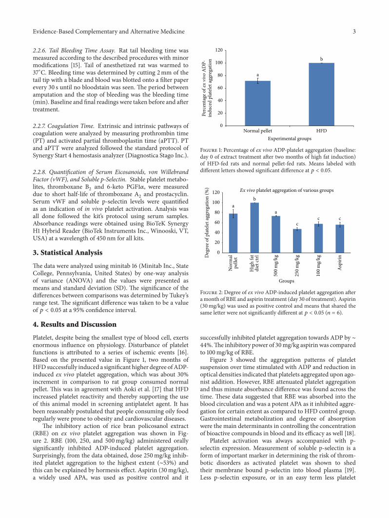
Platelet, despite being the smallest type of blood cell, exertsenormous influence on physiology. Disturbance of plateletfunctions is attributed to a series of ischemic events [16].Based on the presented value in Figure 1, two months ofHFD successfully induced a significant higher degree ofADP-induced ex vivo platelet aggregation, which was about 30%increment in comparison to rat group consumed normalpellet. This was in agreement with Aoki et al. [17] that HFDincreased platelet reactivity and thereby supporting the useof this animal model in screening antiplatelet agent. It hasbeen reasonably postulated that people consuming oily foodregularly were prone to obesity and cardiovascular diseases.
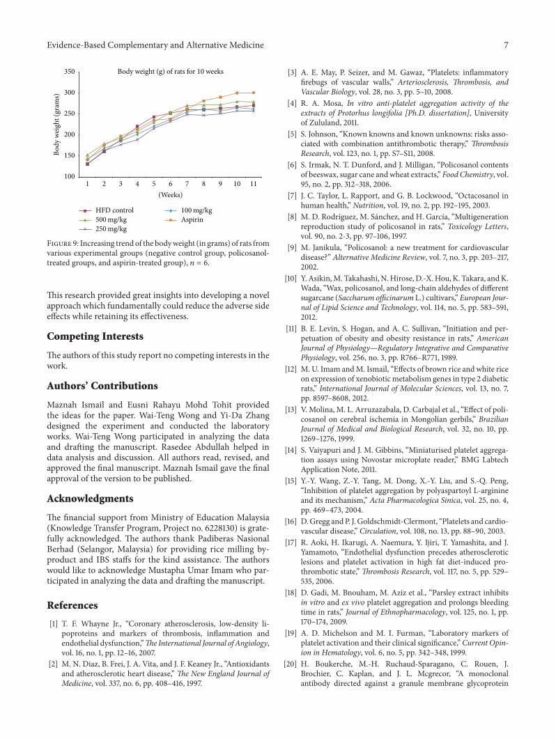
The inhibitory action of rice bran policosanol extract(RBE) on ex vivo platelet aggregation was shown in Fig-ure 2. RBE (100, 250, and 500mg/kg) administered orallysignificantly inhibited ADP-induced platelet aggregation.Surprisingly, from the data obtained, dose 250mg/kg inhib-ited platelet aggregation to the highest extent (∼53%) andthis can be explained by hormesis effect. Aspirin (30mg/kg),a widely used APA, was used as positive control and it
Perc
enta
ge o
f ex
vivo
AD
P-
a
b
HFDNormal pelletExperimental groups
0
20
40
60
80
100
120
indu
ced
plat
elet
aggr
egat
ion
Figure 1: Percentage of ex vivo ADP-platelet aggregation (baseline:day 0 of extract treatment after two months of high fat induction)of HFD-fed rats and normal pellet-fed rats. Means labeled withdifferent letters showed significant difference at 𝑝 < 0.05.
Nor
mal
pelle
t
Hig
h fa
tdi
et ct
rl
Asp
irin
Groups
Ex vivo platelet aggregation of various groups
aa
b
ccc
100
mg/
kg
500
mg/
kg
0
20
40
60
80
100
120
Deg
ree o
f pla
tele
t agg
rega
tion
(%)
250
mg/
kgFigure 2: Degree of ex vivo ADP-induced platelet aggregation afteramonth of RBE and aspirin treatment (day 30 of treatment). Aspirin(30mg/kg) was used as positive control and means that shared thesame letter were not significantly different at 𝑝 < 0.05 (𝑛 = 6).
successfully inhibited platelet aggregation towards ADP by ∼44%.The inhibitory power of 30mg/kg aspirin was comparedto 100mg/kg of RBE.
Figure 3 showed the aggregation patterns of plateletsuspension over time stimulated with ADP and reduction inoptical densities indicated that platelets aggregated upon ago-nist addition. However, RBE attenuated platelet aggregationand thus minute absorbance difference was found across thetime. These data suggested that RBE was absorbed into theblood circulation and was a potent APA as it inhibited aggre-gation for certain extent as compared to HFD control group.Gastrointestinal metabolization and degree of absorptionwere the main determinants in controlling the concentrationof bioactive compounds in blood and its efficacy as well [18].
Platelet activation was always accompanied with p-selectin expression. Measurement of soluble p-selectin is aform of important marker in determining the risk of throm-botic disorders as activated platelet was shown to shedtheir membrane bound p-selectin into blood plasma [19].Less p-selectin exposure, or in an easy term less platelet

4 Evidence-Based Complementary and Alternative Medicine
Ex vivo ADP-induced platelet aggregation
ControlNormal pelletAspirin
5 10 15 20 250Cycles
Extract 250𝜇g/mLExtract 500 𝜇g/mL
0.2
0.3
0.4
0.5
0.6
0.7
0.8
0.9
1
Opt
ical
den
sity
(405
nm)
Extract 100𝜇g/mL
Figure 3: Ex vivo ADP-induced platelet aggregation patternsobtained in double orbital shaking mode using microplate readerat wavelength of 450 nm. Optical densities of 100𝜇L of platelets sus-pensions from each group (𝑛 = 6) were studied for 23 cycles with afixed time interval of one minute.
activation, was important in reducing atherosclerotic lesionsas p-selectinwas a strong adhesivemolecule in forming stableplatelet aggregates and vital in platelet-monocyte interaction[20, 21]. From Figure 4, 250mg/kg oral ingestion of RBEwas shown to reduce soluble p-selectin detected in serumsignificantly by 14%. Influence of other activation factors,for example, centrifugal speed and temperature shock duringblood serum collection, can be ignored as high thresholdwas needed to activate alpha granule and expose p-selectin,thus supporting the validity of the present study [22]. Thesoluble p-selectin detected was assumed to be originatedfrom activated platelets instead of activated endothelial cellsbased on the presented value shown in Figure 5 and this wasconcurred with the study done by Perumal et al. [23]. Therewas no significant difference in vonWillebrand factor (vWF)level detected in the blood serum between various groups at𝑝 < 0.05.This result highlighted that therewas no endothelialactivation and again proved our hypothesis on antiplateletfunction of RBE.
As shown in Table 1, RBE treatments did not modifyplatelet number (109/L) in comparison with HFD controlgroup. This showed that the attenuation of platelet aggrega-tion was not associated with platelet production or destruc-tion in vivo upon RBE treatment. In addition to that, therewere no statistical differences on various blood parametersbetween RBE-treated groups and control group. These sug-gested that RBE was safe for consumption (not toxic to cells).
Bleeding time, the time taken for bleeding to stop, wasan important test in platelet function assessment [24]. Basedon the data depicted in Table 2, RBE did not significantlyprolong bleeding time as compared to control group at 𝑝 <0.05. This result was correlated with other literatures statingthat antiplatelet compounds were not often associated withprolongation of bleeding time [18, 25, 26]. On the other hand,RBE and aspirin did not significantly affect PT and aPTT, and
a, b a, b a, b a, bba
500 250 100 Aspirin HFDNormalpellet
Groups
0
0.2
0.4
0.6
0.8
1
1.2
Seru
m p
-sel
ectin
leve
l (pg
)
Figure 4: Serum level of soluble p-selectin after 30 days of RBEextract or aspirin treatment on experimental rats. Means whichshared the same letter were not significantly different at 𝑝 < 0.05.
a a a a a
Asp
irin
(HFD
)C
ontro
l
100
mg/
kg
250
mg/
kg
500
mg/
kg
Groups
0
0.1
0.2
0.3
0.4
0.5
0.6
vWF
leve
l (ng
/mL)
Figure 5: Serum level of soluble von Willebrand factor (vWF) after30 days of RBE extract or aspirin treatment on experimental rats.Means which shared the same letter were not significantly differentat 𝑝 < 0.05.
this showed that RBE potentiated antiplatelet effect withoutinfluencing coagulation system. These notable findings sup-ported the consumption of brown rice or rice bran-derivedproducts as therapeutic approach without giving adverse sideeffects.
Practically, platelets were activated by numerous phys-iological factors through a multitude of mechanism, forinstance, interaction with platelet receptors or glycoproteins,modulation of intracellular platelet messengers, and regula-tion of platelet signaling products. Despite different pathwayof platelet activation, platelet responded in the same seriesinitiated with shape change, secretion, liberation includ-ing prostaglandins, or lipooxygenase products, followed byaggregation [27]. RBE was a crude extract containing amixture of bioactive compounds and policosanol was under-stood to be the leading compound responsible in exertingantiplatelet function. However, existence of other biocon-stituents, for example, polyphenolic compounds or toco-pherols, might work individually or synergistically in givingantiaggregant activity [28, 29]. The actual mechanism bywhich RBE implicated in antiplatelet function was yet to

Evidence-Based Complementary and Alternative Medicine 5
Table1:Fu
llbloo
dcoun
tanalysis
usinghematolog
yanalyzer
(baseline:day0of
treatment;fin
alreading:day30
oftre
atment).
Group
sRB
C(1012/L)
MCV
RDW
HCT
PLT(109/L)
MPV
(fl)
HGB(g/L)
MCH
(pg)
MCH
C(g/L)
(fl)
(%)
(%)
NP(beforetreatment)
8.005±0.04
a53.95±0.35
b10.7±2.12
c40
.85±3.75
d712±97.58e
6.7±0.14
f148.5±9.1
9g19.75±0.64
h367±14.14
i
HFD
(beforetreatment)
7.49±0.68
a48.58±1.3
6b14.35±1.3
2c36.21±
3.23
d844.25±157.4
6e6.93±0.12
f133.78±10.45g
17.91±0.78
h366.3±6.70
i
NP(afte
rtreatment)
7.21±
1.01a
52.97±1.8
9b13.83±2.46
c38.1±4.42
d730±97.58e
7.27±0.45
f138.67±20.50g
19.27±0.72
h363.67±13.65i
HFD
-NC(afte
rtreatment)
7.8±0.71
a49.47±1.2
9b13.53±0.67
c38.53±2.51
d1155.33±160.55
e6.77±0.06
f143.33±8.08
g18.37±0.67
h372±3.61
i
500m
g/kg
7.66±1.0
4a47.27±1.8
8b15.53±0.67
c36.3±6.16
d756±86.27e
7.53±0.91
f131.6
7±23.44g
17.13±1.0
3h362.33±8.74
i
250m
g/kg
8.29±0.25
a47.37±1.2
5b14.87±0.84
c39.3±1.3
1d961±
147.8
9e7.13±0.40
f143.33±4.62
g17.27±0.55
h365±2i
100m
g/kg
7.86±0.31
a48.23±1.3
2b15.23±1.6
7c37.9±1.4
d772±288.5e
7.13±0.61
f138±6.08
g17.57±0.40
h364±3.46
i
Aspirin
-PC
8.31±0.23
a46
.53±0.81
b15.3±1.4
2c38.67±0.64
d902±106.07
e7±0.7f
140.33±3.5
1g16.87±0.23
h363±3.61
i
NC:
negativ
econtrol;PC
:positive
control;NP:
ratswhich
consum
eno
rmalpellet;HFD
:ratsw
hich
consum
ehigh
fatd
ietthrou
ghou
t3mon
thss
tudy
perio
d,𝑛=6.Th
epresentedvaluewas
mean±sta
ndard
deviation.
Means
thatshared
thes
ameletterw
eren
otsig
nificantly
different
at𝑝<0.05.

6 Evidence-Based Complementary and Alternative Medicine
Table 2: Tail bleeding time (BT), activated partial prothrombin time(aPTT), and prothrombin time (PT) in secs of various experimentalgroups after oral administration of RBE or aspirin for one month,𝑛 = 6.
Groups BT (secs) aPTT (secs) PT (secs)HFD-negative control 484 ± 45.36a 19.7 ± 1.8b 19.8 ± 0.8c
500mg/kg 573.6 ± 39.11a,b 16.8 ± 3.1b 17.7 ± 1.5c
250mg/kg 540.8 ± 93.59a,b 17.5 ± 1.8b 18.8 ± 0.2c
100mg/kg 521.2 ± 97.16a,b 16.3 ± 0.3b 17.7 ± 1.6c
Aspirin-PC 597 ± 96.77b 15.1 ± 0.1b 18.4 ± 0.8c
The presented value was mean ± standard deviation. Means that shared thesame letter were not significantly different at 𝑝 < 0.05.
Animal groups
a, b
a, b
a, ba, b
a
b
Nor
mal
pelle
t
HFD
ctrl
Asp
irin
100
mg/
kg
250
mg/
kg
500
mg/
kg
0100200300400500600700800900
Thro
mbo
xane
B2
leve
l (pg
/mL)
Figure 6: Serum level of thromboxane B2after 30 days of aspirin/
RBE treatment, 𝑛 = 6. Means which shared the same letter were notsignificantly different at 𝑝 < 0.05.
be known. Figures 6 and 7 showed that RBE significantlyreduced serum thromboxane B
2and meanwhile increased
prostacyclin level. These results were concurred with otherstudies [30–32]. The results suggested that RBE exerted itsantiplatelet function through mechanisms different fromaspirin (ASA). Reduced TXA
2/PGI2ratio as compared to
control inhibited pathophysiology ischemic events is associ-ated with platelet activation [13].
From the result obtained, we can boldly hypothesizethat RBE might serve as antagonists of receptor GPIIb/IIIa(fibrinogen receptor), P
2Y1and P
2Y12, thus preventing the
ADP-induced ex vivo platelet aggregation and thromboxaneB2liberation. It was believed that both inside-out signaling
through ADP receptors and outside-in signaling throughfibrinogen receptor were necessary to activate phospholipaseA2activity and result in thromboxane A
2generation [33].
In addition to that, antagonism of receptor P2Y12
increasedintracellular cAMP level and further reduced platelet aggre-gation by regulating a series of enzyme activities [34].
Additionally, antioxidant ability of RBE was a potentialfactor in reducing platelet aggregation. Inhibition powerof RBE on endogenous and exogenous free radicals fromattacking platelets must not be discarded [35]. On the otherhand, long believed hypocholesterolemic action (reduction inLDL-C, total cholesterol, and triglycerides) and endothelialprotecting action of policosanol made RBE a potential agent
Nor
mal
pelle
t
HFD
ctrl
Asp
irin
Animal groups
a, ba, b
b
b
b
a
100
mg/
kg
250
mg/
kg
500
mg/
kg
0
200
400
600
800
1000
1200
1400
6-ke
to P
GF1
a lev
el (p
g/m
L)
Figure 7: Serum level stable metabolite of prostaglandin I2(6-keto
PGF1𝛼) after 30 days of aspirin/extract treatment, 𝑛 = 6. Meanswhich shared the same letter were not significantly different at 𝑝 <0.05.
Heart Kidney Liver SpleenOrgans
Organs weight of various experimental groups
HFD controlAspirin
aa, b a, b b a, b
c c c c c
d
dd d
d
e ee e e
0123456789
Org
an w
eigh
t (gr
ams)
100 mg/kg
250 mg/kg500 mg/kg
Figure 8: Organs weight (heart, liver, spleen, and kidney) of sacri-ficed rats from various experimental groups. The presented valueswere mean ± standard deviation and means denoted with sameletters were not significantly different at 𝑝 < 0.05 (𝑛 = 6).
to reduce in vivo platelet activation [36–38]. RBE showed notoxicity to experimental animals as shown in Figures 8 and 9.Normal increment in rat body weight along the study periodsuggested the safety use of RBE in human. Holistically, thepresent study suggested the feasibility of RBE as antiplateletagent as it selectively inhibited platelet-dependent mecha-nisms without disturbing the normal physiology.
5. Conclusion
The present study demonstrated that rice bran policosanolextract (RBE) potentiates antiplatelet effect using ex vivo andin vivomodels. Antiplatelet effect of RBE was evident after 30days of RBE oral administration. RBE successfully reducedserum thromboxane A
2production but increased prostacy-
clin level. Notably, significant platelet aggregation inhibitiondid not accompany the prolongation of bleeding time andcoagulation time and thus overwhelmed other commonAPA.
Evidence-Based Complementary and Alternative Medicine 7
Body weight (g) of rats for 10 weeks
HFD controlAspirin100 mg/kg
250 mg/kg500 mg/kg
100
150
200
250
300
350
Body
wei
ght (
gram
s)
2 3 4 5 6 7 8 9 10 111(Weeks)
Figure 9: Increasing trend of the bodyweight (in grams) of rats fromvarious experimental groups (negative control group, policosanol-treated groups, and aspirin-treated group), 𝑛 = 6.
This research provided great insights into developing a novelapproach which fundamentally could reduce the adverse sideeffects while retaining its effectiveness.
Competing Interests
The authors of this study report no competing interests in thework.
Authors’ Contributions
Maznah Ismail and Eusni Rahayu Mohd Tohit providedthe ideas for the paper. Wai-Teng Wong and Yi-Da Zhangdesigned the experiment and conducted the laboratoryworks. Wai-Teng Wong participated in analyzing the dataand drafting the manuscript. Rasedee Abdullah helped indata analysis and discussion. All authors read, revised, andapproved the final manuscript. Maznah Ismail gave the finalapproval of the version to be published.
Acknowledgments
The financial support from Ministry of Education Malaysia(Knowledge Transfer Program, Project no. 6228130) is grate-fully acknowledged. The authors thank Padiberas NasionalBerhad (Selangor, Malaysia) for providing rice milling by-product and IBS staffs for the kind assistance. The authorswould like to acknowledge Mustapha Umar Imam who par-ticipated in analyzing the data and drafting the manuscript.
References
[1] T. F. Whayne Jr., “Coronary atherosclerosis, low-density li-poproteins and markers of thrombosis, inflammation andendothelial dysfunction,”TheInternational Journal of Angiology,vol. 16, no. 1, pp. 12–16, 2007.
[2] M. N. Diaz, B. Frei, J. A. Vita, and J. F. Keaney Jr., “Antioxidantsand atherosclerotic heart disease,” The New England Journal ofMedicine, vol. 337, no. 6, pp. 408–416, 1997.
[3] A. E. May, P. Seizer, and M. Gawaz, “Platelets: inflammatoryfirebugs of vascular walls,” Arteriosclerosis, Thrombosis, andVascular Biology, vol. 28, no. 3, pp. 5–10, 2008.
[4] R. A. Mosa, In vitro anti-platelet aggregation activity of theextracts of Protorhus longifolia [Ph.D. dissertation], Universityof Zululand, 2011.
[5] S. Johnson, “Known knowns and known unknowns: risks asso-ciated with combination antithrombotic therapy,” ThrombosisResearch, vol. 123, no. 1, pp. S7–S11, 2008.
[6] S. Irmak, N. T. Dunford, and J. Milligan, “Policosanol contentsof beeswax, sugar cane andwheat extracts,” Food Chemistry, vol.95, no. 2, pp. 312–318, 2006.
[7] J. C. Taylor, L. Rapport, and G. B. Lockwood, “Octacosanol inhuman health,” Nutrition, vol. 19, no. 2, pp. 192–195, 2003.
[8] M. D. Rodrıguez, M. Sanchez, and H. Garcıa, “Multigenerationreproduction study of policosanol in rats,” Toxicology Letters,vol. 90, no. 2-3, pp. 97–106, 1997.
[9] M. Janikula, “Policosanol: a new treatment for cardiovasculardisease?” Alternative Medicine Review, vol. 7, no. 3, pp. 203–217,2002.
[10] Y. Asikin,M. Takahashi, N.Hirose, D.-X.Hou, K. Takara, andK.Wada, “Wax, policosanol, and long-chain aldehydes of differentsugarcane (Saccharum officinarum L.) cultivars,” European Jour-nal of Lipid Science and Technology, vol. 114, no. 5, pp. 583–591,2012.
[11] B. E. Levin, S. Hogan, and A. C. Sullivan, “Initiation and per-petuation of obesity and obesity resistance in rats,” AmericanJournal of Physiology—Regulatory Integrative and ComparativePhysiology, vol. 256, no. 3, pp. R766–R771, 1989.
[12] M. U. Imam andM. Ismail, “Effects of brown rice and white riceon expression of xenobiotic metabolism genes in type 2 diabeticrats,” International Journal of Molecular Sciences, vol. 13, no. 7,pp. 8597–8608, 2012.
[13] V. Molina, M. L. Arruzazabala, D. Carbajal et al., “Effect of poli-cosanol on cerebral ischemia in Mongolian gerbils,” BrazilianJournal of Medical and Biological Research, vol. 32, no. 10, pp.1269–1276, 1999.
[14] S. Vaiyapuri and J. M. Gibbins, “Miniaturised platelet aggrega-tion assays using Novostar microplate reader,” BMG LabtechApplication Note, 2011.
[15] Y.-Y. Wang, Z.-Y. Tang, M. Dong, X.-Y. Liu, and S.-Q. Peng,“Inhibition of platelet aggregation by polyaspartoyl L-arginineand its mechanism,” Acta Pharmacologica Sinica, vol. 25, no. 4,pp. 469–473, 2004.
[16] D.Gregg andP. J. Goldschmidt-Clermont, “Platelets and cardio-vascular disease,” Circulation, vol. 108, no. 13, pp. 88–90, 2003.
[17] R. Aoki, H. Ikarugi, A. Naemura, Y. Ijiri, T. Yamashita, and J.Yamamoto, “Endothelial dysfunction precedes atheroscleroticlesions and platelet activation in high fat diet-induced pro-thrombotic state,” Thrombosis Research, vol. 117, no. 5, pp. 529–535, 2006.
[18] D. Gadi, M. Bnouham, M. Aziz et al., “Parsley extract inhibitsin vitro and ex vivo platelet aggregation and prolongs bleedingtime in rats,” Journal of Ethnopharmacology, vol. 125, no. 1, pp.170–174, 2009.
[19] A. D. Michelson and M. I. Furman, “Laboratory markers ofplatelet activation and their clinical significance,”Current Opin-ion in Hematology, vol. 6, no. 5, pp. 342–348, 1999.
[20] H. Boukerche, M.-H. Ruchaud-Sparagano, C. Rouen, J.Brochier, C. Kaplan, and J. L. Mcgrecor, “A monoclonalantibody directed against a granule membrane glycoprotein

8 Evidence-Based Complementary and Alternative Medicine
(GMP-140/PADGEM, P-selectin, CD62P) inhibits ristocetin-induced platelet aggregation,” British Journal of Haematology,vol. 92, no. 2, pp. 442–451, 1996.
[21] H. M. Rinder, J. L. Bonan, C. S. Rinder, K. A. Ault, and B.R. Smith, “Dynamics of leukocyte-platelet adhesion in wholeblood,” Blood, vol. 78, no. 7, pp. 1730–1737, 1991.
[22] M. B. Holmes, B. E. Sobel, D. B. Howard, and D. J. Schnei-der, “Differences between activation thresholds for platelet p-selectin and glycoprotein IIb-IIIa expression and their clinicalimplications,” Thrombosis Research, vol. 95, no. 2, pp. 75–82,1999.
[23] R. Perumal, M. Rajendran, M. Krishnamurthy, K. K. Ganji, andS. D. Pendor, “Modulation of P-selection and platelet aggrega-tion in chronic periodontitis: a clinical study,” Journal of IndianSociety of Periodontology, vol. 18, no. 3, pp. 293–300, 2014.
[24] C. Praga,M. Cortellaro, and E. Pogliani, “Standardized bleedingtime in the study of drug interfering with platelet function,”in Advances in Experimental Medicine and Biology, vol. 34, pp.146–158, 1972.
[25] K. H. Ryu, H. Y. Han, S. Y. Lee et al., “Ginkgo biloba extractenhances antiplatelet and antithrombotic effects of cilostazolwithout prolongation of bleeding time,” Thrombosis Research,vol. 124, no. 3, pp. 328–334, 2009.
[26] Z. Tian, N. Gao, L. Li, J. Yu, and X. Luo, “Effect of two extractedfraction from Lycopus lucidus on coagulation function,” Journalof Chinese Medicinal Materials, vol. 24, no. 7, pp. 507–508, 2001.
[27] S.Willoughby, A.Holmes, and J. Loscalzo, “Platelets and cardio-vascular disease,” European Journal of Cardiovascular Nursing,vol. 1, no. 4, pp. 273–288, 2002.
[28] A. F. G. Cicero and A. Gaddi, “Rice bran oil and 𝛾-oryzanol inthe treatment of hyperlipoproteinaemias and other conditions,”Phytotherapy Research, vol. 15, no. 4, pp. 277–289, 2001.
[29] H. Kim, D. Lee, J. H. Hong et al., “Inhibitory Effects of RiceBran Water Extract Fermented Lactobacillus plantarum due tocAMP-dependent Phosphorylation of VASP (Ser157) on humanPlatelet Aggregation,” Biomedical Science Letters, vol. 21, no. 2,pp. 103–114, 2015.
[30] M. D. L. Arruzazabala, D. Carbajal, R. Mas, M. Garcia, and V.Fraga, “Effects of policosanol on platelet aggregation in rats,”Thrombosis Research, vol. 69, no. 3, pp. 321–327, 1993.
[31] D. Carbajal, M. L. Arruzazabala, R. Mas, V. Molina, andS. Valdes, “Effect of policosanol on experimental thrombosismodels,” Prostaglandins, Leukotrienes and Essential Fatty Acids,vol. 50, no. 5, pp. 249–251, 1994.
[32] V. Molina, M. L. Arruzazabala, D. Carbajal, and R. Mas, “Syn-ergistic effect of D-003 and aspirin on experimental thrombosismodels,” Prostaglandins, Leukotrienes and Essential Fatty Acids,vol. 68, no. 5, pp. 305–310, 2003.
[33] J. Jin, T.M. Quinton, J. Zhang, S. E. Rittenhouse, and S. P. Kuna-puli, “Adenosine diphosphate (ADP)-induced thromboxane A
2
generation in human platelets requires coordinated signalingthrough integrin 𝛼II
𝑏/𝛽3and ADP receptors,” Blood, vol. 99, no.
1, pp. 193–198, 2002.[34] H. Kim, J. H. Hong, P. Ingkasupart, D. Lee, and H. Park,
“Inhibitory effects of water extract from rice bran due to cAMP-dependent phosphorylation of VASP (Ser 157) onADP-inducedplatelet aggregation,” Biomedical Science Letters, vol. 20, no. 3,pp. 129–138, 2014.
[35] V. Fraga, R.Menendez, A.M.Amor, R.M.Gonzalez, S. Jimenez,and R.Mas, “Effect of policosanol on in vitro and in vivo rat livermicrosomal lipid peroxidation,” Archives of Medical Research,vol. 28, no. 3, pp. 355–360, 1997.
[36] M. L. Arruzazabala, V.Molina, R.Mas et al., “Antiplatelet effectsof policosanol (20 and 40 mg/day) in healthy volunteers anddyslipidaemic patients,” Clinical and Experimental Pharmacol-ogy and Physiology, vol. 29, no. 10, pp. 891–897, 2002.
[37] G. Castano, R. Menendez, R. Mas et al., “Effects of policosanoland lovastatin on lipid profile and lipid peroxidation in patientswith dyslipidemia associated with type 2 diabetes mellitus,”International Journal of Clinical Pharmacology Research, vol. 22,no. 3-4, pp. 89–99, 2002.
[38] K. A.Varady, Y.Wang, and P. J. H. Jones, “Role of policosanols inthe prevention and treatment of cardiovascular disease,” Nutri-tion Reviews, vol. 61, no. 11, pp. 376–383, 2003.

Submit your manuscripts athttp://www.hindawi.com
Stem CellsInternational
Hindawi Publishing Corporationhttp://www.hindawi.com Volume 2014
Hindawi Publishing Corporationhttp://www.hindawi.com Volume 2014
MEDIATORSINFLAMMATION
of
Hindawi Publishing Corporationhttp://www.hindawi.com Volume 2014
Behavioural Neurology
EndocrinologyInternational Journal of
Hindawi Publishing Corporationhttp://www.hindawi.com Volume 2014
Hindawi Publishing Corporationhttp://www.hindawi.com Volume 2014
Disease Markers
Hindawi Publishing Corporationhttp://www.hindawi.com Volume 2014
BioMed Research International
OncologyJournal of
Hindawi Publishing Corporationhttp://www.hindawi.com Volume 2014
Hindawi Publishing Corporationhttp://www.hindawi.com Volume 2014
Oxidative Medicine and Cellular Longevity
Hindawi Publishing Corporationhttp://www.hindawi.com Volume 2014
PPAR Research
The Scientific World JournalHindawi Publishing Corporation http://www.hindawi.com Volume 2014
Immunology ResearchHindawi Publishing Corporationhttp://www.hindawi.com Volume 2014
Journal of
ObesityJournal of
Hindawi Publishing Corporationhttp://www.hindawi.com Volume 2014
Hindawi Publishing Corporationhttp://www.hindawi.com Volume 2014
Computational and Mathematical Methods in Medicine
OphthalmologyJournal of
Hindawi Publishing Corporationhttp://www.hindawi.com Volume 2014
Diabetes ResearchJournal of
Hindawi Publishing Corporationhttp://www.hindawi.com Volume 2014
Hindawi Publishing Corporationhttp://www.hindawi.com Volume 2014
Research and TreatmentAIDS
Hindawi Publishing Corporationhttp://www.hindawi.com Volume 2014
Gastroenterology Research and Practice
Hindawi Publishing Corporationhttp://www.hindawi.com Volume 2014
Parkinson’s Disease
Evidence-Based Complementary and Alternative Medicine
Volume 2014Hindawi Publishing Corporationhttp://www.hindawi.com

![)JOEBXJ1VCMJTIJOH$PSQPSBUJPO …downloads.hindawi.com/journals/grp/2016/7490452.pdf · 2019. 7. 30. · milk products on H. pylori infection have been documented in humans [ , ].](https://static.fdocument.pub/doc/165x107/603bc7df3781810d4124c59c/joebxj1vcmjtijohpsqpsbujpo-2019-7-30-milk-products-on-h-pylori-infection.jpg)


![)JOEBXJ1VCMJTIJOH$PSQPSBUJPO …OmerAbdallaAhmedHamdi, 2 KhalijahAwang, 2 NurfinaAznamNugroho, 3 andSriNurestriAbdMalek 1 ... as temulawak ) for higher pro t margins [ ]. Until now,](https://static.fdocument.pub/doc/165x107/6123450ad37b8f3cfa327a00/joebxj1vcmjtijohpsqpsbujpo-omerabdallaahmedhamdi-2-khalijahawang-2-nurfinaaznamnugroho.jpg)