
20140913 basic musculoskeletal ultrasound abnormalities kailen tsai更正版
IVL2
glomeruläre Erkrankungen II
25.10.2018
Philipp Moog
Abteilung für Nephrologie
Klinikum rechts der Isar
Technische Universität München

Abteilung für Nephrologie
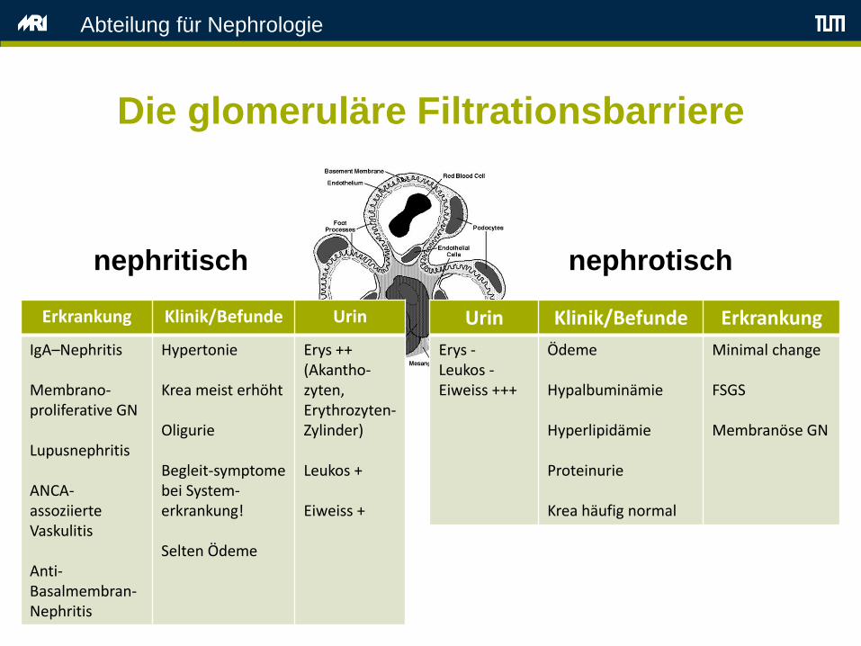
Die glomeruläre Filtrationsbarriere
nephrotischnephritisch
Urin Klinik/Befunde Erkrankung
Erys -Leukos -Eiweiss +++
Ödeme
Hypalbuminämie
Hyperlipidämie
Proteinurie
Krea häufig normal
Minimal change
FSGS
Membranöse GN
Erkrankung Klinik/Befunde Urin
IgA–Nephritis
Membrano-proliferative GN
Lupusnephritis
ANCA-assoziierte Vaskulitis
Anti-Basalmembran-Nephritis
Hypertonie
Krea meist erhöht
Oligurie
Begleit-symptome bei System-erkrankung!
Selten Ödeme
Erys ++(Akantho-zyten,Erythrozyten-Zylinder)
Leukos +
Eiweiss +
Abteilung für Nephrologie
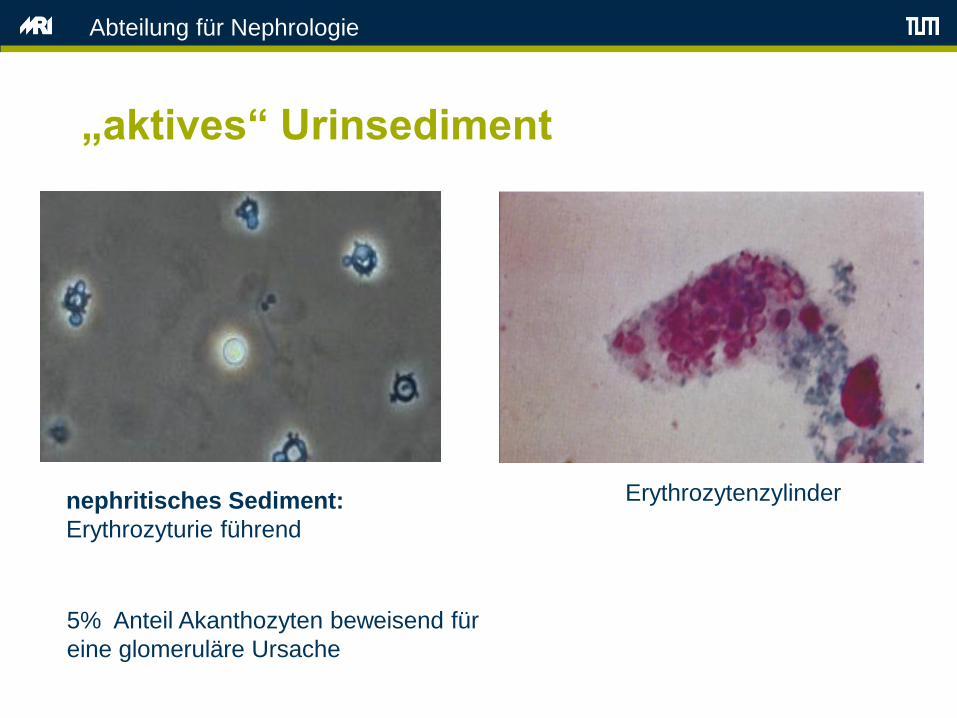
„aktives“ Urinsediment
nephritisches Sediment:
Erythrozyturie führend
5% Anteil Akanthozyten beweisend für
eine glomeruläre Ursache
Erythrozytenzylinder
Abteilung für Nephrologie
Ursachen einer glomerulären Hämaturie
• Hereditäre Erkrankungen
• Alport-Syndrom
• Syndrom der dünnen Basalmembran
• Primäre Glomerulonephritiden
• IgA-Nephritis
• Membranoproliferative GN
• Glomerulonephritiden bei Systemerkrankungen
• SLE
• Vaskulitis-Syndrome
• Goodpasture-Syndrom
• Infektiöse/parinfektiöse/postinfektiöse Glomerulonephritiden
• Post-Streptokokken GN
• Hepatitis B/C
• Malaria
• Endokarditis

Abteilung für Nephrologie
Rapid progressive Glomerulonephritis
• Definition:
Glomerulonephritis mit Nierenfunktionsverlust um 50% innerhalb
weniger Tage bis zu 3 Monaten
„Halbmondbildung“

Abteilung für Nephrologie
„Halbmonde“

Abteilung für Nephrologie
Patient 2
• 58 j. Patient
• AZ-Verschlechterung, Dyspnoe, Hämoptoe
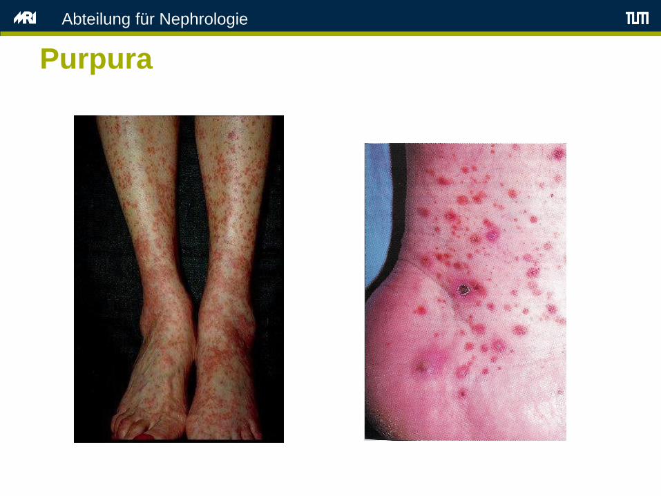
• RR 180/100, Purpura
• Blutwerte: Kreatinin 4,3 mg/dl; Harnstoff-N 68 mg/dl, Hb
9,8 g/l
• Sediment: Erythrozyturie, Akanthozyten, hyaline
Zylinder, Erythrozytenzylinder
• Proteinurie: 0,9 g/d
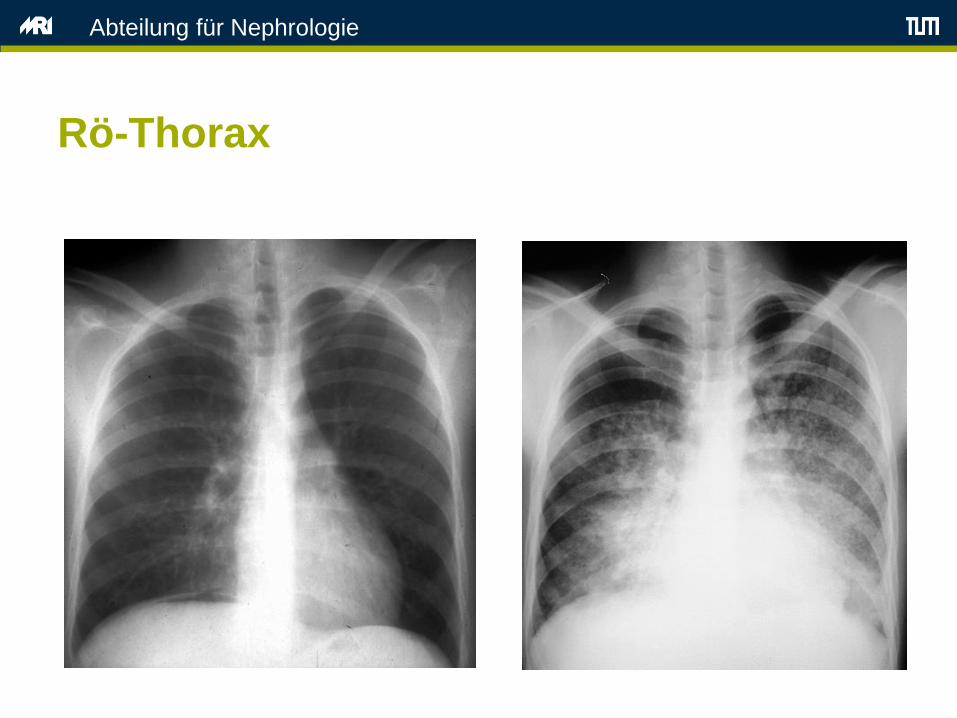
• Röntgen des Thorax: diffuse Verschattungen bds.
Abteilung für Nephrologie
Purpura

Abteilung für Nephrologie
Purpura
Abteilung für Nephrologie
Rö-Thorax
Abteilung für Nephrologie
Patient 2
• Akutes Nierenversagen oder chron. Niereninsuffizienz?
• Weitere Diagnostik zur Eingrenzung der Erkrankung?

Abteilung für Nephrologie
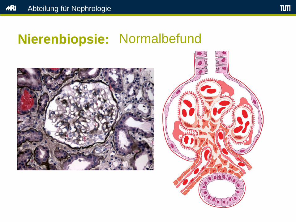
Nierenbiopsie
• Goldstandard für die Diagnose akuter Glomerulonephritiden
• Durchführung in Lokalanästhesie, Ultraschall-gesteuert unter
stationären Bedingungen
• Komplikationen: Makrohämaturie, AV-Fisteln, perirenales
Hämatom, Verletzung von Nachbarorganen (Darm, Leber)
• Histologische Untersuchungsschritte:
– Lichtmikroskopie
– Immunfluoreszenz
– Elektronenmikroskopie
Abteilung für Nephrologie
NierenbiopsieNierenbiopsie: Normalbefund

Abteilung für Nephrologie
Nierenbiopsie Patient 2:
„Halbmondnephritis“ Histologie der RPGN

Abteilung für Nephrologie
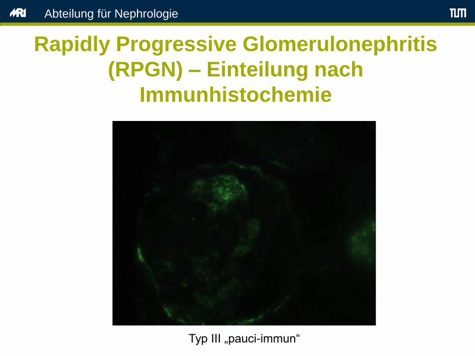
Rapidly Progressive Glomerulonephritis
(RPGN) – Einteilung nach
Immunfluoreszenz
• Type I – Antikörper-vermittelt
– Anti-GBM AK/ Goodpasture Syndrom
• Type II – Immunkomplex-vermittelt
– Lupus nephritis, IgA-Nephropathie, MPGN
• Type III – “pauci-immune”
– Granulomatose mit Polyangiitis (Wegener)
– Mikroskopische Polyangiitis

Abteilung für Nephrologie
Rapidly Progressive Glomerulonephritis
(RPGN) – Einteilung nach
Immunhistochemie
Typ I lineare IgG-Ablagerungen

Abteilung für Nephrologie
Rapidly Progressive Glomerulonephritis
(RPGN) – Einteilung nach
Immunhistochemie
Typ II granuläre Immunkomplexablagerungen
Abteilung für Nephrologie
Rapidly Progressive Glomerulonephritis
(RPGN) – Einteilung nach
Immunhistochemie
Typ III „pauci-immun“
Abteilung für Nephrologie
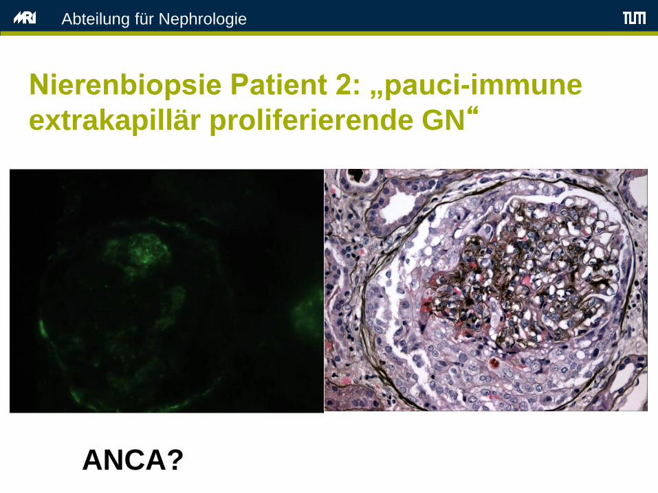
Nierenbiopsie Patient 2: „pauci-immune
extrakapillär proliferierende GN“
ANCA?
Abteilung für Nephrologie
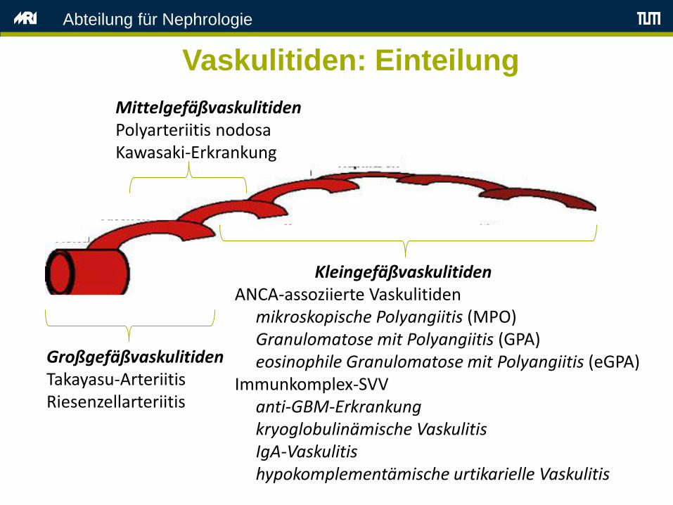
Vaskulitiden: Einteilung
GroßgefäßvaskulitidenTakayasu-ArteriitisRiesenzellarteriitis
MittelgefäßvaskulitidenPolyarteriitis nodosaKawasaki-Erkrankung
KleingefäßvaskulitidenANCA-assoziierte Vaskulitiden
mikroskopische Polyangiitis (MPO)Granulomatose mit Polyangiitis (GPA)eosinophile Granulomatose mit Polyangiitis (eGPA)
Immunkomplex-SVVanti-GBM-Erkrankungkryoglobulinämische VaskulitisIgA-Vaskulitishypokomplementämische urtikarielle Vaskulitis

Abteilung für Nephrologie
Vaskulitiden: Nierenbeteiligung
GroßgefäßvaskulitidenTakayasu-ArteriitisRiesenzellarteriitis
MittelgefäßvaskulitidenPolyarteriitis nodosaKawasaki-Erkrankung
KleingefäßvaskulitidenANCA-assoziierte Vaskulitiden
mikroskopische Polyangiitis (MPO)Granulomatose mit Polyangiitis (GPA)eosinophile Granulomatose mit Polyangiitis (eGPA)
Immunkomplex-Vaskulitidenanti-GBM-Erkrankungkryoglobulinämische VaskulitisIgA-Vaskulitishypokomplementämische urtikarielle Vaskulitis
Abteilung für Nephrologie
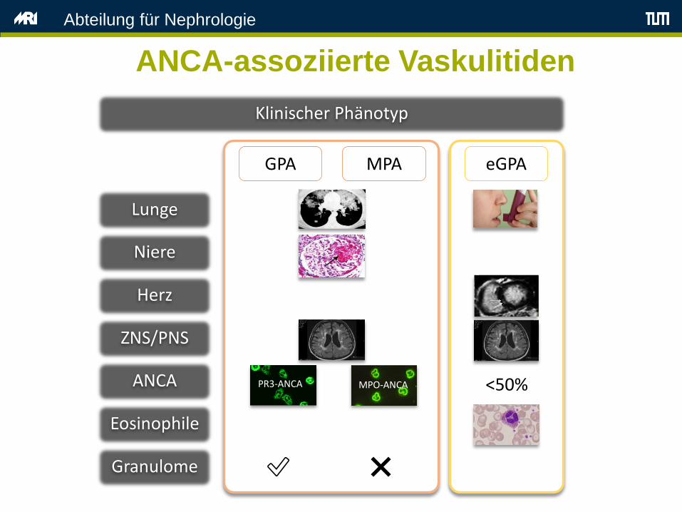
ANCA-assoziierte Vaskulitiden
Pathogenese Klinischer Phänotyp
GPA MPA eGPA
Lunge
Niere
Herz
ANCA
Eosinophile
ZNS/PNS
SEC
TIO
N 1
1 ● T
HE
VA
SCU
LITI
DES
1550
KidneysRenal disease is an ominous clinical manifestation of WG. Although
less than 20% of patients with WG have renal involvement at the time of diagnosis, nearly 80% develop this complication at some point in their course. 9 T he clinical presentation of renal disease in WG is rapidly progressive glomer ulonephritis: hematuria, red blood cell casts, proteinuria (usually non-nephrotic), and rising serum creatinine. In addition to glomer ular disease, patients with WG often have substan -tial intersti tial kidney disease. Without appropriate therapy, ir revers-ible loss of renal function may ensue within days to weeks. T hus the appearance of active urine sediment or a rise in serum creatinine in WG signals the need for prompt, aggressive treatment.
OTHER MANIFESTATIONS
Consti tutional symptoms such as fever and weight loss, common in WG, serve as important indicators of an active inflammator y process.
T hese symptoms, however , rarely occur in isolation. Prominent mus-culosk eletal symptoms occur in 60% of patients. Arthralgias are more common than frank arthri tis but true synovitis does occur. T he typical patter n of joint involvement is migrator y and oligoarticular , often involving large joints, but polyarthri tis also occurs. In addition, cutane-ous nodules (“cutaneous extravascular necrotizing granulomas” 16 (Fig. 153.11) may occur at si tes that are also common locations for rheu -matoid nodules. Because approximately one third of patients with WG are rheumatoid factor positive, rheumatoid arthri tis is a common mis-diagnosis early in the disease course. Assays for antibodies to cyclic ci tr ullinated peptides, which do not occur in WG, can help distinguish the arthri tis of WG from that of rheumatoid arthri tis.
In addition to nodules (which are frequently overlook ed), skin find-
ings in WG include all the potential manifestations of cutaneous vasculi tis: palpable purpura, vesiculobullous lesions, papules, ulcers, digital infarctions, and splinter hemor rhages. N ervous system disease, though present in only a minority of patients at diagnosis, may be severe. Vasculi tic neuropathy may lead to a devastating mononeuritis multiplex (Fig. 153.12) and/or a disabling sensor y polyneuropathy . Central ner vous system abnor mali ties occur in approximately 8% of patients, usually as cranial neuropathies, mass lesions, or pachymen -ingitis (Fig. 153.13). Parenchymal brain involvement by vasculi tis is uncommon in WG but has been described. Rare neuroendocrine com -plications of WG include panhypopituitarism 17 and diabetes insipi -dus.18 WG may also mimic giant cell arteri tis, with prominent headaches and symptoms that recall polymyalgia rheumatica.
Finally, with regard to pulmonar y disease, clinicians must be vigi -lant to the possibi li ty of pulmonar y embolism in WG. Patients with this disease are highly susceptible to deep venous thromboses and pulmonar y emboli . 15 T he risk of venous thrombotic events in WG is believed to result from the involvement of veins by vascular inflamma-tion, as well as the risk factors associated with debi li ty and immobili ty , significant degrees of proteinuria, and possibly other factors.
Fig. 153.8 Subglottic stenosis. A web of scar tissue is eviden t just below the
vocal chords, leading to narrowing of the subglottic area and inspiratory
stridor.
Fig. 153.9 Comput ed tomography scan of the chest in Wegener
granulomatosis. Multiple bilateral pulmonar y nodules can be seen, many of
which have cavitated.
Fig. 153.10 Alveolar hemorrhage in a patient with Wegener granulomatosis.
This has result ed in rapidly chang ing pulmonar y inf ltrates. There is also a
nodular lesion in the right lung.
contrast, T2 values between both AAV subgroups (EGPA
and GPA) showed no significant difference (p = 0.85).
These results suggest a common pathway of myocardial
involvement in AAV patients, reflecting a combination of
both ongoing inflammation and fibrosis, which is sup-
ported by histology in the literature [3, 25].
Values above the 95% percentile of normal
Despite highly significant differences between patients
and controls, there is some overlap in T1, ECV, and T2
values, which seem to lower the diagnostic accuracy in
the individual AAV patient. Defining the 95% percentile
of the control group as threshold for definite abnormal
Fig. 2 CMRof a 77-year-old female with EGPA (BVAS= 4), presenting with palpitations and atrial fibrillation. Cine images (a) revealed normal
LV-EF (66%), LGE(b) detected intramural enhancement in the inferior septum (white arrows), suggestive of cardiac involvement. Native T1 map (c)
showed increased T1 with 980 ms, shortened post-contrast T1 (d) with 452 ms, increased ECV (e) of 37%, and higher T2 (f) (52 ms) compared
to controls
Fig. 3 CMRof a 26-year-old female with a history of EGPA for 3 years with the same BVAS(=4) as the patient from Fig. 2. She was suffering from
palpitations and dyspnea, ECG was normal. Cine images (a) showed a preserved LV-EF (67%), LGE images (b) were negative. However, native T1
(1019 ms, c), ECV (27%, e), and T2 (52 ms, f) were increased compared to controls, suggesting myocardial involvement despite normal LV-EF, and
unremarkable ECG
Greulich et al. Journal of Cardiovascular Magnetic Resonance (2017) 19:6 Page 8 of 12
MPO-ANCAPR3-ANCA
ANCA-assoziierte Vaskulitiden (AAV)
Abteilung für Nephrologie
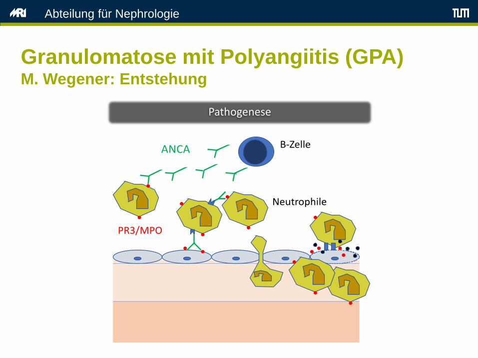
Im m unpathogenese - allgem ein
ANCA
PR3/MPO
B-Zelle
Neutrophile
<50%
Granulome ✅ ❌

Abteilung für Nephrologie
„Neutrophil extracellular traps“
Brinkmann et al.
Science 2004

Abteilung für Nephrologie
NETose bei ANCA Vaskulitis
Kessenbrock et al. Nature Med. 2009

Abteilung für Nephrologie
Granulomatose mit Polyangiitis (GPA)
Wegener Granulomatose
• Männer = Frauen
• mittleres Alter 40 Jahre
• ca. 30 % ätiologisch unklarer ANV sind auf eine RPGN (rapid
progressive Glomerulonephritis) aus dem Formenkreis von
Wegener zurückzuführen.
Definition
– Vaskulitis an kleinen Gefäßen (fokal-nekrotisierend)
– nekrotisierende Granulome (bes. Respirationstrakt)
– keine Immunglobulin-Ablagerungen in Glomeruli („pauci-
immun“)
Abteilung für Nephrologie
Granulomatose mit Polyangiitis (GPA)M. Wegener: Entstehung
• Auslösend möglicherweise Medikamente, Bakterien (Staph. aureus?)
• ANCA (antineutrophile zytoplasmatische Antikörper):
– Zwei Arten: p-ANCA, c-ANCA
– Korrelation mit Krankheitsverlauf
– Aktivierung von neutrophilen Granulozyten und Endothelzellen
notwendig
Abteilung für Nephrologie
Granulomatose mit Polyangiitis (GPA)M. Wegener: Entstehung
Pathogenese Klinischer Phänotyp
GPA MPA eGPA
Lunge
Niere
Herz
ANCA
Eosinophile
ZNS/PNS
SEC
TIO
N 1
1 ● T
HE
VASC
ULI
TID
ES
1550
KidneysRenal disease is an ominous clinical manifestation of WG. Although less than 20% of patients with WG have renal involvement at the time of diagnosis, nearly 80% develop this complication at some point in their course. 9 T he clinical presentation of renal disease in WG is rapidly progressive glomer ulonephritis: hematuria, red blood cell casts, proteinuria (usually non-nephrotic), and rising serum creatinine. In
addition to glomer ular disease, patients with WG often have substan -tial intersti tial kidney disease. Without appropriate therapy, ir revers-ible loss of renal function may ensue within days to weeks. T hus the appearance of active urine sediment or a rise in serum creatinine in WG signals the need for prompt, aggressive treatment.
OTHER MANIFESTATIONS
Consti tutional symptoms such as fever and weight loss, common in WG, serve as important indicators of an active inflammator y process. T hese symptoms, however , rarely occur in isolation. Prominent mus-culosk eletal symptoms occur in 60% of patients. Arthralgias are more common than frank arthri tis but true synovitis does occur. T he typical patter n of joint involvement is migrator y and oligoarticular , often involving large joints, but polyarthri tis also occurs. In addition, cutane-
ous nodules (“cutaneous extravascular necrotizing granulomas” 16 (Fig. 153.11) may occur at si tes that are also common locations for rheu -matoid nodules. Because approximately one third of patients with WG are rheumatoid factor positive, rheumatoid arthri tis is a common mis-diagnosis early in the disease course. Assays for antibodies to cyclic ci tr ullinated peptides, which do not occur in WG, can help distinguish the arthri tis of WG from that of rheumatoid arthri tis.
In addition to nodules (which are frequently overlook ed), skin find-ings in WG include all the potential manifestations of cutaneous vasculi tis: palpable purpura, vesiculobullous lesions, papules, ulcers, digital infarctions, and splinter hemor rhages. N ervous system disease, though present in only a minority of patients at diagnosis, may be severe. Vasculi tic neuropathy may lead to a devastating mononeuritis
multiplex (Fig. 153.12) and/or a disabling sensor y polyneuropathy . Central nervous system abnor mali ties occur in approximately 8% of patients, usually as cranial neuropathies, mass lesions, or pachymen -ingitis (Fig. 153.13). Parenchymal brain involvement by vasculi tis is uncommon in WG but has been described. Rare neuroendocrine com -plications of WG include panhypopituitarism 17 and diabetes insipi -dus.18 WG may also mimic giant cell arteri tis, with prominent headaches and symptoms that recall polymyalgia rheumatica.
Finally, with regard to pulmonar y disease, clinicians must be vigi -
lant to the possibi li ty of pulmonar y embolism in WG. Patients with this disease are highly susceptible to deep venous thromboses and pulmonar y emboli . 15 T he risk of venous thrombotic events in WG is believed to result from the involvement of veins by vascular inflamma-tion, as well as the risk factors associated with debi li ty and immobili ty, significant degrees of proteinuria, and possibly other factors.
Fig. 153.8 Subglottic stenosis. A web of scar tissue is eviden t just below the
vocal chords, leading to narrowing of the subglottic area and inspiratory
stridor.
Fig. 153.9 Comput ed tomography scan of the chest in Wegener
granulomatosis. Multiple bilateral pulmonar y nodules can be seen, many of
which have cavitated.
Fig. 153.10 Alveolar hemorrhage in a patient with Wegener granulomatosis.
This has result ed in rapidly chang ing pulmonar y inf ltrates. There is also a
nodular lesion in the right lung.
contrast, T2 values between both AAV subgroups (EGPA
and GPA) showed no significant difference (p = 0.85).
These results suggest a common pathway of myocardial
involvement in AAV patients, reflecting a combination of
both ongoing inflammation and fibrosis, which is sup-
ported by histology in the literature [3, 25].
Values above the 95% percentile of normal
Despite highly significant differences between patients
and controls, there is some overlap in T1, ECV, and T2
values, which seem to lower the diagnostic accuracy in
the individual AAV patient. Defining the 95% percentile
of the control group as threshold for definite abnormal
Fig. 2 CMRof a 77-year-old female with EGPA (BVAS= 4), presenting with palpitations and atrial fibrillation. Cine images (a) revealed normal
LV-EF (66%), LGE(b) detected intramural enhancement in the inferior septum (white arrows), suggestive of cardiac involvement. Native T1 map (c)
showed increased T1 with 980 ms, shortened post-contrast T1 (d) with 452 ms, increased ECV (e) of 37%, and higher T2 (f) (52 ms) compared
to controls
Fig. 3 CMRof a 26-year-old female with a history of EGPA for 3 years with the same BVAS(=4) as the patient from Fig. 2. She was suffering from
palpitations and dyspnea, ECG was normal. Cine images (a) showed a preserved LV-EF (67%), LGEimages (b) were negative. However, native T1
(1019 ms, c), ECV (27%, e), and T2 (52 ms, f) were increased compared to controls, suggesting myocardial involvement despite normal LV-EF, and
unremarkable ECG
Greulich et al. Journal of Cardiovascular Magnetic Resonance (2017) 19:6 Page 8 of 12
MPO-ANCAPR3-ANCA
ANCA-assoziierte Vaskulitiden (AAV)
Abteilung für Nephrologie
Im m unpathogenese - allgem ein
ANCA
PR3/MPO
B-Zelle
Neutrophile
<50%
Granulome
Abteilung für Nephrologie
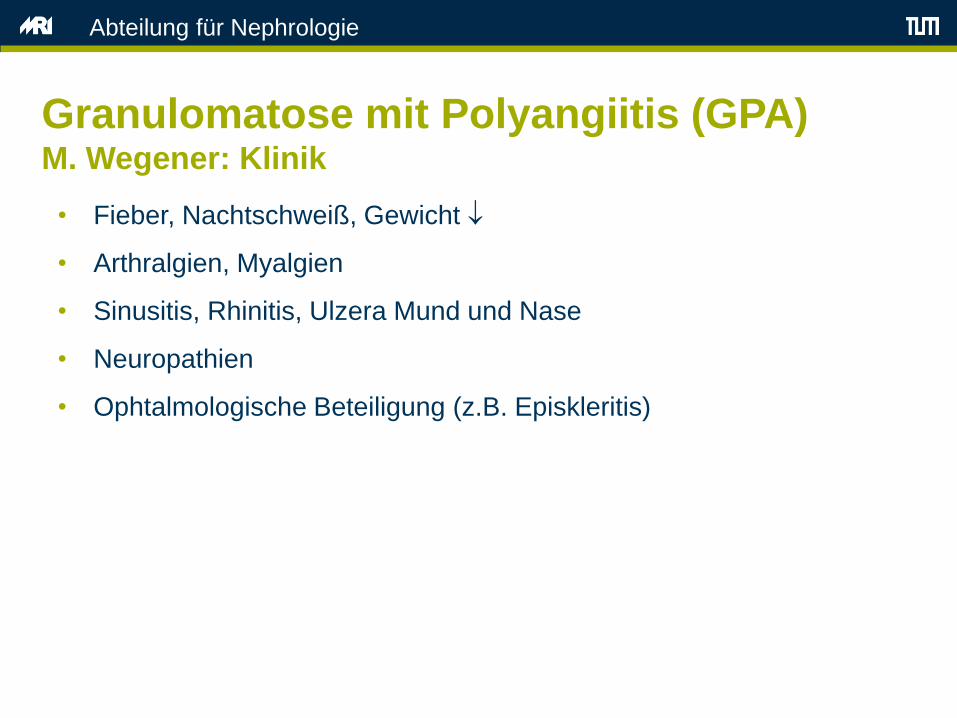
• Fieber, Nachtschweiß, Gewicht
• Arthralgien, Myalgien
• Sinusitis, Rhinitis, Ulzera Mund und Nase
• Neuropathien
• Ophtalmologische Beteiligung (z.B. Episkleritis)
Granulomatose mit Polyangiitis (GPA)M. Wegener: Klinik

Abteilung für Nephrologie
• Renal:
– Mikrohämaturie, RPGN
• Pulmonal:
– Hämoptysen,
– pulmorenales Syndrom (Befall v. Lunge und Niere i. S. einer
Vaskulitis und rasch fortschreitender Niereninsuffizienz).
• Haut:
– Purpura
Granulomatose mit Polyangiitis (GPA)M. Wegener: Klinik

Abteilung für Nephrologie
ANCA(antineutrophile zytoplasmatische Autoantikörper)
• c-ANCA:
Proteinase-3 (Wegener)
• p-ANCA:
Myeloperoxidase
(mikroskop. Polyangiitis)
[aus: Comprehensive Nephrology]
Abteilung für Nephrologie
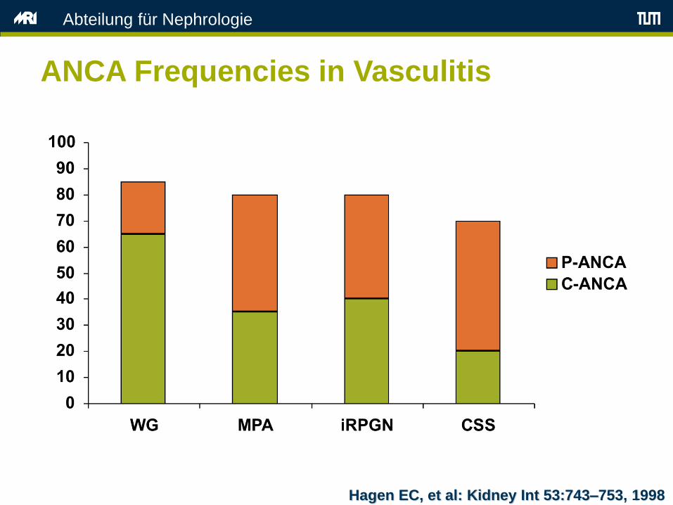
ANCA Frequencies in Vasculitis
Hagen EC, et al: Kidney Int 53:743–753, 1998

Abteilung für Nephrologie
ANCA Vaskulitis - Therapieprinzip
Remissionsinduktion
Ca. 6-9 Monate Remissionserhaltung (mind. 2 Jahre)
• Glukokortikoide
• Cyclophosphamid
• Rituximab
• Plasmapherese
• Avacopan?
• Azathioprin
• Mycophenolat
• Methotrexat
• Leflunomid
• Rituximab
Abteilung für Nephrologie
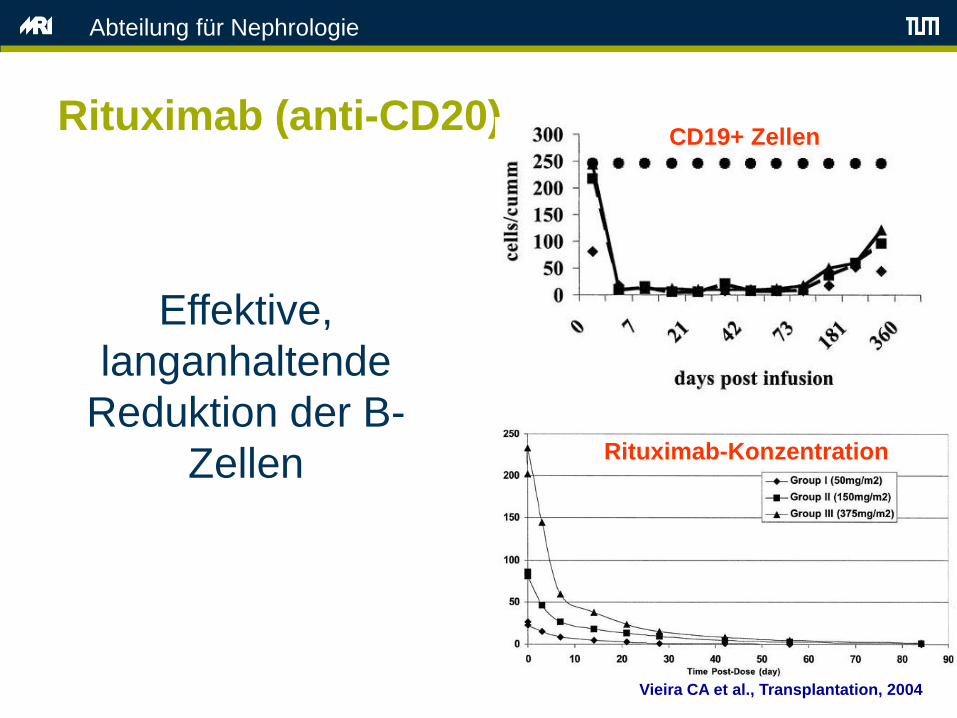
Rituximab (anti-CD20)
Effektive,
langanhaltende
Reduktion der B-
Zellen
Vieira CA et al., Transplantation, 2004
CD19+ Zellen
Rituximab-Konzentration

Abteilung für Nephrologie
Rituximab und Vaskulitis
Eriksson P, J Int Med, 2005
Krankheitsaktivität
vor Therapie
nach Therapie
Abteilung für Nephrologie
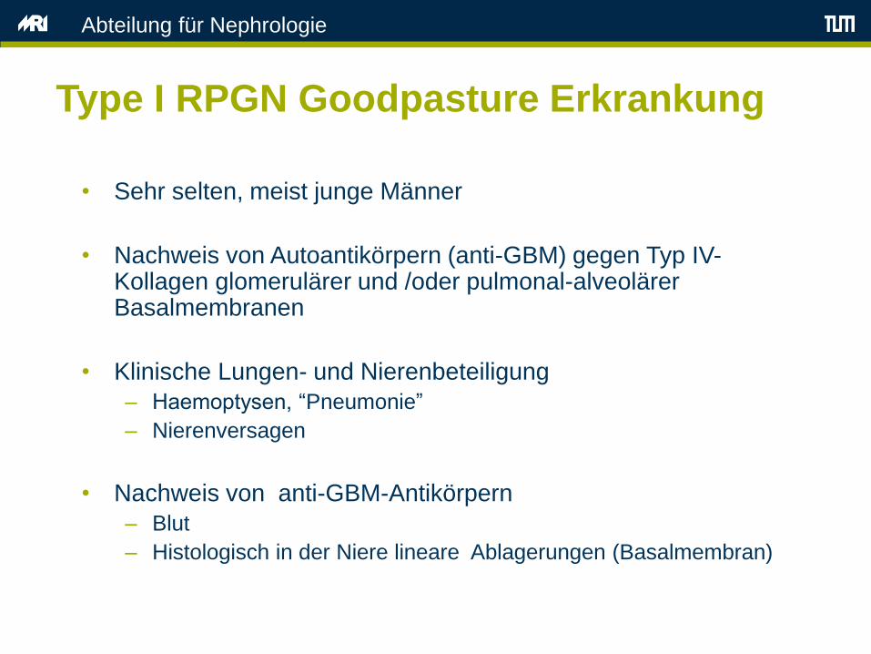
Type I RPGN Goodpasture Erkrankung
• Sehr selten, meist junge Männer
• Nachweis von Autoantikörpern (anti-GBM) gegen Typ IV-Kollagen glomerulärer und /oder pulmonal-alveolärer Basalmembranen
• Klinische Lungen- und Nierenbeteiligung
– Haemoptysen, “Pneumonie”
– Nierenversagen
• Nachweis von anti-GBM-Antikörpern
– Blut
– Histologisch in der Niere lineare Ablagerungen (Basalmembran)

Abteilung für Nephrologie
Type I: Goodpasture Erkrankung
„Halbmond“ anti-GBM--Immunhisochemie

Abteilung für Nephrologie
Type I RPGN Goodpasture Erkrankung
Therapie:
• Steroidbolustherapie
• Plasmapherese zur schnelleren Ak- Eliminination
• Cyclophosphamidbolustherapie für 6-9 Monaten
• Trotz Therapie häufig rasche Progression zur
dialysepflichtigen Niereninsuffizienz
Abteilung für Nephrologie
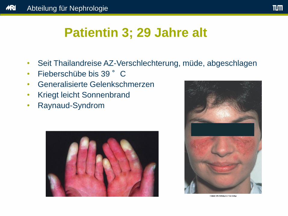
Patientin 3; 29 Jahre alt
• Seit Thailandreise AZ-Verschlechterung, müde, abgeschlagen
• Fieberschübe bis 39 °C
• Generalisierte Gelenkschmerzen
• Kriegt leicht Sonnenbrand
• Raynaud-Syndrom
Abteilung für Nephrologie
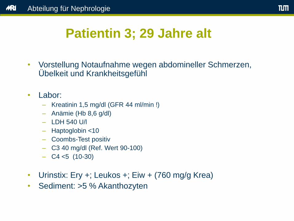
Patientin 3; 29 Jahre alt
• Vorstellung Notaufnahme wegen abdomineller Schmerzen, Übelkeit und Krankheitsgefühl
• Labor: – Kreatinin 1,5 mg/dl (GFR 44 ml/min !)
– Anämie (Hb 8,6 g/dl)
– LDH 540 U/l
– Haptoglobin <10
– Coombs-Test positiv
– C3 40 mg/dl (Ref. Wert 90-100)
– C4 <5 (10-30)
• Urinstix: Ery +; Leukos +; Eiw + (760 mg/g Krea)
• Sediment: >5 % Akanthozyten

Abteilung für Nephrologie
Patientin 3; 29 Jahre alt
• ANA 1:25.000
• ds DNS-Ak positive
• Nukleosomen Ak positive
• Histon Ak positive
• V.a. systemischen Lupus erythematodes mit Lupusnephritis
• Nierenbiopsie
Abteilung für Nephrologie
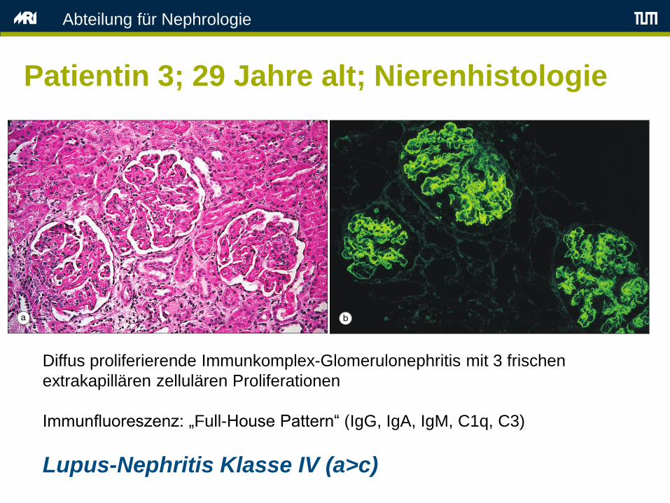
Patientin 3; 29 Jahre alt; Nierenhistologie
Diffus proliferierende Immunkomplex-Glomerulonephritis mit 3 frischen
extrakapillären zellulären Proliferationen
Immunfluoreszenz: „Full-House Pattern“ (IgG, IgA, IgM, C1q, C3)
Lupus-Nephritis Klasse IV (a>c)

Abteilung für Nephrologie
Systemischer Lupus erythematodes -
Pathogenese

Abteilung für Nephrologie
Systemischer Lupus erythematodes -
Manifestationen

Abteilung für Nephrologie
Systemischer Lupus erythematodes -
Manifestationen
Hautbefall (>50%)

Abteilung für Nephrologie
Systemischer Lupus erythematodes -
Nierenbeteiligung
über 70% der SLE-Patienten erleiden im Laufe
ihrer Erkrankung eine Nierenbeteiligung
Abteilung für Nephrologie
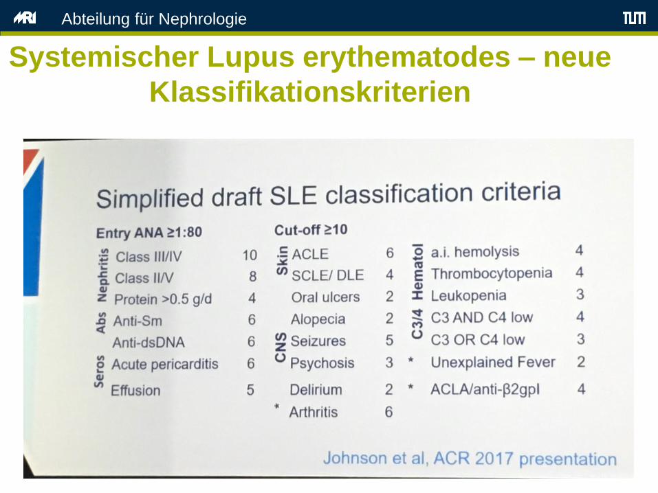
Systemischer Lupus erythematodes – neue
Klassifikationskriterien

Abteilung für Nephrologie
Lupusnephritis - Therapie
Remissionsinduktion
Ca. 3-6 Monate Remissionserhaltung (mind. 2 Jahre)
• Glukokortikoide
• Mycophenolat mofetil
• Cyclophosphamid
• Rituximab
• Mycophenolat mofetil
• Azathioprin
• Rituximab
Abteilung für Nephrologie
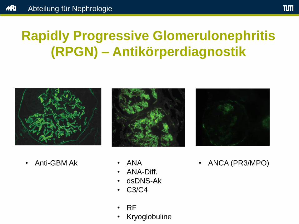
Rapidly Progressive Glomerulonephritis
(RPGN) – Antikörperdiagnostik
• Anti-GBM Ak • ANA
• ANA-Diff.
• dsDNS-Ak
• C3/C4
• RF
• Kryoglobuline
• ANCA (PR3/MPO)

Abteilung für Nephrologie
Take Home Message (RPGN)
1. Die rapid progressive Glomerulonephritis ist ein klinischer Begriff
2. Die RPGN umfasst verschiedene Krankheitsbilder
3. Ursachen sind häufig Systemerkrankungen und Vaskulitiden
4. In der Nierenbiopsie: Halbmond-Bildung
5. Frühzeitige, aggressive Therapie notwendig
Abteilung für Nephrologie
Die glomeruläre Filtrationsbarriere
nephrotischnephritisch
Urin Klinik/Befunde Erkrankung
Erys -Leukos -Eiweiss +++
Ödeme
Hypalbuminämie
Hyperlipidämie
Proteinurie
Krea häufig normal
Minimal change
FSGS
Membranöse GN
Erkrankung Klinik/Befunde Urin
IgA–Nephritis
Membrano-proliferative GN
Lupusnephritis
ANCA-assoziierte Vaskulitis
Anti-Basalmembran-Nephritis
Hypertonie
Krea meist erhöht
Oligurie
Begleit-symptome bei System-erkrankung!
Selten Ödeme
Erys ++(Akantho-zyten,Erythrozyten-Zylinder)
Leukos +
Eiweiss +


![Chapter 11 [blood abnormalities n diseases]](https://static.fdocument.pub/doc/165x107/5477db1fb4af9f7a0f8b45fd/chapter-11-blood-abnormalities-n-diseases.jpg)