ISOLATION AND CHARACTERIZATION OF PSEUDOMONAS …
10
www.ijcrt.org © 2020 IJCRT | Volume 8, Issue 6 June 2020 | ISSN: 2320-2882 IJCRT2006327 International Journal of Creative Research Thoughts (IJCRT) www.ijcrt.org 2399 ISOLATION AND CHARACTERIZATION OF PSEUDOMONAS FLUORESCENS STRAINS FROM THE DIFFERENT REGIONS OF CHAKRATA, JAUNSAR-BAWAR, UTTARAKHAND Saif Ali* 1 , Babali Semwal* 1 , Keerti Singh 1 , Bhavtosh Sharma 2 , Bipin K Sati 2 1. SGRRU (Shri Guru Ram Rai University), Dehradun, Uttarakhand, India 2. USERC (Uttarakhand Science Education and Research Centre), Dehradun, Uttarakhand, India Abstract: Chakrata, Jaunsar-Bawar region is conferred by diverse wealth in terms of flora. The flora is so wealthy just because of the microorganism which inhabits the soil. Pseudomonas fluorescens belongs to the PGPR (plant-growth promoting rhizobacteria), which plays a major role in plant growth promotion and reduce severity of various diseases, bio-control agent as well as antagonist, and a potential biosurfactant producer. A total of six soil samples were collected from different villages of chakrata. Dilutions of soil sample were made and isolated on the nutrient agar medium followed by identification through Gram’s staining and different biochemical analysis. Different tests were performed for each isolated bacterias from different soil samples. Out of which four samples showed the presence of Pseudomonas fluorescens strains according to the Bergey’s manual of determinative bacteriology. Seeds of black-eyed peas(lobia) and okra(bhindi) were treated with isolated strains of P.fluorescens . After treating them, seeds were sown in the pots. A pot as a control is taken for the comparison of growth between inoculated and uninoculated seeds. The inoculated seeds showed positive growth than control. The present study indicates that the presence of Pseudomonas species in soils of chakrata is selective in nature and supports the well growth of selected plants and this is one of the main reasons for the well productivity of different varieties of crops and medicinal plants in chakrata, jaunsar-bawar areas. Keywords- Biofertilizers, PGPR, Chakrata, Pseudomonas fluorescens Introduction: The bacteria which colonize the roots of the plants and enhance plant growth are referred to as plant growth promoting rhizobacteria (PGPR). Pseudomonas fluorescens is one of the PGPR that inhabits soil, plants and water surfaces. This rhizobacterium is well adapted to grow in the rhizosphere. It is a Gram-negative rod shaped bacteria with optimum growth temperature between 25-30 0 C. The Pf-5 strain resides in the plants rhizosphere and produce a variety of secondary metabolites including antibiotic against some soil borne pathogens while PFO-1 strain is well adapted to the soil where it was first isolated in agricultural species that contributes greatly to the turnover of organic matter and while present in soil, is abundant on the surfaces of plants they get nutrients and environmental protection . In exchange, they also destroy things that might be potentially harmful to the plants. These things include toxins and pollutants like TNT, styrene, and polycyclic hydrocarbons. P.fluorescens can multiply very quickly , this means they can rapidly colonize a space and indirectly protect a crop. Certain members of P.fluorescens have been shown to be potential agents for the biocontrol which suppress plant diseases by protecting the seeds and roots from fungal infections (Hoffland et al.1996 , Wei et al.1996). This effect is the result of the antibiotics, siderophores and hydrogen
Transcript of ISOLATION AND CHARACTERIZATION OF PSEUDOMONAS …

www.ijcrt.org © 2020 IJCRT | Volume 8, Issue 6 June 2020 | ISSN: 2320-2882
IJCRT2006327 International Journal of Creative Research Thoughts (IJCRT) www.ijcrt.org 2399
ISOLATION AND CHARACTERIZATION OF
PSEUDOMONAS FLUORESCENS STRAINS FROM
THE DIFFERENT REGIONS OF CHAKRATA,
JAUNSAR-BAWAR, UTTARAKHAND
Saif Ali*1, Babali Semwal*1, Keerti Singh1, Bhavtosh Sharma2, Bipin K Sati2
1. SGRRU (Shri Guru Ram Rai University), Dehradun, Uttarakhand, India
2. USERC (Uttarakhand Science Education and Research Centre), Dehradun, Uttarakhand, India
Abstract:
Chakrata, Jaunsar-Bawar region is conferred by diverse wealth in terms of flora. The flora is so wealthy just because
of the microorganism which inhabits the soil. Pseudomonas fluorescens belongs to the PGPR (plant-growth
promoting rhizobacteria), which plays a major role in plant growth promotion and reduce severity of various
diseases, bio-control agent as well as antagonist, and a potential biosurfactant producer. A total of six soil samples
were collected from different villages of chakrata. Dilutions of soil sample were made and isolated on the nutrient
agar medium followed by identification through Gram’s staining and different biochemical analysis. Different tests
were performed for each isolated bacterias from different soil samples. Out of which four samples showed the
presence of Pseudomonas fluorescens strains according to the Bergey’s manual of determinative bacteriology. Seeds
of black-eyed peas(lobia) and okra(bhindi) were treated with isolated strains of P.fluorescens . After treating them,
seeds were sown in the pots. A pot as a control is taken for the comparison of growth between inoculated and
uninoculated seeds. The inoculated seeds showed positive growth than control. The present study indicates that the
presence of Pseudomonas species in soils of chakrata is selective in nature and supports the well growth of selected
plants and this is one of the main reasons for the well productivity of different varieties of crops and medicinal plants
in chakrata, jaunsar-bawar areas.
Keywords- Biofertilizers, PGPR, Chakrata, Pseudomonas fluorescens
Introduction:
The bacteria which colonize the roots of the plants and enhance plant growth are referred to as plant growth
promoting rhizobacteria (PGPR). Pseudomonas fluorescens is one of the PGPR that inhabits soil, plants and water
surfaces. This rhizobacterium is well adapted to grow in the rhizosphere. It is a Gram-negative rod shaped bacteria
with optimum growth temperature between 25-300 C. The Pf-5 strain resides in the plants rhizosphere and produce
a variety of secondary metabolites including antibiotic against some soil borne pathogens while PFO-1 strain is well
adapted to the soil where it was first isolated in agricultural species that contributes greatly to the turnover of organic
matter and while present in soil, is abundant on the surfaces of plants they get nutrients and environmental protection
. In exchange, they also destroy things that might be potentially harmful to the plants. These things include toxins
and pollutants like TNT, styrene, and polycyclic hydrocarbons. P.fluorescens can multiply very quickly , this means
they can rapidly colonize a space and indirectly protect a crop. Certain members of P.fluorescens have been shown
to be potential agents for the biocontrol which suppress plant diseases by protecting the seeds and roots from fungal
infections (Hoffland et al.1996 , Wei et al.1996). This effect is the result of the antibiotics, siderophores and hydrogen

www.ijcrt.org © 2020 IJCRT | Volume 8, Issue 6 June 2020 | ISSN: 2320-2882
IJCRT2006327 International Journal of Creative Research Thoughts (IJCRT) www.ijcrt.org 2400
cyanide (O’Sullivan & O’Gara 1992). Of the plant colonizing strains, some isolates are known to positively affect
plant health and nutrition. It usually affects the patients which are immuno-compromised and they also produces
phenazine, phenazine carboxylic acid, 2,4-diacetylphloroglucinol and the MRSA (methicillin resistant
staphylococcus aureus) active antibiotic mupirocin.
Materials and Methods
Experiment location and soil sample collection
This experiment was conducted at USERC (Uttarakhand Science Education and Research centre) and SGRR
University, Dehradun, Uttarakhand. The soil samples were collected from the different sites of Chakrata – Kanasar,
Haiya, Sahiya, Jutapa, Kalsi and Ragula. The dilutions of different soil samples were made for isolation of
microorganism.
Morphological Characterization
Samples of different dilutions were spreaded on the nutrient agar and then isolated colonies were streaked on nutrient
gar petri-plates separately for colony development. Streaked plates were observed under UV transilluminator for
fluorescence produced by bacteria.
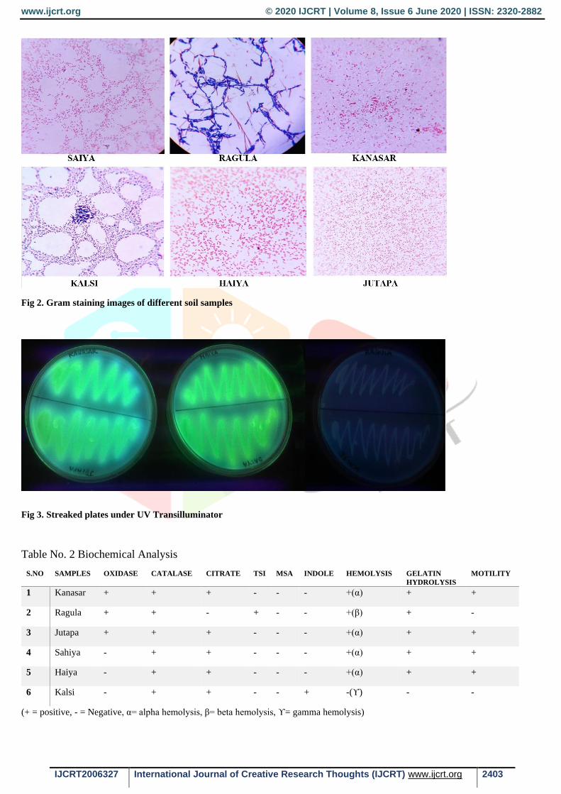
Identification by Gram’s Staining
This method is used to differentiate among Gram- Negative (pink) and Gram-Positive (purple) bacteria. It is based
on the ability of bacteria to retain the color of the stain used in Gram reaction. Gram-Negative bacteria were
decolorized by alcohol, losing the purple color of crystal violet, a counter stain safranin is used to impart a pink color
to the bacteria. But Gram-Positive bacteria were not decolorized by alcohol and remans as purple (Fig.2).
Biochemical test for Pseudomonas fluorescens
For identification of P.fluorescens , certain biochemical test were conducted according to the Bergey’s manual for
determinative bacteriology.
Catalase test
Bacteria produce catalase enzyme to protect themselves from lethal effect of hydrogen peroxide which is
accumulated as end product of aerobic carbohydrate metabolism. Use a sterile loop to transfer a small amount of
colony growth in the surface of a clean, dry glass slide. Add a drop of 3% hydrogen peroxide in the glass slide (Fig.
5). Evolution of bubbles shows positive catalase while no bubble formation shows catalase negative.
Oxidase test
The oxidase test is used to identify bacteria that produce cytochrome c oxidase enzyme. Soak the filter paper
(Whatman’s #1) in a 1% tetramethyl-p-phenylene-diamine dihydrochloride solution. Dry for about half a minute.
With the help of loop (platinum material or plastic stick), pick the colony and rub the culture on the filter paper (Fig.
4). Inoculated area of paper changes color from blue to dark purple within 10-30 seconds showing positive oxidase
test while no appearance of color shows oxidase negative.
Indole Test
It is used to determine the ability of an organism to split amino acid tryphtophan to form compound indole. Inoculate
the tryphtophan broth with broth culture. Incubate at 370 C for 24-48 hours in ambient air. After incubation, add 1-
2 drops of Kovac’s Reagent to the broth culture. Development of cherry-coloured ring shows indole positive.

www.ijcrt.org © 2020 IJCRT | Volume 8, Issue 6 June 2020 | ISSN: 2320-2882
IJCRT2006327 International Journal of Creative Research Thoughts (IJCRT) www.ijcrt.org 2401
Citrate Test
Citrate utilisation test is used to check the ability of an organism to utilize sodium citrate as a sole carbon source and
ammonium salts as a sole source of nitrogen. It is indicated by change in color of bromothymol blue indicator from
green to blue (Fig.6). Inoculate and incubate the simmons citrate agar by touching colony that is 18-24 hours old
with a inoculating loop.
OF(Oxidation-Fermentation) Test
Certain Gram-Negative bacteria metabolizes glucose by aerobic respiration (oxidatively). The high concentration of
acid produced during aerobic respiration will turn the bromothymol blue indicator in OF media from green to yellow
in the presence or absence of oxygen. Inoculate two test-tubes of OF medium with test organism by stabbing with
the help of straight wire and covering one tube of each pair with 1 cm of sterile liquid paraffin wax or sterile mineral
oil, while leave the other tube open to the air. Development of yellow colouration in open tube shows oxidative
utilisation of carbohydrate while fermentative utilisation of carbohydrates shows development of yellow colouration
in both open and closed tubes (Fig.10).
Gelatin Hydrolysis Test
Gelatin hydrolysis test is used to detect the ability of an organism to produce gelatinase (proteolytic enzyme) that
liquefy the gelatin. Inoculate a heavy inoculum of test bacteria (18-24 hours old) by stabbing 4-5 times (half inch)
on the tube containing nutrient gelatin medium. Incubate the inoculated tube along with an uninoculated medium at
350 C, or at the test bacterium’s optimal growth temperature for up to 2 weeks. Remove the tube daily from the
incubator and place in ice bath or refrigerator (40 C) for 15-30 minutes (until control is gelled) every day to check
for gelatin liquefaction (Fig.9). Gelatin normally liquefies at 280 C and above, so to confirm that liquefaction was
due to gelatinase activity (the tubes are immersed in an ice bath or kept in refrigerator at 40 C).
Blood Haemolysis Test
Blood agar contains general nutrients and 5% sheep blood is added to it after autoclaving. It is useful for cultivating
fastidious organisms and determining their haemolytic capabilities. Some bacteria produce different exoenzymes
that lyse red blood cells and degrade haemoglobin and are called haemolysins. Bacteria producing β-haemolysin
break down red blood cells and haemoglobin completely by forming a clear zone around the bacterial growth. α-
haemolysins producing bacteria partially breaks down red blood cells and leaves a greenish color around the colony
while ϒ-haemolysins does not break any blood cells (Fig.8).
Motility test by Hanging drop method
Motility is the ability of an organism to move itself by unique flagella or cilia. The ability of the bacteria to move
has been used as a means of classification and characterization. Place a small drop of freshly prepared suspension
of 24 hours old culture over a clean coverslip. Put a little balls of paraffin wax on the corners of coverslip and place
the coverslip on the concave slide so that the drop of suspension will be hanging upside down from the under surface
of coverslip. Place a drop of immersion oil on the coverslip and focus the edges of the drop under 100X objective
lens. The cells will look like either dark or slightly greenish, very small rods or spheres.
www.ijcrt.org © 2020 IJCRT | Volume 8, Issue 6 June 2020 | ISSN: 2320-2882
IJCRT2006327 International Journal of Creative Research Thoughts (IJCRT) www.ijcrt.org 2402
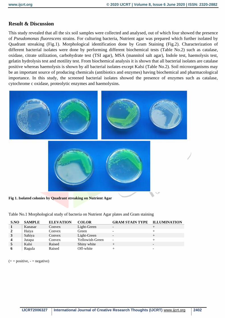
Result & Discussion
This study revealed that all the six soil samples were collected and analysed, out of which four showed the presence
of Pseudomonas fluorescens strains. For culturing bacteria, Nutrient agar was prepared which further isolated by
Quadrant streaking (Fig.1). Morphological identification done by Gram Staining (Fig.2). Characterization of
different bacterial isolates were done by performing different biochemical tests (Table No.2) such as catalase,
oxidase, citrate utilization, carbohydrate test (TSI agar), MSA (mannitol salt agar), Indole test, haemolysis test,
gelatin hydrolysis test and motility test. From biochemical analysis it is shown that all bacterial isolates are catalase
positive whereas haemolysis is shown by all bacterial isolates except Kalsi (Table No.2). Soil microorganisms may
be an important source of producing chemicals (antibiotics and enzymes) having biochemical and pharmacological
importance. In this study, the screened bacterial isolates showed the presence of enzymes such as catalase,
cytochrome c oxidase, proteolytic enzymes and haemolysins.
Fig 1. Isolated colonies by Quadrant streaking on Nutrient Agar
Table No.1 Morphological study of bacteria on Nutrient Agar plates and Gram staining
S.NO SAMPLE ELEVATION COLOR GRAM STAIN TYPE ILLUMINATION
1 Kanasar Convex Light-Green - +
2 Haiya Convex Green - +
3 Sahiya Convex Light-Green - +
4 Jutapa Convex Yellowish-Green - +
5 Kalsi Raised Shiny white + -
6 Ragula Raised Off-white + -
(+ = positive, - = negative)
www.ijcrt.org © 2020 IJCRT | Volume 8, Issue 6 June 2020 | ISSN: 2320-2882
IJCRT2006327 International Journal of Creative Research Thoughts (IJCRT) www.ijcrt.org 2403
Fig 2. Gram staining images of different soil samples
Fig 3. Streaked plates under UV Transilluminator
Table No. 2 Biochemical Analysis
S.NO SAMPLES OXIDASE CATALASE CITRATE TSI MSA INDOLE HEMOLYSIS GELATIN
HYDROLYSIS
MOTILITY
1 Kanasar + + + - - - +(α) + +
2 Ragula + + - + - - +(β) + -
3 Jutapa + + + - - - +(α) + +
4 Sahiya - + + - - - +(α) + +
5 Haiya - + + - - - +(α) + +
6 Kalsi - + + - - + -(ϒ) - -
(+ = positive, - = Negative, α= alpha hemolysis, β= beta hemolysis, ϒ= gamma hemolysis)

www.ijcrt.org © 2020 IJCRT | Volume 8, Issue 6 June 2020 | ISSN: 2320-2882
IJCRT2006327 International Journal of Creative Research Thoughts (IJCRT) www.ijcrt.org 2404
Table No. 3 OF (Oxidative and Fermentative) Test
S.NO SAMPLES OPEN TUBE
(AEROBIC)
CLOSED
TUBE(ANAEROBIC)
METABOLISM
1 Kanasar yellow Green Oxidative
2 Ragula yellow yellow Fermentative
3 Jutapa yellow Green Oxidative
4 Sahiya yellow Green Oxidative
5 Haiya yellow Green Oxidative
6 Kalsi yellow Green Oxidative
(Yellow = Acid, Green = Alkaline)
Fig 4. Oxidase test
Fig 5. Catalase test
Fig 6. Citrate test

www.ijcrt.org © 2020 IJCRT | Volume 8, Issue 6 June 2020 | ISSN: 2320-2882
IJCRT2006327 International Journal of Creative Research Thoughts (IJCRT) www.ijcrt.org 2405
Fig 7. TSI plate
Fig 8. Blood hemolysis by bacteria
Fig 9. Gelatin Hydrolysis
Fig 10. OF test

www.ijcrt.org © 2020 IJCRT | Volume 8, Issue 6 June 2020 | ISSN: 2320-2882
IJCRT2006327 International Journal of Creative Research Thoughts (IJCRT) www.ijcrt.org 2406
Effect of Microorganism on seeds of Black-eyed peas and Okra
Potential for application of bacteria in soil. There are vast possibilities in the application of bacteria for beneficial
purposes; the potential for tailoring organism to specific tasks using genetic engineering techniques has certainly
contributed to this. Many different bacteria, in particular Pseudomonas and Bacillus strains, are known to be able to
control pathogenic agents especially fungi. Microbial inoculants applied as seeds treatments deliver microorganism
directly to plants rhizosphere – the narrow zone of soil that surrounds the root where plants interact directly with
microorganism (Philippot et al.2013). It is a zone of intense microbial activity with growth of plants and
microorganism dependent on reciprocal provision of nutrients and a wide range of other compounds including plant
growth regulators and antibiotics.
Normal soil for pot is collected and autoclaved at 1210 C and then incubated overnight and next day again autoclaved.
This is done to kill the other microbes and endospores in the soil which affect the results and efficiency of isolated
microbes (Fig. 1).
Seeds of black-eyed peas and okra is inoculated with P.fluorescens isolated strains from different sites of Chakrata.
5 ml of each broth culture of bacteria is poured in 5 test tubes. In 4 tubes, 4 different bacterial sample was inoculated
and one tube is taken as control(uninoculated). 2 gm of Okra and Black-eyed peas was weighed respectively and put
it into each tubes containing different bacterial culture. Seeds were soaked in bacterial culture for 2-3 hours. Remove
the seeds and put it into pots and labelled well.
Table No. 4 Comparison of plant growth
S.NO SAMPLES HEIGHT OF PLANT AFTER 7
DAYS FROM ROOT TO TIP
HEIGHT OF PLANT AFTER
15 DAYS
Black-eyed peas Okra Black-eyed peas Okra
1 Haiya 12 cm 8 cm 27 cm 11 cm
2 Sahiya - 9 cm - 17 cm
3 Jutapa 11 cm - 26 cm -
4 Kanasar 15 cm - 38 cm -
5 Control 17cm - 33cm -
(- = No growth)
Fig 11. Growth after 7 days (captured by Saif Ali)

www.ijcrt.org © 2020 IJCRT | Volume 8, Issue 6 June 2020 | ISSN: 2320-2882
IJCRT2006327 International Journal of Creative Research Thoughts (IJCRT) www.ijcrt.org 2407
Fig. 12 Growth of plants after 15 days (captured by Saif Ali)
Fig. 13 Growth of Okra and black-eyed peas after 28 days (captured by Babali Semwal)

www.ijcrt.org © 2020 IJCRT | Volume 8, Issue 6 June 2020 | ISSN: 2320-2882
IJCRT2006327 International Journal of Creative Research Thoughts (IJCRT) www.ijcrt.org 2408
Conclusion
By studying the different soil samples, we conclude that soil of Jutapa , Haiya, Sahiya and Kanasar have the
Pseudomonas fluorescens strains, while Ragula and Kalsi may not having those useful bacterial strains as compared
by the results (Table No.4). Ragula and Kalsi have the most abundant gram-positive long bacilli which may be a
useful species for humans, but for other samples it is cleared by the above results that it having the Pseudomonas sp.
which is useful for agricultural fields.
P.fluorescens is a very wide spread bacteria and not harmful to humans, inhabits soil and water. It multiply very
quickly, this means they can rapidly colonize a space and indirectly protects a crop. It also acts as a bio-control
agent, means it can kill other pathogens in the soil which may harm the crop. In short, we will say that it is a multi-
tasking bacteria which is fully compatible with soil and helpful to human beings.
These four samples were test for the growth of plants and control is taken for the comparison of growth. Kanasar
showed the maximum growth of black-eyed peas and Okra where as control only showed the growth of black-eyed
peas. Jutapa showed the least growth of plants. Haiya and Sahiya showed the growth of Okra only. However, some
seeds were not grown.
From the above research it is cleared that Chakrata soil is rich in microflora. Different sites do not support the growth
of all plants but supports growth of selected plants which shows a large bio-diversity.
Acknowledgement
The authors would like to thanks Department of Microbiology, SGRR University and USERC for providing
necessary facilities for carrying out the study.
References
Kumar A, Kumar A, Devi S, Patil S, Payal C.Negi Isolation S; Screening and characterization of bacteria from
rhizospheric soils for different plant growth promotion (PGP) activities :an in vitro study. Recent Res. Sci.
Technol.,2012:4:01-05.
King A, & Phillips, I, (1978). The Identification of pseudomonads and related bacteria in a clicnical laboratory.J
Med Microbiol. 11(2):165-76.
Bergey, D.H., 1939. Manual of Determinative bacteriology. Williams and Wilkins, Baltimore. 5th Ed.
Palleroni, N.J. (1984) Pseudomonadaceae. Bergey's Manual of Systematic Bacteriology. Krieg, N. R. and Holt J. G.
(editors) Baltimore: The Williams and Wilkins Co., pg. 141 – 199