
InvolvementoftheCalcium-sensingReceptorinHuman ... ·...
8
Involvement of the Calcium-sensing Receptor in Human Taste Perception □ S Received for publication, June 3, 2009, and in revised form, October 19, 2009 Published, JBC Papers in Press, November 5, 2009, DOI 10.1074/jbc.M109.029165 Takeaki Ohsu ‡ , Yusuke Amino § , Hiroaki Nagasaki ¶ , Tomohiko Yamanaka ¶ , Sen Takeshita ‡ , Toshihiro Hatanaka ‡ , Yutaka Maruyama , Naohiro Miyamura ¶ , and Yuzuru Eto ‡1 From the ‡ Pharmaceutical Research Laboratories, § AminoScience Laboratories, ¶ Food Products Company, and Institute of Life Sciences, Ajinomoto Company, Incorporated, Kawasaki 210-8681, Japan By human sensory analyses, we found that various extracellu- lar calcium-sensing receptor (CaSR) agonists enhance sweet, salty, and umami tastes, although they have no taste themselves. These characteristics are known as “kokumi taste” and often appear in traditional Japanese cuisine. Although GSH is a typical kokumi taste substance (taste enhancer), its mode of action is poorly understood. Here, we demonstrate how the kokumi taste is enhanced by the CaSR, a close relative of the class C G-pro- tein-coupled receptors T1R1, T1R2, and T1R3 (sweet and umami receptors). We identified a large number of CaSR ago- nist -glutamyl peptides, including GSH (-Glu-Cys-Gly) and -Glu-Val-Gly, and showed that these peptides elicit the kokumi taste. Further analyses revealed that some known CaSR agonists such as Ca 2 , protamine, polylysine, L-histidine, and cinacalcet (a calcium-mimetic drug) also elicit the kokumi taste and that the CaSR-specific antagonist, NPS-2143, significantly sup- presses the kokumi taste. This is the first report indicating a distinct function of the CaSR in human taste perception. The extracellular calcium-sensing receptor (CaSR) 2 (1) is a class C G-protein-coupled receptor consisting (in humans) of 1078 amino acids. It plays a central role in extracellular calcium homeostasis in mammals (2). An increase in the blood calcium level is sensed by the CaSR, which in turn suppresses parathy- roid hormone secretion, stimulates the secretion of calcitonin, and induces urinary calcium excretion to reduce blood calcium to normal levels. It has become apparent that the CaSR is expressed not only in the parathyroid glands and kidneys but also in many other tissues such as the liver, heart, lungs, alimen- tary canal, lymphocytes, pancreas, and the central and periph- eral nervous systems, thus suggesting that it is involved in a range of biological functions (3). Several types of substances have been reported to possess CaSR activity, including cations such as Ca 2 and Gd 3 , basic peptides such as protamine and polylysine, and polyamines such as spermine (3). In addition, the CaSR is moderately activated by the aromatic amino acids His, Trp, Phe, and Tyr and is weakly activated by other amino acids such as Arg, Lys, Val, and Gly (4, 5). However, the physi- ological relevance of these CaSR agonists, except calcium, is poorly understood. Studies investigating the nutritional significance of calcium, mainly in laboratory animals, suggest the involvement of cal- cium in taste perception. Tordoff and co-workers (6, 7) re- ported the taste perception of calcium and the physiological mechanisms underlying calcium intake, appetite, and homeo- stasis and indicated that calcium deprivation increases the palatability of calcium. Although it is presumed that the CaSR (8) or a specific calcium channel is involved, no research pro- vides direct evidence for the involvement of the CaSR in human taste perception. However, it is believed that calcium usually tastes bitter at a high concentration. Humans can identify five basic tastes, sweet, salty, sour, bit- ter, and umami, which are believed to be recognized by specific receptors and transduction pathways. In addition to the five basic tastes, taste-enhancing substances are often added to foodstuffs. Substances with kokumi taste (9 –12), which is dis- tinct from the five basic tastes, have been used for many years in Japanese cuisine. GSH (-glutamylcysteinylglycine), a typical kokumi taste substance, is abundantly present in food-grade yeast extract, which is commercially available and has been used to make foods taste savory and hearty. The kokumi taste was first characterized by Ueda et al. (9, 10), who isolated a kokumi taste substance from water extracts of garlic and onion and identified GSH as the main active ingredient. GSH itself is tasteless; however, in the presence of small amounts of umami taste substances such as monosodium glutamate (MSG) and IMP, GSH synergistically reinforces those tastes. In this study, we demonstrate that the CaSR is involved in kokumi taste perception in humans and report the discovery of various CaSR agonist peptides, including -glutamylvalylgly- cine (-Glu-Val-Gly), a potent kokumi taste substance. EXPERIMENTAL PROCEDURES Chemicals—The CaSR agonists used in the human sensory analyses were commercially available food additive products such as calcium lactate (Sky Food), protamine (Asama Chemi- cals), and polylysine (Nihon Chisso). Cinacalcet (13) and NPS- 2143 (14) were chemically synthesized by methods described in the literature, and their activity was determined using HEK-293 cells that were transiently transformed with the human CaSR (hCaSR). All other reagents were a special purity grade pur- chased from Sigma-Aldrich Japan. □ S The on-line version of this article (available at http://www.jbc.org) contains supplemental Figs. 1–3 and Table 1. 1 To whom correspondence should be addressed: Inst. of Life Sciences, Ajinomoto Co., Inc., 1-1 Suzuki-cho, Kawasaki-ku, Kawasaki 210-8681, Japan. Tel.: 81-44-210-5845; Fax: 81-44-210-5871; E-mail: yuzuru_eto@ ajinomoto.com. 2 The abbreviations used are: CaSR, calcium-sensing receptor; hCaSR, human CaSR; MSG, monosodium glutamate; Abu, -aminobutyric acid; PSE, point of subjective equivalence; RT, reverse transcription. THE JOURNAL OF BIOLOGICAL CHEMISTRY VOL. 285, NO. 2, pp. 1016 –1022, January 8, 2010 © 2010 by The American Society for Biochemistry and Molecular Biology, Inc. Printed in the U.S.A. 1016 JOURNAL OF BIOLOGICAL CHEMISTRY VOLUME 285 • NUMBER 2 • JANUARY 8, 2010 by guest on October 14, 2018 http://www.jbc.org/ Downloaded from
Transcript of InvolvementoftheCalcium-sensingReceptorinHuman ... ·...
Involvement of the Calcium-sensing Receptor in HumanTaste Perception□S
Received for publication, June 3, 2009, and in revised form, October 19, 2009 Published, JBC Papers in Press, November 5, 2009, DOI 10.1074/jbc.M109.029165
Takeaki Ohsu‡, Yusuke Amino§, Hiroaki Nagasaki¶, Tomohiko Yamanaka¶, Sen Takeshita‡, Toshihiro Hatanaka‡,Yutaka Maruyama�, Naohiro Miyamura¶, and Yuzuru Eto‡�1
From the ‡Pharmaceutical Research Laboratories, §AminoScience Laboratories, ¶Food Products Company, and �Institute of LifeSciences, Ajinomoto Company, Incorporated, Kawasaki 210-8681, Japan
By human sensory analyses, we found that various extracellu-lar calcium-sensing receptor (CaSR) agonists enhance sweet,salty, and umami tastes, although they have no taste themselves.These characteristics are known as “kokumi taste” and oftenappear in traditional Japanese cuisine.AlthoughGSH is a typicalkokumi taste substance (taste enhancer), its mode of action ispoorly understood. Here, we demonstrate how the kokumi tasteis enhanced by the CaSR, a close relative of the class C G-pro-tein-coupled receptors T1R1, T1R2, and T1R3 (sweet andumami receptors). We identified a large number of CaSR ago-nist �-glutamyl peptides, including GSH (�-Glu-Cys-Gly) and�-Glu-Val-Gly, and showed that these peptides elicit the kokumitaste. Further analyses revealed that some knownCaSR agonistssuch as Ca2�, protamine, polylysine, L-histidine, and cinacalcet(a calcium-mimetic drug) also elicit the kokumi taste and thatthe CaSR-specific antagonist, NPS-2143, significantly sup-presses the kokumi taste. This is the first report indicating adistinct function of the CaSR in human taste perception.
The extracellular calcium-sensing receptor (CaSR)2 (1) is aclass C G-protein-coupled receptor consisting (in humans) of1078 amino acids. It plays a central role in extracellular calciumhomeostasis in mammals (2). An increase in the blood calciumlevel is sensed by the CaSR, which in turn suppresses parathy-roid hormone secretion, stimulates the secretion of calcitonin,and induces urinary calcium excretion to reduce blood calciumto normal levels. It has become apparent that the CaSR isexpressed not only in the parathyroid glands and kidneys butalso inmany other tissues such as the liver, heart, lungs, alimen-tary canal, lymphocytes, pancreas, and the central and periph-eral nervous systems, thus suggesting that it is involved in arange of biological functions (3). Several types of substanceshave been reported to possess CaSR activity, including cationssuch as Ca2� and Gd3�, basic peptides such as protamine andpolylysine, and polyamines such as spermine (3). In addition,the CaSR is moderately activated by the aromatic amino acidsHis, Trp, Phe, and Tyr and is weakly activated by other amino
acids such as Arg, Lys, Val, and Gly (4, 5). However, the physi-ological relevance of these CaSR agonists, except calcium, ispoorly understood.Studies investigating the nutritional significance of calcium,
mainly in laboratory animals, suggest the involvement of cal-cium in taste perception. Tordoff and co-workers (6, 7) re-ported the taste perception of calcium and the physiologicalmechanisms underlying calcium intake, appetite, and homeo-stasis and indicated that calcium deprivation increases thepalatability of calcium. Although it is presumed that the CaSR(8) or a specific calcium channel is involved, no research pro-vides direct evidence for the involvement of the CaSR in humantaste perception. However, it is believed that calcium usuallytastes bitter at a high concentration.Humans can identify five basic tastes, sweet, salty, sour, bit-
ter, and umami, which are believed to be recognized by specificreceptors and transduction pathways. In addition to the fivebasic tastes, taste-enhancing substances are often added tofoodstuffs. Substances with kokumi taste (9–12), which is dis-tinct from the five basic tastes, have been used formany years inJapanese cuisine. GSH (�-glutamylcysteinylglycine), a typicalkokumi taste substance, is abundantly present in food-gradeyeast extract, which is commercially available and has beenused to make foods taste savory and hearty. The kokumi tastewas first characterized by Ueda et al. (9, 10), who isolated akokumi taste substance from water extracts of garlic and onionand identified GSH as the main active ingredient. GSH itself istasteless; however, in the presence of small amounts of umamitaste substances such as monosodium glutamate (MSG) andIMP, GSH synergistically reinforces those tastes.In this study, we demonstrate that the CaSR is involved in
kokumi taste perception in humans and report the discovery ofvarious CaSR agonist peptides, including �-glutamylvalylgly-cine (�-Glu-Val-Gly), a potent kokumi taste substance.
EXPERIMENTAL PROCEDURES
Chemicals—The CaSR agonists used in the human sensoryanalyses were commercially available food additive productssuch as calcium lactate (Sky Food), protamine (Asama Chemi-cals), and polylysine (Nihon Chisso). Cinacalcet (13) and NPS-2143 (14) were chemically synthesized bymethods described inthe literature, and their activity was determined usingHEK-293cells that were transiently transformed with the human CaSR(hCaSR). All other reagents were a special purity grade pur-chased from Sigma-Aldrich Japan.
□S The on-line version of this article (available at http://www.jbc.org) containssupplemental Figs. 1–3 and Table 1.
1 To whom correspondence should be addressed: Inst. of Life Sciences,Ajinomoto Co., Inc., 1-1 Suzuki-cho, Kawasaki-ku, Kawasaki 210-8681,Japan. Tel.: 81-44-210-5845; Fax: 81-44-210-5871; E-mail: [email protected].
2 The abbreviations used are: CaSR, calcium-sensing receptor; hCaSR, humanCaSR; MSG, monosodium glutamate; Abu, �-aminobutyric acid; PSE, pointof subjective equivalence; RT, reverse transcription.
THE JOURNAL OF BIOLOGICAL CHEMISTRY VOL. 285, NO. 2, pp. 1016 –1022, January 8, 2010© 2010 by The American Society for Biochemistry and Molecular Biology, Inc. Printed in the U.S.A.
1016 JOURNAL OF BIOLOGICAL CHEMISTRY VOLUME 285 • NUMBER 2 • JANUARY 8, 2010
by guest on October 14, 2018
http://ww
w.jbc.org/
Dow
nloaded from

Peptides—The following peptides were used in the study:�-Glu-Cys-Gly (GSH) and �-Glu-Cys (Sigma-Aldrich Japan);�-Glu-Cys(S-nitroso)-Gly (Dojin Chemical Laboratory); �-Glu-Ala, �-Glu-Gly, �-Glu-Met, and �-Glu-Abu-Gly (BachemFeinchemikalien AG); and �-Glu-Thr and �-Glu-Val (KokusanChemical). Other peptides were chemically synthesized andpurified by a contract manufacturer using well known tech-niques. Solutions of the glutamine- and cysteine-containingpeptides were prepared immediately before use, whereas thoseof other peptides were stored at �20 °C after preparation. Inthe case of acidic and alkaline substances, the solutions wereadjusted to pH 7.0–7.4 by the addition of NaOH or HCl. TheCaSR agonists and antagonist used in this study are listed insupplemental Table 1.Preparation of cRNA—cRNA of the hCaSR was pre-
pared from human kidney cDNA (Clontech) using a PCRmethod. Briefly, primer oligonucleotide DNAs (forwardprimer, 5�-ACTAATACGACTCACTATAGGGACCATGG-CATTTTATAGCTGCTGCTGG-3�, and reverse primer, 5�-TTATGAATTCACTACGTTTTCTGTAACAG-3�; NCBI ac-cession number NM_000388) were synthesized, and PCR wasperformed using PfuUltraDNApolymerase (Stratagene) underthe following conditions. After an initial reaction at 94 °C for 3min, a cycle of reactions at 94 °C for 30 s, 55 °C for 30 s, and72 °C for 2 min was repeated 35 times, and then a final reactionwas performed at 72 °C for 7 min. The plasmid vector pBR322(Takara) was digested with the restriction enzyme EcoRV. ThePCR product was ligated to the EcoRV cleavage site of pBR322using a ligation kit (Promega). hCaSR cRNA was synthesizedusing a cRNA preparation kit (Ambion) with this sequence as atemplate.Determination of CaSR Activity Using Oocytes—CaSR ago-
nist-induced currents were characterized using oocytes micro-injected with hCaSR cRNA. Briefly, Xenopus laevis ovarianlobes were surgically removed, defolliculated, and treated withcollagenase II. Oocytes were thenmicroinjected with 10–20 ngof hCaSR cRNA and incubated for 36–48 h at 15 °C in Barth’ssolution. Activation of the CaSR (Gq class G-protein-coupledreceptor) expressed in oocytes leads to an increase in intercel-lular calcium ions. This increase in free calcium activatesoocyte endogenous calcium-dependent chloride channels con-comitantly with a measurable current. The oocytes wereimpaled by two electrodes in a voltage-clamp configurationwith a GeneClamp 500 (Axon), and responses were recordedusing AxoScope 9.0 recording software (Axon) at a membranepotential of �70 mV. The oocytes were challenged with 0.1–1000�M solutions ofCaSR agonists in perfusion buffer contain-ing 96 mM NaCl, 2 mM KCl, 1 mM MgCl2, 1.8 mM CaCl2, and 5mMHepes (pH 7.2), and the peak recorded current was deemedthe strength of receptor activation.Determination of CaSR Activity Using HEK-293 Cells—
hCaSR cDNA was constructed in the expression vectorpcDNA3.1 and transiently transfected into HEK-293 cells.Briefly, the cDNA was diluted with Opti-MEM I medium(Invitrogen), mixed with FuGENE 6 (Roche Applied Science),and poured onto HEK-293 cells grown at a submaximum con-centration. After 24 h of culture in a 96-well plate, the cells wereincubated with 5 �M Calcium-4 (Calcium-4 assay kit, Molecu-
larDevices) for 45–60min, andmeasurementswere conductedusing an image analyzer (FlexStation, Molecular Devices) andits associated software. Activation of the CaSR expressed inHEK-293 cells leads to an increase in intercellular calcium ions.This increase in free calciumwas determined using the calciumdye Calcium-4. The dye binds the free Ca2�, resulting in anincrease in dye fluorescence, which is excited at 485 nm andemits at 525 nm. The concentration dependence of the fluores-cence intensity was analyzed with various CaSR agonists withor without 0.0002% (4.5 �M) NPS-2143. The assay buffer forcalcium imaging contained 0.9 mM CaCl2.Continuous Kokumi Taste Sensory Analyses of GSH and
�-Glu-Val-Gly—Time course profiles of the kokumi taste ofGSH (0.08%) and �-Glu-Val-Gly (0.01%) were determined bycontinuous human sensory analyses. Briefly, the test samplescontained low concentrations of umami and salty tastes (0.02%MSG, 0.02% IMP, and 0.07% NaCl) with or without kokumitaste substances. The test sample (20 ml) was administeredorally, and the taste assessors were instructed to keep the sam-ple in their mouths for at least 30 s, during which time the tasteintensity was expressed as a percentage score in comparisonwith the maximum intensity obtained with a standard solutioncontaining 0.7%MSG and 0.5%NaCl, taken as 100%. Data wererecorded every second. The analyses were performed by a panelof 20 well trained assessors.Kokumi Taste Determination by Human Sensory Analyses
with a 5-Point Rating Scale—Samples were dissolved in dis-tilled water and adjusted to pH 6.8–7.2 with NaOH. Thehuman sensory analysis of a sample solution was performedusing a previously describedmethod (9). Awell trained panel of20 assessors was asked to evaluate the sample solutions on a5-point rating scale, from �2 (apparently suppressed) to �2(apparently strong). The kokumi taste in each basic taste solu-tion was estimated by the thickness, a measure of the tasteintensity 5 s after tasting. The results were analyzed by Stu-dent’s t test. The kokumi taste in the presence of each basictaste, i.e. sweet, salty, or umami, was determined with GSH(0.1%) or �-Glu-Val-Gly (0.01%) mixed with sucrose (3.3%),NaCl (0.9%), or MSG (0.5%), respectively. The presence of thekokumi taste in foodstuffs was determined as follows. Con-somme containing 2% commercial chicken consomme powder(Ajinomoto Co., Inc.) was prepared. GSH (0.02%), �-Glu-Val-Gly (0.002%), and �-Glu-Val-Leu (0.02%; a low-activity controlpeptide) were then dissolved in the consomme. The kokumitaste was determined for three paired scale terms: thickness,continuity, and mouthfulness. Thickness was expressed interms of increased taste intensity at �5 s after tasting; continu-ity was expressed as the taste intensity at �20 s; and mouthful-ness was expressed as the reinforcement of the taste sensationthroughout themouth and not just on the tongue. KnownCaSRagonists and an antagonist were used at the following final con-centrations: calcium lactate (0.35%), protamine (0.02%), polyly-sine (0.08%), and L-histidine (0.2%). Cinacalcet was dissolved in99.5% ethanol at a concentration of 1% and then diluted to0.0015% (38 �M) with the tasting solution for sensory analysis.NPS-2143 was dissolved in 99.5% ethanol at a concentration of0.1% and then diluted to 0.0002% (4.5�M)with the tasting solu-tion. The remaining ethanol in the tasting solution did not
CaSR in Taste Perception
JANUARY 8, 2010 • VOLUME 285 • NUMBER 2 JOURNAL OF BIOLOGICAL CHEMISTRY 1017
by guest on October 14, 2018
http://ww
w.jbc.org/
Dow
nloaded from

affect the sensory analysis evaluation.Methods used in the humansensory analyses were approved by the Management Committeeof the Food Product Application Center at Ajinomoto Co., Inc.,and informed consent was obtained from all assessors.Quantitative Analyses of the Kokumi Taste Compared with
the GSH Kokumi Standard—Quantitative analyses were con-ducted to compare taste thickness. The strength of the kokumitaste was evaluated as a point of subjective equivalence (PSE) asfollows. Test samples of �-Glu-Cys (0.15%), �-Glu-Val (0.15%),�-Glu-Ala (0.5%), �-Glu-Abu-Gly (0.05%), or �-Glu-Val-Gly(0.01%)weremixedwith umami and salty taste solutions (0.05%MSG, 0.05% IMP, and 0.5% NaCl), and their tastes were com-pared with those of various concentrations of GSH as referencesolutions. Eight concentrations of GSH in logarithmically equalsteps at 50% intervals were used (0.02, 0.03, 0.044, 0.07, 0.10,0.15, 0.23, and 0.34%, w/v). Each test sample was paired twicewith each reference solution.Assessors judged the PSEbetweenthe sample and reference solutions. All judgments were dichot-omous, i.e. the assessors were required to rate a test sample aseither more or less intense than the reference GSH solution.The PSE was defined as the concentration that was judged togive a sensation that was equivalent to that of the standardsolution. The analyses were performed by a panel of 17 welltrained assessors.Reverse Transcription (RT)-PCRAnalyses ofmRNAExtract of
Mouse Taste Buds—C57BL/6N mice (6-week-old males,Charles River) were killed by cervical dislocation. RT-PCRamplification was performed using primers that amplify themouse CaSR, T1R2, and �-actin. Briefly, tissues containing cir-cumvallate or foliate papillae were injected into the submucosallayer with a mixture of 1 mg/ml collagenase A (Roche AppliedScience), 2.5 mg/ml Dispase II (Roche Applied Science), and 1mg/ml trypsin inhibitor (Sigma-Aldrich Japan) and then incu-
bated for 20 min at room temperature. The papilla-containingepithelium was peeled from the underlying connective tissue.The total RNA was isolated from the epithelial papillae andfrom the epithelium without taste buds (RNAmicro kit, Strat-agene). The purified RNA was denatured at 60 °C for 5 min,and first-strand cDNA was synthesized at 50 °C for 60 minusing oligo(dT)12–18 primer and reverse transcriptase (Super-Script III, Invitrogen) in a final volume of 20 �l. After synthe-sizing the cDNA, 1 �l of cDNA was used as a template in 20 �lof PCR mixture with Taq polymerase (Invitrogen). The PCRconditions were as follows: 94 °C for 2 min and 29–35 cycles at94 °C for 30 s, 58 °C for 20 s, and 72 °C for 45 s. The PCR prod-ucts were analyzed by gel electrophoresis (2% agarose gel) withGelRed staining (Biotium). The primers used were as follows:mouse CaSR, 5�-TCGAGACCCCTTACATGGAC-3� (forward)and 5�-AGTAGTTCCCCACCAGGTCA-3� (reverse); mouseT1R2, 5�-TGGCAGCTACTCAGGGAGAT-3� (forward) and5�-GGACAGTCCACACACTCGAA-3� (reverse); and mouse�-actin, 5-CACCCTGTGCTGCTCACC-3 (forward) and 5�-GCACGATTTCCCTCTCAG-3� (reverse).
RESULTS
Discovery of CaSR Agonist �-Glutamyl Peptides—Wescreened di- and tripeptide libraries for CaSR activity anddiscovered 46 �-glutamyl (�-Glu) peptides, including GSH(�-Glu-Cys-Gly) (Table 1). Among them, �-Glu-Val-Gly hadthemost potent CaSR activity. Amino acids have been reportedto bind to the large extracellular Venus flytrap domain of theCaSR, a structure common to allmembers of class CG-protein-coupled receptors. It was suggested that all amino acids bind atthe Venus flytrap domain-binding pocket, through the aminoand carboxyl groups (5). This binding motif is common to freeamino acids and �-Glu peptides (�-glutamic acid (�-Glu) at the
TABLE 1Determination of CaSR activity using Xenopus oocytesA two-electrode voltage-clamp assay was performed using oocytes microinjected with hCaSR cRNA at�70mV. Peptides were added at various concentrations (1000, 500,300, 100, 50, 30, 10, 3, 1, 0.3, and 0.1 �M), and the minimum effective concentration at which a current was detected was a measure of the strength of CaSR activation. Theresults show that 46 �-glutamyl peptides had CaSR activity. No response was observed in oocytes injected with distilled water as a control. In comparison, EC50 values (�M)for six peptides determined byHEK-293 assay are indicated in parentheses. TheHEK-293 assay gave a higher sensitivity than the oocyte assay. SNO, S-nitroso;Met(O),Metsulfoxide; Tau, taurine; tLeu, tert-Leu.
No. �-Glu peptide CaSR activity No. �-Glu peptides CaSR activity
�M �M
1 �-Glu-Gly 1000 24 �-Glu-Ile-Gly 1002 �-Glu-Val-NH2 1000 25 �-Glu-Cys(S-allyl)-Gly 1003 �-Glu-Val-ol 1000 26 �-Glu-Val-His 1004 �-Glu-Met(O) 1000 27 �-Glu-Val-Orn 1005 �-Glu-Val-Val 1000 28 �-Glu-Ala 50 (3.7)6 �-Glu-Val-Glu 1000 29 �-Glu-Thr 507 �-Glu-Val-Lys 1000 30 �-Glu-Cys(S-Me) 308 �-Glu-Val-Arg 1000 31 �-Glu-Cys-Gly 309 �-Glu-Val-Asp 1000 32 �-Glu-Val-Phe 3010 �-Glu-Val-Met 1000 33 �-Glu-Val-Ser 3011 �-Glu-Val-Thr 1000 34 �-Glu-Val-Pro 3012 �-Glu-Met 500 35 �-Glu-Val-Asn 3013 �-Glu-Orn 500 36 �-Glu-Val 10 (1.6)14 �-Glu-Ser 300 37 �-Glu-Ala-Gly 1015 �-Glu-Tau 300 38 �-Glu-Ser-Gly 1016 �-Glu-Leu-Gly 300 39 �-Glu-Val-Gln 1017 �-Glu-Thr-Gly 300 40 �-Glu-Val-Cys 1018 �-Glu-Pro-Gly 300 41 �-Glu-Cys 3 (0.46)19 �-Glu-Val-Ala 300 42 �-Glu-Cys-Gly (GSH) 3 (0.71)20 �-Glu-Cys(S-Me)(O) 300 43 �-Glu-Abu-Gly 3 (0.025)21 �-Glu-Leu 100 44 �-Glu-Cys(S-Me)-Gly 322 �-Glu-Ile 100 45 �-Glu-Cys(SNO)-Gly 323 �-Glu-tLeu 100 46 �-Glu-Val-Gly 0.1 (0.039)
CaSR in Taste Perception
1018 JOURNAL OF BIOLOGICAL CHEMISTRY VOLUME 285 • NUMBER 2 • JANUARY 8, 2010
by guest on October 14, 2018
http://ww
w.jbc.org/
Dow
nloaded from
N-terminal end). In contrast, substitution of the �-Glu residuewith �-Asp or �-Ala resulted in loss of activity (data notshown). Furthermore, those �-Glu tripeptides with a largehydrophobic acid, a basic acid, or an acidic amino acid at thesecond position were inactive. A thiol group of cysteine at thesecond position was not essential for CaSR activity. The third-position analogs of �-Glu-Val-Gly were active, except for Ile,Trp, Tyr, and�-Ala. The presence of the third-position residue,especially one with a free carboxyl terminus and no side chain(Gly), substantially enhanced activity. These results indicate thatan exact alignment and the presence of two charged groups in theN-terminal residue are required for CaSR binding. The putativeinteraction between �-Glu-Val-Gly and the L-amino acid-bindingsite of the CaSR is illustrated in supplemental Fig. 1.Time Course Profiles of Kokumi Taste Intensity of GSH and
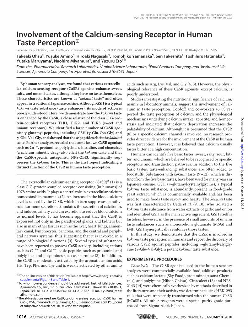
�-Glu-Val-Gly—BecauseGSH is a knownauthentic kokumi tastesubstance, we evaluatedwhether or not the newly identifiedCaSRagonistpeptide�-Glu-Val-Glycould impart akokumi taste. Short-term time course profiles of the kokumi taste of GSH and �-Glu-Val-Gly were determined by human sensory analyses (Fig. 1). Inthe case of control solutions containing low concentrations ofumami and salty substances, the taste intensity increased after oraladministration at 0 s, peaked at �10 s, and then decreased over a40-s period. The peak intensitywas significantly elevated (i.e. dou-bled) in the presence of GSH or �-Glu-Val-Gly, and this elevationremained distinct even after 20 s.Kokumi Taste Intensity of GSH and �-Glu-Val-Gly—We
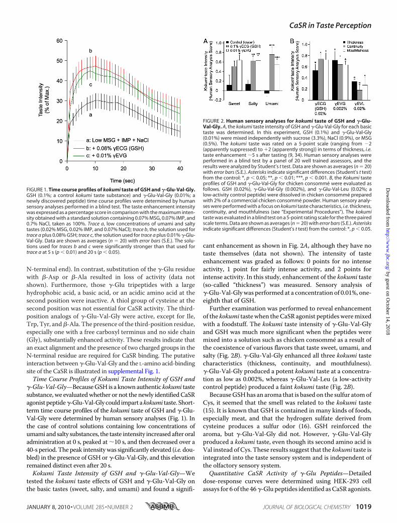
tested the kokumi taste effects of GSH and �-Glu-Val-Gly onthe basic tastes (sweet, salty, and umami) and found a signifi-
cant enhancement as shown in Fig. 2A, although they have notaste themselves (data not shown). The intensity of tasteenhancement was graded as follows: 0 points for no intenseactivity, 1 point for fairly intense activity, and 2 points forintense activity. In this study, enhancement of the kokumi taste(so-called “thickness”) was measured. Sensory analysis of�-Glu-Val-Glywas performed at a concentration of 0.01%, one-eighth that of GSH.Further examination was performed to reveal enhancement
of the kokumi tastewhen theCaSR agonist peptidesweremixedwith a foodstuff. The kokumi taste intensity of �-Glu-Val-Glyand GSH was much more significant when the peptides weremixed into a solution such as chicken consomme as a result ofthe coexistence of various flavors that taste sweet, umami, andsalty (Fig. 2B). �-Glu-Val-Gly enhanced all three kokumi tastecharacteristics (thickness, continuity, and mouthfulness).�-Glu-Val-Gly produced a potent kokumi taste at a concentra-tion as low as 0.002%, whereas �-Glu-Val-Leu (a low-activitycontrol peptide) produced a faint kokumi taste (Fig. 2B).BecauseGSHhas an aroma that is based on the sulfur atomof
Cys, it seemed that the smell was related to the kokumi taste(15). It is known that GSH is contained in many kinds of foods,especially meat, and that the hydrogen sulfate derived fromcysteine produces a sulfur odor (16). GSH reinforced thearoma, but �-Glu-Val-Gly did not. However, �-Glu-Val-Glyproduced a kokumi taste, even though its second amino acid isVal instead of Cys. These results suggest that the kokumi taste isintegrated into the taste sensory system and is independent ofthe olfactory sensory system.Quantitative CaSR Activity of �-Glu Peptides—Detailed
dose-response curves were determined using HEK-293 cellassays for 6 of the 46�-Glu peptides identified asCaSR agonists.
FIGURE 1. Time course profiles of kokumi taste of GSH and �-Glu-Val-Gly.GSH (0.1%; a control kokumi taste substance) and �-Glu-Val-Gly (0.01%; anewly discovered peptide) time course profiles were determined by humansensory analyses performed in a blind test. The taste enhancement intensitywas expressed as a percentage score in comparison with the maximum inten-sity obtained with a standard solution containing 0.07% MSG, 0.07% IMP, and0.7% NaCl, taken as 100%. Trace a, low concentrations of umami and saltytastes (0.02% MSG, 0.02% IMP, and 0.07% NaCl); trace b, the solution used fortrace a plus 0.08% GSH; trace c, the solution used for trace a plus 0.01% �-Glu-Val-Gly. Data are shown as averages (n � 20) with error bars (S.E.). The solu-tions used for traces b and c were significantly stronger than that used fortrace a at 5 s (p � 0.01) and 20 s (p � 0.05).
FIGURE 2. Human sensory analyses for kokumi taste of GSH and �-Glu-Val-Gly. A, the kokumi taste intensity of GSH and �-Glu-Val-Gly for each basictaste was determined. In this experiment, GSH (0.1%) and �-Glu-Val-Gly(0.01%) were mixed independently with sucrose (3.3%), NaCl (0.9%), or MSG(0.5%). The kokumi taste was rated on a 5-point scale (ranging from �2(apparently suppressed) to �2 (apparently strong)) in terms of thickness, i.e.taste enhancement �5 s after tasting (9, 34). Human sensory analyses wereperformed in a blind test by a panel of 20 well trained assessors, and theresults were analyzed by Student’s t test. Data are shown as averages (n � 20)with error bars (S.E.). Asterisks indicate significant differences (Student’s t test)from the control: *, p � 0.05; **, p � 0.01; ***, p � 0.001. B, the Kokumi tasteprofiles of GSH and �-Glu-Val-Gly for chicken consomme were evaluated asfollows. GSH (0.02%), �-Glu-Val-Gly (0.002%), and �-Glu-Val-Leu (0.02%; alow-activity control peptide) were dissolved in chicken consomme preparedwith 2% of a commercial chicken consomme powder. Human sensory analy-ses were performed with a focus on kokumi taste characteristics, i.e. thickness,continuity, and mouthfulness (see “Experimental Procedures”). The kokumitaste was evaluated in a blind test on a 5-point rating scale for the three pairedscale terms. Data are shown as averages (n � 20) with error bars (S.E.). Asterisksindicate significant differences (Student’s t test) from the control: *, p � 0.05.
CaSR in Taste Perception
JANUARY 8, 2010 • VOLUME 285 • NUMBER 2 JOURNAL OF BIOLOGICAL CHEMISTRY 1019
by guest on October 14, 2018
http://ww
w.jbc.org/
Dow
nloaded from

Fluorescence intensities observed with these peptides were26–53% of themaximum intensity observedwith Ca2� or cina-calcet (Fig. 3A and supplemental Fig. 2). The effective concen-tration (EC50) values for the �-Glu peptides, which correspondto the concentrations that produced 50% of the maximumintensity for Ca2�, are as follows: �-Glu-Ala, 3.65 �M/42.6 �Emax; �-Glu-Val, 1.34 �M/53.3; �-Glu-Cys, 458 nM/38.0; �-Glu-Cys-Gly, 76.5 nM/27.6; �-Glu-Abu-Gly, 18.1 nM/25.6; and�-Glu-Val-Gly, 41.9 nM/40.8.Correlations between CaSRActivity and Kokumi Taste Inten-
sity of �-Glu Peptides—Aquantitative sensory evaluation of thekokumi taste was performed with the six �-Glu peptides, andthe results were expressed as PSEs for each �-Glu peptide testsolution in relation to that of the reference solution, GSH (9).The intensity of the kokumi taste was quantified in terms of theGSH concentration required to attain an equal intensity of sen-sation. �-Glu-Val-Gly had the most potent kokumi taste; a0.01% solution produced a kokumi taste equivalent to a GSHconcentration of 0.128%. Therefore, we estimated that thekokumi taste of �-Glu-Val-Gly is 12.8 times stronger than thatof GSH. All five CaSR agonist peptides tested against GSHwere
confirmed to have kokumi taste (Table 2). CaSR activity wasinvestigated in detail using HEK-293 assays to determine EC50values (Fig. 3A). A significant correlationwas observed betweenCaSR activity and kokumi taste intensity when the intensitieswere expressed as a value relative to that of GSH and was plot-ted in two dimensions on a logarithmic scale (Fig. 3B). Thesepeptides were randomly chosen from the CaSR agonist pep-tides listed in Table 1. These results strongly suggest that theCaSR is involved in the perception of kokumi taste in humans.Kokumi Taste Intensity of Known CaSR Agonists and Effect of
the Antagonist—We determined the kokumi taste of knownCaSR agonists and the inhibitory effect of the synthetic CaSRantagonist, NPS-2143. Although calcium lactate, protamine,polylysine, and L-histidine are used as food additives, it is notknown whether they have their own taste. Cinacalcet, a chem-ically synthesized allosteric activator of the CaSR, is useful as adrug for the treatment of secondary hyperparathyroidism andhypercalcemia. In our tests, calcium lactate, protamine, polyly-sine, L-histidine, and cinacalcet showed significant kokumi tasteenhancement at concentrations of 0.35, 0.02, 0.08, 0.2, and0.0015, respectively (Fig. 4A). Some of these substances hadbitter tastes at higher concentrations but were suitable for anal-ysis when tested under our conditions. In contrast, the testedCaSR agonist peptides did not possess a bitter taste at any con-centration, even though kokumi taste intensitywas evaluated byhuman sensory analyses over a wide range of concentrations.Human sensory analyses were performed to determine
whether the kokumi tastes of GSH and �-Glu-Val-Gly weresuppressed in the presence of 0.0002% (4.5 �M) NPS-2143 (Fig.4B). NPS-2143 is a CaSR-specific antagonist that effectivelyinhibits the activity of both GSH (data not shown) and �-Glu-Val-Gly (Fig. 3C). These results clearly indicate that the CaSR isinvolved in kokumi taste perception in humans.Expression of the CaSR in Peripheral Taste Tissue—RT-PCR
analyses in mice have demonstrated that mRNAs of the CaSRand T1R2 (used as the control for taste cells) are expressed inthe taste bud-containing epithelium of the circumvallate andfoliate papillae. The RT-PCR products for the CaSR were notfound in the epithelium surrounding the papillae (supplemen-tal Fig. 2). These findings suggest that the CaSR could be a
FIGURE 3. Correlation between CaSR activity and kokumi taste intensity.A, HEK-293 cells transiently expressing hCaSRs were used in the calcium imag-ing method, and the dose dependence was analyzed for six CaSR agonistpeptides. The effective concentration (EC50) values are as follows: �-Glu-Ala(a), 3.65 �M/42.6 � Emax; �-Glu-Val (b), 1.34 �M/53.3; �-Glu-Cys (c), 458nM/38.0; �-Glu-Cys-Gly (GSH), 76.5 nM/27.6; �-Glu-Abu-Gly (d), 18.1 nM/25.6;and �-Glu-Val-Gly (e), 41.9 nM/40.8. �-Glu peptides showed 26 –53% of themaximum intensity (Emax) observed with Ca2� (calcium � 100%). RFU, relativefluorescence units. B, the relationship between CaSR activity (A, EC50) andkokumi taste intensity (Table 1) of five �-Glu peptides is plotted on logarith-mic scales in comparison with GSH. A Pearson correlation coefficient wasused to calculate the relationship between CaSR activity and kokumi tasteintensity. A significant positive correlation was found (r2 � 0.660, p � 0.0496).C, shown is the strong inhibitory effect of the CaSR antagonist. The CaSRactivity dose response of �-Glu-Val-Gly was analyzed using HEK-293 cellstransiently expressing hCaSR mRNA in the absence and presence of NPS-2143(5 �M).
TABLE 2Quantitative analyses of the kokumi taste intensity of �-glutamylpeptidesA quantitative evaluation of the human sensory analyses was performed by a panelof 17well trained assessors, who judged the PSE between sample and referenceGSHsolutions. A series of GSH concentrations were chosen in logarithmically equalsteps at 50% intervals (0.02, 0.03, 0.044, 0.07, 0.10, 0.15, 0.23, and 0.34%, w/v). Eachtest sample was paired twice with the reference GSH solution, and the assessorswere required to rate the sensation produced by a test sample as either more or lessintense than that of the reference solution. The PSE was defined as the concentra-tion that was judged to produce a sensation equal to that of the standard solution.The sample concentrationwas determined by preliminary tests. Data were analyzedby the probit method.
Sampleconcentration
GSHconcentration
for PSE
Kokumi tasteintensity compared
with GSH
% %GSH (�-Glu-Cys-Gly) 1�-Glu-Ala 0.5 0.074 0.15�-Glu-Val 0.15 0.092 0.61�-Glu-Cys 0.15 0.094 0.63�-Glu-Abu-Gly 0.05 0.085 1.7�-Glu-Val-Gly 0.01 0.128 12.8
CaSR in Taste Perception
1020 JOURNAL OF BIOLOGICAL CHEMISTRY VOLUME 285 • NUMBER 2 • JANUARY 8, 2010
by guest on October 14, 2018
http://ww
w.jbc.org/
Dow
nloaded from

kokumi taste receptor involved in taste perception. On theother hand, mRNA expression of Gprc6a, which is the closestrelative gene of theCaSR,was not detected in circumvallate andfoliate papillae in our experiments with four independent setsof primers.
DISCUSSION
Taste plays a crucial role in the response to sweet, bitter,sour, salty, and umami stimuli and in the detection of nutri-tional status. These stimuli are believed to be recognized byspecific receptors and transduction pathways. It has been pre-viously shown that the sweet receptor is a heterodimer of T1R2and T1R3 (17, 18), that the umami receptor is a heterodimer ofT1R1 and T1R3 (17, 19), and that the bitter receptors are afamily of T2R receptors (20, 21). The epithelial sodium channel(22) and transient receptor potential family members PKD1L3and PKD2L1 (23) are plausible candidates as receptors for thesalty and sour (acid) tastes, respectively. A common character-istic of kokumi taste substances such as GSH is that they aretasteless by themselves but enhance basic tastes. Because GSHappears on the list of identified CaSR agonists (Table 1), wehypothesized that CaSR agonist peptides such as �-Glu-Val-Gly might impart a kokumi taste.
In this study, we have shown that CaSR agonists, includingcalcium, are possible appetite stimulants that enhance basictastes by providing a kokumi taste and have demonstrated thatthe CaSR is involved in human kokumi taste perception. CaSRagonists may directly activate the CaSR expressed on the sur-face of taste cells and subsequently be integrated in the brainthrough the central nervous system. Until now, the physiolog-ical roles of calcium appetite were less well understood thanthose of other appetites. Some behavioral studies have focusedon characterizing the specificity of the calcium appetite, andabundant behavioral experimental data on rats and mice areavailable, although the data are scarce for humans.
Attempts have been made todevelop kokumi taste ingredientsfor commercial use from naturalproducts such as autolyzed yeastextracts and hydrolyzed vegetableproteins (24); however, there arefew examples of the isolation of purekokumi taste substances. Recently,new kokumi taste substances havebeen isolated from an extract of edi-ble beans, and these have been char-acterized by human sensory analysis(10). The active substances wereidentified as the �-Glu dipeptides�-Glu-Val and �-Glu-Leu, whichappear in the list of CaSR agonistpeptides that we identified (Table1). These �-Glu peptides are alsofound in beech fruit (25) and inbovine brain (26); �-Glu-Cys ispresent in yeast extracts (27). Theidentification of the kokumi tastereceptor allowed us to screen for
new kokumi taste substances using in vitro high-throughputassay methods. These kokumi taste substances are useful astaste enhancers in many kinds of food.Wang et al. (28) reported that GSH does not directly activate
the CaSR but potentiates calcium-induced responses. Theyshowed that GSH does not activate the CaSR in an assay buffercontaining 0.5 mM calcium, but activation occurred under sim-ilar conditionswhen the assay buffer was supplementedwith anadditional 0.3mM calcium administered simultaneously. In ourstudy, GSH and �-Glu-Val-Gly activated the CaSR in an assaybuffer containing �0.75 mM calcium (supplemental Fig. 2),thus suggesting that a certain basal level of calcium, corre-sponding to a physiological concentration, is necessary for acti-vation of the CaSR by �-Glu peptides. �-Glu peptides such as�-Glu-Val-Gly may act as partial allosteric agonists in a physi-ological environment, including plasma and saliva, in which thecalcium concentration is �0.75 mM or higher.CaSR gene expression in taste buds was investigated using
mouse tissue specimens. RT-PCR analyses showed that theCaSRmRNAwas expressed in taste bud-containing circumval-late and foliate papilla epithelium (supplemental Fig. 3). How-ever, RT-PCR products for the CaSR were not found in theepithelium surrounding the papillae. The same results wereshown by immunohistological observation of rat and mousetaste buds (29). Among the class C G-protein-coupled recep-tors, the CaSR has the highest homology with GPRC6A. It hasbeen reported that a chimeric receptor consisting of the gold-fish 5.24 (mammalian GPRC6A homolog) outer membranedomain and the mouse GPRC6A trans- and intramembranedomains is activated by GSH but that neither a chimeric recep-tor consisting of the GPRC6A outer membrane domain and5.24 trans- and intramembrane domains nor full-lengthGPRC6A itself is functionally active (28). Wellendorph et al.(30) reported that GPRC6A is expressed in rat taste bud-con-taining epithelia.However, in our experiment,GPRC6AmRNA
FIGURE 4. Kokumi taste profiles of known CaSR agonists and an antagonist. A, human sensory analyses ofthe CaSR agonists were performed to determine the kokumi taste with known CaSR agonists. CaSR agonistswere mixed with a control solution of MSG (0.1%) and NaCl (0.5%) at the following concentrations: GSH, 0.08%(control kokumi taste substance; calcium lactate, 0.35%; protamine, 0.02%; polylysine, 0.08%; L-histidine, 0.2%;cinacalcet, 0.0015%; �-Glu-Val-Gly, 0.01%; and �-Glu-Val-Leu, 0.08% (a low-activity control peptide). Humansensory analyses were performed in a blind test focusing on one of the kokumi taste characteristics, i.e. thick-ness. Data are shown as average scores (n � 20) with error bars (S.E.). Asterisks indicate significant differences(Student’s t test) from the control: ***, p � 0.001. B, human sensory analyses of CaSR agonists were performedto determine kokumi taste in the presence or absence of a CaSR antagonist (NPS-2143) using the methoddescribed for Fig. 3A. The concentrations used in this experiment were as follows: GSH, 0.08%; �-Glu-Val-Gly,0.01%; and NPS-2143, 0.0002% (4.5 �M). Data are shown as average scores (n � 20) with error bars (S.E.).Asterisks indicate significant differences (Student’s t test) from NSP-2143: *, p � 0.05; ***, p � 0.001.
CaSR in Taste Perception
JANUARY 8, 2010 • VOLUME 285 • NUMBER 2 JOURNAL OF BIOLOGICAL CHEMISTRY 1021
by guest on October 14, 2018
http://ww
w.jbc.org/
Dow
nloaded from

expressionwas not detected in circumvallate and foliate epithe-lia from adult mice. These findings suggest that the kokumitaste is mediated by CaSR activation, and it is unlikely thatGPRC6A is involved in human taste perception.Overall, our results show that CaSR agonists are recognized
by the CaSR in taste cells and produce a desirable kokumi tastesensation in humans. Various kinds of CaSR agonists, includingcalcium, exist widely in nature and are found in plants, animals,and microbes. The involvement of calcium in taste perceptionhas been previously suggested by some behavioral studies onmice that focused on calcium appetite, fromwhich the involve-ment of T1R3 in calcium and magnesium recognition was pro-posed (31). No previous research has established direct evi-dence for the involvement of the CaSR in human tasteperception. In addition to being expressed in taste buds, theCaSR is also expressed in the gastrointestinal tract (32, 33), thussuggesting that CaSR agonists present in foodstuffs such as cat-ions, amino acids, and peptides could be determinants of phys-iological processes such as motility, digestion, absorption, andsecretion. Taken together, our findings indicate that there is anappetite for CaSR agonists in humans and highlight the impor-tance of further detailed studies to elucidate the physiologicalsignificance of the palatability of kokumi taste substances inhumans.
Acknowledgments—We sincerely thank Yasuhito Uezono and SeijiFukumoto for valuable comments on electrophysiology and the CaSR,respectively. We thank Kiyoshi Miwa, Tohru Kouda, and HiroakiTakino for encouragement and continued support of this work.Wearegrateful to Hisashi Uneyama, Chiori Ijichi, Mitsuo Takahashi,Sayaka Asari, Kaoru Takenaka, Reiko Yasuda, Seiichi Sato, MegumiKaneko, TakakoHirose, Orie Yokoi, andYasuhisaManabe for helpfuldiscussions and assistance. We are also grateful to members of thetaste panel at the Food Products Global R&D Center in AjinomotoCo., Inc.
REFERENCES1. Brown, E.M., Gamba, G., Riccardi, D., Lombardi,M., Butters, R., Kifor, O.,
Sun, A., Hediger, M. A., Lytton, J., and Hebert, S. C. (1993) Nature 366,575–580
2. Chattopadhyay, N., Vassilev, P. M., and Brown, E. M. (1997) Biol. Chem.378, 759–768
3. Brown, E. M., and MacLeod, R. J. (2001) Physiol. Rev. 81, 239–2974. Conigrave, A. D., Quinn, S. J., and Brown, E. M. (2000) Proc. Natl. Acad.
Sci. U.S.A. 97, 4814–48195. Conigrave, A. D., and Hampson, D. R. (2006) Trends Endocrinol. Metab.
17, 398–4076. McCaughey, S. A., Forestell, C. A., and Tordoff, M. G. (2005) Physiol.
Behav. 84, 335–342
7. Reed, D. R., Li, X., McDaniel, A. H., Lu, K., Li, S., Tordoff, M. G., Price,R. A., and Bachmanov, A. A. (2003)Mamm. Genome 14, 302–313
8. Tordoff, M. G., Reed, D. R., and Shao, H. (2008) Genes Brain Behav. 7,618–628
9. Ueda, Y., Sakaguchi, M., Hirayama, K., Miyajima, R., and Kimizuka, A.(1990) Agric. Biol. Chem. 54, 163–169
10. Ueda, Y., Yonemitsu, M., Tsubuku, T., Sakaguchi, M., and Miyajima, R.(1997) Biosci. Biotechnol. Biochem. 61, 1977–1980
11. Dunkel, A., Koster, J., and Hofmann, T. (2007) J. Agric. Food Chem. 55,6712–6719
12. Toelstede, S., Dunkel, A., andHofmann, T. (2009) J. Agric. Food Chem. 57,1440–1448
13. Rodriguez, M., Nemeth, E., and Martin, D. (2005) Am. J. Physiol. Renal.Physiol. 288, F253–F264
14. Rybczynska, A., Lehmann, A., Jurska-Jasko, A., Boblewski, K., Orlewska,C., Foks, H., and Drewnowska, K. (2006) J. Endocrinol. 191, 189–195
15. Zhang, Y., Chen, M., and Ho, C. (1988) J. Agric. Food Chem. 36, 992–99616. Mecchi, E. P., Pippen, E. L., and Lineweaver, H. (1964) J. Food Sci. 29,
393–39917. Hoon, M. A., Adler, E., Lindemeier, J., Battey, J. F., Ryba, N. J., and Zuker,
C. S. (1999) Cell 96, 541–55118. Nelson, G., Hoon, M. A., Chandrashekar, J., Zhang, Y., Ryba, N. J., and
Zuker, C. S. (2001) Cell 106, 381–39019. Nelson, G., Chandrashekar, J., Hoon, M. A., Feng, L., Zhao, G., Ryba, N. J.,
and Zuker, C. S. (2002) Nature 416, 199–20220. Chandrashekar, J.,Mueller, K. L., Hoon,M.A., Adler, E., Feng, L., Guo,W.,
Zuker, C. S., and Ryba, N. J. (2000) Cell 100, 703–71121. Adler, E., Hoon, M. A., Mueller, K. L., Chandrashekar, J., Ryba, N. J., and
Zuker, C. S. (2000) Cell 100, 693–70222. Rotin, D., Bar-Sagi, D., O’Brodovich, H., Merilainen, J., Lehto, V. P., Can-
essa, C. M., Rossier, B. C., and Downey, G. P. (1994) EMBO J. 13,4440–4450
23. Ishimaru, Y., Inada, H., Kubota, M., Zhuang, H., Tominaga, M., and Mat-sunami, H. (2006) Proc. Natl. Acad. Sci. U.S.A. 103, 12569–12574
24. Aaslyng, M. D., Martens, M., Poll, L., Nielsen, P. M., Flyge, H., and Larsen,L. M. (1998) J. Agric. Food Chem. 46, 481–489
25. Kristensen, I., Larsen, P., and Sørensen, H. (1974) Phytochemistry 13,2803–2811
26. Kanazawa, A., Kakimoto, Y., Nakajima, T., and Sano, I. (1965) Biochim.Biophys. Acta 111, 90–95
27. Li, Y., Wei, G., and Chen, J. (2004) Appl. Microbiol. Biotechnol. 66,233–242
28. Wang, M., Yao, Y., Kuang, D., and Hampson, D. R. (2006) J. Biol. Chem.281, 8864–8870
29. San Gabriel, A., Uneyama, H., Maekawa, T., and Torii, K. (2009) Biochem.Biophys. Res. Commun. 378, 414–418
30. Wellendorph, P., Burhenne, N., Christiansen, B., Walter, B., Schmale, H.,and Brauner-Osborne, H. (2007) Gene 396, 257–267
31. Tordoff, M. G., Shao, H., Alarcon, L. K., Margolskee, R. F., Mosinger, B.,Bachmanov, A. A., Reed, D. R., and McCaughey, S. A. (2008) Physiol.Genomics 34, 338–348
32. Hebert, S. C., Cheng, S., and Geibel, J. (2004) Cell Calcium 35, 239–24733. Conigrave, A. D., and Brown, E. M. (2006) Am. J. Physiol. Gastrointest.
Liver Physiol. 291, G753–G76134. Scheffer, H. (1952) J. Am. Stat. Assoc. 47, 381–400
CaSR in Taste Perception
1022 JOURNAL OF BIOLOGICAL CHEMISTRY VOLUME 285 • NUMBER 2 • JANUARY 8, 2010
by guest on October 14, 2018
http://ww
w.jbc.org/
Dow
nloaded from

Toshihiro Hatanaka, Yutaka Maruyama, Naohiro Miyamura and Yuzuru EtoTakeaki Ohsu, Yusuke Amino, Hiroaki Nagasaki, Tomohiko Yamanaka, Sen Takeshita,
Involvement of the Calcium-sensing Receptor in Human Taste Perception
doi: 10.1074/jbc.M109.029165 originally published online November 5, 20092010, 285:1016-1022.J. Biol. Chem.
10.1074/jbc.M109.029165Access the most updated version of this article at doi:
Alerts:
When a correction for this article is posted•
When this article is cited•
to choose from all of JBC's e-mail alertsClick here
Supplemental material:
http://www.jbc.org/content/suppl/2009/11/05/M109.029165.DC1
http://www.jbc.org/content/285/2/1016.full.html#ref-list-1
This article cites 34 references, 4 of which can be accessed free at
by guest on October 14, 2018
http://ww
w.jbc.org/
Dow
nloaded from