Interleukin-6, interleukin-1β, and tumor necrosis factor α in menstrual effluents as biomarkers of...
6
Interleukin-6, interleukin-1b, and tumor necrosis factor a in menstrual effluents as biomarkers of chronic endometritis Cosimo Tortorella, M.D., a Giuseppina Piazzolla, M.D., a Maria Matteo, M.D., b Vincenzo Pinto, M.D., b Raffaele Tinelli, M.D., b Carlo Sabb a, M.D., a Margherita Fanelli, a and Ettore Cicinelli, M.D. b a Dipartimento Interdisciplinare di Medicina and b Dipartimento di Scienze Biomediche ed Oncologia Umana, University of Bari, Bari, Italy Objective: To assess the relationship between chronic endometritis (CE) and proinflammatory cytokine levels in menstrual effluents and to develop a simple noninvasive test for screening CE. Design: Case-control study. Setting: Academic center. Patient(s): Sixty-four women referred to our center for infertility. Intervention(s): Office hysteroscopy; endometrial biopsy; collection of menstrual blood at subsequent cycle. Main Outcome Measure(s): Interleukin (IL) 6, IL-1b, and tumor necrosis factor (TNF) a concentrations in menstrual effluents. Result(s): Thirty-six out of 64 infertile women had histologically proven CE. The remaining 28 women were included as controls. IL-6, IL-1b, and TNF-a levels were markedly higher in menstrual effluents of women with CE compared with control subjects. Receiver operating characteristic curve analysis revealed a good CE screening capacity for all of the cytokines. The combined evaluation of either IL-6/TNF-a or IL-6/IL-1b increased the diagnostic capacity of the test, which reached a 100% sensitivity and a negative predictive value of 100 when at least one cytokine was found to exceed its cutoff value; it also reached a 100% specificity and a positive predictive value of 100 in cases of positivity of both cytokines. Logistic regression analysis confirmed the IL-6/TNF-a– based model as a significant predictor of CE. Conclusion(s): Proinflammatory cytokine levels are increased in menstrual effluents of women with CE. A test dosing IL-6 and TNF-a seems to have a high screening capacity for CE. (Fertil Steril Ò 2014;101:242–7. Ó2014 by American Society for Reproductive Medicine.) Key Words: Chronic endometritis, menstrual effluents, cytokines, IL-6, IL-1b, TNF-a Discuss: You can discuss this article with its authors and with other ASRM members at http:// fertstertforum.com/tortorellac-il-6-menstrual-effluents-chronic-endometritis/ Use your smartphone to scan this QR code and connect to the discussion forum for this article now.* * Download a free QR code scanner by searching for “QR scanner” in your smartphone’s app store or app marketplace. C hronic endometritis (CE) is a chronic inflammation of the endometrial lining. It is a poorly investigated disorder whose clinical impact, diagnosis, and therapy have not yet been well defined. Clini- cally, CE is frequently asymptomatic or accompanied by only mild distur- bances. Nevertheless, it may account for dysfunctional uterine bleeding (DUB), pelvic pain, and/or reproduc- tive failure (1). With particular refer- ence to this last point, it is worth mentioning that CE was diagnosed in 9.3% of patients with recurrent mis- carriages (in 12.9% of patients with miscarriages of unknown etiology) (2) and in 30.3% of patients with repeated implantation failure after IVF-ET (3). Histologic examination is consid- ered to be the criterion standard for the diagnosis of CE, with plasma cells found in endometrial stroma being the hallmark of the disorder. The presence of lymphocytes, neutrophils, histio- cytes, and eosinophils, on the other hand, is not diagnostic for CE, because they are normal components of the endometrial stroma, especially shortly before menses (1, 4). We have previously demonstrated that fluid office hysteroscopy is a reliable and useful technique for Received May 26, 2013; revised September 9, 2013; accepted September 29, 2013; published online December 5, 2013. C.T. has nothing to disclose. G.P. has nothing to disclose. M.M. has nothing to disclose. V.P. has nothing to disclose. R.T. has nothing to disclose. C.S. has nothing to disclose. M.F. has nothing to disclose. E.C. has nothing to disclose. Reprint requests: Ettore Cicinelli, M.D., Section of Obstetrics and Gynaecology, Department of Biomedical Science and Human Oncology, University of Bari, Policlinico, Piazza Giulio Cesare 11, 70124 Bari, Italy (E-mail: [email protected]). Fertility and Sterility® Vol. 101, No. 1, January 2014 0015-0282/$36.00 Copyright ©2014 American Society for Reproductive Medicine, Published by Elsevier Inc. http://dx.doi.org/10.1016/j.fertnstert.2013.09.041 242 VOL. 101 NO. 1 / JANUARY 2014
Transcript of Interleukin-6, interleukin-1β, and tumor necrosis factor α in menstrual effluents as biomarkers of...
Interleukin-6, interleukin-1b, andtumor necrosis factor a in menstrualeffluents as biomarkers ofchronic endometritis
Cosimo Tortorella, M.D.,a Giuseppina Piazzolla, M.D.,a Maria Matteo, M.D.,b Vincenzo Pinto, M.D.,bRaffaele Tinelli, M.D.,b Carlo Sabb�a, M.D.,a Margherita Fanelli,a and Ettore Cicinelli, M.D.b
a Dipartimento Interdisciplinare di Medicina and b Dipartimento di Scienze Biomediche ed Oncologia Umana, University ofBari, Bari, Italy
Objective: To assess the relationship between chronic endometritis (CE) and proinflammatory cytokine levels in menstrual effluentsand to develop a simple noninvasive test for screening CE.Design: Case-control study.Setting: Academic center.Patient(s): Sixty-four women referred to our center for infertility.Intervention(s): Office hysteroscopy; endometrial biopsy; collection of menstrual blood at subsequent cycle.Main Outcome Measure(s): Interleukin (IL) 6, IL-1b, and tumor necrosis factor (TNF) a concentrations in menstrual effluents.Result(s): Thirty-six out of 64 infertile women had histologically proven CE. The remaining 28 women were included as controls. IL-6,IL-1b, and TNF-a levels were markedly higher in menstrual effluents of women with CE compared with control subjects. Receiveroperating characteristic curve analysis revealed a good CE screening capacity for all of the cytokines. The combined evaluation ofeither IL-6/TNF-a or IL-6/IL-1b increased the diagnostic capacity of the test, which reached a 100% sensitivity and a negativepredictive value of 100 when at least one cytokine was found to exceed its cutoff value; it also reached a 100% specificity and apositive predictive value of 100 in cases of positivity of both cytokines. Logistic regression analysis confirmed the IL-6/TNF-a–
Use your smartphone
based model as a significant predictor of CE.Conclusion(s): Proinflammatory cytokine levels are increased inmenstrual effluents of womenwith CE. A test dosing IL-6 and TNF-a seems to have a high screening capacity for CE. (FertilSteril� 2014;101:242–7. �2014 by American Society for Reproductive Medicine.)Key Words: Chronic endometritis, menstrual effluents, cytokines, IL-6, IL-1b, TNF-a
Discuss: You can discuss this article with its authors and with other ASRM members at http://fertstertforum.com/tortorellac-il-6-menstrual-effluents-chronic-endometritis/
to scan this QR codeand connect to thediscussion forum forthis article now.*
* Download a free QR code scanner by searching for “QRscanner” in your smartphone’s app store or app marketplace.
hronic endometritis (CE) is a bances. Nevertheless, it may account miscarriages of unknown etiology)
C chronic inflammation of theendometrial lining. It is apoorly investigated disorder whoseclinical impact, diagnosis, and therapyhave not yet been well defined. Clini-cally, CE is frequently asymptomaticor accompanied by only mild distur-
Received May 26, 2013; revised September 9, 2013;December 5, 2013.
C.T. has nothing to disclose. G.P. has nothing to dnothing to disclose. R.T. has nothing to discloseto disclose. E.C. has nothing to disclose.
Reprint requests: Ettore Cicinelli, M.D., Section ofBiomedical Science and Human Oncology, Uni11, 70124 Bari, Italy (E-mail: ettore.cicinelli@un
Fertility and Sterility® Vol. 101, No. 1, January 2014Copyright ©2014 American Society for Reproductivehttp://dx.doi.org/10.1016/j.fertnstert.2013.09.041
242
for dysfunctional uterine bleeding(DUB), pelvic pain, and/or reproduc-tive failure (1). With particular refer-ence to this last point, it is worthmentioning that CE was diagnosed in9.3% of patients with recurrent mis-carriages (in 12.9% of patients with
accepted September 29, 2013; published online
isclose. M.M. has nothing to disclose. V.P. has. C.S. has nothing to disclose. M.F. has nothing
Obstetrics and Gynaecology, Department ofversity of Bari, Policlinico, Piazza Giulio Cesareiba.it).
0015-0282/$36.00Medicine, Published by Elsevier Inc.
(2) and in 30.3% of patients withrepeated implantation failure afterIVF-ET (3).
Histologic examination is consid-ered to be the criterion standard forthe diagnosis of CE, with plasma cellsfound in endometrial stroma being thehallmark of the disorder. The presenceof lymphocytes, neutrophils, histio-cytes, and eosinophils, on the otherhand, is not diagnostic for CE, becausethey are normal components of theendometrial stroma, especially shortlybefore menses (1, 4).
We have previously demonstratedthat fluid office hysteroscopy is areliable and useful technique for
VOL. 101 NO. 1 / JANUARY 2014
Fertility and Sterility®
diagnosing CE. Compared with the histologic diagnosis, fluidhysteroscopy showed a diagnostic accuracy of 93.4% (5–7).Furthermore, it is a well tolerated and safe procedure. Yetits invasive nature, the need for special skills, and theimplied costs hinder its use as a screening technique for CE,which has a low prevalence in the general population.
Looking at different ways to screen CE, Yudin et al. (8)showed that finding vaginal neutrophils had a high sensi-tivity and negative predictive value (NPV) but a low speci-ficity and a positive predictive value (PPV) for diagnosingupper genital tract infection, including CE. More recently, Ka-miyama et al. (9) demonstrated that bacterial endotoxin levelsdetected in menstrual effluent were inversely correlated withpregnancy rates after IVF-ET.
Following Kamiyama et al.'s reasoning, we may speculatethat menstrual effluents of women with CE contain inflam-matory mediators that reflect this disorder. Inflammation is,indeed, associated with the release of a number of moleculesthat can amplify the immune response, either activatingdormant cells or recruiting new cells to the area involved inthe inflammatory process, including interleukin (IL) 6, IL-1b, and tumor necrosis factor (TNF) a. On the basis of theabove data, the present study aimed at: 1) assessing the rela-tionship between CE and proinflammatory cytokines in men-strual effluents; and 2) developing a simple noninvasive testfor screening CE on the basis of cytokine levels measured inmenstrual effluents.
MATERIALS AND METHODSPatients
Sixty-four women, referred to the Endoscopic Unit of theGynecology, Obstetrics, and Neonatology Division of theUniversity of Bari for infertility, were enrolled in this case-control study, which was approved by an Institutional Re-view Board, after signing informed consents. All womenhad been trying to conceive for more than 1 year withoutundergoing any hormonal treatment. All of the womenwere free from diseases affecting the immune system, cancer,and systemic infections and were not taking any antiinflam-matory drugs or corticosteroids. In all of the women gyneco-logic evaluation was normal; fat and hair distribution,exploration of external genitalia, and speculum examinationwere normal. Also at bimanual examination, no abnormal-ities were found. In all of the women chlamydia and gonor-rhea testing on cervical swabs was negative. Transvaginalechography was normal as well, and endometriosis hadbeen excluded after diagnostic laparoscopy. Because of thespecific expertise achieved by our unit in the field of diag-nosis and treatment of CE, 19 out of 64 women were referredto our center following a hysteroscopic diagnosis of CEcarried out in other hospitals.
CE was investigated by means of office hysteroscopy andhistology in the follicular phase between days 7 and 10. Dur-ing office hysteroscopy, 0.9% NaCl was used to distend theuterine cavity, with the use of gravity-generated pressure (1meter above the patient). A lens-based 2.9-mm hysteroscope,a 300-W light source with a xenon bulb, and a 3-CCD digitalcamera (Karl Storz) were used. Exploration of the uterine cav-
VOL. 101 NO. 1 / JANUARY 2014
ity consisted of a panoramic view of the cavity followed byexamination of both cornua, tubal ostia, and anterior andposterior walls and thorough assessment of the endometrialmucosa by moving the hysteroscope near to each uterinewall to get a view that paralleled the endometrial surface toidentify any irregularity of the mucosa. All hysteroscopieswere performed by the same operator. The CE diagnosis reliedon the demonstration of specific signs such as micropolyps,stromal edema, and focal or diffuse hyperemia (5–7).
Immediately after the complete hysteroscopy, all womenunderwent an endometrial biopsy with the use of a 3-mm No-vakcurette connected to a20-mLsyringe. Endometrial samplesfor histologic examination were fixed in neutral formalin andlater embedded in paraffin according to routine histologic pro-cedure. Five microsections were stained with hematoxylin-eosin. A single operator, blinded to the hysteroscopic findings,performed a histologic examination following the diagnosticcriteria described in literature (6, 10). Specifically, superficialstromal edema, increased stromal density, pleomorphicstromal inflammatory infiltrate (lymphocytes, neutrophils,eosinophils, and histiocytes) associated with signs ofepithelium and/or vessel aggression, and the presence ofplasma cells were considered to be signs of CE.
Thirty-six out of 64 infertile women were diagnosed withCE at histology. The remaining 28 women, in whom endome-trial biopsy did not reveal any sign of CE, were included ascontrol subjects. In all cases, hysteroscopic findings matchedhistologic diagnosis. Characteristic features of patients andcontrol subjects are summarized in Table 1.
Menstrual Effluent Sample Collection
All women were asked to return on the second–third day ofthe subsequent menstrual phase for collection of a menstrualeffluent sample. Aspiration of the menstrual blood was per-formed by inserting a 16-gauge teflon cannula connected toa 10-mL syringe into the cervical canal up to the internal cer-vical ostium and was absolutely painless. Aspiration wascontinued until R0.5 mL menstrual blood was sampled.Samples were immediately centrifuged at 1,500 rpm for 30minutes and the supernates frozen at �80�C before beingused for functional assays.
Cytokine Analysis
IL-6, IL-1b, and TNF-a concentrations were determined byELISA (Euroclone Life Sciences). To this aim, 96-well flat-bottomed microtiter plates (EIA/RIA plates; Corning Costar)were coated with capture monoclonal antibody in coatingbuffer (1� phosphate-buffered saline solution [PBS], pH7.2-7.4) and incubated overnight at 4�C. After washing withPBS–0.05% Tween-20 (wash buffer), wells were blockedwith PBS–5% bovine serum albumin [BSA]–0.1% Tween-20(blocking buffer) for 2 hours; afterward they were emptiedand left to dry for 24 hours. One hundred microliter per wellof standards or samples, serially diluted in diluent buffer(PBS–1% BSA–0.1% Tween-20), plus 50 mL/well of bio-tinylated monoclonal antibody were then dispensed andplates were incubated for 1 hour at room temperature. After
243

TABLE 1
Characteristics of infertile women with and without a diagnosis ofchronic endometritis (CE).
Positive forCE (n [36)
Negative forCE (n [ 28)
Pvalue
Female age, ya 29.3 � 2.8 30.0 � 2.7 .301c
Male age, ya 32.7 � 3.3 32.3 � 3.4 .604c
Duration of infertility, moa 16.8 � 2.9 17.1 � 2.9 .716c
Female FSH, IU/L 7.5 � 1.5 7.0 � 1.7 .187c
Female BMI, kg/m2 22.5 � 1.5 22.4 � 1.4 .906c
TMSC, �106,b 103.4 � 29.5 97.5 � 22.9 .386c
Obstetric history, n 1d
Primary infertile 36 28Secondary infertile 0 0
Smoker (female), n .622d
Yes 12 11No 24 17
Note: BMI ¼ body mass index; TMSC ¼ total motile sperm count.a Ages and duration of infertility at time of first visit.b TMSC of the first semen analysis.c Student t test for unpaired samples.d c2 test for contingency tables.
Tortorella. Menstrual blood cytokines in CE. Fertil Steril 2014.
FIGURE 1
ORIGINAL ARTICLE: GYNECOLOGY AND MENOPAUSE
extensive washing, 100 mL streptavidin–horseradish peroxi-dase (HRP; 1:100 diluted in HRP-Streptavidin Diluent Buffer[PBS–0.1% Tween-20–1% BSA]) was added to each well for20 minutes at room temperature. The plates were washedagain and developed with 3,30,5,50-tetramethylbenzidine inthe dark for 15 minutes at room temperature. The reactionwas then stopped by adding 100 mL 1 mol/L H2SO4, and theabsorbance at 450 nm (reference filter set to 630 nm) wasread on the microplate reader ETI-System Fast Reader S800(Sorin Biomedica).
Interleukin (IL) 6, IL-1b, and tumor necrosis factor (TNF) a levels inmenstrual effluents of infertile women with chronic endometritis(CE; n ¼ 36) and without signs of endometrial inflammation(n ¼ 28). Each lozenge represents a single subject.Tortorella. Menstrual blood cytokines in CE. Fertil Steril 2014.
Statistical Analyses
Comparisons between groups were performed by means of theunpaired Student t test and c2 test for continuous and categoricdata, respectively. Receiver operating characteristic (ROC)curve analysis was used to evaluate the role of cytokines inmenstrual effluents as a tool for screening or diagnosing CEand to identify a hypothetical cutoff for each cytokine. Rela-tions between cytokines were assessed by Pearson correlationindex. Logistic regression analysis was performed to assessthe diagnostic power of the single cytokines and their combi-nation, and to achieve a bias-adjusted classification table bymeans of the jacknifing method. SAS 9.2 and Medcalc9.2.0.1 were used for the statistic analyses.
RESULTSConcentrations of IL-6, IL-1b, and TNF-a in menstrual efflu-ents were markedly higher in infertile women with CEcompared with control subjects (IL-6: 58,719 � 55,617 pg/mL vs. 8,099 � 9,720 pg/mL; t ¼ 4.75; P¼ .000012; IL-1b:3,219 � 3,358 pg/mL vs. 346 � 491 pg/mL; t ¼ 4.46;P¼ .000036; TNF-a: 2,885 � 3,329 pg/mL vs. 141 � 161pg/mL; t ¼ 4.35; P¼ .000052, respectively; Fig. 1). The possi-bility of using cytokine levels in menstrual effluents to set upa screening test for CE was then evaluated by means of the
244 VOL. 101 NO. 1 / JANUARY 2014
Fertility and Sterility®
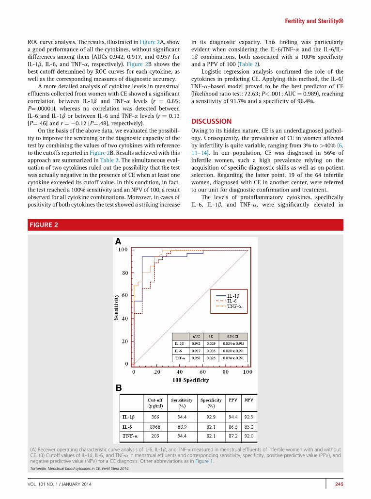
ROC curve analysis. The results, illustrated in Figure 2A, showa good performance of all the cytokines, without significantdifferences among them (AUCs 0.942, 0.917, and 0.957 forIL-1b, IL-6, and TNF-a, respectively). Figure 2B shows thebest cutoff determined by ROC curves for each cytokine, aswell as the corresponding measures of diagnostic accuracy.
A more detailed analysis of cytokine levels in menstrualeffluents collected from women with CE showed a significantcorrelation between IL-1b and TNF-a levels (r ¼ 0.65;P¼ .00001), whereas no correlation was detected betweenIL-6 and IL-1b or between IL-6 and TNF-a levels (r ¼ 0.13[P¼ .46] and r ¼ �0.12 [P¼ .48], respectively).
On the basis of the above data, we evaluated the possibil-ity to improve the screening or the diagnostic capacity of thetest by combining the values of two cytokines with referenceto the cutoffs reported in Figure 2B. Results achieved with thisapproach are summarized in Table 2. The simultaneous eval-uation of two cytokines ruled out the possibility that the testwas actually negative in the presence of CE when at least onecytokine exceeded its cutoff value. In this condition, in fact,the test reached a 100% sensitivity and an NPV of 100, a resultobserved for all cytokine combinations. Moreover, in cases ofpositivity of both cytokines the test showed a striking increase
FIGURE 2
(A) Receiver operating characteristic curve analysis of IL-6, IL-1b, and TNF-aCE. (B) Cutoff values of IL-1b, IL-6, and TNF-a in menstrual effluents and cnegative predictive value (NPV) for a CE diagnosis. Other abbreviations asTortorella. Menstrual blood cytokines in CE. Fertil Steril 2014.
VOL. 101 NO. 1 / JANUARY 2014
in its diagnostic capacity. This finding was particularlyevident when considering the IL-6/TNF-a and the IL-6/IL-1b combinations, both associated with a 100% specificityand a PPV of 100 (Table 2).
Logistic regression analysis confirmed the role of thecytokines in predicting CE. Applying this method, the IL-6/TNF-a–based model proved to be the best predictor of CE(likelihood ratio test: 72.63; P< .001; AUC ¼ 0.989), reachinga sensitivity of 91.7% and a specificity of 96.4%.
DISCUSSIONOwing to its hidden nature, CE is an underdiagnosed pathol-ogy. Consequently, the prevalence of CE in women affectedby infertility is quite variable, ranging from 3% to >40% (6,11–14). In our population, CE was diagnosed in 56% ofinfertile women, such a high prevalence relying on theacquisition of specific diagnostic skills as well as on patientselection. Regarding the latter point, 19 of the 64 infertilewomen, diagnosed with CE in another center, were referredto our unit for diagnostic confirmation and treatment.
The levels of proinflammatory cytokines, specificallyIL-6, IL-1b, and TNF-a, were significantly elevated in
measured in menstrual effluents of infertile women with and withoutorresponding sensitivity, specificity, positive predictive value (PPV), andin Figure 1.
245
TABLE 2
Sensitivity, specificity, positive predictive value (PPV), and negativepredictive value (NPV) for a chronic endometritis diagnosis obtainedby combining the values of two cytokines measured in menstrualeffluents.
Sensitivity(%)
Specificity(%) PPV NPV
1 or 2 positive vs. both negativeIL-6 and TNF-a 100 64.3 78.3 100IL-6 and IL-1b 100 75.0 83.7 100
Both positive vs. 1 or 2 negativeIL-6 and TNF-a 83.3 100 100 82.4IL-6 and IL-1b 83.3 100 100 82.4
Note: Il ¼ interleukin; TNF ¼ tumor necrosis factor.
Tortorella. Menstrual blood cytokines in CE. Fertil Steril 2014.
ORIGINAL ARTICLE: GYNECOLOGY AND MENOPAUSE
menstrual effluents of infertile women with CE comparedwith women with no evidence of CE. These data are inaccordance with those of Boomsma et al., who found asignificant positive association between elevated levels ofIL-1b in endometrial secretions and the presence of bacterialvaginosis (15). These findings are also in keeping withearlier studies indicating that cytokine production in theendometrium may contribute to DUB and reduced endome-trial receptivity. In this regard, DUB in levonorgestrel usershas been correlated with an altered endometrial expressionof IL-13 and IL-15, two key cytokines in inflammatoryand immune cell trafficking (16).
Regarding endometrial receptivity, it is worth noting thatthe endometrium is physiologically colonized by a pleiotropicpopulation of immune-competent cells. The role of these cellsis not only to ensure local defense, but also to allow embryoimplantation and maintenance during pregnancy by modu-lating the host reaction against the embryo. In fact, successfulimplantation and maintenance of pregnancy are the result ofa delicate balance between the embryo and endometrium andboth seem to be closely dependent on the prevalence of a TH2versus a TH1 cytokine profile at endometrial level (17).Accordingly, any condition affecting this balance may impairendometrial receptivity and fertility.
In this context, CE appears to affect the endometrialmicroenvironment significantly. During CE, in fact, notonly the number but also the quality, type, and distributionof leukocytes in the endometrial tissue is remarkably altered.In a previous study, we found that the secretory endometriumof patients with CE displayed significantly lower percentagesof CD56þCD16� and CD56brightCD16� cells and significantlyhigher percentage of CD3þ cells (18). The increase in naturalkiller cells shifts the cytokine balance to a prevalence of theTH1 pathway, with negative effects on implantation and/ortrophoblast invasion, thus determining a higher susceptibilityto miscarriage in the earlier stages of pregnancy (17, 18).
Our data showing very high levels of proinflammatorycytokines in menstrual effluents of women diagnosed withCE suggest that the endometrial production of these mole-cules is overexpressed as well. This is likely to be a reflectionof a TH1/TH2 cytokine imbalance at the endometrial level,where a microenvironment similar to that occurring in other
246
chronic inflammatory diseases, such as rheumatoid arthritis(19, 20) or inflammatory bowel diseases (21), might bepresent. The prevalence of a TH1 cytokine profile, in fact,empowers the recruitment and activation of endometrialmacrophages with an increased release of IL-1, TNF-a, andIL-6 that are, in turn, able to sustain a chronic inflammatoryprocess. Moreover, the long-term consequences of this condi-tion at the endometrial level are still unknown. Salama et al.(22) showed that in endometrial glandular cells exposed toTNF-a, a rise in aromatasic activity takes place, with a conse-quent increase in estrogen synthesis at endometrial level, thisevent being responsible for both functional changes and anenhanced susceptibility to cancer.
Investigating correlations between cytokines, we foundthat IL-1b and TNF-a levels were correlated with each otherbut not with IL-6 concentrations. These findings suggestthat IL-6 responds to different factors compared with theother two proinflammatory cytokines. We may speculatethat IL-6 identifies late stages of inflammation or thatelevated levels of the cytokine are associated with unique in-flammatory processes occurring as a result of peculiar patho-genic noxae. Regardless of the underlying mechanism, highlocal levels of IL-6, a well known differentiation factor ofmature B lymphocytes (23), might largely account for thepresence of plasma cells that is considered to be a histologichallmark of CE.
The study also aimed at assessing whether measuringcytokines in menstrual effluents might provide a simplenoninvasive tool for screening CE. In this regard, the resultsappear to be quite promising, especially when IL-6 and TNF-a or IL-6 and IL-1b are evaluated together. With both com-binations, in fact, if at least one cytokine exceeds its cutoffvalue a sensitivity of 100% and a NPV of 100 are attained,thus ruling out the possibility of a false negative test. Inthese conditions, the test still has a good specificity andPPV and may thus be regarded as a screening test. Accord-ingly, women should be addressed to second-level surveys,such as fluid hysteroscopy combined with endometrial bi-opsy, for a definitive diagnosis. Notably, this step couldeven be omitted should both cytokines measured in men-strual effluents result positive. When this condition occurs,in fact, the test might acquire a high diagnostic value andonly microbiologic analysis would be eventually necessaryto address therapy. Although any test needs to be appliedin an independent similar population to be definitively vali-dated (24), logistic regression analysis may overcome thebias due to the use of the same data. Applying this method,we confirm the role of cytokines measured in menstrual ef-fluents in predicting CE, the best screening for CE beingachieved with the model based on the combined evaluationof IL-6 and TNF-a.
In conclusion, this study provides evidence that CE isassociated with an altered endometrial paracrine milieu, char-acterized by a predominantly TH1 cytokine profile sustainingsignificant levels of proinflammatory cytokines in the men-strual blood. The results of this pilot study may represent afirst step for developing a test based on the combined dosageof IL-6 and TNF-a in menstrual effluents for screening CE inwomen complaining of infertility.
VOL. 101 NO. 1 / JANUARY 2014

Fertility and Sterility®
REFERENCES1. Greenwood SM, Moran JJ. Chronic endometritis: morphologic and clinical
observation. Obstet Gynecol 1981;58:176–84.2. Kitaya K. Prevalence of chronic endometritis in recurrent miscarriages. Fertil
Steril 2011;95:1156–8.3. Johnston-MacAnanny EB, Hartnett J, Engmann LL, Nulsen JC, Sanders MM,
Benadiva CA. Chronic endometritis is a frequent finding in women withrecurrent implantation failure after in vitro fertilization. Fertil Steril 2010;93:437–41.
4. Gilmore H, Fleischhacker D, Hecht JL. Diagnosis of chronic endometritis inbiopsies with stromal breakdown. Hum Pathol 2007;38:581–4.
5. Cicinelli E, Resta L, Nicoletti R, Zappimbulso V, Tartagni M, Saliani N. Endo-metrial micropolyps at fluid hysteroscopy suggest the existence of chronicendometritis. Humanit Rep 2005;20:1386–9.
6. Cicinelli E, de Ziegler D, Nicoletti R, Colafiglio G, Saliani N, Resta L, et al.Chronic endometritis: correlation among hysteroscopic, histologic, andbacteriologic findings in a prospective trial with 2,190 consecutive office his-teroscopies. Fertil Steril 2008;89:677–84.
7. Cicinelli E, Tinelli R, Lepera A, Pinto V, Fucci M, Resta L. Correspondence be-tween hysteroscopic and histologic findings in women with chronic endo-metritis. Acta Obstet Gynecol Scand 2010;89:1061–5.
8. Yudin MH, Hillier SL, Wiesenfeld HC, Krohn MA, Amortegui AA, Sweet RL.Vaginal polymorphonuclear leukocytes and bacterial vaginosis as markersfor histologic endometritis among women without symptoms of pelvic in-flammatory disease. Am J Obstet Gynecol 2003;188:328–33.
9. Kamiyama S, Teruya Y, Nohara M. Impact of detection of bacterial endo-toxin in menstrual effluent on the pregnancy rate in vitro fertilization andembryo transfer. Fertil Steril 2004;82:793–4.
10. Resta L, Palumbo M, Rossi R, Piscitelli D, Fiore GM, Cicinelli E. Histology ofmicro polyps in chronic endometritis. Histopathology 2012;60:670–4.
11. Kasius JC, Fatemi HM, Bourgain C, Sie-Go DM, Eijkemans RJ, Fauser BC,et al. The impact of chronic endometritis on reproductive outcome. FertilSteril 2011;96:1451–6.
12. Carvalho FM, Aguiar FN, Tomioka R, de Oliveira RM, Frantz N, Ueno J. Func-tional endometrial polyps in infertile asymptomatic patients: a possible evo-lution of vascula changes secondary to endometritis. Eur J Obstet GynecolReprod Biol 10.1016/j.ejogrb.2013.05.012.
VOL. 101 NO. 1 / JANUARY 2014
13. Bayer-Gerner IB, Korourian S. Plasma cells in chronic endometritis are easilyidentified when stained with syndecan-1. Mod Pathol 2001;14:877–9.
14. Kitaya K, Yasuo T. Immunohistochemical and clinicopathological char-acterization of chronic endometritis. Am J Reprod Immunol 2011;66:410–5.
15. Boomsma CM, Kavelaars A, Bozkurt N, EijkemansMJ, Fauser BC, Heijnen CJ,et al. Is bacterial vaginosis associated with a pro-inflammatory cytokine pro-file in endometrial secretions of women undergoing IVF? Reprod BiomedOnline 2010;21:133–41.
16. Rhoton-Vlasak A, Chegini N, Hardt N, Williams RS. Histological characteris-tics and altered expression of interleukins (IL) IL-13 and IL-15 in endometriaof levonorgestrel users with different uterine bleeding patterns. Fertil Steril2005;83:659–65.
17. Jin LP, Fan DX, Zhang T, Guo PF, Li DJ. The costimulatory signal upregulationis associated with Th1 bias at the maternal-fetal interface in human miscar-riage. Am J Reprod Immunol 2011;66:270–8.
18. Matteo M, Cicinelli E, Greco P, Massenzio F, Baldini D, Falagario T, et al.Abnormal pattern of lymphocyte subpopulations in the endometrium ofinfertile women with chronic endometritis. Am J Reprod Immunol 2009;61:322–9.
19. Herman S, Zurgil N, Machlav S, Shinberg A, Langevitz P, Ehrenfeld M, et al.Distinct effects of antitumor necrosis factor combined therapy on TH1/TH2balance in rheumatoid arthritis patients. Clin Vaccine Immunol 2011;18:1077–82.
20. Skapenko A, Leipe J, Lipsky PE, Schulze-Koops H. The role of T cell in auto-immune inflammation. Arthritis Res Ther 2005;7(Suppl 2):4–14.
21. Rutgeerts P, Vermeire S, van Assche G. Biological therapies for inflammatorybowel diseases. Gastroenterology 2009;136:1182–97.
22. Salama SA, Kamel MW, Diaz-Arrastia CR, Xu X, Veenstra TD, Salih S, et al.Effect of tumor necrosis factor-alpha on estrogen metabolism and endome-trial cells: potential physiological and pathological relevance. J Clin Endocri-nol Metab 2009;94:285–93.
23. Muraguchi A, Hirano T, Tang B, Matsuda T, Horii Y, Nakajima K, et al. Theessential role of B cell stimulating factor 2 (BSF-2/IL-6) for the terminal differ-entiation of B cells. J Exp Med 1988;167:332–44.
24. Van den Bruel A, Aertgeerts B, Buntinx F. Results of diagnostic accuracystudies are not always validated. J Clin Epidemiol 2006;59:559–66.
247


![Original Article Interleukin-1β induces metabolic and ...excessive apoptosis of disc cells is also pivotal in DDD, which frequently contributes to neck or low back pain [11]. Numerous](https://static.fdocument.pub/doc/165x107/5e8e95c68742d36e0b68f874/original-article-interleukin-1-induces-metabolic-and-excessive-apoptosis-of.jpg)