Influence of the physical and chemical properties of magnetic nanoparticles on their performance in...
7
Influence of the physical and chemical properties of magnetic nanoparticles on their performance in a chemiluminescence immunoassay Xiaoyan Dai a,b , Hong Xu b, ⁎, Yongguang Li a , Hongchen Gu b , Meng Wei a, ⁎⁎ a Division of Cardiology, Shanghai Jiao Tong University Affiliated Sixth People's Hospital, Shanghai, 200233, PR China b Nano Biomedical Research Center, School of Biomedical Engineering, Med-X Research Institute, Shanghai Jiao Tong University, Shanghai 200030, PR China abstract article info Article history: Received 13 October 2013 Received in revised form 2 December 2013 Accepted 7 December 2013 Available online 18 December 2013 Keywords: Magnetic nanoparticle Physicochemical property Chemiluminescence immunoassay Detection performance Myoglobin Objectives: Magnetic nanoparticles (MNPs) are important carriers in immunoassays. In this study, we inves- tigated the influence of the physical and chemical properties of MNPs on their performance in a detection process. Design and methods: A comparative study of the properties of two MNPs with sizes of 200 nm and 1 μm (MNP-200 nm and MNP-1 μm, respectively) was conducted using the following four aspects: nonspecific adsorption to proteins (IgG was used as a model protein), influence of magnetic nanoparticles on the chemilumi- nescence signal, response speed to an external magnetic field, and intensity of the detection signal. Results: MNP-1 μm exhibited lower nonspecific adsorption to IgG in serum, a weaker interference with the chemiluminescence signal, and a higher response speed to the external magnetic field than the same amount of MNP-200 nm. An automated chemiluminescence immunoassay system based on MNP-1 μm was also established. Conclusions: MNP-1 μm acts as an excellent carrier in an automated chemiluminescence immunoassay system for the analysis of serum samples from clinical patients. © 2013 The Canadian Society of Clinical Chemists. Published by Elsevier Inc. All rights reserved. Introduction Magnetic nanoparticles (MNPs), due to their superparamagnetism, suspension stability, large surface area, and easy surface modification, have been widely applied in biomedical fields, including the separation and purification of biomolecules [1,2], immunoassays [3–6], targeted drug delivery [7,8], magnetic hyperthermia [9,10], and MRI contrast agents [11,12]. For in vitro immuno and molecular diagnostics, MNPs are usually used as solid carriers to realize the effective recognition, capture, and manipulation of the target molecules. By virtue of their efficient reaction kinetics and easy automation, MNPs have become an irreplaceable tool for immuno and molecular diagnostics in clinical applications. The current research interest in this field has focused on the develop- ment of new methods and detection agents for better detection perfor- mances [6,13–15]. However, the physical and chemical properties of MNPs, such as size, size distribution, surface structure, and density of functional groups, also have a direct impact on the detection performance because these influence the state of the surface-capped biomolecules and the response speed of MNPs through the external magnetic field. How- ever, few studies have been devoted to the investigation of the relation- ship between the physical and chemical properties of MNPs and their performance in immunoassays. Xie et al. reported that the detection signal obtained by a 100-nm MNP as the carrier is much higher than that obtained by a 1000- or 2800-nm MNP [16]. Harri et al. investigated the effect of the size and number of MNPs on the detection of PSA and concluded that the kinetics of a multiple microparticle assay (particle diameter = 4 μm, 500,000 particles per assay) was rapid due to a favor- able surface-to-volume ratio [17]. Ding et al. studied the influence of the antibody coating density on MNPs on their immunoassay performance. These studies, while presenting some meaningful results concerning the relationship between the given properties of MNPs and their im- munoassay performance, may not be universal considering the great variation in the surface structure of MNPs introduced by the different synthesis methods. Thus, a systematic study of this issue should be addressed. Herein, we present a systematic study of the relationship between the physical and chemical properties of MNPs and their performance in immunoassays. Two MNPs with different sizes (200 nm and 1 μm, denoted MNP-200 nm, MNP-1 μm, respectively) and similar functional group densities and surface structures were prepared through a hybrid miniemulsion method [18], and a comparative study of these two MNPs Clinical Biochemistry 47 (2014) 220–226 ⁎ Correspondence to: H. Xu, Nano Biomedical Research Center, Med-X Research Institute, Shanghai Jiao Tong University, 1954 Huashan Road, Shanghai 200030, PR China. ⁎⁎ Correspondence to: M. Wei, Division of Cardiology, Shanghai Jiao Tong University Affiliated Sixth People's Hospital, 600 Yishan Road, Shanghai 200233, PR China. E-mail addresses: [email protected] (H. Xu), [email protected] (M. Wei). 0009-9120/$ – see front matter © 2013 The Canadian Society of Clinical Chemists. Published by Elsevier Inc. All rights reserved. http://dx.doi.org/10.1016/j.clinbiochem.2013.12.010 Contents lists available at ScienceDirect Clinical Biochemistry journal homepage: www.elsevier.com/locate/clinbiochem
Transcript of Influence of the physical and chemical properties of magnetic nanoparticles on their performance in...
Clinical Biochemistry 47 (2014) 220–226
Contents lists available at ScienceDirect
Clinical Biochemistry
j ourna l homepage: www.e lsev ie r .com/ locate /c l inb iochem
Influence of the physical and chemical properties of magneticnanoparticles on their performance in achemiluminescence immunoassay
Xiaoyan Dai a,b, Hong Xu b,⁎, Yongguang Li a, Hongchen Gu b, Meng Wei a,⁎⁎a Division of Cardiology, Shanghai Jiao Tong University Affiliated Sixth People's Hospital, Shanghai, 200233, PR Chinab Nano Biomedical Research Center, School of Biomedical Engineering, Med-X Research Institute, Shanghai Jiao Tong University, Shanghai 200030, PR China
⁎ Correspondence to: H. Xu, Nano Biomedical ReseInstitute, Shanghai Jiao Tong University, 1954 Huashan Ro⁎⁎ Correspondence to: M. Wei, Division of Cardiology,Affiliated Sixth People's Hospital, 600 Yishan Road, Shang
E-mail addresses: [email protected] (H. Xu), weim.s
0009-9120/$ – see front matter © 2013 The Canadian Sochttp://dx.doi.org/10.1016/j.clinbiochem.2013.12.010
a b s t r a c t
a r t i c l e i n f oArticle history:
Received 13 October 2013Received in revised form 2 December 2013Accepted 7 December 2013Available online 18 December 2013Keywords:Magnetic nanoparticlePhysicochemical propertyChemiluminescence immunoassayDetection performanceMyoglobin
Objectives:Magnetic nanoparticles (MNPs) are important carriers in immunoassays. In this study, we inves-tigated the influence of the physical and chemical properties of MNPs on their performance in a detectionprocess.
Design and methods: A comparative study of the properties of two MNPs with sizes of 200 nm and 1 μm(MNP-200 nm and MNP-1 μm, respectively) was conducted using the following four aspects: nonspecificadsorption to proteins (IgGwas used as amodel protein), influence ofmagnetic nanoparticles on the chemilumi-nescence signal, response speed to an external magnetic field, and intensity of the detection signal.
Results: MNP-1 μm exhibited lower nonspecific adsorption to IgG in serum, a weaker interference withthe chemiluminescence signal, and a higher response speed to the externalmagnetic field than the same amountof MNP-200 nm. An automated chemiluminescence immunoassay system based on MNP-1 μm was alsoestablished.
Conclusions:MNP-1 μmacts as an excellent carrier in an automated chemiluminescence immunoassay systemfor the analysis of serum samples from clinical patients.
© 2013 The Canadian Society of Clinical Chemists. Published by Elsevier Inc. All rights reserved.
Introduction
Magnetic nanoparticles (MNPs), due to their superparamagnetism,suspension stability, large surface area, and easy surface modification,have been widely applied in biomedical fields, including the separationand purification of biomolecules [1,2], immunoassays [3–6], targeteddrug delivery [7,8], magnetic hyperthermia [9,10], and MRI contrastagents [11,12]. For in vitro immuno and molecular diagnostics, MNPsare usually used as solid carriers to realize the effective recognition,capture, and manipulation of the target molecules. By virtue of theirefficient reaction kinetics and easy automation, MNPs have become anirreplaceable tool for immuno and molecular diagnostics in clinicalapplications.
The current research interest in this field has focused on the develop-ment of new methods and detection agents for better detection perfor-mances [6,13–15]. However, the physical and chemical properties ofMNPs, such as size, size distribution, surface structure, and density offunctional groups, also have a direct impact on the detection performance
arch Center, Med-X Researchad, Shanghai 200030, PR China.Shanghai Jiao Tong Universityhai 200233, PR [email protected] (M. Wei).
iety of Clinical Chemists. Published b
because these influence the state of the surface-capped biomolecules andthe response speed of MNPs through the external magnetic field. How-ever, few studies have been devoted to the investigation of the relation-ship between the physical and chemical properties of MNPs and theirperformance in immunoassays. Xie et al. reported that the detectionsignal obtained by a 100-nm MNP as the carrier is much higher thanthat obtained by a 1000- or 2800-nmMNP [16]. Harri et al. investigatedthe effect of the size and number of MNPs on the detection of PSA andconcluded that the kinetics of a multiple microparticle assay (particlediameter = 4 μm, 500,000 particles per assay) was rapid due to a favor-able surface-to-volume ratio [17]. Ding et al. studied the influence of theantibody coating density on MNPs on their immunoassay performance.These studies, while presenting some meaningful results concerningthe relationship between the given properties of MNPs and their im-munoassay performance, may not be universal considering the greatvariation in the surface structure of MNPs introduced by the differentsynthesis methods. Thus, a systematic study of this issue should beaddressed.
Herein, we present a systematic study of the relationship betweenthe physical and chemical properties of MNPs and their performancein immunoassays. Two MNPs with different sizes (200 nm and 1 μm,denoted MNP-200 nm, MNP-1 μm, respectively) and similar functionalgroup densities and surface structures were prepared through a hybridminiemulsionmethod [18], and a comparative study of these twoMNPs
y Elsevier Inc. All rights reserved.

221X. Dai et al. / Clinical Biochemistry 47 (2014) 220–226
was conducted from the following four aspects: (1) nonspecific adsorp-tion to IgG in serum, (2) effects of magnetic nanoparticles in the MNPson the detection signal generated through chemiluminescence, (3) theintensity of the detection signal in myoglobin (model protein), and(4) the response speed to the external magnetic field. Based on theabove-mentioned study, an automated chemiluminescence immu-noassay system for myoglobin was established using MNP-1 μm asthe carrier. In addition, we analyzed patients' serum samples usingthe proposed system and a commercial immunoassay system to in-vestigate the correlation between the detection results obtainedusing the two systems.
Materials
Apparatus
ANano ZS particle size analyzer was obtained fromMalvern (Britain)to analyze MNPs hydrodynamic size, a 1420Multilabel Counter Victor3instrument was purchased from PerkinElmer (Finland) to carry outnonspecific adsorption of MNPs to IgG and CLIA experiment, and amicroplate shaker was obtained from Hengfeng (model WZ-2A, Jintan,China). Mini-table type vacuum pumps (GL-802) and a vortex mixer(QL-901) were purchased from QiLinBeiEr (Haimen, China). A magneticseparation rack and magnetic separation plate were obtained fromAllrun (Shanghai, China), and a constant-temperaturewater bath oscilla-tor was obtained from Bolaite (Taicang, China).
Reagents
Tween-20 was provided by BBI (Canada). MES (2-[N-morpholino]ethane sulfonic acid) was purchased from Alfa Aesar (USA), andpotassium dihydrogen phosphate (KH2PO4), sodium chloride (NaCl),potassium persulfate (KPS), hydroxylamine hydrochloride, and 1,10-phenanthrolinemonohydrate were obtained from Sinopharm ChemicalReagents (Shanghai, China). Potassium chloride (KCl) was providedby SCRC (Shanghai, China), glycine was obtained from Sigma (USA),and bovine serum albumin (BSA) was obtained from Genview (USA).N-Hydroxysuccinimide (NHS), O-phenylenediamine (OPD), hydrogenperoxide (H2O2), the BCA protein assay kit, and the chemiluminescentsubstrates were purchased from Pierce (USA), and 1-ethyl-3-[3-dimethylaminopropyl]carbodiimide hydrochloride (EDC) was obtainedfrom Medpe (Shanghai, China). The quality control serum was pur-chased from Randox (UK), and the myoglobin antigen and two anti-human myoglobin monoclonal antibodies (mAb1-4E2 and mAb2-7C3),one of which (mAb2-7C3) was tagged with horseradish peroxidase(HRP), were purchased from Hytest (Turku, Finland). HRP-labeled goatanti-human IgG was obtained from Bioss (Beijing, China). The testhuman serum was collected from Shanghai Sixth People's Hospital.This study was approved by the Medical Ethics Committee of ShanghaiSixth People's Hospital Affiliated with Shanghai Jiaotong University,and all of the patients provided signed informed consent.
Methods
Synthesis of MNPs
Two carboxylated MNPs with sizes of approximately 200 nm and1 μm (MNP-200 nm and MNP-1 μm, respectively) were synthesizedthrough hybrid miniemulsion polymerization as described in ourprevious work [18]. The MNPs were composed of Fe3O4 nanoparticles,and the content of magnetite nanoparticles (25 wt.%, 48 wt.%, and74 wt.%) was adjusted by changing the concentration of magneticaggregates during the synthesis.
Nonspecific adsorption of MNPs to IgG in serum
In this study, the relative adsorption quantity of MNPs to IgG inserum was measured to evaluate the antifouling property of theMNPs. MNP-200 nm and MNP-1 μm (30, 50, 100, 200, and 300 μg)were introduced into a 96-well microplate and washed twice withPBST (0.05 M, pH 7.5). One hundred microliters of the quality controlserum were added to each well, and the plate was incubated at 37 °Cfor 30 min. After incubation, the MNPs were collected by an externalmagnetic field and washed three times with PBST and deionizedwater. One hundred microliters of a diluted solution of HRP-labeledgoat-anti-human IgG were then added to the MNPs, and the mixtureswere incubated for 30 min at 37 °C. After magnetic separation anddiscarding the supernatant, 150 μL of the substrate solution wasadded (OPD + H2O2) to each well, and the plate was incubated for15 min at 37 °C to afford the color reaction. After magnetic separation,100 μL of the supernatant was transferred to another clean 96-wellmicroplate, and 25 μL of the stopping solution (2 M H2SO4) was addedto each well. The absorption of the solution wasmeasured at the wave-length of 490 nm, and the intensity was used to evaluate the relativequantity of nonspecific adsorption of the MNPs to IgG.
Response speed of MNPs to an external magnetic field
The response speed of MNPs to a magnetic field was evaluated bymonitoring the concentration of Fe at different time points after anexternal magnetic field was applied to an MNP suspension. First, 1 mgof MNPs (MNP-200 nm and MNP-1 μm) suspended in 800 μL of deion-ized water was added to a 1.5-mL centrifuge tube, which was thenplaced in a magnetic separator (a total of 12 samples, six for eachMNP). Then, 700 μL of the supernatant were removed 0, 20, 40, 60, 90,and 120 s after the external magnetic field was applied. Four hundredmicroliters of HCl (37 wt.%) were added to each sample, and the sam-ples were incubated for 48 h to dissolve the MNPs. The concentrationof Fe in the supernatant at the different time points was determinedthrough a colorimetric method: 1 mL of 100 g/L hydroxylamine hydro-chloride solution was added to the supernatant, and the mixture wasshaken for 5 min. Then, 1 mLof 4 g/L 1,10-phenanthrolinemonohydratesolution and 5 mL of HAc–NaAc buffer were successively added. Deion-ized water was then added to obtain a final volume of 50 mL. The absor-bance of the solution was measured at the wavelength of 510 nm.
Influence of MNPs on the signal of a chemiluminescence immunoassay(CLIA)
First, 0–100 μg of the MNPs (MNP-200 nm and MNP-1 μm) wereadded to a 96-well microplate and washed twice with PBST. To eachwell, 100 μL of a mixed solution of enzyme-labeled antibody (1 mg/mL,10,000-fold dilution) and chemiluminescence agent (1:9 v/v) wasadded and mixed with the MNPs. Then, 90 μL of the mixture was trans-ferred to a white microplate and incubated in the dark for 5 min beforemeasuring the chemiluminescence intensity.
To evaluate the effect of the content of the magnetite nanoparticleson the CLIA signal, the same quantity of three MNPs with differentcontents of magnetic nanoparticles (25 wt.%, 48 wt.%, and 74 wt.%)was used. The experimental procedure was the same as that describedabove.
Establishment of the CLIA system
Preparation of immuno-MNPsFirst, 2 mg of MNPs (MNP-200 nm andMNP-1 μm)were added to a
2-mL centrifuge tube and re-dispersed in 200 μL of MEST (25 mM,pH 6) by washing twice with MEST. Then, 2 mg of EDC and NHS wereadded to the tube, and the reaction was allowed to proceed at ambienttemperature for 15 min. After the reaction, the excess reactants were

Table 1Characterization of magnetic nano-particles.
MNP Hydrodynamicdiameter (nm)
Fe3O4 content(wt.%)
Carboxyl groupdensity (mmol/g)
MNP-1 μm 1380 ~50 ~0.5MNP-200 nm 204 ~60 0.57
222 X. Dai et al. / Clinical Biochemistry 47 (2014) 220–226
removed bywashing three timeswithMEST. Then, 300 μg ofmyoglobinmonoclonal antibody was added, and the reaction was allowed to pro-ceed at ambient temperature for 2 h. The MNP-myoglobin complexeswere blockedwith 200 μL of blocking solution (0.05 M PBST containing0.1% BSA and 0.3% Gly) overnight at 4 °C. The complexes were then re-dispersed in 200 μL of storing solution (0.05 M PBST containing 2%BSA) at 4 °C.
The binding capacity of the antibody was determined using a BCAprotein quantification kit with the depletion method. A control tubethat underwent the same procedure without the addition of antibodywas used to eliminate the interference.
Chemiluminescence detection of myoglobinThe obtained immuno-MNPwas applied for the chemiluminescence
detection of myoglobin using the following procedure: To a microplate,30 μg of immuno-MNP (30 μL), 60 μL of antigen solution, and 100 μL ofHRP-labeled antibody solution (1 mg/mL, 2000-fold dilution) wereadded and incubated at 37 °C for 30 min to obtain MNP–immune com-plexes. After the reaction, the obtained MNP–immune complexes werewashed five times with Tris–HCl (0.02 M, pH 7.4), and the supernatantwas discarded with the help of an external magnetic field. Then, 100 μLof the chemiluminescence agent was added and mixed with the MNP–immune complexes. The mixture was placed in the dark for 5 min, andthe chemiluminescence signal was then measured.
Establishment of automated MYO CLIA systemThe myoglobin chemiluminescence enzyme immunoassay system
(Horseradish peroxidase as label enzyme, Luminol as enzyme sub-strate) was established according to the above-mentioned procedureusingMNP-1 μmas the carrier. The detection standard curve, sensitivity,precision, and dynamic range of the system were determined.
Twenty-six split serum samples were collected from 26 patientshospitalized in Shanghai Sixth People's Hospital (two samples fromeach patient). The concentration of myoglobin in one sample from apatient was determined using the system proposed in this paper, andthe concentration of myoglobin in the other sample from the samepatient was determined using the Beckman chemiluminescence immu-noassay kit and instrument. The results obtained by the two systemswere analyzed through a correlation analysis.
Results and discussion
Synthesis and characterization of MNPs
Carboxylated MNPs with sizes of approximately 200 nm and 1 μm(MNP-200 nm, MNP-1 μm, respectively) were synthesized throughhybrid miniemulsion polymerization. Fig. 1 shows the transmission
Fig. 1. TEM of the superparamagnetic beads with diameters of 200 nm (a) a
electron microscope (TEM) image of the MNPs. Both MNPs are com-posed of 10-nm Fe3O4 nanoparticles with a narrow size distribution.In addition, their contents of magnetite nanoparticles and their func-tional group densities are similar (Table 1).
Nonspecific adsorption to IgG
In a clinical immunoassay, the recognition and capture of the targetmolecule byMNPs is usually performed in serumand plasma. Therefore,the nonspecific adsorption to IgG in serum or plasma will influence thespecificity and accuracy of the assay. To evaluate thenonspecific adsorp-tion of MNPs in the detection of serum samples, a comparative study ofthe nonspecific adsorption properties of MNP-200 nm and MNP-1 μmto IgG in serum was performed.
As shown in Fig. 2a, for both MNPs, the optical density (OD) in-creases with an increase in the amount of the MNPs (30 μg–300 μg).When the amounts of the two MNPs were the same, the OD of MNP-200 nmwas always higher than that of MNP-1 μm, indicating a highernonspecific adsorption. This difference can be ascribed to the larger spe-cific surface area ofMNP-200 nmdue to its smaller size, which providesa larger area for protein adsorption.
The results were converted into OD/nm2 to show the nonspecificadsorption capacity of the MNPs per unit area. As shown in Fig. 2b, forboth MNPs, the nonspecific adsorption per unit area decreased withan increase in the MNP amount. In addition, the nonspecific adsorptionof MNP-200 nm was always lower than that of MNP-1 μm. This resultsuggests that the interaction between MNP-200 nm and the protein isweaker, which possibly results from the larger curvature of MNP-200 nm [19,20]. In contrast, althoughMNP-200 nm has a lower nonspe-cific adsorption per unit area, it possesses a higher nonspecific adsorptionper unit mass due to its larger specific surface area. Therefore, a higherbackground was obtained for MNP-200 nm in the immunoassay.
Response speed to the external magnetic field
MNPs act as carriers for the separation of biomolecules in CLIA. Theirresponse speed to the external magnetic field not only influences thedetection time but also plays a decisive role in the realization of theautomated detection procedure. Hereby, we evaluated the responses
nd 1 μm (b) synthesized through hybrid miniemulsion polymerization.

Fig. 2. Non-specific adsorption of MNP-200 nm and MNP-1 μm as a function of the mass of the MNPs. (a) Total adsorption. (b) Adsorption per unit surface area. ** indicates p b 0.01.
223X. Dai et al. / Clinical Biochemistry 47 (2014) 220–226
of MNP-200 nm andMNP-1 μmby detecting the residual Fe concentra-tion in the supernatant as a function of the capture time after applyingthe external magnetic field. As shown in Fig. 3, for both MNPs, the Feconcentration in the supernatant decreases with the prolongation ofthe magnetic capture time. The Fe concentration in the supernatant ofMNP-1 μm decreased more rapidly and almost reached zero at 40 s. Incontrast, MNP-200 nm is not fully captured even at 120 s because themotion velocity of MNP in amagnetic field is proportional to the squareof its radius as well as the magnetic substances [21]. If the contents ofmagnetite nanoparticles of MNP-200 nm and MNP-1 μm are similar(approximately 60 wt.% and 50 wt.%, respectively), the larger MNP-1 μm exhibits a rapid response speed to the external magnetic field.In practical applications of CLIA, the slow response speed of MNP-
Fig. 3.Magnetic response speed ofMNP-200 nm andMNP-1 μm to the external magneticfield. This response was evaluated by the decrease in the residual Fe concentration in thesupernatant as a function of time after applying the external magnetic field.
200 nm will greatly increase the detection time and cannot meetthe demands of automation. Therefore, MNP-1 μm is more suitablefor automated CLIA based on its detection time and the demands ofautomation.
Quenching of the chemiluminescence signal by MNPs
The immunoassay system established in this study is a luminolchemiluminescence system based on horseradish peroxidase (HRP)-labeled IgG. The MNPs were suspended in the chemiluminescencesubstrate throughout the detection process and thus may quench thechemiluminescence signal. In this work, we studied the influence of
Fig. 4.Quenching effect ofMNP-200 nmandMNP-1 μmon the chemiluminescence signal.The MNPs were directly added to a mixture of the enzyme-labeled antibody and thechemiluminescence agent.
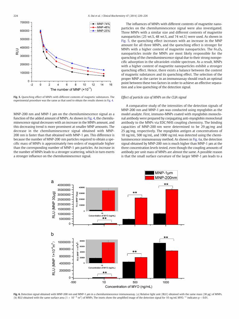
Fig. 5. Quenching effect of MNPs with different contents of magnetic substances. Theexperimental procedure was the same as that used to obtain the results shown in Fig. 4.
224 X. Dai et al. / Clinical Biochemistry 47 (2014) 220–226
MNP-200 nm and MNP-1 μm on the chemiluminescence signal as afunction of the added amount of MNPs. As shown in Fig. 4, the chemilu-minescence signal decreases with an increase in the MNPs amount, andthis decreasing trend is more prominent at smaller MNP amounts. Thedecrease in the chemiluminescence signal obtained with MNP-200 nm is faster than that obtained with MNP-1 μm. This difference isbecause the number of MNP-200 nm particles required to obtain a spe-cific mass of MNPs is approximately two orders of magnitude higherthan the corresponding number of MNP-1 μm particles. An increase inthe number of MNPs leads to a stronger scattering, which in turn exertsa stronger influence on the chemiluminescence signal.
Fig. 6. Detection signal obtained with MNP-200 nm and MNP-1 μm in a chemiluminescence im(b) RLU obtained with the same surface area (1 × 10⁻⁴ m2) of MNPs. The insets show the am
The influences of MNPs with different contents of magnetite nano-particles on the chemiluminescence signal were also investigated.Three MNPs with a similar size and different contents of magnetitenanoparticles (25 wt.%, 48 wt.%, and 74 wt.%) were used. As shown inFig. 5, the quenching effect increases with an increase in the MNPamount for all three MNPs, and the quenching effect is stronger forMNPs with a higher content of magnetite nanoparticles. The Fe3O4
nanoparticles inside the MNPs are most likely responsible for thequenching of the chemiluminescence signal due to their strong nonspe-cific adsorption in the ultraviolet–visible spectrum. As a result, MNPswith a higher content of magnetite nanoparticles exhibit a strongerquenching effect. Hence, there exists a balance between the contentof magnetic substances and its quenching effect. The selection of theproper MNP as the carrier in an immunoassay should reach an optimalpoint between these two factors in order to achieve an effective separa-tion and a low quenching of the detection signal.
Effect of particle size of MNPs on the CLIA signal
A comparative study of the intensities of the detection signals ofMNP-200 nm and MNP-1 μm was conducted using myoglobin as themodel analyte. First, immuno-MNPs coated with myoglobin monoclo-nal antibodywere prepared by conjugating anti-myoglobinmonoclonalantibody to the MNPs via EDC/NHS coupling chemistry. The bindingcapacities of MNP-200 nm were determined to be 29 μg/mg and25 μg/mg, respectively. The myoglobin antigen at concentrations of10 ng/mL, 500 ng/mL, and 1000 ng/mL was detected using the chemi-luminescence immunoassay method. As shown in Fig. 6a, the detectionsignal obtained by MNP-200 nm is much higher than MNP-1 μm at thethree concentration levels tested, even though the coupling amounts ofantibody per unit mass of MNPs are almost the same. A possible reasonis that the small surface curvature of the larger MNP-1 μm leads to a
munoassay. (a) Relative light unit (RLU) obtained with the same mass (30 μg) of MNPs.plified image of the detection signal for 10 ng/mL MYO. ** indicates p b 0.01.
225X. Dai et al. / Clinical Biochemistry 47 (2014) 220–226
stronger interaction with the antibody, which further induces an alter-ation in the protein conformation and thereby reduces its activity[22,23]. As a result, the antibody on MNP-1 μmmay have a lower affin-ity to the myoglobin antigen. Converting the results in Fig. 6a into thedetection signal obtainedwith the sameMNP surface area, we obtainedFig. 6b. As shown, when the surface area of the two MNPs are the same(1 × 10⁻⁴ m2), the detection signal obtainedwithMNP-200was higherthan that obtained with MNP-1 μm in the presence of 10 ng/mL and500 ng/mL antigen, whereas the opposite results were obtained withan antigen concentration of 1000 ng/mL. A possible reason is that theMNP surface antibody at the antigen concentrations of 10 ng/mL and500 ng/mL is found in excess compared with the immune-complex,and the signal is determined by the reactivity of the surface antibody.As a result, a higher signal was obtained for MNP-200 due to the higherreactivity of the antibody on its surface.When the antigen concentrationwas increased to 1000 ng/mL, the number of antigen molecules be-comes larger than the number of surface antibody molecules, and thedensity of the surface antibody becomes a decisive factor. Because theantibody coating amounts per mass of MNP were similar for the twoMNPs, MNP-1 μm possesses a higher density of surface antibody dueto its lower specific surface area. Consequently, a higher signal wasobtained with MNP-1 μm in the presence of an antigen concentrationof 1000 ng/mL.
The described studies evaluated the properties of MNP-200 nm andMNP-1 μm in CLIA from several aspects. Although the detection signalobtained with MNP-1 μm is lower than that obtained with MNP-200 nm, MNP-1 μm can generally meet the demands of the immunoas-say and exhibits a lower background interference. The most importantaspect is that MNP-1 μm can be rapidly captured by the external mag-netic field to fulfill automated detection. Therefore, MNP-1 μm is moresuitable to be used as the carrier in an automated CLIA.
Comparison between the present chemiluminescence immunoassay systemand a commercial system
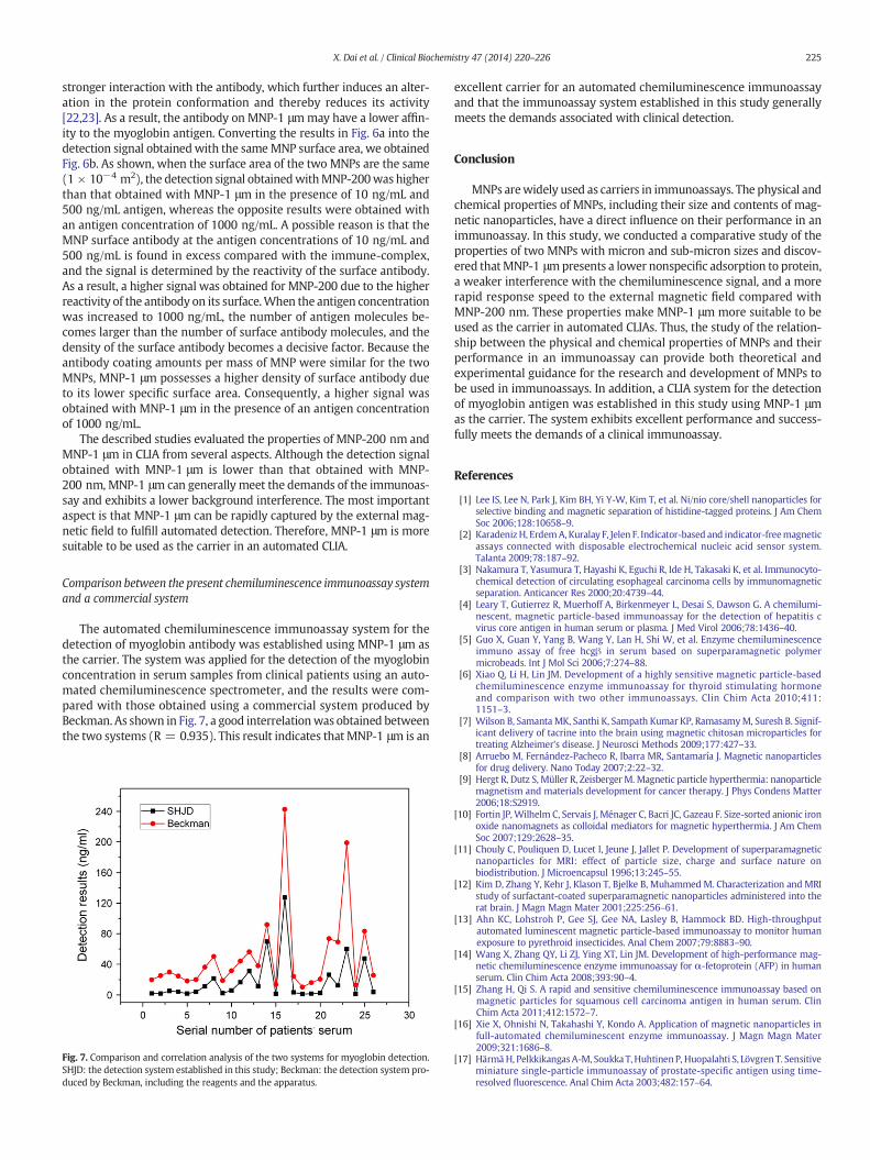
The automated chemiluminescence immunoassay system for thedetection of myoglobin antibody was established using MNP-1 μm asthe carrier. The system was applied for the detection of the myoglobinconcentration in serum samples from clinical patients using an auto-mated chemiluminescence spectrometer, and the results were com-pared with those obtained using a commercial system produced byBeckman. As shown in Fig. 7, a good interrelationwas obtained betweenthe two systems (R = 0.935). This result indicates that MNP-1 μm is an
Fig. 7. Comparison and correlation analysis of the two systems for myoglobin detection.SHJD: the detection system established in this study; Beckman: the detection system pro-duced by Beckman, including the reagents and the apparatus.
excellent carrier for an automated chemiluminescence immunoassayand that the immunoassay system established in this study generallymeets the demands associated with clinical detection.
Conclusion
MNPs arewidely used as carriers in immunoassays. The physical andchemical properties of MNPs, including their size and contents of mag-netic nanoparticles, have a direct influence on their performance in animmunoassay. In this study, we conducted a comparative study of theproperties of two MNPs with micron and sub-micron sizes and discov-ered thatMNP-1 μmpresents a lower nonspecific adsorption to protein,a weaker interference with the chemiluminescence signal, and a morerapid response speed to the external magnetic field compared withMNP-200 nm. These properties make MNP-1 μm more suitable to beused as the carrier in automated CLIAs. Thus, the study of the relation-ship between the physical and chemical properties of MNPs and theirperformance in an immunoassay can provide both theoretical andexperimental guidance for the research and development of MNPs tobe used in immunoassays. In addition, a CLIA system for the detectionof myoglobin antigen was established in this study using MNP-1 μmas the carrier. The system exhibits excellent performance and success-fully meets the demands of a clinical immunoassay.
References
[1] Lee IS, Lee N, Park J, Kim BH, Yi Y-W, Kim T, et al. Ni/nio core/shell nanoparticles forselective binding and magnetic separation of histidine-tagged proteins. J Am ChemSoc 2006;128:10658–9.
[2] KaradenizH, ErdemA,Kuralay F, Jelen F. Indicator-based and indicator-freemagneticassays connected with disposable electrochemical nucleic acid sensor system.Talanta 2009;78:187–92.
[3] Nakamura T, Yasumura T, Hayashi K, Eguchi R, Ide H, Takasaki K, et al. Immunocyto-chemical detection of circulating esophageal carcinoma cells by immunomagneticseparation. Anticancer Res 2000;20:4739–44.
[4] Leary T, Gutierrez R, Muerhoff A, Birkenmeyer L, Desai S, Dawson G. A chemilumi-nescent, magnetic particle-based immunoassay for the detection of hepatitis cvirus core antigen in human serum or plasma. J Med Virol 2006;78:1436–40.
[5] Guo X, Guan Y, Yang B, Wang Y, Lan H, Shi W, et al. Enzyme chemiluminescenceimmuno assay of free hcgβ in serum based on superparamagnetic polymermicrobeads. Int J Mol Sci 2006;7:274–88.
[6] Xiao Q, Li H, Lin JM. Development of a highly sensitive magnetic particle-basedchemiluminescence enzyme immunoassay for thyroid stimulating hormoneand comparison with two other immunoassays. Clin Chim Acta 2010;411:1151–3.
[7] Wilson B, Samanta MK, Santhi K, Sampath Kumar KP, RamasamyM, Suresh B. Signif-icant delivery of tacrine into the brain using magnetic chitosan microparticles fortreating Alzheimer's disease. J Neurosci Methods 2009;177:427–33.
[8] Arruebo M, Fernández-Pacheco R, Ibarra MR, Santamaría J. Magnetic nanoparticlesfor drug delivery. Nano Today 2007;2:22–32.
[9] Hergt R, Dutz S, Müller R, ZeisbergerM. Magnetic particle hyperthermia: nanoparticlemagnetism and materials development for cancer therapy. J Phys Condens Matter2006;18:S2919.
[10] Fortin JP, Wilhelm C, Servais J, Ménager C, Bacri JC, Gazeau F. Size-sorted anionic ironoxide nanomagnets as colloidal mediators for magnetic hyperthermia. J Am ChemSoc 2007;129:2628–35.
[11] Chouly C, Pouliquen D, Lucet I, Jeune J, Jallet P. Development of superparamagneticnanoparticles for MRI: effect of particle size, charge and surface nature onbiodistribution. J Microencapsul 1996;13:245–55.
[12] Kim D, Zhang Y, Kehr J, Klason T, Bjelke B, Muhammed M. Characterization and MRIstudy of surfactant-coated superparamagnetic nanoparticles administered into therat brain. J Magn Magn Mater 2001;225:256–61.
[13] Ahn KC, Lohstroh P, Gee SJ, Gee NA, Lasley B, Hammock BD. High-throughputautomated luminescent magnetic particle-based immunoassay to monitor humanexposure to pyrethroid insecticides. Anal Chem 2007;79:8883–90.
[14] Wang X, Zhang QY, Li ZJ, Ying XT, Lin JM. Development of high-performance mag-netic chemiluminescence enzyme immunoassay for α-fetoprotein (AFP) in humanserum. Clin Chim Acta 2008;393:90–4.
[15] Zhang H, Qi S. A rapid and sensitive chemiluminescence immunoassay based onmagnetic particles for squamous cell carcinoma antigen in human serum. ClinChim Acta 2011;412:1572–7.
[16] Xie X, Ohnishi N, Takahashi Y, Kondo A. Application of magnetic nanoparticles infull-automated chemiluminescent enzyme immunoassay. J Magn Magn Mater2009;321:1686–8.
[17] HärmäH, PelkkikangasA-M, Soukka T,HuhtinenP, Huopalahti S, Lövgren T. Sensitiveminiature single-particle immunoassay of prostate-specific antigen using time-resolved fluorescence. Anal Chim Acta 2003;482:157–64.

226 X. Dai et al. / Clinical Biochemistry 47 (2014) 220–226
[18] Cui L, Xu H, He P, Sumitomo K, Yamaguchi Y, Gu H. Developing a hybrid emulsionpolymerization system to synthesize fe3o4/polystyrene latexes with narrow sizedistribution and high magnetite content. J Polym Sci A Polym Chem 2007;45:5285–95.
[19] Lacerda SHDP, Park JJ, Meuse C, Pristinski D, Becker ML, Karim A, et al. Interaction ofgold nanoparticles with common human blood proteins. ACS Nano 2009;4: 365–79.
[20] Walkey CD, ChanWC. Understanding and controlling the interaction of nanomaterialswith proteins in a physiological environment. Chem Soc Rev 2012;41:2780–99.
[21] Xu H. Controllable preparation and biomedical applications of monodisperse andsuperparamagnetic magnetic microspheres with high saturation magnetization.Shanghai Jiaotong University; 2008.
[22] Gagner JE, Lopez MD, Dordick JS, Siegel RW. Effect of gold nanoparticle morphologyon adsorbed protein structure and function. Biomaterials 2011;32:7241–52.
[23] Shang W, Nuffer JH, Muñiz‐Papandrea VA, Colón W, Siegel RW, Dordick JS. Cyto-chrome c on silica nanoparticles: influence of nanoparticle size on protein structure,stability, and activity. Small 2009;5:470–6.