Immunohistochemical detection of transforming growth factor-β1 in fibrotic liver diseases
5
Immunohistochemical Detection of Transforming Growth Factor+, in Fibrotic Liver Diseases PETER NAGY, ZSUZSA SCHAFF AND KbLROLY LAPIS I. Institute of Pathology and Experimental Cancer Research, Semmelweis Medical School, Budapest, H-1085 Hungary Transforming growth factor-$, was localized by means of immunohistochemical reaction in liver biopsy specimens taken from patients having different chronic liver diseases with extending fibrosis. Two polyclonal antibodies that were produced in rabbits were directed against the amino terminal of trans- forming growth factor-p,. Staining by anti-CC(1-30) was primarily extracel- lular and located in the portal and periportal fibrotic areas of all seven cases with chronic active hepatitis. No staining was noted in the four chronic persistent cases studied. A strong reaction was seen with the antibody in nine of the ten cirrhotic samples, whereas it was negative in one inactive cirrhosis case and in all five cases with normal liver histological findings. No positive stainingcouldbe detected by the anti-LC( 1-30) in any of the liver tissues. Detection of transforming growth factor-$,in active liver diseases at the site of fibrosis suggests that transforminggrowth factor-$, might have a role in the process and progression of fibrosis during the devel- opment of the disease. (HEPATOLOGY 1991;14:269-273.) Increased deposition of extracellular matrix is a characteristic change in several chronic liver diseases, especially in cirrhosis (1-4). The exact factors that are responsible for this accumulation are unknown. Consid- ering the similarities in histopathological features for cirrhosis and chronic hepatitis, as well as the polyetio- logical origin of both, we considered common factors that could be responsible for fibrosis in the liver during several pathological conditions. One candidate for such a factor is transforming growth factor-p, (TGF-@,I. TGF-P, is a multifunctional growth factor. It is presumed to play a role in the regulation of many physiological and pathological processes (5). The first recognized in uiuo effect was the stimulation of wound healing (6). The deposition of extracellular matrix is the most important event during the process of scar for- mation in the late phase of wound healing. For this reason, it was proposed that TGF-P, may have an influence on the metabolism of extracellular matrix Received March 22, 1990; accepted March 19. 1991. Address reprint requests to: Peter Nagy. M.D., I. Institute of Pathology and Experimental Cancer Research, Semmelweis Medical School, iilloi ut 26, Budapest, H-1085 Hungary. 31/1/30136 constituents. In various experimental and in uiuo pro- cesses, TGF-@, has been reported to cause an increase in the synthesis of different matrix components (collagen, fibronectin, etc.) (7) while also inhibiting their proteo- lytic degradation (8,9). Furthermore, TGF-P, has been reported to increase the number of receptors for extra- cellular matrix components on fibroblasts (10). Because the ability of TGF-P, to increase matrix accumulation in various fibrotic processes, TGF-P, may also have a direct function in the regulation of progressive liver fibrosis. There are some data supporting this idea in experi- mental systems (11-14). In this study, we used immu- nohistological methods to detect TGF-P, in human liver biopsy specimens from patients with progressive fibrotic liver diseases. MATERIALS AND METHODS In a retrospective study, we analyzed the needle biopsy specimens from 26 patients; 7 with CAH, 4 with chronic persistent hepatitis, and 10 with alcoholic HBsAg-negative cirrhosis. Five liver biopsy specimens in which no pathological alterations were seen were included as controls. The biopsy specimens were fixed in 8% neutral formaldehyde and em- bedded in paraffin. The diagnosis of liver disease was made from slides stained with hematoxylin and eosin, PAS, Gomory and Mallory’s trichrome procedures. Immunohistochemistry The antibodies used in this study were produced and characterized for TGF-P, specificityby Ellingsworth et al. (15) and Flanders et al. (16). The polyclonal antisera were gen- erated in rabbits by two different synthetic preparations of a peptide correspondingto the amino terminal 30 amino acids of TGF-@, (16, 17). These two antisera give different staining patterns in several tissues and are believed to differentiate between the synthesized and activated forms of TGF-@, in some experimental systems. Staining with anti-LC(1-30)is principally intracellular, whereas that of anti-CC(1-30) is extracellular. It was suggested that anti-LC(l-30)-positive cells could be the sites of TGF-@, synthesis, whereas anti- CC(1-30) may recognize activated TGF-@, (17). A computer search of the immunogen sequence revealed no homology with five or more adjacentamino acids of any protein in the database with the exception of TGF-@, (18). Thereforeit is unlikely that cross-reactivity with other proteins is responsible for the staining patterns. After deparaffinization, the blocking of endogenous perox- idase was performed in H,O,/methanol. Membrane perme- 269
-
Upload
peter-nagy -
Category
Documents
-
view
212 -
download
0
Transcript of Immunohistochemical detection of transforming growth factor-β1 in fibrotic liver diseases

Immunohistochemical Detection of Transforming Growth Factor+, in Fibrotic Liver Diseases
PETER NAGY, ZSUZSA SCHAFF AND KbLROLY LAPIS I . Institute of Pathology and Experimental Cancer Research, Semmelweis Medical School, Budapest, H-1085 Hungary
Transforming growth factor-$, was localized by means of immunohistochemical reaction in liver biopsy specimens taken from patients having different chronic liver diseases with extending fibrosis. Two polyclonal antibodies that were produced in rabbits were directed against the amino terminal of trans- forming growth factor-p,.
Staining by anti-CC(1-30) was primarily extracel- lular and located in the portal and periportal fibrotic areas of al l seven cases with chronic active hepatitis. No staining was noted in the four chronic persistent cases studied. A strong reaction was seen with the antibody in nine of the ten cirrhotic samples, whereas it was negative in one inactive cirrhosis case and in all five cases with normal liver histological findings. No positive staining could be detected by the anti-LC( 1-30) in any of the liver tissues.
Detection of transforming growth factor-$, in active liver diseases at the site of fibrosis suggests that transforming growth factor-$, might have a role in the process and progression of fibrosis during the devel- opment of the disease. (HEPATOLOGY 1991;14:269-273.)
Increased deposition of extracellular matrix is a characteristic change in several chronic liver diseases, especially in cirrhosis (1-4). The exact factors that are responsible for this accumulation are unknown. Consid- ering the similarities in histopathological features for cirrhosis and chronic hepatitis, as well as the polyetio- logical origin of both, we considered common factors that could be responsible for fibrosis in the liver during several pathological conditions. One candidate for such a factor is transforming growth factor-p, (TGF-@,I.
TGF-P, is a multifunctional growth factor. It is presumed to play a role in the regulation of many physiological and pathological processes (5). The first recognized in uiuo effect was the stimulation of wound healing (6). The deposition of extracellular matrix is the most important event during the process of scar for- mation in the late phase of wound healing. For this reason, it was proposed that TGF-P, may have an influence on the metabolism of extracellular matrix
Received March 22, 1990; accepted March 19. 1991. Address reprint requests to: Peter Nagy. M.D., I. Institute of Pathology and
Experimental Cancer Research, Semmelweis Medical School, iilloi ut 26, Budapest, H-1085 Hungary.
31/1/30136
constituents. In various experimental and in uiuo pro- cesses, TGF-@, has been reported to cause an increase in the synthesis of different matrix components (collagen, fibronectin, etc.) (7) while also inhibiting their proteo- lytic degradation (8,9). Furthermore, TGF-P, has been reported to increase the number of receptors for extra- cellular matrix components on fibroblasts (10). Because the ability of TGF-P, to increase matrix accumulation in various fibrotic processes, TGF-P, may also have a direct function in the regulation of progressive liver fibrosis. There are some data supporting this idea in experi- mental systems (11-14). In this study, we used immu- nohistological methods to detect TGF-P, in human liver biopsy specimens from patients with progressive fibrotic liver diseases.
MATERIALS AND METHODS In a retrospective study, we analyzed the needle biopsy
specimens from 26 patients; 7 with CAH, 4 with chronic persistent hepatitis, and 10 with alcoholic HBsAg-negative cirrhosis. Five liver biopsy specimens in which no pathological alterations were seen were included as controls. The biopsy specimens were fixed in 8% neutral formaldehyde and em- bedded in paraffin. The diagnosis of liver disease was made from slides stained with hematoxylin and eosin, PAS, Gomory and Mallory’s trichrome procedures.
Immunohistochemistry The antibodies used in this study were produced and
characterized for TGF-P, specificity by Ellingsworth et al. (15) and Flanders et al. (16). The polyclonal antisera were gen- erated in rabbits by two different synthetic preparations of a peptide corresponding to the amino terminal 30 amino acids of TGF-@, (16, 17). These two antisera give different staining patterns in several tissues and are believed to differentiate between the synthesized and activated forms of TGF-@, in some experimental systems. Staining with anti-LC(1-30) is principally intracellular, whereas that of anti-CC(1-30) is extracellular. It was suggested that anti-LC(l-30)-positive cells could be the sites of TGF-@, synthesis, whereas anti- CC(1-30) may recognize activated TGF-@, (17). A computer search of the immunogen sequence revealed no homology with five or more adjacent amino acids of any protein in the database with the exception of TGF-@, (18). Therefore it is unlikely that cross-reactivity with other proteins is responsible for the staining patterns.
After deparaffinization, the blocking of endogenous perox- idase was performed in H,O,/methanol. Membrane perme-
269

270 NAGY ET AL. HEPATOLOGY
... - .L
. .
FIG. 1. Normal liver. Negative reaction with anti-CC( 1-30) antibody. (H & E; original magnification x 140.)
FIG. 2. Chronic persistent hepatitis. Mild lymphocytic infiltration in the portal area. Negative reaction with anti-CC(1-30) antibody. (H & E; original magnification x 200.)
abilization was achieved by treatment of the sections with proteinase K (2 p,g/ml in 20 mmol/L Tris, 2 mmoVL CaCl,, pH 7.4) for 15 min at room temperature. Sections were then incubated for 2 hr in humidified chambers at room temper- ature with the IgG fraction of anti-CC(1-30) or anti-LC(1-30) antiserum (25 p,g/ml). Binding was visualized by using a biotin-labeled Biogenenex anti-rabbit antibody, with diami- nobenzidine as the chromogen. Sections were counterstained with hematoxylin. On control slides, the primary antibody was replaced by normal rabbit IgG.
RESULTS Anti-LCf1-30) Antibody
Specific binding of anti-LC(1-30) antibody could not be detected on liver sections from healthy or diseased
FIG. 3. CAH. Strongimmunohistochemical reaction (black staining) in the area of periportal fibrosis. (H & E; original magnification x 140.)
patients. Usually, we noted a faint yellow staining overall with some darker spots independent of the histological structures and diagnosis.
Anti-CC(1-30) Antibody Normal Liver. The normal liver findings were essen-
tially negative (Fig. 1). Occasionally, we noted a faint reaction over some sinusoidal cells or yellow stripes above portal triangles, but this staining was very weak and not significant.
Chronic Persistent Hepatitis. In the four cases studied, the result was similar to the normal livers. Positive immunostaining was not observed either on the normal liver structures or on the circumscribed inflam- matory lesions (Fig. 2).
CAH. In each of the seven cases of CAH, a distinct positive reaction was seen in the area of portal and periportal inflammation (Figs. 3 and 4). The staining was almost exclusively extracellular, involving the pe- riphery of the liver lobules corresponding to the areas of the piecemeal necrosis. At times, the staining sur- rounded the trapped hepatocytes. The pattern of immu- nostaining corresponded closely with the distribution of extracellular matrix material seen on slides stained by Mallory's trichrome method. In two cases, mild intra- lobular inflammation without fibrosis was observed. TGF-P, staining was not detected in these areas of inflammation.
Cirrhosis. Seven of the cirrhotic cases corresponded to the active florid cirrhosis. Heavy inflammatory reaction consisting mainly of lymphocytes was present in the thin septa surrounding the pseudolobules. Occasionally, 8 to 10 hepatocytes formed groups that were surrounded by thin connective tissue fibers and infiltrated by inflam- matory cells. In these specimens the increased amount of extracellular matrix associated with inflammation was positively stained. The brownish yellow reaction

Val. 14, No. 2, 1991 TGF-p, AND LIVER FIBROSIS 271
FIG. 4. CAH. Strongimmunohistochemical reaction rhlach staining) FIG. 6. Inactive cirrhosis. Negative reaction in the septa. (H & E; in the area of portal and periportal fibrosis. ( € 1 Br. E: original original magnification x 140.1 magnification x 140.1
FIG. 7. Inactive cirrhosis The thick fibrotic septa are primarily negative. Positive staining can be seen only at the border between the parenchyma and connective tissue. (11 & E: original magnification
FIG. 5. Active cirrhosis. Heavy positive reaction in the connective tissue surrounding the pseuddobulr. (FI & E; original magnification x 140.) x26.1
appeared to circumscribe the pseudolobules (Fig. 5) and also spread into the pseudolobules in places of pericel- lular fibrosis.
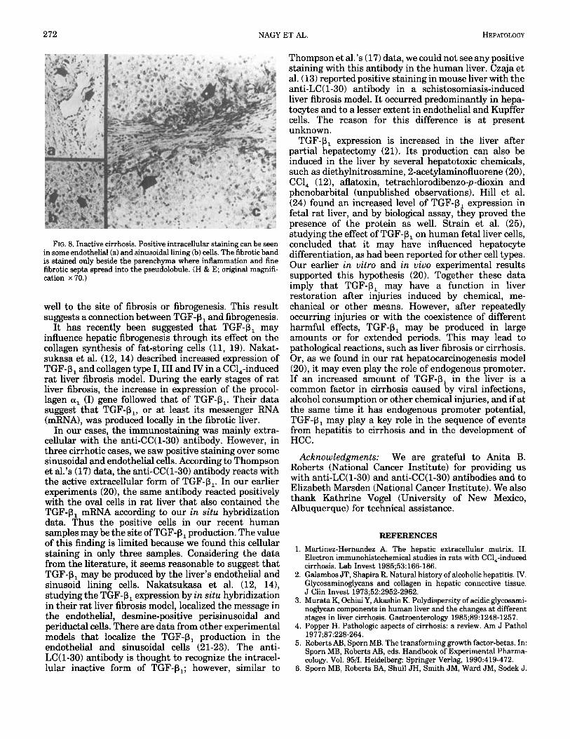
Three cirrhotic patients had “burnout” or inactive cirrhosis. The pseudolobules in these cases were circum- scribed by thick fibrotic bands with a mild lymphocytic infiltration. In one of these cases, the immunostaining was negative in repeated experiments (Fig. 6). TGF-P, could be demonstrated in the two other specimens: however, its distribution was slightly different than in active cirrhosis. The thick fibrotic bands were primar- ily negative except at the border between the paren- chyma and connective tissue (Fig. 7 ) . The inflammatory reaction, and probably the active fibrogenesis, was also localized in this area. Where inflammation and
fine fibrotic septa spread into the pseudolobules, the immunostaining was also positive (Fig. 8c).
In three cirrhotic samples (one inactive and two active), a few endothelial (Fig. 8a) and sinusoidal lining cells (Fig. 8b) also reacted with the anti-CC(1-30) antibody.
DISCUSSION
TGF-P, was not detected by immunohistochemistry in normal human liver or in liver from patients with chronic persistent hepatitis. However, in liver biopsy specimens in which an active fibrotic process was observable, namely in chronic aggressive hepatitis and in cirrhosis, we found strong immunostaining for TGF-P,. The localization of TGF-p, corresponded quite
272 NAGY ET AL. HEPATOLOGY
FIG. 8. Inactive cirrhosis. Positive intracellular staining can be seen in some endothelial (a) and sinusoidal lining (b) cells. The fibrotic band is stained only beside the parenchyma where inflammation and fine fibrotic septa spread into the pseudolobule. (H & E; original magnifi- cation x70.)
well to the site of fibrosis or fibrogenesis. This result suggests a connection between TGF-P, and fibrogenesis.
It has recently been suggested that TGF-P, may influence hepatic fibrogenesis through its effect on the collagen synthesis of fat-storing cells (11, 19). Nakat- sukasa et al. (12, 14) described increased expression of TGF-P, and collagen type I, I11 and N in a CC1,-induced rat liver fibrosis model. During the early stages of rat liver fibrosis, the increase in expression of the procol- lagen 01, (I) gene followed that of TGF-P,. Their data suggest that TGF-P,, or at least its messenger RNA (mRNA), was produced locally in the fibrotic liver.
In our cases, the immunostaining was mainly extra- cellular with the anti-CC(1-30) antibody. However, in three cirrhotic cases, we saw positive staining over some sinusoidal and endothelial cells. According to Thompson et al.’s (17) data, the anti-CC(1-30) antibody reacts with the active extracellular form of TGF-P,. In our earlier experiments (20), the same antibody reacted positively with the oval cells in rat liver that also contained the TGF-P, mRNA according to our in sztu hybridization data. Thus the positive cells in our recent human samples may be the site of TGF-P, production. The value of this finding is limited because we found this cellular staining in only three samples. Considering the data from the literature, it seems reasonable to suggest that TGF-P, may be produced by the liver’s endothelial and sinusoid lining cells. Nakatsukasa et al. (12, 14), studying the TGF-P, eFression by in sztu hybridization in their rat liver fibrosis model, localized the message in the endothelial, desmine-positive perisinusoidal and periductal cells. There are data from other experimental models that localize the TGF-P, production in the endothelial and sinusoidal cells (21-23). The anti- LC(1-30) antibody is thought to recognize the intracel- lular inactive form of TGF-PI; however, similar to
Thompson et al.’s (17) data, we could not see any positive staining with this antibody in the human liver. Czaja et al. (13) reported positive staining in mouse liver with the anti-LC(1-30) antibody in a schistosomiasis-induced liver fibrosis model. It occurred predominantly in hepa- tocytes and to a lesser extent in endothelial and Kupffer cells. The reason for this difference is at present unknown.
TGF-P, expression is increased in the liver after partial hepatectomy (21). Its production can also be induced in the liver by several hepatotoxic chemicals, such as diethylnitrosamine, 2-acetylaminofluorene (201, CCl, (121, aflatoxin, tetrachlorodibenzo-p-dioxin and phenobarbital (unpublished observations). Hill et al. (24) found an increased level of TGF-P, expression in fetal rat liver, and by biological assay, they proved the presence of the protein as well. Strain et al. (25), studying the effect of TGF-6, on human fetal liver cells, concluded that it may have influenced hepatocyte differentiation, as had been reported for other cell types. Our earlier in uitro and in uiuo experimental results supported this hypothesis (20). Together these data imply that TGF-P, may have a function in liver restoration after injuries induced by chemical, me- chanical or other means. However, after repeatedly occurring injuries or with the coexistence of different harmful effects, TGF-P, may be produced in large amounts or for extended periods. This may lead to pathological reactions, such as liver fibrosis or cirrhosis. Or, as we found in our rat hepatocarcinogenesis model (201, it may even play the role of endogenous promoter. If an increased amount of TGF-P, in the liver is a common factor in cirrhosis caused by viral infections, alcohol consumption or other chemical injuries, and if at the same time it has endogenous promoter potential, TGF-P, may play a key role in the sequence of events from hepatitis to cirrhosis and in the development of HCC.
We are grateful to Anita B. Roberts (National Cancer Institute) for providing us with anti-LC(1-30) and anti-CC(1-30) antibodies and to Elizabeth Marsden (National Cancer Institute). We also thank Kathrine Vogel (University of New Mexico, Albuquerque) for technical assistance.
Acknowledgments:
REFERENCES 1. Martinez-Hernandez A. The hepatic extracellular matrix. 11.
Electron immunohistochemical studies in rats with CC1,-induced cirrhosis. Lab Invest 1985;53:166-186.
2. Galambos JT, Shapira R. Natural history of alcoholic hepatitis. N. Glycosaminoglycans and collagen in hepatic connective tissue. J Clin Invest 1973;52:2952-2962.
3. Murata K, Ochiai Y, Akashio K. Polydispersity of acidic glycosami- noglycan components in human liver and the changes at different stages in liver cirrhosis. Gastroenterology 1985;89: 1248-1257.
4. Popper H. Pathologic aspects of cirrhosis: a review. Am J Pathol
5. Roberts AB, Sporn MB. The transforming growth factor-betas. In: Sporn MB, Roberts AB, eds. Handbook of Experimental Pharma- cology. Vol. 95/1. Heidelberg: Springer Verlag, 1990:419-472.
6. Sporn MB, Roberts BA, Shull JH, Smith JM, Ward JM, Sodek J .
1911;87:228-264.
Voi. 14, No. 2, 1991 TGF-p, AND LIVER FIBROSIS 273
Polypeptide transforming growth factors isolated from bovine sources and used for wound healing in vivo. Science 1983;219:
7. Ignotz RA, Massegue J. Transforming growth factor beta stimu- lates the expression of fibronectin and collagen and their incor- poration into the extra-cellular matrix. J Biol Chem 1986;261: 4337-4345.
8. Edwards DR, Murphy G, Reynolds JJ, Whitman SE, Docherty AJP, Angel P, Heath JK. Transforming growth factor beta modulates the expression of collagenase and metalloproteinase inhibitor. EMBO J 1987;6:1899-1904.
9. Laiho M, Sakesela 0, Andreasen PA, Keski-Oja J . Enhanced production and extracellular deposition of the endothelial type plasminogen activator inhibitor in cultured human lung fibro- blasts by transforming growth factor beta. J Cell Biol 1986;103:
10. Ignotz RA, Massegue J. Cell adhesion protein receptors as targets for transforming growth factor beta. Cell 1987;51:189-197.
11. Weiner FR, Gianbrone MA, Takahashi S, Annoni G, Czaja MJ, Zern MA. Modulators of Ito cell collagen synthesis [Abstract]. HEPATOLOGY 1988;8: 1253.
12. Nakatsukasa H, Nagy P, Evarts RP, Hsia C-C, Marsden E, Thorgeisson SS. The cellular distribution of TGF-beta 1 and procollagen I, 111 and N transcripts in carbon tetrachloride induced rat liver fibrosis. J Clin Invest 1991 (in press).
13. Czaja MJ, Weiner FR, Flanders KC, Giambrone M-A, Wind R, Biempica L, Z e n MA. In vitro and in vivo association of transforminggrowth factor beta-1 with hepatic fibrosis. J Cell Biol
14. Nakatsukasa H, Evarts RP, Hsia C, Thorgeirsson SS. TGF-beta-1 and type 1 procollagen transcripts during regeneration and early fibrosis of rat liver. Lab Invest 1990;63:171-181.
15. Ellingsworth LR, Brennan JE, Fok K, Rosen DM, Bentz H, Piez KA, Seyedin SM. Antibodies to the N terminal portion of cartilage inducing factor A and transforming growth factor beta. J Biol Chem 1986;261: 12362-12367.
1329-1331.
2403-2410.
1989;108:2477-2482.
16. Flanders KC, Thompson NL, Cissel DS, Ellingsworth LR, Roberts AB, Sporn MB. Epitope-dependent immunohistochemical local- ization of transforming growth factor beta 1. J Cell Biol 1989;
17. Thompson NL, Flanders KC, Smith JM, Ellingsworth LR, Roberts AB, Sporn MB. Cell type specific expression of transforming growth factor beta 1 in adult and neonatal mouse tissue. J Cell Biol
18. Marquardt H, Lioubin MN, Ikeda T. Complete amino acid sequence of human transforming growth factor type beta 2. J Biol Chem 1987;262:12127-12131.
19. Davis BH. Transforming growth factor beta responsiveness modulated by extracellular collagen matrix during hepatic Ito cell culture. J Cell Physiol 1988;136:547-553.
20. Nagy P, Evarts RP, McMahon JB, Thorgeirsson SS. Role of TGF-beta 1 in normal differentiation and oncogenesis in rat liver. Molec Carcinog 1989;2:345-354.
21. Braun L, Mead JE, Panzica M, Mikumo R, Bell GT, Fausto N. Transforming growth factor beta mRNA increases during liver regeneration: a possible paracrine mechanism of growth regu- lation. Proc Natl Acad Sci USA 1988;85:1539-1543.
22. Braun L, Gruppuso P, Mikumo R, Fausto N. Transforminggrowth factor beta 1 in liver carcinogenesis: messenger RNA expression and growth effects. Cell Growth & Differentiation 199O;l:
23. Evarts RP, Nakatsukasa H, Marsden ER, Hsia C-C, Dunsford HA, Thorgeirsson SS. Cellular and molecular changes in the early stages of chemical hepatocarcinogenesis. Cancer Res 1990;50: 3439-3444.
24. Hill DJ, Strain AJ, Milner RDG. Presence of transforming growth factor beta like activity in multiple fetal rat tissues. Cell Biol Int Rep 1986; 10 :9 15-922.
25. Strain AJ, Hill DJ, Milner RDG. Divergent action of transforming growth factor beta on DNA synthesis in human foetal liver cells. Cell Biol Int Rep 1986;10:855-860.
108:653-660.
1989;108:661-669.
103-1 11.