IGF-1 Regulates the Extracellular Level of Active MMP...
13
Retinal Cell Biology IGF-1 Regulates the Extracellular Level of Active MMP-2 and Promotes M¨ uller Glial Cell Motility Valeria E. Lorenc, 1 Javier R. Jald´ ın-Fincati, 1 Jos´ e D. Luna, 2 Gustavo A. Chiabrando, 1 and Mar´ ıa C. S´ anchez 1 1 Centro de Investigaciones en Bioqu´ ımica Cl´ ınica e Inmunolog´ ıa (CIBICI-CONICET), Departamento de Bioqu´ ımica Cl´ ınica, Facultad de Ciencias Qu´ ımicas, Universidad Nacional de C´ ordoba, C´ ordoba, Argentina 2 Departamento de V´ ıtreo-Retina, Centro Privado de Ojos Romagosa-Fundaci´ on VER, C´ ordoba, Argentina Correspondence: Mar´ ıa C. S´ anchez, Departamento de Bioqu´ ımica Cl´ ıni- ca, Centro de Investigaciones en Bioqu´ ımica Cl´ ınica e Inmunolog´ ıa (CIBICI-CONICET), Facultad de Ciencias Qu´ ımicas, Universidad Na- cional de C´ ordoba, C´ ordoba, Argen- tina; [email protected]. Submitted: June 15, 2015 Accepted: August 31, 2015 Citation: Lorenc VE, Jald´ ın-Fincati JR, Luna JD, Chiabrando GA, S´ anchez MC. IGF-1 regulates the extracellular level of active MMP-2 and promotes M¨ uller glial cell motility. Invest Ophthalmol Vis Sci. 2015;56:6948–6960. DOI:10.1167/iovs.15-17496 PURPOSE. In ischemic proliferative retinopathies, M¨ uller glial cells (MGCs) acquire migratory abilities. However, the mechanisms that regulate this migration remain poorly understood. In addition, proliferative disorders associated with enhanced activities of matrix metal- loproteinases (MMPs) also involve insulin-like growth factor (IGF)-1 participation. Therefore, the main interest of this work was to investigate the IGF-1 effect on the extracellular proteolytic activity in MGCs. METHODS. Cell culture supernatants and cell lysates of the human MGC line MIO-M1 stimulated with IGF-1 were analyzed for MMP-2 by zymographic and Western blot analysis. The MGCs’ motility was evaluated by scratch wound assay. The MMP-2, b1-integrin, and focal adhesions were detected by confocal microscopy. The localization of active MMPs and actin cytoskeleton were evaluated by in situ zymography. RESULTS. The IGF-1 induced the activation of canonical signaling pathways through the IGF-1R phosphorylation. Culture supernatants showed a relative decrease in the active form of MMP- 2, correlating with an increased accumulation of MMP-2 protein in the MGCs’ lysate. The IGF- 1 effect on MMP-2 was abolished by an IGF-1R blocking antibody, aIR3, as well as by the PI3- kinase inhibitor, LY294002. The IGF-1 increased the migratory capacity of MGCs, which was blocked by the GM6001 MMP inhibitor, LY294002 and aIR3. Finally, IGF-1 induced the intracellular distribution of MMP-2 toward cellular protrusions and the partial colocalization with b1-integrin and phospo-focal adhesion kinase signals. Gelatinase activity was concentrated along F-actin filaments. CONCLUSIONS. Taken together, these data indicate that IGF-1, through its receptor activation, regulates MGCs’ motility by a mechanism that involves the MMP-2 and PI3K signaling pathway. Keywords: glial cell, metalloproteinases, cell migration I nsulin-like growth factor 1 (IGF-1) is a 70–amino acid peptide belonging to a family of polypeptide trophic factors with mitogenic, differentiating, antiapoptotic, and metabolic func- tions. 1 These biological actions are mediated by the IGF-1 receptor (IGF-1R), a member of the tyrosine kinase gene family of growth factor receptors. 2 Upon IGF-1 binding, the activated IGF-1R promotes phosphorylation of specific cytosolic sub- strates, then the IGF-1 signal is transmitted to two downstream pathways: extracellular signal–regulated kinases 1 and 2 (ERK 1/2) and phosphatidylinositol 3-kinase (PI3K/AKT). 1 Circulat- ing IGF-1 is mainly released from the liver in response to pituitary growth hormone; however, a number of other tissues can also produce IGF-1 locally, including the central nervous system (CNS). 3–5 Despite its peripheral location, the retina is part of the CNS, and there is considerable evidence that IGF-1 is important for neural and vascular retinal development. 6,7 In addition, the IGF-1/IGF-1R system has also been implicated in retinopathies, and an increasing of IGF-1 levels has been found in vitreous samples from patients with proliferative diabetic retinopathy (PDR), 8–10 whereas its participation in retinopathy of prematurity (ROP) appears to be more complex. 11 M¨ uller glial cells (MGCs) are the main glial cell type in the retina. They constitute an anatomic and functional link between retinal neurons and compartments to exchange molecules and play a crucial role in supporting neuronal development, survival, and information processing. 12–15 The MGCs are also involved in the production and secretion of growth factors, including IGF-1. 16,17 In pathologic conditions, such as PDR and proliferative vitreoretinopathy (PVR), 18 MGCs adopt a number of drastic changes at the level of gene and protein expression, which impact on their morphology, migration, and proliferation, followed by extracellular matrix (ECM) degradation. This results in the formation of retinal membranes and resembles physiological processes of wound healing and fibrosis. 19–21 Both metalloproteinase (MMP)-2 and MMP-9 have been associated with these processes, and MGCs are one type of MMP-producing cells in the retina. 22,23 During proliferative retinopathy, MGCs encounter different ECM proteins, cytokines, and growth factors because these are often present in the vitreous humor and retinal tissues of affected eyes. 24–27 Therefore, MGCs’ functions, including the produc- tion or regulation of matrix-degrading enzymes, may be Copyright 2015 The Association for Research in Vision and Ophthalmology, Inc. iovs.arvojournals.org j ISSN: 1552-5783 6948 Downloaded From: http://iovs.arvojournals.org/pdfaccess.ashx?url=/data/journals/iovs/934564/ on 05/13/2018
Transcript of IGF-1 Regulates the Extracellular Level of Active MMP...
Retinal Cell Biology
IGF-1 Regulates the Extracellular Level of Active MMP-2and Promotes Muller Glial Cell Motility
Valeria E. Lorenc,1 Javier R. Jaldın-Fincati,1 Jose D. Luna,2 Gustavo A. Chiabrando,1
and Marıa C. Sanchez1
1Centro de Investigaciones en Bioquımica Clınica e Inmunologıa (CIBICI-CONICET), Departamento de Bioquımica Clınica, Facultadde Ciencias Quımicas, Universidad Nacional de Cordoba, Cordoba, Argentina2Departamento de Vıtreo-Retina, Centro Privado de Ojos Romagosa-Fundacion VER, Cordoba, Argentina
Correspondence: Marıa C. Sanchez,Departamento de Bioquımica Clıni-ca, Centro de Investigaciones enBioquımica Clınica e Inmunologıa(CIBICI-CONICET), Facultad deCiencias Quımicas, Universidad Na-cional de Cordoba, Cordoba, Argen-tina;[email protected].
Submitted: June 15, 2015Accepted: August 31, 2015
Citation: Lorenc VE, Jaldın-Fincati JR,Luna JD, Chiabrando GA, Sanchez MC.IGF-1 regulates the extracellular levelof active MMP-2 and promotes Mullerglial cell motility. Invest Ophthalmol
Vis Sci. 2015;56:6948–6960.DOI:10.1167/iovs.15-17496
PURPOSE. In ischemic proliferative retinopathies, Muller glial cells (MGCs) acquire migratoryabilities. However, the mechanisms that regulate this migration remain poorly understood. Inaddition, proliferative disorders associated with enhanced activities of matrix metal-loproteinases (MMPs) also involve insulin-like growth factor (IGF)-1 participation. Therefore,the main interest of this work was to investigate the IGF-1 effect on the extracellularproteolytic activity in MGCs.
METHODS. Cell culture supernatants and cell lysates of the human MGC line MIO-M1stimulated with IGF-1 were analyzed for MMP-2 by zymographic and Western blot analysis.The MGCs’ motility was evaluated by scratch wound assay. The MMP-2, b1-integrin, and focaladhesions were detected by confocal microscopy. The localization of active MMPs and actincytoskeleton were evaluated by in situ zymography.
RESULTS. The IGF-1 induced the activation of canonical signaling pathways through the IGF-1Rphosphorylation. Culture supernatants showed a relative decrease in the active form of MMP-2, correlating with an increased accumulation of MMP-2 protein in the MGCs’ lysate. The IGF-1 effect on MMP-2 was abolished by an IGF-1R blocking antibody, aIR3, as well as by the PI3-kinase inhibitor, LY294002. The IGF-1 increased the migratory capacity of MGCs, which wasblocked by the GM6001 MMP inhibitor, LY294002 and aIR3. Finally, IGF-1 induced theintracellular distribution of MMP-2 toward cellular protrusions and the partial colocalizationwith b1-integrin and phospo-focal adhesion kinase signals. Gelatinase activity wasconcentrated along F-actin filaments.
CONCLUSIONS. Taken together, these data indicate that IGF-1, through its receptor activation,regulates MGCs’ motility by a mechanism that involves the MMP-2 and PI3K signalingpathway.
Keywords: glial cell, metalloproteinases, cell migration
Insulin-like growth factor 1 (IGF-1) is a 70–amino acid peptidebelonging to a family of polypeptide trophic factors with
mitogenic, differentiating, antiapoptotic, and metabolic func-tions.1 These biological actions are mediated by the IGF-1receptor (IGF-1R), a member of the tyrosine kinase gene familyof growth factor receptors.2 Upon IGF-1 binding, the activatedIGF-1R promotes phosphorylation of specific cytosolic sub-strates, then the IGF-1 signal is transmitted to two downstreampathways: extracellular signal–regulated kinases 1 and 2 (ERK1/2) and phosphatidylinositol 3-kinase (PI3K/AKT).1 Circulat-ing IGF-1 is mainly released from the liver in response topituitary growth hormone; however, a number of other tissuescan also produce IGF-1 locally, including the central nervoussystem (CNS).3–5 Despite its peripheral location, the retina ispart of the CNS, and there is considerable evidence that IGF-1 isimportant for neural and vascular retinal development.6,7 Inaddition, the IGF-1/IGF-1R system has also been implicated inretinopathies, and an increasing of IGF-1 levels has been foundin vitreous samples from patients with proliferative diabeticretinopathy (PDR),8–10 whereas its participation in retinopathyof prematurity (ROP) appears to be more complex.11
Muller glial cells (MGCs) are the main glial cell type in theretina. They constitute an anatomic and functional linkbetween retinal neurons and compartments to exchangemolecules and play a crucial role in supporting neuronaldevelopment, survival, and information processing.12–15 TheMGCs are also involved in the production and secretion ofgrowth factors, including IGF-1.16,17 In pathologic conditions,such as PDR and proliferative vitreoretinopathy (PVR),18 MGCsadopt a number of drastic changes at the level of gene andprotein expression, which impact on their morphology,migration, and proliferation, followed by extracellular matrix(ECM) degradation. This results in the formation of retinalmembranes and resembles physiological processes of woundhealing and fibrosis.19–21 Both metalloproteinase (MMP)-2 andMMP-9 have been associated with these processes, and MGCsare one type of MMP-producing cells in the retina.22,23 Duringproliferative retinopathy, MGCs encounter different ECMproteins, cytokines, and growth factors because these are oftenpresent in the vitreous humor and retinal tissues of affectedeyes.24–27 Therefore, MGCs’ functions, including the produc-tion or regulation of matrix-degrading enzymes, may be
Copyright 2015 The Association for Research in Vision and Ophthalmology, Inc.
iovs.arvojournals.org j ISSN: 1552-5783 6948
Downloaded From: http://iovs.arvojournals.org/pdfaccess.ashx?url=/data/journals/iovs/934564/ on 05/13/2018
influenced by cytokines and growth factors present in theretinal microenvironment. Whereas the MMP-9 expression wasincreased in MGCs stimulated by TNF-a,22 we previouslydemonstrated that a2-macroglobulin (a2M), an acute-phaseresponse protein involved in retinopathies, induces MMP-2activation and regulates membrane type 1 MMP (MT1-MMP)activity.28
Studies in models of CNS injury have demonstrated that theIGF-1/IGF-1R system stimulates cell migration of oligodendro-cytes,29 astrocytes,30 and neuronal cells.31 However, there isno information about the IGF-1 effect on MGCs’ migration inretinopathies. The evidence accumulated over the past threedecades has demonstrated that the IGF-1 system plays animportant role as a regulator of tumor cell invasion bymodulating the MMP-2 synthesis and activity.32,33 Like othermembers of the MMP family, MMP-2 synthesis and function areregulated at multiple levels, including transcriptional activa-tion, posttranscriptional processing, regulation of proteolyticactivity by MT1-MMP, and inhibition through the tissueinhibitors of MMP (TIMP).34
MMP-2 is constitutively expressed and secreted in a latentform or pro-MMP-2 (72 kDa). Its activation to MMP-2 takesplace at the cell surface and requires the participation of theactive form of MT1-MMP that binds pro-MMP-2 on the cellmembrane in a multimeric complex with TIMP-2.35 Enhancedexpression or activity of MMP-2 was previously reportedwithin retinal neovascular tissue, as well as in vitreous oraqueous samples from patients with PDR,36 suggesting thatMMP-2 is an important therapeutic target in retinopathies. Onthe other hand, serum MMP-2 levels in these same diabeticpatients were almost identical to control patients, indicatingthat increased levels of MMP-2 in the eyes were an ocular andnot systemic consequence. In addition, numerous studies havereported high levels of IGF-1 in vitreous fluid of diabeticpatients.8,9,37 However, the precise role of IGF-1 in thepathogenesis of retinopathies remains unclear. Our hypothesisis that IGF-1 plays a major role on the extracellular MMP-2activity and cellular motility of MGCs during retinal ischemicproliferative disease. Thus, in the present study we investigatedthe IGF-1 effect on MMP-2 activity and cell migration in a well-characterized MGC line, MIO-M1.
MATERIALS AND METHODS
A spontaneously immortalized human MGC line (MIO-M1),kindly provided by G. Astrid Limb (UCL Institute of Ophthal-mology and Moorfields Eye Hospital, London, UK), was used.38
Cells were grown in Dulbecco’s modified Eagle’s medium(DMEM; Invitrogen, Buenos Aires, Argentina) containing 4500mg/L glucose, sodium pyruvate) with 2 mM L-glutamine(GlutaMAX; Invitrogen), 10% vol/vol fetal bovine serum, and50 U/mL penicillin/streptomycin (Invitrogen) at 378C with 5%CO2. Recombinant human IGF-1 was purchased from Sigma-Aldrich Corp. (St. Louis, MO, USA). Immunoblots wereperformed with the following primary monoclonal antibodies:anti-phosphorylated ERK 1/2 (anti-p-ERK 1/2), polyclonalrabbit anti-total ERK 1/2, anti-calreticulin, and anti-MMP-2(specific for the active form), all from Santa Cruz Biotechnol-ogy, Inc. (Santa Cruz, CA, USA). Rabbit monoclonal anti-phospho-IGF-1R antibody was obtained from Cell SignalingTechnologies, Inc. (Danvers, MA, USA). Mouse monoclonalanti-IGF-1R and MT1-MMP antibodies were both from Abcam,Inc. (Cambridge, MA, USA). Secondary antibodies used forimmunoblotting were horseradish peroxidase–conjugatedstreptavidin (Thermo Fisher Scientific, Rockford, IL, USA).Dilutions for primary antibodies were between 1/250 and 1/1000, whereas for secondary antibodies they were 1/5000. To
block IGF-1/IGF-1R binding, cells were pretreated with amouse monoclonal antibody specific against the IGF-1 recep-tor, aIR3 (Calbiochem, San Diego, CA, USA), at a finalconcentration of 16 nM. The inhibitory studies of ligandsignaling included ERK 1/2 inhibitor PD-98059 and PI3Kinhibitor LY-294002, both from Sigma-Aldrich Corp. Collagentype I (Col-I) or laminin used in the wound healing assays werealso from Sigma-Aldrich Corp. To inhibit cell proliferation,hydroxyurea was used (Sigma-Aldrich Corp.). To block MMPs, ahydroxamate-based inhibitor N-[(2R)-2(hydroxamideocarbonyl-methyl)-4-methylpantanoyl]-L-tryptophan methylamide](GM6001) was obtained (Calbiochem). To visualize cellularlocalization of proteins, we used two different monoclonalantibodies against MMP-2 (mouse anti-MMP-2 or rabbit anti-MMP-2): a rabbit anti-b1-integrin antibody (Abcam, Inc.,Cambridge, MA, USA) and a mouse anti-phospo-focal adhesionkinase (p-FAK) antibody (Santa Cruz Biotechnology, Inc.).Secondary antibodies were raised in goat against rabbit ormouse IgG conjugated with Alexa Fluor 488 and 594,respectively (Molecular Probes, Eugene, OR, USA). The FITC-labeled DQ-collagen and Alexa Fluor 594 phalloidin, used in anin situ zymography assay and in actin cytoskeleton labeling,were also purchased from Molecular Probes.
Western Blot Assay
To evaluate the IGF-1 effect on IGF-1R autophosphorylationand to investigate the involvement of the classical signalingpathways, MIO-M1 cells were plated in the presence orabsence of IGF-1 (10 nM) for different periods of times. Toevaluate the MMP-2 and MT1-MMP expression, MIO-M1 cellswere incubated with IGF-1 (10 nM) for up to 8 hours. At theend of each treatment, cells at 70% to 80% confluence werekept on ice, and the medium was aspirated and lysed usingnondenaturalizing buffer (20 mM Tris-HCl, 1% Triton X-100,10% glycerol, 137 mM ClNa, 0.01% wt/vol bromophenolblue) containing phenyl methyl sulfonyl fluoride (1 mM),sodium orthovanadate (10 mM), and a protease inhibitorcocktail (Sigma-Aldrich Corp.). Protein concentrations weredetermined with a BCA protein assay kit (Pierce, BuenosAires, Argentina) using albumin as standard. Aliquotscontaining proteins (50 lm) were resolved on 10% SDS-PAGE and transferred onto nitrocellulose membranes(Amersham Hybond ECL; GE Healthcare Bio-Sciences AB,Uppsala, Sweden). Nonspecific binding was blocked with5% bovine serum albumin (BSA) in Tris-buffered saltcontaining 0.01% Tween-20, and the same solution wasused to prepare primary antibodies, which were incubatedwith the membranes overnight at 48C. Membranes wereincubated with a peroxidase-conjugated secondary antibodyfor 1 hour at room temperature (RT). Finally, the specificbands were revealed using a chemiluminescence kit(Thermo Fisher Scientific) and quantified by densitometricanalysis using analysis software (Gel Pro Analyzer; MediaCybernetics, Inc., Rockville, MD, USA).
Gelatin Zymography Assays
Standard methodology for gelatin zymography was used todetect MMPs gelatinolytic activity in MIO-M1 cell supernatantsas described previously by Kleiner and colleague.39 For thispurpose, MIO-M1 cells (5 3 105 cells/well) were cultured at378C for 24 hours. The cells were then rinsed twice withserum-free medium, and 1 mL of DMEM-high glucose wasadded. Cells were stimulated with 10 nM IGF-1 at differenttimes (4–8 hours), after which the cell culture supernatantswere collected. To block IGF-1/IGF-1R binding, cells werepreviously pretreated with aIR3 in serum-free medium for 30
IGF-1 Regulates MMP-2 and Promotes MGC Motility IOVS j October 2015 j Vol. 56 j No. 11 j 6949
Downloaded From: http://iovs.arvojournals.org/pdfaccess.ashx?url=/data/journals/iovs/934564/ on 05/13/2018
minutes before ligand addition. Capillary blood was used as apositive control of MMP gelatinolytic activity. Aliquots of 30 lLcell supernatants were resolved on 10% SDS-PAGE/1.5% gelatin(Sigma-Aldrich Corp.) in denaturing and nonreducing condi-tions. Gels were soaked for 1 hour with 2.5% Triton X-100 toremove the SDS, and the MMP activity was developed at 378Cin the enzyme buffer (50 mM Tris-HCl, 0.2 M NaCl, and 5 mMCaCl2, pH 7.5) for 24 hours. After incubation, gels were stainedfor 30 minutes in 0.125% Coomassie blue R-250, and the stainwas removed with the same solution without the dye untilclear bands of gelatinolysis appeared on a dark background.The MMP-2 and MMP-9 were identified by molecular size usinghigh-molecular-mass (14.5–200 kDa) standards (Bio-Rad, Her-cules, CA, USA). Images were processed, and the intensity ofthe bands was obtained using the imaging program Gel ProAnalyzer (Media Cybernetics, Inc.).
Biotinylation Assay
A biotin-labeling protein assay using sulfo-NHS-SS-biotin (No-Weigh EZ-Link; Thermo Fisher Scientific) was performedfollowing the manufacturer’s procedure. Briefly, to evaluateMT1-MMP membrane expression, MIO-M1 cells were stimu-lated with IGF-1 (8 hours), washed with PBS, and incubatedwith sulfo-NHS-SS-biotin for 1 hour at 48C. After biotinincubation, cells were again washed and lysed with PBS0.2% Triton X-100 containing a protease cocktail inhibitor.Then 5 lL of 20% agarose-streptavidin beads were incubatedwith the lysates (100 lg) for 2 hours at RT. When theincubation was finished, the lysates were removed, the beadswashed with PBS, and the bound proteins eluted. Equalamounts of eluted proteins were resolved on SDS-PAGE andblotted with an anti-MT1-MMP antibody. Results werequantified by ImageJ software (http://imagej.nih.gov/ij/;provided in the public domain by the National Institutes ofHealth, Bethesda, MD, USA).
Cell Migration Assays
Cell migration activities were examined by a two-dimension-al scratch wound assay in six-well plates coated with Col-I(10 lg/cm2) or laminin (1 lg/cm2; Sigma-Aldrich Corp.). TheMIO-M1 cells (5 3 105 cells/well) were cultured for 24 hours.In each well, a straight lesion was created in the center ofthe MIO-M1 cell monolayer with a sterile 10-lL pipette tip.This technique produced a consistent wound devoid of cells,~35 mm long 3 400 lm wide. Wells were then rinsed twicewith serum-free medium to remove any cell debris, and 2 mLof DMEM-high glucose without red phenol was added. Cellswere treated with 10 nM IGF-1 for 12 hours. To block IGF-1/IGF-1R binding, cells were pretreated with aIR3 for 30minutes. In another set of experiments, to inhibit MMPsactivity, cells were preincubated with GM6001 MMPinhibitor (20 nM) for 30 minutes.28 In addition, to inhibitthe activity of PI3K, cells were also pretreated with LY-294002. In all the migration experiments and to inhibit cellproliferation, cells were preincubated with 0.5 mM hydroxy-urea.
Cellular migration was measured following a procedurepreviously described.28 Briefly, at selected times (0 and 12hours), three random images of the wound per condition wereacquired using a charge-coupled device camera (Nikon; Nikon,Inc., Melville, NY, USA) using bright-field microscopy (invertedmicroscope Nikon TU-2000; Nikon, Inc.) with a 103 objective(0.3 numerical aperture). Each image defined an average areaof the wound equivalent to 5 3 105 6 1 3 104 lm2 recorded tot ¼ 0 hours. Cells invading this area were counted to t ¼ 12hours and results expressed as cells per area.
Immunofluorescence Labeling and ConfocalMicroscopy
To evaluate the cell distribution of MMP-2, b1-integrin, and p-FAK, MIO-M1 cells were grown to 30% to 70% confluence onglass coverslips coated with Col-I (10 lg/cm2) in 24-well platesbefore being stimulated, or not, with IGF-1 (10 nM) for 8 hours.Coverslips containing the cells were washed twice with PBS,fixed in 4% paraformaldehyde (PFA) for 15 minutes at RT,permeabilized with 0.1% (vol/vol) Triton X-100, and blockedwith 2% BSA for 1 hour at 378C to block nonspecific bindingsites. The coverslips were then incubated overnight at 48Cwith primary antibodies diluted 1:50, washed three times withPBS plus 1% BSA, and exposed to secondary antibodies diluted1:800 for 45 minutes at RT. After a thorough rinse with PBSplus 1% BSA, cell nuclei were stained at 1:1000 dilution(Hoechst no. 33258; Molecular Probes). Cells were washedwith PBS and mounted on glass slides with reagent (Mowiol 4-88, Calbiochem; Merck KGaA, Darmstadt, Germany). Fluores-cent images were obtained with a confocal laser-scanningbiological microscope (Olympus FluoView FV1000; OlympusCorp., New York, NY, USA). Finally, images were processedwith microscope software (FV10-ASW Viewer 3.1; OlympusCorp.) and with ImageJ software.
In Situ Zymography
To localize net pericellular proteolysis in MGCs, we used an insitu zymography assay described by Rivera.40 Briefly, after 12hours of IGF-1 stimulus, migrating MGCs were rinsed in PBSand media was replaced by fresh media containing serum-freeDMEM supplemented with calcium (5 mM CaCl2 in 50 mMTris, pH 7.6) and 100 lg/mL of FITC-labeled DQ-collagen that isintramolecularly quenched. Cells were incubated for 6 hours ina dark, humid chamber at 378C, rinsed in PBS, fixed in 4% PFA/sucrose, and permeabilized with 0.1% (vol/vol) Triton X-100.For actin cytoskeleton labeling, nonspecific binding wasblocked with 2% BSA for 1 hour at 378C. Cells were thenincubated with Alexa Fluor 594 phalloidin, rinsed in PBS plus1% BSA, and incubated with Hoechst no. 33258 (1:1000dilution). Finally, cells were mounted on glass slides withMowiol 4-88 reagent. Collagen-FITC cleavage by cellularproteinases releases quenched fluorescence representative ofnet proteolytic activity. Cells incubated without DQ-collagenwere not fluorescent.
Statistical Methods
For the densitometric quantifications and cellular migrationassays, results were expressed as the mean plus or minusstandard deviation of independent experiments; one-wayANOVA and a Tukey comparison posttest were used. Formicroscope quantifications of the level of colocalization, aJACoP plugin from ImageJ software was used.41 At least 50cells per condition were analyzed. Differences from the controlwere considered significant at P < 0.05.
RESULTS
IGF-1 Induces IGF-1R Phosphorylation andIntracellular Signaling Activation in MIO-M1 Cells
To investigate the effect of IGF-1 on the intracellular signalingpathways activation in MGCs, we used the MIO-M1 cell line,which constitutively expresses IGF-1R (Lorenc VE, et al. IOVS
IGF-1 Regulates MMP-2 and Promotes MGC Motility IOVS j October 2015 j Vol. 56 j No. 11 j 6950
Downloaded From: http://iovs.arvojournals.org/pdfaccess.ashx?url=/data/journals/iovs/934564/ on 05/13/2018
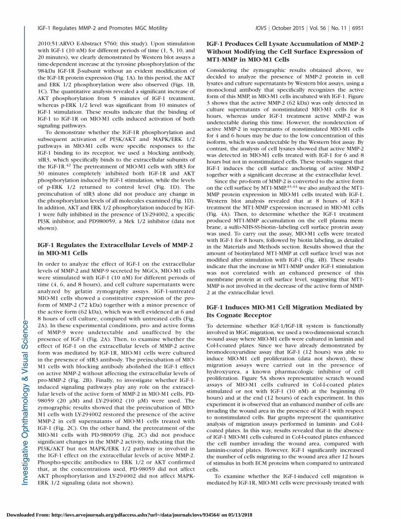
2010;51:ARVO E-Abstract 5760; this study). Upon stimulationwith IGF-1 (10 nM) for different periods of time (1, 5, 10, and20 minutes), we clearly demonstrated by Western blot assays atime-dependent increase at the tyrosine phosphorylation of the98-kDa IGF-1R b-subunit without an evident modification ofthe IGF-1R protein expression (Fig. 1A). In this period, the AKTand ERK 1/2 phosphorylation were also observed (Figs. 1B,1C). The quantitative analysis revealed a significant increase ofAKT phosphorylation from 5 minutes of IGF-1 treatment,whereas p-ERK 1/2 level was significant from 10 minutes ofIGF-1 stimulation. These results indicate that the binding ofIGF-1 to IGF-1R on MIO-M1 cells induced activation of bothsignaling pathways.
To demonstrate whether the IGF-1R phosphorylation andsubsequent activation of PI3K/AKT and MAPK/ERK 1/2pathways in MIO-M1 cells were specific responses to theIGF-1 binding to its receptor, we used a blocking antibody,aIR3, which specifically binds to the extracellular subunits ofthe IGF-1R.42 The pretreatment of MIO-M1 cells with aIR3 for30 minutes completely inhibited both IGF-1R and AKTphosphorylation induced by IGF-1 stimulation, while the levelsof p-ERK 1/2 returned to control level (Fig. 1D). Thepreincubation of aIR3 alone did not produce any change inthe phosphorylation levels of all molecules examined (Fig. 1D).In addition, AKT and ERK 1/2 phosphorylation induced by IGF-1 were fully inhibited in the presence of LY-294002, a specificPI3K inhibitor, and PD980059, a Mek 1/2 inhibitor (data notshown).
IGF-1 Regulates the Extracellular Levels of MMP-2in MIO-M1 Cells
In order to analyze the effect of IGF-1 on the extracellularlevels of MMP-2 and MMP-9 secreted by MGCs, MIO-M1 cellswere stimulated with IGF-1 (10 nM) for different periods oftime (4, 6, and 8 hours), and cell culture supernatants wereanalyzed by gelatin zymography assays. IGF-1-untreatedMIO-M1 cells showed a constitutive expression of the pro-form of MMP-2 (72 kDa) together with a minor presence ofthe active form (62 kDa), which was well evidenced at 6 and8 hours of cell culture, compared with untreated cells (Fig.2A). In these experimental conditions, pro- and active formsof MMP-9 were undetectable and unaffected by thepresence of IGF-1 (Fig. 2A). Then, to examine whether theeffect of IGF-1 on the extracellular levels of MMP-2 activeform was mediated by IGF-1R, MIO-M1 cells were culturedin the presence of aIR3 antibody. The preincubation of MIO-M1 cells with blocking antibody abolished the IGF-1 effecton active MMP-2 without affecting the extracellular levels ofpro-MMP-2 (Fig. 2B). Finally, to investigate whether IGF-1-induced signaling pathways play any role on the extracel-lular levels of the active form of MMP-2 in MIO-M1 cells, PD-98059 (20 lM) and LY-294002 (10 lM) were used. Thezymographic results showed that the preincubation of MIO-M1 cells with LY-294002 restored the presence of the activeMMP-2 in cell supernatants of MIO-M1 cells treated withIGF-1 (Fig. 2C). On the other hand, the pretreatment of theMIO-M1 cells with PD-980059 (Fig. 2C) did not producesignificant changes in the MMP-2 activity, indicating that thePI3K/AKT but not MAPK/ERK 1/2 pathway is involved inthe IGF-1 effect on the extracellular levels of active MMP-2.Phospho-specific antibodies to ERK 1/2 or AKT confirmedthat, at the concentrations used, PD-98059 did not affectAKT phosphorylation and LY-294002 did not affect MAPK-ERK 1/2 signaling (data not shown).
IGF-1 Produces Cell Lysate Accumulation of MMP-2Without Modifying the Cell Surface Expression ofMT1-MMP in MIO-M1 Cells
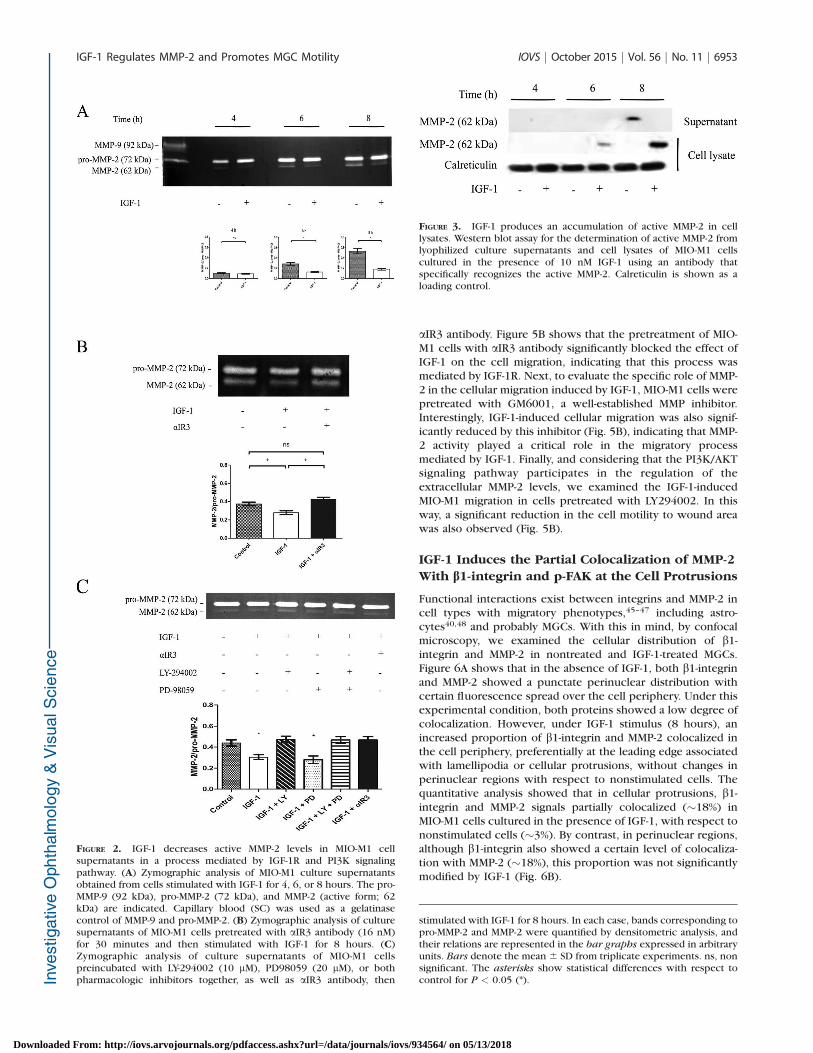
Considering the zymographic results obtained above, wedecided to analyze the presence of MMP-2 protein in celllysates and culture supernatants by Western blot assays, using amonoclonal antibody that specifically recognizes the activeform of this MMP, in MIO-M1 cells incubated with IGF-1. Figure3 shows that the active MMP-2 (62 kDa) was only detected inculture supernatants of nonstimulated MIO-M1 cells for 8hours, whereas under IGF-1 treatment active MMP-2 wasundetectable during this time. However, the nondetection ofactive MMP-2 in supernatants of nonstimulated MIO-M1 cellsfor 4 and 6 hours may be due to the low concentration of thisisoform, which was undetectable by the Western blot assay. Bycontrast, the analysis of cell lysates showed that active MMP-2was detected in MIO-M1 cells treated with IGF-1 for 6 and 8hours but not in nonstimulated cells. These results suggest thatIGF-1 induces the cell surface anchoring of active MMP-2together with a significant decrease at the extracellular level.
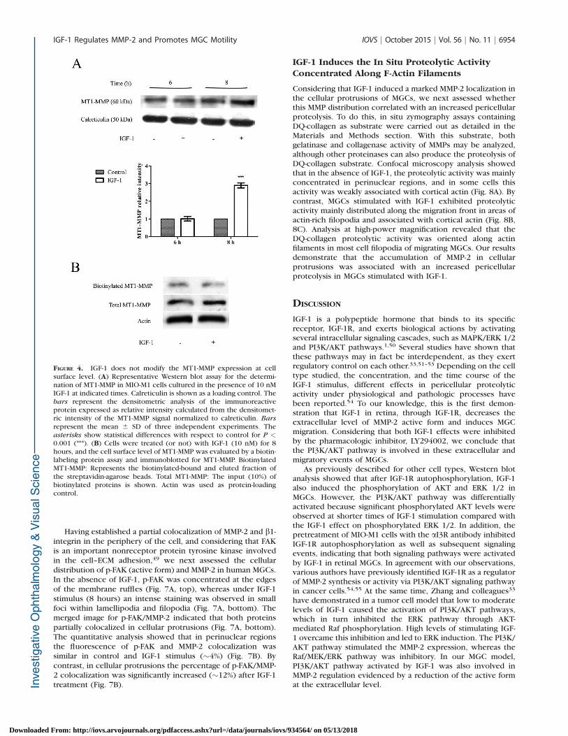
Since the pro-form of MMP-2 is converted to the active formon the cell surface by MT1-MMP,43,44 we also analyzed the MT1-MMP protein expression in MIO-M1 cells treated with IGF-1.Western blot analysis revealed that at 8 hours of IGF-1treatment the MT1-MMP expression increased in MIO-M1 cells(Fig. 4A). Then, to determine whether the IGF-1 treatmentproduced MT1-MMP accumulation on the cell plasma mem-brane, a sulfo-NHS-SS-biotin–labeling cell surface protein assaywas used. To carry out the assay, MIO-M1 cells were treatedwith IGF-1 for 8 hours, followed by biotin labeling, as detailedin the Materials and Methods section. Results showed that theamount of biotinylated MT1-MMP at cell surface level was notmodified after stimulation with IGF-1 (Fig. 4B). These resultsindicate that the increase in MT1-MMP under IGF-1 stimulationwas not correlated with an enhanced presence of thismembrane protein at cell surface level, suggesting that MT1-MMP is not involved in the decrease of the active form of MMP-2 at the extracellular level.
IGF-1 Induces MIO-M1 Cell Migration Mediated byIts Cognate Receptor
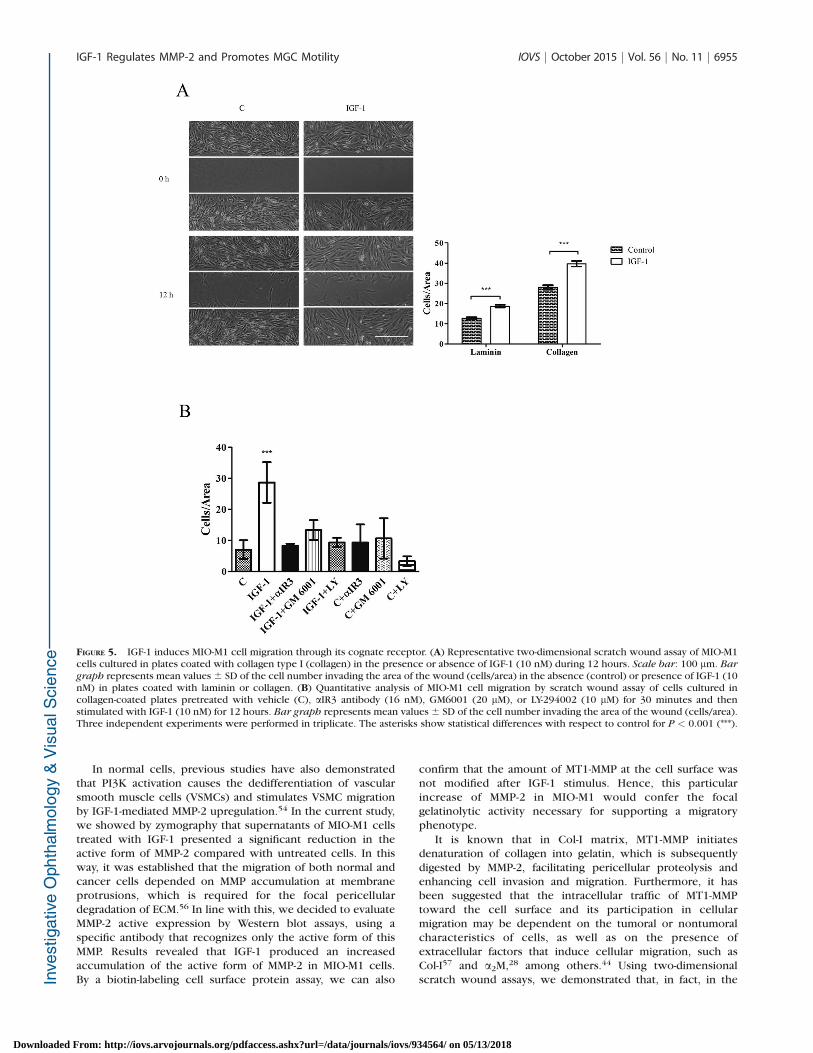
To determine whether IGF-1/IGF-1R system is functionallyinvolved in MGC migration, we used a two-dimensional scratchwound assay where MIO-M1 cells were cultured in laminin andCol-I-coated plates. Since we have already demonstrated bybromodeoxyuridine assay that IGF-1 (12 hours) was able toinduce MIO-M1 cell proliferation (data not shown), thesemigration assays were carried out in the presence ofhydroxyurea, a known pharmacologic inhibitor of cellproliferation. Figure 5A shows representative scratch woundassays of MIO-M1 cells cultured in Col-I-coated platesstimulated or not with IGF-1 (10 nM) at the beginning (0hours) and at the end (12 hours) of each experiment. In thisexperiment it is observed that an enhanced number of cells areinvading the wound area in the presence of IGF-1 with respectto nonstimulated cells. Bar graphs represent the quantitativeanalysis of migration assays performed in laminin- and Col-I-coated plates. In this way, results revealed that in the absenceof IGF-1 MIO-M1 cells cultured in Col-I-coated plates enhancedthe cell number invading the wound area, compared withlaminin-coated plates. However, IGF-1 significantly increasedthe number of cells migrating to the wound area after 12 hoursof stimulus in both ECM proteins when compared to untreatedcells.
To examine whether the IGF-1-induced cell migration ismediated by IGF-1R, MIO-M1 cells were previously treated with
IGF-1 Regulates MMP-2 and Promotes MGC Motility IOVS j October 2015 j Vol. 56 j No. 11 j 6951
Downloaded From: http://iovs.arvojournals.org/pdfaccess.ashx?url=/data/journals/iovs/934564/ on 05/13/2018
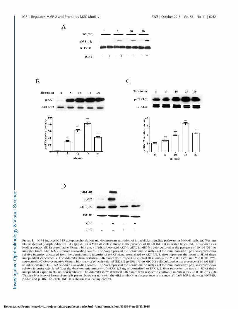
FIGURE 1. IGF-1 induces IGF-1R autophosphorylation and downstream activation of intracellular signaling pathways in MIO-M1 cells. (A) Westernblot analysis of phosphorylated IGF-1R (p-IGF-1R) in MIO-M1 cells cultured in the presence of 10 nM IGF-1 at indicated times. IGF-1R is shown as aloading control. (B) Representative Western blot assay of phosphorylated AKT (p-AKT) in MIO-M1 cells cultured in the presence of 10 nM IGF-1 atindicated times. AKT 1/2/3 is shown as a loading control. The bars represent the densitometric analysis of the immunoreactive protein expressed asrelative intensity calculated from the densitometric intensity of p-AKT signal normalized to AKT 1/2/3. Bars represent the mean 6 SD of threeindependent experiments. The asterisks show statistical differences with respect to control (0 minutes) for P < 0.01 (**) and P < 0.001 (***),respectively. (C) Representative Western blot assay of phosphorylated ERK 1/2 (p-ERK 1/2) in MIO-M1 cells cultured in the presence of 10 nM IGF-1at indicated times. ERK 1/2 is shown as a loading control. The bars represent the densitometric analysis of the immunoreactive protein expressed asrelative intensity calculated from the densitometric intensity of p-ERK 1/2 signal normalized to ERK 1/2. Bars represent the mean 6 SD of threeindependent experiments. ns, nonsignificant. The asterisks show statistical differences with respect to control (0 minutes) for P < 0.001 (***). (D)Western blot assay of lysates from cells preincubated (or not) with the aIR3 antibody in the presence or absence of 10 nM IGF-1, showing p-IGF-1R,p-AKT, and p-ERK 1/2 levels. IGF-1R is shown as a loading control.
IGF-1 Regulates MMP-2 and Promotes MGC Motility IOVS j October 2015 j Vol. 56 j No. 11 j 6952
Downloaded From: http://iovs.arvojournals.org/pdfaccess.ashx?url=/data/journals/iovs/934564/ on 05/13/2018
aIR3 antibody. Figure 5B shows that the pretreatment of MIO-M1 cells with aIR3 antibody significantly blocked the effect ofIGF-1 on the cell migration, indicating that this process wasmediated by IGF-1R. Next, to evaluate the specific role of MMP-2 in the cellular migration induced by IGF-1, MIO-M1 cells werepretreated with GM6001, a well-established MMP inhibitor.Interestingly, IGF-1-induced cellular migration was also signif-icantly reduced by this inhibitor (Fig. 5B), indicating that MMP-2 activity played a critical role in the migratory processmediated by IGF-1. Finally, and considering that the PI3K/AKTsignaling pathway participates in the regulation of theextracellular MMP-2 levels, we examined the IGF-1-inducedMIO-M1 migration in cells pretreated with LY294002. In thisway, a significant reduction in the cell motility to wound areawas also observed (Fig. 5B).
IGF-1 Induces the Partial Colocalization of MMP-2
With b1-integrin and p-FAK at the Cell Protrusions
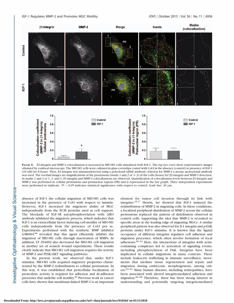
Functional interactions exist between integrins and MMP-2 incell types with migratory phenotypes,45–47 including astro-cytes40,48 and probably MGCs. With this in mind, by confocalmicroscopy, we examined the cellular distribution of b1-integrin and MMP-2 in nontreated and IGF-1-treated MGCs.Figure 6A shows that in the absence of IGF-1, both b1-integrinand MMP-2 showed a punctate perinuclear distribution withcertain fluorescence spread over the cell periphery. Under thisexperimental condition, both proteins showed a low degree ofcolocalization. However, under IGF-1 stimulus (8 hours), anincreased proportion of b1-integrin and MMP-2 colocalized inthe cell periphery, preferentially at the leading edge associatedwith lamellipodia or cellular protrusions, without changes inperinuclear regions with respect to nonstimulated cells. Thequantitative analysis showed that in cellular protrusions, b1-integrin and MMP-2 signals partially colocalized (~18%) inMIO-M1 cells cultured in the presence of IGF-1, with respect tononstimulated cells (~3%). By contrast, in perinuclear regions,although b1-integrin also showed a certain level of colocaliza-tion with MMP-2 (~18%), this proportion was not significantlymodified by IGF-1 (Fig. 6B).
FIGURE 2. IGF-1 decreases active MMP-2 levels in MIO-M1 cellsupernatants in a process mediated by IGF-1R and PI3K signalingpathway. (A) Zymographic analysis of MIO-M1 culture supernatantsobtained from cells stimulated with IGF-1 for 4, 6, or 8 hours. The pro-MMP-9 (92 kDa), pro-MMP-2 (72 kDa), and MMP-2 (active form; 62kDa) are indicated. Capillary blood (SC) was used as a gelatinasecontrol of MMP-9 and pro-MMP-2. (B) Zymographic analysis of culturesupernatants of MIO-M1 cells pretreated with aIR3 antibody (16 nM)for 30 minutes and then stimulated with IGF-1 for 8 hours. (C)Zymographic analysis of culture supernatants of MIO-M1 cellspreincubated with LY-294002 (10 lM), PD98059 (20 lM), or bothpharmacologic inhibitors together, as well as aIR3 antibody, then
FIGURE 3. IGF-1 produces an accumulation of active MMP-2 in celllysates. Western blot assay for the determination of active MMP-2 fromlyophilized culture supernatants and cell lysates of MIO-M1 cellscultured in the presence of 10 nM IGF-1 using an antibody thatspecifically recognizes the active MMP-2. Calreticulin is shown as aloading control.
stimulated with IGF-1 for 8 hours. In each case, bands corresponding topro-MMP-2 and MMP-2 were quantified by densitometric analysis, andtheir relations are represented in the bar graphs expressed in arbitraryunits. Bars denote the mean 6 SD from triplicate experiments. ns, nonsignificant. The asterisks show statistical differences with respect tocontrol for P < 0.05 (*).
IGF-1 Regulates MMP-2 and Promotes MGC Motility IOVS j October 2015 j Vol. 56 j No. 11 j 6953
Downloaded From: http://iovs.arvojournals.org/pdfaccess.ashx?url=/data/journals/iovs/934564/ on 05/13/2018
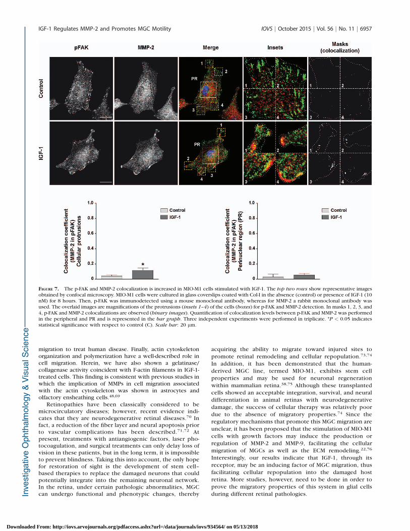
Having established a partial colocalization of MMP-2 and b1-integrin in the periphery of the cell, and considering that FAKis an important nonreceptor protein tyrosine kinase involvedin the cell–ECM adhesion,49 we next assessed the cellulardistribution of p-FAK (active form) and MMP-2 in human MGCs.In the absence of IGF-1, p-FAK was concentrated at the edgesof the membrane ruffles (Fig. 7A, top), whereas under IGF-1stimulus (8 hours) an intense staining was observed in smallfoci within lamellipodia and filopodia (Fig. 7A, bottom). Themerged image for p-FAK/MMP-2 indicated that both proteinspartially colocalized in cellular protrusions (Fig. 7A, bottom).The quantitative analysis showed that in perinuclear regionsthe fluorescence of p-FAK and MMP-2 colocalization wassimilar in control and IGF-1 stimulus (~4%) (Fig. 7B). Bycontrast, in cellular protrusions the percentage of p-FAK/MMP-2 colocalization was significantly increased (~12%) after IGF-1treatment (Fig. 7B).
IGF-1 Induces the In Situ Proteolytic ActivityConcentrated Along F-Actin Filaments
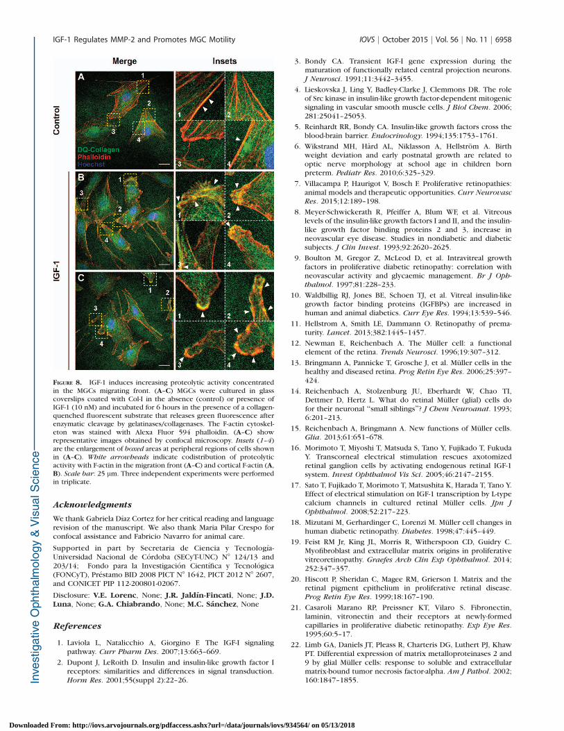
Considering that IGF-1 induced a marked MMP-2 localization inthe cellular protrusions of MGCs, we next assessed whetherthis MMP distribution correlated with an increased pericellularproteolysis. To do this, in situ zymography assays containingDQ-collagen as substrate were carried out as detailed in theMaterials and Methods section. With this substrate, bothgelatinase and collagenase activity of MMPs may be analyzed,although other proteinases can also produce the proteolysis ofDQ-collagen substrate. Confocal microscopy analysis showedthat in the absence of IGF-1, the proteolytic activity was mainlyconcentrated in perinuclear regions, and in some cells thisactivity was weakly associated with cortical actin (Fig. 8A). Bycontrast, MGCs stimulated with IGF-1 exhibited proteolyticactivity mainly distributed along the migration front in areas ofactin-rich filopodia and associated with cortical actin (Fig. 8B,8C). Analysis at high-power magnification revealed that theDQ-collagen proteolytic activity was oriented along actinfilaments in most cell filopodia of migrating MGCs. Our resultsdemonstrate that the accumulation of MMP-2 in cellularprotrusions was associated with an increased pericellularproteolysis in MGCs stimulated with IGF-1.
DISCUSSION
IGF-1 is a polypeptide hormone that binds to its specificreceptor, IGF-1R, and exerts biological actions by activatingseveral intracellular signaling cascades, such as MAPK/ERK 1/2and PI3K/AKT pathways.1,50 Several studies have shown thatthese pathways may in fact be interdependent, as they exertregulatory control on each other.33,51–53 Depending on the celltype studied, the concentration, and the time course of theIGF-1 stimulus, different effects in pericellular proteolyticactivity under physiological and pathologic processes havebeen reported.54 To our knowledge, this is the first demon-stration that IGF-1 in retina, through IGF-1R, decreases theextracellular level of MMP-2 active form and induces MGCmigration. Considering that both IGF-1 effects were inhibitedby the pharmacologic inhibitor, LY294002, we conclude thatthe PI3K/AKT pathway is involved in these extracellular andmigratory events of MGCs.
As previously described for other cell types, Western blotanalysis showed that after IGF-1R autophosphorylation, IGF-1also induced the phosphorylation of AKT and ERK 1/2 inMGCs. However, the PI3K/AKT pathway was differentiallyactivated because significant phosphorylated AKT levels wereobserved at shorter times of IGF-1 stimulation compared withthe IGF-1 effect on phosphorylated ERK 1/2. In addition, thepretreatment of MIO-M1 cells with the aI3R antibody inhibitedIGF-1R autophosphorylation as well as subsequent signalingevents, indicating that both signaling pathways were activatedby IGF-1 in retinal MGCs. In agreement with our observations,various authors have previously identified IGF-1R as a regulatorof MMP-2 synthesis or activity via PI3K/AKT signaling pathwayin cancer cells.54,55 At the same time, Zhang and colleagues33
have demonstrated in a tumor cell model that low to moderatelevels of IGF-1 caused the activation of PI3K/AKT pathways,which in turn inhibited the ERK pathway through AKT-mediated Raf phosphorylation. High levels of stimulating IGF-1 overcame this inhibition and led to ERK induction. The PI3K/AKT pathway stimulated the MMP-2 expression, whereas theRaf/MEK/ERK pathway was inhibitory. In our MGC model,PI3K/AKT pathway activated by IGF-1 was also involved inMMP-2 regulation evidenced by a reduction of the active format the extracellular level.
FIGURE 4. IGF-1 does not modify the MT1-MMP expression at cellsurface level. (A) Representative Western blot assay for the determi-nation of MT1-MMP in MIO-M1 cells cultured in the presence of 10 nMIGF-1 at indicated times. Calreticulin is shown as a loading control. Thebars represent the densitometric analysis of the immunoreactiveprotein expressed as relative intensity calculated from the densitomet-ric intensity of the MT1-MMP signal normalized to calreticulin. Bars
represent the mean 6 SD of three independent experiments. Theasterisks show statistical differences with respect to control for P <0.001 (***). (B) Cells were treated (or not) with IGF-1 (10 nM) for 8hours, and the cell surface level of MT1-MMP was evaluated by a biotin-labeling protein assay and immunoblotted for MT1-MMP. BiotinylatedMT1-MMP: Represents the biotinylated-bound and eluted fraction ofthe streptavidin-agarose beads. Total MT1-MMP: The input (10%) ofbiotinylated proteins is shown. Actin was used as protein-loadingcontrol.
IGF-1 Regulates MMP-2 and Promotes MGC Motility IOVS j October 2015 j Vol. 56 j No. 11 j 6954
Downloaded From: http://iovs.arvojournals.org/pdfaccess.ashx?url=/data/journals/iovs/934564/ on 05/13/2018
In normal cells, previous studies have also demonstratedthat PI3K activation causes the dedifferentiation of vascularsmooth muscle cells (VSMCs) and stimulates VSMC migrationby IGF-1-mediated MMP-2 upregulation.54 In the current study,we showed by zymography that supernatants of MIO-M1 cellstreated with IGF-1 presented a significant reduction in theactive form of MMP-2 compared with untreated cells. In thisway, it was established that the migration of both normal andcancer cells depended on MMP accumulation at membraneprotrusions, which is required for the focal pericellulardegradation of ECM.56 In line with this, we decided to evaluateMMP-2 active expression by Western blot assays, using aspecific antibody that recognizes only the active form of thisMMP. Results revealed that IGF-1 produced an increasedaccumulation of the active form of MMP-2 in MIO-M1 cells.By a biotin-labeling cell surface protein assay, we can also
confirm that the amount of MT1-MMP at the cell surface wasnot modified after IGF-1 stimulus. Hence, this particularincrease of MMP-2 in MIO-M1 would confer the focalgelatinolytic activity necessary for supporting a migratoryphenotype.
It is known that in Col-I matrix, MT1-MMP initiatesdenaturation of collagen into gelatin, which is subsequentlydigested by MMP-2, facilitating pericellular proteolysis andenhancing cell invasion and migration. Furthermore, it hasbeen suggested that the intracellular traffic of MT1-MMPtoward the cell surface and its participation in cellularmigration may be dependent on the tumoral or nontumoralcharacteristics of cells, as well as on the presence ofextracellular factors that induce cellular migration, such asCol-I57 and a2M,28 among others.44 Using two-dimensionalscratch wound assays, we demonstrated that, in fact, in the
FIGURE 5. IGF-1 induces MIO-M1 cell migration through its cognate receptor. (A) Representative two-dimensional scratch wound assay of MIO-M1cells cultured in plates coated with collagen type I (collagen) in the presence or absence of IGF-1 (10 nM) during 12 hours. Scale bar: 100 lm. Bar
graph represents mean values 6 SD of the cell number invading the area of the wound (cells/area) in the absence (control) or presence of IGF-1 (10nM) in plates coated with laminin or collagen. (B) Quantitative analysis of MIO-M1 cell migration by scratch wound assay of cells cultured incollagen-coated plates pretreated with vehicle (C), aIR3 antibody (16 nM), GM6001 (20 lM), or LY-294002 (10 lM) for 30 minutes and thenstimulated with IGF-1 (10 nM) for 12 hours. Bar graph represents mean values 6 SD of the cell number invading the area of the wound (cells/area).Three independent experiments were performed in triplicate. The asterisks show statistical differences with respect to control for P < 0.001 (***).
IGF-1 Regulates MMP-2 and Promotes MGC Motility IOVS j October 2015 j Vol. 56 j No. 11 j 6955
Downloaded From: http://iovs.arvojournals.org/pdfaccess.ashx?url=/data/journals/iovs/934564/ on 05/13/2018
absence of IGF-1 the cellular migration of MIO-M1 cells wasincreased in the presence of Col-I with respect to laminin.However, IGF-1 increased the migratory ability of MGCindependently from the ECM proteins used as cell support.The blockade of IGF-1R autophosphorylation with aIR3antibody inhibited the migratory process, which indicates thatIGF-1 is an extracellular factor inducing cell motility of MIO-M1cells independently from the presence of Col-I per se.Experiments performed with the synthetic MMP inhibitorGM600158 revealed that this agent efficiently inhibits themigration of MIO-M1 cells through inactivation of MMPs. Inaddition, LY 294002 also decreased the MIO-M1 cell migrationin another set of scratch wound experiments. These resultsclearly indicate that MIO-M1 cell migration requires the activityof MMP-2 and PI3K/AKT signaling pathways.
In the present work, we observed that, under IGF-1stimulus, MIO-M1 cells acquired migratory properties charac-terized by the MMP-2 redistribution to cellular protrusions. Inthis way, it was established that pericellular focalization ofproteolytic activity is required for adhesion and de-adhesionprocesses that underlie cell motility.59 Previous work in cancercells have shown that membrane-linked MMP-2 is an important
element for tumor cell invasion through its link withintegrins.45,47 Herein, we showed that IGF-1 induced theredistribution of MMP-2 in migrating cells. In these conditions,a focalized peripheral distribution of MMP-2 across the cellularprotrusions replaced the pattern of distribution observed incontrol cells, supporting the idea that MMP-2 is recruited tospecific areas at the leading edge of migrating MGCs. A similarperipheral pattern was also observed for b-1 integrin and p-FAKproteins under IGF-1 stimulus. It is known that the ligandoccupancy of different integrins regulates cell adhesion andmigration processes, which often involve formation of focaladhesions.60–62 Here, the interactions of integrins with actin-containing complexes led to activation of signaling events,including phosphorylation of FAK. Integrins have beenimplicated in cellular migrations in many contexts. Theseinclude leukocyte trafficking in immune surveillance, move-ments that mediate tissue regeneration and repair, andmigration during embryonic morphogenesis, among oth-ers.63–65 Many human diseases, including retinopathies, havebeen associated with altered integrin-mediated adhesion andmigration.66–68 Therefore, there has been strong interest inunderstanding and potentially targeting integrin-mediated
FIGURE 6. b1-integrin and MMP-2 colocalization is increased in MIO-M1 cells stimulated with IGF-1. The top two rows show representative imagesobtained by confocal microscopy. The MIO-M1 cells were cultured in glass coverslips coated with Col-I in the absence (control) or presence of IGF-1(10 nM) for 8 hours. Then, b1-integrin was immunodetected using a polyclonal rabbit antibody, whereas for MMP-2 a mouse monoclonal antibodywas used. The overlaid images are magnifications of the protrusions (insets 1 and 2 or 1–3) of the cells (boxes) for b1-integrin and MMP-2 detection.In masks 1 and 2 or 1, 2, and 3, b1-integrin and MMP-2 colocalizations are observed. Quantification of colocalization levels between b1-integrin andMMP-2 was performed in cellular protrusions and perinuclear regions (PR) and is represented in the bar graph. Three independent experimentswere performed in triplicate. *P < 0.05 indicates statistical significance with respect to control. Scale bar: 20 lm.
IGF-1 Regulates MMP-2 and Promotes MGC Motility IOVS j October 2015 j Vol. 56 j No. 11 j 6956
Downloaded From: http://iovs.arvojournals.org/pdfaccess.ashx?url=/data/journals/iovs/934564/ on 05/13/2018
migration to treat human disease. Finally, actin cytoskeletonorganization and polymerization have a well-described role incell migration. Herein, we have also shown a gelatinase/collagenase activity coincident with F-actin filaments in IGF-1-treated cells. This finding is consistent with previous studies inwhich the implication of MMPs in cell migration associatedwith the actin cytoskeleton was shown in astrocytes andolfactory ensheathing cells.48,69
Retinopathies have been classically considered to bemicrocirculatory diseases; however, recent evidence indi-cates that they are neurodegenerative retinal diseases.70 Infact, a reduction of the fiber layer and neural apoptosis priorto vascular complications has been described.71,72 Atpresent, treatments with antiangiogenic factors, laser pho-tocoagulation, and surgical treatments can only delay loss ofvision in these patients, but in the long term, it is impossibleto prevent blindness. Taking this into account, the only hopefor restoration of sight is the development of stem cell–based therapies to replace the damaged neurons that couldpotentially integrate into the remaining neuronal network.In the retina, under certain pathologic abnormalities, MGCcan undergo functional and phenotypic changes, thereby
acquiring the ability to migrate toward injured sites to
promote retinal remodeling and cellular repopulation.73,74
In addition, it has been demonstrated that the human-
derived MGC line, termed MIO-M1, exhibits stem cell
properties and may be used for neuronal regeneration
within mammalian retina.38,75 Although these transplanted
cells showed an acceptable integration, survival, and neural
differentiation in animal retinas with neurodegenerative
damage, the success of cellular therapy was relatively poor
due to the absence of migratory properties.74 Since the
regulatory mechanisms that promote this MGC migration are
unclear, it has been proposed that the stimulation of MIO-M1
cells with growth factors may induce the production or
regulation of MMP-2 and MMP-9, facilitating the cellular
migration of MGCs as well as the ECM remodeling.22,76
Interestingly, our results indicate that IGF-1, through its
receptor, may be an inducing factor of MGC migration, thus
facilitating cellular repopulation into the damaged host
retina. More studies, however, need to be done in order to
prove the migratory properties of this system in glial cells
during different retinal pathologies.
FIGURE 7. The p-FAK and MMP-2 colocalization is increased in MIO-M1 cells stimulated with IGF-1. The top two rows show representative imagesobtained by confocal microscopy. MIO-M1 cells were cultured in glass coverslips coated with Col-I in the absence (control) or presence of IGF-1 (10nM) for 8 hours. Then, p-FAK was immunodetected using a mouse monoclonal antibody, whereas for MMP-2 a rabbit monoclonal antibody wasused. The overlaid images are magnifications of the protrusions (insets 1–4) of the cells (boxes) for p-FAK and MMP-2 detection. In masks 1, 2, 3, and4, p-FAK and MMP-2 colocalizations are observed (binary images). Quantification of colocalization levels between p-FAK and MMP-2 was performedin the peripheral and PR and is represented in the bar graph. Three independent experiments were performed in triplicate. *P < 0.05 indicatesstatistical significance with respect to control (C). Scale bar: 20 lm.
IGF-1 Regulates MMP-2 and Promotes MGC Motility IOVS j October 2015 j Vol. 56 j No. 11 j 6957
Downloaded From: http://iovs.arvojournals.org/pdfaccess.ashx?url=/data/journals/iovs/934564/ on 05/13/2018
Acknowledgments
We thank Gabriela Dıaz Cortez for her critical reading and languagerevision of the manuscript. We also thank Maria Pilar Crespo forconfocal assistance and Fabricio Navarro for animal care.
Supported in part by Secretaria de Ciencia y Tecnologıa-Universidad Nacional de Cordoba (SECyT-UNC) N8 124/13 and203/14; Fondo para la Investigacion Cientıfica y Tecnologica(FONCyT), Prestamo BID 2008 PICT N8 1642, PICT 2012 N8 2607,and CONICET PIP 112-200801-02067.
Disclosure: V.E. Lorenc, None; J.R. Jaldın-Fincati, None; J.D.Luna, None; G.A. Chiabrando, None; M.C. Sanchez, None
References
1. Laviola L, Natalicchio A, Giorgino F. The IGF-I signalingpathway. Curr Pharm Des. 2007;13:663–669.
2. Dupont J, LeRoith D. Insulin and insulin-like growth factor Ireceptors: similarities and differences in signal transduction.Horm Res. 2001;55(suppl 2):22–26.
3. Bondy CA. Transient IGF-I gene expression during thematuration of functionally related central projection neurons.J Neurosci. 1991;11:3442–3455.
4. Lieskovska J, Ling Y, Badley-Clarke J, Clemmons DR. The roleof Src kinase in insulin-like growth factor-dependent mitogenicsignaling in vascular smooth muscle cells. J Biol Chem. 2006;281:25041–25053.
5. Reinhardt RR, Bondy CA. Insulin-like growth factors cross theblood-brain barrier. Endocrinology. 1994;135:1753–1761.
6. Wikstrand MH, Hard AL, Niklasson A, Hellstrom A. Birthweight deviation and early postnatal growth are related tooptic nerve morphology at school age in children bornpreterm. Pediatr Res. 2010;6:325–329.
7. Villacampa P, Haurigot V, Bosch F. Proliferative retinopathies:animal models and therapeutic opportunities. Curr Neurovasc
Res. 2015;12:189–198.
8. Meyer-Schwickerath R, Pfeiffer A, Blum WF, et al. Vitreouslevels of the insulin-like growth factors I and II, and the insulin-like growth factor binding proteins 2 and 3, increase inneovascular eye disease. Studies in nondiabetic and diabeticsubjects. J Clin Invest. 1993;92:2620–2625.
9. Boulton M, Gregor Z, McLeod D, et al. Intravitreal growthfactors in proliferative diabetic retinopathy: correlation withneovascular activity and glycaemic management. Br J Oph-
thalmol. 1997;81:228–233.
10. Waldbillig RJ, Jones BE, Schoen TJ, et al. Vitreal insulin-likegrowth factor binding proteins (IGFBPs) are increased inhuman and animal diabetics. Curr Eye Res. 1994;13:539–546.
11. Hellstrom A, Smith LE, Dammann O. Retinopathy of prema-turity. Lancet. 2013;382:1445–1457.
12. Newman E, Reichenbach A. The Muller cell: a functionalelement of the retina. Trends Neurosci. 1996;19:307–312.
13. Bringmann A, Pannicke T, Grosche J, et al. Muller cells in thehealthy and diseased retina. Prog Retin Eye Res. 2006;25:397–424.
14. Reichenbach A, Stolzenburg JU, Eberhardt W, Chao TI,Dettmer D, Hertz L. What do retinal Muller (glial) cells dofor their neuronal ‘‘small siblings’’? J Chem Neuroanat. 1993;6:201–213.
15. Reichenbach A, Bringmann A. New functions of Muller cells.Glia. 2013;61:651–678.
16. Morimoto T, Miyoshi T, Matsuda S, Tano Y, Fujikado T, FukudaY. Transcorneal electrical stimulation rescues axotomizedretinal ganglion cells by activating endogenous retinal IGF-1system. Invest Ophthalmol Vis Sci. 2005;46:2147–2155.
17. Sato T, Fujikado T, Morimoto T, Matsushita K, Harada T, Tano Y.Effect of electrical stimulation on IGF-1 transcription by L-typecalcium channels in cultured retinal Muller cells. Jpn J
Ophthalmol. 2008;52:217–223.
18. Mizutani M, Gerhardinger C, Lorenzi M. Muller cell changes inhuman diabetic retinopathy. Diabetes. 1998;47:445–449.
19. Feist RM Jr, King JL, Morris R, Witherspoon CD, Guidry C.Myofibroblast and extracellular matrix origins in proliferativevitreoretinopathy. Graefes Arch Clin Exp Ophthalmol. 2014;252:347–357.
20. Hiscott P, Sheridan C, Magee RM, Grierson I. Matrix and theretinal pigment epithelium in proliferative retinal disease.Prog Retin Eye Res. 1999;18:167–190.
21. Casaroli Marano RP, Preissner KT, Vilaro S. Fibronectin,laminin, vitronectin and their receptors at newly-formedcapillaries in proliferative diabetic retinopathy. Exp Eye Res.1995;60:5–17.
22. Limb GA, Daniels JT, Pleass R, Charteris DG, Luthert PJ, KhawPT. Differential expression of matrix metalloproteinases 2 and9 by glial Muller cells: response to soluble and extracellularmatrix-bound tumor necrosis factor-alpha. Am J Pathol. 2002;160:1847–1855.
FIGURE 8. IGF-1 induces increasing proteolytic activity concentratedin the MGCs migrating front. (A–C) MGCs were cultured in glasscoverslips coated with Col-I in the absence (control) or presence ofIGF-1 (10 nM) and incubated for 6 hours in the presence of a collagen-quenched fluorescent substrate that releases green fluorescence afterenzymatic cleavage by gelatinases/collagenases. The F-actin cytoskel-eton was stained with Alexa Fluor 594 phalloidin. (A–C) showrepresentative images obtained by confocal microscopy. Insets (1–4)are the enlargement of boxed areas at peripheral regions of cells shownin (A–C). White arrowheads indicate codistribution of proteolyticactivity with F-actin in the migration front (A–C) and cortical F-actin (A,B). Scale bar: 25 lm. Three independent experiments were performedin triplicate.
IGF-1 Regulates MMP-2 and Promotes MGC Motility IOVS j October 2015 j Vol. 56 j No. 11 j 6958
Downloaded From: http://iovs.arvojournals.org/pdfaccess.ashx?url=/data/journals/iovs/934564/ on 05/13/2018
23. Miyata Y, Kase M, Sugita Y, et al. Protein kinase C-mediatedregulation of matrix metalloproteinase and tissue inhibitor ofmetalloproteinase production in a human retinal Muller cells.Curr Eye Res. 2012;37;842–849.
24. Limb GA, Little BC, Meager A, et al. Cytokines in proliferativevitreoretinopathy. Eye (Lond). 1991;5(pt 6):686–693.
25. Franks WA, Limb GA, Stanford MR, et al. Cytokines in humanintraocular inflammation. Curr Eye Res. 1992;11(suppl):187–191.
26. Limb GA, Alam A, Earley O, Green W, Chignell AH, DumondeDC. Distribution of cytokine proteins within epiretinalmembranes in proliferative vitreoretinopathy. Curr Eye Res.1994;13:791–798.
27. Limb GA, Chignell AH, Green W, LeRoy F, Dumonde DC.Distribution of TNF alpha and its reactive vascular adhesionmolecules in fibrovascular membranes of proliferative diabeticretinopathy. Br J Ophthalmol. 1996;80:168–173.
28. Barcelona PF, Jaldın-Fincati JR, Sanchez MC, Chiabrando GA.Activated alpha2-macroglobulin induces Muller glial cellmigration by regulating MT1-MMP activity through LRP1.FASEB J. 2013;27:3181–3197.
29. Chesik D, De Keyser J, Bron R, Fuhler GM. Insulin-like growthfactor binding protein-1 activates integrin-mediated intracellu-lar signaling and migration in oligodendrocytes. J Neurochem.2010;113:1319–1330.
30. Faber-Elman A, Solomon A, Abraham JA, Marikovsky M,Schwartz M. Involvement of wound-associated factors in ratbrain astrocyte migratory response to axonal injury: in vitrosimulation. J Clin Invest. 1996;97:162–171.
31. Jiang J, McMurtry J, Niedzwiecki D, Goldman SA. Insulin-likegrowth factor-1 is a radial cell-associated neurotrophin thatpromotes neuronal recruitment from the adult songbirdedpendyma/subependyma. J Neurobiol. 1998;36:1–15.
32. Long L, Navab R, Brodt P. Regulation of the Mr 72,000 type IVcollagenase by the type I insulin-like growth factor receptor.Cancer Res. 1998;58:3243–3247.
33. Zhang D, Bar-Eli M, Meloche S, Brodt P. Dual regulation ofMMP-2 expression by the type 1 insulin-like growth factorreceptor: the phosphatidylinositol 3-kinase/Akt and Raf/ERKpathways transmit opposing signals. J Biol Chem. 2004;279:19683–19690.
34. Stetler-Stevenson WG. The role of matrix metalloproteinasesin tumor invasion, metastasis, and angiogenesis. Surg Oncol
Clin N Am. 2001;10:383–392, x.
35. Seiki M Yana I. Roles of pericellular proteolysis by membranetype-1 matrix metalloproteinase in cancer invasion andangiogenesis. Cancer Sci. 2003;94:569–574.
36. Sanchez MC, Luna JD, Barcelona PF, et al. Effect of retinal laserphotocoagulation on the activity of metalloproteinases and thealpha(2)-macroglobulin proteolytic state in the vitreous ofeyes with proliferative diabetic retinopathy. Exp Eye Res.2007;85:644–650.
37. Grant MB, Schmetz I, Russell B, Harwood HJ Jr, Silverstein J,Merimee TJ. Changes in insulin-like growth factors I and IIand their binding protein after a single intramuscularinjection of growth hormone. J Clin Endocrinol Metab.1986;63:981–984.
38. Limb GA, Salt TE, Munro PM, Moss SE, Khaw PT. In vitrocharacterization of a spontaneously immortalized humanMuller cell line (MIO-M1). Invest Ophthalmol Vis Sci. 2002;43:864–869.
39. Kleiner DE, Stetler-Stevenson, WG. Quantitative zymography:detection of picogram quantities of gelatinases. Anal Bio-
chem. 1994;218:325–329.
40. Rivera S, Ogier C, Jourquin J, et al. Gelatinase B and TIMP-1 areregulated in a cell- and time-dependent manner in associationwith neuronal death and glial reactivity after global forebrainischemia. Eur J Neurosci. 2002;15:19–32.
41. Bolte S, Cordelieres, FP. A guided tour into subcellularcolocalization analysis in light microscopy. J Microsc. 2006;224(pt 3):213–232.
42. Li YM, Schacher DH, Liu Q, et al. Regulation of myeloidgrowth and differentiation by the insulin-like growth factor Ireceptor. Endocrinology. 1997;138:362–368.
43. Strongin AY. Proteolytic and non-proteolytic roles of mem-brane type-1 matrix metalloproteinase in malignancy. Biochim
Biophys Acta. 2010;1803:133–141.
44. Frittoli E, Palamidessi A, Disanza A, Scita G. Secretory andendo/exocytic trafficking in invadopodia formation: the MT1-MMP paradigm. Eur J Cell Biol. 2011;90:108–114.
45. Brooks PC, Stromblad S, Sanders LC, et al. Localization ofmatrix metalloproteinase MMP-2 to the surface of invasivecells by interaction with integrin alpha v beta 3. Cell. 1996;85:683–693.
46. Deryugina EI, Bourdon MA, Luo GX, Reisfeld RA, Strongin A.Matrix metalloproteinase-2 activation modulates glioma cellmigration. J Cell Sci. 1997;110(pt 19):2473–2482.
47. Mitra A, Chakrabarti J, Chatterjee A. Binding of alpha5monoclonal antibody to cell surface alpha5beta1 integrinmodulates MMP-2 and MMP-7 activity in B16F10 melanomacells. J Environ Pathol Toxicol Oncol. 2003;22:167–178.
48. Ogier C, Bernard A, Chollet AM, et al. Matrix metalloprotei-nase-2 (MMP-2) regulates astrocyte motility in connection withthe actin cytoskeleton and integrins. Glia. 2006;54:272–284.
49. Mitra SK, Hanson DA, Schlaepfer DD. Focal adhesion kinase: incommand and control of cell motility. Nat Rev Mol Cell Biol.2005;6:56–68.
50. LeRoith D, Werner H, Beitner-Johnson D, Roberts CT Jr.Molecular and cellular aspects of the insulin-like growth factorI receptor. Endocr Rev. 1995;16:143–163.
51. Lehman JA, Gomez-Cambronero J. Molecular crosstalk be-tween p70S6k and MAPK cell signaling pathways. Biochem
Biophys Res Commun. 2002;293:463–469.
52. Moelling K, Schad K, Bosse M, Zimmermann S, Schweneker M.Regulation of Raf-Akt cross-talk. J Biol Chem. 2002;277:31099–31106.
53. Zimmermann S, Moelling K. Phosphorylation and regulation ofRaf by Akt (protein kinase B). Science. 1999;286:1741–1744.
54. Risinger GM Jr, Hunt TS, Updike DL, Bullen EC, Howard EW.Matrix metalloproteinase-2 expression by vascular smoothmuscle cells is mediated by both stimulatory and inhibitorysignals in response to growth factors. J Biol Chem. 2006;281:25915–25925.
55. Stawowy P, Kallisch H, Kilimnik A, et al. Proproteinconvertases regulate insulin-like growth factor 1-inducedmembrane-type 1 matrix metalloproteinase in VSMCs viaendoproteolytic activation of the insulin-like growth factor-1receptor. Biochem Biophys Res Commun. 2004;321:31–538.
56. Artym VV, Zhang Y, Seillier-Moiseiwitsch F, Yamada KM,Mueller SC. Dynamic interactions of cortactin and membranetype 1 matrix metalloproteinase at invadopodia: defining thestages of invadopodia formation and function. Cancer Res.2006;66:3034–3043.
57. Bravo-Cordero JJ, Marrero-Diaz R, Megıas D, et al. MT1-MMPproinvasive activity is regulated by a novel Rab8-dependentexocytic pathway. EMBO J. 2007;26:1499–1510.
58. Galardy RE, Cassabonne ME, Giese C, et al. Low molecularweight inhibitors in corneal ulceration. Ann N Y Acad Sci.1994;732:315–323.
59. Sternlicht MD, Werb Z. How matrix metalloproteinasesregulate cell behavior. Annu Rev Cell Dev Biol. 2001;17:463–516.
60. Huttenlocher A, Horwitz, AR. Integrins in cell migration. Cold
Spring Harb Perspect Biol. 2011;3:a005074.
IGF-1 Regulates MMP-2 and Promotes MGC Motility IOVS j October 2015 j Vol. 56 j No. 11 j 6959
Downloaded From: http://iovs.arvojournals.org/pdfaccess.ashx?url=/data/journals/iovs/934564/ on 05/13/2018
61. Wozniak MA, Modzelewska K, Kwong L, Keely PJ. Focaladhesion regulation of cell behavior. Biochim Biophys Acta.2004;1692:103–119.
62. Gleeson LM, Chakraborty C, McKinnon T, Lala PK. Insulin-likegrowth factor-binding protein 1 stimulates human trophoblastmigration by signaling through alpha 5 beta 1 integrin viamitogen-activated protein Kinase pathway. J Clin Endocrinol
Metab. 2001;86:2484–2493.
63. Webb DJ, Parsons JT, Horwitz AF. Adhesion assembly,disassembly and turnover in migrating cells—over and overand over again. Nat Cell Biol. 2002;4:E97–E100.
64. Ridley AJ, Schwartz MA, Burridge K, et al. Cell migration:integrating signals from front to back. Science. 2003;302:1704–1709.
65. Friedl P, Wolf K. Plasticity of cell migration: a multiscale tuningmodel. J Cell Biol. 2010;188:11–19.
66. Moreno-Layseca P, Streuli CH. Signalling pathways linkingintegrins with cell cycle progression. Matrix Biol. 2014;34:144–153.
67. Madamanchi A, Capozzi M, Geng L, et al. Mitigation of oxygen-induced retinopathy in alpha2beta1 integrin-deficient mice.Invest Ophthalmol Vis Sci. 2014;55:4338–4347.
68. Park SW, Yun JH, Kim JH, Kim KW, Cho CH, Kim JH.Angiopoietin 2 induces pericyte apoptosis via alpha3beta1integrin signaling in diabetic retinopathy. Diabetes. 2014;63:3057–3068.
69. Gueye Y, Ferhat L, Sbai O, et al. Trafficking and secretion ofmatrix metalloproteinase-2 in olfactory ensheathing glial cells:A role in cell migration? Glia. 2011;59:750–770.
70. Barber AJ. A new view of diabetic retinopathy: a neurodegen-erative disease of the eye. Prog Neuropsychopharmacol Biol
Psychiatry. 2003;27:283–290.
71. Barber AJ, Gardner TW, Abcouwer SF. The significance ofvascular and neural apoptosis to the pathology of diabeticretinopathy. Invest Ophthalmol Vis Sci. 2011;52:1156–1163.
72. Sennlaub F, Courtois Y, Goureau O. Inducible nitric oxidesynthase mediates retinal apoptosis in ischemic proliferativeretinopathy. J Neurosci. 2002;22:3987–3993.
73. Romo P, Madigan MC, Provis JM, Cullen KM. Differentialeffects of TGF-beta and FGF-2 on in vitro proliferation andmigration of primate retinal endothelial and Muller cells. Acta
Ophthalmol. 2011;89:e263–e268.
74. Bull ND, Limb GA, Martin KR. Human Muller stem cell (MIO-M1) transplantation in a rat model of glaucoma: survival,differentiation, and integration. Invest Ophthalmol Vis Sci.2008;49:3449–3456.
75. Lawrence JM, Singhal S, Bhatia B, et al. MIO-M1 cells andsimilar Muller glial cell lines derived from adult human retinaexhibit neural stem cell characteristics. Stem Cells. 2007;25:2033–2043.
76. Tackenberg MA, Tucker BA, Swift JS, et al. Muller cellactivation, proliferation and migration following laser injury.Mol Vis. 2009;15:1886–1896.
IGF-1 Regulates MMP-2 and Promotes MGC Motility IOVS j October 2015 j Vol. 56 j No. 11 j 6960
Downloaded From: http://iovs.arvojournals.org/pdfaccess.ashx?url=/data/journals/iovs/934564/ on 05/13/2018