Identifying Genetic Basis and Molecular Mechanisms in ...hss.ulb.uni-bonn.de/2013/3308/3308.pdf ·...
102
Identifying Genetic Basis and Molecular Mechanisms in Different Types of von Willebrand Disease (VWD) Dissertation zur Erlangung des Doktorgrades (Dr. rer. nat.) der Mathematisch-Naturwissenschaftlichen Fakultät der Rheinischen Friedrich-Wilhelms-Universität Bonn vorgelegt von Hamideh Yadegari aus Esfahan, Iran Bonn, März 2013
Transcript of Identifying Genetic Basis and Molecular Mechanisms in ...hss.ulb.uni-bonn.de/2013/3308/3308.pdf ·...
Identifying Genetic Basis and
Molecular Mechanisms in
Different Types of
von Willebrand Disease (VWD)
Dissertation zur
Erlangung des Doktorgrades (Dr. rer. nat.) der
Mathematisch-Naturwissenschaftlichen Fakultät der
Rheinischen Friedrich-Wilhelms-Universität Bonn
vorgelegt von
Hamideh Yadegari
aus
Esfahan, Iran
Bonn, März 2013
Angefertigt mit Genehmigung der
Mathematisch Naturwissenschaftlichen Fakultät der
Rheinischen Friedrich-Wilhems-Universität Bonn
1. Gutachter: Prof. Dr. Johannes Oldenburg
2. Gutachter: Prof. Dr. Evi Kostenis
Tag der Promotion: 17.07.2013
Erscheinungsjahr: 2013
Dedicated to my parents
For their endless love, support and encouragement
Abbreviations
- I -
Abbreviations
aa amino-acid ADAMTS13 a disintegrin-like and metalloprotease domain
with thrombospondin type-1 motif, number 13
aPTT Activated partial thromboplastine time
ASA Average surface accessible area
Bp Base pair
BS Bleeding score
BSS Bleeding severity score
cAMP Cyclic adenosine monophosphate
cDNA Complementary Deoxynucleic acid
CK Cysteine knot
C-terminal Carboxy-terminal
C8 Cysteine 8
Da Dalton
db Database
DDAVP Vasopressin analogue, 1-desamino-8-D-
arginine-vasopressin
dl Deciliter
DMEM Dulbecco’s Modified Eagle’s Medium
DNA Deoxynucleic acid
DSS Donor splice site
EDTA Ethylenediamintetraacetic acid
ELISA Enzyme-linked immunosorbent assay
ER Endoplasmic reticulum
FBS Fetal bovine serum
FVIII Factor VIII
FVIII:C Factor VIII activity
F8 Factor VIII gene
GPIb Platelet glycoprotein Ib
GPIIb–IIIa Glycoprotein IIb–IIIa
HEK 293 Human embryonic kidney 293 cells
Abbreviations
- II -
HMW High-molecular weight
HMWM High-molecular weight multimers
Hr Hour
IP Index patient
ISTH SSC International Society on Thrombosis and
Haemostasis Scientific and Standardisation
Committee
IU International unit
kb Kilo base pair
kDa Kilo Dalton
MD Molecular dynamic
mg Miligram
min Minute
ml Milliliter
MLPA Multiplex ligation-dependent probe amplification
mRNA Messenger RNA
µg Microgram
µm Micrometer
N Normandy
ns Nanosecond
nt Nucleotide
PBS Phosphate Buffered Saline
PCR Polymerase chain reaction
PDI Protein disulphide isomerase
PFA-100 Platelet function analyzer 100
PolyPhen Polymorphism Phenotyping
RER Rough endoplasmic reticulum
RGD Arg-Gly-Asp
RIPA Ristocetin induced platelet aggregation
RNA Ribonucleic acid
ROG Radius of gyration
rVWF Recombinant von Willebrand factor
SDS Sodium dodecyl sulfate
SIFT Sorting Intolerant From Tolerant
Abbreviations
- III -
TGN Trans-Golgi network
TIL Trypsin-inhibitor-like
UL-VWF Ultra large von Willebrand factor
VWC -domain von Willebrand C-domain
VWD von Willebrand disease
VWD-domain von Willebrand D-domain
VWF von Willebrand factor
VWF VWF gene
VWF:Ag von Willebrand factor antigen
VWF:CB von Willebrand factor: collagen binding
VWF:FVIIIB VWF: FVIII binding
VWF:GPIbB von Willebrand factor: GPIb binding
VWF:RCo von Willebrand factor: ristocetin cofactor
WPB Weibel-Palade body
WT Wild type
Summary
- IV -
Summary
Von Willebrand disease (VWD) is the most common inherited bleeding disorder.
It is caused by quantitative or qualitative defects of the von Willebrand factor
(VWF) which has crucial roles in hemostasis. VWD is classified into three
primary categories. Types 1 and 3 represent partial and total quantitative
deficiency of VWF, respectively. Type 2 is due to qualitative defects of VWF,
and is divided into four secondary categories 2A, 2B, 2M and 2N. In this study
we explored genotype and phenotype characteristics of a cohort of VWD
patients with the aim of dissecting the distribution of mutations in different types
of VWD. Mutation analysis of 114 patients diagnosed to have VWD was
performed by direct sequencing of the VWF gene (VWF). Large deletions were
investigated by multiplex ligation-dependent probe amplification (MLPA)
analysis. The results showed a mutation detection rate of 68%, 94% and 94%
for VWD type 1, 2 and 3, respectively. In total, 68 different putative mutations
were detected. Twenty six of these mutations were novel. In type 1 and type 2
VWD, the majority of identified mutations (74% vs 88.1%) were missense
substitutions while mutations in type 3 VWD mostly caused null alleles (82%). In
addition, the impact of five detected novel cysteine missense mutations residing
in D4-CK domains was characterized on conformation and biosynthesis of
VWF. Transient expression of human cell lines with wild-type or five mutant
VWF constructs was done. Quantitative and qualitative assessment of mutated
recombinant VWF was performed. Storage of VWF in pseudo-Weible-Palade
bodies (WPBs) was studied with confocal microscopy. Moreovere, structural
impact of the mutations was analyzed by homology modeling. Homozygous
expressions showed that these mutations caused defects in multimerization,
elongation of pseudo-WPBs and consequently secretion of VWF. Co-
expressions of wild-type VWF and 3 of the mutants demonstrated defect in
multimer assembly, suggesting a new pathologic mechanism for dominant type
2A VWD due to mutations in D4 and B domains. Structural analysis revealed
that mutations either disrupt intra-domain disulfide bonds or might affect an
inter-domain disulfide bond.
Summary
- V -
In conclusion, our study extends the mutational spectrum of VWF, and improves
the knowledge of the genetic basis of different types of VWD. The gene
expression studies highlight the importance of cysteine residues within the C-
terminal of VWF on the structural conformation of the protein and consequently
multimerization, storage, and secretion of VWF.
Table of contents
- VI -
Table of Contents
Abbreviations ................................................................................................. I
Summary ....................................................................................................... IV
Table of Contents ......................................................................................... VI
Chapter 1: General introduction and outline .............................................. 1
1.1 VWF....................................................................................................... 2
1.1.1 VWF gene ....................................................................................... 2
1.1.2 VWF protein structure and domain organization ....................... 3
1.1.3 VWF biosynthesis ........................................................................ 5
1.1.4 VWF functions ............................................................................. 10
1.2 VWD .................................................................................................... 12
1.2.1 Diagnosis and classification ...................................................... 13
1.2.2 Characterization of VWD subtypes ........................................... 16
1.2.3 Treatment ..................................................................................... 18
1.3 Aim and outline of the thesis ............................................................ 19
Chapter 2 : Mutation distribution in the von Willebrand factor gene related
to the different von Willebrand disease (VWD) types in a cohort of VWD
patients ......................................................................................................... 21
2.1 Abstract .............................................................................................. 22
2.2 Introduction ........................................................................................ 23
2.3 Materials and Methods ...................................................................... 24
2.3.1 Patients ........................................................................................ 24
2.3.2 Phenotypic Analysis ................................................................... 24
2.3.3 VWF analysis .............................................................................. 25
2.3.4 Structure analysis of VWF .......................................................... 26
2.4 Results ................................................................................................ 26
2.4.1 Mutations in Type 1 VWD ........................................................... 27
2.4.2 Mutations in type 2 VWD ............................................................ 29
2.4.3 Mutations in type 3 VWD ............................................................ 32
2.4.4 Prediction of impact of novel substitutions .............................. 33
Table of contents
- VII -
2.4.5 Structure analysis of four missense mutations located in A1 and
A2 domains of VWF .............................................................................. 35
2.5 Discussion .......................................................................................... 37
Chapter 3 : Large deletions identified in patients with von Wil lebrand
disease using multiple ligation-dependent probe amp lification ............. 41
3.1 Abstract .............................................................................................. 42
3.2 Introduction ........................................................................................ 43
3.3 Method and materials ........................................................................ 43
3.4 Results and Discussion .................................................................... 44
Chapter 4: Insights into pathological mechanisms of missense mutations
in C-terminal domains of von Willebrand factor caus ing qualitative or
quantitative von Willebrand disease ......................................................... 52
4.1 Abstract .............................................................................................. 53
4.2 Introduction ........................................................................................ 54
4.3 Materials and Methods ...................................................................... 55
4.3.1 Patients ........................................................................................ 55
4.3.2 Expression studies ..................................................................... 55
4.3.3 Quantitative and qualitative analysis of rVWF .......................... 56
4.3.4 ADAMTS13 assay ........................................................................ 57
4.3.5 Immunofluorescence analysis ................................................... 57
4.3.6 Structure analysis ....................................................................... 58
4.4 Results ................................................................................................ 59
4.4.1 Characterization of patients ....................................................... 59
4.4.2 Expression of VWF mutations in human cell lin es................... 61
4.4.3 Functional characterization of recombinant VW F mutants ..... 62
4.4.4 Susceptibility of variants to cleavage by ADA MTS13 .............. 65
4.4.5 Intracellular localization ............................................................. 66
4.4.6 Impact of the mutations on VWF structure ............................... 68
4.5 Discussion .......................................................................................... 72
References ................................................................................................... 76
List of publications ..................................................................................... 88
Table of contents
- VIII -
Oral and poster presentations ................................................................... 89
Acknowledgements ..................................................................................... 92
Chapter 1.General introduction and outline
- 1 -
Chapter 1
General introduction and outline
Chapter 1. General introduction and outline
- 2 -
1.1 VWF
Von Willebrand factor (VWF) is a large multimeric plasma glycoprotein that first
was identified by Zimmermann in 1971.1, 2 The VWF plays a crucial role in
hemostasis. It mediates platelet adhesion in primary hemostasis. In addition,
VWF carries factor VIII (FVIII) in the circulation, protecting it from rapid
proteolytic degradation and delivering it to sites of vascular damage for
secondary hemostasis.1, 3 Normal circulating VWF is composed of a series of
heterogeneous multimers ranging in size from about 500 kDa to over 20000
kDa.4 The mean plasma level of VWF is 100 IU dL-1 but the population
distribution is between 50 IU dL-1 and 200 IU dL-1.5 Many factors such as ABO
blood group, gender, age, hormonal regulation, and inflammatory states have
been described to have an impact on VWF levels.6
Deficient or defective VWF results in von Willebrand disease (VWD), a common
inherited bleeding disorder.7
1.1.1 VWF gene
The VWF gene (VWF) is located on the short arm of chromosome 12 (12p
13.2). It spans approximately 178 kb of genomic DNA, and is transcribed into an
8.8 kb mRNA. The VWF gene comprises 52 exons, most exons are ranging
from 40-342 bp, but exon 28 is exceptionally larger (1.4 kb in size).1, 8 A non-
coding partial VWF pseudogene has been identified on chromosome 22
(22q11.22 to 22q11.23). The pseudogene is 97% similar in sequence to the
coding gene, corresponding to exons 23 to 34 of VWF. Gene conversion events
between VWF and VWF pseudogene have been demonstrated.9, 10 VWF is
highly polymorphic with about 200 reported polymorphic variations in VWF
recorded in the International Society on Thrombosis and Haemostasis Scientific
and Standardisation Committee on VWD database (ISTH SSC-VWD db
http://vwf.group.shef.ac.uk/index.html accessed January 2013). This highly
polymorphic nature of VWF, along with its large size and the presence of a
partial pseudogene make genetic analysis of VWF challenging.10, 11
Chapter 1. General introduction and outline
- 3 -
1.1.2 VWF protein structure and domain organizatio n
The VWF monomer is comprised of 2813 amino acids (aa). The VWF protein is
remarkably rich in cysteines, which comprise 234 of the 2813 (8,3%) residues in
prepro VWF.3, 12 The VWF protein is extensively glycosylated, oligosaccharide
side chains make up approximately 20% of the mass of VWF, and are believed
to affect its structural and functional integrity .13
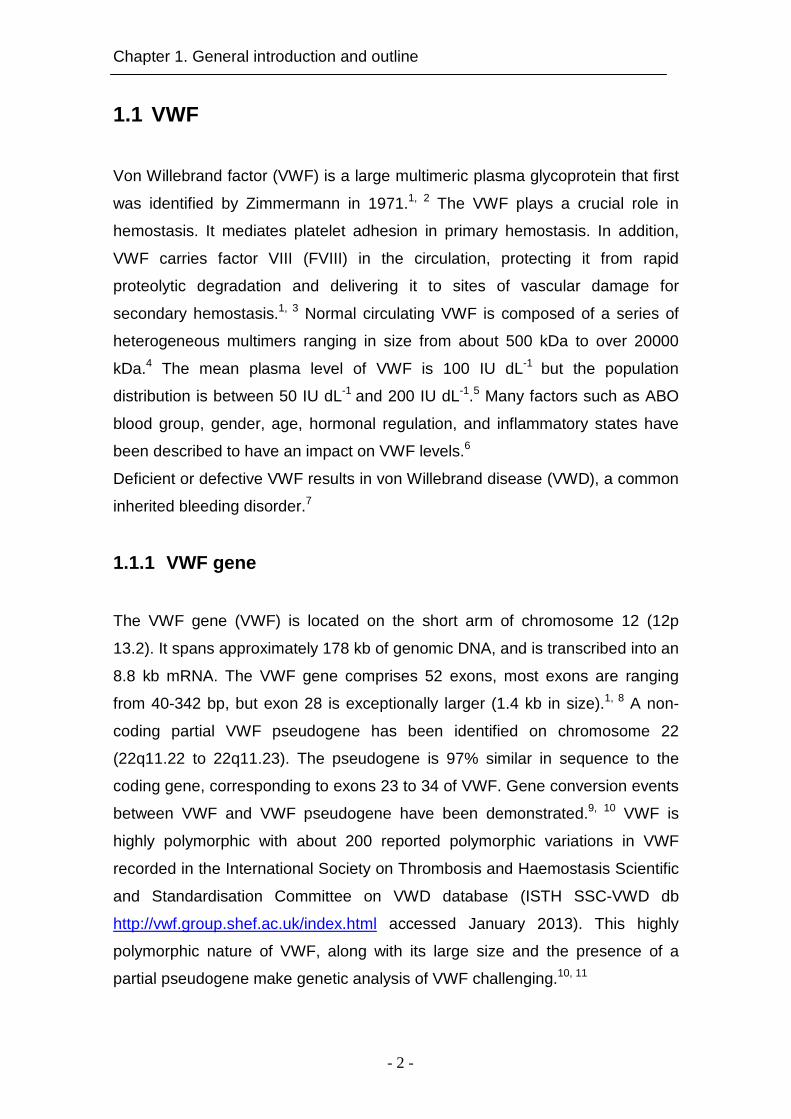
The prepro VWF precursor is composed of five types of domains that are
constructed as repeats in the following order: D1-D2-D’-D3-A1-A2-A3-D4-B1-
B2-B3-C1-C2-CK (Figure 1.1 A).14 The D1 and D2 domains comprise the
propeptide and are cleaved during proteolytic processing to generate the
mature VWF. The remaining domains in the mature VWF carry out specific
functions. 2, 15
The domains may be characterized as structural or functional, depending on
their role in VWF structure or its interaction with other factors. Structural
domains are involved in the post-translational processing of VWF, for example
the cysteine knot (CK) domain is required for dimerization of VWF monomers
and the D1, D2 and D3 domains for proper multimerization of VWF dimers.16 All
‘D’ domains (except D4) contain a CGLC consensus sequence that is highly
homologous to the active site of proteins harbouring protein disulphide
isomerase (PDI) activity.16, 17, 18 PDI is the enzyme that catalyses thiol-
disulphide interchange reactions in protein substrates, leading to disulphide
formation and folding of the protein.17 In this respect, ability of the D1, D2 and
D3 domains to promote interchain disulphide bonding and multimerization can
be ascribed to the intrinsic PDI activity of these domains.17, 18
Functional domains include those that contain cleavage sites for proteolysis
(domain A2) and binding sites for collagen (domains A1 and A3), platelets
(domain A1 for platelet glycoprotein Ib (GPIb) receptors), and FVIII (domains D3
and D’) (Figure 1.1A).16, 18 Moreover, the C1 domain contains the RGD
sequence (Arg-Gly-Asp) recognized by platelet integrin GPIIb/IIIa receptors.
Another RGD sequence is present in the propeptide, but no role in integrin
binding has been observed. 3, 17 The carboxy-terminal domains, namely D4-B1-
3-C1-2, are cysteine-rich, which may suggest structural importance.16
Chapter 1. General introduction and outline
- 4 -
Recently, Zhou et al. have re-evaluated the VWF domain structure, using the
updated information on the structure of these homologous domains in
combination with electron microscopy techniques.14, 19 The previous B and C
regions of VWF are re-annotated as 6 tandem von Willebrand C (VWC) and
VWC-like domains.19 Moreover, the VWF D domains are annotated as
containing von Willebrand D (VWD-domain), cysteine 8 (C8), trypsin-inhibitor-
like (TIL), E or fibronectin type 1-like domains and a unique D4N sequence in
D4 (Figure 1.1B). However, the original domain designations still predominate in
the VWF literature.
Figure 1.1. Structural and functional domains of vo n Willebrand factor (VWF). Part A illustrates the original domain assignment of VWF along with binding and cleavage sites, as well as dimerization and multimerization regions. Exons encoding each domain are shown (adapted from Goodeve AC)1. Part B illustrates the updated domain annotation of VWF (adapted from Zhou YF et al.)19.
Chapter 1. General introduction and outline
- 5 -
1.1.3 VWF biosynthesis
VWF is produced exclusively by endothelial cells present in different tissues and
by the platelet precursor, megakaryocytes.2, 17 Platelet VWF is stored within
cells and has not been shown to contribute significantly to plasma VWF.13
The VWF primary translation product is a 2813-aa pre-pro-polypeptide (350
kDa) which encompasses a 22-aa classic signal sequence, a 741-aa propeptide
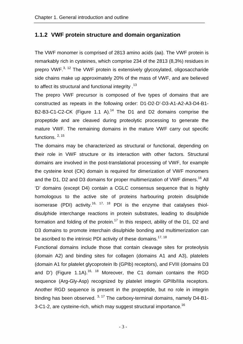
and a 2050-aa mature VWF protein.17, 20 Posttranslational processing of VWF
includes dimerization, glycosylation, sulfation, propeptide cleavage, and
multimerization, followed by storage or secretion (Figure 1.2).3
Dimerization and multimer assembly
After the cleavage of the signal peptide and translocation into the endoplasmic
reticulum (ER), pro-VWF subunits dimerize in a ‘tail-to tail’ manner through
disulfide bonds that form between the C-terminal residues (Figure 1.2).2, 3 The
dimerization function requires only sequences within the last 150 residues. The
cysteins within the last 150 aa residues of the VWF subunit form intersubunit or
intrasubunit disulfide bonds. The carboxyl-terminal 90 residues comprise the
“CK” domain that is homologous to the “Cysteine knot” superfamily of proteins.
The family members share a tendency to dimerize, often through disulfide
bonds.15, 21
The pro-VWF dimers are subsequently transported to the Golgi complex, where
multimerization and the proteolytic removal of the large VWF peptide take place
(Figure 1.2).2 In the Golgi apparatus and post-Golgi compartments, dimers
undergo multimerization to form tetramers, hexamers and so on, generating
high-molecular weight (HMW) multimers containing up to 100 monomers that
may exceed 20 million Da in size.18 Multimerization occurs through additional
head-to-head disulfide bonds near the amino-termini of the subunits.3 The VWF
multimer assembly appears to involve an unique oxidoreductase mechanism
that is activated by the low pH of the Golgi apparatus.15 The key to multimer
assembly is the N-terminal D1-D2-D’-D3 region of proVWF. The reaction
appears to proceed through a transient, disulfide-linked intermediate between

Chapter 1. General introduction and outline
- 6 -
the propeptide and the D3 region of VWF that forms in the ER and resolves in
the Golgi to yield disulfide-linked VWF multimers.22
The polymerization process is accompanied by proteolytic cleavage (probably
by furin) of the propeptide in the trans-Golgi network, yielding mature VWF
multimers and propeptide dimers. The propeptide cleavage occurs after the
argenine residue at position 763. After cleavage, VWF and the VWF propeptide
remain non-covalently associated within the cell.15, 17
Figure 1.2. Biosynthes is of VWF (adapted from Millar CM et al.).13 After the cleavage of the signal peptide and translocation into the rough endoplasmic reticulum (RER), initial glycosylation and dimerization of pro-VWF takes place. The pro-VWF dimers are subsequently transported to the Golgi complex, where they undergo post-translational glycosylation, sulphation and multimerization. Finally, the propeptide is cleaved in the trans-Golgi network (TGN) and fully functional VWF multimers are stored in Weibel-Palade bodies (WPB).
Chapter 1. General introduction and outline
- 7 -
Glycosylation, sulfation
The mature subunit is extensively glycosylated with 12 N-linked and 10 O-linked
oligosaccharides, and the propeptide has three more potential N-glycosylation
sites.12 The N-linked carbohydrates are added in high-mannose form in the ER
and are further processed to complex forms in the Golgi apparatus. O-
glycosylation and sulfation of some N-linked oligosaccharides also occur in
Golgi compartments.23, 24 The N-linked oligosaccharides of VWF are unusual
compared to those of other plasma glycoproteins because they contain ABO
blood group oligisacharides.12 The antigens of the ABO blood group system (A,
B, and H determinants, respectively) consist of complex carbohydrate
molecules. It is demonstrated that ABH structures are carried on the N-linked
oligosaccharide chains of VWF according to the blood type of the individual.23, 25
Intracellular storage
VWF produced by endothelial cells is either secreted constitutively or stored in
Weibel-Palade bodies (WPBs), while VWF produced by megakaryocytes that
are later converted into platelets, is stored in α-granules.12
WPBs are rod-shaped, membrane enclosed organelles, approximately 0.2 µm
wide and up to 5 µm long.15, 17 They consist of densely packed tubular arrays of
VWF which are composed of VWF multimers and propeptide. Tubulation allows
a 100-fold compaction of VWF, without which intracellular storage of VWF
would be impossible. In addition, the tubular storage of VWF multimers is critical
for the orderly secretion of long VWF strings without tangling.12, 26
WPBs initially form at the TGN and subsequently undergo maturation, before
accumulating within the cytoplasm of endothelial cells. The VWF multimers and
propeptide condense into tubules and are incorporated into nascent vesicles
that protrude from the TGN. After budding from the TGN, immature WPBs
remain in a perinuclear location, where they acquire additional membrane
proteins and then disperse throughout the cytoplasm.20, 27
The VWF is a prerequisite for the existence of WPBs.28 It has been proposed
that the targeting of VWF to the WPBs occurs as a consequence of selective
Chapter 1. General introduction and outline
- 8 -
aggregation, by means of multimerization of this protein in the trans-Golgi
network.27, 29 The N-terminal D1-D2-D’-D3 domains and the acidic pH of the
trans-Golgi are required to target and package VWF into WPBs. Thus, multimer
assembly and tubular packing both depend on the propeptide, D’D3 domains
and acidic pH.15, 30
Recently, it has been demonstrated that the VWF C-terminals zip up and form a
structure resembling a bouquet of flowers, where the A2, A3, and D4 domains
comprise the flower part and the six VWC domains configure the stem. Dimeric
bouquets are essential for correct VWF dimer incorporation into growing tubules
and to prevent crosslinking between neighbouring tubules.31, 32 Moreover, it was
found that the last steps in VWF biosynthesis, interdimer N-terminal disulphide
bond formation and cleavage by furin, appear to occur during or after assembly
of individual VWF dimers onto the growing ends of tubules in nascent WPBs.31
Helical assembly thus provides a template for disulphide bond formation during
N-terminal concatamerization.31 The lack of crosslinking between tubules
promotes orderly unfurling of VWF from the ends of helices similarly to orderly
uncoiling of a rope.26 Dimeric bouquets are undoubtedly also important in
compaction of WPBs during maturation and their expansion during secretion.31
Megakaryocytes synthesize large VWF multimers and package them into
platelet α-granules that are spherical rather than cigar-shaped. The VWF
multimers in α-granules are organized into clusters of tubules with dimensions
similar to those of VWF tubules in Weibel–Palade bodies.27, 30
VWF secretion and catabolism
Secretion of stored VWF from endothelial cells occurs through both a
constitutive and a regulated pathway.2 The majority of endothelial-derived VWF
is secreted via the constitutive pathway, which contributes to approximately
95% of plasma VWF; the remaining VWF is stored within WPBs of endothelial
cells, and secreted via the regulated pathway on stimulation by
secretagogues.13 Megakaryocytes, however, lack the regulatory pathway, VWF
is constitutively secreted and has not been shown to contribute significantly to
plasma VWF.2, 13
Chapter 1. General introduction and outline
- 9 -
It would be of great interest to define in detail the molecular mechanisms and
machinery that underlie VWF secretion in health and disease.33 In response to
pathological stimuli, such as vascular injury and inflammation, the circulatory
concentration of VWF increases rapidly mediated by secretagogues. The
secretagogues can be divided into two distinct groups: Ca2+-raising agonists
(thrombin and histamine) and cAMP-raising agonists (epinephrine and
vasopressin).28, 33 In vivo, plasma VWF and FVIII levels rise rapidly after
administration of the vasopressin analogue, 1-desamino-8-D-arginine-
vasopressin (DDAVP), mediated by an increase in cAMP. The ability of DDAVP
to raise plasma VWF and FVIII levels has made it a major drug for the treatment
of VWD and hemophilia A.3, 12
Upon activation of endothelial cells by agonists, the WPBs fuse with the plasma
membrane and release their contents into the blood circulation.20, 33 During
normal exocytosis, the shift in pH from pH 5.5 within the WPBs to the neutral pH
of plasma leads to the unfolding of compact VWF tubules to VWF strings 100
times longer than the WPBs.15, 26
The length of the released VWF strings is typically several 100µm long but can
reach extraordinary lengths of up to 1mm. These long VWF strings could arise
through end-to-end self-association of VWF multimers.34 Presumably, the
unpaired cysteine thiols localized to the N-terminal D3 and C-terminal C
domains are involved in VWF self association.14, 35 Moreover, high shear stress
changes the shape of the molecule from a globular form to an elongated or
stretched form that is crucial for the interaction with platelets.18
The molecular size of VWF is critical for its physiologic function as a mediator of
platelet adhesion and aggregation in primary hemostasis. The high-molecular-
weight-multimers (HMWMs) are biologically most active and are essential to
maintain hemostasis under conditions of high shear stress in the
microvasculature.36 However, the ultra large VWF (UL-VWF) is potentially
harmful in the normal circulation and must be processed into smaller, less
reactive molecules that will not precipitate unwanted platelet aggregation.34 The
highly pro-thrombotic UL-VWF is cleaved by the specific VWF cleaving protease
ADAMTS13 (a disintegrin-like and metalloprotease domain with
thrombospondin type-1 motif, number 13).36 The ADAMTS13 cleavage site in
Chapter 1. General introduction and outline
- 10 -
VWF (Met1605-Tyr1606) is normally hidden within the middle of the folded A2
domain.34 High shear stresses can unfold the ADAMTS13 cleavage site in the
A2 domain.2
The half-life of the circulating VWF multimers is ~ 12h.12 It is demonstrated that
macrophages and FVIII, mainly in the liver, mediate the removal of VWF from
circulating blood. However, the precise cellular membrane receptors for VWF
remain unknown.2
1.1.4 VWF functions
The VWF protein has three main recognized hemostatic functions, which are to
mediate interactions of platelets to subendothelial matrix and platelets to
platelets (platelet adhesion and platelet aggregation), and to serve as a carrier
of factor VIII in the circulation system.2 Recent studies have stated that VWF
may have other, non hemostatic functions in angiogenesis, cell proliferation,
inflammation, and tumor cell survival.2, 14, 37
Interactions between VWF and the subendothelial matrix
VWF performs a bridging function by binding the platelets to the extracellular
matrix at sites of vascular injury.3 When vessel wall subendothelium is exposed,
VWF binds to collagen in the subendothelial matrix. The high shear stress
allows conformational changes within VWF, which can result in the exposure of
binding sites for platelets or collagen.2, 34 The A1 domain of VWF interacts
mainly with collagen type VI but can also bind, with less affinity, to collagen
types I and III. The A3 domain can also bind to collagens, namely collagen
types I and III.2
Role of VWF in platelet adhesion and aggregation
Under physiological conditions, circulating platelets are recruited from the blood
flow to injury sites, where they act to prevent excessive bleeding. The highly
polymerized VWF molecules are required to achieve efficient platelet adhesion
Chapter 1. General introduction and outline
- 11 -
and aggregation.17 The plasma VWF adheres to circulating platelets through
two membrane receptors: glycoprotein Ib (GPIb) and the glycoprotein IIb–IIIa
(GPIIb–IIIa) complex.2,3 Upon exposure of vessel subendothelial matrix, VWF
undergoes a conformational change after matrix binding and subsequently the
binding site for the platelet receptor GPIb, located in the A1 domain of VWF,
becomes accessible.
The platelet GPIIb/IIIa receptor only becomes available for VWF binding after
platelet activation. Platelets are activated at sites of vascular injury by a variety
of agonists. In addition, binding of VWF to the platelet GPIb receptor appears to
activate platelets, making the GpIIb/IIIa receptor available for ligand binding.
The RGD sequence near the C-terminus of VWF seems to be responsible for
VWF binding to GpIIb/IIIa.3
GPIb participates mainly in platelet-vessel wall adhesion, while the GPIIb–IIIa
complex is involved in both platelet-vessel wall adhesion and crosstalk between
platelets. VWF is the only ligand that promotes platelet adhesion by attaching to
both of these receptors.2 Addition of a second layer of platelets (aggregation)
involves binding of VWF to the GPIb and GPIIb/IIIa platelet receptors and of
fibrin to GPIIb/IIIa. This results in forming a layer of VWF and fibrin coating the
adhered platelets which functions as a platform for recruiting more circulating
platelets. The aggregation of platelets continues until the injury is sealed off and
a stable platelet plug has been formed.38
Carrier of factor VIII
VWF forms a complex with FVIII in plasma through a non-covalent interaction.3
FVIII is a cofactor of the intrinsic clotting cascade, and its deficiency manifests
as hemophilia A. VWF protects FVIII from degradation and may also serve to
localize FVIII to the site of the clot. This phenomenon may be mediated by one
or a combination of the following mechanisms: (1) structural stabilization of
FVIII; (2) inhibition of phospholipid-binding proteins that target FVIII for
proteolytic degradation; (3) inhibition of FVIII binding to activated FIX to
stimulate the coagulation pathway, and (4) prevention of FVIII cellular uptake
via scavenger cell receptors.2
Chapter 1. General introduction and outline
- 12 -
FVIII binds to VWF within the N terminal of D’ and D3 domains of VWF
corresponding to residues 763-1035.39
1.2 VWD
Von Willebrand disease (VWD) is the most common inherited bleeding disorder.
VWD has a prevalence of about 1% in the general population, but only
approximately 1 in 10,000 individuals has clinically significant bleedings.1, 40
VWD was first described in 1926 by Erik von Willebrand in a Finnish medical
journal.40 He described a young woman who bled to death at the time of her
fourth menstrual period. Since then, significant advances in clinical and
laboratory phenotypic description of patients and understanding of genetics of
VWD has increased our knowledge on this disorder and its pathophysiology.16,
40 However, the molecular basis of VWD remains the subject of ongoing
investigations.7
VWD is caused by quantitative and/or functional deficits of VWF.16
Characteristic bleeding symptoms include frequent nose bleeding, easy
bruising, oral cavity bleeding, menorrhagia, bleeding after dental extraction,
surgery, and childbirth.7 Patients with severe forms of VWD may also suffer
from joint, muscle and central nervous system bleeding.16
VWD has been classified into three primary categories. Types 1 and 3 VWD
represent quantitative variants; type 1 VWD is a partial quantitative deficiency
and type 3 VWD is a virtually complete quantitative deficiency of VWF. Type 2
VWD is characterized by a qualitative deficiency and defective VWF and is
further classified into the four types 2A, 2B, 2M, and 2N.7, 18 The functional
defects lead to enhanced (2B) or reduced (2A, 2M) platelet interaction or
impaired binding to FVIII (2N).1 Generally, type 1, type 2A, type 2M and type 2B
VWD are inherited in an autosomal dominant pattern, while type 3 and type 2N
VWD are inherited in an autosomal recessive pattern.
Chapter 1. General introduction and outline
- 13 -
1.2.1 Diagnosis and classification
The diagnosis and classification of VWD requires a combination of patients’
medical history, specialized laboratory tests which can determine quantitative or
qualitative defects in VWF and genetic testing.18, 41 The correct classification of
VWD facilitates the treatment and counseling of patients with VWD. In practice,
distinctions between certain VWD types are not always easy to make.42
Bleeding history
A standardized bleeding history of the patient is a prerequisite for proper
diagnosis of a patient with presumed VWD. This may be carried out by a
standard questionnaire.43 In addition, the utility of standard clinical assessment
tools like bleeding severity score (BSS) to quantify bleeding symptoms
facilitates diagnosis.7 The BSS is a summary score from -3 to 45, based on the
presence of various types of bleedings.41
Laboratory testing
For patients suspected of having a bleeding disorder, a number of laboratory
tests may be necessary for proper diagnosis. Initial standard tests include
activated partial thromboplastin time (aPTT), FVIII activity (FVIII:C), bleeding
time and platelet function analyzer 100 (PFA100) assay. The specific assays
important for VWD diagnosis are VWF antigen (VWF:Ag) determination,
VWF:Ristocetin Cofactor activity (VWF:RCo), VWF:collagen binding (VWF:CB)
and VWF:GPIb binding (VWF:GPIb).18, 44 If abnormal results are obtained,
specific assays should be performed to determine the subtype of VWD, such as
multimer analysis, RIPA (Ristocetin induced platelet aggregation), VWF:FVIIIB
(binding of FVIII to VWF) assay, and if indicated, molecular testing.6, 7, 18
VWF antigen (VWF:Ag): Quantity of VWF protein (antigen) in the plasma,
measured antigenically using enzyme-linked immunosorbent assay (ELISA) or
latex immunoassay. The normal range that should be determined independently
by each laboratory is approximately 50–200 IU/dL.7
Chapter 1. General introduction and outline
- 14 -
Ristocetin cofactor activity assay (VWF:RCo): This assay determines the
ability of plasma VWF to interact with platelet GPIb, initiated by the antibiotic
ristocetin. The normal range is approximately 50–200 IU/dL.7
Collagen binding assay (VWF:CB): This test determines the ability of VWF to
bind to collagen (a subendothelial matrix component). The normal range is
approximately 50–200 IU/dL.7
FVIII activity (FVIII:C): Functional FVIII assay that determines the activity of
FVIII in the coagulation cascade. The normal range is approximately 50–150
IU/dL.7
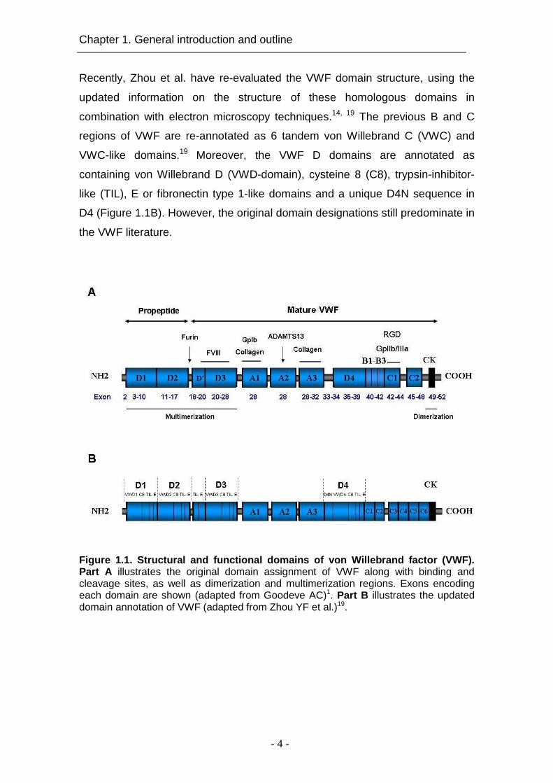
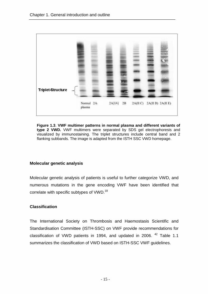
VWF multimer analysis: SDS-agarose gel electrophoresis is used to
determine the distribution of VWF oligomers in the plasma. High-resolution
agarose-gel electrophoresis demonstrated that circulating VWF has a complex
multimeric structure (Figure 1.3). The multimeric pattern of VWF that is seen on
SDS/agarose-gel electrophoresis consists of regularly spaced bands while each
band exhibits a triplet structure appearance. The smallest detectable subunit is
a dimer with a molecular mass of ~540 kDa. The largest molecules exceed
10,000 kDa. The triplet bands of VWF are composed of mature, central band,
and cleaved subunits by ADAMTS13 represented by a faster migrating satellite
band and a slower migrating satellite band (Figure 1.3).5, 45, 46
Ristocetin induced platelet aggregation (RIPA): The RIPA test evaluates the
ristocetin-induced aggregation of platelet-rich plasma. It is used to identify
patients with type 2B and discriminates patients with type 2 VWD from those
with type 1 VWD.43
Binding of FVIII to VWF (VWF:FVIIIB): This test that determines the ability of
VWF to bind FVIII is useful for the identification and differential diagnosis of type
2N VWD.
Chapter 1. General introduction and outline
- 15 -
Molecular genetic analysis
Molecular genetic analysis of patients is useful to further categorize VWD, and
numerous mutations in the gene encoding VWF have been identified that
correlate with specific subtypes of VWD.18
Classification
The International Society on Thrombosis and Haemostasis Scientific and
Standardisation Committee (ISTH-SSC) on VWF provide recommendations for
classification of VWD patients in 1994, and updated in 2006. 42 Table 1.1
summarizes the classification of VWD based on ISTH-SSC VWF guidelines.
Figure 1.3 . VWF multimer patterns in normal plasma and differen t vari ants of type 2 VWD. VWF multimers were separated by SDS gel electrophoresis and visualized by immunostaining. The triplet structures include central band and 2 flanking subbands. The image is adapted from the ISTH SSC VWD homepage.
Chapter 1. General introduction and outline
- 16 -
1.2.2 Characterization of VWD subtypes
VWD type 1
Type 1 VWD is the most common form of the disorder and accounts for up to
70% of all VWD. A VWF level less than 30–40 IU/dL indicates type 1 VWD.18
However, it can be difficult to diagnose due to factors influencing VWF levels in
plasma including environmental and genetics factors.47 One of the major
determinants of plasma VWF:Ag levels is the ABO blood group of an
individual.13, 48 ABO blood group O has been known to be more prevalent in
type 1 VWD than in the normal population and is associated with VWF levels
approximately 25% lower than the population average. The effect of blood-
VWD Type
Description
1
Partial quantitative deficiency of VWF Reduction of VWF:Ag, VWF:Ag/VWF:RCo>0.6, normal VWF multimer structure
2
Qualitative defects of VWF
2A
Decreased VWF-dependent platelet adhesion Associated with deficiency of HMW-multimers, VWF:Ag/VWF:Rco<0.6.
2M
Decreased VWF-dependent platelet adhesion. VWF:Ag/VWF:Rco<0.6 and/or reduced VWF:CB, and normal VWF multimer pattern
2B
Increased affinity for platelet GPIb; Characterized by increased RIPA at low ristocetin concentrations, and deficiency of HMW-multimers
2N Markedly decreased binding affinity for FVIII (reduction in VWF:FVIIIB).
3
Virtually complete deficiency of VWF VWF:Ag <5 IU/dL and FVIII:C<10 IU/dL.
Table 1.1. Classification of VWD (adapted from Sadler JE, et al.)42
Chapter 1. General introduction and outline
- 17 -
group O appears to be due to increased VWF clearance from plasma; A and B
blood-group glycosylation protects VWF from clearance,.1, 49, 50
The potential pathogenetic mechanisms characterized in type1 VWD to date
include impaired biosynthesis, clearance (decreased survival) and intracellular
retention of VWF.51
VWD Type 2
Type 2 VWD accounts for approximately 25% of all VWD.7 Type 2 VWD is
characterized by qualitative VWF deficiency. Levels of VWF:Ag may be
decreased, normal or even elevated, but structural or functional defects lead to
impaired activity.16 Many type 2 VWD phenotypes can be distinguished from
VWD type 1 by their decreased ratios of functional parameters, i.e. VWF:RCo,
VWF:CB, VWF:GPIbB, VWF:FVIIIB, respectively, to VWF:Ag. Type 2 VWD
subtypes include types 2A, 2B, 2M and 2N.51 However, reliable classification of
patients with VWD types 2A, 2B and 2M additionally requires multimer
analysis.16
Type 2A VWD is the most common form of type 2 VWD which is associated
with reduced platelet binding due to significant reduction or absence of VWF
HMWM.16 The hallmark of type 2A disease is a low VWF:RCo to VWF:Ag ratio
(< 0.6) and impaired RIPA.52 Four different subtypes of VWD type 2A including
IIA, IIC, IID, IIE can be distinguished by multimer analysis indicating different
underlying mechanisms (Figure 1.3).1, 16 Type 2A (IIA) VWD results from
mutations located in the A1 and A2 domains, which cause intracellular retention
of HMWM (Group I) or enhanced susceptibility to ADAMTS13 cleavage (Group
II). Type 2A VWD can also be caused by mutations interfering with
multimerization assembly which either located in multimerization region (IIC and
IIE) or in dimerization region (IID).53, 54
In type 2M VWD , platelet adhesion is defective despite a normal multimeric
structure of VWF. The reduced capability for platelet adhesion is reflected by a
low level of VWF:RCo or/and VWF:CB.53, 55 Type 2M VWD typically results from
Chapter 1. General introduction and outline
- 18 -
mutations disrupting binding of VWF to platelets (in A1 domain) or impairing
collagen binding (mostly in A3 domain).53
Type 2B VWD is due to a gain-of-function mutation associated with increased
affinity for platelet GPIb.52 Although patients with type 2B VWD show loss of
HMWM like type 2A VWD patients (Figure 1.3), they can be diagnosed
phenotypically by enhanced RIPA at low concentration of ristocetin. In patients
with classic type 2B, development of thrombocytopenia may occure during
stressful situations, such as severe infection, surgery or pregnancy.1 Type 2B is
the result of missense substitutions in the GPIb binding region of VWF, the A1
domain.
Type 2N VWD (‘N’ for Normandy) represents forms of the disease with
defective VWF:FVIII binding,thereby mimicking haemophilia A.16 Symptoms
largely result from reduced FVIII levels. VWF:RCo and VWF:Ag levels can be
within the normal range, while FVIII:C is typically 5–40 IU/dL.
VWD Type 3
Type 3 VWD is the most severe and infrequent form of the disease,55 and
accounts for <5% of VWD patients.7 Type 3 VWD is identified by VWF:Ag
levels< 1% and moderate deficiency of FVIII (levels <10%).
It is an autosomal-recessive form of the disease due to homozygosity or
compound heterozygosity of two mutations resulting in lack of VWF expression.
In most cases, heterozygous mutation carriers of VWD type 3 are unaffected.16
Phenotypic analysis is generally sufficient for diagnosis of the disorder, although
molecular analysis can be useful where carrier status determination or prenatal
diagnosis is required.
1.2.3 Treatment
The most important treatment options available for patients with VWD include
stimulating the release of VWF from endogenous storage organelles by
DDAVP, or replacement with plasma-derived VWF/FVIII concentrates.16, 53, 56
Chapter 1. General introduction and outline
- 19 -
Other treatments that can reduce symptoms include fibrinolytic inhibitors and
hormones for menorrhagia.7
DDAVP induces a prompt two- to fourfold increase in VWF plasma
concentration and has therefore a significant role in the prevention and
treatment of bleeding episodes in patients with VWD. The effectiveness of
DDAVP is very variable between patients and dependent on the type of genetic
defect in VWF.28 Stimulation of endogenous VWF with DDAVP usually works
well in patients with type 1 VWD.53 Response to DDAVP among patients with
type 2 VWD is heterogenous. In general, most type 2 patients produce
dysfunctional VWF, so increasing levels of dysfunctional endogenous VWF with
DDAVP might not be a viable strategy. Specifically, patients with type 2B VWD
are usually not treated with DDAVP due to the occurrence of severe
thrombocytopenia.16 DDAVP is not effective in patients with type 3 VWD
because these patients cannot produce any VWF. Type 3 VWD patients must
be treated with VWF-containing concentrates.57
Therefore, the determination of correct subtype of VWD is important for
selecting the available treatment options.
1.3 Aim and outline of the thesis
Diagnosis and classification of VWD can be complex because of variability in
clinical manifestation and considerable heterogeneity in its molecular basis.
Increasing our knowledge of the molecular biology and etiology of VWD will
allow improvements in diagnosis, classification and consequently treatment of
VWD patients. The main aim of the study was expanding our understanding of
the molecular basis of different types of VWD to establish phenotype-genotype
correlations. In addition, we aimed to elucidate the pathophysiological
mechanisms of novel cysteine missense mutations within C-terminal domains of
VWF which cause quantitative or qualitative VWD.
In chapter 2, we explored genotype and phenotype characteristics of a cohort
of patients with VWD with the aim of dissecting the distribution of mutations in
different types of VWD and correlate them to the clinical disease severity.
Chapter 1. General introduction and outline
- 20 -
Moreover, the pathogenicity of novel candidate missense mutations and
potential splice site mutations was predicted by in silico assessments.
In chapter 3, the presence of large deletions in VWF was investigated by
multiple ligation-dependent probe amplification (MLPA), a gene dosage-based
analysis method. The large deletions are likely to be missed by direct
sequencing of VWF gene, where a heterozygous deletion is masked by the
presence of an intact second allele. The MLPA analysis was performed for
patients in whom no causative mutation was found, or homozygous causative
gene alterations together with homozygous known polymorphisms were
identified.
In chapter 4, the impact of five novel cysteine missense mutations residing in
D4-CK domains on conformation and biosynthesis of VWF was characterized.
These variants were identified as heterozygous in type 1 (p.Cys2619Tyr and
p.Cys2676Phe), type 2A (p.Cys2085Tyr and p.Cys2327Trp) and as compound
heterozygous in type 3 (p.Cys2283Arg) VWD. The effects of mutations on the
processing of VWF including multimer assembly, its storage in Weibel-Palade
bodies and secretion have been studied by transient expression in mammalian
cell lines. Possible structural impact of these cysteine mutations was
additionally studied by homology modeling.
Chapter 2. Distribution of VWF mutations in a cohort of VWD patients
- 21 -
Chapter 2
Mutation distribution in the von Willebrand factor
gene related to the different von Willebrand
disease (VWD) types in a cohort of VWD patients
Adapted from
Yadegari H., Driesen J., Pavlova A., Biswas A., Hertfelder H.-J., Oldenburg J.
Thrombosis and Haemostasis. 2012;108: 108(4):662-671
Chapter 2. Distribution of VWF mutations in a cohort of VWD patients
- 22 -
2.1 Abstract
In this study we explored genotype and phenotype characteristics of patients
with VWD with the aim of dissecting the distribution of mutations in different
types of VWD. One hundred fourteen patients belonging to 78 families
diagnosed to have VWD were studied. Mutation analysis was performed by
direct sequencing of the VWF. Large deletions were investigated by MLPA
analysis. The impact of novel candidate missense mutations and potential
splice site mutations was predicted by in silico assessments. We identified
mutations in 66 index patients (IPs) (84.6%). Mutation detection rate was 68%,
94% and 94% for VWD type 1, 2 and 3, respectively. In total, 68 different
putative mutations were detected comprising 37 missense mutations (54.4%),
10 small deletions (14.7%), 2 small insertions (2.9%), 7 nonsense mutations
(10.3%), 5 splice-site mutations (7.4%), 6 large deletions (8.8%) and 1 silent
mutation (1.5%). Twenty six of these mutations were novel. Furthermore, in
type 1 and type 2 VWD, the majority of identified mutations (74% vs 88.1%)
were missense substitutions while mutations in type 3 VWD mostly caused null
alleles (82%). Genotyping in VWD is a helpful tool to further elucidate the
pathogenesis of VWD and to establish the relationship between genotype and
phenotype.
Chapter 2. Distribution of VWF mutations in a cohort of VWD patients
- 23 -
2.2 Introduction
Correct diagnosis and classification of VWD is important to provide the best
therapeutic approaches to these patients.18 However, diagnosis and
classification of VWD can be complex because of clinical and laboratory
variability and considerable heterogeneity in its molecular basis.43 VWF plasma
levels are affected by both inherited and acquired factors as ABO blood group,
age, illness, pregnancy, and medication, making diagnosis of VWD, particularly
type 1, difficult.43, 58-61 On the other hand, in some occasions, compound
heterozygosity for VWF mutations, or presence of mutations in the
multifunctional domains of the VWF molecule cause pleiotropic effects and
produce unique phenotype characterizations. Previous studies have
demonstrated the challenge for a clear discrimination between type 1, 2A and
2M because of the overlap of these types.62-64 Molecular genetic analysis in the
last two decades has greatly enhanced our knowledge of the molecular biology
of the disorder allowing improvement in diagnosis and management of patients
with VWD. More than 500 different mutations are reported until now in the VWF
database.1 Nevertheless, for each of the three VWD types important clinical and
biologic questions currently remain unanswered. Expanding our understanding
of the molecular basis of VWD helps to find out the pathophysiological
mechanisms of VWF mutations that will allow a more refined classification of
VWD and establishment of phenotype-genotype correlations in the future.
In this study we explored genotype and phenotype characteristics of 114
patients with VWD with the aim of dissecting the distribution of mutations in
different types of VWD and correlate them to the clinical disease severity.
Chapter 2. Distribution of VWF mutations in a cohort of VWD patients
- 24 -
2.3 Materials and Methods
2.3.1 Patients
A total of 114 patients belonging to 78 families from the Bonn Haemophilia
Center (80% of patients) and also centers from different regions of Germany
(20% of patients) with different types of VWD were recruited in our study. The
patients were classified after laboratory and clinical investigation by the treating
physician based on ISTH-SSC VWF guidelines.42 Informed consent according
to the declaration of Helnsinki was obtained from all index patients (IPs) and
their family members. The majority of individuals collected for this study
recorded their race/ethnic origin as Caucasian. Whole blood samples from IPs
and their available family members were collected in both sodium citrate and
EDTA tubes.
2.3.2 Phenotypic analysis
Coagulation assays: VWF antigen levels were measured using a particle-based
turbidimetric assay, and procoagulant FVIII:C by an in-house clotting assay.
Both assays were performed on a BCS XP coagulation analyzer according to
the manufacturer’s instructions (Siemens Healthcare, Marburg, Germany). The
VWF:RCo assay and RIPA in platelet-rich plasma with final concentrations of
ristocetin of 0,5 mg/ml and 1,2 mg/ml were performed using aggregometry-
based in-house assays. The VWF:FVIIIB assay was performed at the source
clinic attended by the patient. Multimer assay: VWF multimer composition was evaluated by sodium dodecyl
sulfate agarose gel electrophoresis followed by Western blotting and detection
with rabbit anti-human VWF antibody (Dako, Glostrup, Denmark). The multimers
were then visualized by luminescence (FluorChem 8000; Alpha Innotech Corp,
Ca, USA).65
Chapter 2. Distribution of VWF mutations in a cohort of VWD patients
- 25 -
2.3.3 VWF analysis
DNA isolation: Genomic DNA was isolated from peripheral whole blood by
standard salting out procedure. DNA purity and concentration were determined
and standardized to 100 ng/µl.66
DNA sequencing: Fifty-six primer pairs were used to amplify VWF, including
exons 2 to 52 and exon/intron boundaries. The promoter region and exon 1
were also investigated by 8 primer pairs in IPs without any clear causative
mutation. DNA sequencing was performed on both strands, using the BigDye
Terminator Cycle Sequencing V1.1 Ready Reaction kit and an automated ABI-
3130 DNA sequencer (Applied Biosystems, CA, USA). Sequence Analysis
software package (Applied Biosystems, CA, USA) was applied for final
sequence reading and mutation documentation.
Any VWF sequence variation that was identified was noted. The ISTH SSC
VWD homepage (http://vwf.group.shef.ac.uk/index.html accessed May 2012),
VWFdb Hemobase (http://www.vwf.hemobase.com accessed May 2012) and
the published literature were checked to see if the variation had been previously
recorded and if the molecular mechanisms had been elucidated. SNPdb was
checked for presence of novel substitutions through NCBI
(http://www.ncbi.nlm.nih.gov/SNP accessed May 2012). We evaluated the
likelihood that novel candidate missense mutations could be pathogenic with
Sorting Intolerant From Tolerant (SIFT, http://sift.jcvi.org accessed January
2011),67 Poly morphism Phenotyping (PolyPhen,
http://genetics.bwh.harvard.edu/pph accessed January 2011), and Align GVGD
(http://agvgd.iarc.fr/agvgd_input.php accessed May 2012). Four splice site
prediction software programs, Neural Network
(http://www.fruitfly.org/seq_tools/splice.html accessed January 2011),
NETGENE2 (http://www.cbs.dtu.dk/services/NetGene2/ accessed January
2011), WebGene Splice View (http://zeus2.itb.cnr.it/%7Ewebgene/
wwwspliceview.html accessed May 2012), and MaxEntScan
(http://genes.mit.edu/burgelab/maxent/Xmaxentscan_scoreseq_acc.html
accessed May 2012) were used to investigate the possible pathogenicity of
potential splice site mutations.
Chapter 2. Distribution of VWF mutations in a cohort of VWD patients
- 26 -
MLPA: Investigation for large deletions was performed by MLPA for patients
without clear causative mutations. Patients were analyzed by MLPA (Kit P011
and P012, MRC Holland, Amsterdam, The Netherlands) according to the
manufacturer’s instructions.68
2.3.4 Structure analysis of VWF
The crystallographic models of the recombinant human VWF A2 domain
(resolution solved to 2.3Å) and the A1 domain/GPIb complex (resolution solved
to 3.1Å) were downloaded from the Protein Data Bank for viewing, analysis and
graphical rendering using UCSF Chimera 1.2306.69-71 Ribbon models were
rendered with the A1/GPIb and A2 domains in different colors. The A1/GPIb
complex is represented in bicoloured ribbons, the A1 domain turquoise colored
and the Gp1b domain red colored. The side chains of the native residues at the
positions of the reported human mutations were depicted as van der Waals
spheres or sticks. Hydrogen bonds were inferred using the H-bond distance
calculation algorithm of Mills & Dean (1996) relaxing constraints by 0.4
angstroms and 20 degrees.72
2.4 Results
One hundred fourteen patients belonging to 78 families were investigated in this
study. Twenty-eight IPs with low VWF levels met the initial type 1 VWD
diagnostic criteria according to ISTH-SSC VWF guidelines. The phenotypic
characteristics of 32 IPs were in agreement with type 2 VWD of which 18 were
assigned as type 2A, 7 as type 2M, 3 type 2B and 4 type 2N VWD. Eighteen IPs
were diagnosed with type 3 VWD.
We identified putative mutations in 66 IPs (84.6%). In total, 68 different
mutations were detected comprising 37 missense mutations (54.4%), 10 small
deletions (14.7%), 2 small insertions (2.9%), 7 nonsense mutations (10.3%), 5
splice-site mutations (7.4%), 6 large deletions (8.8%) (20) and 1 silent mutation
(1.5%). Of these 68 gene variants, 26 variants (38%) are reported for the first
time.
Chapter 2. Distribution of VWF mutations in a cohort of VWD patients
- 27 -
2.4.1 Mutations in Type 1 VWD
We identified VWF mutations in 19 (68%) of the 28 studied type 1 VWD
patients. No VWF mutations were detected in 9 (32%) IPs, presenting with
VWF:Ag level higher than 30%. In 3 (11%) IPs more than one putative mutation
was identified. The mutations were distributed along all regions of the VWF
(Figure 2.1A). However, a substantial number of candidate mutations was
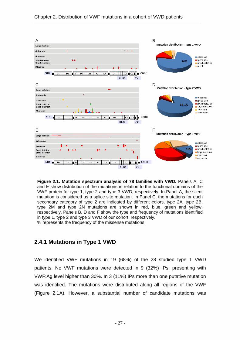
Figure 2.1. Mutation spectrum analysis of 78 famili es with VWD. Panels A, C and E show distribution of the mutations in relation to the functional domains of the VWF protein for type 1, type 2 and type 3 VWD, respectively. In Panel A, the silent mutation is considered as a splice site mutation. In Panel C, the mutations for each secondary category of type 2 are indicated by different colors, type 2A, type 2B, type 2M and type 2N mutations are shown in red, blue, green and yellow, respectively. Panels B, D and F show the type and frequency of mutations identified in type 1, type 2 and type 3 VWD of our cohort, respectively. % represents the frequency of the missense mutations.
Chapter 2. Distribution of VWF mutations in a cohort of VWD patients
- 28 -
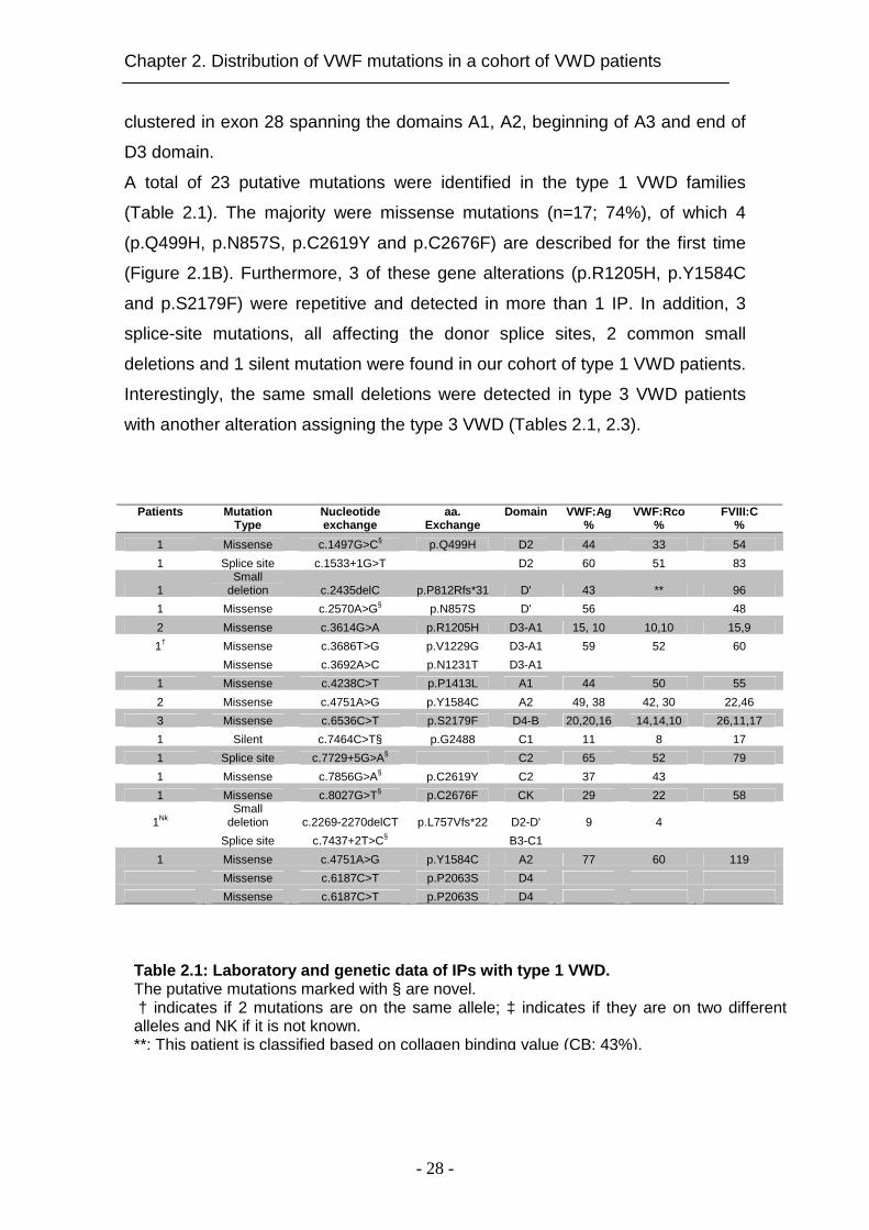
Table 2.1: Laboratory and genetic data of IPs with type 1 VWD. The putative mutations marked with § are novel. † indicates if 2 mutations are on the same allele; ‡ indicates if they are on two different alleles and NK if it is not known. **: This patient is classified based on collagen binding value (CB: 43%).
clustered in exon 28 spanning the domains A1, A2, beginning of A3 and end of
D3 domain.
A total of 23 putative mutations were identified in the type 1 VWD families
(Table 2.1). The majority were missense mutations (n=17; 74%), of which 4
(p.Q499H, p.N857S, p.C2619Y and p.C2676F) are described for the first time
(Figure 2.1B). Furthermore, 3 of these gene alterations (p.R1205H, p.Y1584C
and p.S2179F) were repetitive and detected in more than 1 IP. In addition, 3
splice-site mutations, all affecting the donor splice sites, 2 common small
deletions and 1 silent mutation were found in our cohort of type 1 VWD patients.
Interestingly, the same small deletions were detected in type 3 VWD patients
with another alteration assigning the type 3 VWD (Tables 2.1, 2.3).
Patients
Mutation Type
Nucleotide exchange
aa. Exchange
Domain
VWF:Ag %
VWF:Rco %
FVIII:C %
1 Missense c.1497G>C§ p.Q499H D2 44 33 54
1 Splice site c.1533+1G>T D2 60 51 83
1 Small
deletion c.2435delC p.P812Rfs*31 D' 43 ** 96
1 Missense c.2570A>G§ p.N857S D' 56 48
2 Missense c.3614G>A p.R1205H D3-A1 15, 10 10,10 15,9
1† Missense c.3686T>G p.V1229G D3-A1 59 52 60
Missense c.3692A>C p.N1231T D3-A1
1 Missense c.4238C>T p.P1413L A1 44 50 55
2 Missense c.4751A>G p.Y1584C A2 49, 38 42, 30 22,46
3 Missense c.6536C>T p.S2179F D4-B 20,20,16 14,14,10 26,11,17
1 Silent c.7464C>T§ p.G2488 C1 11 8 17
1 Splice site c.7729+5G>A§ C2 65 52 79
1 Missense c.7856G>A§ p.C2619Y C2 37 43
1 Missense c.8027G>T§ p.C2676F CK 29 22 58
1Nk Small
deletion c.2269-2270delCT p.L757Vfs*22 D2-D' 9 4
Splice site c.7437+2T>C§ B3-C1
1 Missense c.4751A>G p.Y1584C A2 77 60 119
Missense c.6187C>T p.P2063S D4
Missense c.6187C>T p.P2063S D4
Chapter 2. Distribution of VWF mutations in a cohort of VWD patients
- 29 -
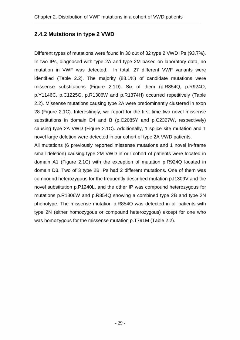
2.4.2 Mutations in type 2 VWD
Different types of mutations were found in 30 out of 32 type 2 VWD IPs (93.7%).
In two IPs, diagnosed with type 2A and type 2M based on laboratory data, no
mutation in VWF was detected. In total, 27 different VWF variants were
identified (Table 2.2). The majority (88.1%) of candidate mutations were
missense substitutions (Figure 2.1D). Six of them (p.R854Q, p.R924Q,
p.Y1146C, p.C1225G, p.R1306W and p.R1374H) occurred repetitively (Table
2.2). Missense mutations causing type 2A were predominantly clustered in exon
28 (Figure 2.1C). Interestingly, we report for the first time two novel missense
substitutions in domain D4 and B (p.C2085Y and p.C2327W, respectively)
causing type 2A VWD (Figure 2.1C). Additionally, 1 splice site mutation and 1
novel large deletion were detected in our cohort of type 2A VWD patients.
All mutations (6 previously reported missense mutations and 1 novel in-frame
small deletion) causing type 2M VWD in our cohort of patients were located in
domain A1 (Figure 2.1C) with the exception of mutation p.R924Q located in
domain D3. Two of 3 type 2B IPs had 2 different mutations. One of them was
compound heterozygous for the frequently described mutation p.I1309V and the
novel substitution p.P1240L, and the other IP was compound heterozygous for
mutations p.R1306W and p.R854Q showing a combined type 2B and type 2N
phenotype. The missense mutation p.R854Q was detected in all patients with
type 2N (either homozygous or compound heterozygous) except for one who
was homozygous for the missense mutation p.T791M (Table 2.2).
Chapter 2. Distribution of VWF mutations in a cohort of VWD patients
- 30 -
Patients
Type 2 categories
Mutation Type
Nucleotide exchange
aa. exchange
Domain
VWF:Ag %
VWF:Rco %
FVIII:C %
4 2A Missense c.3437A>G p.Y1146C D3-A1 17-33 <10-18 21-32
1 2A Splice site c.3538+1G>A D3 11 6 10
2 2A Missense c.3673T>G p.C1225G D3-A1 3 <6 5-35
(atypical 2N) Missense c.3673T>G p.C1225G D3-A1
1 2A Missense c.3863T>G p.L1288R D3-A1 33 15 30
1 2A Missense c.4571T>G§ p.V1524G A2 90 25 45
1 2A Missense c.4789C>T p.R1597W A2 25 13 20
1 2A Missense c.4883T>C p.I1628T A2 20 8 39
1 2A Large deletion Exons 33-34 deletion§ A3-D4 17 9 1.9
1 2A Missense c.6254G>A§ p.C2085Y D4 22 13 42
1 2A Missense c.6981T>G§ p.C2327W B 13 7 22
1 2A Missense c.4121G>A p.R1374H A1 12 <6 27
1NK 2A Missense c.4121G>A p.R1374H A1 42 17 46
Missense c.2220G>A p.M740I D2-D'
1‡ 2A Missense c.4121G>A p.R1374H A1 39 <6 22
Missense c.2771G>A p.R924Q D3
1 2B Missense c.3916C>T p.R1306W A1 50 26 57
1NK 2B Missense c.3916C>T p.R1306W A1 50 24 29
Missense c.2561G>A p.R854Q D'
1‡ 2B Missense c.3925A>G p.I1309V A1 75 56 55
Missense c.3719C>T§ p.P1240L D3-A1
1 2M Missense c.3943C>G p.R1315G A1 25 11 37
1‡ 2M Small Deletion c.3964_3966delCAC§ p.H1322del A1 21 8 19
Missense c.2771G>A p.R924Q D3
1 2M Missense c.4120C>T p.R1374C A1 22 11 38
1 2M Missense c.4121G>T p.R1374L A1 20 7 20
1 2M Missense c.4105T>A p.F1369I A1 30 15 30
1 2M Missense c.4225G>T p.V1409F A1 27 <6 23
Chapter 2. Distribution of VWF mutations in a cohort of VWD patients
- 31 -
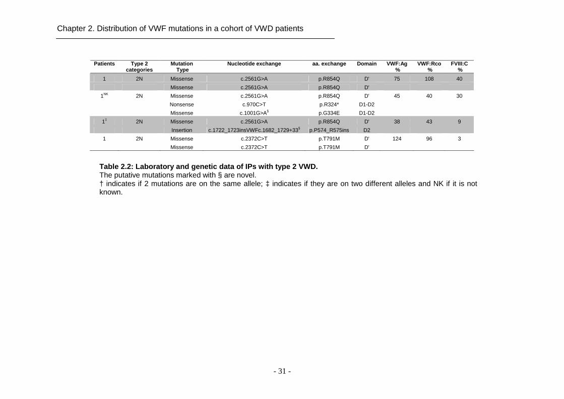
Table 2.2: Laboratory and genetic data of IPs with type 2 VWD. The putative mutations marked with § are novel. † indicates if 2 mutations are on the same allele; ‡ indicates if they are on two different alleles and NK if it is not known.
Patients
Type 2 categories
Mutation Type
Nucleotide exchange
aa. exchange
Domain
VWF:Ag %
VWF:Rco %
FVIII:C %
1 2N Missense c.2561G>A p.R854Q D' 75 108 40
Missense c.2561G>A p.R854Q D'
1NK 2N Missense c.2561G>A p.R854Q D' 45 40 30
Nonsense c.970C>T p.R324* D1-D2
Missense c.1001G>A§ p.G334E D1-D2
1‡ 2N Missense c.2561G>A p.R854Q D' 38 43 9
Insertion c.1722_1723insVWFc.1682_1729+33§ p.P574_R575ins D2
1 2N Missense c.2372C>T p.T791M D' 124 96 3
Missense c.2372C>T p.T791M D'
Chapter 2. Distribution of VWF mutations in a cohort of VWD patients
- 32 -
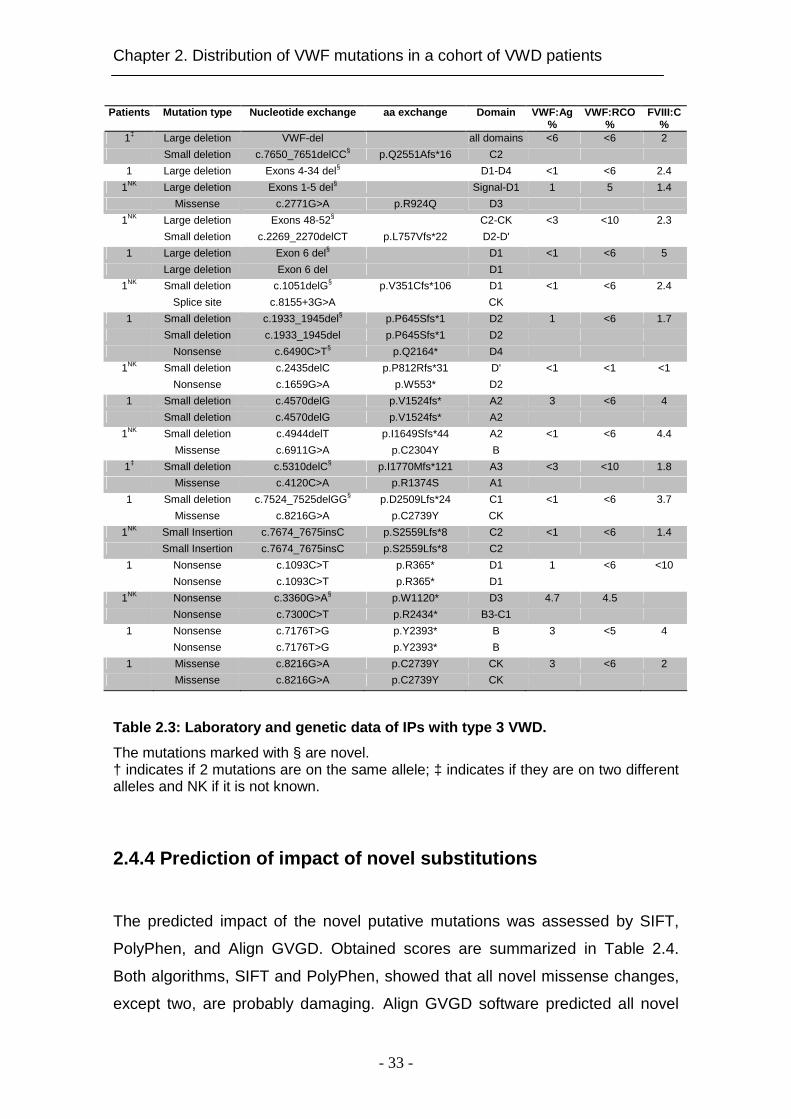
2.4.3 Mutations in type 3 VWD
We could detect mutations in 17 of 18 IPs (94.4%). All IPs except one were
homozygous or compound heterozygous for given mutations (Table 2.3). In
total, we identified 26 different VWF mutations in type 3 VWD families. The
majority of mutations (82%) were null mutations (Figure 2.1F) resulting in
severely decreased VWF:Ag levels. In total, we found 4 previously reported
missense mutations, 5 large deletions (4 novel), 9 small deletions (5 novel), 1
insertion, 1 splice site mutation and 6 nonsense mutations (2 novel) (Figure
2.1E). All small deletions detected in our cohort caused a shift of the reading
frame and determined a premature termination codon. The small deletions
c.2435delC and c.7650-7651delCC were located in a stretch of cytosine
residues while c.7524-7525delGG was in a stretch of guanine residues. One
previously described cytosine insertion in a stretch of four cytosines (c.7671-
7674) in exon 45 was detected (c.7674_7675insC) in a homozygous status.
Chapter 2. Distribution of VWF mutations in a cohort of VWD patients
- 33 -
Table 2.3: Laboratory and genetic data of IPs with type 3 VWD.
The mutations marked with § are novel. † indicates if 2 mutations are on the same allele; ‡ indicates if they are on two different alleles and NK if it is not known.
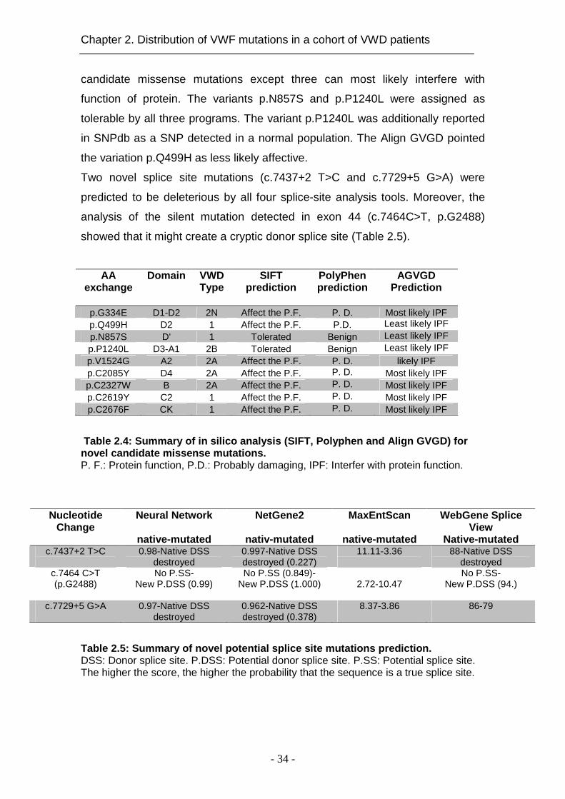
2.4.4 Prediction of impact of novel substitutions
The predicted impact of the novel putative mutations was assessed by SIFT,
PolyPhen, and Align GVGD. Obtained scores are summarized in Table 2.4.
Both algorithms, SIFT and PolyPhen, showed that all novel missense changes,
except two, are probably damaging. Align GVGD software predicted all novel
Patients Mutation type Nucleotide exchange aa exchange Domai n VWF:Ag %
VWF:RCO %
FVIII:C %
1‡ Large deletion VWF-del all domains <6 <6 2
Small deletion c.7650_7651delCC§ p.Q2551Afs*16 C2
1 Large deletion Exons 4-34 del§ D1-D4 <1 <6 2.4
1NK Large deletion Exons 1-5 del§ Signal-D1 1 5 1.4
Missense c.2771G>A p.R924Q D3
1NK Large deletion Exons 48-52§ C2-CK <3 <10 2.3
Small deletion c.2269_2270delCT p.L757Vfs*22 D2-D'
1 Large deletion Exon 6 del§ D1 <1 <6 5
Large deletion Exon 6 del D1
1NK Small deletion c.1051delG§ p.V351Cfs*106 D1 <1 <6 2.4
Splice site c.8155+3G>A CK
1 Small deletion c.1933_1945del§ p.P645Sfs*1 D2 1 <6 1.7
Small deletion c.1933_1945del p.P645Sfs*1 D2
Nonsense c.6490C>T§ p.Q2164* D4
1NK Small deletion c.2435delC p.P812Rfs*31 D' <1 <1 <1
Nonsense c.1659G>A p.W553* D2
1 Small deletion c.4570delG p.V1524fs* A2 3 <6 4
Small deletion c.4570delG p.V1524fs* A2
1NK Small deletion c.4944delT p.I1649Sfs*44 A2 <1 <6 4.4
Missense c.6911G>A p.C2304Y B
1‡ Small deletion c.5310delC§ p.I1770Mfs*121 A3 <3 <10 1.8
Missense c.4120C>A p.R1374S A1
1 Small deletion c.7524_7525delGG§ p.D2509Lfs*24 C1 <1 <6 3.7
Missense c.8216G>A p.C2739Y CK
1NK Small Insertion c.7674_7675insC p.S2559Lfs*8 C2 <1 <6 1.4
Small Insertion c.7674_7675insC p.S2559Lfs*8 C2
1 Nonsense c.1093C>T p.R365* D1 1 <6 <10
Nonsense c.1093C>T p.R365* D1
1NK Nonsense c.3360G>A§ p.W1120* D3 4.7 4.5
Nonsense c.7300C>T p.R2434* B3-C1
1 Nonsense c.7176T>G p.Y2393* B 3 <5 4
Nonsense c.7176T>G p.Y2393* B
1 Missense c.8216G>A p.C2739Y CK 3 <6 2
Missense c.8216G>A p.C2739Y CK
Chapter 2. Distribution of VWF mutations in a cohort of VWD patients
- 34 -
candidate missense mutations except three can most likely interfere with
function of protein. The variants p.N857S and p.P1240L were assigned as
tolerable by all three programs. The variant p.P1240L was additionally reported
in SNPdb as a SNP detected in a normal population. The Align GVGD pointed
the variation p.Q499H as less likely affective. Two novel splice site mutations (c.7437+2 T>C and c.7729+5 G>A) were
predicted to be deleterious by all four splice-site analysis tools. Moreover, the
analysis of the silent mutation detected in exon 44 (c.7464C>T, p.G2488)
showed that it might create a cryptic donor splice site (Table 2.5).
AA exchange
Domain
VWD Type
SIFT prediction
PolyPhen prediction
AGVGD Prediction
p.G334E D1-D2 2N Affect the P.F. P. D. Most likely IPF p.Q499H D2 1 Affect the P.F. P.D. Least likely IPF p.N857S D' 1 Tolerated Benign Least likely IPF p.P1240L D3-A1 2B Tolerated Benign Least likely IPF p.V1524G A2 2A Affect the P.F. P. D. likely IPF p.C2085Y D4 2A Affect the P.F. P. D. Most likely IPF p.C2327W B 2A Affect the P.F. P. D. Most likely IPF p.C2619Y C2 1 Affect the P.F. P. D. Most likely IPF p.C2676F CK 1 Affect the P.F. P. D. Most likely IPF
Table 2.4: Summary of in silico analysis (SIFT, Pol yphen and Align GVGD) for novel candidate missense mutations. P. F.: Protein function, P.D.: Probably damaging, IPF: Interfer with protein function.
Nucleotide Change
Neural Network
native-mutated
NetGene2
nativ-mutated
MaxEntScan
native-mutated
WebGene Splice View
Native-mutated c.7437+2 T>C
0.98-Native DSS
destroyed 0.997-Native DSS destroyed (0.227)
11.11-3.36
88-Native DSS destroyed
c.7464 C>T (p.G2488)
No P.SS- New P.DSS (0.99)
No P.SS (0.849)- New P.DSS (1.000)
2.72-10.47
No P.SS- New P.DSS (94.)
c.7729+5 G>A
0.97-Native DSS
destroyed 0.962-Native DSS destroyed (0.378)
8.37-3.86
86-79
Table 2.5: Summary of novel potential splice site m utations prediction. DSS: Donor splice site. P.DSS: Potential donor splice site. P.SS: Potential splice site. The higher the score, the higher the probability that the sequence is a true splice site.
Chapter 2. Distribution of VWF mutations in a cohort of VWD patients
- 35 -
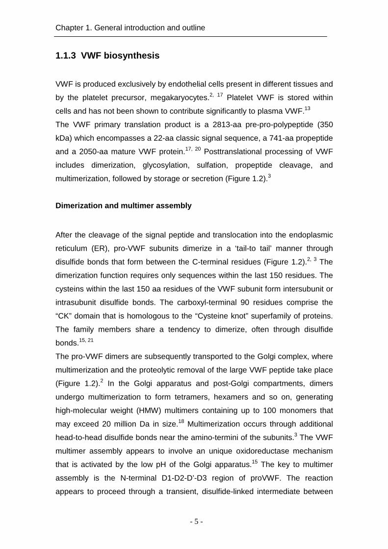
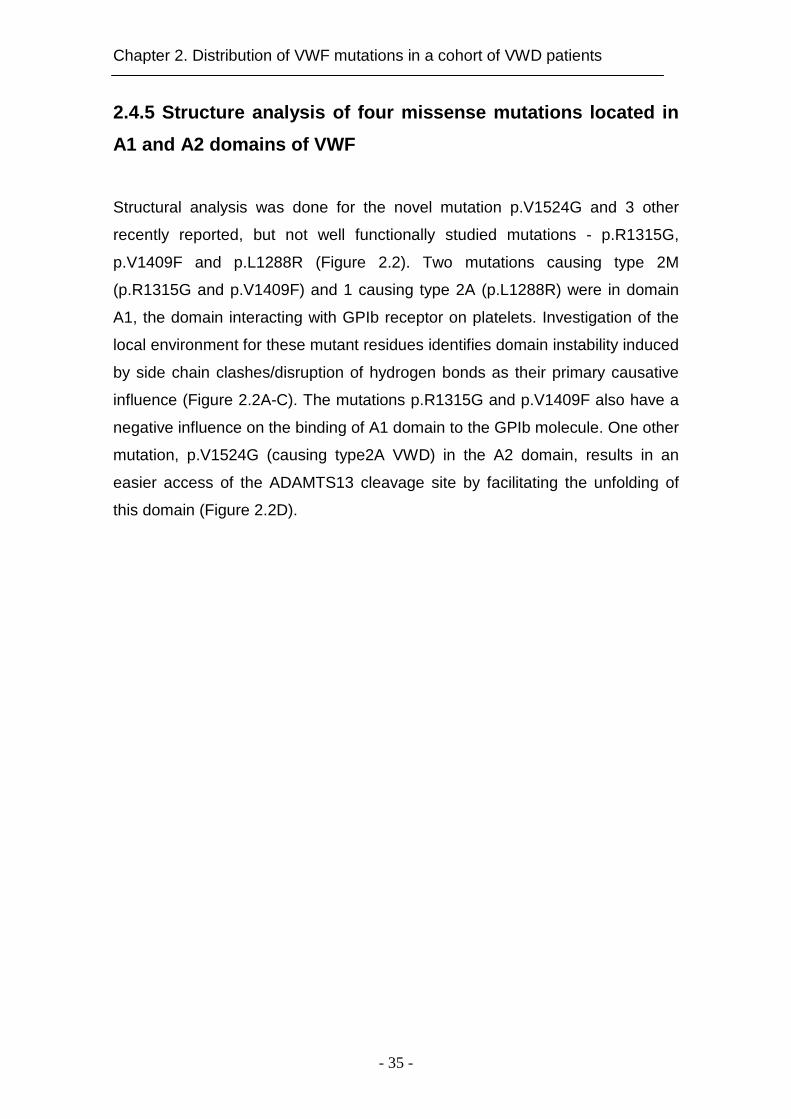
2.4.5 Structure analysis of four missense mutations located in
A1 and A2 domains of VWF
Structural analysis was done for the novel mutation p.V1524G and 3 other
recently reported, but not well functionally studied mutations - p.R1315G,
p.V1409F and p.L1288R (Figure 2.2). Two mutations causing type 2M
(p.R1315G and p.V1409F) and 1 causing type 2A (p.L1288R) were in domain
A1, the domain interacting with GPIb receptor on platelets. Investigation of the
local environment for these mutant residues identifies domain instability induced
by side chain clashes/disruption of hydrogen bonds as their primary causative
influence (Figure 2.2A-C). The mutations p.R1315G and p.V1409F also have a
negative influence on the binding of A1 domain to the GPIb molecule. One other
mutation, p.V1524G (causing type2A VWD) in the A2 domain, results in an
easier access of the ADAMTS13 cleavage site by facilitating the unfolding of
this domain (Figure 2.2D).
Chapter 2. Distribution of VWF mutations in a cohort of VWD patients
- 36 -
Figure 2.2. Close-up views of the local molecular e nvironments of four human VWFA1 and VWFA2 domain missense mutations depicted on structural models of VWFA1 /GPIb and VWFA2 domains (PDB ID: 1M10; res olution: 3.10A °°°° and PDB ID: 1AUQ; resolution: 2.30A °°°° respectively). The figure is divided up into four panels representing four different mutations. Panel A, B and C depict mutations in VWFA1 domain while Panel D corresponds to mutation in VWFA2 domain. Panel A. The panel is split into three images. The top image depicts the interface between the VWFA1 domain and GPIb molecule. The contacts (H-bonds) across the interface are represented by red lines. The affected residue is depicted as a dark blue CPK sphere. The image on the left shows the H-bonds formed by the wild type p.R1315 residue. The image on the right shows the H-bonds formed by the mutated p.G1315 residue. Both residues are depicted as ball and stick models. No structure is apparent for the side-chain of glycine since it is a single hydrogen atom (not resolved in the 3.10 Å X-ray crystallographic data). The backbone of the VWFA1 domain and GPIb molecule are represented as grey ribbons respectively. The effective surface of these residues is depicted in magenta. Panel B. The panel is split into two images. The image on the left shows the wild type p.V1409 residue and the image on the right shows the mutated p.F1409 residue. The backbone of the VWFA1 domain and GPIb molecule are represented as grey ribbons. The residues are represented as CPK spheres. The red lines which are indicated with a black arrow depict clashes with p.I617 and p.A618. Panel C. The panel is split into two images. The residues of interest are represented as dark blue sticks. The left image depicts local environment of the wild type p.L1288 residue. The positive charge on the residues proximal to the affected p.L1288 are represented in red surface colors while the neutral charge of the p.L1288 residue is depicted in green. The right image represents the mutated p.R1288 residue which brings another positive charge in proximity to the other two charged residues giving a reddish hue to the original green colored surface. Panel D p.V1524 (red sticks) is shown near the hydrophobic plug formed by the vicinal disulphides (green ribbon and stick) and 8 other hydrophilic residues (deep blue sticks). The backbone of the VWFA1 domain is represented as a turquoise ribbon.
Chapter 2. Distribution of VWF mutations in a cohort of VWD patients
- 37 -
2.5 Discussion In the present cohort, we investigated VWD patients for variations within VWF
to elucidate the molecular basis for each type in order to enhance
understanding of the disorder. The majority of mutant alleles (74% vs 88.1%)
identified in our patients with type 1 and type 2 VWD were missense mutations,
while null alleles were predominant (82%) in patients with type 3 VWD (Figure
2.1 B, D and F). The sequence variants identified in our cohort of type 1 and
type 3 VWD patients spread throughout the whole VWF, but as expected, were
confined to a particular region of the VWF in the different types of our patients
with type 2 VWD (Figure 2.1A, C and E). These findings are in line with
previous reports.1, 51, 73-75 The VWF:Ag level was variable in our cohort of type 1 VWD patients. A mean
VWF:Ag of 39.5% was registered, ranging from 9% to 77%. Our data showed a
correlation between VWF:Ag level and mutation detection rate. In all patients
with type 1 VWD and VWF:Ag level less than 30% mutations were identified. In
contrast, in only 53% of patients a mutation was detected when VWF:Ag was
higher than 37%. Our data supported the finding of other groups,73, 74 indicating
that in milder cases of VWD, the genetic determinants are more complex and
might involve other factors outside the VWF. In our study, VWD type 1 patients
bearing p.S2179F and p.R1205H expressed the lowest VWF antigen levels (10-
20%) compared to the antigen levels of patients with other missense mutations.
These data are in concordance with the literature.62-64, 76, 77
Although the pattern of mutations found in type 3 VWD patients was similar to
previous studies, only few of the mutations identified so far have been found
repetitively in analogous studies from different populations.78-80 The presence of
11 novel mutations in 17 IPs diagnosed as type 3 VWD in our cohort shows a
high degree of genetic heterogeneity in the molecular pathology of type 3 VWD.
VWD is known for a high degree of variability in clinical presentation and a
considerable heterogeneity of its molecular basis. It has been shown that within
the phenotypic classification of VWD, variations within a single mutation can be
responsible for different types of the disease. Thus, some substitutions are
discussed controversially. The missense mutations affecting p.R1374 is
Chapter 2. Distribution of VWF mutations in a cohort of VWD patients
- 38 -
presented with different variants depending on the nucleotide substitution. In
our study the variant p.R1374H led to type 2A VWD. Two other variants of
p.R1374, p.R1374L and p.R1374C, were considered to be responsible for type
2M VWD (Table 2.2). Although the ISTH registry of VWD assigns these variants
as unclassified (U), they have been classified by several investigators into
different VWD types due to the patient´s pleiotropic phenotype.1, 75, 81-83
In some cases an equal mutation contributes to expression of different
phenotypes. The variant p.R924Q is listed as a polymorphism, as a type 2N
mutation and also as a type 1 mutation in the ISTH SSC VWF database. In our
study this gene alteration was associated with type 2M (2 patients) and type 3
(1 Patient) VWD. However, p.R924Q was found as a second causative mutation
in a patient with type 3 VWD. Berber et al. demonstrated through in vitro
expression studies that the p.R924Q variation does not significantly affect
biosynthesis of VWF, but they suggested that this variant allele may mark a null
allele in some instances.84 Hickson et al also reported that its inheritance alone
may be insufficient for VWD diagnosis, but it appears to be associated with a
further VWF level reduction in individuals with a second VWF mutation.85
Therefore, we cannot exclude the possibility of a second mutation in intronic
regions or outside of the VWF for the IP with type 3 VWD.
The variant p.P2063S is listed in the ISTH SSC VWF database as a type 3
mutation, and it has been identified in Canadian and Spanish populations in
type 1 VWD patients.63, 82 Moreover, p.P2063S is found in a Spanish population
in compound heterozygous state with p.V1409F in a type 2M patient with Blood
group A, VWF:Ag 9% and VWF:RCo 3%.82 The patient heterozygous for
p.V1409F variant with Blood group A classified as type 2M in our cohort shows
VWF:Ag 27%, VWF:RCo <6. Comparison of the laboratory data of these 2
patients indicates a possible influence of p.P2063S as a second variant for the
quantitative defect in the Spanish patient. However, the functional study of this
substitution by Eikenboom et al. showed that p.P2063S is possibly a
polymorphism.86 In our cohort, p.P2063S was detected in homozygous state in
combination with p.Y1584C in a patient with type 1 VWD exhibiting VWF:Ag
level of 77% and VWF:RCo of 60%. Since p.Y1584C is also known as a variant
Chapter 2. Distribution of VWF mutations in a cohort of VWD patients
- 39 -
with incomplete penetrance, determination of causative mutation explaining the
phenotype of this patient is ambiguous.
An interesting mutation is p.C1225G listed as a type 2N mutation in the ISTH
SSc database. In our cohort the patients carrying p.C1225G are classified as
type 2A VWD owing to relative reduction of big multimers and normal values of
VWF:FVIIIB (in one patient). However, Allen et al. classified this mutation as an
atypical type 2N mutation.87 They discovered by in-vitro expression of the
recombinant VWF that this mutation affects the level of secreted VWF, the
binding of VWF to factor VIII and the ability of VWF to form multimers. The
pleiotropic effects of this mutation on the VWF molecule make its classification
complicated.
The novel variant p.N857S is predicted to be benign by all predictive tools, but it
is one of the twelve N-linked glycosylation sites of the mature VWF protein.88
Therefore, its causality cannot be excluded prior to expression studies. In total,
we found 5 splice site mutations and 1 silent mutation. The previous studies
have proved the causality of 3 of these splice site mutations by mRNA
analysis.77, 89 Both, two novel splice site mutations and 1 novel silent mutation,
were predicted to be causative by all splice-site analysis tools used in our study.
Structural interpretation
The A1 domain structure can be divided into six unique faces or fronts.70 The
p.R1315 residue is located on a beta strand from the previously defined “lower
face”. It is part of an elaborate set of domain stabilizing salt bridge networks and
forms hydrogen bonds with an adjacent strand and a loop. Mutation to a much
smaller glycine residue would result in the disruption of these H-bonds resulting
in domain instability (Figure 2.2A). Additionally, since the confirmation of this
region is essential for binding to platelets, the altered domain structure would
also negatively influence the same. The mutation p.V1409F is also situated in
proximity of this surface and the resulting longer Phenylalanine side chain may
result in side chain clashes and disturb the binding of this surface to the Gp1b
molecule (Figure 2.2B). The mutation p.L1288R is located on the previously
defined “upper face” of the A1 domain which is uncharged surface of approx.
Chapter 2. Distribution of VWF mutations in a cohort of VWD patients
- 40 -
20A° diameter with charged residues only at the periphery.70 The only charged
residues on this surface are the p.R1287 and p.H1419 (Figure 2.2C). The
mutation of p.L1288 to a large positively charged arginine residue over this
surface will destabilize this region and result in an unstable molecule. The
mutation also disrupts a domain stabilizing salt bridge network (loss of H-bonds
to a connecting loop and helix) resulting in an unstable A1 domain.
The shear induced unfolding of the VWF A2 domain to expose the p.Y1605-
p.M1606 ADAMTS13 cleavage site, is a well investigated phenomena.90 In the
A2 domain crystal structure 3GXB, the p.V1524 residue is proximal to a
hydrophobic plug formed by the hydrophobic interaction of the vicinal disulfides
and eight residues which forms the first high energy barrier against the
unfolding (entry of water molecule) of the A2 domain prior to the final rate
limiting step (Figure 2.2D).90 The mutant polar p.G1524 residue in the proximity
of this hydrophobic plug will reduce the energy barrier for this step and result in
easier access to the ADAMTS13 cleavage site.
In conclusion, our study extends the mutational spectrum of VWF, and improves
the knowledge on the genetic basis of different types of VWD. This data
provides the opportunity to possibly design functional studies to understand
molecular mechanisms underlying this disease and establish genotype-
phenotype correlations for proper classification and improving clinical
management. Additionally, our study also shows that the combination of
mutations causing different types of VWD within one patient causes complex
phenotypes. Finally, the availability of well defined crystal structures for some of
the VWF domains is useful in understanding the molecular etiology of some of
the variants and their impact on protein structure. The understanding of the
molecular etiology of these variants is crucial in determining their causality and
therefore establishing their credentials as mutations or mere neutral
polymorphisms.
Chapter 3. Identification of large deletions in VWD
- 41 -
Chapter 3
Large deletions identified in patients with von
Willebrand disease using multiple ligation-dependen t
probe amplification
Adapted from
Yadegari H., Driesen J., Hass M., Budde U., ,Pavlova A., and Oldenburg J.
Journal of Thrombosis and haemostasis. 2011;9:1083-1086
Chapter 3. Identification of large deletions in VWD
- 42 -
3.1 Abstract
Large deletions are considered as being a rare cause of VWD. In the VWF
database only 12 large deletions are reported ranging in size from a single exon
to the entire gene. The aim of our study was screening for large deletions of
VWF in VWD patients where direct sequencing of VWF failed to reveal
causative VWF gene mutations or showed homozygousity for a given mutation
and polymorphisms. Twenty-three patients were investigated, including 11
patients with type 1, 3 patients with type 2 and 9 patients with type 3 VWD.
Genomic DNA was isolated by standard methods and investigation for large
deletions was performed by MLPA. Six large deletions were identified in our
cohort of which five were not previously described. Five large gene deletions
were detected in patients with type 3 VWD and one in type 2 VWD. A
homozygous deletion of exon 6 was identified in 2 patients with type 3 VWD
belonging to the same family. The deletion segregates within the family and was
detected in another four family members in heterozygous state. The deletions
spanning the exons 1-5, whole gene and exons 48-52 were combined with a
second mutation affecting the other allele – a missense mutation, a small
deletion in exon 45, or a small deletion in exon 17, respectively. In the patient
with the heterozygous large deletion of exon 4-34, no second mutation was
identified. Interestingly a large deletion of exons 33-34 of VWF along with a
missense mutation in FVIII gene (F8) was detected in a combined type 2A
VWD/hemophilia A patient. Our results indicate that investigation for presence
of heterozygous large deletions should be taken into consideration when no
causative mutations are identified, or homozygous polymorphisms and
homozygous mutations are detected.
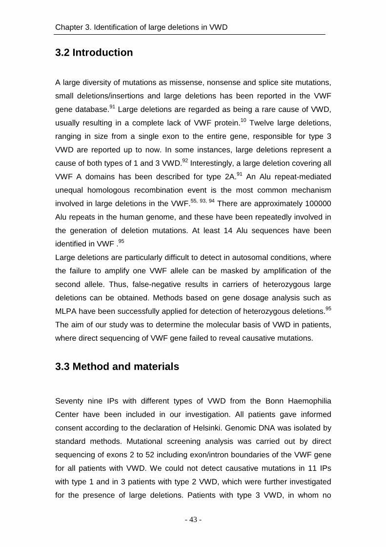
Chapter 3. Identification of large deletions in VWD
- 43 -
3.2 Introduction
A large diversity of mutations as missense, nonsense and splice site mutations,
small deletions/insertions and large deletions has been reported in the VWF
gene database.91 Large deletions are regarded as being a rare cause of VWD,
usually resulting in a complete lack of VWF protein.10 Twelve large deletions,
ranging in size from a single exon to the entire gene, responsible for type 3
VWD are reported up to now. In some instances, large deletions represent a
cause of both types of 1 and 3 VWD.92 Interestingly, a large deletion covering all
VWF A domains has been described for type 2A.91 An Alu repeat-mediated
unequal homologous recombination event is the most common mechanism
involved in large deletions in the VWF.55, 93, 94 There are approximately 100000
Alu repeats in the human genome, and these have been repeatedly involved in
the generation of deletion mutations. At least 14 Alu sequences have been
identified in VWF .95
Large deletions are particularly difficult to detect in autosomal conditions, where
the failure to amplify one VWF allele can be masked by amplification of the
second allele. Thus, false-negative results in carriers of heterozygous large
deletions can be obtained. Methods based on gene dosage analysis such as
MLPA have been successfully applied for detection of heterozygous deletions.95
The aim of our study was to determine the molecular basis of VWD in patients,
where direct sequencing of VWF gene failed to reveal causative mutations.
3.3 Method and materials
Seventy nine IPs with different types of VWD from the Bonn Haemophilia
Center have been included in our investigation. All patients gave informed
consent according to the declaration of Helsinki. Genomic DNA was isolated by
standard methods. Mutational screening analysis was carried out by direct
sequencing of exons 2 to 52 including exon/intron boundaries of the VWF gene
for all patients with VWD. We could not detect causative mutations in 11 IPs
with type 1 and in 3 patients with type 2 VWD, which were further investigated
for the presence of large deletions. Patients with type 3 VWD, in whom no
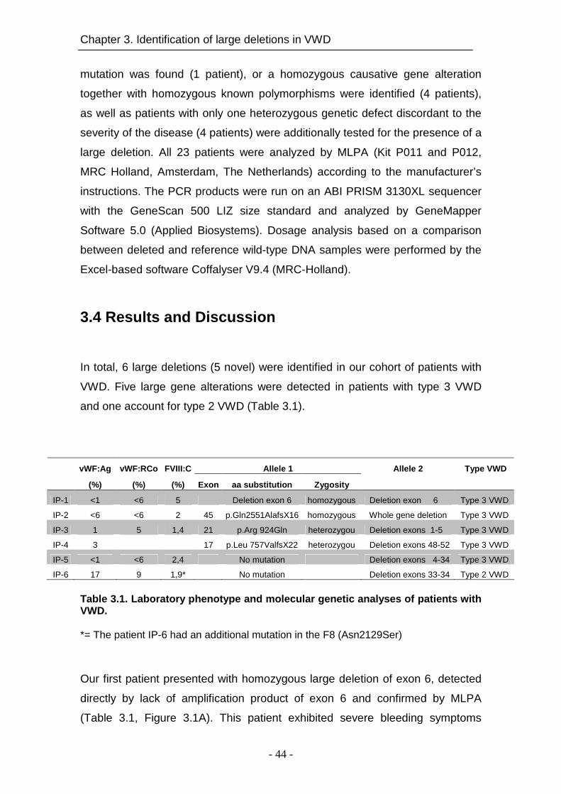
Chapter 3. Identification of large deletions in VWD
- 44 -
mutation was found (1 patient), or a homozygous causative gene alteration
together with homozygous known polymorphisms were identified (4 patients),
as well as patients with only one heterozygous genetic defect discordant to the
severity of the disease (4 patients) were additionally tested for the presence of a
large deletion. All 23 patients were analyzed by MLPA (Kit P011 and P012,
MRC Holland, Amsterdam, The Netherlands) according to the manufacturer’s
instructions. The PCR products were run on an ABI PRISM 3130XL sequencer
with the GeneScan 500 LIZ size standard and analyzed by GeneMapper
Software 5.0 (Applied Biosystems). Dosage analysis based on a comparison
between deleted and reference wild-type DNA samples were performed by the
Excel-based software Coffalyser V9.4 (MRC-Holland).
3.4 Results and Discussion
In total, 6 large deletions (5 novel) were identified in our cohort of patients with
VWD. Five large gene alterations were detected in patients with type 3 VWD
and one account for type 2 VWD (Table 3.1).
Table 3.1. Laboratory phenotype and molecular genet ic analyses of patients with VWD. *= The patient IP-6 had an additional mutation in the F8 (Asn2129Ser)
Our first patient presented with homozygous large deletion of exon 6, detected
directly by lack of amplification product of exon 6 and confirmed by MLPA
(Table 3.1, Figure 3.1A). This patient exhibited severe bleeding symptoms
vWF:Ag vWF:RCo FVIII:C Allele 1 Allele 2 Type VWD
(%) (%) (%) Exon aa substitution Zygosity
IP-1 <1 <6 5 Deletion exon 6 homozygous Deletion exon 6 Type 3 VWD
IP-2 <6 <6 2 45 p.Gln2551AlafsX16 homozygous Whole gene deletion Type 3 VWD
IP-3 1 5 1,4 21 p.Arg 924Gln heterozygou Deletion exons 1-5 Type 3 VWD
IP-4 3 17 p.Leu 757ValfsX22 heterozygou Deletion exons 48-52 Type 3 VWD
IP-5 <1 <6 2,4 No mutation Deletion exons 4-34 Type 3 VWD
IP-6 17 9 1,9* No mutation Deletion exons 33-34 Type 2 VWD
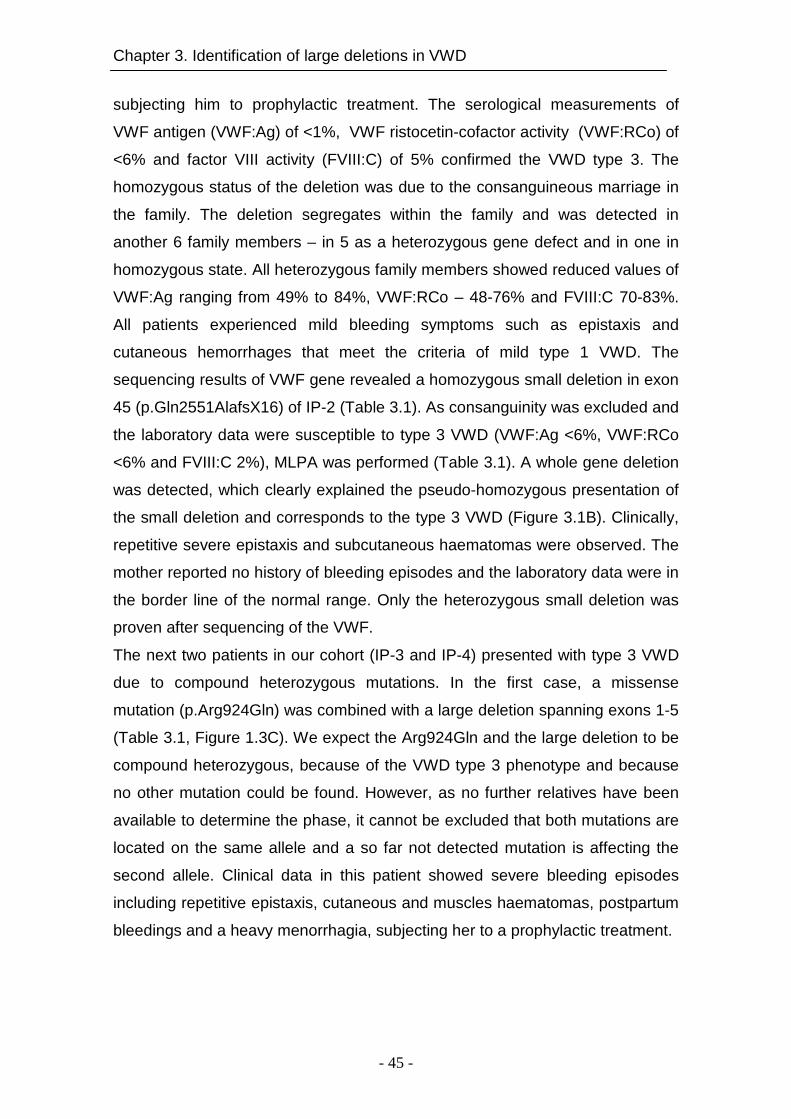
Chapter 3. Identification of large deletions in VWD
- 45 -
subjecting him to prophylactic treatment. The serological measurements of
VWF antigen (VWF:Ag) of <1%, VWF ristocetin-cofactor activity (VWF:RCo) of
<6% and factor VIII activity (FVIII:C) of 5% confirmed the VWD type 3. The
homozygous status of the deletion was due to the consanguineous marriage in
the family. The deletion segregates within the family and was detected in
another 6 family members – in 5 as a heterozygous gene defect and in one in
homozygous state. All heterozygous family members showed reduced values of
VWF:Ag ranging from 49% to 84%, VWF:RCo – 48-76% and FVIII:C 70-83%.
All patients experienced mild bleeding symptoms such as epistaxis and
cutaneous hemorrhages that meet the criteria of mild type 1 VWD. The
sequencing results of VWF gene revealed a homozygous small deletion in exon
45 (p.Gln2551AlafsX16) of IP-2 (Table 3.1). As consanguinity was excluded and
the laboratory data were susceptible to type 3 VWD (VWF:Ag <6%, VWF:RCo
<6% and FVIII:C 2%), MLPA was performed (Table 3.1). A whole gene deletion
was detected, which clearly explained the pseudo-homozygous presentation of
the small deletion and corresponds to the type 3 VWD (Figure 3.1B). Clinically,
repetitive severe epistaxis and subcutaneous haematomas were observed. The
mother reported no history of bleeding episodes and the laboratory data were in
the border line of the normal range. Only the heterozygous small deletion was
proven after sequencing of the VWF.
The next two patients in our cohort (IP-3 and IP-4) presented with type 3 VWD
due to compound heterozygous mutations. In the first case, a missense
mutation (p.Arg924Gln) was combined with a large deletion spanning exons 1-5
(Table 3.1, Figure 1.3C). We expect the Arg924Gln and the large deletion to be
compound heterozygous, because of the VWD type 3 phenotype and because
no other mutation could be found. However, as no further relatives have been
available to determine the phase, it cannot be excluded that both mutations are
located on the same allele and a so far not detected mutation is affecting the
second allele. Clinical data in this patient showed severe bleeding episodes
including repetitive epistaxis, cutaneous and muscles haematomas, postpartum
bleedings and a heavy menorrhagia, subjecting her to a prophylactic treatment.
Chapter 3. Identification of large deletions in VWD
- 46 -
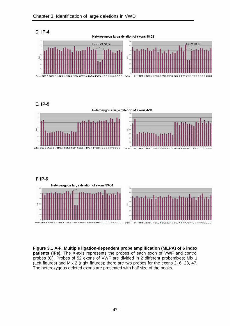
Chapter 3. Identification of large deletions in VWD
- 47 -
Figure 3.1 A-F. Multiple ligation-dependent probe a mplification (MLPA) of 6 index patients (IPs). The X-axis represents the probes of each exon of VWF and control probes (C). Probes of 52 exons of VWF are divided in 2 different probemixes; Mix 1 (Left figures) and Mix 2 (right figures); there are two probes for the exons 2, 6, 28, 47. The heterozygous deleted exons are presented with half size of the peaks.
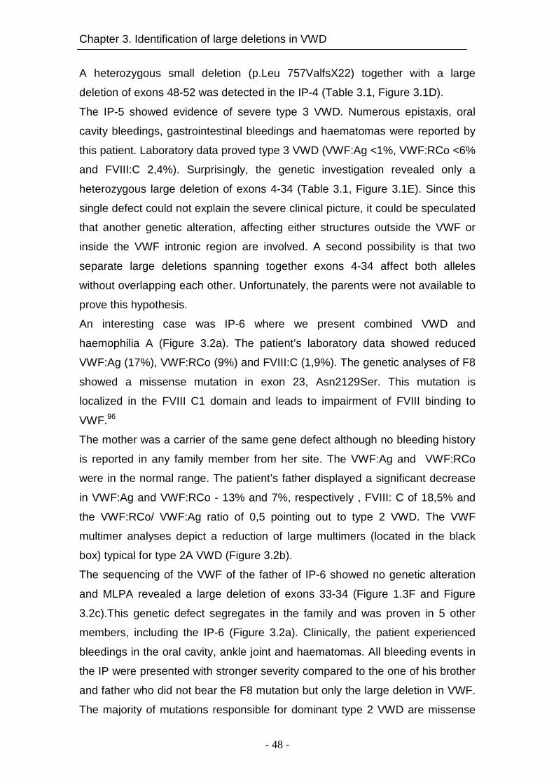
Chapter 3. Identification of large deletions in VWD
- 48 -
A heterozygous small deletion (p.Leu 757ValfsX22) together with a large
deletion of exons 48-52 was detected in the IP-4 (Table 3.1, Figure 3.1D).
The IP-5 showed evidence of severe type 3 VWD. Numerous epistaxis, oral
cavity bleedings, gastrointestinal bleedings and haematomas were reported by
this patient. Laboratory data proved type 3 VWD (VWF:Ag <1%, VWF:RCo <6%
and FVIII:C 2,4%). Surprisingly, the genetic investigation revealed only a
heterozygous large deletion of exons 4-34 (Table 3.1, Figure 3.1E). Since this
single defect could not explain the severe clinical picture, it could be speculated
that another genetic alteration, affecting either structures outside the VWF or
inside the VWF intronic region are involved. A second possibility is that two
separate large deletions spanning together exons 4-34 affect both alleles
without overlapping each other. Unfortunately, the parents were not available to
prove this hypothesis.
An interesting case was IP-6 where we present combined VWD and
haemophilia A (Figure 3.2a). The patient’s laboratory data showed reduced
VWF:Ag (17%), VWF:RCo (9%) and FVIII:C (1,9%). The genetic analyses of F8
showed a missense mutation in exon 23, Asn2129Ser. This mutation is
localized in the FVIII C1 domain and leads to impairment of FVIII binding to
VWF.96
The mother was a carrier of the same gene defect although no bleeding history
is reported in any family member from her site. The VWF:Ag and VWF:RCo
were in the normal range. The patient’s father displayed a significant decrease
in VWF:Ag and VWF:RCo - 13% and 7%, respectively , FVIII: C of 18,5% and
the VWF:RCo/ VWF:Ag ratio of 0,5 pointing out to type 2 VWD. The VWF
multimer analyses depict a reduction of large multimers (located in the black
box) typical for type 2A VWD (Figure 3.2b).
The sequencing of the VWF of the father of IP-6 showed no genetic alteration
and MLPA revealed a large deletion of exons 33-34 (Figure 1.3F and Figure
3.2c).This genetic defect segregates in the family and was proven in 5 other
members, including the IP-6 (Figure 3.2a). Clinically, the patient experienced
bleedings in the oral cavity, ankle joint and haematomas. All bleeding events in
the IP were presented with stronger severity compared to the one of his brother
and father who did not bear the F8 mutation but only the large deletion in VWF.
The majority of mutations responsible for dominant type 2 VWD are missense
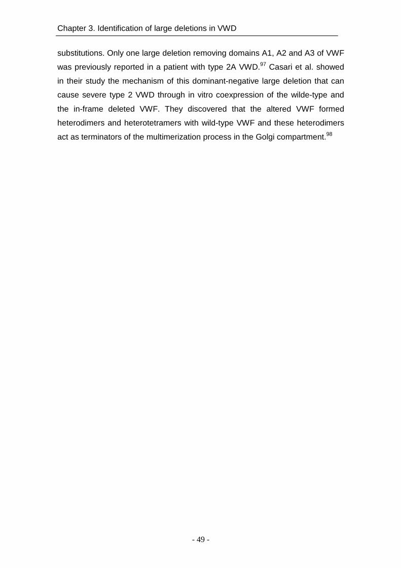
Chapter 3. Identification of large deletions in VWD
- 49 -
substitutions. Only one large deletion removing domains A1, A2 and A3 of VWF
was previously reported in a patient with type 2A VWD.97 Casari et al. showed
in their study the mechanism of this dominant-negative large deletion that can
cause severe type 2 VWD through in vitro coexpression of the wilde-type and
the in-frame deleted VWF. They discovered that the altered VWF formed
heterodimers and heterotetramers with wild-type VWF and these heterodimers
act as terminators of the multimerization process in the Golgi compartment.98
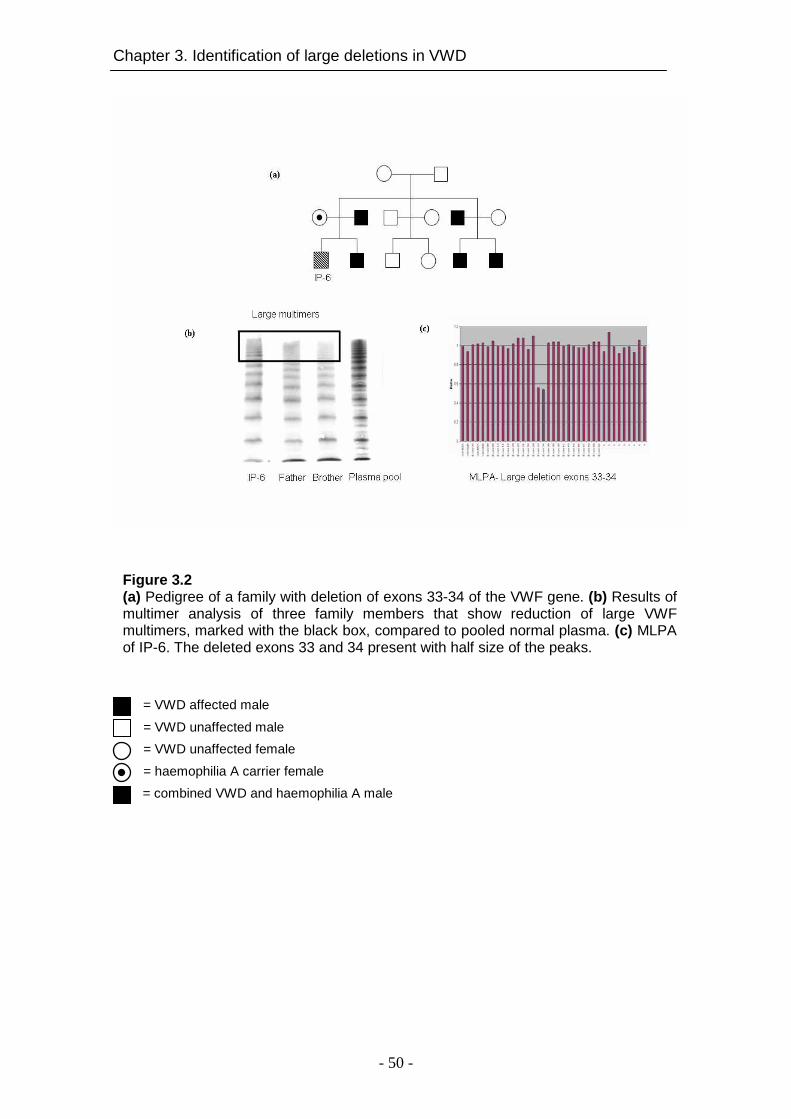
Chapter 3. Identification of large deletions in VWD
- 50 -
Figure 3.2 (a) Pedigree of a family with deletion of exons 33-34 of the VWF gene. (b) Results of multimer analysis of three family members that show reduction of large VWF multimers, marked with the black box, compared to pooled normal plasma. (c) MLPA of IP-6. The deleted exons 33 and 34 present with half size of the peaks.
= VWD affected male
= VWD unaffected male
= VWD unaffected female
= haemophilia A carrier female
= combined VWD and haemophilia A male
Chapter 3. Identification of large deletions in VWD
- 51 -
No large deletion was detected in 11 index patients with type 1 VWD in whom
VWF antigen level was more than 33 IU/dL. Previous studies have reported that
VWF levels below the reference range but above 30IU/dL may often not be in
linkage to the VWF locus.42 , 99
In conclusion, our results indicate that large deletions should be taken into
consideration for the diagnosis of type 3 VWD especially in patients without two
clear causative mutations. The possibility of a large deletion should always be
taken into consideration if sequencing results reveal the presence of
homozygous polymorphisms or a homozygous mutation. Moreover, not only
type 3 VWD patients but also patients diagnosed to have type 2 VWD in which
no VWF mutation could be identified, should be analyzed for the presence of
large deletions. Despite the fact that no large deletions were detected in
patients with type 1 VWD in our study, we propose to investigate these patients
for the presence of possible heterozygous large deletions when no causative
mutations are identified. Finally, the MLPA assay is a rapid, simple and robust
method for the routine diagnosis of large deletions.
Chapter 4. Cysteine missense mutations and VWF biosynthesis
- 52 -
Chapter 4
Insights into pathological mechanisms of
missense mutations in C-terminal domains of
von Willebrand factor causing qualitative or
quantitative von Willebrand disease
Adapted from
Yadegari H., Driesen J., Pavlova A., Biswas A., Ivaskevicius V., Klamroth R.,
and Oldenburg J.,
Accepted to be published in Haematologica [In Press]
Chapter 4. Cysteine missense mutations and VWF biosynthesis
- 53 -
4.1 Abstract The carboxy-terminal domains of VWF, D4-CK, are cysteine rich, implying their
structural importance. The present study characterized the impact of five
cysteine missense mutations residing in D4-CK domains on conformation and
biosynthesis of VWF. These variants were identified as heterozygous in type 1
(p.Cys2619Tyr and p.Cys2676Phe), type 2A (p.Cys2085Tyr and p.Cys2327Trp)
and as compound heterozygous in type 3 (p.Cys2283Arg) VWD. Transient
expression of human cell lines with wild-type or mutant VWF constructs was
performed. Quantitative and qualitative assessment of mutated recombinant
VWF was done in comparison with recombinant wild-type VWF. Storage of
VWF in pseudo-Weible-Palade bodies was studied with confocal microscopy.
Structural impact of the mutations was analyzed by homology modeling.
Homozygous expressions showed that these mutations caused defects in
multimerization, elongation of pseudo-Weible-Palade and consequently
secretion of VWF. Co-expressions of wild-type VWF and p.Cys2085Tyr,
p.Cys2327Trp and p.Cys2283Arg demonstrated defect in multimer assembly,
suggesting a new pathologic mechanism for dominant type 2A VWD due to
mutations in D4 and B domains. Structural analysis revealed that mutations
p.Cys2283Arg, p.Cys2619Tyr and p.Cys2676Phe disrupted intra-domain
disulfide bonds, whereas p.Cys2327Trp might affect an inter-domain disulfide
bond. The p.Cys2327Trp variant is distinguished from the other mutants by an
electrophoretic mobility shift of the multimer bands. The results highlight the
importance of cysteine residues within the C-terminal of VWF on structural
conformation of protein and consequently multimerization, storage, and
secretion of VWF.
Chapter 4. Cysteine missense mutations and VWF biosynthesis
- 54 -
4.2 Introduction Types 1 and 3 VWD are characterized by quantitative deficiencies of VWF. This
can resulte from relative (type 1) or absolute (type 3) deficiency in VWF
production due to impaired synthesis, secretion, or half-life of the molecule.3, 18,
100 The VWD type 2A, characterized by the absence of HMWM and decreased
platelet-dependent function, is the most common form of VWD type 2.51, 59 The
loss of HMWM in type 2A VWD results from either mutations that impair
assembly and secretion of VWF multimers or variants that increased
susceptibility to proteolytic cleavage by ADAMTS13. 9, 101, 102
The pre-pro-VWF precursor comprises of a signal peptide and repeated
domains arranged in the order D1-D2-D′-D3-A1-A2-A3-D4-B1-B2-B3-C1-C2-
CK. In the ER, pro-VWF assembles into dimers through disulfide bonds
between CK domains, and is then transported to the TGN. There the dimers
assemble into multimers by N-terminal disulfide bonds aligned with formation of
helical tubules in nascent WPB.27, 32 The domains may be characterized as
functional or structural, depending on whether they have specific functions in
haemostasis or in forming ultralong concatamers. Although carboxy-terminal (C-
terminal) of VWF, including D4, B and C domains, is a cysteine-rich area, which
may imply structural importance, no particular function for most of these
domains is assigned.16 Recently, the classical annotation of the C-terminal
domain has been updated by Zhou et al., where the previous B and C regions
of VWF are re-annotated as 6 tandem von Willebrand C (VWC) and VWC-like
domains, C1-C6.19 Additionally, it has been demonstrated that the VWF C-
terminals zip up and form a structure resembling a bouquet of flowers, where
the A2, A3, and D4 domains comprise the flowers part and the six VWC
domains configure the stem.19, 31
In this study we analyzed five different VWF mutations, affecting cysteine amino
acids in the D4, B, C2 and N- terminal of CK domains, four of which have been
previously reported in our work.103 Interestingly, these gene alterations were
associated with either quantitative (types 1 and 3) or qualitative VWD (type 2A).
The mutations have been transiently expressed in vitro, and their effect on the
multimer assembly, biogenesis of WPB and secretion has been characterized.
Possible structural impact of these cysteine mutations was additionally studied
Chapter 4. Cysteine missense mutations and VWF biosynthesis
- 55 -
by homology modeling. The results of the present study expand our
understanding of the pathophysiological mechanisms of C-terminal domains
VWF mutations which will help in establishment of phenotype-genotype
correlations.
4.3 Materials and Methods
4.3.1 Patients
Five patients, diagnosed with VWD (2 patients with type 1, 2 patients with type
2A and 1 with type 3) were included in the study. Bleeding severity was
quantified by the bleeding score (BS) calculated on the basis of condensed
MCMDM -1 VWD questionnaire.104 This study was approved by the local ethics
committee and informed consent was obtained from all patients (vote 091/09).
Coagulation and multimer analysis
Laboratory investigation of VWF antigen (VWF:Ag), VWF ristocetin cofactor
activity (VWF:RCo), FVIII coagulant activity (FVIII:C) and VWF multimers (1.2%
[w/v] and 1,6% [w/v] agarose gels) was performed.60, 103 VWF multimer profiles
were classified as normal or abnormal.
Mutation detection
All mutations were identified by direct sequencing of VWF coding region, exon–
intron boundaries and 5’ and 3’ non-coding regions.103
4.3.2 Expression studies
Plasmid constructs
Plasmid pMT2-VWF containing the human full-length wild-type (WT) VWF
cDNA was kindly provided by Professor Schneppenheim (Department of
Chapter 4. Cysteine missense mutations and VWF biosynthesis
- 56 -
Pediatric Hematology and Oncology, University Medical Center Hamburg-
Eppendorf, Hamburg, Germany). The mutant constructs were generated using
a QuickChange II XL site-directed mutagenesis kit (Stratagene, La Jolla, CA,
USA). The sequences of all constructs were verified by DNA sequencing.
Cell culture and transfection
Human embryonic kidney cell lines (HEK 293T; DSMZ, Braunschweig,
Germany) were used to evaluate the secretion and intracellular retention of WT
and mutants recombinant VWF (rVWF). HEK293 cell lines (DSMZ,
Braunschweig, Germany) were used to analyze the intracellular location of VWF
by confocal immunofluorescence microscopy. Both cell lines were cultured in
Dulbecco’s modified Eagel medium (DMEM) containing 4.5g L¯1 glucose and
2mM L-glutamine supplemented with 10% (v/v) fetal bovine serum, 100IU mL¯1
penicillin, 100 µg mL¯1 streptomycin and 0.25 UG/ML Fungizone (all reagents
supplied by Life Technologies, CA, USA) at 37°C in 5% CO2 atmosphere.
Cells were transiently transfected with 8µg of WT-VWF or mutated VWF
constructs using liposomal transfer (Lipofectamine 2000; Life Technologies, CA,
USA) according to the manufacturer’s instructions. Co-transfections were
performed using an equal amount of WT and mutant vectors (4 µg each of WT
and mutant DNA) to reproduce the patients’ heterozygous state. Seventy-two
hours after transfection of HEK293T cells, supernatants were collected and
cells were lysed for analysis of intracellular VWF. VWF secreted in the medium
was concentrated on Amicon Centrifugal 50K filter devices (Millipore, USA) to
one-forth of the original volume before subsequent analysis. The transfected
cells were lysed by 3 rounds of freezing (-80˚C) and thawing in lysis buffer
(0.1M Tris/HCl pH 8.0; 0.6 [v/v] Triton X-100).105
4.3.3 Quantitative and qualitative analysis of rVWF
The amount of VWF:Ag secreted into the medium and VWF:Ag present in the
cellular lysate were determined, and the values were expressed as a
percentage of the corresponding WT recombinant VWF. Activity of secreted
Chapter 4. Cysteine missense mutations and VWF biosynthesis
- 57 -
rVWF was assessed by binding to platelet GPIb, collagen type III (VWF:CB),
and VWF:RCo. The VWF activities were expressed as ratios to secreted VWF.
VWF:GPIb binding was determined using a particle enhanced assay (Siemens
Healthcare, Marburg, Germany) and VWF:CB was assessed by ELISA method
(Technoclone, Vienna, Austria) according to the manufacturer’s instructions.
Comparison between the mean values of the groups was performed with the
Student t-test. Multimer analysis of concentrated secreted rVWF was performed
as described above.
4.3.4 ADAMTS13 assay
To assess the susceptibility of mutants of VWF to proteolysis, full-length WT
and mutated rVWF (6µg/ml) were cleaved by 3µg/ml of recombinant
ADAMTS13 (R&D systems, USA) in the presence of 1.5M urea at 37˚ C for 2
hours following the manufacturer’s instructions. The multimer patterns of
mutated and rVWF-WT were visually compared on agarose gel electrophoresis
after ADAMTS13 digestion.
4.3.5 Immunofluorescence analysis
HEK293 cells were grown on gelatin pre-coated glass coverslips in 24-well
plates. Cells were fixed 48 hr after transfection by 4% (w/v) paraformaldehyde
in PBS, blocked with 0.1% (v/v) Triton-X 100 in PBS azide supplemented with
10% (v/v) FBS. Cells were incubated with first antibodies for 2 hr and then with
fluorescence-conjugated secondary antibodies for 1 hr. Polyclonal sheep to
human VWF (abcam, England) and rabbit anti–human VWF antibodies (Dako,
Glostrup, Denmark) were used to visualize VWF. Monoclonal anti-GM130 (BD
Bioscience, CA) and polyclonal rabbit anti-human TGN46 (Sigma-Aldrich, USA)
were used to stain cis- and trans-Golgi networks, respectively.106 To visualize
the endoplasmic reticulum (ER), the HEK293 cells were prepared and stained
with rabbit anti-human protein-disulfide isomerase (PDI) antibody using
SelectFX® Endoplasmic Reticulum Labeling kit (Life Technologies, CA, USA)
according to the manufacturer’s instructions. Secondary antibodies were
Chapter 4. Cysteine missense mutations and VWF biosynthesis
- 58 -
conjugated with either AlexaFluor-488 or AlexaFluor-594 (Life Technologies,
CA, USA). The coverslips were mounted onto microscope slides with
Vectorshield (Vector Labs, Burlingame, CA, USA) and analyzed with the
Olympus Fluo View FV1000 or Leica SL confocal microscope.
4.3.6 Structure analysis
Homology modeling of VWF C1, C5, C6 domains
Homology models for the C1, C5, and C6 domains were constructed using
various software packages. The domain boundaries for C1, C5, and C6
domains were based on domain annotations from a recently published article.19
The sequences for the respective domains were from the VWF (Accession
number P04275) full length sequence. Templates for each of the domains were
searched on the LOMETS server from Zhang.107 Close packed alignments
(using the conserved cysteines as guideposts) were generated between the
best LOMETS templates and the original domain sequences using Jalview. The
alignments as well as the templates were entered into YASARA version
12.8.6108 to generate full-length models. The resulting models were further
refined by a 500ps MD simulation (AMBER03 force field) in YASARA version
12.8.6. The models with the lowest force field energies were chosen as the final
models for the respective domains. The stereochemical quality of these models
were checked on the MOLPROBITY server
[http://molprobity.biochem.duke.edu/;accessed between 15.07.2012 and
15.09.2012].109 The monomer models were run on the online server CLUSPRO
in dimer mode (http://cluspro.bu.edu/home.php;accessed between 15.08.2012
and 15.10.2012) and the best dimers were chosen based on cluster size for
further interface analysis.110 Impact of mutation on folding was calculated using
the FOLDX plugin incorporated in YASARA version 12.8.6.111
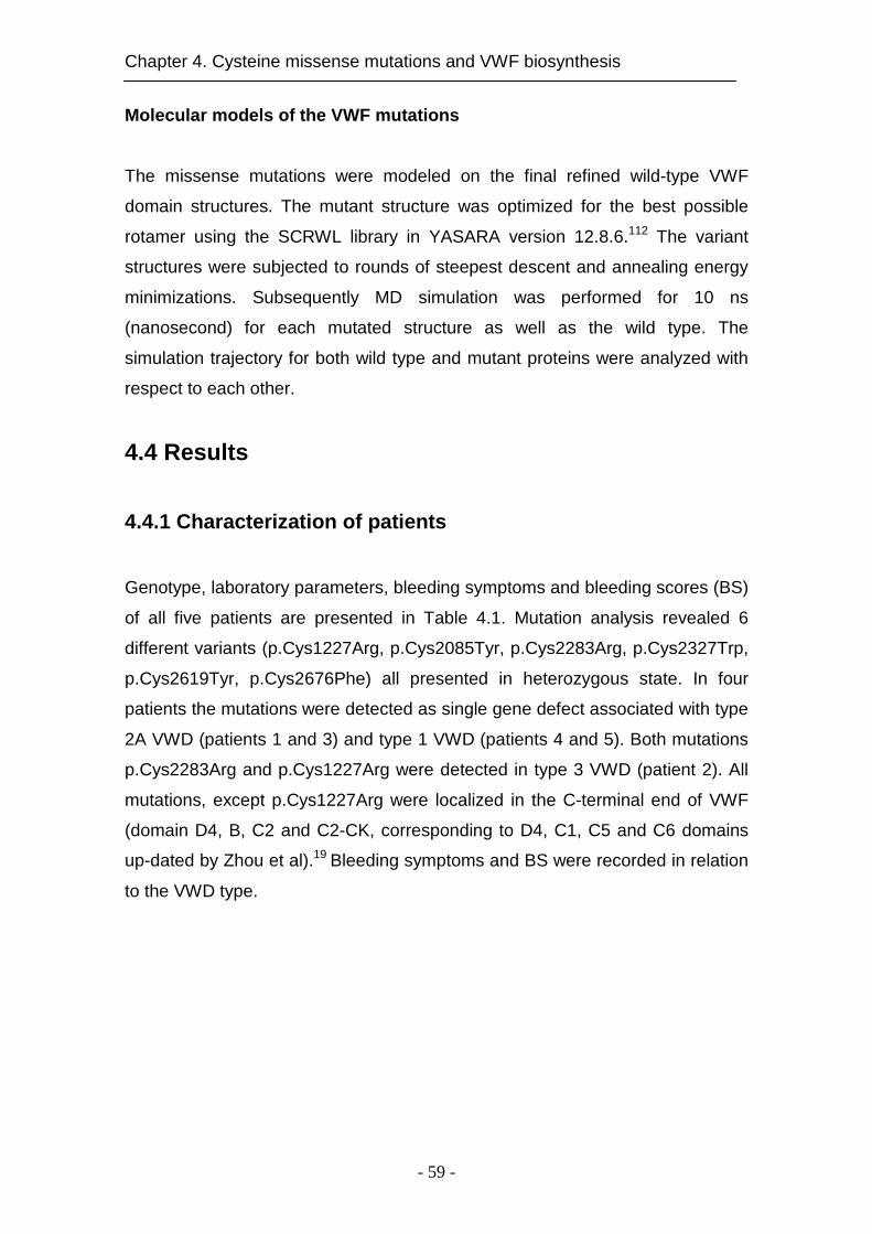
Chapter 4. Cysteine missense mutations and VWF biosynthesis
- 59 -
Molecular models of the VWF mutations
The missense mutations were modeled on the final refined wild-type VWF
domain structures. The mutant structure was optimized for the best possible
rotamer using the SCRWL library in YASARA version 12.8.6.112 The variant
structures were subjected to rounds of steepest descent and annealing energy
minimizations. Subsequently MD simulation was performed for 10 ns
(nanosecond) for each mutated structure as well as the wild type. The
simulation trajectory for both wild type and mutant proteins were analyzed with
respect to each other.
4.4 Results
4.4.1 Characterization of patients Genotype, laboratory parameters, bleeding symptoms and bleeding scores (BS)
of all five patients are presented in Table 4.1. Mutation analysis revealed 6
different variants (p.Cys1227Arg, p.Cys2085Tyr, p.Cys2283Arg, p.Cys2327Trp,
p.Cys2619Tyr, p.Cys2676Phe) all presented in heterozygous state. In four
patients the mutations were detected as single gene defect associated with type
2A VWD (patients 1 and 3) and type 1 VWD (patients 4 and 5). Both mutations
p.Cys2283Arg and p.Cys1227Arg were detected in type 3 VWD (patient 2). All
mutations, except p.Cys1227Arg were localized in the C-terminal end of VWF
(domain D4, B, C2 and C2-CK, corresponding to D4, C1, C5 and C6 domains
up-dated by Zhou et al).19 Bleeding symptoms and BS were recorded in relation
to the VWD type.
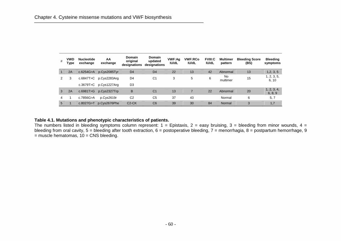
Chapter 4. Cysteine missense mutations and VWF biosynthesis
- 60 -
# VWD Type
Nucleotide exchange
AA exchange
Domain original
designations
Domain updated
designations
VWF:Ag IU/dL
VWF:RCo IU/dL
FVIII:C IU/dL
Multimer pattern
Bleeding Score (BS)
Bleeding symptoms
1 2A c.6254G>A p.Cys2085Tyr D4 D4 22 13 42 Abnormal 13 1,2, 3, 5
2 3 c.6847T>C p.Cys2283Arg D4 C1 3 5 6 No multimer
15 1, 2, 3, 5, 6, 10
c.3679T>C p.Cys1227Arg D3
3 2A c.6981T>G p.Cys2327Trp B C1 13 7 22 Abnormal 20 1, 2, 3, 4, 6, 8, 9
4 1 c.7856G>A p.Cys2619r C2 C5 37 43 Normal 6 5, 7
5 1 c.8027G>T p.Cys2676Phe C2-CK C6 39 30 84 Normal 3 1,7
Table 4.1. Mutations and phenotypic characteristics of patients. The numbers listed in bleeding symptoms column represent: 1 = Epistaxis, 2 = easy bruising, 3 = bleeding from minor wounds, 4 = bleeding from oral cavity, 5 = bleeding after tooth extraction, 6 = postoperative bleeding, 7 = menorrhagia, 8 = postpartum hemorrhage, 9 = muscle hematomas, 10 = CNS bleeding.
Chapter 4. Cysteine missense mutations and VWF biosynthesis
- 61 -
4.4.2 Expression of VWF mutations in human cell lin es
To characterize the effect of the identified mutations on VWF processing,
transient transfections were performed and rVWF:Ag levels were measured in
both the conditioned culture media and cell lysates and expressed as a
percentage of the corresponding wild-type rVWF levels. Five expression vectors
corresponding to each mutation were homozygously expressed in HEK293T
cells. All mutants demonstrated severely impaired secretion. The detected
rVWF:Ag levels in medium were significantly lower compared to the WT,
ranging from 7% to 23% of rVWF-WT (Figure 4.1, left side). To mimic the
heterozygous patient state, co-expression of four mutants (p.Cys2085Tyr,
p.Cys2327Trp, p.Cys2619Tyr, p.Cys2676Phe) together with WT were
additionally performed. The rVWF:Ag values showed reduction in secretion, in
the range of 45.7 to 59.5% of WT (Figure 4.1, right side).
Furthermore, co-expression of variants p.Cys2283Arg and p.Cys1227Arg
(representing the compound heterozygous state of type 3 VWD patient) resulted
in strongly reduced secretion of VWF to 19% (Figure 4.1, right side).
The measurement of intracellular rVWF:Ag of all mutants showed values higher
than that of the WT, indicating intracellular retention of rVWF-mutants (Figure
4.1).
Chapter 4. Cysteine missense mutations and VWF biosynthesis
- 62 -
Figure 4.1. Recombinant VWF levels in medium and ly sates. Recombinant VWF antigen levels (VWF:Ag) in medium (white bars) and lysate (black bars) are expressed as percentage relative to the amount of VWF:Ag in the medium and lysate of cells expressing WT-VWF. Each column represents the mean and standard deviations of at least three independent experiments in triplicate. Co-transfections of WT and mutant VWF cDNA were done with a 1:1 ratio.
4.4.3 Functional characterization of recombinant VW F mutants
The multimer distribution of the recombinant variants demonstrated a range of
structural abnormalities (Figure 4.2). Single transfections rVWF-p.Cys2085Tyr,
p.Cys2283Arg and p.Cys2676Phe exhibited only dimers and tetramers.
Multimer analysis of secreted rVWF-p.Cys2327Trp revealed a complete lack of
intermediate and high molecular weight multimers, whereas rVWF-
p.Cys2619Tyr showed loss of most of intermediate multimers and lack of
HMWM (Figure 4.2 A). Interestingly, variant C2327W additionally showed shift
in mobility of multimer bands (Figure 4.2 A).
Figure 4.2B illustrates the multimer patterns for co-expressed mutants and WT
as well as for co-expression of the two mutants p.Cys2283Arg and
p.Cys1227Arg, identified in the type 3 VWD patient. The co-expression of WT
with mutants p.Cys2619Tyr and p.Cys2676Phe led to secretion of the full range
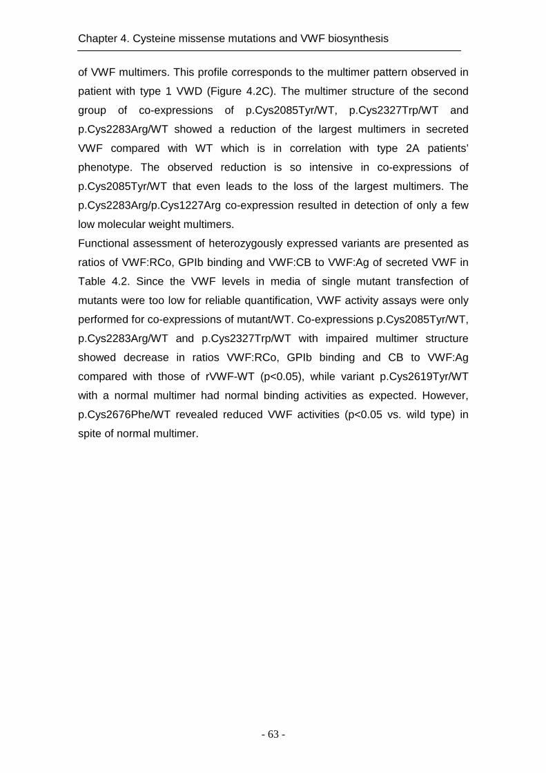
Chapter 4. Cysteine missense mutations and VWF biosynthesis
- 63 -
of VWF multimers. This profile corresponds to the multimer pattern observed in
patient with type 1 VWD (Figure 4.2C). The multimer structure of the second
group of co-expressions of p.Cys2085Tyr/WT, p.Cys2327Trp/WT and
p.Cys2283Arg/WT showed a reduction of the largest multimers in secreted
VWF compared with WT which is in correlation with type 2A patients’
phenotype. The observed reduction is so intensive in co-expressions of
p.Cys2085Tyr/WT that even leads to the loss of the largest multimers. The
p.Cys2283Arg/p.Cys1227Arg co-expression resulted in detection of only a few
low molecular weight multimers.
Functional assessment of heterozygously expressed variants are presented as
ratios of VWF:RCo, GPIb binding and VWF:CB to VWF:Ag of secreted VWF in
Table 4.2. Since the VWF levels in media of single mutant transfection of
mutants were too low for reliable quantification, VWF activity assays were only
performed for co-expressions of mutant/WT. Co-expressions p.Cys2085Tyr/WT,
p.Cys2283Arg/WT and p.Cys2327Trp/WT with impaired multimer structure
showed decrease in ratios VWF:RCo, GPIb binding and CB to VWF:Ag
compared with those of rVWF-WT (p<0.05), while variant p.Cys2619Tyr/WT
with a normal multimer had normal binding activities as expected. However,
p.Cys2676Phe/WT revealed reduced VWF activities (p<0.05 vs. wild type) in
spite of normal multimer.
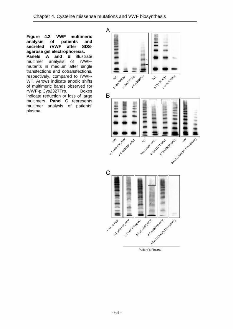
Chapter 4. Cysteine missense mutations and VWF biosynthesis
- 64 -
Figure 4.2. VWF multimeric analysis of patients and secreted rVWF after SDS-agarose gel electrophoresis. Panels A and B illustrate multimer analysis of rVWF-mutants in medium after single transfections and cotransfections, respectively, compared to rVWF-WT. Arrows indicate anodic shifts of multimeric bands observed for rVWF-p.Cys2327Trp. Boxes indicate reduction or loss of large multimers. Panel C represents multimer analysis of patients’ plasma.
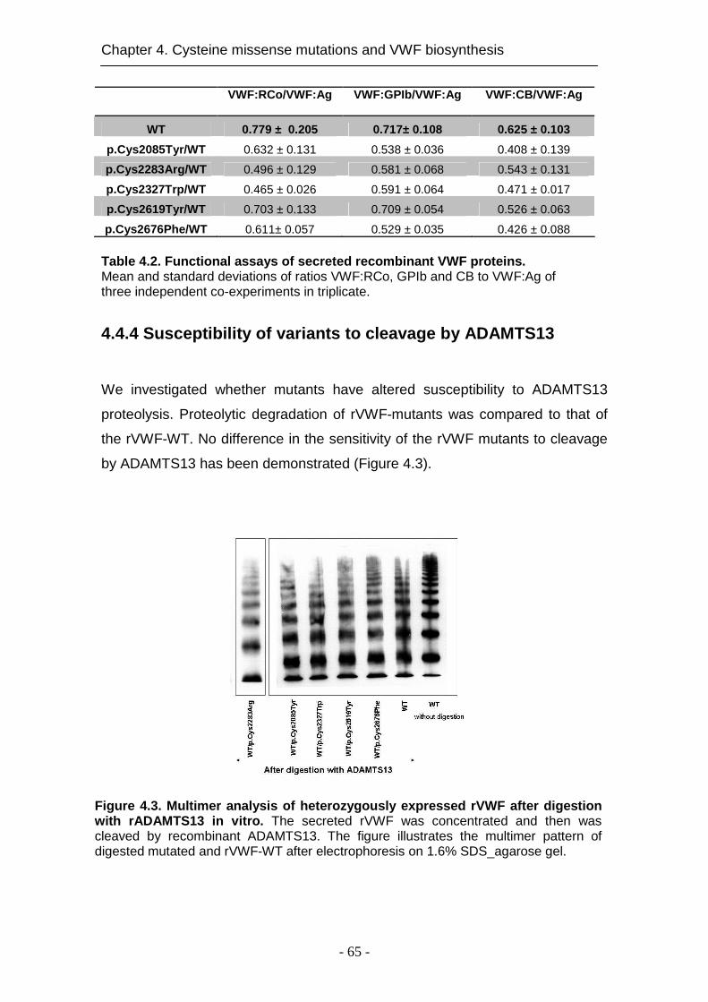
Chapter 4. Cysteine missense mutations and VWF biosynthesis
- 65 -
Table 4.2. Functional assays of secreted recombinan t VWF proteins. Mean and standard deviations of ratios VWF:RCo, GPIb and CB to VWF:Ag of three independent co-experiments in triplicate.
4.4.4 Susceptibility of variants to cleavage by ADA MTS13
We investigated whether mutants have altered susceptibility to ADAMTS13
proteolysis. Proteolytic degradation of rVWF-mutants was compared to that of
the rVWF-WT. No difference in the sensitivity of the rVWF mutants to cleavage
by ADAMTS13 has been demonstrated (Figure 4.3).
VWF:RCo/VWF:Ag
VWF:GPIb/VWF:Ag
VWF:CB/VWF:Ag
WT 0.779 ± 0.205 0.717± 0.108 0.625 ± 0.103
p.Cys2085Tyr/WT 0.632 ± 0.131 0.538 ± 0.036 0.408 ± 0.139
p.Cys2283Arg/WT 0.496 ± 0.129 0.581 ± 0.068 0.543 ± 0.131
p.Cys2327Trp/WT 0.465 ± 0.026 0.591 ± 0.064 0.471 ± 0.017
p.Cys2619Tyr/WT 0.703 ± 0.133 0.709 ± 0.054 0.526 ± 0.063
p.Cys2676Phe/WT 0.611± 0.057 0.529 ± 0.035 0.426 ± 0.088
Figure 4.3. Multimer analysis of heterozygously expressed rVWF after digestion with rADAMTS13 in vitro. The secreted rVWF was concentrated and then was cleaved by recombinant ADAMTS13. The figure illustrates the multimer pattern of digested mutated and rVWF-WT after electrophoresis on 1.6% SDS_agarose gel.
Chapter 4. Cysteine missense mutations and VWF biosynthesis
- 66 -
4.4.5 Intracellular localization
Intracellular trafficking and the impact of mutations on granule formation were
analyzed after expression of mutants in HEK293 cells. HEK293 cells form
pseudo-WPB granules when transfected with rVWF-WT.101, 113 Confocal
microscopy showed no retention of all mutant rVWF in either the ER or in the
cis- or trans-Golgi compartments (Figures 4.4 and 4.5). Each of the VWF
mutants was able to form pseudo-WPB storage granules (Figure 4.6, presented
as small dots), although those granules had different morphology from normal
cigar-shaped granules formed by rVWF-WT. The majority of the pseudo-WPB
granules formed by mutants were relatively shorter. We also investigated the
impact of VWF variant heterozygosity on storage in pseudo-WPB granules.
Upon co-transfection with rVWF-WT, the defects in the elongation of the
pseudo-WPB granules caused by mutations were partially corrected.
Figure 4.4 Subce llular localization of rVWF-WT and variants in relation to endoplasmic reticulum (ER) in transfected HEK293 cell lines. Fixed cells were stained for VWF (green channel, left panel) and for PDI (ER marker, red channel, middle panel). Right panel illustrates the merge of green and red channels. Scale bars, 10 µl.
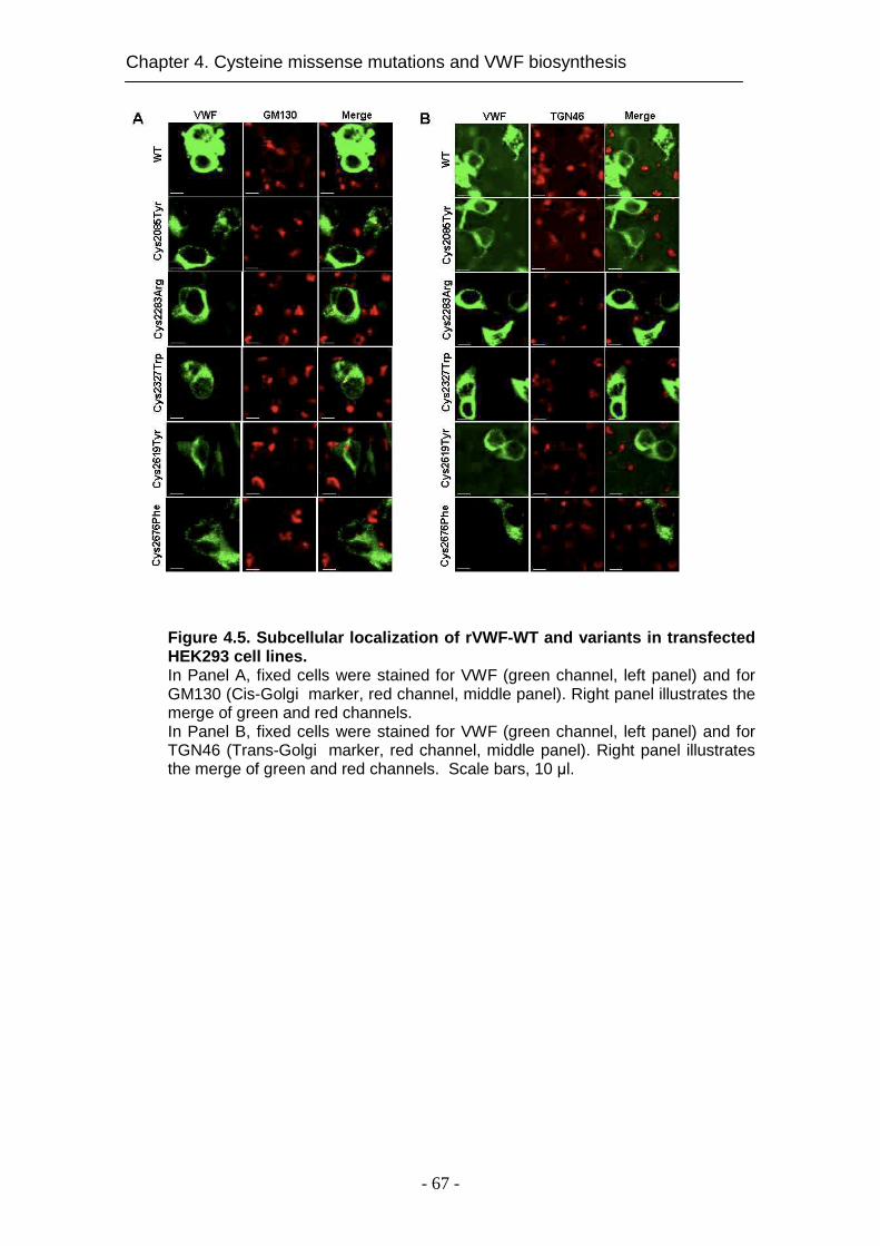
Chapter 4. Cysteine missense mutations and VWF biosynthesis
- 67 -
Figure 4.5. Subcellular localization of rVWF-WT and variants in transfected HEK293 cell lines. In Panel A, fixed cells were stained for VWF (green channel, left panel) and for GM130 (Cis-Golgi marker, red channel, middle panel). Right panel illustrates the merge of green and red channels. In Panel B, fixed cells were stained for VWF (green channel, left panel) and for TGN46 (Trans-Golgi marker, red channel, middle panel). Right panel illustrates the merge of green and red channels. Scale bars, 10 µl.
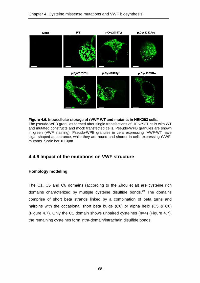
Chapter 4. Cysteine missense mutations and VWF biosynthesis
- 68 -
Figure 4.6. Intracellular storage of rVWF-WT and mu tants in HEK293 cells. The pseudo-WPB granules formed after single transfections of HEK293T cells with WT and mutated constructs and mock transfected cells. Pseudo-WPB granules are shown in green (VWF staining). Pseudo-WPB granules in cells expressing rVWF-WT have cigar-shaped appearance, while they are round and shorter in cells expressing rVWF-mutants. Scale bar = 10µm.
4.4.6 Impact of the mutations on VWF structure
Homology modeling
The C1, C5 and C6 domains (according to the Zhou et al) are cysteine rich
domains characterized by multiple cysteine disulfide bonds.19 The domains
comprise of short beta strands linked by a combination of beta turns and
hairpins with the occasional short beta bulge (C6) or alpha helix (C5 & C6)
(Figure 4.7). Only the C1 domain shows unpaired cysteines (n=4) (Figure 4.7),
the remaining cysteines form intra-domain/intrachain disulfide bonds.
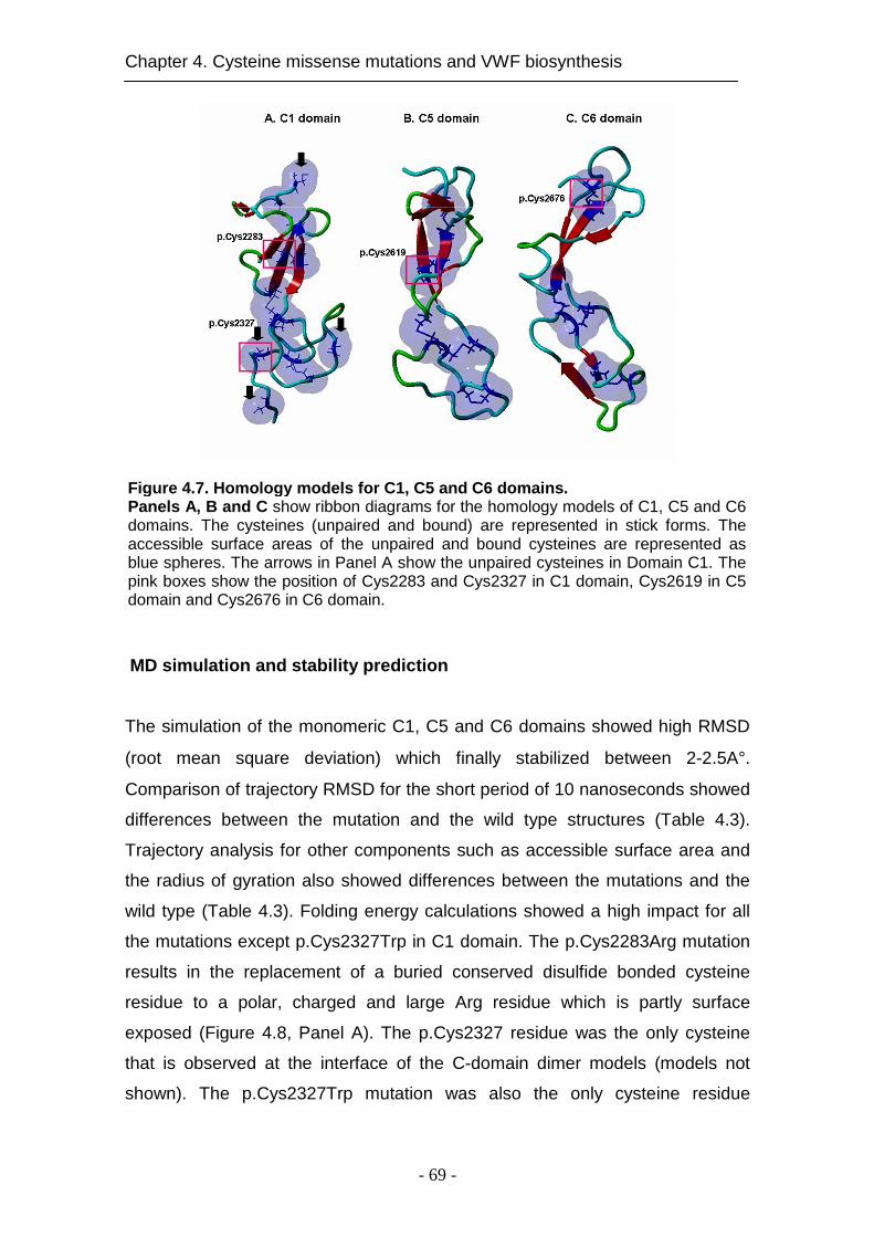
Chapter 4. Cysteine missense mutations and VWF biosynthesis
- 69 -
MD simulation and stability prediction
The simulation of the monomeric C1, C5 and C6 domains showed high RMSD
(root mean square deviation) which finally stabilized between 2-2.5A°.
Comparison of trajectory RMSD for the short period of 10 nanoseconds showed
differences between the mutation and the wild type structures (Table 4.3).
Trajectory analysis for other components such as accessible surface area and
the radius of gyration also showed differences between the mutations and the
wild type (Table 4.3). Folding energy calculations showed a high impact for all
the mutations except p.Cys2327Trp in C1 domain. The p.Cys2283Arg mutation
results in the replacement of a buried conserved disulfide bonded cysteine
residue to a polar, charged and large Arg residue which is partly surface
exposed (Figure 4.8, Panel A). The p.Cys2327 residue was the only cysteine
that is observed at the interface of the C-domain dimer models (models not
shown). The p.Cys2327Trp mutation was also the only cysteine residue
Figure 4.7. Homology models for C1, C5 and C6 domai ns. Panels A, B and C show ribbon diagrams for the homology models of C1, C5 and C6 domains. The cysteines (unpaired and bound) are represented in stick forms. The accessible surface areas of the unpaired and bound cysteines are represented as blue spheres. The arrows in Panel A show the unpaired cysteines in Domain C1. The pink boxes show the position of Cys2283 and Cys2327 in C1 domain, Cys2619 in C5 domain and Cys2676 in C6 domain.
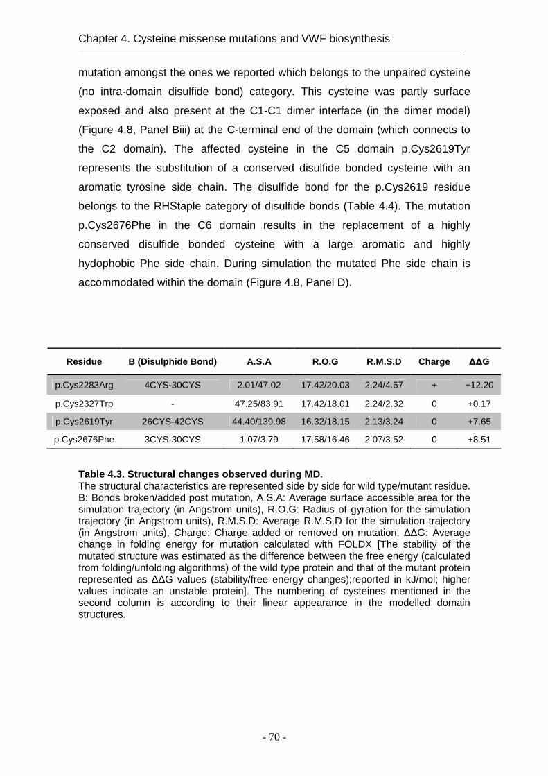
Chapter 4. Cysteine missense mutations and VWF biosynthesis
- 70 -
mutation amongst the ones we reported which belongs to the unpaired cysteine
(no intra-domain disulfide bond) category. This cysteine was partly surface
exposed and also present at the C1-C1 dimer interface (in the dimer model)
(Figure 4.8, Panel Biii) at the C-terminal end of the domain (which connects to
the C2 domain). The affected cysteine in the C5 domain p.Cys2619Tyr
represents the substitution of a conserved disulfide bonded cysteine with an
aromatic tyrosine side chain. The disulfide bond for the p.Cys2619 residue
belongs to the RHStaple category of disulfide bonds (Table 4.4). The mutation
p.Cys2676Phe in the C6 domain results in the replacement of a highly
conserved disulfide bonded cysteine with a large aromatic and highly
hydophobic Phe side chain. During simulation the mutated Phe side chain is
accommodated within the domain (Figure 4.8, Panel D).
Table 4.3. Structural changes observed during MD . The structural characteristics are represented side by side for wild type/mutant residue. B: Bonds broken/added post mutation, A.S.A: Average surface accessible area for the simulation trajectory (in Angstrom units), R.O.G: Radius of gyration for the simulation trajectory (in Angstrom units), R.M.S.D: Average R.M.S.D for the simulation trajectory (in Angstrom units), Charge: Charge added or removed on mutation, ∆∆G: Average change in folding energy for mutation calculated with FOLDX [The stability of the mutated structure was estimated as the difference between the free energy (calculated from folding/unfolding algorithms) of the wild type protein and that of the mutant protein represented as ∆∆G values (stability/free energy changes);reported in kJ/mol; higher values indicate an unstable protein]. The numbering of cysteines mentioned in the second column is according to their linear appearance in the modelled domain structures.
Residue B (Disulphide Bond) A.S.A R.O.G R.M.S.D Cha rge ∆∆G
p.Cys2283Arg 4CYS-30CYS 2.01/47.02 17.42/20.03 2.24/4.67 + +12.20
p.Cys2327Trp - 47.25/83.91 17.42/18.01 2.24/2.32 0 +0.17
p.Cys2619Tyr 26CYS-42CYS 44.40/139.98 16.32/18.15 2.13/3.24 0 +7.65
p.Cys2676Phe 3CYS-30CYS 1.07/3.79 17.58/16.46 2.07/3.52 0 +8.51
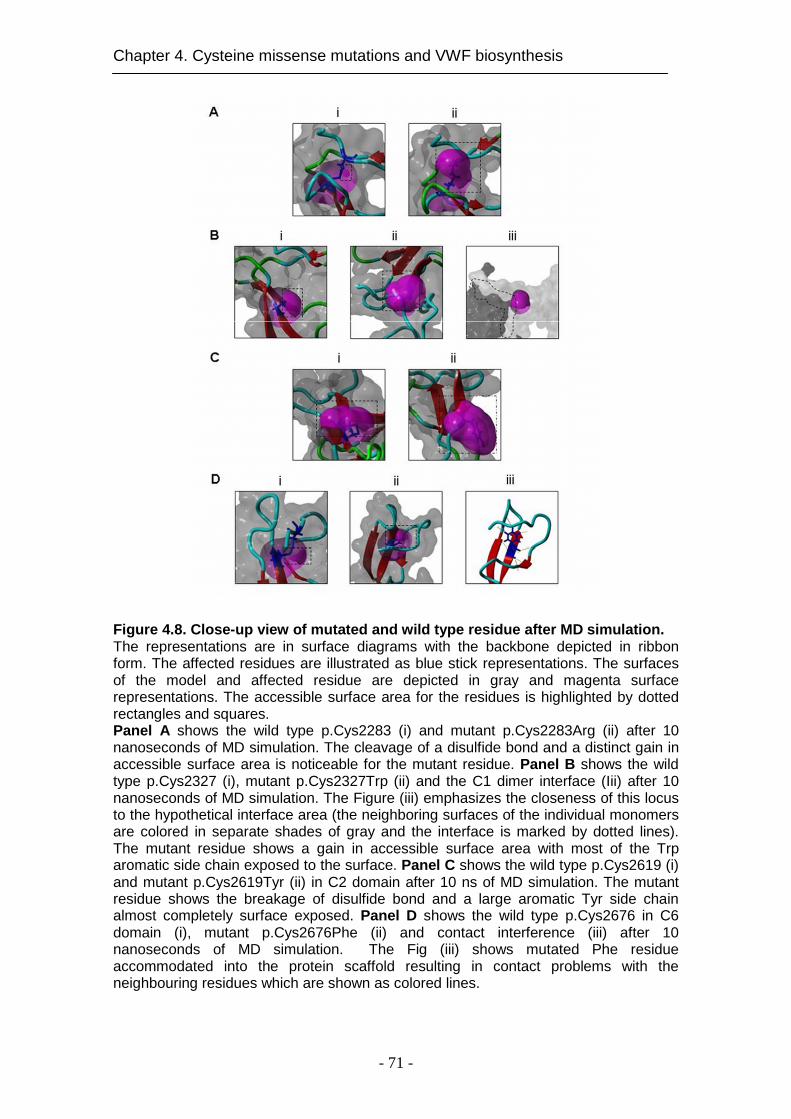
Chapter 4. Cysteine missense mutations and VWF biosynthesis
- 71 -
Figure 4.8. Close-up view of mutated and wild type residue afte r MD simulation. The representations are in surface diagrams with the backbone depicted in ribbon form. The affected residues are illustrated as blue stick representations. The surfaces of the model and affected residue are depicted in gray and magenta surface representations. The accessible surface area for the residues is highlighted by dotted rectangles and squares. Panel A shows the wild type p.Cys2283 (i) and mutant p.Cys2283Arg (ii) after 10 nanoseconds of MD simulation. The cleavage of a disulfide bond and a distinct gain in accessible surface area is noticeable for the mutant residue. Panel B shows the wild type p.Cys2327 (i), mutant p.Cys2327Trp (ii) and the C1 dimer interface (Iii) after 10 nanoseconds of MD simulation. The Figure (iii) emphasizes the closeness of this locus to the hypothetical interface area (the neighboring surfaces of the individual monomers are colored in separate shades of gray and the interface is marked by dotted lines). The mutant residue shows a gain in accessible surface area with most of the Trp aromatic side chain exposed to the surface. Panel C shows the wild type p.Cys2619 (i) and mutant p.Cys2619Tyr (ii) in C2 domain after 10 ns of MD simulation. The mutant residue shows the breakage of disulfide bond and a large aromatic Tyr side chain almost completely surface exposed. Panel D shows the wild type p.Cys2676 in C6 domain (i), mutant p.Cys2676Phe (ii) and contact interference (iii) after 10 nanoseconds of MD simulation. The Fig (iii) shows mutated Phe residue accommodated into the protein scaffold resulting in contact problems with the neighbouring residues which are shown as colored lines.
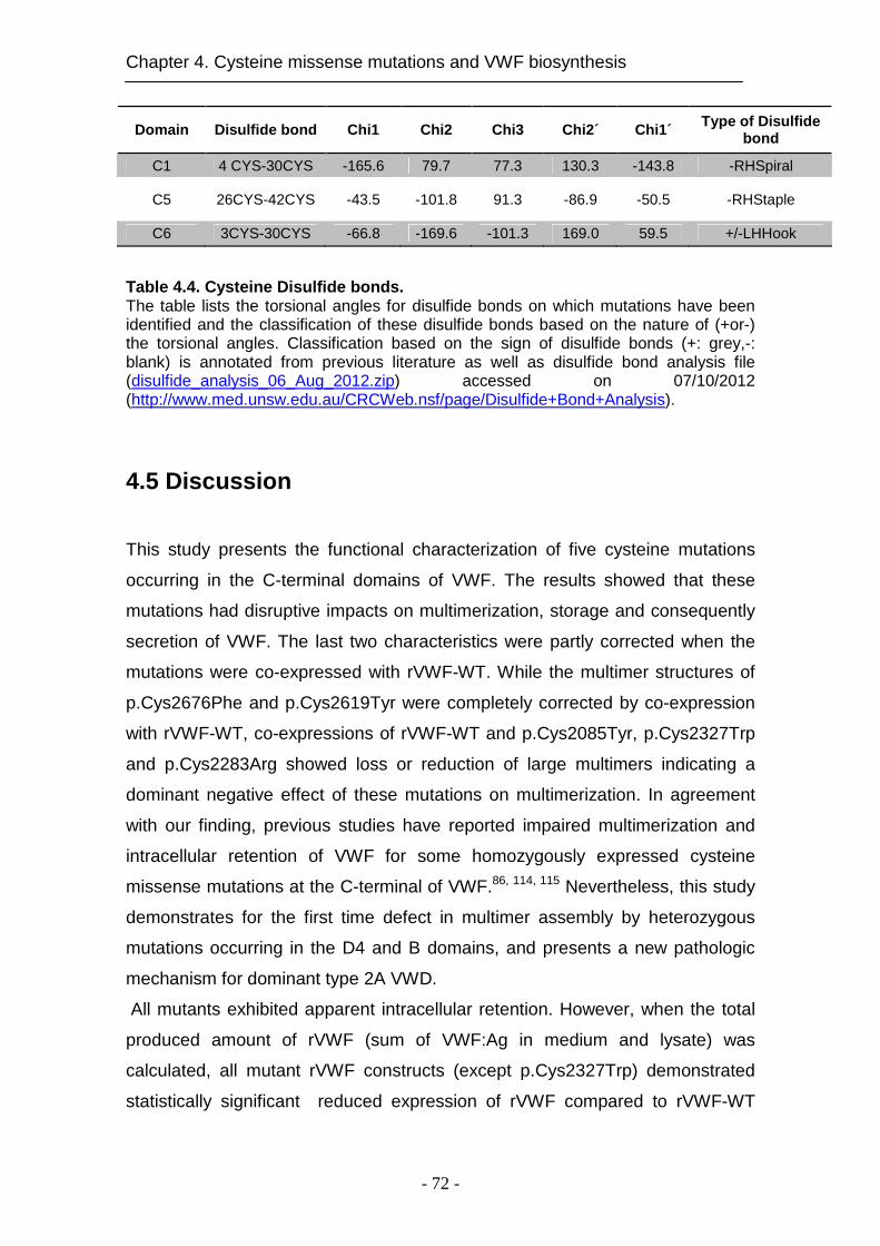
Chapter 4. Cysteine missense mutations and VWF biosynthesis
- 72 -
Domain Disulfide bond Chi1 Chi2 Chi3 Chi2´ Chi1´ Type of Disulfide bond
C1 4 CYS-30CYS -165.6 79.7 77.3 130.3 -143.8 -RHSpiral
C5 26CYS-42CYS -43.5 -101.8 91.3 -86.9 -50.5 -RHStaple
C6 3CYS-30CYS -66.8 -169.6 -101.3 169.0 59.5 +/-LHHook
Table 4.4. Cysteine Disulfide bonds. The table lists the torsional angles for disulfide bonds on which mutations have been identified and the classification of these disulfide bonds based on the nature of (+or-) the torsional angles. Classification based on the sign of disulfide bonds (+: grey,-: blank) is annotated from previous literature as well as disulfide bond analysis file (disulfide_analysis_06_Aug_2012.zip) accessed on 07/10/2012 (http://www.med.unsw.edu.au/CRCWeb.nsf/page/Disulfide+Bond+Analysis).
4.5 Discussion
This study presents the functional characterization of five cysteine mutations
occurring in the C-terminal domains of VWF. The results showed that these
mutations had disruptive impacts on multimerization, storage and consequently
secretion of VWF. The last two characteristics were partly corrected when the
mutations were co-expressed with rVWF-WT. While the multimer structures of
p.Cys2676Phe and p.Cys2619Tyr were completely corrected by co-expression
with rVWF-WT, co-expressions of rVWF-WT and p.Cys2085Tyr, p.Cys2327Trp
and p.Cys2283Arg showed loss or reduction of large multimers indicating a
dominant negative effect of these mutations on multimerization. In agreement
with our finding, previous studies have reported impaired multimerization and
intracellular retention of VWF for some homozygously expressed cysteine
missense mutations at the C-terminal of VWF.86, 114, 115 Nevertheless, this study
demonstrates for the first time defect in multimer assembly by heterozygous
mutations occurring in the D4 and B domains, and presents a new pathologic
mechanism for dominant type 2A VWD.
All mutants exhibited apparent intracellular retention. However, when the total
produced amount of rVWF (sum of VWF:Ag in medium and lysate) was
calculated, all mutant rVWF constructs (except p.Cys2327Trp) demonstrated
statistically significant reduced expression of rVWF compared to rVWF-WT
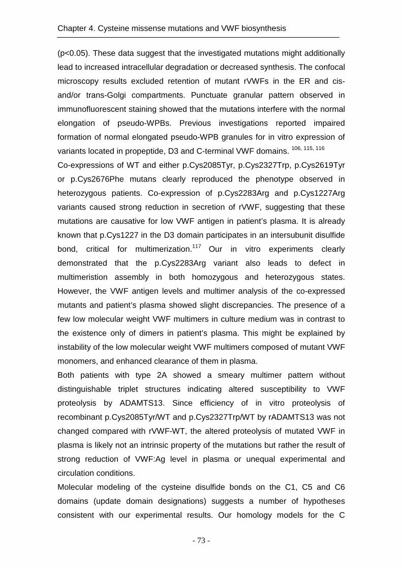
Chapter 4. Cysteine missense mutations and VWF biosynthesis
- 73 -
(p<0.05). These data suggest that the investigated mutations might additionally
lead to increased intracellular degradation or decreased synthesis. The confocal
microscopy results excluded retention of mutant rVWFs in the ER and cis-
and/or trans-Golgi compartments. Punctuate granular pattern observed in
immunofluorescent staining showed that the mutations interfere with the normal
elongation of pseudo-WPBs. Previous investigations reported impaired
formation of normal elongated pseudo-WPB granules for in vitro expression of
variants located in propeptide, D3 and C-terminal VWF domains. 106, 115, 116
Co-expressions of WT and either p.Cys2085Tyr, p.Cys2327Trp, p.Cys2619Tyr
or p.Cys2676Phe mutans clearly reproduced the phenotype observed in
heterozygous patients. Co-expression of p.Cys2283Arg and p.Cys1227Arg
variants caused strong reduction in secretion of rVWF, suggesting that these
mutations are causative for low VWF antigen in patient’s plasma. It is already
known that p.Cys1227 in the D3 domain participates in an intersubunit disulfide
bond, critical for multimerization.117 Our in vitro experiments clearly
demonstrated that the p.Cys2283Arg variant also leads to defect in
multimeristion assembly in both homozygous and heterozygous states.
However, the VWF antigen levels and multimer analysis of the co-expressed
mutants and patient’s plasma showed slight discrepancies. The presence of a
few low molecular weight VWF multimers in culture medium was in contrast to
the existence only of dimers in patient’s plasma. This might be explained by
instability of the low molecular weight VWF multimers composed of mutant VWF
monomers, and enhanced clearance of them in plasma.
Both patients with type 2A showed a smeary multimer pattern without
distinguishable triplet structures indicating altered susceptibility to VWF
proteolysis by ADAMTS13. Since efficiency of in vitro proteolysis of
recombinant p.Cys2085Tyr/WT and p.Cys2327Trp/WT by rADAMTS13 was not
changed compared with rVWF-WT, the altered proteolysis of mutated VWF in
plasma is likely not an intrinsic property of the mutations but rather the result of
strong reduction of VWF:Ag level in plasma or unequal experimental and
circulation conditions.
Molecular modeling of the cysteine disulfide bonds on the C1, C5 and C6
domains (update domain designations) suggests a number of hypotheses
consistent with our experimental results. Our homology models for the C
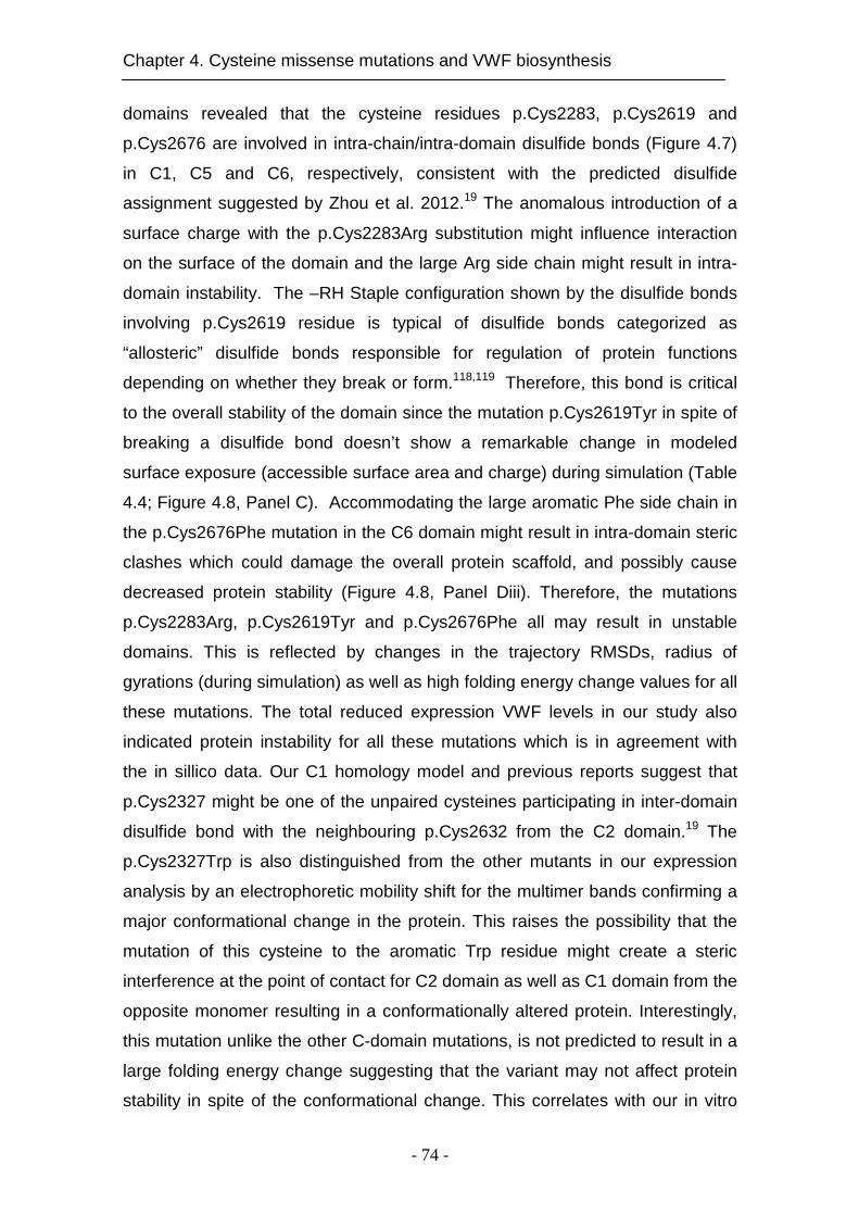
Chapter 4. Cysteine missense mutations and VWF biosynthesis
- 74 -
domains revealed that the cysteine residues p.Cys2283, p.Cys2619 and
p.Cys2676 are involved in intra-chain/intra-domain disulfide bonds (Figure 4.7)
in C1, C5 and C6, respectively, consistent with the predicted disulfide
assignment suggested by Zhou et al. 2012.19 The anomalous introduction of a
surface charge with the p.Cys2283Arg substitution might influence interaction
on the surface of the domain and the large Arg side chain might result in intra-
domain instability. The –RH Staple configuration shown by the disulfide bonds
involving p.Cys2619 residue is typical of disulfide bonds categorized as
“allosteric” disulfide bonds responsible for regulation of protein functions
depending on whether they break or form.118,119 Therefore, this bond is critical
to the overall stability of the domain since the mutation p.Cys2619Tyr in spite of
breaking a disulfide bond doesn’t show a remarkable change in modeled
surface exposure (accessible surface area and charge) during simulation (Table
4.4; Figure 4.8, Panel C). Accommodating the large aromatic Phe side chain in
the p.Cys2676Phe mutation in the C6 domain might result in intra-domain steric
clashes which could damage the overall protein scaffold, and possibly cause
decreased protein stability (Figure 4.8, Panel Diii). Therefore, the mutations
p.Cys2283Arg, p.Cys2619Tyr and p.Cys2676Phe all may result in unstable
domains. This is reflected by changes in the trajectory RMSDs, radius of
gyrations (during simulation) as well as high folding energy change values for all
these mutations. The total reduced expression VWF levels in our study also
indicated protein instability for all these mutations which is in agreement with
the in sillico data. Our C1 homology model and previous reports suggest that
p.Cys2327 might be one of the unpaired cysteines participating in inter-domain
disulfide bond with the neighbouring p.Cys2632 from the C2 domain.19 The
p.Cys2327Trp is also distinguished from the other mutants in our expression
analysis by an electrophoretic mobility shift for the multimer bands confirming a
major conformational change in the protein. This raises the possibility that the
mutation of this cysteine to the aromatic Trp residue might create a steric
interference at the point of contact for C2 domain as well as C1 domain from the
opposite monomer resulting in a conformationally altered protein. Interestingly,
this mutation unlike the other C-domain mutations, is not predicted to result in a
large folding energy change suggesting that the variant may not affect protein
stability in spite of the conformational change. This correlates with our in vitro
Chapter 4. Cysteine missense mutations and VWF biosynthesis
- 75 -
expression results where total expression level was similar to that of the wild
type VWF. Interestingly, Tjernberg et al have reported mobility shift in multimer
for substitutions at p.Cys2362 in the C2 domain which is most likely the
disulfide-bond partner of p.Cys2327 in C1 domain .120 Although we did not
perform any structural modeling for the D4 domain, it was predicted that
p.Cys2085 residue in D4 domain participate in intrachain disulfide bond.19 The
introduction of large Arg side chain will disrupt the disulfide bond, and might
cause domain instability.
In summary, the current structural analysis showed that mutations eliminating
these cysteine residues could induce misfolding of the respective domains. We
speculate that such domain misfoldings might influence dimeric bouquet
formation and consequently helical assembly of tubules and storage. Therefore,
VWF secretion can be defected since tubular packing of VWF is prerequisite for
physiological VWF unfurling upon exocytosis.26, 30, 31 Moreover, recent findings
stated that dimeric bouquet formation facilitates N-terminal disulphide linkage
between VWF dimers which is colinear with assembly in tubules.31, 32
Accordingly, we assume that defects in multimerization might be attributed to
disruption in zipping up of misfolded C domains into dimeric bouquet structures.
In conclusion, our results indicates that the above described cysteine residues
have a crucial role in accurate folding of D4 and C1, C5 and C6 domains
(corresponding to original D4, B and C domains) required for proper
arrangement of VWF dimer bouquets and their incorporation into growing
tubules in nascent WPBs. This study demonstrates that these mutations disrupt
storage, and secretion of VWF through a common pathological mechanism,
although their eventual outcome on multimerization might differ because of sub-
molecular differences in structural alteratnations.
References
- 76 -
References
1. Goodeve AC. The genetic basis of von Willebrand disease. Blood Rev
2010; 24: 123-34.
2. Luo GP, Ni B, Yang X, Wu YZ. Von Willebrand factor: more than a
regulator of hemostasis and thrombosis. Acta Haematol 2012;128:158-69.
3. Lyons SE, Ginsburg D. Molecular and cellular biology of von Willebrand
factor. Trends Cardiovasc Med 1994;4(1):34-9.
4. Hohenstein K, Griesmacher A, Weigel G, Golderer G, Ott HW. Native
multimer analysis of plasma and platelet von Willebrand factor compared to
denaturing separation: implication for the interpretation of satellite bands.
Electrophoresis 2011;32(13):1684-91.
5. Sadler JE. von Willebrand factor: two sides of a coin. J Thromb Haemost
2005;3:1702-9.
6. Szántό T, Joutsi-Korhonen L, Deckmyn H, Lassila R. New insights into von
Willebrand disease and platelet function. Semin Thromb Hemost 2012;38:55-
63.
7. James PD, Goodeve AC. von Willebrand disease. Genet Med
2011;13(5):365-76.
8. Mancuso DJ, Tuley EA, Westfield LA, Worrall NK, Shelton-Inloes BB,
Sorace JM, et al. Structure of the gene for human von Willebrand factor. J Biol
Chem 1989; 264: 19514-27.
9. Castaman G, Federici AB, Rodeghiero F, Mannucci PM. Von Willebrand’s
disease in the year 2003: towards the complete identification of gene defects for
correct diagnosis and treatment. Haematologica 2003; 88:94-108.
10. Keeney S, Bowen D, Cumming A, Enayat S, Goodeve A, Hill M. The
molecular analysis of von Willebrand disease: a guideline from the UK
Haemophilia Centre Doctors' Organisation Haemophilia Genetics Laboratory
Network. Haemophilia 2008; 14: 1099-111.
11. Kasatkar P, Shetty S, Ghosh K. VWF pseudogene: Mimics, masks and
spoils. Clin Chim Acta 2010;411(7-8):607-9.
12. Sadler JE. Biochemistry and genetics of von Willebrand factor. Annu Rev
Biochem 1998;67:395-424.
References
- 77 -
13. Millar CM, Brown SA. Oligosaccharide structures of von Willebrand factor
and their potential role in von Willebrand disease. Blood Rev 2006;20(2):83-92.
14. Lenting PJ, Casari C, Christophe OD, Denis CV. von Willebrand factor: the
old, the new and the unknown. J Thromb Haemost 2012;10(12):2428-37.
15. Sadler JE. von Willebrand factor assembly and secretion. J Thromb
Haemost 2009;7(Suppl. 1):24-7.
16. Schneppenheim R, Budde U. von Willebrand factor: the complex
molecular genetics of a multidomain and multifunctional protein. J Thromb
Haemost 2011;9 Suppl 1:209-15.
17. de Wit RT, van Mourik JA. Biosynthesis, processing and secretion of von
Willebrand factor: biological implications. Best practice and research clonical
haematology 2001;14(2):241-55.
18. Schneppenheim R. The pathophysiology of von Willebrand disease:
therapeutic implications. Thrombosis Research 2011; 128: S3-S7.
19. Zhou YF, Eng ET, Zhu J, Lu C, Walz T, Springer TA. Sequence and
structure relationships within von Willebrand factor. Blood 2012;120(2):449-58.
20. Metcalf DJ, Nightingale TD, Zenner HL, Lui-Roberts WW, Cutler DF.
Formation and function of Weibel-Palade bodies. Journal of Cell Science
2008;121(1):19-27.
21. Katsumi A, Tuley EA, Bodó I, Sadler JE. Localization of disulfide bonds in
the cystine knot domain of human von Willebrand factor. J Biol Chem
2000;275(33):25585-94.
22. Purvis AR, Gross J, Dang LT, Huang RH, Kapadia M, Townsend RR, et
al.. Two Cys residues essential for von Willebrand factor multimer assembly in
the Golgi. Proc Natl Acad Sci U S A 2007;104(40):15647-52.
23. McGrath RT, McRae E, Smith OP, O’Donnell JS. Platelet von Willebrand
factor-structure, function and biological importance. British Journal of
Haematology 2010;148(6):834-43.
24. Jenkins PV, O'Donnell JS. ABO blood group determines plasma von
Willebrand factor levels: a biologic function after all? Transfusion
2006;46(10):1836-44.
25. O'Donnell J, Boulton FE, Manning RA, Laffan MA. Amount of H antigen
expressed on circulating von Willebrand factor is modified by ABO blood group
References
- 78 -
genotype and is a major determinant of plasma von Willebrand factor antigen
levels. Arterioscler Thromb Vasc Biol 2002 ;22(2):335-41.
26. Michaux G, Abbitt KB, Collinson LM, Haberichter SL, Norman KE, Cutler
DF. The physiological function of von Willebrand's factor depends on its tubular
storage in endothelial Weibel-Palade bodies. Dev Cell 2006;10(2):223-32.
27. Valentijn KM, Sadler JE, Valentijn JA, Voorberg J, Eikenboom J.
Functional architecture of Weibel-Palade bodies. Blood 2012;117(19):5033-43.
28. Blombäck M, Eikenboom J, Lane D, Denis C, Lillicrap D. Von Willebrand
disease biology. Haemophilia 2012;18 Suppl 4:141-7.
29. Hannah MJ, Williams R, Kaur J, Hewlett LJ, Cutler DF. Biogenesis of
Weibel-Palade bodies. Seminars in Cell & Development Biology 2002;13: 313-
24.
30. Huang RH, Wang Y, Roth R,Yu X, Purvis AR, Heuser JE, et al. Assembly
of Weibel-Palade body-like tubules from N-terminal domains of von Willebrand
factor. Proc Natl Acad Sci U S A 2008;105(2):482-7.
31. Zhou YF, Eng ET, Nishida N, Lu C, Walz T, Springer TA. A pH-regulated
dimeric bouquet in the structure of von Willebrand factor. EMBO J
2011;30(19):4098-111.
32. Springer TA. Biology and physics of von Willebrand factor concatamers. J
Thromb Haemost 2011;9 Suppl 1:130-43.
33. van Mourik JA, de Wit TR, Voorberg J. Biogenesis and exocytosis of
Weibel-Palade bodies. Histochem Cell Biol 2002;117:113-22.
34. De Ceunynck K, De Meyer SF, Vanhoorelbeke K. Unwinding the von
Willebrand factor strings puzzle Blood. 2013;121(2):270-7.
35. Ganderton T, Wong JW, Schroeder C, Hogg PJ. Lateral self-association of
VWF involves the Cys2431-Cys2453 disulfide/dithiol in the C2 domain. Blood
2011;118(19):5312-8.
36. Hassenpflug WA, Budde U, Obser T, Angerhaus D, Drewke E,
Schneppenheim S, et al. Impact of mutations in the von Willebrand factor A2
domain on ADAMTS13-dependent proteolysis. Blood 2006;107(6):2339-45.
37. Starke RD, Ferraro F, Paschalaki KE, Dryden NH, McKinnon TA, Sutton
RE, et al. Endothelial von Willebrand factor regulates angiogenesis. Blood
2011;117(3):1071-80.
References
- 79 -
38. Tjernberg AP. 2007. The effect of mutated cysteine residues in von
Willebrand factor. Ph.D. thesis, Leiden University, Netherlands.192pp.
39. Terraube V, O'Donnell JS, Jenkins PV. Factor VIII and von Willebrand
factor interaction: biological, clinical and therapeutic importance. Haemophilia
2010;16(1):3-13.
40. James PD, Lillicrap D, Mannucci PM. Alloantibodies in von Willebrand
Disease. Blood 2013 Jan [Epub ahead of print].
41. Federici AB, Canciani MT. Clinical and laboratory versus molecular
markers for a correct classification of von Willebrand disease. Haematologica
2009;94(5):610-5.
42. Sadler JE, Budde U, Eikenboom JC, Favaloro EJ, Hill FG, Holmberg L, et
al. Update on the pathophysiology and classification of von Willebrand disease:
a report of the Subcommittee on von Willebrand Factor. J Thromb Haemost
2006; 4: 2103-14.
43. Budde U. Diagnosis of von Willebrand disease subtypes: implications for
treatment. Haemophilia 2008; 14: 27-38.
44. Castaman G, Montgomery RR, Meschengieser SS, Haberichter SL,
Woods AI, Lazzari MA. Von Willebrand’s disease diagnosis and laboratory
issues. Haemophilia 2010;16(Supple. 5):67-73.
45. Budde U, Pieconka A, Will K, Schneppenheim R. Laboratory testing for
von Willebrand disease: contribution of multimer analysis to diagnosis and
classification. Semin Thromb Hemost 2006;32:514-21.
46. Fischer BE, Thomas KB, Schlokat U, Dorner F. Triplet structure of human
von Willebrand factor. Biochem J 1998;331:483-8.
47. Starke RD, Paschalaki KE, Dyer CE, Harrison-Lavoie KJ, Cutler JA,
McKinnon TA, et al. Cellular and molecular basis of von Willebrand disease:
studies on blood outgrowth endothelial cells. Blood. 2013 Jan [Epub ahead of
print].
48. Franchini M, Capra F, Targher G, Montagnana M, Lippi G. Relationship
between ABO blood group and von Willebrand factor levels: from biology to
clinical implications.Thromb J 2007; 5: 14.
49. Sadler JE. Low von Willebrand factor: sometimes a risk factor and
sometimes a disease. Hematology Am Soc Hematol Educ Program 2009:106-
12.
References
- 80 -
50. van Schooten CJ, Tjernberg P, Westein E, Terraube V, Castaman G,
Mourik JA, et al. Cysteine-mutations in von Willebrand factor associated with
increased clearance. J Thromb Haemost 2005;3(10):2228-37.
51. Lillicrap D. Genotype/phenotype association in von Willebrand disease: is
the glass half full or empty? J Thromb Haemost 2009; 7: 65-70.
52. Lillicrap D. Von Willebrand disease - phenotype versus genotype:
deficiency versus disease. Thromb Res 2007; 120 Suppl 1: S11-6.
53. Federici AB. Classification of inherited von Willebrand disease and
implications in clinical practice. Thrombosis Research 2009; 124: S2-S6.
54. Xu AJ, Springer TA. Mechanisms By Which von Willebrand Disease
Mutations Destabilize the A2 Domain. J Biol Chem. 2013 Jan [Epub ahead of
print]
55. James PD, Lillicrap D. von Willebrand disease: Clinical and laboratory
lessons learned from the large von Willebrand disease studies. Am. J. Hematol
2012; 87:S4-S11.
56. Castaman G, Lethagen S, Federici AB, Tosetto A, Goodeve A, Budde U,
et al. Response to desmopressin is influenced by the genotype and phenotype
in type 1 von Willebrand disease (VWD): results from the European Study
MCMDM-1VWD. Blood 2008;111(7):3531-9.
57. Tuohy E, Litt E, Alikhan R. Treatment of patients with von Willebrand
disease. Journal of blood medicine 2011;2:49-57.
58. Peake I, Goodeve A. Type 1 von Willebrand disease. Journal of
Thrombosis and Haemostasis 2007; 5: 7–11.
59. Schneppenheim R, Budde U, Obser T, Brassard J, Mainusch K, Ruggeri
ZM et al. Expression and characterization of von Willebrand factor dimerization
defects in different types of von Willebrand disease. Blood 2001; 97: 2059-66.
60. Schneppenheim R, Lenk H, Obser T, Oldenburg J, Oyen F,
Schneppenheim S, et al. Recombinant expression of mutations causing von
Willebrand disease type Normandy: characterization of a combined defect of
factor VIII binding and multimerization. Thromb Haemost 2004;92(1):36-41.
61. Collins PW, Cumming AM, Goodive AC, et al. Type 1 von willebrand
disease: application of emerging data to clinical practice. Haemophilia 2008; 14:
685-696.
References
- 81 -
62. Goodive A, Eikenboom J, Castaman G, Lillicrap D. Phenotype and
genotype of a cohort of families historically diagnosed with type 1 von
Willebrand disease in the European study, Molecular and Clinical Markers for
the Diagnosis and Management of Type 1 von Willebrand Disease (MCMDM-
1VWD). Blood 2007; 109:112-21.
63. James PD, Notley C, Hegadorn C, Leggo J, Tuttle A, Tinlin S, et al. The
mutational spectrum of type 1 von Willebrand disease: results from a Canadian
cohort study. Blood 2007; 109: 145-54.
64. Cumming A, Grundy P, Keeney S, Lester W, Enayat S, Guilliatt A ,et al.
An investigation of the von Willebrand factor genotype in UK patients diagnosed
to have type 1 von Willebrand disease. Thromb Haemost 2006; 96: 630-41.
65. Budde U, Schneppenheim R, Eikenboom J, Goodeve A, Will K, Drewke E,
Castaman G et al. Detailed von Willebrand factor multimer analysis in patients
with von Willebrand disease in the European study, molecular and clinical
markers for the diagnosis and management of type 1 von Willebrand disease
(MCMDM-1VWD). J Thromb Haemost 2008; 6:762-71.
66. Miller SA, Dykes DD, Polesky HF. A simple salting out procedure for
extracting DNA from human nucleated cells. Nucleic Acids Res 1988; 16: 1215.
67. Ng PC, Henikoff S. SIFT: Predicting amino acid changes that affect
protein function. Nucleic Acids Res 2003; 31: 3812-4.
68. Yadegari H, Driesen J, Hass M, Budde U, Pavlova A, Oldenburg J. Large
deletions identified in patients with von Willebrand disease using multiple
ligation-dependent probe amplification. J Thromb Haemost 2011; 9:1083-6.
69. Huizinga EG, Tsuji S, Romijn RA, Schiphorst ME, de Groot PG, Sixma JJ,
et al. Structures of glycoprotein Ib alpha and its complex with von Willebrand
factor A1 domain. Science 2002; 297:1176-9.
70. Emsley J, Cruz M, Handin R, Liddington R. Crystal structure of the von
Willebrand Factor A1 domain and implications for the binding of platelet
glycoprotein Ib. J Biol Chem 1998; 273:10396-401.
71. Pettersen EF, Goddard TD, Huang CC, Couch GS, Greenblatt DM, Meng
EC, et al. UCSF Chimera-visualization system for exploratory research and
analysis. J Comput Chem 2004; 25:1605-12. http://www.cgl.ucsf.edu/chimera/ ,
Accessed November 6, 2006.
References
- 82 -
72. Mills J.E.J, Dean P.M. Three-dimensional hydrogen-bond geometry and
probability information from a crystal survey J Comput-Aided Mol Des 1996; 10:
607.
73. James PD, Paterson AD, Notley C, Cameron C, Hegadorn C, Tinlin S, et
al. Genetic linkage and association analysis in type 1 von Willebrand disease:
results from the Canadian Type 1 VWD Study. J Thromb Haemost 2006; 4:
783–92.
74. Eikenboom J, Van Marion V, Putter H, Goodeve A, Rodeghiero F,
Castaman G, et al. Linkage analysis in families diagnosed with type 1 von
Willebrand disease in the European study, molecular and clinical markers for
the diagnosis and management of type 1 VWD. J Thromb Haemost 2006; 4:
774–82.
75. Hermans C, Batlle J. Autosomal dominant von Willebrand disease type
2M. Acta Haematol 2009; 121: 139-44.
76. Corrales I, Catarino S, Ayats J, Arteta D, Altisent C, Parra R, et al. High-
throughput molecular diagnosis of von Willebrand disease by next generation
sequencing methods. Haematologica 2012; 97 [Epub ahead of print].
77. Robertson JD, Yenson PR, Rand ML, Blanchette VS, Carcao MD, Notley
C, et al. Expanded phenotype correlations in a pediatric population with type 1
von Willebrand disease. J Thromb Haemost 2011; 9: 1752-60.
78. Sutherland MS, keeney S, Bolton-Maggs PHB, Hay CR, Will A, Cumming
AM. The mutation spectrum associated with type 3 von Willebrand disease in a
cohort of patients from the North West of England. Haemophilia 2009; 15: 1048-
57.
79. Baronciani L, Cozzi G, Canciani MT, Peyvandi F, Srivastava A, Federici
AB, et. al. Molecular defects in type 3 von Willebrand disease: updated results
from 40 multiethnic patients. Blood Cells, Molecules, and Diseases 2003; 30:
264-70.
80. Baronciani L, Cozzi G, Canciani MT, Peyvandi F, Srivastava A, Federici
AB, et. al. Molecular characterization of a multiethnic group of 21 patients with
type 3 von Willebrand disease. Thromb Haemost 2000; 84: 536-40.
81. Gadisseur A, van der Planken M, Schroyens W, Berneman Z, Michiels JJ.
Dominant von Willebrand disease type 2M and 2U are variable expressions of
References
- 83 -
one distinct disease entity caused by loss-of-function mutations in the A1
domain of the von Willebrand factor gene. Acta Haematol 2009; 121:145-53.
82. Corrales I, Ramirez L, Altisent C, Parra R, Vidal F. Rapid molecular
diagnosis of von Willebrand disease by direct sequencing. Detection of 12 novel
putative mutations in VWF gene. Thromb Haemost 2009; 101: 570-6.
83. James PD, Notley C, Hegadorn C, Poon MC, Walker I, Rapson D et al.
Challenges in defining type 2M von Willebrand disease: results from a
Canadian cohort study. J Thromb haemost 2007; 5: 1914-22.
84. Berber E, James PD, Hough C, Lillicrap D. An assessment of the
pathologenic significance of the R924Q von Willebrand factor substitution. J
Thromb Haemost 2009; 7: 1672-9.
85. Hickson N, Hampshire D, Winship P, Goudemand J, Schneppenheim R,
Budde U, et al, on behalf of the MCMDM-1VWD and ZPMCB-VWD Study
Groups. von Willebrand factor variant p.Arg924Gln marks an allele associated
with reduced von Willebrand factor and factor VIII levels. J Thromb Haemost
2010; 8: 1986–93.
86. Eikenboom J, Hilbert L, Ribba AS, Hommais A, Habart D, Messenger S, et
al. Expression of 14 von Willebrand factor mutations identified in patients with
type 1 von Willebrand disease from the MCMDM-1VWD study. J Thromb
Haemost 2009; 7:1304-12.
87. Allen S, Abuzenadah AM, Blagg JL, Hinks J, Nesbitt IM, Goodeve AC, et
al. Two novel type 2N von Willebrand disease-causing mutations that result in
defective factor VIII binding, multimerization, and secretion of von Willebrand
factor. Blood 2000; 95: 2000-7.
88. McKinnon TA, Goode EC, Birdsey GM, Nowak AA, Chan AC, Lane DA, et
al. Specific N-linked glycosylation sites modulate synthesis and secretion of von
Willebrand factor. Blood 2010;116: 640-8.
89. Corrales I, Ramirez L, Altisent C, Parra R, Vidal F. The study of the effect
of splicing mutations in von Willebrand factor using RNA isolated from patients’
platelets and leukocytes. J Thromb Haemost 2011; 9: 679-88.
90. Zhang Q, Zhou YF, Zhang CZ, Zhang X, Lu C, Springer TA. Structural
specializations of A2, a force-sensing domain in the ultralarge vascular protein
von Willebrand factor. Proc Natl Acad Sci U S A 2009;106: 9226-31.
References
- 84 -
91. Schneppenheim R, Castaman G, Federici AB, Kreuz W, Marschalek R,
Oldenburg J et al. A common 253-kb deletion involving VWF and TMEM16B in
German and Italian patients with severe von Willebrand disease type 3. J
Thromb Haemost. 2007; 5: 722-8.
92. Sutherland MS, Cumming AM, Bowman M, Bolton-Maggs PHB, Bowen
DJ, Collins PW et al. A novel deletion mutation is recurrent in von Willebrand
disease types 1 and 3. Blood. 2009; 114 (5): 1091-8.
93. Xie F, Wang X, Cooper DN, Chuzhanova N, Fang Y, Cai X, et al. A novel
Alu-mediated 61-kb deletion of the von Willebrand factor (VWF) gene whose
breakpoints co-locate with putative matrix attachment regions. Blood Cells,
Molecules, and Diseases 2006;36: 385-91.
94. Mohl A, Marschalek R, Masszi T, Nagy E, Obser T, Oyen F, et al. An Alu-
mediated novel large deletion is the most frequent cause of type 3 von
Willebrand disease in Hungary. J Thromb Haemost 2008;6:1729-35.
95. Goodeve AC. When 1 plus 1 equals 3 in VWD. Blood 2009;114(5):933-4.
96. Jacquemin M, Lavend'homme R, Benhida A, Vanzieleghem B, d'Oiron R,
Lavergne JM, et al. A novel cause of mild/moderate hemophilia A: mutations
scattered in the factor VIII C1 domain reduce factor VIII binding to von
Willebrand factor. Blood 2000;96(3):958-65.
97. Bernardi F, Patracchini P, Gemmati D, Pinotti M, Schwienbacher C,
Ballerini G, et al. In-frame deletion of von Willebrand factor A domains in a
dominant type of von Willebrand disease. Hum Mol Genet 1993; 2: 545-8.
98. Casari C, Pinotti M, Lancellotti S, Adinolfi E, Casonato A, Cristofaro RD, et
al. The dominant-negative von Willebrand factor gene deletion
p.P1127_C1948delinsR: molecular mechanism and modulation. Blood 2010;
116(24): 5371-6.
99. Eikenboom J, Van Marion V, Putter H, Goodeve A, Rodeghiero F,
Castaman G, et al. Linkage analysis in families diagnosed with type 1 von
Willebrand disease in the European study, molecular and clinical markers for
the diagnosis and management of type 1 VWD. J Thromb Haemost 2006;
4(4):774-82.
100. Haberichter SL, Fahs SA, Montgomery RR. von Willebrand factor storage
and multimerization: 2 independent intracellular processes. Blood
2000;96(5):1808-15.
References
- 85 -
101. Jacobi PM, Gill JC, Flood VH, Jakab DA, Friedman KD, Haberichter SL.
Intersection of mechanisms of type 2A VWD through defects in VWF
multimerization, secretion, ADAMTS-13 susceptibility, and regulated storage.
Blood 2012;119(19):4543-53.
102. Hilbert L, Federici AB, Baronciani L, Dallagiovanna S, Mazurier C. A new
candidate mutation, G1629R, in a patient with type 2A von Willebrand's
disease: basic mechanisms and clinical implications. Haematologica
2004;89(9):1128-33.
103. Yadegari H, Driesen J, Pavlova A, Biswas A, Hertfelder HJ, Oldenburg J.
Mutation distribution in the von Willebrand factor gene related to the different
von Willebrand disease (VWD) types in a cohort of VWD patients. Thromb
Haemost 2012;108(4):662-71.
104. Bowman M, Mundell G, Grabell J, Hopman WM, Rapson D, Lillicrap D et
al. Generation and validation of the Condensed MCMDM-1VWD Bleeding
Questionnaire for von Willebrand disease. J Thromb Haemost 2008;6(12):2062-
6.
105. Schneppenheim R, Michiels JJ, Obser T, Oyen F, Pieconka A,
Schneppenheim S, et al. A cluster of mutations in the D3 domain of von
Willebrand factor correlates with a distinct subgroup of von Willebrand disease:
type 2A/IIE. Blood 2010;115(23):4894-901.
106. Wang JW, Valentijn KM, de Boer HC, Dirven RJ, van Zonneveld AJ,
Koster AJ, et al. Intracellular storage and regulated secretion of von Willebrand
factor in quantitative von Willebrand disease. J Biol Chem. 2011;286(27):24180-
8.
107. Wu S, Zhang Y. LOMETS: a local meta-threading-server for protein
structure prediction. Nucleic Acids Res. 2007;35(10):3375-3382.
108. Krieger E, Koraimann G, Vriend G. Increasing the precision of comparative
models with YASARA NOVA--a self-parameterizing force field. Proteins
2002;47(3):393-402.
109. Davis IW, Leaver-Fay A, Chen VB, Block JN, Kapral GJ, Wang X, et al.
MolProbity: all-atom contacts and structure validation for proteins and nucleic
acids. Nucleic Acids Res 2007;35:W375-83.
References
- 86 -
110. Comeau SR, Gatchell DW, Vajda S, Camacho CJ. ClusPro: an automated
docking and discrimination method for the prediction of protein complexes.
Bioinformatics 2004;20(1):45-50.
111. Van Durme J, Delgado J, Stricher F, Serrano L, Schymkowitz J, Rousseau
F. A graphical interface for the FoldX forcefield. Bioinformatics
2011;27(12):1711-2.
112. Krieger E, Joo K, Lee J, Lee J, Raman S, Thompson J, et al. Improving
physical realism, stereochemistry, and side-chain accuracy in homology
modeling: Four approaches that performed well in CASP8. Proteins. 2009;77
Suppl 9:114-22.
113. Castaman G, Giacomelli SH, Jacobi P, Obser T, Budde U, Rodeghiero F,
et al. Homozygous type 2N R854W von Willebrand factor is poorly secreted and
causes a severe von Willebrand disease phenotype. J Thromb Haemost
2010;8:2011-6.
114. Wang JW, Groeneveld DJ, Cosemans G, Dirven RJ, Valentijn KM,
Voorberg J et al. Biogenesis of Weibel-Palade bodies in von Willebrand's
disease variants with impaired von Willebrand factor intrachain or interchain
disulfide bond formation. Haematologica 2012 ;97(6):859-66.
115. Castaman G, Giacomelli SH, Jacobi PM, Obser T, Budde U, Rodeghiero
F, et al. Reduced von Willebrand factor secretion is associated with loss of
Weibel-Palade body formation. J Thromb Haemost 2012;10:951-8.
116. Haberichter SL, Budde U, Obser T, Schneppenheim S, Wermes C,
Schneppenheim R. The mutation N528S in the von Willebrand factor (VWF)
propeptide causes defective multimerization and storage of VWF. Blood
2010;115(22):4580-7.
117. Michaux G, Hewlett LJ, Messenger SL, Goodeve AC, Peake IR, Daly ME,
et al. Analysis of intracellular storage and regulated secretion of 3 von
Willebrand disease-causing variants of von Willebrand factor. Blood
2003;102(7):2452-8.
118. Schmidt B, Ho L, Hogg PJ. Allosteric disulfide bonds. Biochemistry
2006;45(24):7429-33.
119. Chen VM, Hogg PJ. Allosteric disulfide bonds in thrombosis and
thrombolysis. J Thromb Haemost 2006;4(12):2533-41.
References
- 87 -
120. Tjernberg P, Castaman G, Vos HL, Bertina RM, Eikenboom JC.
Homozygous C2362F von Willebrand factor induces intracellular retention of
mutant von Willebrand factor resulting in autosomal recessive severe von
Willebrand disease. Br J Haematol 2006;133(4):409-18.
List of Publications
- 88 -
List of publications
- Yadegari H , Driesen J, Pavlova A, Biswas A, Ivaskevicius V, Klamroth R,
Oldenburg J. Insights into pathological mechanisms of missense mutations in
C-terminal domains ff von Willebrand factor causing qualitative or quantitative
von Willebrand disease. Haematologica 2013 [In Press]. Impact factor of
journal: 6.4.
- Yadegari H , Driesen J, Pavlova A, Biswas A, Hertfelder HJ, Oldenburg J.
Mutation distribution in the von Willebrand factor gene related to the different
von Willebrand disease (VWD) types in a cohort of VWD patients. Thromb
Haemost. 2012;108(4):662-671. Impact factor of journal: 5.04.
- Yadegari H , Driesen J, Hass M, Budde U, Pavlova A, Oldenburg J. Large
deletions identified in patients with von Willebrand disease using multiple
ligation-dependent probe amplification. J Thromb Haemost. 2011;9:1083-1086.
Impact factor of journal: 5.7.
- Sabouri A, Fazilati M, Yadegari H . Comparison the effectiveness of extraction
protocols phenol-chloroform-silica and guanidinium thiocyanate-silica in
recovering DNA from human skeletal remains. Scientific Journal of Forensic
Medicine of Iran, 2008; 14(51):159-165.
- Sabouri A, Yadegari H. Estimation of efficiency of blood group in comparison
with DNA Typing tests in disputed paternity. Scientific Journal of Forensic
Medicine of Iran, 2007; 13(1):25-29.
- Sabouri A, Ghodousi A, Yadegari H , Moshksar E . Extraction and evaluation
of DNA from tooth using modified Guanidinium Thiocyanate_Silica method in
unknown cadaver referred to the legal medicine center of Esfahan. Scientific
Journal of Forensic Medicine of Iran, 2005;11(2):71-76.
Oral and poster presentations
- 89 -
Oral and poster presentations
Oral presentations:
- Yadegari H , Driesen J, Pavlova A, Biswas A, Ivaskevicius V, Klamroth R,
Oldenburg J. Functional characterization of five von Willebrand factor missense
mutations affecting cysteine residues. 57th Annual Meeting of the Gesellschaft
für Thrombose- und Hämostaseforschung (GTH): Munich, Germany, February
20 -23. 2013.
- Yadegari H , Driesen J, Pavlova A, Biswas A, Hertfelder HJ, Oldenburg J. Von
Willebrand disease: genotype-phenotype correlation. 44. Jahreskongress der
Deutschen Gesellschaft für Transfusionsmedizin und Immunhämatologie
(DGTI): Hannover. Germany, 27 – 30 September 2011.
- Alireza Sabouri, Arash Ghodousi, Hamideh Yadegari , Elham Moshksar.
Extraction and evaluation of DNA from tooth using modified Guanidinium
Thiocyanate_Silica method in unknown cadaver referred to the legal medicine
center of Esfahan. 17th meeting of the international association forensic
sciences: Hong Kong, China, 21-26 August 2005.
Poster:
- Yadegari H., Driesen J., Pavlova A., Oldenburg J. Expression Studies of von
Willebrand Factor Missense Mutations Causing Type 1 or Type 2 VWD.
International Congress of the World Federation of Hemophilia: Paris, France, 8-
12 Juli 2012.
- Yadegari H. , Driesen J., Pavlova A., Dermer H., Schüring I., Vidovic N.
Hertfelder H.-J., Oldenburg J. Genotype-Phenotype Correlation in Von
Willebrand Disease. Congress of the International Society on Thrombosis and
Haemostasis: Kyoto, Japan, 23-28 July 2011.
Oral and poster presentations
- 90 -
- Yadegary H , Y Shafeghati, H Najmabadi, M Ohadi. Combined analysis of the
factor VIII gene intragenic markers for carrier detection and prenatal diagnosis of
haemophilia A in Iran. International Genetic Congress,United Arab Emirate, 9-
11 December 2003.
- Yadegari H . Preimplantation Genetic Diagnosis. The First Students Congress
of Biology and Biotechnology, Esfahan, Iran 2001.
Acknowledgements
- 91 -
Acknowledgements First of all I would like to express my sincerest gratitude to my supervisor Prof.
Dr Johannes Oldenburg for giving me the opportunity to perform my PhD thesis
in institute of Experimental Haematology and Transfusion Medicine. His ability
to transfer enthusiasm and knowledge has provided the basis for me to
accomplish this work.
Moreover, I am grateful to Dr. Julia Driesen for her guidance and support in
preparing this thesis.
I sincerely thank PD Dr. med Anni Pavlova for scientific advices, teaching me
her valuable experiences and her support in accompanying my thesis.
I am grateful to Dr. Arijit Biswas for his support for doing in Silico structural
analysis and his assistance during my work.
I wish to thank Ms Helena Dermer and Ms Inga Schüring for their excellent
technical assistance. I am also thankful to Prof. Dr. med. B. Pötzsch and his lab
technicians for help in measuring the antigen levels and activity levels of my
samples in their laboratory.
Sincere thanks to Dr. Zaid Aburubaiha and Priv. Doz. Dr. Rainer Schwaab for
their help in cell culture lab over the last years.
I also thank all my colleagues in institute of Experimental Haematology and
Transfusion Medicine of University clinic Bonn for their support, understanding
and making a friendly working environment.
Last but not least I wish to express my deepest gratitude to my family, my
parents and my siblings who have supported and encouraged me entire my life
with care, love and good advice.