


IAI Accepts, published online ahead of print on 13 August...
42
1 The role of Interleukin-32 in Helicobacter pylori–induced gastric inflammation 1 Running Title: H. pylori induces IL-32 in gastritis. 2 Key Words: Helicobacter pylori, interleukin-32, gastritis, gastric cancer 3 4 Kosuke Sakitani 1 , Yoshihiro Hirata 1 , Yoku Hayakawa 1 , Takako Serizawa 2 , Wachiko Nakata 1 , 5 Ryota Takahashi 1 , Hiroto Kinoshita 1 , Kei Sakamoto 2 , Hayato Nakagawa 1 , Masao Akanuma 2 , 6 Haruhiko Yoshida 1 , Shin Maeda 3 , Kazuhiko Koike 1 7 1 Department of Gastroenterology, Graduate School of Medicine, The University of Tokyo, Tokyo, 8 Japan 9 2 Division of Gastroenterology, Institute for Adult Diseases, Asahi Life Foundation, Tokyo, Japan 10 3 Gastroenterology Division, Yokohama City University Graduate School of Medicine, Yokohama, 11 Japan 12 Correspondence: 13 Yoshihiro Hirata, Department of Gastroenterology, Graduate School of Medicine, The University of 14 Tokyo, 7-3-1 Hongo, Bunkyo-ku, Tokyo 113-8655, Japan 15 Email: [email protected] 16 Fax: +81-3-3814-0021 17 Tel: +81-3-3815-5411 18 Copyright © 2012, American Society for Microbiology. All Rights Reserved. Infect. Immun. doi:10.1128/IAI.00637-12 IAI Accepts, published online ahead of print on 13 August 2012 on July 13, 2018 by guest http://iai.asm.org/ Downloaded from
Transcript of IAI Accepts, published online ahead of print on 13 August...

1
The role of Interleukin-32 in Helicobacter pylori–induced gastric inflammation 1
Running Title: H. pylori induces IL-32 in gastritis. 2
Key Words: Helicobacter pylori, interleukin-32, gastritis, gastric cancer 3
4
Kosuke Sakitani1, Yoshihiro Hirata1, Yoku Hayakawa1, Takako Serizawa2, Wachiko Nakata1, 5
Ryota Takahashi1, Hiroto Kinoshita1, Kei Sakamoto2, Hayato Nakagawa1, Masao Akanuma2, 6
Haruhiko Yoshida1, Shin Maeda3, Kazuhiko Koike1 7
1 Department of Gastroenterology, Graduate School of Medicine, The University of Tokyo, Tokyo, 8
Japan 9
2 Division of Gastroenterology, Institute for Adult Diseases, Asahi Life Foundation, Tokyo, Japan 10
3 Gastroenterology Division, Yokohama City University Graduate School of Medicine, Yokohama, 11
Japan 12
Correspondence: 13
Yoshihiro Hirata, Department of Gastroenterology, Graduate School of Medicine, The University of 14
Tokyo, 7-3-1 Hongo, Bunkyo-ku, Tokyo 113-8655, Japan 15
Email: [email protected] 16
Fax: +81-3-3814-0021 17
Tel: +81-3-3815-5411 18
Copyright © 2012, American Society for Microbiology. All Rights Reserved.Infect. Immun. doi:10.1128/IAI.00637-12 IAI Accepts, published online ahead of print on 13 August 2012
on July 13, 2018 by guesthttp://iai.asm
.org/D
ownloaded from

2
ABSTRACT 19
Helicobacter pylori (H. pylori) infection is associated with gastritis and gastric cancer. An H. pylori 20
virulence factor, the cag pathogenicity island (PAI), is related to host cell cytokine induction and gastric 21
inflammation. Since elucidation of the mechanisms of inflammation is important for therapy, the 22
associations between cytokines and inflammatory diseases have been vigorously investigated. Levels of 23
interleukin (IL)-32, a recently described inflammatory cytokine, are increased in various inflammatory 24
diseases, such as rheumatoid arthritis and Crohn’s disease, and in malignancies, including gastric cancer. 25
In this report, we examined IL-32 expression in human gastric disease. We also investigated the function 26
of IL-32 in activation of the inflammatory cytokines in gastritis. IL-32 expression paralleled human 27
gastric tissue pathology, with low IL-32 expression in H. pylori–uninfected gastric mucosa, and more in 28
gastritis and gastric cancer tissue. H. pylori infection increased IL-32 expression in human gastric 29
epithelial cell lines. H. pylori–induced IL-32 expression was dependent on the bacterial cagPAI genes 30
and activation of nuclear factor-κB (NF-κB). IL-32 expression induced by H. pylori was not detected in 31
the supernatant of AGS cells, but was found in the cytosol. Expression of H. pylori–induced cytokines, 32
CXCL1, CXCL2, and IL-8, was decreased in IL-32-knockdown AGS cell lines compared to a control 33
AGS cell line. We also found that NF-κB activation was decreased in H. pylori–infected 34
IL-32-knockdown cells. These results suggest that IL-32 has important functions in the regulation of 35
cytokine expression in H. pylori–infected gastric mucosa.36
on July 13, 2018 by guesthttp://iai.asm
.org/D
ownloaded from

3
INTRODUCTION 37
The relationship between microbial infections, inflammatory disorders, and cancer have been 38
extensively investigated and the associations are now widely accepted, partly owing to the discovery of 39
Helicobacter pylori (H. pylori) (5, 19). H. pylori was first isolated from gastritis patients in 1983 by 40
Warren and Marshall (52). Gastric inflammation caused by H. pylori infection increases the risk of 41
gastric cancer, the common cause of cancer death worldwide (8, 42, 51). One of the virulence factors 42
responsible for the progression of gastric diseases is the cag pathogenicity island (PAI) of H. pylori, a 43
cluster of approximately 30 genes. H. pylori and its cagPAI genes are associated with severe gastric 44
diseases, including gastric cancer and gastric mucosa-associated lymphoid tissue (MALT) lymphoma 45
(29, 40). 46
H. pylori cagPAI activates nuclear factor-κB (NF-κB) and mitogen-activated protein kinases 47
(MAPKs) signaling pathways in infected epithelial cells. NF-κB is a transcription factor that regulates 48
various cellular responses including inflammation, cell survival or death, and cell proliferation. NF-κB 49
activation induces production of inflammatory cytokines, such as interleukin (IL)-1β, IL-8, and TNF-α. 50
IL-8, which is induced by H. pylori infection via NF-κB activation in gastric epithelial cells, plays a 51
critical role in gastritis and gastric carcinogenesis (6, 39, 49). IL-8 causes neutrophil infiltration into 52
gastric tissue, which elicits additional inflammation. In Japanese populations, a single polymorphism in 53
the IL-8 gene is associated with up-regulation of IL-8 and is associated with an increased risk of 54
on July 13, 2018 by guesthttp://iai.asm
.org/D
ownloaded from

4
atrophic gastritis, and gastric cancer (50). Similarly, polymorphisms in the IL-1β and TNF-α genes have 55
been associated with gastritis and gastric cancer (9, 47). 56
The importance of understanding inflammation has recently been highlighted. First, particular 57
cytokines induced in inflammatory diseases are good therapeutic targets; for example, tumor necrosis 58
factor (TNF)-α, which leads to the sequential release of cytokines and causes inflammatory reactions. 59
Antibodies used for anti-TNF therapy have been shown to control rheumatoid arthritis and Crohn’s 60
disease (34). Also, the identification and elimination of pathogens in inflammatory disease have 61
decreased the incidence of inflammation-associated cancer. Indeed, some studies on H. pylori have 62
shown that eradication therapy reduces the risk of gastric cancer (10, 38, 53). 63
IL-32, formerly called NK-4, is a newly described inflammatory cytokine, and is reported to induce 64
the production of several other cytokines, such as TNF-α and IL-1 (7, 23). IL-32 does not share 65
sequence homology with other cytokines, and no homolog has been found in rodents. Previous reports 66
showed that IL-32 expression is increased in various inflammatory diseases, and it is involved in the 67
pathogenesis of rheumatoid arthritis and Crohn’s disease (14, 44, 45). IL-32 expression is induced by 68
hepatitis B virus, hepatitis C virus, and Mycobacterium tuberculosis (3, 32, 35, 41). Furthermore, IL-32 69
expression is associated with several malignancies, including lung cancer, pancreatic cancer, and gastric 70
cancer (22, 36, 46). 71
The mechanisms underlying IL-32 expression in gastric tissues as well as the roles of IL-32 in the 72
on July 13, 2018 by guesthttp://iai.asm
.org/D
ownloaded from

5
development of gastric disease have not been fully clarified. In this study, we investigated IL-32 73
expression in H. pylori–infected gastric epithelial cells. We further examined the association of IL-32 74
and other inflammatory cytokines induced in gastritis. 75
76
MATERIALS AND METHODS 77
Human gastric tissue samples. 78
Gastric tissues from patients were obtained from the archives of the University of Tokyo Hospital and 79
Motojima General Hospital with the approval of the medical ethics committee and with informed 80
consent. H. pylori–induced gastritis was defined by positive H. pylori test results, a rapid urease test 81
(Helicocheck, Otsuka Pharmaceuticals, Tokyo, Japan), and microscopic verification. Normal gastric 82
mucosa was defined by the absence of pathological inflammation and negative results for the H. pylori 83
test. 84
85
Cell lines. 86
Three gastric cancer cell lines, AGS, TMK-1, and MKN45, were described previously (15, 30, 31). AGS 87
cells were maintained in Ham’s F12 (Sigma, St. Louis, MO), which contained 10% fetal bovine serum 88
(FBS). TMK-1 and MKN45 cells were maintained in RPMI 1640 (Sigma) contained 10% FBS. 89
90
on July 13, 2018 by guesthttp://iai.asm
.org/D
ownloaded from

6
Helicobacter pylori strains. 91
H. pylori strain TN2, which is positive for cagPAI, was maintained under microaerobic conditions in 92
Brucella broth. TN2 isogenic cagPAI-negative and cagE-negative mutants (TN2-ΔcagPAI and 93
TN2-ΔcagE, respectively) were constructed as described previously (39). In co-culture experiments, H. 94
pylori was washed with PBS, resuspended in Ham’s F12 (Sigma), and used in the assays. The bacteria to 95
cell ratio was approximately 100:1 in all assays. 96
97
Reagents. 98
Recombinant human IL-1β, recombinant human TNF-α, and recombinant IL-32β were purchased from 99
R&D Systems (Minneapolis, MN). Chemical inhibitors for IKKβ (SC-514) and p38 inhibitor 100
(SB203580) were purchased from Merck (Nottingham, U.K). SC-514 and SB203580 were dissolved in 101
4% dimethyl sulfoxide and added to 12-well plates at concentration of 20 μM, one hour before H. pylori 102
infection. 103
104
Plasmids. 105
The luciferase reporter plasmids, -133-IL-8-Luc (a gift from Dr. K Matsushima), pNF-κB-Luc 106
(Stratagene, La Jolla, CA), and pRL-TK (Promega, Madison, WI) were described previously (1, 31). 107
pSilencer vectors (Ambion, Austin, TX) encoding siRNA for IL-32 were constructed using sequences 108
on July 13, 2018 by guesthttp://iai.asm
.org/D
ownloaded from

7
reported previously (3). Two siRNA sequences were used to generate pSi-IL-32-6 and pSi-IL-32-7, 109
respectively. The IL-32 expression vector (pcDNA-IL32β) was constructed by cloning IL-32β cDNA 110
into the pcDNA3.1 vector (Invitrogen, Carlsbad, CA). Full-length IL-32β was amplified by RT-PCR 111
from RNA obtained from AGS cells infected with H. pylori and sequenced. For IL-32 rescue 112
experiments, a mutant IL-32β expression vector (pcDNA-mIL32β) was generated by mutagenesis. We 113
constructed primers to insert three mutations in the target sequence of pSi-IL32-6, and mutagenesis was 114
performed on pcDNA-IL32β using QuickChange Site-Directed Mutagenesis Kit (Stratagene) according 115
to the manufacture’s protocol. 116
117
Immunoblotting and immunoprecipitation. 118
Cells were disrupted in lysis buffer (20 mM Tris, pH 7.5, 150 mM NaCl, 1 mM EDTA, 1 mM EGTA, 119
1% Triton X (Sigma), 2.5 mM sodium pyrophosphate, 1 mM glycerophosphate, 1 mM Na3VO4, 1 μg/ml 120
leupeptin). CagA immunoprecipitation was performed as described previously (16). Cell lysates were 121
separated by SDS-PAGE, transferred to a PVDF membrane (Amersham Pharmacia Biotech, UK), and 122
blocked for one hour with TBST plus 5% bovine serum albumen (BSA). The membrane was incubated 123
overnight at 4°C with the primary antibody, and subsequently washed and incubated with secondary 124
HRP-conjugated antibody. The immunocomplexes were detected with Immunostar LD (Wako, Tokyo, 125
Japan). Images were obtained using the LAS 3000 image analyzer (Fujifilm, Tokyo, Japan). The primary 126
on July 13, 2018 by guesthttp://iai.asm
.org/D
ownloaded from
8
antibodies used for immunoblotting were: anti-human IL32 (R&D), anti-human β-actin (Sigma), 127
anti-urease (Santa Cruz, Santa Cruz, CA), anti-phospho-IκBα (Ser32, Cell Signaling, Beverly, MA), 128
anti-phospho-JNK (Cell Signaling), anti-phospho-p38 (Cell Signaling), anti-phosphotyrosine antibody 129
(PY99, Santa Cruz), and anti-CagA antibody (16). 130
131
Immunohistochemistry. 132
IL-32 expression in human gastric tissues was examined immunohistochemically. Formalin-fixed 133
paraffin-embedded gastric specimens were cut at a thickness of 3 μm, and de-paraffinized. Endogenous 134
peroxidase activity was blocked using 3% H2O2. The primary anti-IL-32 antibody (KU32-52; 135
BioLegend, San Diego, CA) was added and the tissue sections were incubated overnight. For the second 136
antibody, the tissues were exposed to Histofine Simple Stain MAX PO (Nichirei, Tokyo, Japan) for 30 137
min. Mouse IgG1 isotype antibody (R&D) was used as the negative control. 138
IL-32 expression in AGS was examined by immunofluorescence staining. AGS cells were seeded 139
into four-well chamber slides (Nalge Nunc International, Naperville, IL), and infected with H. pylori for 140
24 h. The cells were washed with PBS, fixed with 3% paraformaldehyde for 30 min, incubated with 141
0.5% Triton X (Sigma) for 10 min, and then blocked with 2% BSA for 10 min. IL-32 antibody 142
(KU32-52) was added and incubated overnight. Cells were incubated with Alexa Flour 555-conjugated 143
goat anti-rabbit secondary antibody (Molecular Probes, Eugene, OR) for 30 min, and the nuclei were 144
on July 13, 2018 by guesthttp://iai.asm
.org/D
ownloaded from
9
visualized by staining with Hoechst 33342 (Takara, Tokyo, Japan) for one min. 145
146
ELISA. 147
IL-32, IL-8, CXCL1, CXCL2, and TNF-α levels in culture supernatants or cell lysates were quantitated 148
by an enzyme-linked immunosorbent assay (ELISA). IL-32 levels were measured as described 149
previously (22), KU32-56 (BioLegend) was used as a capture antibody and biotinylated KU32-52 150
(BioLegend) as the detection antibody. IL-8, CXCL1, and TNF-α levels were measured with Quantikine 151
ELISA kits (R&D), according to the manufacturer’s protocol. CXCL2 levels were measured with a 152
human CXCL2 assay kit (IBL, Gunma, Japan). 153
154
Cloning and sequencing. 155
Cloning and sequence analysis was conducted to identify IL-32 isoforms in human gastritis tissue. Total 156
RNA was extracted from human gastric tissue of H. pylori-positive gastritis patients using NucleoSpin 157
RNAII (Takara), and cDNA was synthesized from 1 μg of RNA using the ImProm-II Reverse 158
Transcription System (Promega). The full-length coding region of IL-32 cDNA was amplified by PCR, 159
and cloned into the TOPO TA cloning vector (Invitrogen). Plasmid DNAs from 20 colonies were 160
sequenced using the ABI PRISM 310 genetic analyzer (Applied Biosystems, Foster City, CA). 161
162
on July 13, 2018 by guesthttp://iai.asm
.org/D
ownloaded from

10
Real-time RT-PCR. 163
Total cellular RNA samples were isolated from AGS cells using NucleoSpin RNA II (Takara). The 164
cDNAs were generated from 1 μg of total RNA by reverse transcription using the ImProm-II Reverse 165
Transcription System (Promega). The mRNA expression levels of total IL-32, IL-32β, IL-8, CXCL1, 166
CXCL2, TNF-α, IL-11, and GAPDH was determined by quantitative real-time RT-PCR using the ABI 167
PRISM 7000 Quantitative PCR System (Applied Biosystems). GAPDH mRNA was used as the internal 168
control. The primer sequences used are available on request. 169
170
RNA interference. 171
The siRNAs for IKKβ (5’-GGUGAAGAGGUGGUGGUGAGC-3’) and IL-8 (Qiagen, Chatsworth, CA) 172
were used. The siRNAs or the non-silencing control (5’-AATTCTCCGAACGTGTCACGT-3’) were 173
transfected with Lipofectamine RNAimax (Invitrogen) for 48 h, and cells were infected with H. pylori 174
for the indicated times. 175
176
Construction of stable cell lines. 177
To establish IL-32-knockdown stable cell lines and a control cell line, AGS cells were transfected with 178
pSi-IL-32-6 and pSi-IL-32-7, or a scrambled DNA sequence for control cells, using Effectene 179
Transfection Reagent (Qiagen). The transfectants were cultured with G418 (50 μg/ml) and selected. We 180
on July 13, 2018 by guesthttp://iai.asm
.org/D
ownloaded from
11
established two independent knockdown cell lines, AGS-IL32KD1 and AGS-IL32KD2, using 181
pSi-IL-32-6 and pSi-IL-32-7, respectively. 182
To obtain IL-32 over-expressing cell lines, AGS cells were transfected with pcDNA-IL32β and 183
selected with G418. Two cell lines were established (AGS-IL-32 clone 15 and clone 19). The control 184
cell line was generated by transfection with empty pcDNA3.1 and selection with G418. 185
186
Luciferase assay. 187
AGS cells, cultured in 24-well tissue culture plates, were transfected with 100 ng/μl -133-IL-8-Luc or 188
pNF-κB-Luc and 10 ng/μl pRL-TK using the Effectene transfection reagent (Qiagen). In the rescue 189
experiment, AGS-IL32KD1 cells were transfected with 100 ng of pcDNA-mIL32β or the empty 190
pcDNA3.1 vector. After 24 h, H. pylori was added (cells: bacteria = 1: 100) and co-cultured for a further 191
24 h. Cells were washed with PBS and lysed in luciferase lysis buffer. The lysates were assayed using a 192
Pica-Gene Dual-Luciferase Reporter Assay System (Toyo ink, Tokyo, Japan) as described previously 193
(31). All assays were performed at least three times independently in triplicate. 194
195
Statistical analysis. 196
Statistical analysis was performed using Student’s t-test, two-sided or one-way ANOVA with Dunnett’s 197
multiple comparison, or Tukey’s HSD. Differences were considered statistically significant at values of 198
on July 13, 2018 by guesthttp://iai.asm
.org/D
ownloaded from
12
p <0.05. 199
200
RESULTS 201
IL-32 expression correlates with the severity of gastric disease. 202
To investigate the involvement of IL-32 in H. pylori–induced gastric disease, we first examined the 203
expression levels of IL-32 in human gastric tissues. IL-32 levels were elevated in gastritis tissues and 204
gastric cancer tissues compared to those in H. pylori–negative normal gastric mucosa, as determined by 205
immunoblot analysis (Fig. 1A). Immunohistochemical analysis indicated that high levels of IL-32 were 206
present in the cytoplasm of gastric epithelial cells of gastritis patients (Fig. 1B, right), while the negative 207
isotype control with mouse IgG1 antibody exhibited negative staining (Fig. 1B, left). IL-32 expression 208
was weak or not detectable in normal gastric mucosa (Fig. 1C, left). IL-32 was strongly positive in the 209
cytoplasm of gastric cancer cells (Fig. 1C, right). 210
To quantitate IL-32 expression, IL-32 concentrations in gastric tissues were determined by ELISA. 211
As shown in Fig. 1D, IL-32 protein levels increased depending on the pathology, ranging from very low 212
in normal gastric mucosa to intermediate in H. pylori–infected gastric mucosa, and high in gastric cancer 213
tissue. IL-32 concentration in gastritis (643 pg/ml ± 492) was significantly higher than that of normal 214
gastric tissue (208 pg/ml ± 133). Gastric cancer tissue exhibited an even higher IL-32 expression (1651 215
pg/ml ± 488), which was statistically significant compared to that in gastritis. 216
on July 13, 2018 by guesthttp://iai.asm
.org/D
ownloaded from

13
To determine the IL-32 isoforms that are expressed in gastric tissue, full-length IL-32 mRNA was 217
amplified from the cDNA obtained from gastritis tissue. As shown in Fig. 1E (left), human gastritis 218
tissue, as well as the AGS cells, expressed full-length IL-32. By cloning the IL-32 mRNA from gastritis 219
tissue, we found that 90% of IL-32 isoforms were IL-32β (18/20), 10% were IL-32ε (2/20); no other 220
isoforms were detected (Fig. 1E, right). 221
222
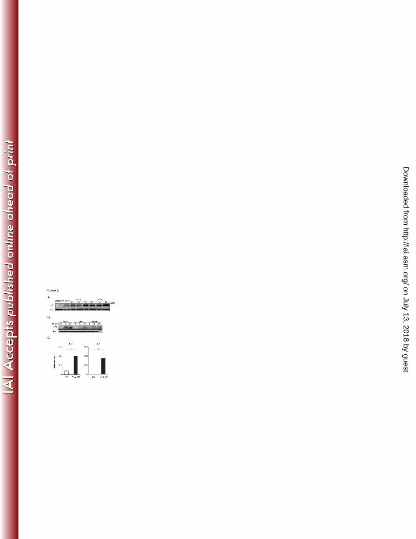
H. pylori infection induces IL-32 expression in gastric cancer cells. 223
Next, we investigated the expression of IL-32 in vitro using the gastric cancer cell lines. IL-1β and 224
TNF-α have been reported to strongly induce IL-32 in several cell lines (21, 37, 54). Twenty-four hours 225
after stimulation with IL-1β, TNF-α, and cagPAI-positive H. pylori strain TN2, IL-32 expression was 226
induced in AGS cells (Fig. 2A). Western blot analysis revealed that IL-32 was also induced by H. pylori 227
in the TMK-1 and MKN45 gastric cancer cell lines, in a time-dependent manner (Fig.2 B). IL-32 mRNA 228
levels were then determined by real-time RT-PCR. IL-32 mRNA levels in AGS cells increased after 229
infection with H. pylori for three hours, similar to the increase in IL-8 mRNA levels (Fig. 2C). These 230
results indicate that H. pylori enhances IL-32 mRNA and protein expression in infected gastric cells in 231
vitro. 232
233
IL-32 is induced by the NF-κB signaling pathway in AGS co-cultured with H. pylori. 234
on July 13, 2018 by guesthttp://iai.asm
.org/D
ownloaded from
14
Progression of gastric disease is affected by the presence of H. pylori virulence factors, such as cagPAI. 235
We next examined the role of cagPAI on IL-32 expression using isogenic mutant strains of TN2. 236
Immunoblot analysis showed that IL-32 expression after infection with TN2-ΔcagPAI and TN2-ΔcagE 237
was significantly weaker than that induced by wild-type H. pylori (Fig. 3A), suggesting that these 238
virulence factors up-regulate IL-32 expression. 239
Since cagPAI-positive H. pylori exhibits a higher capacity to activate NF-κB and MAPK, especially 240
in epithelial cells (11, 16), we next investigated the involvement of these signaling pathways in the 241
expression of IL-32 in H. pylori–infected cells. When we examined the effects of the chemical inhibitors 242
of IKKβ (SC-514) and p38 (SB203580) on H. pylori–induced IL-32 expression, H. pylori–induced 243
IL-32 mRNA expression was significantly reduced in cells pre-incubated with SC-514, but not with 244
SB203580 (Fig. 3B). To confirm the involvement of IKKβ in H. pylori–induced IL-32 expression, 245
siRNA for IKKβ was transfected into cells. H. pylori–induced IL-32 levels were reduced by IKKβ 246
siRNA (Fig. 3C). These results indicate that IL-32 is induced via the NF-κB signaling pathway in AGS 247
cells infected with cagPAI-positive H. pylori. 248
249
IL-32 is intracellular in H. pylori–infected cells. 250
Next, we investigated IL-32 expression by gastric cells. First, we examined IL-32 levels in the 251
supernatant of H. pylori–infected AGS cells. While 24 hours infection with wild-type H. pylori typically 252
on July 13, 2018 by guesthttp://iai.asm
.org/D
ownloaded from

15
produces 1000 pg/ml of IL-8 protein in AGS cell supernatants (data not shown), our ELISA results 253
showed that IL-32 concentrations were below the detection limit (< 16 pg/ml) in the same supernatant in 254
our ELISA. When we disrupted the cell membrane by freeze-thawing, IL-32 protein was detected in the 255
supernatant of AGS cells, suggesting an intracellular accumulation of IL-32 in H. pylori–infected cells 256
(Fig. 4A, left). We also examined intracellular protein levels in AGS cells with or without H. pylori 257
infection by ELISA. Lysates of uninfected AGS cells contained 800 pg/mg of IL-32. However, H. pylori 258
infection increased the intracellular IL-32 level significantly (Fig. 4A, right). 259
To address the localization of IL-32 in AGS cells, we performed immunofluorescence staining. As 260
shown in Fig. 4B, uninfected AGS cells contained low levels of IL-32 in the cytosol. H. pylori infection 261
increased both cytosolic and nuclear IL-32 levels, questioning whether IL-32 in gastric cells functions as 262
a typical cytokine. Thus, we examined whether extracellular IL-32 could activate intracellular signaling 263
in gastric epithelial cells, as it has been reported to do in macrophages (23). In contrast to TNF-α, 264
stimulation of AGS cells with recombinant IL-32β did not enhance IκBα, JNK or p38 phosphorylation in 265
AGS cells, suggesting that IL-32 has a specific intracellular function in gastric cells (Fig. 4C). 266
267
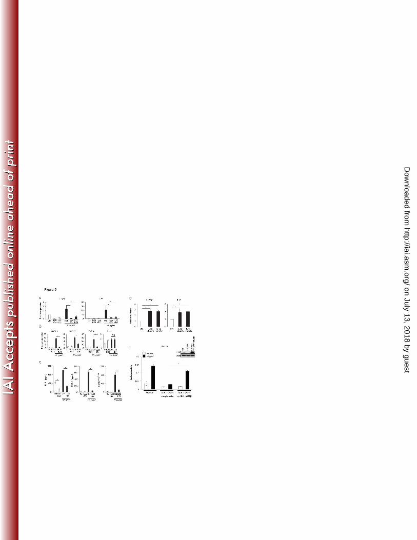
IL-32 expression up-regulates IL-8, CXCL1, CXCL2, and TNF-α expression. 268
Previous reports showed that IL-32 functions as a proinflammatory cytokine, which elicits production of 269
other inflammatory cytokines in diseases such as rheumatoid arthritis. Due to its intracellular 270
on July 13, 2018 by guesthttp://iai.asm
.org/D
ownloaded from

16
localization in AGS cells, next we determined whether IL-32 regulates inflammatory cytokines. We 271
established two stable IL-32-knockdown AGS cell lines, AGS-IL32KD1 and AGS-IL32KD2, and 272
decreased IL-32 mRNA expression was confirmed by real-time RT-PCR in both (Fig. 5A, left). Next, we 273
examined whether IL-32 affects the expression of cytokines in H. pylori–infected AGS cells. As shown 274
in Fig. 5A (right), the expression of IL-8 mRNA was significantly decreased in both AGS-IL32KD1 and 275
AGS-IL32KD2 cells co-cultured with H. pylori, compared with the control cell line. We also measured 276
the expression of other cytokines that are induced by H. pylori (17, 28, 48). CXCL1, CXCL2, and 277
TNF-α mRNA levels were also decreased in the IL-32-knockdown cells (Fig. 5B). IL-11 mRNA levels 278
were not decreased in the IL-32-knockdown cells (Fig. 5B). ELISA analysis indicated that IL-8, CXCL1, 279
and CXCL2 levels in supernatants of AGS-IL32KD1 cells co-cultured with H. pylori were decreased 280
compared to the control cell line (Fig. 5C). The levels of TNF-α were below the detection limit of the 281
ELISA (< 16 pg/ml, data not shown). To confirm the role of IL-32 in regulation of the expression of 282
other cytokines, we established two stable AGS cell lines (AGS IL-32 clone15, and AGS IL-32 clone19) 283
that over-expressed IL-32. IL-8 mRNA levels were increased in these IL-32 over-expressing cells. In the 284
two IL-32 over-expressing lines, IL-8 levels were two-fold higher than that in control cells (Fig. 5D). 285
Next we asked whether the reduced IL-8 levels in IL-32-knockdown AGS cells could be rescued by 286
over-expressing IL-32 cDNA. We re-introduced a mutant IL-32 cDNA (pcDNA-mIL32β), which was 287
genetically modified to be resistant to the inhibitory effects of the siRNA, into AGS-IL32KD1 cells and 288
on July 13, 2018 by guesthttp://iai.asm
.org/D
ownloaded from

17
examined IL-8 activity using reporter assays. Immunoblot analysis confirmed that IL-32 was suppressed 289
in AGS-IL32KD1, and could be rescued by over-expression of mIL-32 cDNA (Fig. 5E, upper right). The 290
suppression of H. pylori–induced IL-8 reporter activation in AGS-IL32KD1 was rescued by 291
over-expression of mIL-32 in AGS-IL32KD1 cells (Fig. 5E). These results suggest that IL-32 functions 292
as an intracellular regulator of cytokine expression in H. pylori–infected gastric cells. 293
294
IL-32 regulates NF-κB activation in H. pylori–infected AGS cells. 295
We examined whether IL-32 suppression impairs the type IV secretion system of H. pylori, which is 296
essential for induction of cytokine production in epithelial cells. CagA phosphorylation and the 297
subsequent hummingbird phenotype of infected AGS cells are the hallmark of the type IV secretion 298
system (43). Microscopic analyses indicated that both AGS-IL32KD1 and AGS control cells co-cultured 299
with H. pylori developed the hummingbird phenotype (Fig. 6A, upper). CagA was phosphorylated in 300
AGS-IL32KD1 and AGS control cells (Fig. 6A, lower), indicating that IL-32 expression does not affect 301
type IV secretion. 302
Next, we examined NF-κB activation in AGS IL-32 stable-knockdown cells. A luciferase reporter 303
assay indicated that NF-κB activation induced by H. pylori was decreased in AGS-IL32KD cells 304
compared with the control cell line (Fig. 6B). Immunoblot analysis showed that phosphorylation of IκBα 305
was decreased in AGS-IL32KD1 co-cultured with H. pylori, compared with the control cell line. This 306
on July 13, 2018 by guesthttp://iai.asm
.org/D
ownloaded from

18
indicates that NF-κB signaling is defective in AGS-IL32KD1 cells (Fig. 6C). 307
Finally, we tested the contribution of down-regulation of IL-8 mRNA to the level of IL-32 mRNA. 308
The siRNA for IL-8 was transfected into AGS cell lines, and decreased IL-8 mRNA expression was 309
confirmed by real-time RT-PCR (Fig. 6D, left). H. pylori–induced IL-32 levels were not significantly 310
different in AGS transfected with scramble siRNA and AGS transfected with siRNA for IL-8 (Fig. 6D, 311
right). 312
These results indicate that intracellular IL-32 expression is associated with NF-κB activation and 313
subsequent cytokine production in gastric epithelial cells infected with H. pylori. 314
315
DISCUSSION 316
In this study we show that expression of IL-32 is elevated in human gastritis and gastric cancer tissue. 317
We also demonstrate that IL-32 is induced by H. pylori in vitro. In addition, our results indicate that 318
IL-32 expression activates NF-κB and induces cytokine production, including IL-8. These results 319
suggest that IL-32 is fundamental to the gastric inflammation caused by H. pylori. H. pylori induces 320
NF-κB activation in gastric epithelial cells in a cagPAI-dependent manner, which is required for IL-32 321
up-regulation. In turn, intracellular IL-32 amplifies NF-κB activity and cytokine production, which 322
accelerates the inflammatory responses of gastric tissue infected with H. pylori (Fig. 6D). 323
IL-32 has six described mRNA splice variants, named IL-32α, β, γ, δ, ε, and ζ (12, 23). In our 324
on July 13, 2018 by guesthttp://iai.asm
.org/D
ownloaded from

19
analysis, IL-32β was the major isoform detected in the gastritis tissue. IL-32β is reportedly a major 325
isoform in endothelial cells, and it has a critical function in vascular inflammation and sepsis (25). The 326
longest IL-32 isoform, IL-32γ, was the most abundant in rheumatoid arthritis synovial tissue (14). 327
IL-32γ exerted the greatest induction of TNF-α in human peripheral blood mononuclear cells (4). 328
However, IL-32γ mRNA was not detected in gastritis tissue. A possible explanation for the differing 329
IL-32 isoform abundance among disease tissues may be that the splicing of IL-32 differs in different cell 330
types or due to varying inductive stimuli. The biological differences and the mechanisms of 331
tissue-specific expression of the IL-32 isoforms need to be investigated to understand the roles of this 332
cytokine in humans. 333
We also investigated the mechanisms of IL-32 induction by H. pylori in AGS cells. Previous reports 334
on IL-32 expression suggested the involvement of the JNK (33), p38 (24), ERK (37), and the NF-κB 335
(37) pathway. We found that cagPAI-positive H. pylori induced production of IL-32, in agreement with 336
the activation of NF-κB by cagPAI-positive H. pylori in the pathogenesis of gastric diseases (2, 11). As 337
expected, inhibition of the NF-κB pathway resulted in decreased IL-32 induction in the H. 338
pylori–infected gastric epithelial cell model. These results indicate that IL-32 is induced in gastric 339
epithelial cells through the activation of NF-κB by H. pylori infection. 340
To determine its role in gastritis, we examined the pattern of IL-32 expression in AGS cells, and 341
found it to be retained intracellularly. This is in contrast to most cytokines, which are generally secreted 342
on July 13, 2018 by guesthttp://iai.asm
.org/D
ownloaded from
20
from cells. In our investigation, recombinant IL-32β, unlike TNF-α, failed to activate IκBα, JNK, and 343
p38 in AGS cells (Fig. 4C), which differed from the situation in macrophages (23). Furthermore, IL-32 344
was not detected by ELISA in supernatants of AGS cells co-cultured with H. pylori. Thus, autocrine 345
signaling activation by IL-32 in gastric cells seems unlikely. Instead, disruption of the cell membrane 346
increased the IL-32 concentration in the supernatant to detectable levels (Fig. 4A). These results are 347
consistent with previous reports that described intracellular IL-32 expression and leakage from apoptotic 348
cells (12, 13, 26). Of note, neutrophil proteinase 3, an IL-32 binding protein, that is expressed in 349
neutrophils and monocytes is highly expressed in H. pylori–related gastritis (18). IL-32 from damaged 350
epithelial cells may activate myeloid cells through an IL-32-neutrophil-proteinase 3-interaction to 351
enhance inflammation of gastric tissue. Although intracellular receptors for IL-32 has not been 352
discovered, some interleukins such as IL-1 and IL-33 is known to have intracellular targets and to play 353
roles as both intracellular and secreted mediators (27). Thus, IL-32 may act intracellularly in gastric 354
epithelial cells and act extracellularly on myeloid cells. 355
We found that IL-32 expression levels increased concurrent with disease progression in the gastric 356
mucosa. IL-32 staining was increased in inflamed lesions of chronic pancreatitis, and strongly positive 357
in pancreatic cancer cells, suggesting the involvement of this cytokine in pancreatic carcinogenesis (36). 358
However, the mechanism by which IL-32 expression modifies the inflammatory processes ultimately 359
leading to malignancy is largely unknown. Our results showing that H. pylori–induced NF-κB activation 360
on July 13, 2018 by guesthttp://iai.asm
.org/D
ownloaded from

21
enhances IL-32 expression, and that IL-32 in turn amplifies the NF-κB signaling pathway, may 361
contribute to inflammation-induced carcinogenesis. Interestingly, when IL-32 was knocked down, 362
expression of the anti-apoptotic genes, Bcl-2 and Bcl-xL, in pancreatic cell lines was reduced (36); these 363
are regulated by the NF-κB pathway (20). Thus, it is plausible that IL-32 expression also amplifies 364
NF-κB signaling in pancreatic cancer cells. Taken together, our findings suggest that IL-32 is involved 365
in the inflammation-cancer process through amplification of NF-κB expression and up-regulation of its 366
targets, including IL-32 itself. 367
In conclusion, the inflammatory molecule IL-32 plays an important role in gastritis. H. pylori 368
infection induces IL-32 through NF-κB activation, in a cag-dependent manner. IL-32 amplifies the 369
NF-κB pathway and induces production of other inflammatory cytokines, including IL-8. IL-32 may be 370
a future target for medical treatment of gastric inflammation and carcinogenesis. 371
372
ACKNOWLEDGEMENTS 373
We thank Mitsuko Tsubouchi for technical assistance. We thank Dr. Takaaki Sano and Dr. Teiji 374
Motojima (Motojima General Hospital, Gumma, Japan) for provision of gastric carcinoma specimens. 375
This work was partly supported by a Grant-in-Aid for Scientific Research from the Ministry of 376
Education, Culture, Sports, Science and Technology of Japan to Y. Hirata. 377
378
on July 13, 2018 by guesthttp://iai.asm
.org/D
ownloaded from

22
REFERENCES 379
1. Aihara, M., D. Tsuchimoto, H. Takizawa, A. Azuma, H. Wakebe, Y. Ohmoto, K. Imagawa, 380
M. Kikuchi, N. Mukaida, and K. Matsushima. 1997. Mechanisms involved in Helicobacter 381
pylori-induced interleukin-8 production by a gastric cancer cell line, MKN45. Infection and 382
Immunity 65:3218-3224. 383
2. Backert, S., and M. Naumann. 2010. What a disorder: proinflammatory signaling pathways 384
induced by Helicobacter pylori. Trends in Microbiology 18:479-486. 385
3. Bai, X. Y., S. H. Kim, T. Azam, M. T. McGibney, H. Huang, C. A. Dinarello, and E. D. Chan. 386
2010. IL-32 Is a Host Protective Cytokine against Mycobacterium tuberculosis in Differentiated 387
THP-1 Human Macrophages. Journal of Immunology 184:3830-3840. 388
4. Choi, J. D., S. Y. Bae, J. W. Hong, T. Azam, C. A. Dinarello, E. Her, W. S. Choi, B. K. Kim, 389
C. K. Lee, D. Y. Yoon, S. J. Kim, and S. H. Kim. 2009. Identification of the most active 390
interleukin-32 isoform. Immunology 126:535-542. 391
5. Correa, P. 1992. Human gastric carcinogenesis - A multistep and multifactorial process - 1st 392
American-Cancer-Society award lecture on cancer-epidemiology and prevention. Cancer 393
Research 52:6735-6740. 394
6. Crabtree, J. E., A. Covacci, S. M. Farmery, Z. Xiang, D. S. Tompkins, S. Perry, I. J. D. 395
Lindley, and R. Rappuoli. 1995. Helicobacter-pylori induced interleukin-8 expression in gastric 396
on July 13, 2018 by guesthttp://iai.asm
.org/D
ownloaded from

23
epithelial-cells is associated with caga positive phenotype. Journal of Clinical Pathology 397
48:41-45. 398
7. Dahl, C. A., R. P. Schall, H. L. He, and J. S. Cairns. 1992. Identification of a novel gene 399
expressed in activated natural-killer-cells and t-cells. Journal of Immunology 148:597-603. 400
8. de Vries, A. C., N. C. T. van Grieken, C. W. N. Looman, M. K. Casparie, E. de Vries, G. A. 401
Meijer, and E. J. Kuipers. 2008. Gastric cancer risk in patients with premalignant gastric 402
lesions: A nationwide cohort study in the Netherlands. Gastroenterology 134:945-952. 403
9. El-Omar, E. M., M. Carrington, W. H. Chow, K. E. L. McColl, J. H. Bream, H. A. Young, J. 404
Herrera, J. Lissowska, C. C. Yuan, N. Rothman, G. Lanyon, M. Martin, J. F. Fraumeni, 405
and C. S. Rabkin. 2000. Interleukin-1 polymorphisms associated with increased risk of gastric 406
cancer. Nature 404:398-402. 407
10. Fukase, K., M. Kato, S. Kikuchi, K. Inoue, N. Uemura, S. Okamoto, S. Terao, K. Amagai, S. 408
Hayashi, M. Asaka, and G. Japan Gast Study. 2008. Effect of eradication of Helicobacter 409
pylori on incidence of metachronous gastric carcinoma after endoscopic resection of early gastric 410
cancer: an open-label, randomised controlled trial. Lancet 372:392-397. 411
11. Glocker, E., C. Lange, A. Covacci, S. Bereswill, M. Kist, and H. L. Pahl. 1998. Proteins 412
encoded by the cag pathogenicity island of Helicobacter pylori are required for NF-kappa B 413
activation. Infection and Immunity 66:2346-2348. 414
on July 13, 2018 by guesthttp://iai.asm
.org/D
ownloaded from

24
12. Goda, C., T. Kanaji, S. Kanaji, G. Tanaka, K. Arima, S. Ohno, and K. Izuhara. 2006. 415
Involvement of IL-32 in activation-induced cell death in T cells. International Immunology 416
18:233-240. 417
13. Hasegawa, H., H. J. Thomas, K. Schooley, and T. L. Born. 2011. Native IL-32 is released 418
from intestinal epithelial cells via a non-classical secretory pathway as a membrane-associated 419
protein. Cytokine 53:74-83. 420
14. Heinhuis, B., M. I. Koenders, F. A. van de Loo, M. G. Netea, W. B. van den Berg, and L. A. 421
B. Joosten. 2011. Inflammation-dependent secretion and splicing of IL-32 gamma in rheumatoid 422
arthritis. Proceedings of the National Academy of Sciences of the United States of America 423
108:4962-4967. 424
15. Hirata, Y., S. Maeda, W. Mitsuno, M. Akanuma, Y. Yamaji, K. Ogura, H. Yoshida, Y. 425
Shiratori, and M. Omata. 2001. Helicobacter pylori activates the cyclin D1 gene through 426
mitogen-activated protein kinase pathway in gastric cancer cells. Infection and Immunity 427
69:3965-3971. 428
16. Hirata, Y., S. Maeda, Y. Mitsuno, K. Tateishi, A. Yanai, M. Akanuma, H. Yoshida, T. 429
Kawabe, Y. Shiratori, and M. Omata. 2002. Helicobacter pylori CagA protein activates serum 430
response element-driven transcription independently of tyrosine phosphorylation. 431
Gastroenterology 123:1962-1971. 432
on July 13, 2018 by guesthttp://iai.asm
.org/D
ownloaded from

25
17. Hirata, Y., S. Maeda, T. Ohmae, W. Shibata, A. Yanai, K. Ogura, H. Yoshida, T. Kawabe, 433
and M. Omata. 2006. Helicobacter pylori induces I kappa B kinase at nuclear translocation and 434
chemokine production in gastric epithelial cells. Infection and Immunity 74:1452-1461. 435
18. Hong, S. N., S. Jo, J. H. Jang, J. Choi, S. Kim, S. Y. Ahn, J. H. Kim, W. H. Choe, S. Y. Lee, I. 436
K. Sung, H. S. Park, and C. S. Shim. 2012. Clinical Characteristics and the Expression Profiles 437
of Inflammatory Cytokines/Cytokine Regulatory Factors in Asymptomatic Patients with Nodular 438
Gastritis. Dig Dis Sci. 6:1486-95 439
19. Karin, M., T. Lawrence, and V. Nizet. 2006. Innate immunity gone awry: Linking microbial 440
infections to chronic inflammation and cancer. Cell 124:823-835. 441
20. Karin, M., and A. Lin. 2002. NF-kappa B at the crossroads of life and death. Nature 442
Immunology 3:221-227. 443
21. Kato, A., A. Keswani, R. T. Chustz, A. Peters, R. Chandra, R. Carter, L. Suh, J. Norton, K. 444
E. Harris, D. Conley, L. C. Grammer, S. Kim, C. A. Dinarello, R. C. Kern, and R. P. 445
Schleimer. 2010. IL-32 Expression in Chronic Rhinosinusitis. Journal of Allergy and Clinical 446
Immunology 125:AB61-AB61. 447
22. Kim, K. H., J. H. Shim, E. H. Seo, M. C. Cho, J. W. Kang, S. H. Kim, D. Y. Yu, E. Y. Song, 448
H. G. Lee, J. H. Sohn, J. Kim, C. A. Dinarello, and D. Y. Yoon. 2008. Interleukin-32 449
monoclonal antibodies for Immunohistochemistry, Western blotting, and ELISA. Journal of 450
on July 13, 2018 by guesthttp://iai.asm
.org/D
ownloaded from

26
Immunological Methods 333:38-50. 451
23. Kim, S. H., S. Y. Han, T. Azam, D. Y. Yoon, and C. A. Dinarello. 2005. Interleukin-32: A 452
cytokine and inducer of TNF alpha. Immunity 22:131-142. 453
24. Ko, N. Y., S. H. Mun, S. H. Lee, J. W. Kim, D. K. Kim, H. S. Kim, E. Her, S. H. Kim, H. S. 454
Won, H. S. Shin, Y. M. Kim, and W. S. Choi. 2011. Interleukin-32 alpha production is 455
regulated by MyD88-dependent and independent pathways in IL-1 beta-stimulated human 456
alveolar epithelial cells. Immunobiology 216:32-40. 457
25. Kobayashi, H., J. H. Huang, F. Ye, Y. Shyr, T. S. Blackwell, and P. C. Lin. 2010. 458
Interleukin-32 beta Propagates Vascular Inflammation and Exacerbates Sepsis in a Mouse Model. 459
Plos One 5. 460
26. Kobayashi, H., and P. C. Lin. 2009. Molecular characterization of IL-32 in human endothelial 461
cells. Cytokine 46:351-358. 462
27. Luheshi, N. M., N. J. Rothwell, and D. Brough. 2009. Dual functionality of interleukin-1 463
family cytokines: implications for anti-interleukin-1 therapy. British Journal of Pharmacology 464
157:1318-1329. 465
28. Maeda, S., M. Akanuma, Y. Mitsuno, Y. Hirata, K. Ogura, H. Yoshida, Y. Shiratori, and M. 466
Omata. 2001. Distinct mechanism of helicobacter pylori-mediated NF-kappa B activation 467
between gastric cancer cells and monocytic cells. Journal of Biological Chemistry 468
on July 13, 2018 by guesthttp://iai.asm
.org/D
ownloaded from

27
276:44856-44864. 469
29. Maeda, S., and A. F. Mentis. 2007. Pathogenesis of Helicobacter pylori Infection. Helicobacter 470
12:10-14. 471
30. Maeda, S., H. Yoshida, K. Ogura, Y. Mitsuno, Y. Hirata, Y. Yamaji, M. Akanuma, Y. 472
Shiratori, and M. Omata. 2000. H-pylori activates NF-kappa B through a signaling pathway 473
involving I kappa B kinases, NF-kappa B-inducing kinase, TRAF2, and TRAF6 in gastric cancer 474
cells. Gastroenterology 119:97-+. 475
31. Mitsuno, Y., H. Yoshida, S. Maeda, K. Ogura, Y. Hirata, T. Kawabe, Y. Shiratori, and M. 476
Omata. 2001. Helicobacter pylori induced transactivation of SRE and AP-1 through the ERK 477
signalling pathway in gastric cancer cells. Gut 49:18-22. 478
32. Moschen, A. R., T. Fritz, A. D. Clouston, I. Rebhan, O. Bauhofer, H. D. Barrie, E. E. Powell, 479
S. H. Kim, C. A. Dinarello, R. Bartenschlager, J. R. Jonsson, and H. Tilg. 2011. 480
Interleukin-32: A New Proinflammatory Cytokine Involved in Hepatitis C Virus-Related Liver 481
Inflammation and Fibrosis. Hepatology 53:1819-1829. 482
33. Mun, S. H., J. W. Kim, S. S. Nah, N. Y. Ko, J. H. Lee, J. D. Kim, D. K. Kim, H. S. Kim, J. D. 483
Choi, S. H. Kim, C. K. Lee, S. H. Park, B. K. Kim, Y. M. Kim, and W. S. Choi. 2009. Tumor 484
Necrosis Factor alpha-Induced Interleukin-32 Is Positively Regulated via the Syk/Protein Kinase 485
C delta/JNK Pathway in Rheumatoid Synovial Fibroblasts. Arthritis and Rheumatism 486
on July 13, 2018 by guesthttp://iai.asm
.org/D
ownloaded from

28
60:678-685. 487
34. Nahar, I. K., K. Shojania, C. A. Marra, A. H. Alamgir, and A. H. Anis. 2003. Infliximab 488
treatment of rheumatoid arthritis and Crohn's disease. Annals of Pharmacotherapy 37:1256-1265. 489
35. Netea, M. G., T. Azam, E. C. Lewis, L. A. B. Joosten, M. R. Wang, D. Langenberg, E. D. 490
Chan, D. Y. Yoon, T. Ottenhoff, S. H. Kim, and C. A. Dinarello. 2006. Mycobacterium 491
tuberculosis induces interleukin-32 production through a 492
caspase1/IL-18/interferon-gamma-dependent mechanism. Plos Medicine 3:1310-1319. 493
36. Nishida, A., A. Andoh, O. Inatomi, and Y. Fujiyama. 2009. Interleukin-32 Expression in the 494
Pancreas. Journal of Biological Chemistry 284:17868-17876. 495
37. Nold-Petry, C. A., M. F. Nold, J. A. Zepp, S. H. Kim, N. F. Voelkel, and C. A. Dinarello. 496
2009. IL-32-dependent effects of IL-1 beta on endothelial cell functions. Proceedings of the 497
National Academy of Sciences of the United States of America 106:3883-3888. 498
38. Ogura, K., Y. Hirata, A. Yanai, W. Shibata, T. Ohmae, Y. Mitsuno, S. Maeda, H. Watabe, Y. 499
Yamaji, M. Okamoto, H. Yoshida, T. Kawabe, and M. Omata. 2008. The effect of 500
Helicobacter pylori eradication on reducing the incidence of gastric cancer. Journal of Clinical 501
Gastroenterology 42:279-283. 502
39. Ogura, K., M. Takahashi, S. Maeda, T. Ikenoue, F. Kanai, H. Yoshida, Y. Shiratori, K. Mori, 503
K. I. Mafune, and M. Omata. 1998. Interleukin-8 production in primary cultures of human 504
on July 13, 2018 by guesthttp://iai.asm
.org/D
ownloaded from
29
gastric epithelial cells induced by Helicobacter pylori. Digestive Diseases and Sciences 505
43:2738-2743. 506
40. Ohmae, T., Y. Hirata, S. Maeda, W. Shibata, A. Yanai, K. Ogura, H. Yoshida, T. Kawabe, 507
and M. Omata. 2005. Helicobacter pylori activates NF-kappa B via the alternative pathway in B 508
lymphocytes. Journal of Immunology 175:7162-7169. 509
41. Pan, X. F., H. Cao, J. X. Lu, X. Shu, X. J. Xiong, X. L. Hong, Q. H. Xu, H. F. Zhu, G. Li, 510
and G. X. Shen. 2011. Interleukin-32 expression induced by hepatitis B virus protein X is 511
mediated through activation of NF-kappa B. Molecular Immunology 48:1573-1577. 512
42. Sakitani, K., Y. Hirata, H. Watabe, A. Yamada, T. Sugimoto, Y. Yamaji, H. Yoshida, S. 513
Maeda, M. Omata, and K. Koike. 2011. Gastric cancer risk according to the distribution of 514
intestinal metaplasia and neutrophil infiltration. Journal of Gastroenterology and Hepatology 515
26:1570-1575. 516
43. Segal, E. D., J. Cha, J. Lo, S. Falkow, and L. S. Tompkins. 1999. Altered states: Involvement 517
of phosphorylated CagA in the induction of host cellular growth changes by Helicobacter pylori. 518
Proceedings of the National Academy of Sciences of the United States of America 519
96:14559-14564. 520
44. Shioya, M., A. Nishida, Y. Yagi, A. Ogawa, T. Tsujikawa, S. Kim-Mitsuyama, A. Takayanagi, 521
N. Shimizu, Y. Fujiyama, and A. Andoh. 2007. Epithelial overexpression of interleukin-32 522
on July 13, 2018 by guesthttp://iai.asm
.org/D
ownloaded from
30
alpha in inflammatory bowel disease. Clinical and Experimental Immunology 149:480-486. 523
45. Shoda, H., K. Fujio, Y. Yamaguchi, A. Okamoto, T. Sawada, Y. Kochi, and K. Yamamoto. 524
2006. Interactions between IL-32 and tumor necrosis factor alpha contribute to the exacerbation 525
of immune-inflammatory diseases. Arthritis Research & Therapy 8. 526
46. Sorrentino, C., and E. Di Carlo. 2009. Expression of IL-32 in Human Lung Cancer Is Related 527
to the Histotype and Metastatic Phenotype. American Journal of Respiratory and Critical Care 528
Medicine 180:769-779. 529
47. Sugimoto, M., T. Furuta, N. Shirai, A. Nakamura, F. Xiao, M. Kajimura, H. Sugimura, and 530
A. Hishida. 2007. Different effects of polymorphisms of tumor necrosis factor-alpha and 531
interleukin-1 beta on development of peptic ulcer and gastric cancer. Journal of Gastroenterology 532
and Hepatology 22:51-59. 533
48. Sugimoto, M., T. Ohno, D. Y. Graham, and Y. Yamaoka. 2011. Helicobacter pylori outer 534
membrane proteins on gastric mucosal interleukin 6 and 11 expression in Mongolian gerbils. 535
Journal of Gastroenterology and Hepatology 26:1677-1684. 536
49. Suzuki, M., M. Mori, A. Miyayama, N. Iwai, N. Tsunematsu, M. Oonuki, H. Suzuki, T. Hibi, 537
and H. Ishii. 1997. Enhancement of neutrophil infiltration in the corpus after failure of 538
Helicobacter pylori eradication. Journal of Clinical Gastroenterology 25:S222-S228. 539
50. Taguchi, A., N. Ohmiya, K. Shirai, N. Mabuchi, A. Itoh, Y. Hirooka, Y. Niwa, and H. Goto. 540
on July 13, 2018 by guesthttp://iai.asm
.org/D
ownloaded from

31
2005. Interleukin-8 promoter polymorphism increases the risk of atrophic gastritis and gastric 541
cancer in Japan. Cancer Epidemiology Biomarkers & Prevention 14:2487-2493. 542
51. Uemura, N., S. Okamoto, S. Yamamoto, N. Matsumura, S. Yamaguchi, M. Yamakido, K. 543
Taniyama, N. Sasaki, and R. J. Schlemper. 2001. Helicobacter pylori infection and the 544
development of gastric cancer. New England Journal of Medicine 345:784-789. 545
52. Warren, J. R., Marshall, B. J. 1983. Unidentified curved bacilli on gastric epithelium in active 546
chronic gastritis. Lancet 1:1273-1273. 547
53. Wong, B. C. Y., S. K. Lam, W. M. Wong, J. S. Chen, T. T. Zheng, R. E. Feng, K. C. Lai, W. 548
H. C. Hu, S. T. Yuen, S. Y. Leung, D. Y. T. Fong, J. Ho, C. K. Ching, and G. China Gastric 549
Canc Study. 2004. Helicobacter pylori eradication to prevent gastric cancer in a high-risk region 550
of China - A randomized controlled trial. Jama-Journal of the American Medical Association 551
291:187-194. 552
54. Yagi, Y., A. Andoh, H. Imaeda, T. Aomatsu, R. Ohsaki, O. Inatomi, S. Bamba, T. Tsujikawa, 553
T. Shimizu, and Y. Fujiyama. 2011. Interleukin-32 alpha expression in human colonic 554
subepithelial myofibroblasts. International Journal of Molecular Medicine 27:263-268. 555
556
FIGURE LEGENDS 557
FIG 1. Expression of IL-32 in human gastric tissues. 558
on July 13, 2018 by guesthttp://iai.asm
.org/D
ownloaded from
32
(A) Immunoblot analysis of IL-32 protein in normal human gastric tissues, gastritis tissues, and gastric 559
cancer tissues. (B) Representative immunohistochemical analysis of IL-32 expression in human gastritis 560
tissue (right). Mouse IgG1 isotype antibody was the negative control (left). (C) Typical examples of 561
immunohistochemical analysis of IL-32 expression in normal human gastric tissue (left), gastritis tissue 562
(center), and gastric cancer tissue (right). (D) Graphic representation of IL-32 expression in human 563
gastric samples, including normal gastric mucosa, gastritis tissues and gastric cancer tissues. Expression 564
was quantified by ELISA. The box plot indicates the median (horizontal line), inter-quartile range (the 565
box itself), and the sample minimum and maximum (bars). *, p <0.05 by Tukey’s HSD. (E) The 566
full-length coding region of IL-32 cDNA, synthesized from RNA extracted from human gastritis tissue 567
and AGS cells infected with H. pylori, was amplified by PCR (left). The known structure of IL-32 568
isoforms (splice variants) and the number of each clone detected in human gastritis tissue are depicted 569
(right). The exon numbers are shown beneath the boxes. 570
FIG 2. H. pylori infection induces IL-32 expression in gastric cancer cells. 571
(A) AGS cells were infected with cagPAI-positive H. pylori strains TN2 for 24 h, or stimulated with 572
IL-1β, and TNF-α for 24 h at the indicated concentrations. IL-32 expression was detected by 573
immunoblot analysis. (B) AGS, TMK-1 and MKN45, gastric cancer cell lines, were co-cultured with H. 574
pylori for the indicated hours, and IL-32 expression was evaluated by immunoblot analysis. (C) IL-32 575
on July 13, 2018 by guesthttp://iai.asm
.org/D
ownloaded from

33
and IL-8 mRNA levels in AGS cells after infection with H. pylori for six hours were determined by 576
real-time RT-PCR. Data are shown as means ± SEM (n=3). *, p <0.05 by Student’s t-test. 577
FIG 3. H. pylori infection induces IL-32 production in AGS cells via the NF-κB signaling pathway. 578
(A) Levels of Il-32 in AGS cells infected with wild-type H. pylori (TN2-WT), TN2-ΔcagPAI, and 579
TN2-ΔcagE for the indicated times were determined by immunoblot analysis. Anti-urease staining was 580
used to control for H. pylori strain variations. (B) H. pylori–induced IL-32 mRNA expression with or 581
without chemical inhibitors was examined by real-time RT-PCR. The IKKβ inhibitor SC-514 (SC) and 582
p38 inhibitor SB203580 (SB) were added at a concentration of 20 μM, one-hour before H. pylori 583
infection. Cells were co-cultured with H. pylori for 24 h. Data are the means ± SEM (n=3). *, p <0.05 by 584
Dunnett’s multiple comparison. NS indicates not significant. (C) H. pylori-induced IL-32 expression in 585
AGS cells treated with control siRNA and IKKβ siRNA as determined by immunoblot analysis. AGS 586
cells were transfected with the siRNAs for 48 h and then infected with H. pylori for the indicated times. 587
588
FIG 4. IL-32 is intracellular in H. pylori–infected cells. 589
(A) IL-32 concentrations in the supernatant were quantitated by ELISA. Supernatants were collected 590
from AGS cells infected with H. pylori for 24 h, and the same cells that had been freeze-thawed to 591
facilitate cell membrane destruction (Fig. 4A, left). IL-32 concentrations in AGS cell lysates with or 592
on July 13, 2018 by guesthttp://iai.asm
.org/D
ownloaded from

34
without H. pylori infection for 24 h were determined by ELISA (Fig. 4A, right). Data are shown as 593
means ± SEM (n=3). * p <0.05 by Student’s t-test. (B) AGS cells were co-cultured with H. pylori for 24 594
h, and IL-32 expression was analyzed by immunofluorescence staining. Nuclear staining was conducted 595
using Hoechst 33342. (C) Immunoblots of the indicated proteins in AGS cells stimulated with TNF-α (1 596
ng/ml), and recombinant IL-32β (1 ng/ml) for 0, 15, 30, and 60 min. 597
FIG 5. IL-32 expression affects induction of other cytokines involved in gastritis. 598
(A) Control (Ctrl) and stable IL-32 knockdown AGS cell lines (IL32KD1 and IL32KD2) were cultured 599
with or without H. pylori for six hours. IL-32β mRNA and IL-8 mRNA levels were determined by 600
real-time RT-PCR. Data are shown as means ± SEM (n=3). *, p <0.05 by Dunnett’s multiple comparison. 601
(B) CXCL1, CXCL2, TNF-α and IL-11 mRNA levels were evaluated in AGS control cells (Ctrl) and 602
IL32KD1 cells cultured with or without H. pylori for six hours by real-time RT-PCR. Data are shown as 603
means ± SEM (n=3). *, p <0.05 by Student’s t-test. NS indicates not significant. (C) ELISA of IL-8, 604
CXCL1, and CXCL2 expression in AGS cell supernatants of control (Ctrl) and IL32KD1 cells cultured 605
with or without H. pylori for 24 h. Data are shown as means ± SEM (n=3). *, p <0.05 by Student’s t-test. 606
(D) Two stable IL-32-overexpressing AGS cell lines (AGS IL-32 clone 15, and AGS IL-32 clone 19) 607
were established. The increased IL-32 mRNA level was confirmed by real-time RT-PCR (left). IL-8 608
mRNA levels in the control (Ctrl), IL-32 clone 15, and IL-32 clone 19 cells were determined by 609
on July 13, 2018 by guesthttp://iai.asm
.org/D
ownloaded from
35
real-time RT-PCR (right). Data are shown as means ± SEM (n=3). *, p <0.05 by Dunnett’s multiple 610
comparison. (E) IL-8 reporter activity was examined in AGS control (Ctrl), IL32KD1, and IL32KD1 611
cells transfected with IL-32β cDNA (pcDNA-mIL32β). Cells were left uninfected or infected with H. 612
pylori for 24 h. Data are shown as means ± SEM (n=3). *, p <0.05 by Student’s t-test. IL-32 levels in 613
each cell are shown in the upper right panel. 614
615
FIG 6. Role of IL-32 in H. pylori–induced cell signaling. 616
(A) Micrographs of uninfected AGS cells transfected with the non-silencing control (AGS Ctrl), AGS 617
Ctrl cells infected with H. pylori, and stable IL-32 knockdown AGS (AGS-IL32KD1) cells infected with 618
H. pylori for 24 h (Fig. 6A, upper). Cell lysates of uninfected AGS Ctrl cells, AGS Ctrl cells infected 619
with H. pylori, and AGS-IL32KD1 cells infected with H. pylori for 24 h were immunoprecipitated using 620
anti-CagA antibody and the immunoprecipitates were immunoblotted using anti-phosphotyrosine and 621
anti-CagA antibodies (Fig. 6A, lower). (B) NF-κB activation induced by 24-h H. pylori infection of AGS 622
Ctrl cells and AGS-IL32KD1 was determined using luciferase activity assays. Data are shown as means 623
± SEM (n=3). *, p <0.05 by Student’s t-test. (C) Immunoblot analysis of IL-32 and pIκBα levels in AGS 624
Ctrl cells and AGS-IL32KD1 cells infected with H. pylori for the indicated times. Anti-urease staining 625
was used to control the H. pylori-load. (D) H. pylori-induced IL-32β mRNA (Fig. 6D right) and IL-8 626
mRNA (Fig. 6D left) levels in AGS cells treated with control siRNA (AGS Ctrl) and IL-8 siRNA 627
on July 13, 2018 by guesthttp://iai.asm
.org/D
ownloaded from

36
(AGS-siIL8) as determined by real-time RT-PCR. AGS cells were transfected with the siRNAs for 48 h 628
and then infected with H. pylori for an additional 24 h. Data are shown as means ± SEM (n=3). *, p 629
<0.05 by Student’s t-test. NS indicates not significant. (E) Summary of the role of IL-32 in gastritis. H. 630
pylori induces NF-κB activation in gastric epithelial cells in a cagPAI-dependent manner. NF-κB 631
activation is required for IL-32 expression. Intracellular IL-32 expression amplifies both NF-κB 632
activation and IL-8 expression. 633
634
635
on July 13, 2018 by guesthttp://iai.asm
.org/D
ownloaded from