HyProCure published studies
19
Published Studies on the Safety and Efficacy of the Sinus Tarsi Stent. October - 2016
-
Upload
gramedica -
Category
Health & Medicine
-
view
120 -
download
0
Transcript of HyProCure published studies
Published Studies on the Safety and Efficacy of the
Sinus Tarsi Stent.
October - 2016
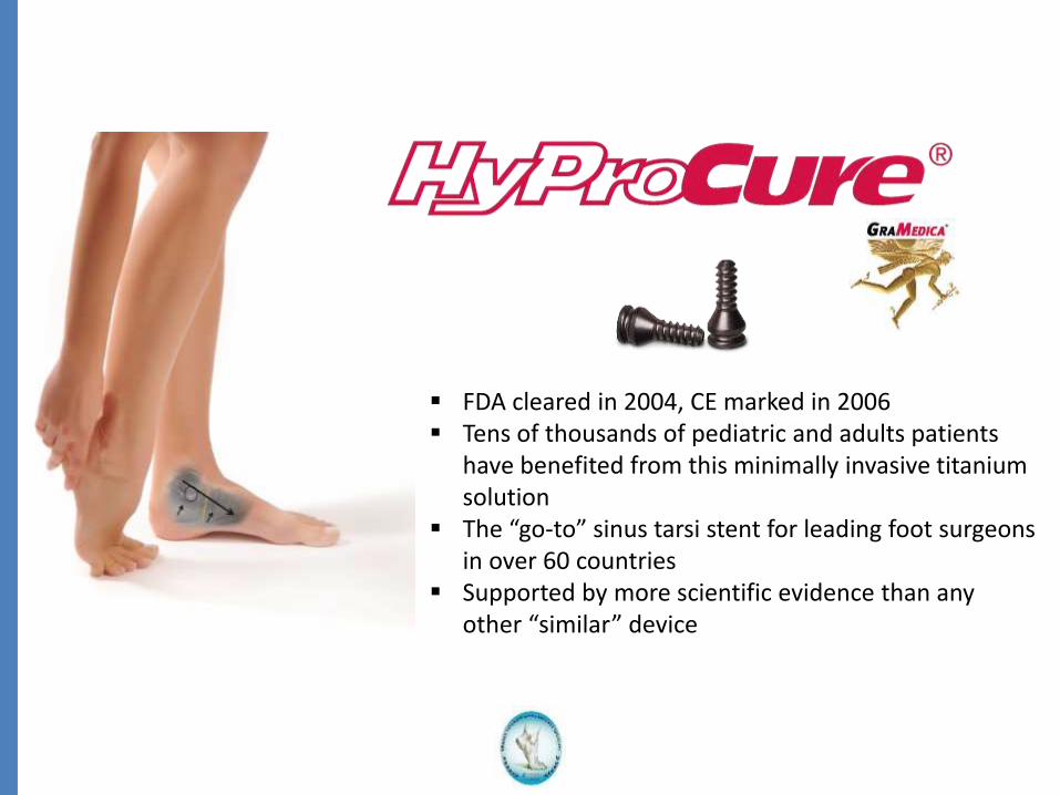
FDA cleared in 2004, CE marked in 2006 Tens of thousands of pediatric and adults patients
have benefited from this minimally invasive titanium solution
The “go-to” sinus tarsi stent for leading foot surgeons in over 60 countries
Supported by more scientific evidence than any other “similar” device

HyProCure® is proven to
Decrease tibialis posterior nerve
strain/elongation
Decrease pressures in the tarsal tunnel
& porta pedis
Decrease strain on the plantar fascia
Decrease strain on the posterior tibial tendon
Normalize both sagittal and transverse plane
displacement
Restore/maintain navicular height
Decrease forces acting on the medial column
Normalize plantar forefoot pressures

HyProCure®
Classified as a Type II, non-arthroereisis sinus tarsi stent
Stabilizes the talocalcaneal joint entrance of the canalis tarsi to re-establish the talotarsal joint center of axis.
Rebalances forces acting on the tarsal mechanism, reduces the forces acting on medial column of the foot.
Reduces strain to the tibialis posterior tendon by a mean of 51%
Reduces strain to the medial band of the plantar fascia by a mean of 33%
Decreases strain on the tibialis posterior nerve by a mean of15%
decreases the pressures within the tarsal tunnel by a mean of 34% and the porta pedis by a mean of 38%
prevents navicular sag/drop by 26%
normalize plantar foot pressures – prospective study
Restores both transverse and sagittal plane talotarsal joint displacement deformities
6% permanent removal rate in adults as a stand alone procedure – retrospective analysis
has a 4% removal rate in pediatric and adult patients- prospective analysis
superior option to realign the hindfoot over arch supports/foot orthotics
superior option in the treatment of pediatric talotarsal joint displacement
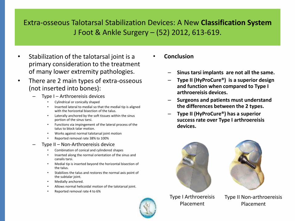
Extra-osseous Talotarsal Stabilization Devices: A New Classification SystemJ Foot & Ankle Surgery – (52) 2012, 613-619.
• Stabilization of the talotarsal joint is a primary consideration to the treatment of many lower extremity pathologies.
• There are 2 main types of extra-osseous (not inserted into bones):– Type I – Arthroereisis devices
• Cylindrical or conically shaped
• Inserted lateral to medial so that the medial tip is aligned with the horizontal bisection of the talus.
• Laterally anchored by the soft tissues within the sinus portion of the sinus tarsi.
• Functions via impingement of the lateral process of the talus to block talar motion.
• Works against normal talotarsal joint motion
• Reported removal rate 38% to 100%
– Type II – Non-Arthroereisis device• Combination of conical and cylindered shapes
• Inserted along the normal orientation of the sinus and canalis tarsi.
• Medial tip is inserted beyond the horizontal bisection of the talus.
• Stabilizes the talus and restores the normal axis point of the subtalar joint.
• Medially anchored.
• Allows normal helicoidal motion of the talotarsal joint.
• Reported removal rate 4 to 6%
• Conclusion
– Sinus tarsi implants are not all the same.
– Type II (HyProCure®) is a superior design and function when compared to Type I arthroereisis devices.
– Surgeons and patients must understand the differences between the 2 types.
– Type II (HyProCure®) has a superior success rate over Type I arthroereisis devices.
Type I ArthroereisisPlacement
Type II Non-arthroereisisPlacement
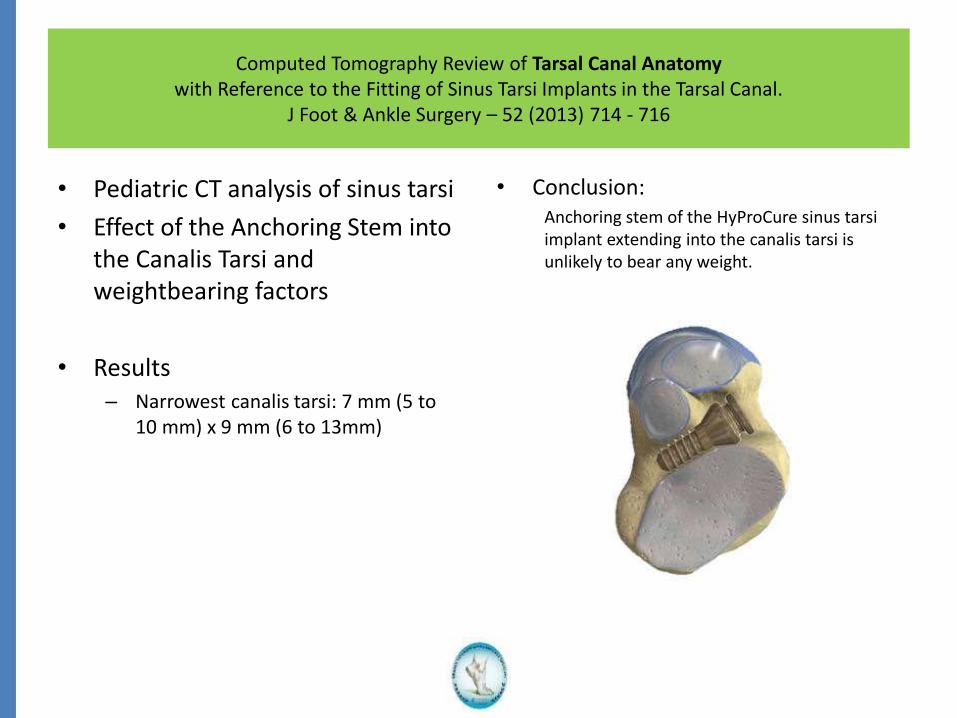
Computed Tomography Review of Tarsal Canal Anatomy with Reference to the Fitting of Sinus Tarsi Implants in the Tarsal Canal.
J Foot & Ankle Surgery – 52 (2013) 714 - 716
• Pediatric CT analysis of sinus tarsi
• Effect of the Anchoring Stem into the Canalis Tarsi and weightbearing factors
• Results – Narrowest canalis tarsi: 7 mm (5 to
10 mm) x 9 mm (6 to 13mm)
• Conclusion:Anchoring stem of the HyProCure sinus tarsi implant extending into the canalis tarsi is unlikely to bear any weight.

Stabilization of Joint Forces of the Subtalar Complex via HyProCure Sinus Tarsi Stent.
J American Podiatric Medical Association 101 (5) 2011 390 - 399
• Recurrent talotarsal joint instability leads to an anteriomedial displacement of the talus on the calcaneus.
• Excessive forces are acting anteriomedially rather than posteriolateral.
• Results– RTTJD directly increases the forces acting
on the medial structures of the foot.
– HyProCure® stabilized the talus on the calcaneus (subtalar joints).
– There was a reduction of anteriorly acting forces by an average of 24%.
– There was an increase of posterior forces by 21%
• Conclusion
– Subtalar joint instability leads to increased forces acting anteriomedially.
– HyProCure® stabilized the talus on the calcaneus keeping the joints in constant congruent contact.
– Forces acting on the medial column of the foot are reduced with HyProCure®
– Patients who have symptoms to medial column related tissues/structures should be evaluated for subtalar joint instability and HyProCure has been proven to stabilize the joint and normalize joint forces.
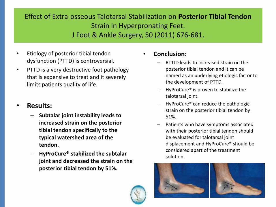
Effect of Extra-osseous Talotarsal Stabilization on Posterior Tibial Tendon Strain in Hyperpronating Feet.
J Foot & Ankle Surgery, 50 (2011) 676-681.
• Etiology of posterior tibial tendon dysfunction (PTTD) is controversial.
• PTTD is a very destructive foot pathology that is expensive to treat and it severely limits patients quality of life.
• Results:– Subtalar joint instability leads to
increased strain on the posterior tibial tendon specifically to the typical watershed area of the tendon.
– HyProCure® stabilized the subtalar joint and decreased the strain on the posterior tibial tendon by 51%.
• Conclusion:– RTTJD leads to increased strain on the
posterior tibial tendon and it can be named as an underlying etiologic factor to the development of PTTD.
– HyProCure® is proven to stabilize the talotarsal joint.
– HyProCure® can reduce the pathologic strain on the posterior tibial tendon by 51%.
– Patients who have symptoms associated with their posterior tibial tendon should be evaluated for talotarsal joint displacement and HyProCure® should be considered apart of the treatment solution.
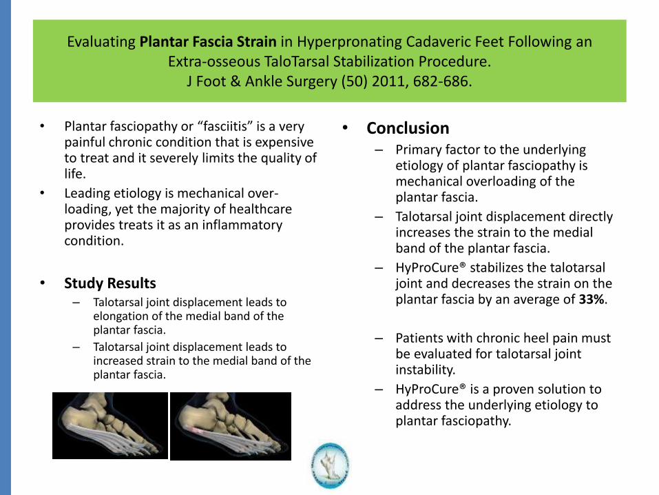
Evaluating Plantar Fascia Strain in Hyperpronating Cadaveric Feet Following an Extra-osseous TaloTarsal Stabilization Procedure.
J Foot & Ankle Surgery (50) 2011, 682-686.
• Plantar fasciopathy or “fasciitis” is a very painful chronic condition that is expensive to treat and it severely limits the quality of life.
• Leading etiology is mechanical over-loading, yet the majority of healthcare provides treats it as an inflammatory condition.
• Study Results– Talotarsal joint displacement leads to
elongation of the medial band of the plantar fascia.
– Talotarsal joint displacement leads to increased strain to the medial band of the plantar fascia.
• Conclusion– Primary factor to the underlying
etiology of plantar fasciopathy is mechanical overloading of the plantar fascia.
– Talotarsal joint displacement directly increases the strain to the medial band of the plantar fascia.
– HyProCure® stabilizes the talotarsal joint and decreases the strain on the plantar fascia by an average of 33%.
– Patients with chronic heel pain must be evaluated for talotarsal joint instability.
– HyProCure® is a proven solution to address the underlying etiology to plantar fasciopathy.

The Effect of HyProCure® on Tarsal Tunnel Compartment Pressures in Hyperpronating Feet.
J Foot and Ankle Surgery, (50) 2011, 44-49.
• Recurrent talotarsal dislocation leads to a prolonged period of pronation.
• Hyperpronation is a lead etiology of tarsal tunnel syndrome.
• Traditional tarsal tunnel surgery has a recurrence rate of 60%, possibly due to the fact the underlying etiology was not adequately reduced/eliminated.
• Pressures within the tarsal tunnel and porta pedis of cadaver feet were examined.
• Results:Tarsal Tunnel
TTJ in neutral position was 3 mmHgTTJ maximally pronated jumped to 32 mmHg (9 to 72 mmHg).After insertion of HyProCure® with maximum pronation were 21 mmHg (10-53mmHg). Reduction by 34%.
Porta PedisTTJ in neutral position was 2 mmHgTTJ maximally pronated jumped to 29 mmHg (10 – 73 mmHg)After insertion of HyProCure® with maximum pronation were 18mmHg (5 - 51. Reduction by 38%.
Clinical Significance:Pressures of 20-30 mmHg
Inhibits intra-neural blood flow
Alters nerve conduction
Leads to neural ischemia
Impairs axonal flow
Leads to demyelination
Hyperpronation/RTTJD leads to neuropathic changes to the posterior tibial nerve resulting in pain and numbness to the plantar aspect of the foot.
Conclusion:EOTTS with HyProCure stabilizes the talotarsal joint, decreasing the pressures within both the tarsal tunnel and porta pedis. This could help to reduce the symptoms associated with posterior tibial neuropathy and tarsal tunnel syndrome.
EOTTS with HyProCure® could be performed as a primary intervention or in combination with tarsal tunnel/porta pedis decompression surgery.

Effect of Extra-osseous Talotarsal Stabilization on Posterior Tibial Nerve Strain in Hyperpronating Feet: A Cadaveric Evaluation.
J Foot Ankle Surgery, (50) 2011, 672 – 675.
• Posterior tibial nerve neuropathy leads to numbness and pain to the plantar aspect of the foot.
• Pressures within the tarsal tunnel and porta pedis can adversely affect nerve function/health.
• Elongation of posterior tibial nerve is rarely taken into consideration.
• Talotarsal joint instability could lead to increased elongation of the posterior tibial nerve.
• Since HyProCure® is a proven solution to stabilize the talotarsal joint there could be a benefit to the decrease the elongation of the nerve.
• Results:– Maximum pronation of the talotarsal joint
instability lead to 6mm elongation of the nerve (range: 3 – 7mm).
– Upon insertion of HyProCure® the maximum elongation was reduced to 3mm (range: 1 to 5mm).
– Nerve strain with no intervention had a mean 27% (range: 13 – 34%)
– Nerve strain after placement of HyProCure® was reduced to a mean of 15% (range: 5 – 24%).
• Clinical significance:
6% nerve stain leads to a decreased action potential.8% strain leads to impaired venular flow.12% strain lead to complete nerve block.15% strain leads to impaired arterial flow and can lead to a irreversible functional deficit.
Conclusion:RTTJD leads to pathologic increased pressures on the posterior tibial nerve within the tarsal tunnel and porta pedis.
RTTJD also leads to an elongation and increased strain on the posterior tibial nerve.
Stabilization of the TTJ with HyProCure® has proven to significantly decrease/normalize both elongation and strain on the nerve.
EOTTS-HyProCure® offers a proven rational in the treatment of plantar foot neuropathy.
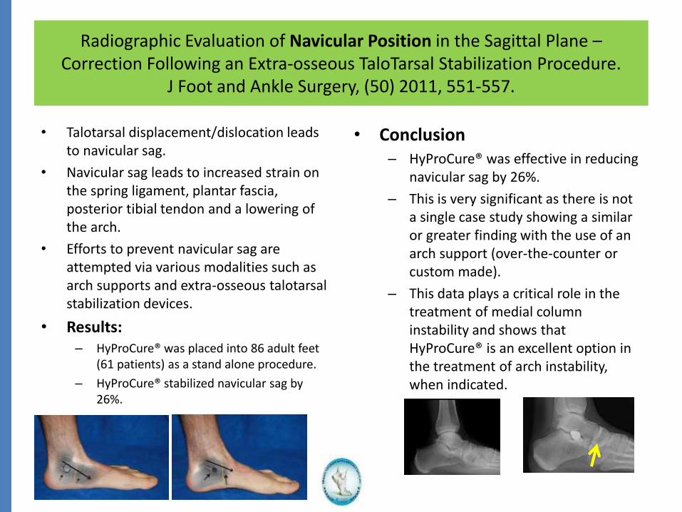
Radiographic Evaluation of Navicular Position in the Sagittal Plane –Correction Following an Extra-osseous TaloTarsal Stabilization Procedure.
J Foot and Ankle Surgery, (50) 2011, 551-557.
• Talotarsal displacement/dislocation leads to navicular sag.
• Navicular sag leads to increased strain on the spring ligament, plantar fascia, posterior tibial tendon and a lowering of the arch.
• Efforts to prevent navicular sag are attempted via various modalities such as arch supports and extra-osseous talotarsal stabilization devices.
• Results:– HyProCure® was placed into 86 adult feet
(61 patients) as a stand alone procedure.
– HyProCure® stabilized navicular sag by 26%.
• Conclusion– HyProCure® was effective in reducing
navicular sag by 26%.
– This is very significant as there is not a single case study showing a similar or greater finding with the use of an arch support (over-the-counter or custom made).
– This data plays a critical role in the treatment of medial column instability and shows that HyProCure® is an excellent option in the treatment of arch instability, when indicated.
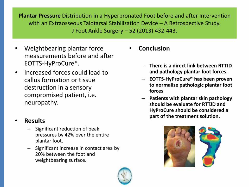
Plantar Pressure Distribution in a Hyperpronated Foot before and after Intervention with an Extraosseous Talotarsal Stabilization Device – A Retrospective Study.
J Foot Ankle Surgery – 52 (2013) 432-443.
• Weightbearing plantar force measurements before and after EOTTS-HyProCure®.
• Increased forces could lead to callus formation or tissue destruction in a sensory compromised patient, i.e. neuropathy.
• Results– Significant reduction of peak
pressures by 42% over the entire plantar foot.
– Significant increase in contact area by 20% between the foot and weightbearing surface.
• Conclusion
– There is a direct link between RTTJD and pathology plantar foot forces.
– EOTTS-HyProCure® has been proven to normalize pathologic plantar foot forces
– Patients with plantar skin pathology should be evaluate for RTTJD and HyProCure should be considered a part of the treatment solution.
Surgical Treatment of Hyperpronation Using an Extra-osseous Talotarsal Stabilization Device: Radiographic Outcomes in Adult Patients.
J Foot and Ankle Surgery, (51) 2012, 548-555.
• The use of HyProCure® has been well tolerated and shown to decrease strain and forces to many areas of the foot.
• This study reviewed DP/lateral weightbearing x-rays of 95 feet (70 patients) who underwent an EOTTS with HyProCure® as a stand-alone procedure.
• Mean age was 58 (22 – 85)
• X-rays were taken once patient had not pain or guarding from standing/walking that could compromise the findings.
• Results:– All 95 feet had a transverse plane component
– 65 of the 95 feet also had a sagittal plane component
– All patients had normalization/reduction of the transverse plane component.
– All patients had normalization/reduction of the sagittal plane component.
– No patient had normalization of one plane component where another plane became pathologic
– There was no significant effect to calcaneal inclination angle.
• Results -Continued– Transverse plane correction via talar 2nd
metatarsal angle via DP x-ray• Pre-op = 25 degrees
• Post-HyProCure® = 6 degrees
• Mean normalization = 19 degrees (77%)
• Maximum correction =
– Sagittal Plane correction via talar declination angle via lateral x-ray
• Pre-op = 25 degrees
• Post-HyProCure® = 19 degrees
• Mean normalization = 6 degrees (23%)
• Maximum correction =
– Not a single measurement showed an over-correction
• Conclusion– Transverse and sagittal plane normalization
with HyProCure® as a stand alone procedure is proven via weightbearing radiographic evidence in adult patients.
– Average and maximum guidelines on the amount of expected correction is provided.
Extra-osseous TaloTarsal Stabilization using HyProCure® in Adults: A 5 year Retrospective follow up.
J Foot & Ankle Surgery (51) 2012, 23-29.
• 1st review of EOTTS as a stand-alone procedure in adults.
• 83 adults 18 or older at time of surgery
• 117 feet/cases
• Mean age 58 (22-85)
• Average follow up 51 months
• Results:– 6% removal rate (compared to 40% or
greater with Type I arthroereisis devices)
– Patient satisfaction score showed excellent long-term results
• Conclusion– HyProCure® has been proven to have
the highest success rate over any other EOTTS device .
– HyProCure® has the lowest removal rate over any EOTTS device (6% compared to 38% to 100% of arthroereisis devices).
– Even though there were removals and/or revisions there were no long-term complications.
– Not a single patient/foot developed chronic pain post-HyProCure® removal.
– Patients who required a revision went on to a successful outcome.
– HyProCure® is a safe and effective option for adult patients with recurrently talotarsal dislocation, when indicated.
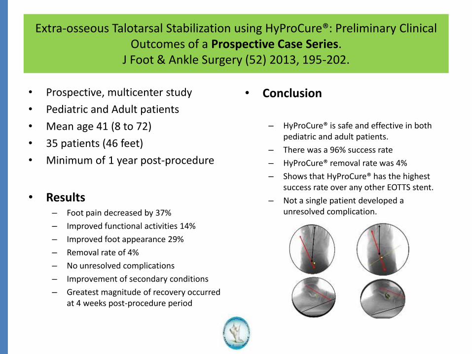
Extra-osseous Talotarsal Stabilization using HyProCure®: Preliminary Clinical Outcomes of a Prospective Case Series.
J Foot & Ankle Surgery (52) 2013, 195-202.
• Prospective, multicenter study
• Pediatric and Adult patients
• Mean age 41 (8 to 72)
• 35 patients (46 feet)
• Minimum of 1 year post-procedure
• Results– Foot pain decreased by 37%
– Improved functional activities 14%
– Improved foot appearance 29%
– Removal rate of 4%
– No unresolved complications
– Improvement of secondary conditions
– Greatest magnitude of recovery occurred at 4 weeks post-procedure period
• Conclusion
– HyProCure® is safe and effective in both pediatric and adult patients.
– There was a 96% success rate
– HyProCure® removal rate was 4%
– Shows that HyProCure® has the highest success rate over any other EOTTS stent.
– Not a single patient developed a unresolved complication.

Analysis of Radiographic Outcomes Comparing Foot Orthosis to Extra-osseous Talotarsal Stabilization in the Treatment of Recurrent Talotarsal Joint Dislocation.
J Minimally Invasive Orthopedics – January 2015
• Multicenter Prospective Study
• Compared weightbearing radiographs
barefoot – no intervention
barefoot - orthosis
barefoot - EOTTS-HyProCure®
Results:
Transverse plane improvement
orthosis = 3.2% average change
EOTTS = 58.9% average change
Sagittal plane improvement
orthosis = 2.2 % average change
EOTTS = 28.3% average change
• Conclusions:
– EOTTS-HyProCure® is a very effective form of treatment to realign and stabilize RTTJD.
– Orthosis are ineffective in the realignment and stabilization of RTTJD
– EOTTS is a superior option over an arch supports/orthosis in realigning and stabilizing the TTJ.

Pediatric Congenital TaloTarsal Joint Displacement and Pes Planovalgus Evaluation, Conservative Management, and Surgical Management.
Clinics Podiatric Medicine and Surgery – (30) 2013, 567-581.
• Recurrent Talotarsal joint displacement/dislocation/instability is a pathologic deformity that does not improve with age.
• RTTJD does not get better, it gets worse.
• RTTJD leads to many secondary pathologies to the lower extremity and can adversely affect health in general.
• Results:– RTTJD is diagnosed via physical examination (non-
weightbearing/weightbearing) and confirmed by weightbearing radiographs.
– RTTJD is a condition that does not present an immediate life-threatening disease, yet it slowly leads to the destructive effects to the musculoskeletal chain and to the body/health in general.
• Conclusion– RTTJD is a deformity that must be diagnosed and
explained to pediatric/adult patients/guardians.
– Observation is a poor-treatment choice simply due to the fact that further destruction occurs to the lower extremity.
– Arch supports are not capable of realigning and/or stabilizing the talotarsal joint.
– Traditional reconstructive surgery is simply too aggressive for most patients.
– EOTTS-HyProCure® is proven safe and effective device that is capable of realigning and stabilizing the talotarsal joint without long-term complications.
Stay tuned - more are on their way.
Please visit:
www.HyProCure.comwww.HyperPronation.com
www.HyProCureDoctors.com