GLOBAL CHANGE STRESS ON SYMBIONT- BEARING BENTHIC...
168
GLOBAL CHANGE STRESS ON SYMBIONT- BEARING BENTHIC FORAMINIFERA Dissertation zur Erlangung des Doktorgrades in den Naturwissenschaften (Dr. rer. nat.) vorgelegt von Christiane Schmidt Bremen, Januar 2015
Transcript of GLOBAL CHANGE STRESS ON SYMBIONT- BEARING BENTHIC...

GLOBAL CHANGE STRESS ON SYMBIONT-
BEARING BENTHIC FORAMINIFERA
Dissertation
zur Erlangung des
Doktorgrades in den Naturwissenschaften (Dr. rer. nat.)
vorgelegt von
Christiane Schmidt
Bremen, Januar 2015

GLOBAL CHANGE STRESS ON SYMBIONT-
BEARING BENTHIC FORAMINIFERA
Dissertation
zur Erlangung des
Doktorgrades in den Naturwissenschaften (Dr. rer. nat.)
im Fachbereich Geowissenschaften
der Universität Bremen
vorgelegt von
Christiane Schmidt
Bremen, Januar 2015

1
1. Gutachter: Professor Dr. Michal Kucera, Mikropaläontologie, Zentrum für
Marine Umweltwissenschaften, MARUM, Universität Bremen
2. Gutachter: Professor Dr. Kai Bischof, Marine Botanik, Universität Bremen
Datum der Verteidigung: 18. März 2015
1
ContentsZusammenfassung................................................................................................................ 5
Summary............................................................................................................................... 9
1. Introduction ...................................................................................................................11
1.1. The ecology of symbiont-bearing benthic foraminifera............................................11
1.2. The Identity and diversity of symbionts in benthic foraminifera ...............................15
1.3. Bleaching in symbiont-bearing foraminifera............................................................17
1.4. Combined effects of global change ........................................................................20
1.5. Culturing of symbiont-bearing foraminifera .............................................................22
1.6. Aims and interdisciplinary research context............................................................25
1.7. Working hypotheses...............................................................................................26
1.8. Outline of the thesis................................................................................................30
1.9. References.............................................................................................................32
2. Publication I: Combined effects of warming and ocean acidification on coral reef
foraminifera Marginopora vertebralis and Heterostegina depressa Coral Reefs ...................43
2.1. Abstract..................................................................................................................45
2.2. Introduction ............................................................................................................45
2.3. Material and Methods.............................................................................................46
2.3.1. Species selection and sample collection.............................................................46
2.3.2. Experimental approach mimicking ‘natural’ conditions ........................................47
2.3.3. Experimental design and carbonate system parameters.....................................47
2.3.4. Survivorship and growth .....................................................................................48
2.3.5. Photobiology, oxygen consumption and chlorophyll a concentration...................48
2.3.6. Experimental light levels .....................................................................................48
2.3.7. Data analysis ......................................................................................................49
2.4. Results ...................................................................................................................50
2.5. Discussion..............................................................................................................52
2.5.1. Photosynthesis, respiration, and chlorophyll a content........................................52
2.5.2. Survivorship and growth .....................................................................................53

2
2.5.3. Combined effects of key global change stressors ...............................................55
2.6. References.............................................................................................................56
3. Publication II: Recent invasion of the symbiont-bearing foraminifera Pararotalia into the
Eastern Mediterranean facilitated by the ongoing warming trend..........................................59
3.1. Abstract..................................................................................................................61
3.2. Introduction ............................................................................................................62
3.3. Material and Methods.............................................................................................64
3.3.1. Sample collection and maintenance of cultures ..................................................64
3.3.2. Taxonomic identification and habitat ...................................................................65
3.3.3. Symbiont culturing and preparation for SEM microscopy ....................................66
3.3.4. PAM Fluorometry................................................................................................67
3.3.5. Reproduction and offspring experiments.............................................................68
3.3.6. Growth measurements........................................................................................69
3.3.7. DNA extraction, amplification, cloning and sequencing .......................................70
3.3.8. Sequence analysis..............................................................................................71
3.3.9. Computation of the habitat model .......................................................................71
3.4. Results and Discussion ..........................................................................................74
3.4.1. The identity of the invasive species and its current distribution ...........................74
3.4.2. Characterization of diatom endosymbionts and their photochemistry..................77
3.4.3. Reproduction, growth and temperature sensitivity of asexual offspring ...............82
3.4.4. Modelling the present and future distribution of the species................................87
3.5. Conclusions............................................................................................................90
3.6. Acknowledgments ..................................................................................................91
3.7. References.............................................................................................................92
3.8. Supporting Information .........................................................................................100
4. Publication III: Extreme heat tolerance of a foraminifera–diatom photo-symbiosis.......109
4.1. Abstract................................................................................................................111
4.2. Introduction ..........................................................................................................112
4.3. Methods ...............................................................................................................114
4.3.1. Sample collection..............................................................................................114

3
4.3.2. Experimental design and sea water parameters ...............................................115
4.3.3. Photochemistry measurements.........................................................................118
4.3.4. Survivorship & Growth measurements..............................................................118
4.3.5. Statistics ...........................................................................................................119
4.4. Results .................................................................................................................119
4.4.1. Seawater data ..................................................................................................121
4.4.2. Symbiont data: Photochemistry ........................................................................121
4.4.3. Holobiont data: Survivorship & Growth .............................................................124
4.5. Discussion............................................................................................................127
4.6. References...........................................................................................................134
4.7. Supporting Information .........................................................................................141
5. Concluding remarks and perspectives............................................................................144
5.1. General discussion and conclusions ........................................................................144
5.2. Implications of the results: the future of symbiont-bearing foraminifera ....................153
5.3. References ..............................................................................................................156
6. Acknowledgements ........................................................................................................161
7. Schriftliche Erklärung......................................................................................................165

4

5
ZusammenfassungSymbionten-tragende benthische Foraminiferen sind wichtige Kalzit
Produzenten in den Weltmeeren, welche zu einem Drittel an der Produktion des
Karbonat-Sediments der Korallenriffgemeinschaften beteiligt sind. Die ansteigenden
Meerwassertemperaturen verursacht durch den globalen Klimawandel, führen zu
oxidativem Stress in den Symbiosen der Korallenrifforganismen, welche sehr
temperatursensitiv sind. Das führt zur sogenannten „Korallen Bleiche“, welche auch
Foraminiferen betrifft. Dieser Prozess ist gekennzeichnet durch den Farbverlust der
Organismen, welcher mit einem Pigmentverlust einhergeht und zum Tod der
Gemeinschaften führen kann. Weiterhin wird die Physiologie von Foraminiferen
durch die Ozeanversauerung negativ beeinflusst. Dieser Prozess lässt den pH Wert
des Meeres absinken und führt zu einer Abnahme der gelösten Carbonat-Ionen im
Wasser, was entscheidend für Kalkbildungsprozesse der kalkschaligen Gehäuse ist.
In dieser Studie geht es um die gleichzeitigen Effekte von ansteigenden
Meerwassertemperaturen und Ozeanversauerung auf Foraminiferen, welche in
anderen Organismen gezeigt haben, dass sie sich gegenseitig verstärken. Frühere
Studien an symbionten-tragenden Foraminiferen haben gezeigt, dass es
artenspezifische Unterscheide in der Hitzetoleranz gibt. Das Ziel war es mehr über
Hitzetoleranz in Foraminiferen zu erfahren, um diese besser in Bezug zu anderen
Eukaryoten mit Photosymbiosen stellen zu können. Dazu haben wir an einer
angeblich sehr hitze-stabilen Art Pararotalia calcariformata gearbeitet, welche lebend
in einer thermalen Fahne eines Stromkraftwerks unter Temperaturen von 36°C im
östlichen Mittelmeer an der Küste Israels gefunden wurde. Wir nutzten diese Art, um
Temperaturstressexperimente im Bereich 20-42°C durchzuführen. Gleichzeitig
exponierten wir eine zweite Art, Amphistegina lobifera, in Experimenten von 20-36°C,
welche in der thermalen Fahne nicht beobachtet wurde, aber häufig an der
Mittelmeerküste vorkommt. Wir führten die Experimente an natürlichen Populationen
aus dem israelischen Nationalpark Nachsholim durch um zu testen ob P.
calcariformata in einem Habitat ausserhalb der thermalen Fahne auch diese extreme
Toleranz aufweist, oder ob sich dieses Phänomen auf die Population in der thermale
Fahne beschränkt. Wir vermuteten, dass die ungewöhnlich gute Anpassung der Art
an hohe Temperaturen auch mit deren geographischen Ursprung oder deren
Einwanderung ins Mittelmeer zu tun haben könnte. Deshalb führten wir weitere
ökologische, genetische und physiologische Studien an dieser Art und an ihren

6
Symbionten durch. Seit der Öffnung des Sueskanals sind viele Arten kürzlich ins
Mittelmeer eingewandert und haben sich dort ausgebreitet, daher wollten wir auch
testen ob sich die Verbreitung der Art P. calcariformata sich mit dem globalen
Wandel nach Westen ausdehnen wird. Somit stellten wir experimentell fest, ob die
Minimaltemperatur von 20°C, ein Hindernis für die Ausbreitung der Art darstellt,
indem wir die Entwicklung von juvenilen Foraminiferen unter drei Temperaturen
experimentell verfolgten.
Die Studie an den gleichzeitigen Effekten von Klimawandel und
Ozeanversauerung zeigte, dass erhöhte Temperaturen die Arten Heterostegina
depressa und Marginopora vertebralis negativ beeinflussen. Ergebnisse früherer
Studien wurden somit bestätigt. Das zusätzliche Einwirken von Effekten der
Ozeanversauerung, verstärkt den physiologische Stress auf die Arten und wirkt oft
auch synergistisch. Weiterhin zeigen wir an Hand von
Temperaturstressexperimenten, dass die Art Amphistegina lobifera ab einer
Temperatur von 32°C photosynthetischem Stress ausgesetzt ist, und bestätigen
damit frühere Studien aus Australien und Florida. Wir bestätigen für P. calcariformata
eine extreme Hitzetoleranz für die Population aus dem israelischen Nationalpark
Nachsholim in mehreren Laborexperimenten und zeigten damit, dass die
Hitzetoleranz der Art nicht auf eine Hitzefahne eines Kraftwerks beschränkt ist. In der
Population haben wir eine signifikante Reduktion der photosynthethischen Aktivität
beginnend ab 36°C nach drei Wochen Exposition gemessen und permanente
Photoinhibition bei 42° ab einer Woche. Wir zeigen, dass das Wachstum der
Juvenilen am besten zwischen 24-28°C stattfindet und, dass es bei 20°C und 35°C
inhibiert ist. Dies lässt vermuten, dass die Temperaturempfindlichkeit von P.
calcariformata gegenüber niedrigen Temperaturen ein Grund ist, wieso sich die Art
nicht bereits ins westliche Mittelmeer ausgebreitet hat, sondern bislang nur im
östlichen Mittelmeer zu finden ist. Molekulare und taxonomische Analysen lassen
weiterhin vermuten, dass die Art eine eingewanderte Art aus dem Indopazifik ist und
dass der Temperaturanstieg im östlichen Mittelmeer, welcher auf den globalen
Wandel zurückzuführen ist, entscheidend zur Ausbreitung der Art beigetragen hat.
Zusammenfassend zeigen wir, dass der globale Wandel die Physiologie von
benthischen symbiont-tragenden Foraminiferen stärker beeinflusst, als die isolierten
Effekte von Temperaturanstieg und Ozeanversauerung einzeln betrachtet. Wir

7
bestätigen artenspezifische Unterschiede in der Hitzetoleranz von symbionten-
tragenden Foraminiferen. Weiterhin beschreiben wir die Physiologie von P.
calcariformata, welche unter 36°C für mehrere Wochen photosynthetisch aktiv ist,
und welche somit hitzetoleranter ist als die meisten Korallen und andere
eukaryotischen Photosymbiosen. Die Ergebnisse lassen deuten, dass manche Arten
bereits jetzt schon an höhere Temperaturen von 1-2°C über Sommermaxima
adaptiert sind und somit besser mit den Folgen des Klimawandels umgehen können
als andere Arten, welche unter diesen Bedingungen nicht existieren können.

8

9
SummarySymbiont-bearing benthic foraminifera are important calcite producers
accounting for one third of the production of carbonate sediment in coral reef
environments. Like other coral reef organisms with endosymbionts, they are sensitive
to oxidative stress, induced by ongoing anthropogenic global climate change.
Elevated temperatures affect the symbiotic relationship with marine microalgae,
resulting in bleaching; defined as the loss of pigments from the host and eventually
induce mortality. Additionally, foraminifera’s physiology is negatively affected by
ocean acidification, a process which results from increasing atmospheric carbon
dioxide emissions, which lowers the pH of the ocean and reduces the availability of
carbonate ions for the calcification processes of marine organisms.
This study uses foraminifera to establish whether elevated temperatures and
ocean acidification acting in concert exaggerate the negative effects of these factors
singularly, as shown in other studies. Previous studies on symbiont-bearing benthic
foraminifera showed that there are species-specific differences in bleaching
thresholds in foraminifera. To find out more about bleaching thresholds in
foraminifera and to compare them to other eukaryotic symbioses, especially corals,
we study an apparently very heat-tolerant foraminifer Pararotalia calcariformata
which was recently observed to survive temperatures of 36°C inside a heat plume at
a power plant in the eastern Mediterranean Sea, Israel. We conducted temperature
exposure experiments in the range of 20-42°C on this species and in the range of 20-
36°C on another abundant species Amphistegina lobifera in the Eastern
Mediterranean which was not found to occur in the heat plume. We conducted the
experiments on a population from outside of the heat plume to see if its unique
thermal tolerance would be limited to the heat plume population or is also present in
populations from thermally unpolluted habitat. As its apparently innate resistance to
elevated temperatures could also have to do with the origin of P. calcariformata or its
recent invasion in the Mediterranean, we conducted combined ecological, genetic
and physiological studies on this species and its symbionts. As many species have
recently invaded the Mediterranean and spread westwards with ongoing global
warming, we wanted to test, if this is also likely for P. calcariformata. We also
experimentally observed shell development and growth rates of juveniles under
different temperatures to evaluate if the distribution of this species is constrained by
colder temperatures to spread in the western Mediterranean.

10
The study on the combined effects of global warming and ocean acidification
showed that temperature negatively affected Heterostegina depressa and
Marginopora vertebralis, confirming previous results. In combination with ocean
acidification the effects were stronger and often even synergistic. In temperature
sensitivity experiments on Amphistegina we showed that its bleaching threshold is
similar to earlier studies from Florida and Australia, and that temperatures above
32°C put stress on the photosynthetic activity of its symbiosis. For a Pararotalia
population from a thermally unpolluted habitat a unique thermal tolerance was
confirmed by laboratory experiments. This confirms that this thermal tolerance is not
limited to the heat plume. We observed a significant reduction in photosynthetic
activity first at 36°C after three weeks and chronic photoinhibition at 42°C after one
week of exposure. We show that juvenile development is best between 24-28°C and
inhibited at 20°C and 35°C, indicating that lower temperature in the western
Mediterranean are a limiting factor for the establishment of new populations. Our
molecular and taxonomic identification show that Pararotalia is a likely invader
species from the Indo-pacific and that it could establish a recent population in the
eastern Mediterranean Sea because of the ongoing warming trend. In conclusion we
show that the combined effects of climate change and ocean acidification impact the
physiology of symbiont-bearing foraminifera stronger than the individual effects and
those are likely underestimated when stressors are evaluated in isolation. We
confirmed species-specific differences in the thermal tolerance of symbiont-bearing
foraminifera to the extent that we found an active photosymbiosis under 36° in the
foraminifer Pararotalia for 3 weeks, which is higher than most corals symbioses and
other eukaryote-eukaryote symbioses. Our results point out that some foraminiferal
species seem to be well adapted to conditions 1-2°C above current summer maxima
and they are likely to persist under global change conditions, where other species will
not.

11
1. Introduction
1.1. The ecology of symbiont-bearing benthic foraminifera
Symbiont-bearing foraminifera are important producers of reef carbonate
(Scoffin and Tudhope 1985; Langer et al. 1997; Doo et al. 2012). They act as
ecosystem engineers (Langer et al. 2012; Weinmann et al. 2013) making up 30-90%
of reef deposits in some Indo-pacific coral reefs as, for example, on Green Island,
Australia (Fig. 1, 2 a, 2b) (Yamano et al. 2000) or Rain Island, Australia (Dawson et
al. 2014). Along these islands empty shells are transported down slope and partly
constitute coral sand, which help stabilize reef structures (Yamano et al. 2000;
Hohenegger 2006). Stabilization of reef structures by calcium carbonate is crucial for
coral reefs ecosystems, as it enables optimal light regimes for the different coral reef
organisms (Liquete et al. 2013). A large fraction of the current carbonate production
by larger benthic foraminifera (LBF) can only be achieved, because of the photo-
symbiotic relationship with microalgae (Müller-Merz and Lee. 1976), which help them
to obtain more energy for calcification (de Nooijer et al. 2009) in oligotrophic waters,
and in return provide a protected and nutrient-enriched microenvironment for the
symbionts (Lee and Hallock 1987). In addition to LBF stabilization and production of
reef carbonate, the nocturnal dissolution of their empty Mg-calcite shells also acts as
a buffer to daily pH changes in shallow reef environments, because dissolution of
carbonates locally elevates alkalinity (Yamamoto et al. 2012).
In the tropical realm LBF inhabit the reef crest and slope and can be found
until the end of the photic zone, as waters are usually more oligotrophic (Hohenegger
1994). In the tropics they live epiphytically on substrate which can be marine turf
algae growing on dead coral reef rubble or stones (Nobes et al. 2008; Schmidt et al.
2011) and sea-grasses (Marginopora vertebralis from Chapter 1) (Fig.1, 2b).
Individual species of LBF are distributed within a strict depth and habitat zonation
reflecting light preferences and energetic water conditions (Hallock 1984; Baker et al.
2009). Outside of the tropics and in non-reef settings, they are found mainly on
macro or turf algae growing epiphytically on bedrock environments or overhanging
underwater cliffs. For example, they have been collected on filamentous coralline
algae such as Jania sp. (Amphistegina lobifera studied in Chapter 2-4) (Fig. 1), or
other seaweeds such as Sargassum sp. or Cystoceira sp. (Bresler and Yanko 1995).

12
It has been suggested that structural features in the habitat, nutritional and
environmental gradients strongly shape biodiversity of epiphytic assemblages, as life
spans of epiphytes might be dependent on seasonal availability of the substrate
(Langer 1993). The substrate has multiple benefits for the LBF, it provides shelter
against currents, it may provide additional food for LBF as they can graze on a
surface layer of biofilms, and it enables the foraminifer to expose themselves
optimally to the light and finally provides a nursing ground. Symbiont-bearing
foraminifera are known to grow also without additional feeding, being nourished
solely by their algal endosymbionts (Lee et al. 1991; Schmidt et al. 2011; Uthicke et
al. 2012; Schmidt et al. 2014). Experiments showed that about 90% of the carbon
requirements of the Soritid host can be covered through the symbionts (Lee and
Bock 1976). Providing a mixture of living micro-algal diets isolated from natural
habitat showed species-specific results (Lee et al. 1991) suggesting a high specificity
in the host-symbiont relationship.
Foraminifera reproduce through a complex life cycle of asexual and sexual
reproduction, varying a diploid (2n) agamont generation with a haploid (n) gamont
generation (Grell 1973; Rottger 1974). The asexual mode of reproduction dominates
the life cycle of LBF and facilitates the vertical transmission of the symbionts from
mother to daughter cell (Pochon et al. 2006; Lee et al. 2009). When the mother
organism undergoes asexual reproduction, multiple fission (meiosis) takes place
dividing the cell and its nuclei to produce offspring with a haploid (n) set of
chromosomes. The asexual juveniles usually leave the mother shell when they have
calcified 2-4 chambers and may remain attached in brooding chambers at their adult
individual before they are released in the sea water (Rottger 1974; Hohenegger
2011). Every young asexual gamont obtains the organelles and symbionts from the
parent. Several hundred young gamonts have been observed to be released near
simultaneously from the adult individuals (Hohenegger 2011). This form of
reproduction is most frequently observed under culturing conditions (see Chapter 2),
(e.g. Röttger and Berger 1972; Glas et al. 2012b; Hosono et al. 2014), indicating a
higher success rate than sexual mode of reproduction.
In sexual reproduction, gametes (1-2μm size plus flagella) are released in the
water, and fuse their nuclei by multiple fission and form a diploid (2n) zygote, from
which a diploid (2n) agamont develops (Röttger et al. 1990; Dettmering et al. 1998).

13
This form has several disadvantages compared to asexual reproduction: gametes
from different organisms need to be synchronically “spawned” in the water so that the
chance of finding a partner is higher, especially as the survival time for the gametes
is only a few days (Hohenegger 2011). As the diploid zygote does not contain
symbionts the agamont must obtain new symbionts from the environment (Lee and
Anderson 1991). In many foraminifera the agamont arising from the zygote has a
smaller initial chamber (proloculus) than the gamont arising from the zygote (Grell
1973).

14
Figure 1. Natural habitats of symbiont bearing benthic foraminifera from temperature and tropical locations A) Heterostegina depressa and B) several genus (Calcarina, Bachologypsina, Operculina, Peneropolis) from Orpheus Island, Australia, several scale bar: 1 mm, 2 A) Carbonate sediments and B) Marginopora vertebralis on sea grass at Green Island, 2 cm, 3 A,B, several Foraminifera on filamentous corralling algae Jania sp., form Nachsholim Park, Israel, A) Sorites and B Amphistegina lobifera and Textularia, scale bar: 2 mm.

15
1.2. The Identity and diversity of symbionts in benthic foraminifera
The symbiosis in benthic foraminifera is mutualistic, being beneficial for both
partners (Lee and Hallock 1987). The symbionts are protected in the foraminiferal
test, as “naked” cells, exposed to higher nutrient concentrations than when occurring
free-living in the ocean. This is because the host facilitates internal recycling of
nutrients, allowing high spatial density of the symbionts to exist inside the cell. In this
way, the symbiosis represents an adaptation to oligotrophic conditions. Thus,
photosymbiosis has been a driving force in foraminiferal evolution (Lee and Hallock
1987; Lee et al. 2010).
In comparison to other eukaryotes in shallow coastal seas, such as corals, sea
anemones and giant clams which all host endosymbiotic dinoflagellates, LBF
collectively host a broader taxonomic spectrum of endosymbionts including several
microalgae and cyanobacteria (Lee and Anderson 1991). The microalgae diversity in
LBF symbioses ranges from diatoms, dinoflagellates, red algae and green algae (Lee
and Anderson 1991; Lee 2006). A single LBF genus is usually found to be associated
with one type of microalgae at any one time (Lee 2006; Lee et al. 2010). The
chambers of LBF are often highly compartmentalized and the compartments are
connected by openings and foramina which allow the cell to distribute the symbionts
in different zones (Müller-Merz and Lee. 1976; Lee and Anderson 1991). The
symbionts are kept away from the digestive activities and are most densely
concentrated around the inner zone (Fig. 2) (Müller-Merz and Lee. 1976; Fay et al.
2009). It has been shown that the “naked” endosymbionts have special surface
antigens which prevent them to be digested by their host (Chai and Lee 2000).
Before molecular methods existed transmission electron microscopy was used
to examine symbiont diversity in LBF (Müller-Merz and Lee. 1976; Gastrich 1987).
The first morphological identification of the diatom symbionts outside of the host was
possible as the “naked” endosymbiotic diatoms have been shown to form silica
frustules when grown in antibiotic media. This approach has led to the
characterization of the endosymbiotic diversity in diatom-bearing foraminifera (Lee et
al. 1989). In total about 20 small diatoms (<10μm) have been found to be associated
with LBF, most of them contain one or two, and occasionally a third species at any

16
one time. The most commonly observed symbiotic diatoms belong to the species
Nitzschia frustulum var. symbiotica which has been isolated in one third of the
investigated specimens (Lee et al. 1980; Lee 1991; Lee and Correia 2005). Other
common species are Nanofrustulum shiloi, Nitzschia laevis, Nitzschia panduriformis
and several species belonging to the genus Amphora (Lee and Correia 2005).
Furthermore, eight smaller foraminiferal genera such as Elphidium have been
found to contain isolated diatom chloroplasts as symbionts, a phenomenon known as
kleptoplastidy (Lee and Lee 1990; Bernhard and Bowser 1999; Pillet et al. 2011). The
diatom organelles have a half-life of 9.5 weeks when cultured in the dark (Correia
and Lee 2000; Correia and Lee 2002).
The disc-shaped foraminifera, for example the genus Sorites, Amphisorus,
and Marginopora, contain endosymbiotic dinoflagellates of the genus Symbiodinium
(Lee et al. 1997; Pawlowski et al. 2001). Genetic analysis of foraminiferal symbionts
suggests that multiple Symbiodinium lineages uniquely associate with foraminifera
(clades F3-F5, G1, H, I) (Garcia-Cuetos et al. 2005; Pochon et al. 2007) but dominant
Symbiodinium types C3 and C15, which are common in corals (e.g. LaJeunesse et
al. 2003; Cooper et al. 2011; Putnam et al. 2012) are also found in Marginopora
vertebralis (Momigliano and Uthicke 2013). In comparison to most corals, the disk-
shaped LBF host a more genetically diverse symbiont community (Pochon et al.
2007), suggesting that they may be a reservoir for Symbiodinium communities in the
coral reef environment (Fay et al. 2009). The genus Sorites has been shown to also
contain red cyanobacteria (Lee et al. 1997) or unicellular green algae
Chlamydomonas provasolii (Müller-Merz and Lee. 1976).
Host-symbiont relationship in LBF seems to be flexible (Lee 2006; Momigliano
and Uthicke 2013). It is not clear whether or not adult LBF can re-capture symbionts
from the environment after symbiont-loss. An experiment by Lee et al. (1986)
suggested that it is possible, however no symbionts could be isolated again from the
re-browned hosts, suggesting that possibly symbionts where digested (Lee et al.
1986).
17
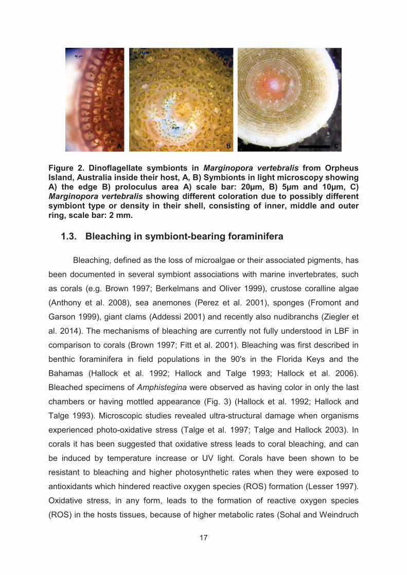
Figure 2. Dinoflagellate symbionts in Marginopora vertebralis from Orpheus Island, Australia inside their host, A, B) Symbionts in light microscopy showing A) the edge B) proloculus area A) scale bar: 20μm, B) 5μm and 10μm, C)Marginopora vertebralis showing different coloration due to possibly different symbiont type or density in their shell, consisting of inner, middle and outer ring, scale bar: 2 mm.
1.3. Bleaching in symbiont-bearing foraminifera
Bleaching, defined as the loss of microalgae or their associated pigments, has
been documented in several symbiont associations with marine invertebrates, such
as corals (e.g. Brown 1997; Berkelmans and Oliver 1999), crustose coralline algae
(Anthony et al. 2008), sea anemones (Perez et al. 2001), sponges (Fromont and
Garson 1999), giant clams (Addessi 2001) and recently also nudibranchs (Ziegler et
al. 2014). The mechanisms of bleaching are currently not fully understood in LBF in
comparison to corals (Brown 1997; Fitt et al. 2001). Bleaching was first described in
benthic foraminifera in field populations in the 90's in the Florida Keys and the
Bahamas (Hallock et al. 1992; Hallock and Talge 1993; Hallock et al. 2006).
Bleached specimens of Amphistegina were observed as having color in only the last
chambers or having mottled appearance (Fig. 3) (Hallock et al. 1992; Hallock and
Talge 1993). Microscopic studies revealed ultra-structural damage when organisms
experienced photo-oxidative stress (Talge et al. 1997; Talge and Hallock 2003). In
corals it has been suggested that oxidative stress leads to coral bleaching, and can
be induced by temperature increase or UV light. Corals have been shown to be
resistant to bleaching and higher photosynthetic rates when they were exposed to
antioxidants which hindered reactive oxygen species (ROS) formation (Lesser 1997).
Oxidative stress, in any form, leads to the formation of reactive oxygen species
(ROS) in the hosts tissues, because of higher metabolic rates (Sohal and Weindruch

18
1996). Reactive oxygen species are damaging to membrane function and in high
concentrations may lead to bleaching and be lethal to the organisms (Sohal and Orr
2012). In the first laboratory experiment on bleaching in Amphistegina gibbosa,
elevated temperature induced symbiont loss at 32°C, however at very low light
intensities of 6-8 μmol photons m2 s-1, which might have additionally stressed the
symbiosis (Talge and Hallock 2003). Similar levels of deterioration of the cytoplasm
has been observed among field-stressed specimens of Amphistegina gibbosa, but
also shell-breakage, symbiont-digestion, deformed tests and reproductive
dysfunction, suggesting the possibility of other diseases affecting these populations
(Talge et al. 1997) (Fig.3).
Physiological stress associated with elevated temperatures and subsequent
bleaching was investigated using Pulse Amplitude Modulated (PAM) Fluorometry and
photometric pigment measurements, which revealed that symbiont performance and
chlorophyll a content was significantly reduced under temperatures >31°C in several
LBF (Schmidt et al. 2011; van Dam et al. 2012a). Furthermore, these studies
revealed species-specific results, as for example the species Calcarina mayorii did
not show signs of stress under 32°C for 30 days (Schmidt et al. 2011). This suggests
that responses do not generally depend on the microalgae type, as all species
examined host endosymbiotic diatoms (Schmidt et al. 2011). Furthermore, bleaching
was also observed under temperature stress in the dinoflagellate-bearing disk-
shaped foraminifer Marginopora vertebralis, which showed increased mortality at
34°C and reduced photosynthesis at 31-32°C after one week of exposure (Uthicke et
al. 2012) (Fig. 3). Bleaching has also been observed in the disk-shaped Sorites
population from Florida and Belize (Richardson 2006; Richardson 2009), where local
conditions suggested that fresh water runoff, hurricane events and high irradiance
impact these populations. It has been suggested that bleaching susceptibility could
depend on the collection depth (van Dam et al. 2012a), as species collected from
slightly deeper habitats showed greater risk of bleaching (Schmidt et al. 2011; van
Dam et al. 2012a; Schmidt et al. 2014). Marine invertebrates living in the inter-tidal
zone have been shown to be more resistant to short-term temperature stress due to
hosting a higher stability form of anti-oxidant enzymes than the invertebrates living in
the sub-tidal zone (Regoli et al. 1997).

19
Figure 3. Examples of bleaching in larger benthic foraminifera: A Amphistegina radiata and B Heterostegina depressa exposed to 32°C for 6 days from Whitsundays, Australia, symbiont loss is highlighted by arrows, see Schmidt et al. (2011), scale bar: 2 mm, C Examples of field-bleached Amphistegina, from Tennessee Reef, Florida Keys, USA, from summer 1999, images provided by P. Hallock, scale bar: 1mm, D Bleaching in Marginopora vertebralis from the Whitsundays, Australia (Shaw Island) after 7 days of exposure to treatment temperatures, images provided by S. Uthicke, see Uthicke et al. (2012), scale bar: 2 mm.

20
1.4. Combined effects of global change
Since the industrial revolution, the world’s oceans have been a sink for
anthropogenic carbon dioxide. Under common carbon emission scenarios, this
process is predicted to lower the oceanic pH between 0.3-0.5 units by 2100 (Caldeira
and Wickett 2005; IPCC 2013). As a c
calcium carbonate minerals (Orr et al. 2005; Doney et al. 2009) is lowered, reducing
the carbonate ion concentration [CO23], which is essential for marine calcification
(Feely et al. 2004; Kleypas and Langdon 2006). In addition, oceans are expected to
warm at an increasingly rapid rate (IPCC 2013). Thus, ocean warming and ocean
acidification (OA) are likely to act in combination impacting the calcification potential
of many coral reefs (De'ath et al. 2009). It is not known whether the two stressors will
combine, enhance or cancel the individual effects. Several studies suggested
species-specific responses to combined effect of warming and acidification (Martin
and Gattuso 2009; Koch et al. 2013). Recent meta-analysis combining those results
showed the vulnerability of many species to a combination of stressors reducing key
physiologic parameters such as growth, survival and calcification (Kroeker et al.
2013). The study by Kroeker et al. (2013) also showed that enhanced sensitivity of
early life history stages is not universal among taxa. On the ecosystem level OA and
warming are in combination lowering coral reef resilience (Anthony et al. 2011). The
loss of ecological resilience occurs because coral regrowth is slow and disturbance
with macro-algae increase in duration and frequency, so that coral-reef ecosystems
are expected to shift from coral-dominated to algae-dominated reefs as a result of
combined OA and warming (Hoegh-Guldberg et al. 2007; Carilli et al. 2009).
If ecosystem shifts from coral-reefs to algae-dominated reefs occur, we are
expecting a change in light regimes and substrate, which will impact foraminifera
associated with this habitat. Thus far, few studies with a focus on important
carbonate sediment producers, such as LBF, have been conducted (Sinutok et al.
2011; Sinutok et al. 2014). The first study showed comprised photosynthetic health in
the controls and needed to be repeated. The latter study showed combined effects of
OA and warming reduced calcification and photosynthesis in M. vertebralis compared
to the controls (Sinutok et al. 2014). Studies on the isolated effects of OA alone
already showed mixed responses in LBF, which are best characterized as species-
specific and dose dependent. LBF either show no reaction in manipulative
21
experiments (Vogel and Uthicke 2012; McIntyre-Wressnig et al. 2013), or reduced
calcification (Kuroyanagi et al. 2009; Haynert et al. 2011; Reymond et al. 2013).
Enhanced calcification in LBF was found until 770 μatm, possibly stimulated by
carbon dioxide fertilization of the symbiont-population, but negative effects on
calcification were observed >970 μatm (Fujita et al. 2011). This shows that the
investigation of combined stressors on several different species of LBF is needed,
especially with regard to the role different symbiont-types play in the stress response.

22
1.5. Culturing of symbiont-bearing foraminifera
Several laboratories have successfully cultured LBF to date, demonstrating that
LBF are suitable organisms for answering ecological questions, which require
experiments under controlled environmental conditions (Fujita et al. 2000; Talge and
Hallock 2003; Fujita and Fujimura 2008; Nobes et al. 2008; Fujita et al. 2011; Hikami
et al. 2011; Reymond et al. 2011; Schmidt et al. 2011; Uthicke et al. 2012; van Dam
et al. 2012b; van Dam et al. 2012a; Reymond et al. 2013; Fujita et al. 2014; Schmidt
et al. 2014). To be kept alive in culture LBF need light for their symbionts (Hallock
1981; Nobes et al. 2008), near constant salinity, nutrients in the form of nitrate or
phosphate, or food given as living or dead microalgae (Lee et al. 1991) and
temperatures not exceeding the summer maxima by 1-2°C of their natural habitat
(Schmidt et al. 2011).
Röttger (1972a) started in the early 70's to culture LBF and described their life-
cycle, chamber formation and feeding behavior (Röttger 1972c,a,b; Röttger and
Berger 1972; Röttger 1973,1976). His work gave particular insights in biological
functioning of the canal system in the species Heterostegina depressa, which is
important for the motility, growth, reproduction and excretion (Röttger et al. 1984).
Lee et al. (1979) reported the first successful isolation of the “naked” symbionts from
LBF and grew them into culture. Diatoms have reduced their silica frustule inside
their host, but re-grow it after death of their host when cultured in sterile media (Lee
1980; Lee et al. 1980). Experiments on the nutritional requirements of several LBF
showed that those are species-specific and that they can take up nitrate and
phosphate from the culturing seawater (Lee et al. 1991). Culturing experiments on
Amphistegina showed that they develop a different test thickness when cultured
under different water motions (Hallock et al. 1986). Moreover, LBF have been
cultured to determine their response to different natural light levels, to test their
photosynthetic and growth responses grown under daily fluctuating conditions
(Nobes et al. 2008). Highest growth and photosynthetic rates (Fv:Fm) where found in
all taxa in tanks when 90% of the incoming natural light was blocked by shade-cloth,
with high light peaks of 60μmol photons m2 s-1 (Nobes et al. 2008). If LBF are
cultured under light conditions similar to average light levels recorded by loggers in
their shallow habitat (e.g. 200-300 μmol photons m2 s-1) they show reduced
photosynthetic responses (reduced Fv:Fm), possibly because of photo-oxidative

23
stress (Sinutok et al. 2011). High irradiances exert stress on the photosymbiosis in
LBF (Nobes et al. 2008), and based on their photosynthetic response to light, LBF
can be categorized as being light-sensitive or light-tolerant (Ziegler and Uthicke
2011). As LBF are motile organisms, they can, unlike corals, shade themselves from
light in their natural habitat during midday, similar to the cover behavior observed in
many sea urchins (Adams 2001). With regard to adaptations for global change, mass
culturing of the species Baculogypsina sphaerulata has been carried out and has
successfully shown that it is a possible way to produce large amount of biogenic
carbonate “artificially” which can be used for additional coastal stabilization (Hosono
et al. 2014).
Several studies used flow-through aquaria systems for culturing LBF, which
constantly provide new input of fresh seawater and/or nutrients (Reymond et al.
2011; Uthicke et al. 2012; van Dam et al. 2012a; Vogel and Uthicke 2012; Schmidt et
al. 2014). This culturing method has been shown to be well suited for culturing over
several weeks to month, especially because rapid water movement are also
observed in the habitat of LBF (Williams and Carpenter 1998; Cornelisen and
Thomas 2009) and high growth rate were reported in several species (Reymond et
al. 2013; Schmidt et al. 2014). Earlier studies also reported positive influence of water
motion on growth and calcification of LBF (ter Kuile and Erez 1984; Hallock et al.
1986). When large LBF are cultured in static-conditions, the lack of water movement
around the shell could create boundary layer conditions, which can be thinned when
water movement is applied. Increasing water exchange with the surrounding
environment and can lead to changes in the pH induced by photosynthesis and
calcification (Glas et al. 2012a). Thus, flow-through conditions for culturing, in
contained in houses (Schmidt et al. 2014) or in free floating culture cages (Fujita et
al. 2011), can be of advantage, especially with regard to testing effects of OA. For
smaller LBF and juveniles the well-plate approach has been successfully used in
studies monitoring the development of shells in asexual offspring (Chapter 2), for
conducting direct measurements of photosynthesis using a Pulse Amplitude
Fluorometer (Schmidt et al. 2011) or exposure tests to toxic metals (Prazeres et al.
2011) or herbicides (van Dam et al. 2012a), which provide a clear advantage during
handling, reducing the loss of specimens.
24
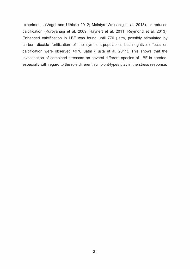
Figure 4. Culturing setups for larger benthic foraminifera, A) Flow-through plate system used in Chapter 1 for culturing Heterostegina depressa and Marginopora vertebralis, made out of two standard 6-well plates, connected by plastic tubing to a small aquaria pump, H. depressa was contained in the lower level and M. vertebralis in the upper level for their optimal exposure to light, flow-through plates constructed to be submerged inside aquaria, B Aquaria setup to manipulate temperatures in each aquaria seperatly, which was used in Chapter 3 for temperature stress experiments (20-36°C).

25
1.6. Aims and interdisciplinary research context
The overall goal of the thesis was to determine the physiological response of
several species of LBF to individual and combined effects of global change stress.
This was achieved by measuring the organisms’ physiological responses by a variety
of parameters, such as photosynthesis, respiration, chlorophyll a content,
survivorship and growth. Understanding the effects of individual and combined
stressors on marine ecosystems are increasingly important under global change
scenarios, because stressors such as OA and warming are likely to occur
simultaneously (Caldeira and Wickett 2005; IPCC 2013). We need to know whether
theses stressors act additive (equal the sum of the individual effects), synergistic
(larger than the sum of the individual effects) or antagonistic (erasing the individual
effects) on marine species. We chose LBF because they are important calcium
carbonate producers in the oceans and need further study. We wanted to test if
differences exist between bleaching thresholds in different species and if they are
lowered under a combination of stressors. Thus, we conducted a study on two LBF,
hosting different photo-symbiotic dinoflagellates (Marginopora vertebralis) and
diatoms (Heterostegina depressa). In addition to species from the Indo-pacific region
LBF from the eastern Mediterranean Sea were chosen (Chapter 2) to test thresholds
to bleaching on a larger geographic area. The eastern Mediterranean Sea is
predicted to be a miniature model of ocean warming in the near future (Lejeusne et
al. 2010; Shaltout and Omstedt 2014). In particular our attention was drawn to this
area because an extreme thermally tolerant species was described by Arieli et al.
(2011). They showed that the foraminifer Pararotalia calcariformata, can survive
temperatures of 36°C, occurring in a heat plume originating from a power plant and
that it likely contains endosymbionts (Arieli et al. 2011). This is remarkable, as all
other eukaryote-eukaryote endosymbiosis including the most heat-tolerant corals are
shown to bleach at 36°C (Coles 1988; Coles and Riegl 2013). We wanted to test
whether the heat tolerance observed in situ is limited to the heat plume or also occurs
in an unaffected population originating from a National Park. We wanted to test if the
bleaching threshold of P. calcariformata is really higher than that of another abundant
species Amphistegina lobifera, which has been shown to bleach starting at 31°C, and
was not found in the heat plume (Talge and Hallock 2003; Schmidt et al. 2011).
Therefore, we conducted several experiments, exposing a summer and winter
26
population of both species in the range from 20-36°C for up to three weeks and
conducted an extreme heat-test experiment ranging from temperatures of 20-42°C
on Pararotalia. We measured the species response by determining survival, growth
and photosynthetic efficiency using Pulse Amplitude Modulated Fluorometry (PAM).
We hypothesize that P. calcariformata is an invader species in the Mediterranean, as
it was only recently described in the Levant (Reinhardt et al. 1994) and Turkish coast
(Meriç et al. 2013). Hence, the aims were to characterize P. calcariformata combining
morphological and molecular tools and describe its general ecology and reproduction
cycle. We aimed to evaluate the species current and future distribution by 2100 using
a species distribution model, previously used on a the genus Amphistegina in the
Mediterranean (Langer et al. 2012; Weinmann et al. 2013). Furthermore, we wanted
to test the temperature effect on the shell development of asexual juveniles to find
out if P. calcariformata is restricted to a specific thermal tolerance window in their
habitat, or if it is able to tolerate cold (20°C) or extremely warm (35°C) conditions in
early life stages.
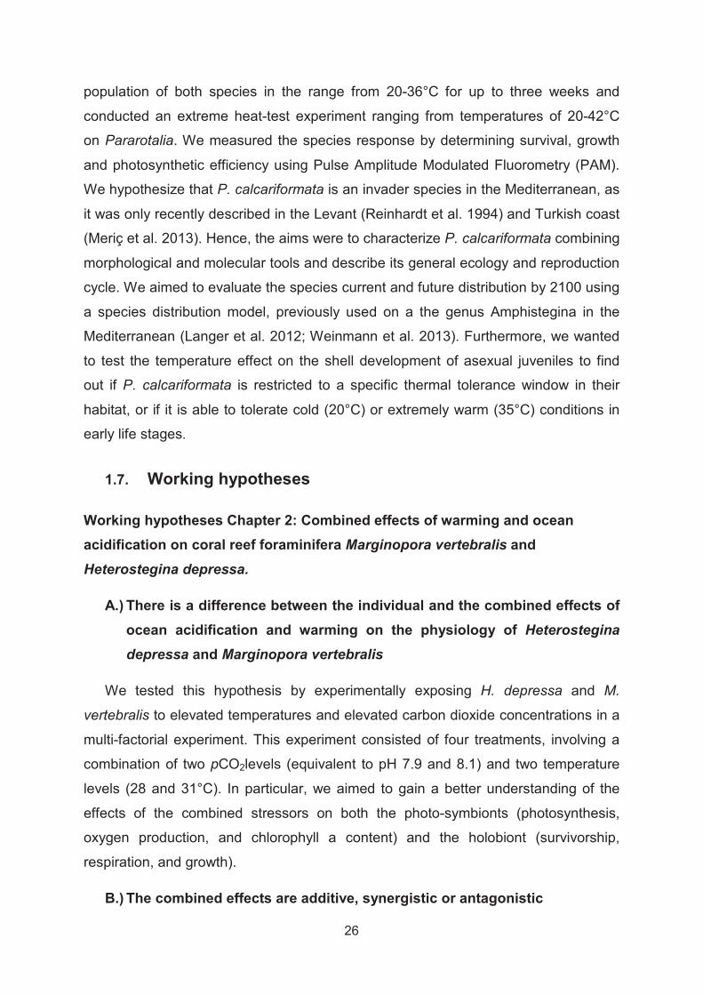
1.7. Working hypotheses
Working hypotheses Chapter 2: Combined effects of warming and ocean acidification on coral reef foraminifera Marginopora vertebralis and Heterostegina depressa.
A.) There is a difference between the individual and the combined effects of ocean acidification and warming on the physiology of Heterostegina depressa and Marginopora vertebralis
We tested this hypothesis by experimentally exposing H. depressa and M.
vertebralis to elevated temperatures and elevated carbon dioxide concentrations in a
multi-factorial experiment. This experiment consisted of four treatments, involving a
combination of two pCO2levels (equivalent to pH 7.9 and 8.1) and two temperature
levels (28 and 31°C). In particular, we aimed to gain a better understanding of the
effects of the combined stressors on both the photo-symbionts (photosynthesis,
oxygen production, and chlorophyll a content) and the holobiont (survivorship,
respiration, and growth).
B.) The combined effects are additive, synergistic or antagonistic

27
When combined effects were significant based on general linear models, we
calculated the observed inhibition compared to the control treatment, based on this
we described if the effect is additive (the sum of the individual effects), synergistic
(larger than the sum of the individual effects) or antagonistic.
C.) The response to the combined effects is species-specific
We chose particular species with different symbiont types (diatoms and
dinoflagellates) to evaluate the difference between species with regard to
differentiated response to the interaction of stressors.
Working hypotheses Chapter 3: Recent invasion of the symbiont-bearing foraminifera Pararotalia into the Eastern Mediterranean facilitated by the ongoing warming trend
A.) Based on its current distribution, the recently discovered foraminifer P. calcariformata in the eastern Mediterranean Sea is an invader species
We characterized its morphology and current biogeography by identifying its
current distribution in the eastern Mediterranean Sea. To investigate whether this
species is a likely invader we analysed the relatedness of Mediterranean and Indo-
Pacific population using phylogenetic inference. Using a compilation of all occurrence
records of the species in the Mediterranean, we model its likely current distribution.
B.) The foraminifer P. calcariformata contains permanent diatom endosymbionts
We also identified its endosymbiotic microalgae by molecular and standard algae
culturing and measured its photosynthetic activity under controlled environmental
condition.

28
C.) The symbionts in P. calcariformata are photosynthetically active over several month in culture
We cultured this species over five month and monitored its photosynthetic activity
using Pulse Amplitude modulated Fluorometry, as well measured its response to
rapidly increasing light conditions to evaluate its optimal light properties for culturing.
D.) The foraminifer P. calcariformata will spread to currently colder regions in the Mediterranean based on global warming
Based on the current occurrence records and the predictions that the distribution
of species is likely impacted by the minimal temperature, turbidity and radiation we
model its future spread under a global change scenario.
E.) Pararotalia calcariformata has a narrow reproductive window for asexual offspring development and is currently restricted by minimum temperatures to spread westwards
We hypothesized that temperature is the main factor for the establishment of new
populations, as it is seen worldwide in most shallow marine fauna (Belanger et al.
2012). To test if there are temperature differences between the developmental rates
in Pararotalia juveniles, we exposed them to three different temperatures (20°C,
28°C and 35°C). We measured growth rates to find out which temperatures promote
shell development and growth in this species.
29
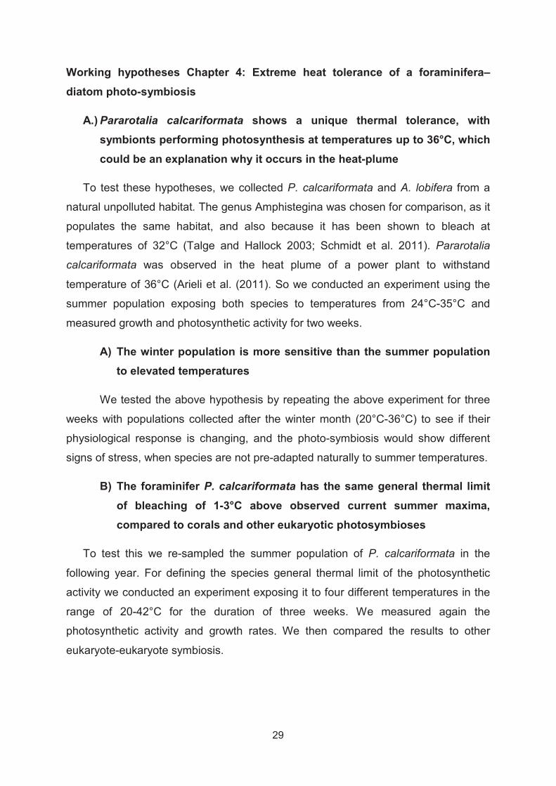
Working hypotheses Chapter 4: Extreme heat tolerance of a foraminifera–diatom photo-symbiosis
A.) Pararotalia calcariformata shows a unique thermal tolerance, with symbionts performing photosynthesis at temperatures up to 36°C, which could be an explanation why it occurs in the heat-plume
To test these hypotheses, we collected P. calcariformata and A. lobifera from a
natural unpolluted habitat. The genus Amphistegina was chosen for comparison, as it
populates the same habitat, and also because it has been shown to bleach at
temperatures of 32°C (Talge and Hallock 2003; Schmidt et al. 2011). Pararotalia
calcariformata was observed in the heat plume of a power plant to withstand
temperature of 36°C (Arieli et al. (2011). So we conducted an experiment using the
summer population exposing both species to temperatures from 24°C-35°C and
measured growth and photosynthetic activity for two weeks.
A) The winter population is more sensitive than the summer population to elevated temperatures
We tested the above hypothesis by repeating the above experiment for three
weeks with populations collected after the winter month (20°C-36°C) to see if their
physiological response is changing, and the photo-symbiosis would show different
signs of stress, when species are not pre-adapted naturally to summer temperatures.
B) The foraminifer P. calcariformata has the same general thermal limit of bleaching of 1-3°C above observed current summer maxima, compared to corals and other eukaryotic photosymbioses
To test this we re-sampled the summer population of P. calcariformata in the
following year. For defining the species general thermal limit of the photosynthetic
activity we conducted an experiment exposing it to four different temperatures in the
range of 20-42°C for the duration of three weeks. We measured again the
photosynthetic activity and growth rates. We then compared the results to other
eukaryote-eukaryote symbiosis.
30
1.8. Outline of the thesis
Publication 1:Schmidt, C., Kucera, M., Uthicke, S.
Combined effects of warming and ocean acidification on coral reef Foraminifera Marginopora vertebralis and Heterostegina depressa. The article
has been published in the Journal Coral Reefs (2014) 33(3):805-818.
Contributions: The project on interactive effects of ocean acidification and
warming on LBF was initiated by S. Uthicke and the AIMS water quality team.
Planning of the particular project on foraminifera was done by S. Uthicke and C.
Schmidt. The experiment was carried out by S. Uthicke, C. Schmidt , M. Liddy, A.
Negri, N. Webber, K. Fabricius, S. Noonan, as a collaborative project. The
physiological data on LBF was collected by C. Schmidt. Data analysis was done by
C. Schmidt with help of S. Uthicke. Writing of the manuscript was done by C. Schmidt
with improvements by M. Kucera and S. Uthicke.
Publication 2: Schmidt, C, Morard, R, Almogi-Labin, A, Weinmann, A, Titelboim, D,
Abramovich, S, Kucera, M
Recent invasion of the symbiont-bearing Foraminifera Pararotalia into the Eastern Mediterranean facilitated by the ongoing warming trend
Contributions: The hypotheses tested in this paper were developed by M.
Kucera and C. Schmidt. Molecular data collection and analysis was conducted by R.
Morard. Data collection on the biology of Pararotalia (photosynthetic measurements,
symbiont isolation, juvenile development, occurrence records) was done by C.
Schmidt. A. Weinmann conducted species distribution modelling based on
occurrence records. D. Titelboim contributed to the description of the taxonomy.
Writing of the manuscript was done by C. Schmidt and M. Kucera with improvements
by A. Almogi-Labin and S. Abramovich.

31
Publication 3: Schmidt, C., Brandt, J., Barak, H., Abramovich, S., and Kucera, M.
Extreme heat tolerance of a foraminifera–diatom photo-symbiosis
Contributions: The hypotheses and experimental design were developed by C.
Schmidt and M. Kucera. Experimental data was collected by C. Schmidt and J.
Brandt. B. Herut and S. Abramovich contributed to field background temperature data
collection. Data analysis was done by C. Schmidt, and M. Kucera. C. Schmidt wrote
the paper, with improvements by M. Kucera.
32
1.9. References
Adams NL (2001) UV radiation evokes negative phototaxis and covering behavior in the sea urchin Strongylocentrotus droebachiensis. Marine Ecology Progress Series 213:87-95
Addessi L (2001) Giant clam bleaching in the lagoon of Takapoto atoll (French Polynesia). Coral Reefs 19:220-220
Anthony KRN, Kline DI, Diaz-Pulido G, Dove S, Hoegh-Guldberg O (2008) Ocean acidification causes bleaching and productivity loss in coral reef builders. Proceedings of the National Academy of Sciences of the United States of America 105:17442-17446
Anthony KRN, Maynard JA, Diaz-Pulido G, Mumby PJ, Marshall PA, Cao L, Hoegh-Guldberg OVE (2011) Ocean acidification and warming will lower coral reef resilience. Global Change Biology 17:1798-1808
Arieli RN, Almogi-Labin A, Abramovich S, Herut B (2011) The effect of thermal pollution on benthic foraminiferal assemblages in the Mediterranean shoreface adjacent to Hadera power plant (Israel). Marine Pollution Bulletin 62:1002-1012
Baker RD, Hallock P, Moses EF, Williams DE, Ramirez A (2009) Larger Foraminfers of the Florida Reef Tract, USA: Distribution patterns on reef-rubble habitats. Journal of Foraminiferal Research 39:267-277
Belanger CL, Jablonski D, Roy K, Berke SK, Krug AZ, Valentine JW (2012) Global environmental predictors of benthic marine biogeographic structure. Proceedings of the National Academy of Sciences of the United States of America 109:14046-14051
Berkelmans R, Oliver JK (1999) Large-scale bleaching of corals on the Great Barrier Reef. Coral Reefs 18:55-60
Bernhard JM, Bowser SS (1999) Benthic foraminifera of dysoxic sediments: chloroplast sequestration and functional morphology. Earth-Science Reviews 46:149-165
Bresler V, Yanko V (1995) Acute toxicity of heavy-metals for benthi epiphytic foraminifera Pararotalia spinigera (LeCalvez) and influence of seaweed-derived DOC. Environmental Toxicology and Chemistry 14:1687-1695
Brown BE (1997) Coral bleaching: causes and consequences. Coral Reefs 16:S129-S138
Caldeira K, Wickett ME (2005) Ocean model predictions of chemistry changes from carbon dioxide emissions to the atmosphere and ocean. Journal of Geophysical Research: Oceans 110:C09S04

33
Carilli JE, Norris RD, Black BA, Walsh SM, McField M (2009) Local Stressors Reduce Coral Resilience to Bleaching. PLoS ONE 4
Chai JY, Lee JJ (2000) Recognition, establishment and maintenance of diatom endosymbiosis in foraminifera. Micropaleontology 46:182-195
Coles SL, Riegl BM (2013) Thermal tolerances of reef corals in the Gulf: A review of the potential for increasing coral survival and adaptation to climate change through assisted translocation. Marine Pollution Bulletin 72:323-332
Cooper TF, Berkelmans R, Ulstrup KE, Weeks S, Radford B, Jones AM, Doyle J, Canto M, O'Leary RA, van Oppen MJH (2011) Environmental Factors Controlling the Distribution of Symbiodinium Harboured by the Coral Acropora millepora on the Great Barrier Reef. PLoS ONE 6
Cornelisen CD, Thomas FIM (2009) Prediction and validation of flow-dependent uptake of ammonium over a seagrass-hardbottom community in Florida Bay. Marine Ecology Progress Series 386:71-81
Correia MJ, Lee JJ (2000) Chloroplast retention by Elphidium excavatum (Terquem). Is it a selective process? Symbiosis 29:343-355
Correia MJ, Lee JJ (2002) How long do the plastids retained by Elphidium excavatum (Terquem) last in their host? Symbiosis 32:27-37
Dawson JL, Smithers SG, Hua Q (2014) The importance of large benthic foraminifera to reef island sediment budget and dynamics at Raine Island, northern Great Barrier Reef. Geomorphology 222:68-81
De'ath G, Lough JM, Fabricius KE (2009) Declining coral calcification on the Great Barrier Reef. Science 323:116-119
de Nooijer LJ, Toyofuku T, Kitazato H (2009) Foraminifera promote calcification by elevating their intracellular pH. Proceedings of the National Academy of Sciences 106:15374-15378
Dettmering C, Rottger R, Hohenegger J, Schmaljohann R (1998) The trimorphic life cycle in foraminifera: Observations from cultures allow new evaluation. European Journal of Protistology 34:363-368
Doney SC, Fabry VJ, Feely RA, Kleypas JA (2009) Ocean Acidification: The Other CO2 Problem Annual Review of Marine Science. Annual Reviews, Palo Alto, pp169-192
Doo SS, Hamylton S, Byrne M (2012) Reef-Scale Assessment of Intertidal Large Benthic Foraminifera Populations on One Tree Island, Great Barrier Reef and Their Future Carbonate Production Potential in a Warming Ocean. Zoological Studies 51:1298-1307
Fay SA, Weber MX, Lipps JH (2009) The distribution of Symbiodinium diversity within individual host foraminifera. Coral Reefs 28:717-726

34
Feely RA, Sabine CL, Lee K, Berelson W, Kleypas J, Fabry VJ, Millero FJ (2004) Impact of anthropogenic CO2 on the CaCO3 system in the oceans. Science 305:362-366
Fitt W, Brown B, Warner M, Dunne R (2001) Coral bleaching: interpretation of thermal tolerance limits and thermal thresholds in tropical corals. Coral Reefs 20:51-65
Fromont J, Garson M (1999) Sponge bleaching on the West and East coasts of Australia. Coral Reefs 18:340-340
Fujita K, Nishi H, Saito T (2000) Population dynamics of Marginopora kudakajimensis Gudmundsson (Foraminifera: Soritidae) in the Ryukyu Islands, the subtropical northwest Pacific. Marine Micropaleontology 38:267-284
Fujita K, Fujimura H (2008) Organic and inorganic carbon production by algal symbiont-bearing foraminifera on northwest Pacific coral-reef flats. Journal of Foraminiferal Research 38:117-126
Fujita K, Hikami M, Suzuki A, Kuroyanagi A, Sakai K, Kawahata H, Nojiri Y (2011) Effects of ocean acidification on calcification of symbiont-bearing reef foraminifers. Biogeosciences 8:2089-2098
Fujita K, Okai T, Hosono T (2014) Oxygen Metabolic Responses of Three Species of Large Benthic Foraminifers with Algal Symbionts to Temperature Stress. PLoS ONE 9
Garcia-Cuetos L, Pochon X, Pawlowski J (2005) Molecular evidence for host-symbiont specificity in soritid Foraminifera. Protist 156:399-412
Gastrich MD (1987) Ultrastructure of a new intracellular symbiotic alga found within planktonic foraminifera. Journal of Phycology 23:623-632
Glas MS, Fabricius KE, de Beer D, Uthicke S (2012a) The O2, pH and Ca2+
Microenvironment of Benthic Foraminifera in a High CO2 World. PLoS ONE 7
Glas MS, Langer G, Keul N (2012b) Calcification acidifies the microenvironment of a benthic foraminifer (Ammonia sp.). Journal of Experimental Marine Biology and Ecology 424:53-58
Grell GK (1973) Protozoology. Springer Heidelberg
Hallock P (1981) Light dependence in Amphistegina. Journal of Foraminiferal Research 11:40-46
Hallock P (1984) Distribution of selected species of living algal symbiont-bearing foraminifera on two Pacific coral reefs. Journal of Foraminiferal Research 14:250-261
Hallock P, Forward LB, Hansen HJ (1986) Influence of environment on the test shape of Amphistegina. Journal of Foraminiferal Research 16:224-231
35
Haynert K, Schonfeld J, Riebesell U, Polovodova I (2011) Biometry and dissolution features of the benthic foraminifer Ammonia aomoriensis at high pCO2. Marine Ecology Progress Series 432:53-67
Hikami M, Ushie H, Irie T, Fujita K, Kuroyanagi A, Sakai K, Nojiri Y, Suzuki A, Kawahata H (2011) Contrasting calcification responses to ocean acidification between two reef foraminifers harboring different algal symbionts. Geophysical Research Letters 38
Hoegh-Guldberg O, Mumby PJ, Hooten AJ, Steneck RS, Greenfield P, Gomez E, Harvell CD, Sale PF, Edwards AJ, Caldeira K, Knowlton N, Eakin CM, Iglesias-Prieto R, Muthiga N, Bradbury RH, Dubi A, Hatziolos ME (2007) Coral reefs under rapid climate change and ocean acidification. Science 318:1737-1742
Hohenegger J (1994) Distribution of living larger foraminifera NW of Sesoko-Jima, Okinawa, Japan. Marine Ecology-Pubblicazioni Della Stazione Zoologica Di Napoli I 15:291-334
Hohenegger J (2006) The importance of symbiont-bearing benthic foraminifera for West Pacific carbonate beach environments. Marine Micropaleontology 61:4-39
Hohenegger J (2011) Large Foraminifera - Greenhouse constructions and gardeners in the oceanic microcosm. The Kagoshima University Muesum, Kagoshima, Bulletin No. 5
Hosono T, Lopati P, Makolo F, Kayanne H (2014) Mass culturing of living sands (Baculogypsina sphaerulata) to protect island coasts against sea-level rise. Journal of Sea Research 90:121-126
IPCC (2013) Climate Change 2013: The Physical Science Basis. Contribution of Working Group I to the Fifth Assessment Report of the Intergovernmental Panel on Climate Change. In: Stocker TF, D. Qin, G.-K. Plattner, M. Tignor, S.K. Allen, J. Boschung, A. Nauels, Y. Xia, V. Bex and P.M. Midgley (ed). Cambridge University Press, Cambridge, United Kingdom and New York, NY, USA, pp1535
Kleypas JA, Langdon C (2006) Coral reefs and changing seawater carbonate. In: Phinney JT, Guldberg, O.H., Kleypas, J., Skirving, W., Strong, A. (ed) Coastal and Estuarine Studies: Coral Reefs and Climate Change Science and Management. Coastal. American Geophysical Union, Washington DC, pppp. 73–110
Koch M, Bowes G, Ross C, Zhang XH (2013) Climate change and ocean acidification effects on seagrasses and marine macroalgae. Global Change Biology 19:103-132
Kroeker KJ, Kordas RL, Crim R, Hendriks IE, Ramajo L, Singh GS, Duarte CM, Gattuso J-P (2013) Impacts of ocean acidification on marine organisms: quantifying sensitivities and interaction with warming. Global Change Biology:n/a-n/a

36
Kuroyanagi A, Kawahata H, Suzuki A, Fujita K, Irie T (2009) Impacts of ocean acidification on large benthic foraminifers: Results from laboratory experiments. Marine Micropaleontology 73:190-195
LaJeunesse TC, Loh WKW, van Woesik R, Hoegh-Guldberg O, Schmidt GW, Fitt WK (2003) Low symbiont diversity in southern Great Barrier Reef corals, relative to those of the Caribbean. Limnology and Oceanography 48:2046-2054
Langer MR (1993) Epiphytic foraminifera. Marine Micropaleontology 20:235-265
Langer MR, Silk MT, Lipps JH (1997) Global ocean carbonate and carbon dioxide production; the role of reef Foraminifera. Journal of Foraminiferal Research 27:271-277
Langer MR, Weinmann AE, Lötters S, Rödder D (2012) "Strangers" in Paradise: Modeling the Biogeographic Range Expansion of the Foraminifera Amphistegina in the Mediterranean Sea. Journal of Foraminiferal Research 42:234-244
Lee J, Reimer C, McEnergy M (1980) The Identification of diatoms isolated as endosymbionts from larger foraminifera from the Gulf of Eilat (Red Sea) and the description of two new species, Fragilaria shiloi sp. nov. and Navicula reissii sp. nov. Botanica Marina 23:41-48
Lee J, Erez J, McEnery M, Lagziel A, Xenophontos X (1986) Experiments on persistence of endosymbiotic diatoms in the larger foraminifer: Amphistegina lessonii. Symbiosis 1:211-226
Lee JJ, Bock WD (1976) The Importance of Feeding in Two Species of Soritid Foraminifera with Algal Symbionts. Bulletin of Marine Science 26:530-537
Lee JJ, McEnery ME, Shilo M, Reiss Z (1979) Isolation and cultivation of diatom symbionts from larger Foraminifera (Protozoa). Nature 280:57-58
Lee JJ, Hallock P (1987) Algal symbiosis as the driving force in the evolution of larger Foraminifera. Annals of the New York Academy of Sciences 503:330-347
Lee JJ, McEnergy ME, Ter Kuile B, Erez J, Roetger R, Rockwell RF, Faber WW, Lagziel A (1989) Identification and distribution of endosymbiotic diatoms in larger foraminifera. Micropaleontology 35:353-366
Lee JJ (1991) Isolation from endosymbiotic algae from larger Foraminifera. In: Lee JJ, Soldo A (eds) Protocols in Protozoology. Society Protozoologists, Lawrence, Kansas, ppA5
Lee JJ, Anderson OR (1991) Symbiosis in Foraminifera. In: Lee JJ, Anderson OR (eds) Biology of Foraminifera. Academic Press Limited, San Diego, pp157-220
Lee JJ, Sang K, Terkuile B, Strauss E, Lee PJ, Faber WW (1991) Nutritional and related experiments on laboratory maintenance of 3 species of symbiont-bearing, large foraminifera. Marine Biology 109:417-425

37
Lee JJ, Morales J, Bacus S, Diamont A, Hallock P, Pawlowski J, Thorpe J (1997) Progress in characterizing the endosymbiotic dinoflagellates of soritid foraminifera and related studies on some stages in the life cycle of Marginopora vertebralis. Journal of Foraminiferal Research 27:254-263
Lee JJ, Correia M (2005) Endosymbiotic diatoms from previously unsampled habitats. Symbiosis 38:251-260
Lee JJ (2006) Algal symbiosis in larger foraminifera. Symbiosis 42:63-75
Lee JJ, Fine M, Levy O, Morales J (2009) A note on asexual reproduction of a Marginopora sp. from a modern deep-water population in the Heron-Wistari Channel, Australia. The Journal of Foraminiferal Research 39:4-7
Lee JJ, Cervasco MH, Morales J, Billik M, Fine M, Levy O (2010) Symbiosis drove cellular evolution. Symbiosis 51:13-25
Lee JJ, McEnergy, M. E., Röttger, R., Reimer, Ch. W. (1980) The isolation, culture and identification of endosymbiotic diatoms from Heterostegina depressaD'Orbigny and Amphistegina lessonii D'Orbigny (Larger Foraminifer) from Hawaii. Botanica Marina 23:297-302
Lejeusne C, Chevaldonné P, Pergent-Martini C, Boudouresque CF, Pérez T (2010) Climate change effects on a miniature ocean: the highly diverse, highly impacted Mediterranean Sea. Trends in Ecology & Evolution 25:250-260
Lesser MP (1997) Oxidative stress causes coral bleaching during exposure to elevated temperatures. Coral Reefs 16:187-192
Liquete C, Piroddi C, Drakou EG, Gurney L, Katsanevakis S, Charef A, Egoh B (2013) Current Status and Future Prospects for the Assessment of Marine and Coastal Ecosystem Services: A Systematic Review. PLoS ONE 8
Martin S, Gattuso JP (2009) Response of Mediterranean coralline algae to ocean acidification and elevated temperature. Global Change Biology 15:2089-2100
McIntyre-Wressnig A, Bernhard JM, McCorkle DC, Hallock P (2013) Non-lethal effects of ocean acidification on the symbiont-bearing benthic foraminifer Amphistegina gibbosa. Marine Ecology Progress Series 472:45-60
Meriç E, Yokes MB, Avsar KN, Kirki-Elmas E, Dinçer F, Karhan SU, Kalkan E, Demir V (2013) First report of Pararotalia calcariformata from the Hatay coastline (Turkey—north-eastern Mediterranean). Marine Biodiversity Records 6:e31
Momigliano P, Uthicke S (2013) Symbiosis in a giant protist (Marginopora vertebralis,Soritinae): flexibility in symbiotic partnerships along a natural temperature gradient. Marine Ecology Progress Series 491:33–46
Müller-Merz E, Lee. JJ (1976) Symbiosis in the Larger Foraminiferan Sorites marginalis (with Notes on Archaias spp.). Journal of Protozoology 23:390– 396

38
Nobes K, Uthicke S, Henderson R (2008) Is light the limiting factor for the distribution of benthic symbiont bearing foraminifera on the Great Barrier Reef? Journal of Experimental Marine Biology and Ecology 363:48-57
Orr JC, Fabry VJ, Aumont O, Bopp L, Doney SC, Feely RA, Gnanadesikan A, Gruber N, Ishida A, Joos F, Key RM, Lindsay K, Maier-Reimer E, Matear R, Monfray P, Mouchet A, Najjar RG, Plattner G-K, Rodgers KB, Sabine CL, Sarmiento JL, Schlitzer R, Slater RD, Totterdell IJ, Weirig M-F, Yamanaka Y, Yool A (2005) Anthropogenic ocean acidification over the twenty-first century and its impact on calcifying organisms. Nature 437:681-686
Pawlowski JAN, Holzmann M, Fahrni JF, Pochon X, Lee JJ (2001) Molecular Identification of Algal Endosymbionts in Large Miliolid Foraminifera: 2. Dinoflagellates. Journal of Eukaryotic Microbiology 48:368-373
Perez SF, Cook CB, Brooks WR (2001) The role of symbiotic dinoflagellates in the temperature-induced bleaching response of the subtropical sea anemone Aiptasia pallida. JExp Mar Biol Ecol 256:1-14
Pillet L, de Vargas C, Pawlowski J (2011) Molecular Identification of Sequestered Diatom Chloroplasts and Kleptoplastidy in Foraminifera. Protist 162:394-404
Pochon X, Montoya-Burgos JI, Stadelmann B, Pawlowski J (2006) Molecular phylogeny, evolutionary rates, and divergence timing of the symbiotic dinoflagellate genus Symbiodinium. Molecular Phylogenetics and Evolution 38:20-30
Pochon X, Garcia-Cuetos L, Baker AC, Castella E, Pawlowski J (2007) One-year survey of a single Micronesian reef reveals extraordinarily rich diversity of Symbiodinium types in soritid foraminifera. Coral Reefs 26:867-882
Prazeres MD, Martins SE, Bianchini A (2011) Biomarkers response to zinc exposure in the symbiont-bearing foraminifer Amphistegina lessonii (Amphisteginidae, Foraminifera). Journal of Experimental Marine Biology and Ecology 407:116-121
Putnam HM, Stat M, Pochon X, Gates RD (2012) Endosymbiotic flexibility associates with environmental sensitivity in scleractinian corals. Proceedings of the Royal Society B-Biological Sciences 279:4352-4361
Regoli F, Principato GB, Bertoli E, Nigro M, Orlando E (1997) Biochemical characterization of the antioxidant system in the scallop Adamussium colbecki, a sentinel organism for monitoring the Antarctic environment. Polar Biology 17:251-258
Reinhardt EG, Patterson RT, Schroeder-Adams CJ (1994) Geoarchaeology of the ancient harbor site of Caesarea Maritima, Israel; evidence from sedimentology and paleoecology of benthic foraminifera. The Journal of Foraminiferal Research 24:37-48
Reymond CE, Uthicke S, Pandolfi JM (2011) Inhibited growth in the photosymbiont-bearing foraminifer Marginopora vertebralis from the nearshore Great Barrier

39
Reef, Australia (vol 435, pg 97, 2011). Marine Ecology-Progress Series 441:302-302
Reymond CE, Lloyd A, Kline DI, Dove SG, Pandolfi JM (2013) Decline in growth of foraminifer Marginopora rossi under eutrophication and ocean acidification scenarios. Global Change Biology 19:291-302
Richardson SD (2006) Endosymbiont-bleaching in epiphytic populations of Sorites dominicensis. Symbiosis 42:101 - 117
Rottger R (1974) Larger foraminifera - reproduction and early stages of development in Heterostegina depressa. Marine Biology 26:5-12
Röttger R (1972a) Die Kultur von Heterostegina depressa (Foraminifera: Nummulitidae). Marine Biology 15:150-159
Röttger R (1972b) Analyse von Wachstumskurven von Heterostegina depressa(Foraminifera: Nummulitidae). Marine Biology 17:228-242
Röttger R (1972c) Die Bedeutung der Symbiose von Heterostegina depressa(Foraminifera, Nummulitidae) für hohe Siedlungsdichte und Karbonatproduktion. Abhandlungen deutscher Zoologischer Gesellschaft 1971:42 - 47
Röttger R, Berger WH (1972) Benthic Foraminifera: morphology and growth in clone cultures of Heterostegina depressa. Marine Biology 15:89-94
Röttger R (1973) Die Ektoplasmahülle von Heterostegina depressa (Foraminifera: Numlitidae). Marine Biology 21:127-138
Röttger R (1976) Ecological observations Heterostegina depressa in the laboratory and in its natural habitat. Maritim sediments Spec. Publ. 1:75-79
Röttger R, Spindler M, Schmaljohann R, Richwien M, Fladung M (1984) Functions of the canal system in the rotaliid foraminifer, Heterostegina depressa. Nature 309:789-791
Röttger R, Krüger R, de Rijk S (1990) Trimorphism in foraminifera (protozoa) —verification of an old hypothesis. European Journal of Protistology 25:226-228
Schmidt C, Heinz P, Kucera M, Uthicke S (2011) Temperature-induced stress leads to bleaching in larger benthic foraminifera hosting endosymbiotic diatoms. Limnology and Oceanography 56:1587-1602
Schmidt C, Kucera M, Uthicke S (2014) Combined effects of warming and ocean acidification on coral reef Foraminifera Marginopora vertebralis and Heterostegina depressa. Coral Reefs 33:805-818
Scoffin TP, Tudhope AW (1985) Sedimentary environments of the Central Region of the Great Barrier Reef of Australia. Coral Reefs 4:81-93
Shaltout M, Omstedt A (2014) Recent sea surface temperature trends and future scenarios for the Mediterranean Sea. Oceanologia 56:411-443

40
Sinutok S, Hill R, Doblin MA, Wuhrer R, Ralph PJ (2011) Warmer more acidic conditions cause decreased productivity and calcification in subtropical coral reef sediment-dwelling calcifiers. Limnology and Oceanography 56:1200-1212
Sinutok S, Hill R, Kühl M, Doblin M, Ralph P (2014) Ocean acidification and warming alter photosynthesis and calcification of the symbiont-bearing foraminifera Marginopora vertebralis. Marine Biology:1-12
Sohal RS, Weindruch R (1996) Oxidative stress, caloric restriction, and aging. Science 273:59-63
Sohal RS, Orr WC (2012) The redox stress hypothesis of aging. Free Radical Biology and Medicine 52:539-555
Talge HK, Hallock P (2003) Ultrastructural responses in field-bleached andexperimentally stressed Amphistegina gibbosa (Class Foraminifera). Journal of Eukaryotic Microbiology 50:324-333
ter Kuile B, Erez J (1984) In situ growth rate experiments on the symbiont-bearing foraminifera Amphistegina lobifera and Amphisorus hemprichii. Journal of Foraminiferal Research 14:262-276
Uthicke S, Vogel N, Doyle J, Schmidt C, Humphrey C (2012) Interactive effects of climate change and eutrophication on the dinoflagellate-bearing benthic foraminifer Marginopora vertebralis. Coral Reefs 31:401-414
van Dam JW, Negri AP, Mueller JF, Altenburger R, Uthicke S (2012a) Additive pressures of elevated sea surface temperatures and herbicides on symbiont-bearing foraminifera. PLoS ONE 7:e33900
van Dam JW, Negri AP, Mueller JF, Uthicke S (2012b) Symbiont-specific responses in foraminifera to the herbicide diuron. Marine Pollution Bulletin 65:373-383
Vogel N, Uthicke S (2012) Calcification and photobiology in symbiont-bearing benthic foraminifera and responses to a high CO2 environment. Journal of Experimental Marine Biology and Ecology 424–425:15-24
Weinmann AE, Rodder D, Lotters S, Langer MR (2013) Traveling through time: The past, present and future biogeographic range of the invasive foraminifera Amphistegina spp. in the Mediterranean Sea. Marine Micropaleontology 105:30-39
Williams SL, Carpenter RC (1998) Effects of unidirectional and oscillatory water flow on nitrogen fixation (acetylene reduction) in coral reef algal turfs, Kaneohe Bay, Hawaii. Journal of Experimental Marine Biology and Ecology 226:293-316
Yamano H, Miyajima T, Koike I (2000) Importance of foraminifera for the formation and maintenance of a coral sand cay: Green Island, Australia. Coral Reefs 19:51-58

41
Ziegler M, Uthicke S (2011) Photosynthetic plasticity of endosymbionts in larger benthic coral reef Foraminifera. Journal of Experimental Marine Biology and Ecology 407:70-80
Ziegler M, FitzPatrick SK, Burghardt I, Liberatore KL, Leffler AJ, Takacs-Vesbach C, Shepherd U (2014) Thermal stress response in a dinoflagellate-bearing nudibranch and the octocoral on which it feeds. Coral Reefs 33:1085-1099

42

43
Chapter 2
2. Publication I: Combined effects of warming and ocean acidification on coral reef foraminifera Marginopora vertebralis and Heterostegina depressa Coral Reefs
Christiane Schmidt1, Michal Kucera1 and Sven Uthicke2
1MARUM, University of Bremen, Leobener Str., 28359 Bremen, Bremen, Germany
2Australian Institute of Marine Science, PMB No 3, Townsville, QLD, Australia
Published in Coral Reefs (2014) 33:805–818

44

REPORT
Combined effects of warming and ocean acidification on coral reefForaminifera Marginopora vertebralis and Heterostegina depressa
Christiane Schmidt • Michal Kucera •
Sven Uthicke
Received: 4 October 2013 / Accepted: 2 April 2014 / Published online: 19 April 2014
� The Author(s) 2014. This article is published with open access at Springerlink.com
Abstract Warming and changes in ocean carbonate
chemistry alter marine coastal ecosystems at an accelerat-
ing pace. The interaction between these stressors has been
the subject of recent studies on reef organisms such as
corals, bryozoa, molluscs, and crustose coralline algae.
Here we investigated the combined effects of elevated sea
surface temperatures and pCO2 on two species of photo-
symbiont-bearing coral reef Foraminifera: Heterostegina
depressa (hosting diatoms) and Marginopora vertebralis
(hosting dinoflagellates). The effects of single and com-
bined stressors were studied by monitoring survivorship,
growth, and physiological parameters, such as respiration,
photochemistry (pulse amplitude modulation fluorometry
and oxygen production), and chl a content. Specimens
were exposed in flow-through aquaria for up to seven
weeks to combinations of two pCO2 (*790 and
*490 latm) and two temperature (28 and 31 �C) regimes.
Elevated temperature had negative effects on the physiol-
ogy of both species. Elevated pCO2 had negative effects on
growth and apparent photosynthetic rate in H.depressa but
a positive effect on effective quantum yield. With
increasing pCO2, chl a content decreased in H. depressa
and increased in M. vertebralis. The strongest stress
responses were observed when the two stressors acted in
combination. An interaction term was statistically signifi-
cant in half of the measured parameters. Further explora-
tion revealed that 75 % of these cases showed a synergistic
(= larger than additive) interaction between the two
stressors. These results indicate that negative physiological
effects on photosymbiont-bearing coral reef Foraminifera
are likely to be stronger under simultaneous acidification
and temperature rise than what would be expected from the
effect of each of the stressors individually.
Keywords Climate change � Ocean acidification �Benthic Foraminifera � Diatoms � Dinoflagellates �Symbiosis
Introduction
Coral reef ecosystems react sensitively to rapid climatic
events and changes in ocean carbonate chemistry (Hoegh-
Guldberg et al. 2007; Wernberg et al. 2013). It is
increasingly acknowledged that an understanding of the
effect of these stressors on marine organisms requires
experiments investigating the combined effects of multiple
stressors. In theory, when the stressors individually have a
negative effect, then their combined effect can be either
additive (total effect C = A ? B), antagonistic (C\A ?
B), or synergistic (C[A ? B) (Crain et al. 2008). In the
context of global change, it is especially important to
understand whether synergistic effects are likely to occur
under the combination of stressors. Interactive effects of
warming and rising pCO2 have been observed in marine
organisms, such as corals, bryozoa, molluscs, and crustose
coralline algae (Reynaud et al. 2003; Anthony et al. 2008;
Communicated by Biology Editor Dr. Anastazia Banaszak
Electronic supplementary material The online version of thisarticle (doi:10.1007/s00338-014-1151-4) contains supplementarymaterial, which is available to authorized users.
C. Schmidt (&) � M. Kucera
MARUM, University of Bremen, Leobener Str., 28359 Bremen,
Bremen, Germany
e-mail: [email protected]
S. Uthicke
Australian Institute of Marine Science, PMB No 3, Townsville,
QLD, Australia
123
Coral Reefs (2014) 33:805–818
DOI 10.1007/s00338-014-1151-4
45

Rodolfo-Metalpa et al. 2010, 2011). A meta-analysis of a
decade of climate change experiments on marine organ-
isms indicated that environmental stressors can have sig-
nificant combined effects, often not detectable in single-
stressor studies (Wernberg et al. 2012). The negative
effects of raised pCO2 are amplified by simultaneously
elevated temperatures in marine crustose coralline algae
and macroalgae, but indicate that the response to the
combined treatment is species specific (Martin and Gattuso
2009; Koch et al. 2013). In contrast, McCulloch et al.
(2012) showed that increasing temperature may counteract
the negative effects of acidification on calcification in
corals, by facilitating upregulation of pH at the site of
calcification. Collectively, these studies illustrate that coral
reef organisms exhibit significant interactive effects in
response to key global change stressors, but the strength
and direction of response differ among taxa.
Portner (2002, 2008) argued that protists and especially
prokaryotes might be less vulnerable to pCO2 and tem-
perature stress than more complex macro-organisms
because the latter are more specialised on a molecular
level. An ecologically significant group of protists on coral
reefs are benthic Foraminifera. Foraminifera are major
ecosystem engineers in coral reefs because they contribute
significantly to the carbonate sediment production (Langer
et al. 1997), providing substrata for other coral reef
organisms. Larger Foraminifera host photosymbionts,
which facilitate growth to cell sizes 10–100 times larger
than their asymbiotic relatives (Hallock 1985; Lee and
Hallock 1987; Lee 1995). The endosymbiosis is sensitive
to thermal and light stress, leading to bleaching analogous
to that in corals (Hallock et al. 1992; Hallock and Talge
1993). The effect of thermal stress in Foraminifera has
been documented in laboratory studies (Talge and Hallock
2003; Schmidt et al. 2011; Uthicke et al. 2011). In the field,
thermal pollution was documented to effect foraminiferal
species composition (Arieli et al. 2011).
The effect of changes in ocean carbonate chemistry on
Foraminifera due to raised pCO2 levels in the atmosphere
is not yet fully understood. Experimental manipulations of
various species of Foraminifera using pCO2 levels of up to
2,000 latm have shown no evidence for reduced survi-
vorship (McIntyre-Wressnig et al. 2013), nor any effects on
photobiology and calcification (Vogel and Uthicke 2012),
supporting the hypothesis of Portner (2002, 2008). Other
studies, however, have shown reduced calcification of coral
reef Foraminifera at elevated pCO2 levels (Kuroyanagi
et al. 2009; Haynert et al. 2011; Reymond et al. 2013).
Fujita et al. (2011) reported that calcification in two trop-
ical Foraminifera hosting diatoms and exhibiting a hyaline
shell increased with elevated pCO2 up to 770 latm and
decreased at pCO2 levels up to 970 latm. One species with
a porcelaneous shell decreased in size with increasing
pCO2. Species-specific responses have been confirmed in a
study by Hikami et al. (2011). Studies of Foraminifera
from natural CO2 seeps with locally decreased pH reported
increasing incidences of altered shell structure, decreasing
population densities, declining diversity in calcifying
Foraminifera, and increasing proportion of Foraminifera
with agglutinated shells towards low pH/high pCO2 (Dias
et al. 2010; Fabricius et al. 2011; Uthicke and Fabricius
2012; Uthicke et al. 2013). Test dissolution has been
reported under elevated pCO2 (Sinutok et al. 2011).
Interactive effects of elevated pCO2 and eutrophica-
tion have been shown to impact growth in Marginopora
rossi (Reymond et al. 2013). Elevated temperatures and
eutrophication can also have interactive negative effects
on growth and survivorship in Marginopora vertebralis
(Uthicke et al. 2011). Negative effects of elevated tem-
peratures are more severe in the presence of low con-
centrations of the herbicide Diuron (van Dam et al.
2012).
The main aim of this study was to investigate the indi-
vidual effects of pCO2 and temperature and their combined
effects on coral reef Foraminifera. In particular, we aimed
to gain a better understanding of parameters targeting the
foraminiferal photosymbionts (photosynthesis, oxygen
production, and chl a content) and the holobiont (survi-
vorship, respiration, and growth) in response to a combi-
nation of stressors. The multi-factorial experiment
consisted of two pCO2 levels (pH 7.9 and 8.1) and two
temperature levels (28 and 31 �C). The elevated treatments
(31 �C and pH 7.9) simulate levels that are predicted to
occur by the end of the century in Australian coastal waters
(Lough and Hobday 2011; Redondo-Rodriguez et al.
2012). Since investigations of multiple stressors on other
marine organisms showed significant differences among
taxa, the experiment included two species of Foraminifera.
Heterostegina depressa and M. vertebralis represent two
phylogenetically distinct clades that appear to have
diverged [500 million years ago, evolved calcification
independently of each other (Pawlowski et al. 2003) and
host different types of photosymbionts (Lee 2006).
Materials and methods
Species selection and sample collection
Two species of Foraminifera were investigated: M. ver-
tebralis and H. depressa. M. vertebralis represents the
family Soritidae, which produces imperforate, porcelane-
ous tests made of high-Mg calcite (Blackmon and Todd
1959). This species is abundant in shallow reef settings of
the Indo-Pacific Ocean (Langer and Lipps 2003). It har-
bours dinoflagellate symbionts of the genus Symbiodinium
806 Coral Reefs (2014) 33:805–818
123
46

(Pochon et al. 2007; Momigliano and Uthicke 2013). H.
depressa is a representative of the family Nummulitidae,
which produces multi-layered, perforate low Mg-calcite
tests and harbours endosymbiotic diatoms (Lee et al. 1980;
Leutenegger 1984). This species occurs in highest abun-
dance below 10-m water depth and is less abundant in
highly energetic shallow habitats (Hohenegger et al. 1999;
Renema 2006; Nobes et al. 2008). Specimens were col-
lected from Orpheus Island in the central Great Barrier
Reef in September 2011. H. depressa was collected at a
depth of 8–12 m from coral rubble (Cattle Bay, 18�3400800S146�2805500E) and M. vertebralis at a depth of 0–1 m
(below Lowest Astronomical Tide) from turf algae-covered
rocks (Hazard Bay, 18�3805800S 146�2901100E). Both species
were acclimated to laboratory conditions in tanks with
moderate flow-through conditions (same as used in experi-
mental setup) under low-light conditions (10 lmol pho-
tons m-2 s-1) for a period of 3 weeks.
Experimental design and carbonate system parameters
12 flow-through aquaria (working volume 17.5 L) were
installed in a constant temperature room, and the experi-
ment was carried out over a period of 53 d. The duration of
experimental exposure was adapted to the species’ bio-
logical response in order to perform final respiration and
production measurements on all treatments. The exposure
time of 53 d for M. vertebralis and 35 d for H. depressa
was deemed adequate to observe the effects of long-term
stress. For each temperature (28 and 31 �C) and pCO2 level
(*790 latm, pHNIST 7.9 and *490 latm, pHNIST 8.1),
three replicate tanks were used; replicate tanks were ran-
domly allocated to treatments. Seawater was pumped from
the ocean into the laboratory, filtered to 5 lm, and stored in
four header tanks where it was modified to the desired
experimental conditions. Temperature was manipulated
through a computer-controlled data logger (CR 1000,
Campbell Scientific, Australia). Titanium heating rods in
the four header tanks heated the incoming seawater to the
set temperatures, which were monitored by the data logger
in each header tank and in one aquarium per treatment. The
seawater was then pumped into the aquaria at a flow rate of
450–500 mL min-1 (determined by flow indicators, RS
Components, Ltd., UK). Manual temperature and pH
measurements were performed once to twice per day
(Table 1), using a Eutech, USA, probe and Oakton, USA,
console. For the increased pCO2 treatment, water chemistry
was manipulated by bubbling analytical CO2 into the
header tanks. The water chemistry was controlled by a
computer aquarium system (Aquamedic, Germany), as
described in Uthicke et al. (2011) and Vogel and Uthicke
(2012). Water samples for total alkalinity (AT) and dis-
solved inorganic carbon (DIC) determinations were taken
weekly and analysed by AIMS Laboratory Services (Vin-
dta 3C). The program CO2 SYSCALC.EXE (Lewis and
Wallace 1997) was used to calculate carbonate system
parameters from AT, DIC, salinity, and temperature values
(Table 1).
Experimental approach mimicking ‘natural’ conditions
Specimens were kept inside custom made flow-through
housings in each aquarium to achieve higher flow condi-
tions more closely mimicking their habitat than in previous
experiments (Schmidt et al. 2011; Uthicke et al. 2011;
Vogel and Uthicke 2012). Flow-through housings con-
tained two levels made from two standard 6-well cell-
culturing plates with flow-through lids (Electronic Sup-
plementary Material, ESM Fig. S1). Twenty-four speci-
mens (four specimens per well) of H. depressa were put in
the lower level and the same number of M. vertebralis in
the top level. Foraminifera were contained in the housings
by placing a plankton mesh (Ø 0.5 mm: H. depressa, Ø
1 mm: M. vertebralis) and additional shading cloth
between plate and lid, held tight by rubber bands (ESM
Fig. S2). For the construction of the housings, six larger
circles (Ø 3.5 cm) were cut into the lids as water outlets
and six smaller circles (Ø 0.3 cm) on each side as water
inlets. Black plastic tubing was used to space the plates 1
cm apart and to connect them horizontally and vertically.
At the top end, a small aquarium pump delivered a constant
flow of water from the aquarium to the inside of the
housings. Flow into each well was visible because small
flow indicators (1-cm-long red ribbons glued at one end
inside each inlet of the housings) were constantly held in
place by the flowing water. Flow rates into the individual
wells were recorded before and after the experiment,
ranging between 180 and 220 mL min-1. Velocity of the
water flow varied from 4.2–5.2 9 10-1 m s-1 at the inlet
(Ø 0.3 cm) to 3.1–3.8 9 10-3 m s-1 at the outlet (Ø
3.5 cm). The flow rates between inlet and outlet are in the
same range as those measured in situ over dead coral
rubble (Williams and Carpenter 1998) and sea grass
(Cornelisen and Thomas 2009). Both species kept in the
flow-through housings appeared to thrive as indicated by
healthy colouration, minimal shell breakage, and the
development of firm attachment to the walls of the hous-
ings by the pseudopodial network. H. depressa was
attached to the walls of the housings, whereas M. verteb-
ralis was attached to the flat bottom of the housings,
mimicking its firm attachment and its natural position on
sea grass leaves. The flow-through system had the disad-
vantage that a small number of specimens were lost during
the experiment (mean specimen loss per treatment: H.
depressa: 5–13 %, SD = 0–10 %; M. vertebralis 0–6 %,
SD = 0–7 %).
Coral Reefs (2014) 33:805–818 807
123
47

Experimental light levels
Flow-through housings were made containing two levels so
that one species could be kept in the top level exposed to
higher light levels than the species in the lower level. Light
levels were chosen for each species separately because of
their known distributions and different light saturation
points determined by pulse amplitude modulation (PAM)
fluorometry (Hohenegger 2004; Ziegler and Uthicke 2011;
Vogel and Uthicke 2012). PAM fluorometry results for M.
vertebralis indicated a maximum saturating irradiance (Ek)
between 100 and 140 lmol photons m-2 s-1 and for H.
depressa between 40 and 60 lmol photons m-2 s-1 (Zie-
gler and Uthicke 2011). The light levels used in the
experiment were chosen to fall below the Ek values by
Ziegler and Uthicke (2011) and P–I curve Pmax values
determined by Vogel and Uthicke (2012) and were selected
to correspond to levels which have shown no changes in
mortality rates or chlorophyll a concentrations in previ-
ous experimental manipulations of the studied species
(Schmidt et al. 2011; Uthicke et al. 2011; Vogel and
Uthicke 2012; Reymond et al. 2013). H. depressa was put
in the lower level (10–17 lmol photons m-2 s-1) of the
flow-through housings and M. vertebralis in the top level
(38–45 lmol photons m-2 s-1) because the latter has a
higher Ek point, compared with the diatom-bearing spe-
cies (Ziegler and Uthicke 2011). Overall, light levels
inside the tanks (140–150 lmol photons m-2 s-1) were
recorded at the beginning and the end of the experiment
using a light quantum sensor (Apogee MQ-200, USA).
Light was supplied by 50:50 actinic 420 nm/10 K tri-
chromatic daylight fluorescent grow tubes (Catalina
Compact, 12-h dark/12-h light cycle). Green shade cloth
(light reduction by *30 %) was used for M. vertebralis,
and black shade cloth (light reduction by *50 %) was
used for H. depressa. Shade cloth and plankton mesh were
exchanged every week to keep light levels constant over
the experimental period.
Survivorship and growth
To determine % survivorship, specimens were examined
twice per week and recorded as living, when their cytoplasm
exhibited colour, or dead, when the shells and cytoplasm
were pale and no cytoplasmic activity was observed (Bern-
hard 2000). Growth of H. depressa and M. vertebralis was
expressed as the increase in cross-sectional surface area per
day over the experimental period. Procedures of high-reso-
lution photography before and at the end of the experiment
were the same as previously published (Uthicke and Alten-
rath 2010; Schmidt et al. 2011; Vogel and Uthicke 2012).
Surface area (mm2) of individual Foraminifera was mea-
sured and analysed as described in Schmidt et al. (2011). H.
depressa growth rates have been based on tracking individ-
uals within one well from the beginning to the end of the
experiment. This was possible by following distinct shell
features and overall size among the images through time.M.
vertebralis growth rates had to be based on overall means of
wells because the shells of this species do not possess char-
acteristic differences that would allow the tracking of indi-
vidual specimens.Wells whereM. vertebraliswere lost were
excluded from the data set, as were wells where mechanical
damage to the specimens occurred. Growth rates (% d-1)
were determined following the equation of terKuile andErez
(1984). Average initial surface area of analysed specimens
did not deviate between treatments both with respect to the
mean values and the variance (H. depressa: one-way
ANOVA, F1,143 = 0.29, p = 0.8346; Levene’s Test,
F3,142 = 1.22, p = 0.304;M. vertebralis: one-way ANOVA,
F1,57 = 1.10, p = 0.3559; Levene’s Test, F3,53 = 1.077,
p = 0.366).
Photobiology, oxygen consumption, and chlorophyll a
concentration
Photochemical performance of Photosystem II (PSII) was
measured by obtaining the maximum quantum yield
Table 1 Carbonate system parameters over the course of the experiment
Treatment Measured parameters Calculated parameters
pHNIST
(SD)
Temperature
(�C) (SD)AT
(lmol kg-1 SW)
DIC
(lmol kg-1 SW)
pCO2 (latm)
(SD)
XCa
(SD)
XAr
(SD)
28, 8.1 control 8.15 (0.05) 28.1 (0.2) 2332 (24) 2031 (10) 479 (38) 5.1 (0.3) 3.4 (0.2)
31, 8.1 elevated T 8.14 (0.05) 30.8 (0.3) 2338 (20) 2025 (6) 499 (32) 5.4 (0.3) 3.6 (0.2)
28, 7.9 elevated pCO2 7.98 (0.05) 27.9 (0.3) 2335 (22) 2134 (19) 738 (65) 3.8 (0.2) 2.5 (0.1)
31, 7.9 elevated temp &
pCO2
7.96 (0.03) 30.8 (0.4) 2337 (22) 2142 (16) 835 (85) 3.8 (0.3) 2.6 (0.2)
pH and temperature values were derived from individual daily measurements (N = 49), including light and dark cycle, whereas water chemistry
parameters AT and DIC were measured from two sets of experimental samples taken over the course of the experiment to calculate pCO2, XCa
and XAr
808 Coral Reefs (2014) 33:805–818
123
48

(MQY, dark adapted yield, Fv:Fm) and the effective
quantum yield (EQY, light adapted yield, UPSII) of indi-vidual H. depressa and M. vertebralis before and at the end
of the experiment with an Imaging-PAM Fluorometer
(WALZ, Unit IMAG-CM, Maxi Head, Germany; Schmidt
et al. 2011; Uthicke et al. 2011; Vogel and Uthicke 2012).
Foraminifera were transferred into 6-well plates containing
the respective treatment water and dark adapted for 20 min
prior to measuring MQY. Similar light conditions as in the
aquaria, supplied by the LED unit of the Maxi head
(H. depressa, 10–14 lmol photons m2 s-1, M. vertebralis
35–40 lmol photons m2 s-1), were used to measure EQY.
Calculations of EQY (UPSII) and MQY (Fv:Fm) were
conducted by the software Imaging Win (WALZ, Ger-
many) after independent AOI (Areas of Interest) were put
on the individual Foraminifera from which the measure-
ments were read. For further information on the Imaging-
PAM, details are described in Hill et al. (2004). Addi-
tionally, PAR-absorptivity (Abs) was measured as the ratio
of reflectance of red light (650 nm, R) to the reflectance of
non-absorbed near-infrared light (780 nm, NIR) from
individual Foraminifera at the end of the experiment. This
is based on the assumption that the change in photosyn-
thetic pigments within the same species will alter the
absorption of red light and change the ratio. For calculating
the apparent photosynthetic rate (APR, Ps), we used the
formula: Abs 9 EQY 9 light intensity (Cooper and Ulst-
rup 2009). Absorptance measurements are used to calculate
APR and are influenced by differences in the calcareous
shells of the species. Therefore, affecting absolute and
relative differences and are thus only discussed in light of
relative changes in response to the tested environmental
parameter and not used to compare absolute values
between the species.
Respiration and photosynthesis rates were determined in
incubation chambers by measuring changes in oxygen
concentration during a 15-min dark phase followed by a
15-min light phase, using a custom-build respirometer
(Uthicke et al. 2011; Vogel and Uthicke 2012), before and
after the experiment. Non-invasive oxygen sensor spots
(‘optodes’, Ø 5 mm) were attached to closed glass vials
(volume 6.6 mL) containing the Foraminifera. Fibre optic
cables connected these to an OXY-4 mini transmitter
(Presens, Germany). Prior to measurements, specimens
were incubated in the dark for a minimum of 25 min to
stabilise temperatures in the flow-through water bath. For
photosynthesis measurements, Foraminifera were exposed
to the same light conditions as used in the experiment (light
sources: Catalina compact: 420 nm actinic/1,000 K). In
each run, one out of four vials did not contain any species,
to test for potential respiration not caused by the Forami-
nifera. Prior to the experiment, baseline respiration and
production rates were measured in the ambient pCO2 and
temperature treatment. For each of these replicates, prior to
and at the end of the experiment, 3 specimens of M. ver-
tebralis and 6–8 specimens of H. depressa were pooled to
obtain a sufficiently strong signal. For final measurements,
five replicates were measured per aquarium for M. ver-
tebralis and two for H. depressa. Respiration rates were
normalised to wet weight (determined to 0.01 mg accuracy
with balance, Mettler-Toledo), which is known to be highly
correlated with dry weight in Foraminifera (Schmidt et al.
2011). Daily net production rates were calculated assuming
that the respiration rates reflect a 12-h night cycle and
production rates the 12-h day cycle. To determine how the
observed changes in photosynthetic performance are linked
with pigment content of the whole organism, the average
concentration of chlorophyll a was determined at the end of
the experiment for a subset (four specimens) per aquaria.
Chlorophyll a was extracted and quantified following the
protocol described in Schmidt et al. (2011).
Data analysis
For statistical analyses, MQY (Fv:Fm), EQY (UPSII),growth, and survivorship were arc sine transformed
because they represent proportions or percentages. Oxygen
respiration and production were log (x ? 1) transformed
and chl a pigment data were log transformed to meet the
assumption of the ANOVA. Residual and normality plots
on transformed data indicated that assumptions of equal
group variance and normality were not violated. Changes
in all parameters under the respective temperature and
pCO2 treatments were analysed using linear models, into
which average temperature and pH for each individual
aquarium obtained through manual measurements were
inserted as factors including their interaction term. The
analysis of MQY, EQY, and APR was based on aver-
age values per well, yielding each a total of 72 values
(6 wells * 4 treatments * 3 replicate tanks). Analysis of
respiration parameters was based on two replicate mea-
surements per tank for H. depressa (N = 24) and four rep-
licate measurements per tank for M. vertebralis (N = 48).
Analysis of chl a content was based on four replicate mea-
surements per tank (4 specimen * 4 treatments * 3 repli-
cates, N = 48). Analysis of survivorship was based on
values for each of the three replicate tanks (N = 3). Analysis
of growth rates was based on averages per well for H. de-
pressa (N = 68) and for M. vertebralis (N = 57).
All analyses were conducted in Jmp, Version 10 (SAS
2012). In cases where linear models indicated significant
effects in both parameters, or a significant interaction
between the stressors, we calculated the expected additive
inhibition from each individual parameter according to a
standard ecotoxicological model (Bliss 1939) to compare
observed effects with expected effects. We determined
Coral Reefs (2014) 33:805–818 809
123
49

combined effects from fraction changes of the treatments
compared with the control (28 �C, 479 latm pCO2).
Additivity can be presumed when both individual factors
are significant, but their interaction is not. In cases where a
significant interaction term is present, comparison of the
predicted with the observed effect of the combined
stressors can reveal if antagonism (C\A ? B), or syner-
gism is indicated (C[A ? B; Crain et al. 2008).
All experimental data will be made available via www.
pangaea.de.
Results
Daily temperature and pCO2 measurements inside each
aquarium are summarised in Table 1. The actual values
Table 2 Linear model analysis of the effect of all physiological parameters based on values at the end of the experiment with Heterostegina
depressa (after 35 days) and Marginopora vertebralis (after 53 days)
Parameter Factor Heterostegina depressa Marginopora vertebralis
Estimate SE t p R2 Estimate SE t p R2
MQY (Fv:Fm) Intercept 0.794 0.100 7.920 0.000 0.512 0.915 0.105 8.720 0.000 0.674
Temp -0.005 0.001 -7.280 0.000 -0.009 0.001 -11.420 0.000
pCO2 0.022 0.012 1.870 0.066 0.015 0.013 1.220 0.228
Temp 9 pCO2 0.021 0.008 2.560 0.013 0.013 0.009 1.510 0.135
EQY (UPSII) Intercept 2.095 0.319 6.580 0.000 0.367 1.341 0.300 4.460 0.000 0.838
Temp -0.025 0.002 -10.530 0.000 -0.042 0.002 -18.530 0.000
pCO2 -0.089 0.038 -2.340 0.022 0.039 0.036 1.080 0.286
Temp 9 pCO2 -0.077 0.027 -2.900 0.005 -0.092 0.025 -3.680 0.001
APR (Ps) Intercept 11.594 3.398 3.410 0.001 0.851 49.862 16.798 2.970 0.004 0.781
Temp -0.489 0.026 -19.100 0.000 -1.949 0.127 -15.400 0.000
pCO2 0.931 0.404 2.300 0.025 2.296 2.006 1.140 0.256
Temp 9 pCO2 -0.014 0.283 -0.050 0.961 -1.039 1.399 -0.740 0.460
Respiration Intercept -0.001 0.024 -0.050 0.963 0.304 0.000 0.043 0.010 0.992 0.608
Temp 0.000 0.000 2.650 0.016 0.002 0.000 5.960 0.000
pCO2 -0.002 0.003 -0.730 0.474 -0.009 0.005 -1.760 0.086
Temp 9 pCO2 0.001 0.002 0.540 0.593 -0.012 0.004 -3.440 0.001
Production Intercept 0.037 0.135 0.270 0.788 0.587 0.150 0.136 1.110 0.274 0.399
Temp -0.005 0.001 -4.820 0.000 -0.005 0.001 -4.910 0.000
pCO2 0.016 0.016 1.000 0.329 0.002 0.016 0.150 0.882
Temp 9 pCO2 0.005 0.011 0.470 0.644 0.009 0.011 0.780 0.441
Net production Intercept 0.038 0.146 0.260 0.796 0.592 0.162 0.148 1.090 0.282 0.521
Temp -0.005 0.001 -4.860 0.000 -0.007 0.001 -6.070 0.000
pCO2 0.018 0.017 1.040 0.313 0.009 0.018 0.530 0.602
Temp 9 pCO2 0.004 0.012 0.350 0.730 0.020 0.012 1.620 0.114
Chl a content Intercept 2.377 2.609 0.910 0.368 0.799 -11.910 5.006 -2.380 0.022 0.712
Temp -0.233 0.020 -11.880 0.000 -0.316 0.038 -8.370 0.000
pCO2 0.285 0.312 0.920 0.366 2.334 0.595 3.930 0.000
Temp 9 pCO2 0.620 0.219 2.840 0.007 1.222 0.416 2.940 0.005
Growth Intercept -3.503 1.961 -1.790 0.079 0.219 0.755 0.466 1.620 0.111 0.176
Temp -0.044 0.015 -2.960 0.004 -0.006 0.003 -1.810 0.076
pCO2 0.633 0.236 2.690 0.009 -0.060 0.054 -1.110 0.270
Temp 9 pCO2 0.161 0.133 1.210 0.228 0.096 0.038 2.550 0.014
Survivorship Intercept -0.431 1.407 -0.310 0.767 0.866 1.746 1.263 1.380 0.204 0.349
Temp -0.062 0.011 -5.810 0.000 -0.019 0.010 -2.040 0.075
pCO2 0.384 0.168 2.290 0.051 -0.026 0.151 -0.170 0.867
Temp 9 pCO2 0.311 0.117 2.660 0.029 0.018 0.105 0.170 0.867
Please refer to the method section for data transformations, R2 is the overall amount of variance explained by the model, significant effects at the
level of a\ 0.05 are in bold
810 Coral Reefs (2014) 33:805–818
123
50
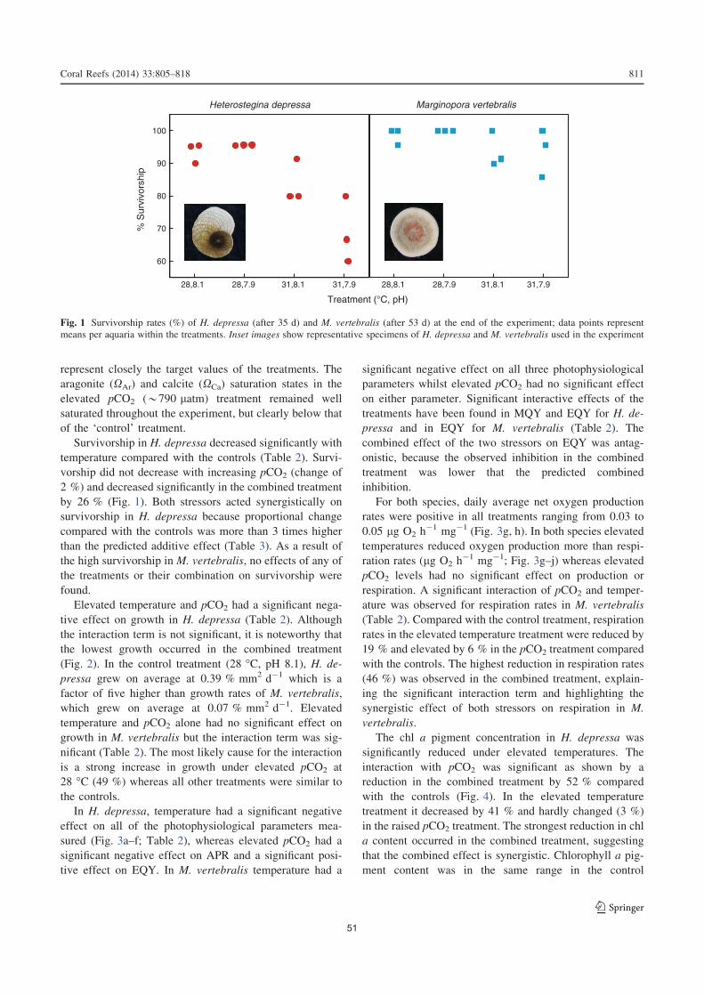
represent closely the target values of the treatments. The
aragonite (XAr) and calcite (XCa) saturation states in the
elevated pCO2 (*790 latm) treatment remained well
saturated throughout the experiment, but clearly below that
of the ‘control’ treatment.
Survivorship in H. depressa decreased significantly with
temperature compared with the controls (Table 2). Survi-
vorship did not decrease with increasing pCO2 (change of
2 %) and decreased significantly in the combined treatment
by 26 % (Fig. 1). Both stressors acted synergistically on
survivorship in H. depressa because proportional change
compared with the controls was more than 3 times higher
than the predicted additive effect (Table 3). As a result of
the high survivorship in M. vertebralis, no effects of any of
the treatments or their combination on survivorship were
found.
Elevated temperature and pCO2 had a significant nega-
tive effect on growth in H. depressa (Table 2). Although
the interaction term is not significant, it is noteworthy that
the lowest growth occurred in the combined treatment
(Fig. 2). In the control treatment (28 �C, pH 8.1), H. de-
pressa grew on average at 0.39 % mm2 d-1 which is a
factor of five higher than growth rates of M. vertebralis,
which grew on average at 0.07 % mm2 d-1. Elevated
temperature and pCO2 alone had no significant effect on
growth in M. vertebralis but the interaction term was sig-
nificant (Table 2). The most likely cause for the interaction
is a strong increase in growth under elevated pCO2 at
28 �C (49 %) whereas all other treatments were similar to
the controls.
In H. depressa, temperature had a significant negative
effect on all of the photophysiological parameters mea-
sured (Fig. 3a–f; Table 2), whereas elevated pCO2 had a
significant negative effect on APR and a significant posi-
tive effect on EQY. In M. vertebralis temperature had a
significant negative effect on all three photophysiological
parameters whilst elevated pCO2 had no significant effect
on either parameter. Significant interactive effects of the
treatments have been found in MQY and EQY for H. de-
pressa and in EQY for M. vertebralis (Table 2). The
combined effect of the two stressors on EQY was antag-
onistic, because the observed inhibition in the combined
treatment was lower that the predicted combined
inhibition.
For both species, daily average net oxygen production
rates were positive in all treatments ranging from 0.03 to
0.05 lg O2 h-1 mg-1 (Fig. 3g, h). In both species elevated
temperatures reduced oxygen production more than respi-
ration rates (lg O2 h-1 mg-1; Fig. 3g–j) whereas elevated
pCO2 levels had no significant effect on production or
respiration. A significant interaction of pCO2 and temper-
ature was observed for respiration rates in M. vertebralis
(Table 2). Compared with the control treatment, respiration
rates in the elevated temperature treatment were reduced by
19 % and elevated by 6 % in the pCO2 treatment compared
with the controls. The highest reduction in respiration rates
(46 %) was observed in the combined treatment, explain-
ing the significant interaction term and highlighting the
synergistic effect of both stressors on respiration in M.
vertebralis.
The chl a pigment concentration in H. depressa was
significantly reduced under elevated temperatures. The
interaction with pCO2 was significant as shown by a
reduction in the combined treatment by 52 % compared
with the controls (Fig. 4). In the elevated temperature
treatment it decreased by 41 % and hardly changed (3 %)
in the raised pCO2 treatment. The strongest reduction in chl
a content occurred in the combined treatment, suggesting
that the combined effect is synergistic. Chlorophyll a pig-
ment content was in the same range in the control
Treatment (°C, pH)
31,8.1 31,7.9
% S
urvi
vors
hip
60
70
80
90
100
28,8.1 28,7.9 28,8.1 28,7.9 31,8.1 31,7.9
Heterostegina depressa Marginopora vertebralis
Fig. 1 Survivorship rates (%) of H. depressa (after 35 d) and M. vertebralis (after 53 d) at the end of the experiment; data points represent
means per aquaria within the treatments. Inset images show representative specimens of H. depressa and M. vertebralis used in the experiment
Coral Reefs (2014) 33:805–818 811
123
51

treatments before and after the experiment with initial
concentrations of 0.13 (SD = 0.03) lg mg-1 wet weight
and final 0.16 (SD = 0.03) lg mg-1 wet weight in H.
depressa and initial 0.12 (SD = 0.008) lg mg-1 wet
weight final 0.15 (SD = 0.02) lg mg-1 wet weight in M.
vertebralis. This indicates that symbiont bleaching did not
occur in the control treatments.
Temperature, pCO2, and their interaction had significant
effects on the chl a content in M. vertebralis (Table 2).
Compared with the control treatment, chl a content in the
temperature treatment was reduced by 41 %. In the pCO2
treatment chl a content was elevated by 8 % compared
with the controls. Despite the apparent positive effect of
pCO2 alone, the highest reduction (72 %) was observed in
the combined treatment, suggesting a synergistic effect of
the stressors on this variable.
In summary, due to temperature elevation, 100 % of the
parameters in H. depressa and 75 % of parameters in M.
vertebralis were reduced compared with the controls
(Table 3). Elevated pCO2 alone had a significant negative
effect on 13 % of the parameters among both species but in
combination with temperature, 50 % of all measured
parameters showed significant negative effects (Table 3).
The strongest reductions in the studied parameters were
observed in the treatments where the two stressors acted in
combination. An interaction term was statistically signifi-
cant in half of the measured parameters (Table 2). Of these,
75 % showed a synergistic interaction between the two
stressors whereas the remaining 25 % of the interactions
were antagonistic (Table 3).
Discussion
Photosynthesis, respiration, and chlorophyll a content
Elevated levels of temperature had a significant effect on
all photosynthetic parameters, which is consistent with
results of previous studies of temperature-induced bleach-
ing in benthic Foraminifera (Schmidt et al. 2011; Uthicke
et al. 2011). Temperatures above 30 �C appear to lead to
damage on the protein level in the holobiont and reduce
carbon fixation rates of the symbionts due to reduced
expression of the RuBisCO enzyme (Doo et al. 2012). In
contrast to the strong effect of temperature, elevated levels
of pCO2 had mixed influences on photosynthetic parame-
ters (Table 3). Negative effects of elevated pCO2 on pho-
tosynthetic rates of Symbiodinium are known to occur at
high pCO2 levels, but at intermediate levels, pCO2, even in
combination with raised temperature, induced an increase
in symbiont oxygen production in Acropora intermedia
(Anthony et al. 2008).
In our experiment, oxygen production and dark respi-
ration rates were negatively affected by temperature.
Table 3 Summary of the significance of individual effects and their
interaction based on general linear models given in Table 2 (e
significant, d non-significant; p\ 0.05) and its interpretation based
calculations of predicted additive inhibition and fraction changes
compared with the controls
Species Parameter Summary of general linear
models
Observed inhibition compared with the
control treatment
Predicted inhibition (additive) Combined effect
Temp pCO2 Interaction A: Temp B: pCO2 C: Interaction A ? B-(A*B)
Heterostegina
depressa
MQY e d e -0.010 0.002 -0.020 -0.008 Synergistic
EQY e e e -0.112 0.004 -0.064 -0.108 Antagonistic
APR e e d -0.246 -0.012 -0.270 -0.260 Additive
Production e d d -0.379 n.a. n.a. n.a. Only temp effect
Chl a content e d e -0.414 0.032 -0.517 -0.368 Synergistic
Respiration e d d -0.319 n.a. n.a. n.a. Only temp effect
Growth e e d -0.264 -0.083 -0.587 -0.369 Additive
Survivorship e d e -0.105 0.022 -0.264 -0.081 Synergistic
Marginopora
vertebralis
MQY e d d -0.023 n.a. n.a. n.a. Only temp effect
EQY e d e -0.273 -0.042 -0.235 -0.327 Antagonistic
APR e d d -0.412 n.a. n.a. n.a. Only temp effect
Production e d d -0.436 n.a. n.a. n.a. Only temp effect
Chl a content e e e -0.406 0.080 -0.724 -0.294 Synergistic
Respiration e d e -0.187 0.060 -0.463 -0.115 Synergistic
Growth d d e 0.079 0.489 -0.140 0.529 Synergistic
Survivorship d d d n.a. n.a. n.a. n.a. No sign. effect
n.a. denotes cases where only a single factor and not the interaction term were significant
812 Coral Reefs (2014) 33:805–818
123
52

Previous work suggested that respiration rates in M. ver-
tebralis increases with increasing temperatures before and
after exposure to 31 �C for several weeks (Uthicke et al.
2011). Despite the fact that an increase in temperature
speeds up enzymatic reactions, explaining increased res-
piration rates in the short term, in the long term, a decline
in respiration rates was observed. This shows that the ho-
lobiont was weakened in the 31 �C compared with the
controls at the end of the experiment. Studies on corals also
report reduced respiration rates with increasing tempera-
ture (Faxneld et al. 2011; Agostini et al. 2013).
We did not observe any effect of raised pCO2 alone on
oxygen production and dark respiration. The result that
dark respiration was not affected by pCO2 increase alone,
is similar to studies in corals (Langdon et al. 2003; Rey-
naud et al. 2003). However, our results imply a significant
synergistic interaction of the two stressors on the reduction
of respiration rates in M. vertebralis. The synergistic
inhibition of respiration observed in the combined treat-
ment indicates that the metabolic rate of the holobiont is
reduced, and this effect may propagate into other vital
parameters.
Elevated pCO2 alone increased the amount of chl a in
M. vertebralis but not in H. depressa. CO2 fertilisation is
known to increase cell numbers, pigments, and productiv-
ity in marine unicellular plankton, for example cyanobac-
teria (Riebesell et al. 1993) and diatoms (Yang and Gao
2012). Productivity increase due to pCO2 increase has also
been demonstrated in imperforate Foraminifera (Uthicke
and Fabricius 2012) and cell counts of dinoflagellate
symbionts in M. rossi showed higher symbiont density
under elevated pCO2 (Reymond et al. 2013). It is therefore
likely that the increased chl a content inM. vertebralis with
pCO2 enhancement in our study reflects higher photo-
symbiont density. When Foraminifera were simultaneously
exposed to elevated temperature, chlorophyll a content did
decrease significantly, indicating that the hypothesised
increase in photosymbiont density due to pCO2 fertilisation
was counteracted by temperature, leading to trade-offs in
the photosymbiont efficiency and density.
Survivorship and growth
Elevated temperature reduced survivorship in H. depressa
but did not affect M. vertebralis, indicating that the latter
species is more tolerant. Previous work on the temperature
response of M. vertebralis also showed no effect on sur-
vivorship rate under exposure at 31 �C for several months,
but reported reduced growth at this temperature (Uthicke
et al. 2011). Elevated pCO2 did not influence survivorship
in either species, which is similar to previous laboratory
studies that reported no significant effect of pCO2 elevation
on survivorship in several Foraminifera species (Vogel and
Uthicke 2012; McIntyre-Wressnig et al. 2013). However,
in H. depressa the combined effect of pCO2 and temper-
ature on survivorship was significant and synergistic indi-
cating that this species is especially vulnerable to the
combination of stressors.
In our experiment, temperature and elevated pCO2
(738 latm) in isolation reduced growth in the hyaline
species H. depressa. In contrast, Vogel and Uthicke (2012)
did not report negative effects on growth in H. depressa up
to pCO2 levels of 1,600 latm. The reason for these dif-
ferences is not clear but we note that in our experiment the
growth rate of H. depressa was higher by almost a factor of
two than in the experiments by Vogel and Uthicke (2012).
Gro
wth
(% d
-1)
0.0
0.2
0.4
0.6
0.8
1.0
1.2
28,8.1 28,7.9 31,8.1 31,7.9 28,8.1 28,7.9 31,8.1 31,7.9
Heterostegina depressa Marginopora vertebralis
Treatment (°C, pH)
Fig. 2 Growth rates (% surface area increase d-1) of H. depressa and
M. vertebralis during the exposure to experimental conditions for a
period of 35 and 53 d, respectively. Data are represented as box-and-
whisker plots, the top and bottom of the box (3rd and 1st quartile), the
2nd quartile (median), the lines extending from the box (whiskers)
extend to the outermost data that fall within the distance computed
as follows: 3rd quartile ? 1.5*(interquartile range); 1st quartile
- 1.5*(interquartile range)
Coral Reefs (2014) 33:805–818 813
123
53
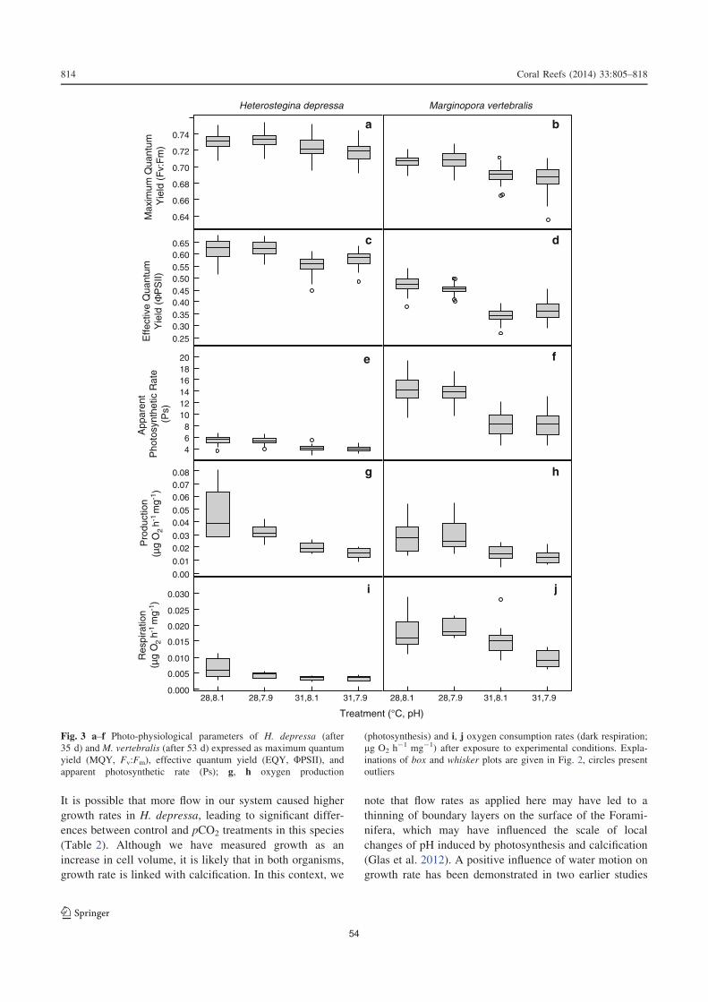
It is possible that more flow in our system caused higher
growth rates in H. depressa, leading to significant differ-
ences between control and pCO2 treatments in this species
(Table 2). Although we have measured growth as an
increase in cell volume, it is likely that in both organisms,
growth rate is linked with calcification. In this context, we
note that flow rates as applied here may have led to a
thinning of boundary layers on the surface of the Forami-
nifera, which may have influenced the scale of local
changes of pH induced by photosynthesis and calcification
(Glas et al. 2012). A positive influence of water motion on
growth rate has been demonstrated in two earlier studies
[Fv/
Fm
][?
PS
II]A
ppar
ent
Pho
tosy
nthe
tic R
ate
(Ps)
[μg
O2
hR
espi
ratio
n (μ
g O
2 h-1
mg-1
) P
rodu
ctio
n (μ
g O
2 h-1
mg-1
)
Effe
ctiv
e Q
uant
umY
ield
(Φ
PS
II)M
axim
um Q
uant
um
Yie
ld (
Fv:
Fm
)
Treatment (°C, pH)
0.64
0.66
0.68
0.70
0.72
0.74
0.250.300.350.400.450.500.550.600.65
468
101214161820
0.00
0.01
0.020.03
0.040.05
0.060.070.08
0.000
0.005
0.010
0.015
0.020
0.025
0.030
28,8.1 28,7.9 31,8.1 31,7.9 28,8.1 28,7.9 31,8.1 31,7.9
ba
dc
e f
hg
i j
Heterostegina depressa Marginopora vertebralis
Fig. 3 a–f Photo-physiological parameters of H. depressa (after
35 d) and M. vertebralis (after 53 d) expressed as maximum quantum
yield (MQY, Fv:Fm), effective quantum yield (EQY, UPSII), andapparent photosynthetic rate (Ps); g, h oxygen production
(photosynthesis) and i, j oxygen consumption rates (dark respiration;
lg O2 h-1 mg-1) after exposure to experimental conditions. Expla-
inations of box and whisker plots are given in Fig. 2, circles present
outliers
814 Coral Reefs (2014) 33:805–818
123
54

(ter Kuile and Erez 1984; Hallock et al. 1986). On the other
hand, the response of growth and calcification in benthic
Foraminifera may be decoupled with respect to the effect
of pCO2 and the reaction might be species specific. For
example, Amphistegina gibbosa did not exhibit negative
effects on growth of up to 2,000 latm pCO2, but showed
patchy test dissolution (McIntyre-Wressnig et al. 2013).
Elevated calcification rates were observed under interme-
diate pCO2 levels (580–770 latm) for the clonal popula-
tion of Baculogypsina sphaerulata, whereas the second
hyaline species in the same study Calcarina gaudichaudii,
showed an inconsistent reaction (Fujita et al. 2011). Fur-
thermore, genotype-specific responses to ocean acidificat-
ion have been reported for other marine organisms, such as
coccolithophorids (Langer et al. 2009) and diatoms (Kremp
et al. 2012).
In contrast, the porcelanous M. vertebralis showed no
significant growth response to elevated temperature and
pCO2 alone. For this species, growth rates in this study
were in the same range as in the previous lower-flow setup
(Uthicke et al. 2011; Vogel and Uthicke 2012). A small but
not significant increase in growth was observed in M.
vertebralis in the treatment exposed to elevated pCO2
(738 latm) at 28 �C. A slight growth enhancement was
observed in other porcelaneous species (Vogel and Uthicke
2012), whereas other studies reported reduced growth at
medium to high pCO2 levels (700–1,100 latm) in Am-
phisorus hemprichii (Fujita et al. 2011; Hikami et al. 2011)
and M. rossii (Reymond et al. 2013).
Differences in the growth and calcification responses to
pCO2 enhancement are known between and within taxo-
nomic groups (Ries et al. 2009). Varying responses in
calcification processes under intermediate scenarios of up
to 900 latm may reflect differential development of pro-
tective layers around the precipitated biomineral and dif-
ferent abilities to regulate the pH at the site of calcification
(Ries et al. 2009). In our experiment, the moderately ele-
vated pCO2 level acting alone may have released the
photosymbiont population of M. vertebralis from CO2
limitation (Allemand et al. 2004), which would increase
carbon translocation to the host and lead to a growth
enhancement.
In all treatments in our study, the XCa of the seawater
remained above two (Table 1) and no test dissolution was
observed in any of the treatments. Typically, test dissolu-
tion is observed in benthic Foraminifera exposed to much
higher pCO2 conditions (2,000 latm) and at much lower
XAr and XCa of sea water than in our experiment (Haynert
et al. 2011; McIntyre-Wressnig et al. 2013), although at
natural CO2 vents, dissolution was observed already at pH
*7.9 (Uthicke et al. 2013). Test dissolution at pCO2 levels
equivalent to a pH of 7.4–7.9 was also observed in M.
vertebralis in laboratory culture (Sinutok et al. 2011), but
specimens in the control treatment in that study also
showed slight test dissolution; indicating that the cultured
Foraminifera were not physiologically fit.
In summary, our results indicate that, at least in the short
term, coral reef Foraminifera are likely to continue to grow
under conditions predicted for the end of the century.
However, the fact that in both studied species, growth was
inhibited in the combined treatment indicates that in the
long term, growth and by inference calcification under
lower saturation may become more difficult. This may
ultimately lead to ecological exclusion of these species as
observed at present in CO2 seep systems (Dias et al. 2010;
Uthicke and Fabricius 2012; Uthicke et al. 2013).
Combined effects of key global change stressors
Significant interactive effects between pCO2 and warming
were observed for 50 % of the parameters investigated. In
75 % of these, for each species, the combined effects were
Chl
α (
μg m
gww
-1)
0.00
0.05
0.10
0.15
0.20
0.25
28,8.1 28,7.9 31,8.1 31,7.9 28,8.1 28,7.9 31,8.1 31,7.9
Heterostegina depressa Marginopora vertebralis
Treatment (°C, pH)
Fig. 4 Chlorophyll a content (lg mg wet weight-1) at the end of the experiment in H. depressa (35 d) and M. vertebralis (53 d) after exposure
to experimental conditions, box and whisker plots are explained in Fig. 2, circles represent outliers
Coral Reefs (2014) 33:805–818 815
123
55

synergistic—the effects of warming and elevated pCO2 led
to a stronger physiological response than the sum of the
effects of the individual parameters (Table 3). Because
temperature and pCO2 may affect the holobiont as a whole,
it is difficult to offer a physiological explanation for the
prevalence of the synergistic effects. As a likely explana-
tion, we suggest that the enhanced negative effects of
multiple stressors could reflect trade-offs in resource allo-
cation, where the costs of counteracting the effect of one
stressor reduce the ability to counteract the effects of the
additional stressor. Similarly, it has been suggested that
under stressful environmental conditions, such as elevated
inorganic nutrient levels, the pressure on the holobiont to
control the population size of its photosymbionts might
increase, reducing the capacity of the holobiont to respond
to stress (Uthicke et al. 2011). The stress caused by over-
growing population of photosymbionts has been described
by Wooldridge (2009) to physiologically affect corals and
lower their bleaching thresholds. Furthermore, a 3-yr field
experiment demonstrated the same effect, where corals
bleached more in artificial nutrient enrichment treatments
(Vega Thurber et al. 2013).
Interactive negative effects between pCO2 and temper-
ature have been shown to affect the calcification rate in
corals, but the response of the corals was highly species
specific (Edmunds et al. 2012). In an experiment increasing
nutrient levels and elevating pCO2, photosymbiont con-
centration in M. rossi was reduced more under the com-
bined stress than when individual stressors acted in
isolation (Reymond et al. 2013). van Dam et al. (2012)
showed that populations of Foraminifera exposed to the
herbicide Diuron become disproportionately more sensitive
to temperature and both factors acted additively on the
foraminiferal photosynthetic response. Ecotoxicological
studies on the interaction of climate change with additional
stressors, such as pesticide exposure, indicate a prevalence
of synergistic interactions across different organisms
(Holmstrup et al. 2010; Kohler and Triebskorn 2013). In
this respect, the observation on the numerous synergistic
interactions in our study indicates that the physiology of
the unicellular Foraminifera and/or their symbiosis with
algae may be affected in a similar way as that of other
organisms, contrary to the hypothesis by Portner (2002)
that protists are less vulnerable to the studied stressors.
Irrespective of the exact mechanism responsible for the
existence of strong synergistic effects between the key
global change stressors tested in this study, our results
indicate that the effects of environmental change in the
shallow marine realm under expected CO2 emission sce-
narios are likely to be underestimated when the effects of
elevated pCO2 and temperature are investigated in
isolation.
Acknowledgments The study was funded by the Australian Insti-
tute of Marine Science and conducted with the support of funding
from the Australian Government’s National Environmental Research
Program. We are grateful for the assistance of M. Takahashi for
developing the design and constructions of the flow-through housings.
J. Brandt assisted with digital image analysis. The study benefited
greatly for the help of F. Flores, S. Noonan, N. Webster, and A. Negri.
Open Access This article is distributed under the terms of the
Creative Commons Attribution License which permits any use, dis-
tribution, and reproduction in any medium, provided the original
author(s) and the source are credited.
References
Agostini S, Fujimura H, Higuchi T, Yuyama I, Casareto BE, Suzuki
Y, Nakano Y (2013) The effects of thermal and high-CO2
stresses on the metabolism and surrounding microenvironment
of the coral Galaxea fascicularis. C R Biol 336:384–391
Allemand D, Ferrier-Pages C, Furla P, Houlbreque F, Puverel S,
Reynaud S, Tambutte E, Tambutte S, Zoccola D (2004)
Biomineralisation in reef-building corals: from molecular mech-
anisms to environmental control. C R Palevol 3:453–467
Anthony KRN, Kline DI, Diaz-Pulido G, Dove S, Hoegh-Guldberg O
(2008) Ocean acidification causes bleaching and productivity
loss in coral reef builders. Proc Natl Acad Sci U S A 105:
17442–17446
Arieli RN, Almogi-Labin A, Abramovich S, Herut B (2011) The
effect of thermal pollution on benthic foraminiferal assemblages
in the Mediterranean shoreface adjacent to Hadera power plant
(Israel). Mar Pollut Bull 62:1002–1012
Bernhard JM (2000) Distinguishing live from dead foraminifera:
Methods review and proper applications. Micropaleontology
46:38–46
Blackmon PD, Todd R (1959) Mineralogy of some foraminifera as
related to their classification and ecology. J Paleontol 1–15
Bliss CI (1939) The toxicity of poisons applied jointly. Ann Appl Biol
26:585–615
Cooper TF, Ulstrup KE (2009) Mesoscale variation in the photophy-
siology of the reef building coral Pocillopora damicornis along
an environmental gradient. Estuar Coast Shelf Sci 83:186–196
Cornelisen CD, Thomas FIM (2009) Prediction and validation of
flow-dependent uptake of ammonium over a seagrass-hardbot-
tom community in Florida Bay. Mar Ecol Prog Ser 386:71–81
Crain CM, Kroeker K, Halpern BS (2008) Interactive and cumulative
effects of multiple human stressors in marine systems. Ecol Lett
11:1304–1315
Dias BB, Hart B, Smart CW, Hall-Spencer JM (2010) Modern
seawater acidification: the response of foraminifera to high-CO2
conditions in the Mediterranean Sea. J Geol Soc London
167:843–846
Doo SS, Mayfield AB, Byrne M, Chen HK, Nguyen HD, Fan TY
(2012) Reduced expression of the rate-limiting carbon fixation
enzyme RuBisCO in the benthic foraminifer Baculogypsina
sphaerulata holobiont in response to heat shock. J Exp Mar Biol
Ecol 430:63–67
Edmunds PJ, Brown D, Moriarty V (2012) Interactive effects of ocean
acidification and temperature on two scleractinian corals from
Moorea, French Polynesia. Glob Chang Biol 18:2173–2183
Fabricius KE, Langdon C, Uthicke S, Humphrey C, Noonan S, De’ath
G, Okazaki R, Muehllehner N, Glas MS, Lough JM (2011)
816 Coral Reefs (2014) 33:805–818
123
56
Losers and winners in coral reefs acclimatized to elevated carbon
dioxide concentrations. Nat Clim Chang 1:165–169
Faxneld S, Jorgensen TL, Nguyen ND, Nystrom M, Tedengren M
(2011) Differences in physiological response to increased
seawater temperature in nearshore and offshore corals in
northern Vietnam. Mar Environ Res 71:225–233
Fujita K, Hikami M, Suzuki A, Kuroyanagi A, Sakai K, Kawahata H,
Nojiri Y (2011) Effects of ocean acidification on calcification of
symbiont-bearing reef foraminifers. Biogeosciences 8:2089–2098
Glas MS, Fabricius KE, de Beer D, Uthicke S (2012) The O2, pH and
Ca2? Microenvironment of Benthic Foraminifera in a High CO2
World. PLoS One 7:e50010
Hallock P (1985) Why are larger foraminifera large? Paleobiology
11:195–208
Hallock P, Talge HK (1993) Symbiont loss (‘‘bleaching’’) in the reef-
dwelling benthic foraminifera Amphistegina gibbosa in the
Florida Keys in 1991-92. Proceedings of the Colloquium on
Global Aspects of Coral Reefs: Health, Hazards and History,
Rosenstiel School of Marine and Atmospheric Science, Univer-
sity of Miami, pp 94–100
Hallock P, Forward LB, Hansen HJ (1986) Influence of environ-
ment on the test shape of Amphistegina. J Foraminiferal Res
16:224–231
Hallock P, Talge HK, Smith KM, Cockey EM (1992) Bleaching in the
reef-dwelling foraminifera Amphistegina gibbosa. Proc 7th Int
Coral Reef Symp 1:44–49
Haynert K, Schonfeld J, Riebesell U, Polovodova I (2011) Biometry
and dissolution features of the benthic foraminifer Ammonia
aomoriensis at high pCO2. Mar Ecol Prog Ser 432:53–67
Hikami M, Ushie H, Irie T, Fujita K, Kuroyanagi A, Sakai K, Nojiri
Y, Suzuki A, Kawahata H (2011) Contrasting calcification
responses to ocean acidification between two reef foraminifers
harboring different algal symbionts. Geophys Res Lett 38
Hill R, Schreiber U, Gademann R, Larkum AWD, Kuhl M, Ralph PJ
(2004) Spatial heterogeneity of photosynthesis and the effect of
temperature-induced bleaching conditions in three species of
corals. Mar Biol 144:633–640
Hoegh-Guldberg O, Mumby PJ, Hooten AJ, Steneck RS, Greenfield
P, Gomez E, Harvell CD, Sale PF, Edwards AJ, Caldeira K,
Knowlton N, Eakin CM, Iglesias-Prieto R, Muthiga N, Bradbury
RH, Dubi A, Hatziolos ME (2007) Coral reefs under rapid
climate change and ocean acidification. Science 318:1737–1742
Hohenegger J (2004) Depth coenoclines and environmental consid-
erations of western pacific larger Foraminifera. J Foraminiferal
Res 34:9–33
Hohenegger J, Yordanova E, Nakano Y, Tatzreiter F (1999) Habitats
of larger foraminifera on the upper reef slope of Sesoko Island,
Okinawa, Japan. Mar Micropaleontol 36:109–168
Holmstrup M, Bindesbøl A, Oostingh GJ, Duschl A, Scheil V, Kohler
H, Loureiro S, Soares AMVM, Ferreira ALG, Kienle C,
Gerhardt A, Laskowski R, Kramarz PE, Bayley M, Svendsen
C, Spurgeon DJ (2010) Interactions between effects of environ-
mental chemicals and natural stressors: A review. Sci Total
Environ 408:3746–3762
Koch M, Bowes G, Ross C, Zhang XH (2013) Climate change and
ocean acidification effects on seagrasses and marine macroalgae.
Glob Chang Biol 19:103–132
Kohler HR, Triebskorn R (2013) Wildlife Ecotoxicology of Pesti-
cides: Can We Track Effects to the Population Level and
Beyond? Science 341:759–765
Kremp A, Godhe A, Egardt J, Dupont S, Suikkanen S, Casabianca S,
Penna A (2012) Intraspecific variability in the response of
bloom-forming marine microalgae to changed climate condi-
tions. Ecol Evol 2:1195–1207
Kuroyanagi A, Kawahata H, Suzuki A, Fujita K, Irie T (2009)
Impacts of ocean acidification on large benthic foraminifers:
Results from laboratory experiments. Mar Micropaleontol 73:
190–195
Langdon C, Broecker WS, Hammond DE, Glenn E, Fitzsimmons K,
Nelson SG, Peng TH, Hajdas I, Bonani G (2003) Effect of
elevated CO2 on the community metabolism of an experimental
coral reef. Global Biogeochem Cycles 17
Langer G, Nehrke G, Probert I, Ly J, Ziveri P (2009) Strain-specific
responses of Emiliania huxleyi to changing seawater carbonate
chemistry. Biogeosciences 6:2637–2646
Langer MR, Lipps JH (2003) Foraminiferal distribution and diversity,
Madang Reef and Lagoon, Papua New Guinea. Coral Reefs 22:
143–154
Langer MR, Silk MT, Lipps JH (1997) Global ocean carbonate and
carbon dioxide production; the role of reef Foraminifera.
J Foraminiferal Res 27:271–277
Lee JJ (1995) Living Sands - The symbiosis of protists and their algae
can provide good models for the study of host/symbiont
interactions. Bioscience 45:252–261
Lee JJ (2006) Algal symbiosis in larger foraminifera. Symbiosis
42:63–75
Lee JJ, Hallock P (1987) Algal symbiosis as the driving force in the
evolution of larger Foraminifera. Ann N Y Acad Sci 503:
330–347
Lee JJ, McEnergy ME, Rottger R, Reimer CW (1980) The isolation,
culture and identification of endosymbiotic diatoms from
Heterostegina depressa D’Orbigny and Amphistegina lessonii
D’Orbigny (Larger Foraminifer) from Hawaii. Botanica Marina
23:297–302
Leutenegger S (1984) Symbiosis in benthic Foraminifera: specificity
and host adaptations. J Foraminiferal Res 14:16–35
Lewis E, Wallace D (1997) CO2 System Calculations Software
CO2SYSTM.EXE. Global Ocean Data Analysis centre
Lough JM, Hobday AJ (2011) Observed climate change in
Australian marine and freshwater environments. Mar Freshw
Res 62:984–999
Martin S, Gattuso JP (2009) Response of Mediterranean coralline
algae to ocean acidification and elevated temperature. Glob
Chang Biol 15:2089–2100
McCulloch M, Falter J, Trotter J, Montagna P (2012) Coral resilience
to ocean acidification and global warming through pH up-
regulation. Nat Clim Chang 2:623–633
McIntyre-Wressnig A, Bernhard JM, McCorkle DC, Hallock P (2013)
Non-lethal effects of ocean acidification on the symbiont-bearing
benthic foraminifer Amphistegina gibbosa. Mar Ecol Prog Ser
472:45–60
Momigliano P, Uthicke S (2013) Symbiosis in a giant protist
(Marginopora vertebralis, Soritinae): flexibility in symbiotic
partnerships along a natural temperature gradient. Mar Ecol Prog
Ser 491:33–46
Nobes K, Uthicke S, Henderson R (2008) Is light the limiting factor
for the distribution of benthic symbiont bearing foraminifera on
the Great Barrier Reef? J Exp Mar Biol Ecol 363:48–57
Pawlowski J, Holzmann M, Berney C, Fahrni J, Gooday AJ,
Cedhagen T, Habura A, Bowser SS (2003) The evolution of
early Foraminifera. Proc Natl Acad Sci U S A 100:11494–11498
Pochon X, Garcia-Cuetos L, Baker AC, Castella E, Pawlowski J
(2007) One-year survey of a single Micronesian reef reveals
extraordinarily rich diversity of Symbiodinium types in soritid
foraminifera. Coral Reefs 26:867–882
Portner HO (2002) Climate variations and the physiological basis of
temperature dependent biogeography: systemic to molecular
hierarchy of thermal tolerance in animals. Comp Biochem
Physiol A Mol Integr Physiol 132:739–761
Portner HO (2008) Ecosystem effects of ocean acidification in times
of ocean warming: a physiologist’s view. Mar Ecol Prog Ser
373:203–217
Coral Reefs (2014) 33:805–818 817
123
57
Redondo-Rodriguez A, Weeks SJ, Berkelmans R, Hoegh-Guldberg O,
Lough JM (2012) Climate variability of the Great Barrier Reef in
relation to the tropical Pacific and El Nino-Southern Oscillation.
Mar Freshw Res 63:34–47
Renema W (2006) Habitat variables determining the occurrence of
large benthic foraminifera in the Berau area (East Kalimantan,
Indonesia). Coral Reefs 25:351–359
Reymond CE, Lloyd A, Kline DI, Dove SG, Pandolfi JM (2013)
Decline in growth of foraminifer Marginopora rossi under
eutrophication and ocean acidification scenarios. Glob Chang
Biol 19:291–302
Reynaud S, Leclercq N, Romaine-Lioud S, Ferrier-Pages C, Jaubert J,
Gattuso JP (2003) Interacting effects of CO2 partial pressure and
temperature on photosynthesis and calcification in a scleractinian
coral. Glob Chang Biol 9:1660–1668
Riebesell U, Wolf-Gladrow DA, Smetacek V (1993) Carbon dioxide
limitation of marine phytoplankton growth rates. Nature 361:
249–251
Ries JB, Cohen AL, McCorkle DC (2009) Marine calcifiers exhibit
mixed responses to CO2-induced ocean acidification. Geology
37:1131–1134
Rodolfo-Metalpa R, Lombardi C, Cocito S, Hall-Spencer JM, Gambi
MC (2010) Effects of ocean acidification and high temperatures
on the bryozoan Myriapora truncata at natural CO2 vents. Mar
Ecol 31:447–456
Rodolfo-Metalpa R, Houlbreque F, Tambutte E, Boisson F, Baggini
C, Patti FP, Jeffree R, Fine M, Foggo A, Gattuso JP, Hall-
Spencer JM (2011) Coral and mollusc resistance to ocean
acidification adversely affected by warming. Nat Clim Chang
1:308–312
SAS (2012) JMP statistics software, Version 10. SAS
Schmidt C, Heinz P, Kucera M, Uthicke S (2011) Temperature-
induced stress leads to bleaching in larger benthic foraminifera
hosting endosymbiotic diatoms. Limnol Oceanogr 56:1587–1602
Sinutok S, Hill R, Doblin MA, Wuhrer R, Ralph PJ (2011) Warmer
more acidic conditions cause decreased productivity and calci-
fication in subtropical coral reef sediment-dwelling calcifiers.
Limnol Oceanogr 56:1200–1212
Talge HK, Hallock P (2003) Ultrastructural responses in field-
bleached and experimentally stressed Amphistegina gibbosa
(Class Foraminifera). J Eukaryot Microbiol 50:324–333
ter Kuile B, Erez J (1984) In situ growth rate experiments on the
symbiont-bearing foraminifera Amphistegina lobifera and Am-
phisorus hemprichii. J Foraminiferal Res 14:262–276
Uthicke S, Altenrath C (2010) Water column nutrients control growth
and C: N ratios of symbiont-bearing benthic foraminifera on the
Great Barrier Reef, Australia. Limnol Oceanogr 55:1681–1696
Uthicke S, Fabricius KE (2012) Productivity gains do not compensate
for reduced calcification under near-future ocean acidification in
the photosynthetic benthic foraminifer species Marginopora
vertebralis. Glob Chang Biol 18:2781–2791
Uthicke S, Momigliano P, Fabricius KE (2013) High risk of
extinction of benthic foraminifera in this century due to ocean
acidification. Sci Rep 3
Uthicke S, Vogel N, Doyle J, Schmidt C, Humphrey C (2011)
Interactive effects of climate change and eutrophication on the
dinoflagellate-bearing benthic foraminifer Marginopora verteb-
ralis. Coral Reefs:1–14
van Dam JW, Negri AP, Mueller JF, Altenburger R, Uthicke S (2012)
Additive pressures of elevated sea surface temperatures and
herbicides on symbiont-bearing foraminifera. PLoS One 7:e33900
Vega Thurber RL, Burkepile DE, Fuchs C, Shantz AA, McMinds R,
Zaneveld JR (2013) Chronic nutrient enrichment increases
prevalence and severity of coral disease and bleaching. Glob
Chang Biol 20:544–554
Vogel N, Uthicke S (2012) Calcification and photobiology in
symbiont-bearing benthic foraminifera and responses to a high
CO2 environment. J Exp Mar Biol Ecol 424:15–24
Wernberg T, Smale DA, Thomsen MS (2012) A decade of climate
change experiments on marine organisms: procedures, patterns
and problems. Glob Chang Biol 18:1491–1498
Wernberg T, Smale DA, Tuya F, Thomsen MS, Langlois TJ, de
Bettignies T, Bennett S, Rousseaux CS (2013) An extreme
climatic event alters marine ecosystem structure in a global
biodiversity hotspot. Nat Clim Chang 3:78–82
Williams SL, Carpenter RC (1998) Effects of unidirectional and
oscillatory water flow on nitrogen fixation (acetylene reduction)
in coral reef algal turfs, Kaneohe Bay, Hawaii. J Exp Mar Biol
Ecol 226:293–316
Wooldridge SA (2009) Water quality and coral bleaching thresholds:
Formalising the linkage for the inshore reefs of the Great Barrier
Reef, Australia. Mar Pollut Bull 58:745–751
Yang GY, Gao KS (2012) Physiological responses of the marine
diatom Thalassiosira pseudonana to increased pCO2 and
seawater acidity. Mar Environ Res 79:142–151
Ziegler M, Uthicke S (2011) Photosynthetic plasticity of endos-
ymbionts in larger benthic coral reef Foraminifera. J Exp Mar
Biol Ecol 407:70–80
818 Coral Reefs (2014) 33:805–818
123
58

59
Chapter 3
3. Publication II: Recent invasion of the symbiont-bearing foraminifera Pararotalia into the Eastern
Mediterranean facilitated by the ongoing warming trend
Christiane Schmidt1, Raphael Morard1, Ahuva Almogi-Labin2, Anna Weinmann3,4,
Danna Titelboim5, Sigal Abramovich5, Michal Kucera1
1MARUM, Centre of Marine Environmental Science, University of Bremen, Bremen, Germany
2Geological Survey of Israel, Jerusalem, Israel3Department of Geology, University of Georgia, Athens, Georgia, United States of
America4Steinmann-Institute for Geology, Mineralogy und Palaeontology, University of Bonn,
Bonn, Germany5Ben Gurion University of the Negev, Beer-Sheva, Israel

60

61
3.1. Abstract
The eastern Mediterranean is a hotspot of biological invasions. Numerous species
of Indopacific origin appear to have colonized the Mediterranean in historical times,
including tropical symbiont-bearing foraminifera. Among these is the species
Pararotalia calcariformata. Unlike other invasive foraminifera, this species has been
discovered only two decades ago and is restricted to the eastern Mediterranean
coast. Combining ecological, genetic and physiological observations, we attempt to
explain the recent invasion of this species in the Mediterranean Sea. Using
morphological and genetic data, we confirm the species attribution to P.
calcariformata McCulloch 1977 and identify its symbionts as a consortium of diatom
species dominated by Minutocellus polymorphus. We document photosynthetic
activity of its endosymbionts using Pulse Amplitude Modulated Fluorometry and test
the effects of elevated temperatures on growth rates of asexual offspring. The
culturing of asexual offspring for 120 days shows a 30-day period of rapid growth
followed by a slower growth phase. A subsequent 48-day temperature sensitivity
experiment indicates a similar developmental pathway and high growth rate at 28°C,
whereas an almost complete inhibition of growth was observed at 20°C and 35°C.
This indicates that the species may have unexpectedly low tolerance to cold
temperatures. We expand this hypothesis by applying a Species Distribution Model
(SDM) based on modern occurrences in the Mediterranean and three environmental
variables (irradiance, turbidity and yearly minimum temperature). The model
reproduces the observed restricted distribution and indicates that the range of the
species will drastically expand westwards under future global change scenarios. We
conclude that P. calcariformata established a population in the Levant because of the
recent warming in the region. In line with observations from other groups of
organisms, our results indicate that continued warming of the eastern Mediterranean
will facilitate the invasion of progressively more tropical marine taxa into the
Mediterranean, disturbing local biodiversity and ecosystem structure.

62
3.2. Introduction
Human activities can induce invasions of marine species in two ways:
indirectly, by altering the climate and ecosystems (Vitousek et al. 1997), facilitating
range expansions (Sorte et al. 2010; Chen et al. 2011), or directly, by mediating
species dispersal through anthropogenic means (Carlton 1999).The latter can be
realized either as active transport, such as ship traffic, or as removal of barriers to
dispersal. The ongoing anthropogenic global change is altering ecosystems at a
faster rate than seen in the recent geological past. In consequence, many species
are unable to adapt to locally changing conditions through phenotypic plasticity and
evolutionary processes and respond by shifting their geographical ranges (Parmesan
2006; Hiddink et al. 2012). This is particularly relevant for marine ecosystems, where
species appear to spread an order of magnitude faster than in the terrestrial realm
(Sorte et al. 2010). Temperature is the key variable controlling the spread of species
and can be used to predict biogeographic range expansions in shallow marine
communities (Belanger et al. 2012).
An excellent example of directly mediated invasion of marine species is the
opening of the Suez Canal. This event facilitated what is known as the Lessepsian
invasion, describing the movement of species from the Red Sea into the
Mediterranean after the opening of the Canal in 1869 (Rilov and Galil 2009; Zenetos
et al. 2012). If the opening of the Suez Canal was the only factor needed to trigger
the range extension of Indopacific species into the Mediterranean, thus all
Lessepsian migrants should have appeared near simultaneously. However, the
eastern Mediterranean has been experiencing a strong warming trend over the last
20 years (Macias et al. 2013) altering the environmental conditions at the exit of the
Lessepsian corridor. The expansion of Indopacific species into the Mediterranean is
thus likely exacerbated by climate change and many of the migrating marine species
including fish, algae, plants and invertebrates, which continue to expand their range
(Hiddink et al. 2012). As a result, the Levantine ecosystem is already dominated by
non-native fish species leading to a significant decline of the indigenous populations
(Edelist et al. 2013).
A particularly successful group among the Lessepsian migrants are symbiont-
bearing benthic foraminifera (Langer et al. 2012; Merkado et al. 2013). The passive
dispersal of these organisms appears to be facilitated through the transport of

63
propagules, which include asexually and sexually reproduced offspring (Alve 1999;
Alve and Goldstein 2003). Alternatively, like in many other Lessepsian migrants, the
association with marine macro algae represents another means of passive dispersal
of benthic foraminifera into the Mediterranean (Zenetos et al. 2012; Caruso and
Cosentino 2014). Also the introduction of foraminiferal species in new habitats via
ballast waters has been documented (McGann et al. 2000). Symbiont-bearing
foraminifera have well defined, species-specific temperature tolerances (Langer and
Hottinger 2000). Temperatures exceeding the upper thermal threshold cause
symbiont bleaching (Schmidt et al. 2011), whilst low temperatures prevent the
establishment of populations (Langer and Hottinger 2000). The symbiosis in benthic
foraminifera provides an energetic advantage (Hallock 1981), with the photosymbiont
having a double role, by providing nutrition (Lee and Hallock 1987) and promoting
calcification (de Nooijer et al. 2009). Symbiont-bearing foraminifera are thus ideally
adapted to the oligotrophic conditions of the eastern Mediterranean (Sisma-Ventura
et al. 2014). Indeed, the appearance of invasive symbiont-bearing foraminifera in the
eastern Mediterranean is documented at least since the 1960s (Langer 2008) and
their ongoing proliferation has significant impact on coastal ecosystems (Mouanga
and Langer 2014).
The most recently described migration of symbiont-bearing benthic
foraminifera into the Levantine basin involves a species of the Indopacific genus
Pararotalia. This species has been first reported in the Levant in 1994 by Reinhardt
et al. (1994) and Yanko et al. (1994). It has since then been found to proliferate along
the Mediterranean coast from Israel (Hyams-Kaphzan et al. 2008; Arieli et al. 2011)
to southern Turkey (Meriç et al. 2013). The modern foraminiferal fauna of the
Mediterranean Sea is mostly of Atlantic origin (Langer 1993; Langer 2008). After the
opening of the Suez Canal in 1869, many tropical symbiont-bearing foraminifera
migrated into the Mediterranean Sea, including amphisteginids, soritids, and
heterosteginids (Hyams et al. 2002). The apparently later invasion and more
restricted occurrence of Pararotalia contrasts with other symbiont-bearing
foraminifera, implying that the invasion of Pararotalia was not facilitated solely by the
physical connection of the Suez Canal. We note that the invasive Pararotalia is not a
“classical” Lessepsian species, as it has not yet been found in the Red Sea (Reiss
and Hottinger 1984; Parker et al. 2012; Madkour 2013). However, with the exception
of the Gulf of Aqaba (Hottinger L et al. 1993), the diversity of foraminifera in the Red

64
Sea is not well known and considering the habitat of the species and its distribution in
the Indopacific, it is likely that it also occurs in the Red Sea (Hesemann 2014).
To understand the explosive recent invasion of Pararotalia in the
Mediterranean, we carried out an investigation of the ecology and physiology of this
species. We investigated its genetic relationship with Pacific populations of the
genus, identified its endosymbiotic microalgae, determined its photosynthetic activity
from freshly collected specimens and monitored their photosynthetic activity in the
laboratory cultures. We hypothesized that like in other foraminifera (Goldstein and
Alve 2011) and in many other marine species (Belanger et al. 2012) temperature is
the main factor controlling the establishment of new populations. Therefore, we
carried out an experiment exposing asexual offspring of P. calcariformata originating
from the invasive population in the Mediterranean to three temperatures (20°C, 28°C
and 35°C) to determine their survival and growth rates under these conditions. Using
a compilation of all occurrence records of the species in the Mediterranean, we
model its likely spread under future climate change.
3.3. Material and Methods
3.3.1. Sample collection and maintenance of cultures
Living specimens of Pararotalia calcariformata were collected during four field
campaigns on 23/10/2012, 01/11/2012, 08/04/2013 and 14/04/2013 at Nachsholim,
Israel (32°37'25.0"N, 34°55'11.4"E) at 0.5-2m. From Hadera, Israel samples were
obtained from two shallow locations at depths of 0.5-1m (32°27'40.9"N,34°52'57.0"E
and 32°26'49.1"N, 34°52'40.8"E) and at two deep locations from 5-7m (32°27'40.0"N,
34°52'46.4"E and 32°27'15.9"N, 34°52'35.5"E). Samples were collected by sampling
macroalgae substrate (either turf algae or the filamentous coralline algae Jania sp.)
by snorkeling or SCUBA diving. Samples were transported in large plastic bottles
filled with algae and sediment to the laboratory, where the algae and sediment was
rinsed with sea water and specimens were picked from the concentrated sediment
and put to a maximum number of 50 specimens in screw capped plastic jars (volume
100 mL). The jars were shipped inside an insolation container to Germany (express
shipment time 48 h). The specimens were cultured in plastic containers at in-situ sea
water temperatures (23-24°C in December, 20-21°C in April) under a diurnal 12 h/12
h light cycle, salinity 38.5-40 ‰ and irradiance of <30 μmol photons m-2 s-2. The

65
seawater was prepared from artificial sea salt (Tropic Marin® Sea Salt, Germany)
every two weeks. Approximately 50% seawater was exchanged weekly and the
cultures were fed with Nannochloropsis food mixture every 6-8 weeks. Food mixture
contained algae concentrate (12 x 109 cells/mL, BlueBioTech GmbH, Germany),
which was diluted with artificial seawater (30 μL Nannochloropsis concentrate: 200
mL artificial seawater) and autoclaved. The sample of Pararotalia sp. from Pelican
Island, Australia was collected on the 05/05/2014, sent by express shipment to
Bremen and cultured under the above conditions and salinity of 35-36 ‰ prior to
DNA analysis.
3.3.2. Taxonomic identification and habitat
The morphology of P. calcariformata from the Israeli coast has been
documented using SEM (Scanning electron microscopy) and light microscopy with a
digital camera (Leica, DFC290HD) (Fig. 1). A detailed taxonomic description of P.
calcariformata is provided in Supporting Information S1, including SEM images
showing adult and juvenile stages. All samples originated from Nachsholim and
represent specimen grown under natural conditions. Occurrence records in the
Mediterranean were obtained by literature search and own sampling and compiled
(shown in Fig 6). We also carried out a literature search for Pararotalia morphological
forms named as or resembling P. calcariformata in the Indopacific (shown in color
map Fig. 2).

66
Figure 1. Shell morphology and live appearance of Pararotalia calcariformatafrom Nachsholim, Israel, collected in April 2013. A) Light microscope image of a natural population, scale bar 2 mm. B) Scanning electron images of a representative specimen in spiral, umbilical and lateral views, arrows indicate septal interlocular spaces (B2) and keel (B3), features supporting the identification of the species in line with the original description by McCulloch (1977), scale bar 100μm. C) and D) Light microscopy images of specimens attached on Jania rubens coralline algae, scale bar in C) 200 μm, in D) 1 mm.
3.3.3. Symbiont culturing and preparation for SEM microscopy
We have isolated and cultured the symbionts of five specimens of P.
calcariformata collected in Nachsholim, Israel on 23/10/2013. The specimens were
taken from cultures collected on 8/11/2013 and the isolates were grown in standard
culture media (Guillard’s (f/2) Marine Water Enrichment Solution, Sigma Aldrich) (Lee
et al. 1979; Lee and Anderson 1991). Cultures were terminated after 4 weeks of
growth, oxidized by H2O2, filtered onto Nucleopore Track Etch Polycarbonate filters
(Whatman) that were cut to fit the size of the metallic stubs used for examination in
the SEM.

67
3.3.4. PAM Fluorometry
To prove that P. calcariformata establishes a permanent photosymbiosis with
algae we characterized its photosynthetic activity by Pulse Amplitude Modulated
(PAM) fluorometry. For measurements of dark adapted yield (maximum quantum
yield, MQY, Fv:Fm) of Photosystem (PS) II, IMAGING-PAM M-Series fluorometer
(IPAM, WALZ GmbH, Germany) was used. It was equipped with MAXI-Head, 1/2’’
CCD camera and zoom objective (F1.0/f=8-48 mm). Specimens were transferred in
Petri dishes containing fresh seawater and dark-adapted 10-15 minutes before
measuring Fv:Fm. We elevated the Petri dishes closer to the zoom objective on a 1.5
cm high stand and used the Leaf Holder IMAG-MIN/BK to allow best possible
imaging for all specimens (size 0.3-0.4 μm). Other procedure followed the protocols
previously published (Hill et al. 2004; Schmidt et al. 2011; Uthicke et al. 2012;
Schmidt et al. 2014).
Using the IPAM we also measured rapid light curves (RLC) to access
photosynthetic activity in the symbionts in P. calcariformata (Ralph and Gademann
2005) under different light intensities. After each light intensity step, the effective
quantum efficiency (Y(II)) of the symbionts is automatically measured by the Imaging
Win Software (WALZ, Germany). Dark adaptation was chosen to be for 10 s as in
Ralph and Gademann (2005) followed by several increasing light intensities for 10 s
each. The irradiance steps emitted by the LEDs of the IPAM instrument were
calibrated using a hand-held PAR Light Meter (Apogee, USA) and were as follows: 0,
11, 26, 42, 65, 92, 125, 164, 213, 264, 313, 385, 450, 534, 604, 682, 780 μmol
photons m-2 s-1. For every irradiance level, the relative electron transport rate (rETR =
E × Y(II)) was calculated and rETR versus Apogee PAR light intensity (E) were
plotted (Fig. 4A). Several photosynthetic parameters were drawn in the curve for
illustration such as ETRmax (maximum height of the curve), the Ek (minimum
saturating irradiance level) and the slope of the cu
the photosynthetic activity level. However, as we do not intent to compare the
response between different light levels or species, we simply estimated them based
on a standard curve (cubic spline with a default lambda of 0.05) fitted by Jmp 11
(SAS 2014). Measurements on 17/04/2013for the RLC were conducted on 30
randomly selected specimens three days after sampling, pre-adapted to light levels
<30 μmol photons m-2 s-1 levels.
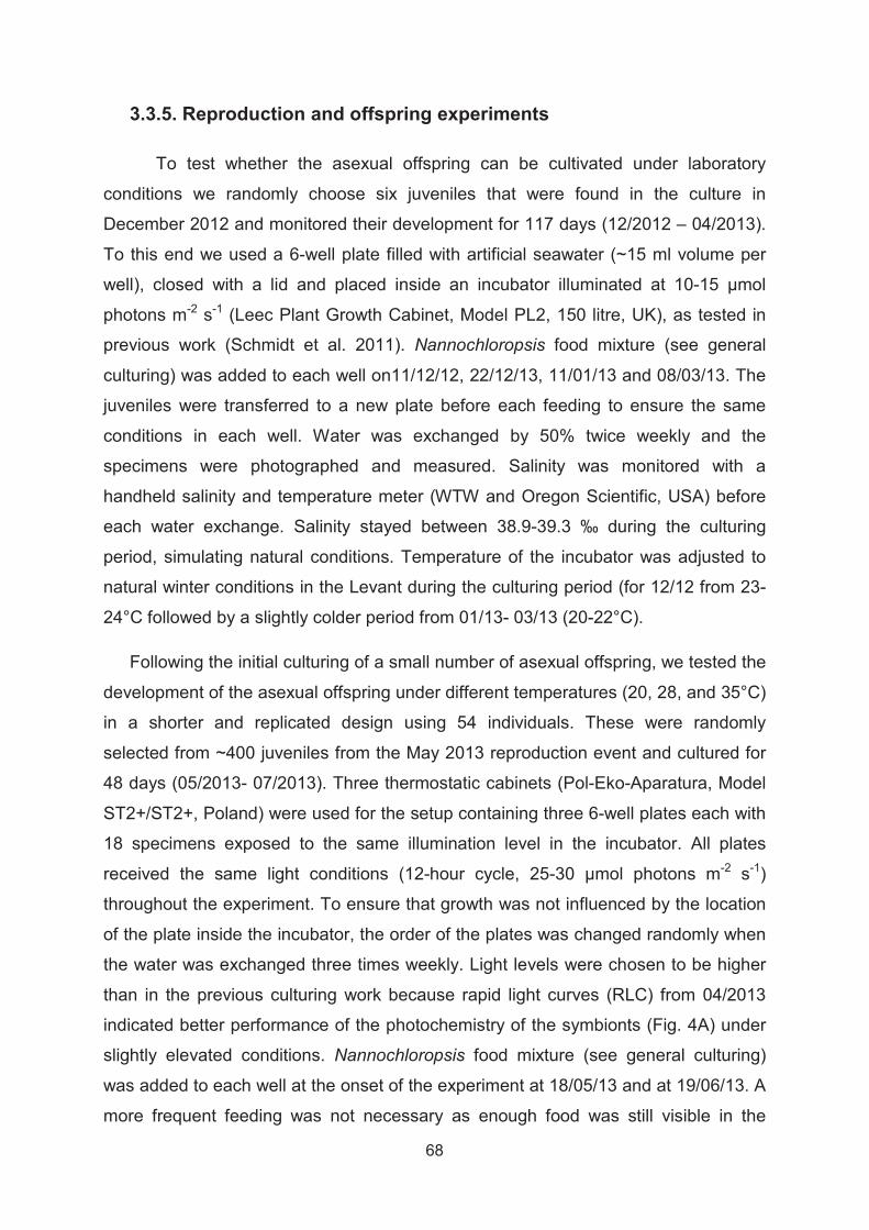
68
3.3.5. Reproduction and offspring experiments
To test whether the asexual offspring can be cultivated under laboratory
conditions we randomly choose six juveniles that were found in the culture in
December 2012 and monitored their development for 117 days (12/2012 – 04/2013).
To this end we used a 6-well plate filled with artificial seawater (~15 ml volume per
well), closed with a lid and placed inside an incubator illuminated at 10-15 μmol
photons m-2 s-1 (Leec Plant Growth Cabinet, Model PL2, 150 litre, UK), as tested in
previous work (Schmidt et al. 2011). Nannochloropsis food mixture (see general
culturing) was added to each well on11/12/12, 22/12/13, 11/01/13 and 08/03/13. The
juveniles were transferred to a new plate before each feeding to ensure the same
conditions in each well. Water was exchanged by 50% twice weekly and the
specimens were photographed and measured. Salinity was monitored with a
handheld salinity and temperature meter (WTW and Oregon Scientific, USA) before
each water exchange. Salinity stayed between 38.9-39.3 ‰ during the culturing
period, simulating natural conditions. Temperature of the incubator was adjusted to
natural winter conditions in the Levant during the culturing period (for 12/12 from 23-
24°C followed by a slightly colder period from 01/13- 03/13 (20-22°C).
Following the initial culturing of a small number of asexual offspring, we tested the
development of the asexual offspring under different temperatures (20, 28, and 35°C)
in a shorter and replicated design using 54 individuals. These were randomly
selected from ~400 juveniles from the May 2013 reproduction event and cultured for
48 days (05/2013- 07/2013). Three thermostatic cabinets (Pol-Eko-Aparatura, Model
ST2+/ST2+, Poland) were used for the setup containing three 6-well plates each with
18 specimens exposed to the same illumination level in the incubator. All plates
received the same light conditions (12-hour cycle, 25-30 μmol photons m-2 s-1)
throughout the experiment. To ensure that growth was not influenced by the location
of the plate inside the incubator, the order of the plates was changed randomly when
the water was exchanged three times weekly. Light levels were chosen to be higher
than in the previous culturing work because rapid light curves (RLC) from 04/2013
indicated better performance of the photochemistry of the symbionts (Fig. 4A) under
slightly elevated conditions. Nannochloropsis food mixture (see general culturing)
was added to each well at the onset of the experiment at 18/05/13 and at 19/06/13. A
more frequent feeding was not necessary as enough food was still visible in the

69
plates. Water exchange was performed three times weekly and manual monitoring of
salinity and temperature was done in the same way as described before. At the onset
of the experiment temperatures in incubators were slowly ramped up to prevent acute
temperature stress. The 28°C and 35°C incubator were automatically ramped up to
final temperature over a period of 12 h (2°C every 2-3 h). Manual temperature
measurements (n=57) conducted inside the plates showed that actual temperatures
in the incubators varied minimally 20.3°C (SD 0.1°C), 27.7°C (SD 0.2°C) and 34.7°C
(SD 0.3°C). During the first 72 h of the experiment salinity peaked by +2-4 ‰ above
the target levels at 38.5-40‰, which otherwise remained constant inside the wells
throughout the experiment (Mean 39.7 ‰, SD 1.1). This short time span of elevated
salinity did not cause mortality or bleaching, as the species has been shown to
inhabit high saline areas (Reinhardt et al. 2003) and is generally adapted to very
saline water of the Levant (Herut et al. 2000). The pH of the artificial ocean water was
monitored weekly with a handheld meter (WTW, Germany) and stayed between 8.1-
8.2 units.
3.3.6. Growth measurements
Twice weekly during culturing experiments specimens were photographed inside
each well (Fig. 5B) with an inverse microscrope (Zeiss PrimoVert) and images were
taken at the resolution of 5184 x 3456 pixel (Canon SLR camera). Largest diameters
were measured starting at the last build chamber diagonally across the shell, using
image analyzing software (ImageJ). Growth rates (% diameter increase day-1) were
calculated using the formula given in Schmidt et al. (2011) modified from ter Kuile
and Erez (1984).
At the end of the temperature sensitivity experiment 46 of initially 54 could be
retrieved for measurements (1/18 missing in 20°C, 3/18 in 28°C, 4/18 in 35°).
Although, the shell diameter increased in most specimens throughout the experiment
(=positive growth, indicated in % in Fig. 5 A), we observe apparent negative growth
rates in 35% of the specimens in the 20°C treatment (6 out of 17 specimens) and
36% in the 35°C treatment (5 out of 14 specimens). The negative growth reflects
measurement uncertainty due to uneven orientation and attached debris, and is
interpreted to indicate lack of growth. To exclude the possibility that the observed
positive growth rate also represent measurement uncertainty, the total number of

70
chambers was counted for specimens where the shell was best exposed. In both the
20°C and the 35°c treatments, we could identify specimens that added at least one
chamber during the experiment. One-way Analysis of Variance (ANOVA) was used to
test the effect of temperature on growth rates of asexual offspring. Statistics were
performed on all individuals exhibiting positive growth rates (Fig. 5 A). Growth data
was 3rd root transformed and residual and normality plots indicated that equal group
variance was not violated. ANOVA was performed using Jmp 11 (SAS 2014).
3.3.7. DNA extraction, amplification, cloning and sequencing
Three specimens of P. calcariformata collected from Hadera Ridge, Israel
(C50) and in Nachsholim, Israel (C228 and C229) on 23/10/2013 and one specimen
of Pararotalia sp. collected in Pelican Island, Australia (C538) on 05/2014 were
isolated from cultures into 50 μl of GITC* on 07/11/2013 and 26/05/2014. Because
of the thickness of the shell, the specimens were crushed with a metal rod prior to
DNA extraction. A fragment of ~1000 bp located at the 3’ end of the SSU rDNA of the
foraminifer was amplified using the primer couple S14F1 (5'-
AAGGGCACCACAAGAACGC-3’) – 1528R (5'-TGATCCTTCTGCAGGTTCACCTAC-
3’) by Polymerase chain reaction (PCR) (Medlin et al. 1988; de Vargas et al. 1997)
using the GoTag (Promega, USA) or PHUSION (Thermo Scientific, USA)
polymerase. The PCR products were purified using the QIAquick PCR purification kit
(Qiagen, Netherlands) and cloned with the Zero Blunt® TOPO® PCR Cloning Kit
(Invitrogen, USA) with TOP10 chemically competent cells following manufacturer’s
instructions. Three to six clones were sequenced per individual by an external
provider (LGC Genomics, Berlin).
A fragment of ~400 bp of the 3’ end of the SSU rDNA of foraminifers’
symbionts was obtained from aliquots of the same DNA extractions using the GoTaq
polymerase (Promega, USA) with the symbionts specific forward primer SymSF1 (5'-
GGTTAATTCCGTTAACGAACGAGA-3’) coupled with the universal reversed primer
1528R (5'-TGATCCTTCTGCAGGTTCACCTAC-3’) for the specimen C228 and C229.
No multiple bands have been observed after migration of the PCR product on
agarose gel. The PCR products were purified using the QIAquick PCR purification kit
(Qiagen, Netherlands) and directly sequenced. The sequence chromatograms were
carefully checked and no sign of multiple signals was detected.
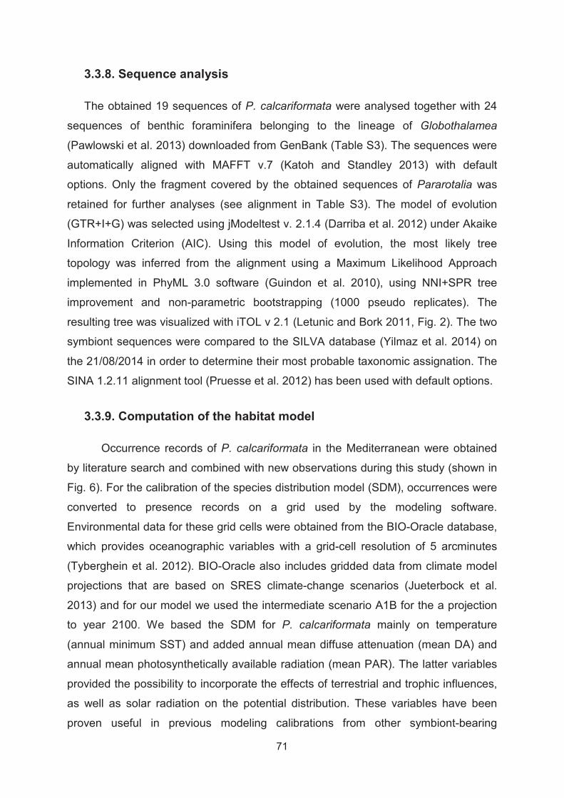
71
3.3.8. Sequence analysis
The obtained 19 sequences of P. calcariformata were analysed together with 24
sequences of benthic foraminifera belonging to the lineage of Globothalamea
(Pawlowski et al. 2013) downloaded from GenBank (Table S3). The sequences were
automatically aligned with MAFFT v.7 (Katoh and Standley 2013) with default
options. Only the fragment covered by the obtained sequences of Pararotalia was
retained for further analyses (see alignment in Table S3). The model of evolution
(GTR+I+G) was selected using jModeltest v. 2.1.4 (Darriba et al. 2012) under Akaike
Information Criterion (AIC). Using this model of evolution, the most likely tree
topology was inferred from the alignment using a Maximum Likelihood Approach
implemented in PhyML 3.0 software (Guindon et al. 2010), using NNI+SPR tree
improvement and non-parametric bootstrapping (1000 pseudo replicates). The
resulting tree was visualized with iTOL v 2.1 (Letunic and Bork 2011, Fig. 2). The two
symbiont sequences were compared to the SILVA database (Yilmaz et al. 2014) on
the 21/08/2014 in order to determine their most probable taxonomic assignation. The
SINA 1.2.11 alignment tool (Pruesse et al. 2012) has been used with default options.
3.3.9. Computation of the habitat model
Occurrence records of P. calcariformata in the Mediterranean were obtained
by literature search and combined with new observations during this study (shown in
Fig. 6). For the calibration of the species distribution model (SDM), occurrences were
converted to presence records on a grid used by the modeling software.
Environmental data for these grid cells were obtained from the BIO-Oracle database,
which provides oceanographic variables with a grid-cell resolution of 5 arcminutes
(Tyberghein et al. 2012). BIO-Oracle also includes gridded data from climate model
projections that are based on SRES climate-change scenarios (Jueterbock et al.
2013) and for our model we used the intermediate scenario A1B for the a projection
to year 2100. We based the SDM for P. calcariformata mainly on temperature
(annual minimum SST) and added annual mean diffuse attenuation (mean DA) and
annual mean photosynthetically available radiation (mean PAR). The latter variables
provided the possibility to incorporate the effects of terrestrial and trophic influences,
as well as solar radiation on the potential distribution. These variables have been
proven useful in previous modeling calibrations from other symbiont-bearing
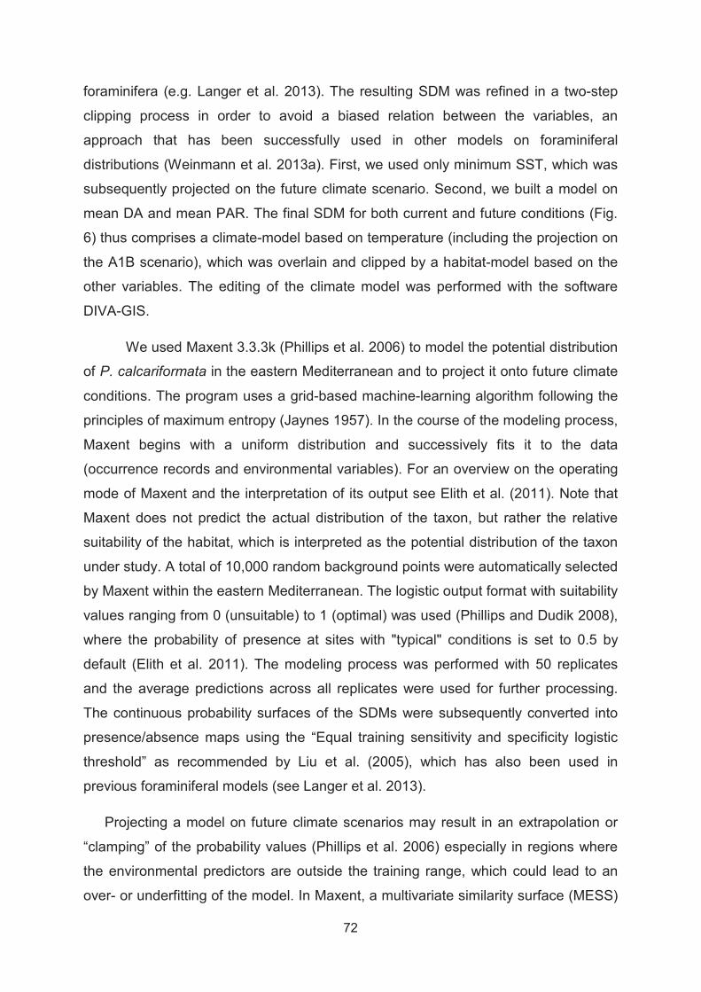
72
foraminifera (e.g. Langer et al. 2013). The resulting SDM was refined in a two-step
clipping process in order to avoid a biased relation between the variables, an
approach that has been successfully used in other models on foraminiferal
distributions (Weinmann et al. 2013a). First, we used only minimum SST, which was
subsequently projected on the future climate scenario. Second, we built a model on
mean DA and mean PAR. The final SDM for both current and future conditions (Fig.
6) thus comprises a climate-model based on temperature (including the projection on
the A1B scenario), which was overlain and clipped by a habitat-model based on the
other variables. The editing of the climate model was performed with the software
DIVA-GIS.
We used Maxent 3.3.3k (Phillips et al. 2006) to model the potential distribution
of P. calcariformata in the eastern Mediterranean and to project it onto future climate
conditions. The program uses a grid-based machine-learning algorithm following the
principles of maximum entropy (Jaynes 1957). In the course of the modeling process,
Maxent begins with a uniform distribution and successively fits it to the data
(occurrence records and environmental variables). For an overview on the operating
mode of Maxent and the interpretation of its output see Elith et al. (2011). Note that
Maxent does not predict the actual distribution of the taxon, but rather the relative
suitability of the habitat, which is interpreted as the potential distribution of the taxon
under study. A total of 10,000 random background points were automatically selected
by Maxent within the eastern Mediterranean. The logistic output format with suitability
values ranging from 0 (unsuitable) to 1 (optimal) was used (Phillips and Dudik 2008),
where the probability of presence at sites with "typical" conditions is set to 0.5 by
default (Elith et al. 2011). The modeling process was performed with 50 replicates
and the average predictions across all replicates were used for further processing.
The continuous probability surfaces of the SDMs were subsequently converted into
presence/absence maps using the “Equal training sensitivity and specificity logistic
threshold” as recommended by Liu et al. (2005), which has also been used in
previous foraminiferal models (see Langer et al. 2013).
Projecting a model on future climate scenarios may result in an extrapolation or
“clamping” of the probability values (Phillips et al. 2006) especially in regions where
the environmental predictors are outside the training range, which could lead to an
over- or underfitting of the model. In Maxent, a multivariate similarity surface (MESS)

73
analysis is implemented, which shows how similar predictor variables within future
climate scenarios are seen during model training (Elith et al. 2011). We added the
result of the MESS analysis to our future model, highlighting areas of possible
extrapolation of the model due to minimum temperature values of the future scenario
being outside the training range.

74
3.4. Results and Discussion
3.4.1. The identity of the invasive species and its current distribution
Pararotalia calcariformata McCulloch 1977 has been only very recently added to
the list of over 700 marine species that appear to have invaded the Mediterranean in
historical times (Zenetos et al. 2012). The species has been reported from littoral
environments of the Israeli coast as Eponides by Yanko et al. (1994) and as
Pararotalia spinigera by Reinhardt et al. (1994). The earliest description in the Levant
of the genus Pararotalia is by Reiss et al. (1961), who reported in 1961 on the
occurrence of Pararotalia cf. ozawai (Graham & Millitante (non Asano)) from a locality
near Haifa. Unfortunately, he provides no illustration and the material of the collection
is not available. Therefore, we cannot confirm this record and conclude that the exact
time of introduction of the species cannot be constrained beyond occurring prior to
1994. However, we note that the species must be a very recent invader, as the study
by Hyams-Kaphzan et al. (2014) found specimens attributed to Pararotalia
calcariformata only in surface sediments and never in historical layers. Other studies
confirmed the proliferation of the species after 1994, identifying large populations
ranging from the southernmost Israeli coast (Hyams-Kaphzan et al. 2008; Arieli et al.
2011) to southern Turkey (Meriç et al. 2013). All localities where this species has
been reported up to now in the literature as well as through our investigations in the
eastern Mediterranean are given in Figure 6.
Considering the large abundance of the species and its distinctive shape, it is
unlikely that it has been overlooked in earlier studies or not seen at other localities in
the eastern Mediterranean (Meric et al. 2008; Langer et al. 2012). Collectively, the
existing evidence implies that the species has established the Levant only recently
and began to proliferate and expand its range within the last two decades (Meriç et
al. 2013). The majority of the invasive species in the eastern Mediterranean
represent Lessepsian migrations through the Suez Canal (Zenetos et al. 2012). The
invasive Pararotalia has not yet been described from potential source locations in the
Red Sea such as the Gulf of Aqaba (Reiss and Hottinger 1984), the Gulf of Suez
(Madkour 2013) or the South Sinai Coast (Parker et al. 2012). On the other hand,
Pararotalia appears to be an Indopacific genus (Langer and Hottinger 2000) and
specimens assignable to P. calcariformata have been found along the coast of the

75
Arabian Sea, Oman. Therefore, it is most likely that P. calcariformata also followed
the Lessepsian route. Under this scenario, the species could have invaded the
Mediterranean across the Suez Canal, for example attached to gastropod larvae
(Cedhagen and Middelfart 1998). Alternatively, it could have been introduced with
ballast waters, as has been hypothesized for the red alga Grateloupia yinggehaiensis
possibly introduced to the Mediterranean from China by ship traffic (Wolf et al. 2014).
This alga appears to have established a continuous population in the vicinity of a
thermoelectric power plant (Wolf et al. 2014). We note that P. calcariformata has
been found to occur prolifically in the heat plume of the thermal power plant in
Hadera (Arieli et al. 2011), which might also have has served as a stepping stone for
its invasion. The exact origin of P. calcariformata and its dispersal route can only be
established by the identification of a potential parent population, such as it has been
shown for the invasive Sorites by Merkado et al. (2013), who found a potential parent
population in the Gulf of Aqaba. Therefore, we conducted a taxonomical and genetic
investigation to characterize the relationship between the Indopacific and Levantine
populations of Pararotalia.
The morphological taxonomy of the invasive Pararotalia population has been
confusing, because the first specimens found and identified in the Mediterranean
were mistakenly classified as Eponides repandus (Fichtel and Moll, 1798) (Yanko et
al. 1994) and only later assigned to Pararotalia spinigera Le Calvez 1949 (Bresler
and Yanko 1995a,b). The generic classification has been stable since, but the
species level taxonomy within this morphologically variable genus is in need of
revision. Previous studies by e.g. Reinhardt et al. (1994); Hyams-Kaphzan et al.
(2008); and Arieli et al. (2011) designated recent specimens from the Israeli coast as
Pararotalia spinigera Le Calvez 1949, following Loeblich and Tappan (1987) and
Hottinger et al. (1991). Meriç et al. (2013) first suggested that the specimens may
rather represent P. calcariformata, an extant species described from the Indian
Ocean by McCulloch (1977). Pararotalia calcariformata McCulloch 1977 is mainly
distinguished from P. spinigera (Le Calvez 1949) as described by Loeblich and
Tappan (1987) and Hottinger et al. (1991) in having a distinct peripheral keel and
deep septal interlocular spaces on the umbilical side (Fig. 1B2-3). A comparison of
the morphology of the Levantine species with this description confirms that the
assignment of the invasive species to Pararotalia spinigera is incorrect, because a
keel and deep septal interlocular space are clearly present among the Levantine
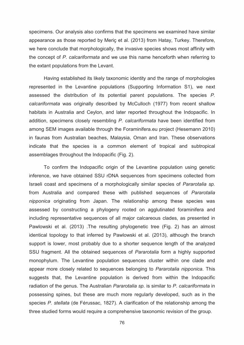
76
specimens. Our analysis also confirms that the specimens we examined have similar
appearance as those reported by Meriç et al. (2013) from Hatay, Turkey. Therefore,
we here conclude that morphologically, the invasive species shows most affinity with
the concept of P. calcariformata and we use this name henceforth when referring to
the extant populations from the Levant.
Having established its likely taxonomic identity and the range of morphologies
represented in the Levantine populations (Supporting Information S1), we next
assessed the distribution of its potential parent populations. The species P.
calcariformata was originally described by McCulloch (1977) from recent shallow
habitats in Australia and Ceylon, and later reported throughout the Indopacific. In
addition, specimens closely resembling P. calcariformata have been identified from
among SEM images available through the Foraminifera.eu project (Hesemann 2010)
in faunas from Australian beaches, Malaysia, Oman and Iran. These observations
indicate that the species is a common element of tropical and subtropical
assemblages throughout the Indopacific (Fig. 2).
To confirm the Indopacific origin of the Levantine population using genetic
inference, we have obtained SSU rDNA sequences from specimens collected from
Israeli coast and specimens of a morphologically similar species of Pararotalia sp.
from Australia and compared these with published sequences of Pararotalia
nipponica originating from Japan. The relationship among these species was
assessed by constructing a phylogeny rooted on agglutinated foraminifera and
including representative sequences of all major calcareous clades, as presented in
Pawlowski et al. (2013) .The resulting phylogenetic tree (Fig. 2) has an almost
identical topology to that inferred by Pawlowski et al. (2013), although the branch
support is lower, most probably due to a shorter sequence length of the analyzed
SSU fragment. All the obtained sequences of Pararotalia form a highly supported
monophylum. The Levantine population sequences cluster within one clade and
appear more closely related to sequences belonging to Pararotalia nipponica. This
suggests that, the Levantine population is derived from within the Indopacific
radiation of the genus. The Australian Pararotalia sp. is similar to P. calcariformata in
possessing spines, but these are much more regularly developed, such as in the
species P. stellata (de Férussac, 1827). A clarification of the relationship among the
three studied forms would require a comprehensive taxonomic revision of the group.

77
Figure 2. Phylogenetic tree (Maximum Likelihood, GTR+I+G) showing the evolutionary relationships of Pararotalia with other benthic foraminifera belonging to the order Globothalamea. Bootstrap scores (1000 replicates) higher than 50% are shown next to the branches. The tree is rooted on the genus Reophax. The occurrence of the genus in the Indo-Pacific is shown on the map against winter sea surface temperature (data extracted from the World Ocean Atlas 2005 Locarnini (2006). Open circles represent the occurrences of Pararotalia reported in the literature and stars denote the location of the sequenced specimens.
3.4.2. Characterization of diatom endosymbionts and their photochemistry
The ecology of symbiont-bearing benthic foraminifera is closely tied to the function of
their algal symbionts. Although the genus Pararotalia together with the genus
Neorotalia is considered to belong to the informal group of larger benthic foraminifera
(LBF), which are typically associated with algal symbionts (Langer and Hottinger
2000), the presence and identity of the symbionts in Pararotalia has never been
formally established. In addition, the Mediterranean Pararotalia is smaller (typically
<400 μm) than other symbiont-bearing benthic foraminifera. One study reporting the
occurrence of the invasive Pararotalia inside a thermally polluted site along the Israeli
coast (Arieli et al. 2011) noted a distinct coloration of its cytoplasm, which is often an
indicator for the presence of algal symbionts. Therefore, prior to further physiological
experiments, we investigated the presence and nature of symbionts in the studied
Levantine population.
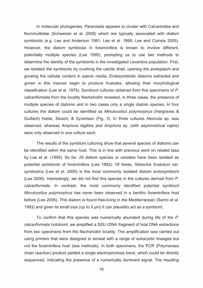
78
In molecular phylogenies, Pararotalia appears to cluster with Calcarinidae and
Nummulitidae (Schweizer et al. 2008) which are typically associated with diatom
symbionts (e.g. Lee and Anderson 1991; Lee et al. 1995; Lee and Correia 2005).
However, the diatom symbiosis in foraminifera is known to involve different,
potentially multiple species (Lee 1995), prompting us to use two methods to
determine the identity of the symbionts in the investigated Levantine population. First,
we isolated the symbionts by crushing the calcite shell, opening the protoplasm and
growing the cellular content in axenic media. Endosymbiotic diatoms extracted and
grown in this manner begin to produce frustules, allowing their morphological
classification (Lee et al. 1979). Symbiont cultures obtained from five specimens of P.
calcariformata from the locality Nachsholim revealed, in three cases, the presence of
multiple species of diatoms and in two cases only a single diatom species. In four
cultures the diatom could be identified as Minutocellus polymorphus (Hargraves &
Guillard) Hasle, Stosch, & Syvertsen (Fig. 3). In three cultures Navicula sp. was
observed, whereas Amphora bigibba and Amphora sp. (with asymmetrical raphe)
were only observed in one culture each.
The results of the symbiont culturing show that several species of diatoms can
be identified within the same host. This is in line with previous work on related taxa
by Lee et al. (1995). So far, 20 diatom species or varieties have been isolated as
potential symbionts of foraminifera (Lee 1992). Of these, Nitzschia frustulum var.
symbiotica (Lee et al. 2000) is the most commonly isolated diatom endosymbiont
(Lee 2006). Interestingly, we did not find this species in the cultures derived from P.
calcariformata. In contrast, the most commonly identified potential symbiont
Minutocellus polymorphus has never been observed in a benthic foraminifera host
before (Lee 2006). This diatom is found free-living in the Mediterranean (Sarno et al.
1993) and given its small size (up to 3 μm) it can plausibly act as a symbiont.
To confirm that this species was numerically abundant during life of the P.
calcariformata holobiont, we amplified a SSU rDNA fragment of total DNA extractions
from two specimens from the Nachsholim locality. The amplification was carried out
using primers that were designed to anneal with a range of eukaryotic lineages but
not the foraminifera host (see methods). In both specimens, the PCR (Polymerase
chain reaction) product yielded a single electrophoresis band, which could be directly
sequenced, indicating the presence of a numerically dominant signal. The resulting

79
sequences could be unambiguously identified as Minutocellus by comparison with
the SILVA database (Yilmaz et al. 2014). Therefore, we conclude that the diatom
endosymbiont consortium in the investigated specimens of P. calcariformata was
likely dominated by M. polymorphus.
Figure 3. Scanning electron microscopy (SEM) images of frustules of the diatom Minutocellus polymorphus found in cultures isolated from Pararotaliacalcariformata. Scale bars represent 200 nm.
To characterize the photosynthetic functioning of the discovered diatom
endosymbiont, we measured the photosynthetic activity of the symbionts inside the
host by Pulse Amplitude Modulated Fluorometry (PAM) (Fig. 4). To achieves this we
conducted Rapid Light Curves (RLCs) after the protocol of Ralph and Gademann
(2005), allowing us to assess the response of PS II (Photosystem II) to elevated light
levels. The measurements on freshly collected specimens yielded an RLC (Fig. 4A),
most similar to intermediate-light adapted Amphistegina (Nobes et al. 2008; Ziegler
and Uthicke 2011) but we observed PSII photoinhibition (light adapted yields, Y(II)=0)
at 166 μmol photons m-2 s-1. The RLC of P. calcariformata thus reveals an unusual
sensitivity to high irradiance among the foraminifera, which we attribute to the
combination of the different nature of the symbiont and the mid-latitude setting of the
locality. Alternatively, the observed lack of fluorescence at higher irradiance levels
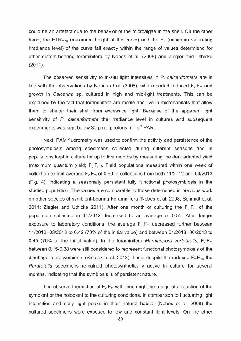
80
could be an artefact due to the behavior of the microalgae in the shell. On the other
hand, the ETRmax (maximum height of the curve) and the Ek (minimum saturating
irradiance level) of the curve fall exactly within the range of values determiend for
other diatom-bearing foraminifera by Nobes et al. (2008) and Ziegler and Uthicke
(2011).
The observed sensitivity to in-situ light intensities in P. calcariformata are in
line with the observations by Nobes et al. (2008), who reported reduced Fv:Fm and
growth in Calcarina sp. cultured in high and mid-light treatments. This can be
explained by the fact that foraminifera are motile and live in microhabitats that allow
them to shelter their shell from excessive light. Because of the apparent light
sensitivity of P. calcariformata the irradiance level in cultures and subsequent
experiments was kept below 30 μmol photons m-2 s-1 PAR.
Next, PAM fluorometry was used to confirm the activity and persistence of the
photosymbiosis among specimens collected during different seasons and in
populations kept in culture for up to five months by measuring the dark adapted yield
(maximum quantum yield; Fv:Fm). Field populations measured within one week of
collection exhibit average Fv:Fm of 0.60 in collections from both 11/2012 and 04/2013
(Fig. 4), indicating a seasonally persistent fully functional photosymbiosis in the
studied population. The values are comparable to those determined in previous work
on other species of symbiont-bearing Foraminifera (Nobes et al. 2008; Schmidt et al.
2011; Ziegler and Uthicke 2011). After one month of culturing the Fv:Fm of the
population collected in 11/2012 decreased to an average of 0.55. After longer
exposure to laboratory conditions, the average Fv:Fm decreased further between
11/2012 -03/2013 to 0.42 (70% of the initial value) and between 04/2013 -06/2013 to
0.45 (76% of the initial value). In the foraminifera Marginopora vertebralis, Fv:Fm
between 0.15-0.38 were still considered to represent functional photosymbiosis of the
dinoflagellates symbionts (Sinutok et al. 2013). Thus, despite the reduced Fv:Fm, the
Pararotalia specimens remained photosynthetically active in culture for several
months, indicating that the symbiosis is of persistent nature.
The observed reduction of Fv:Fm with time might be a sign of a reaction of the
symbiont or the holobiont to the culturing conditions. In comparison to fluctuating light
intensities and daily light peaks in their natural habitat (Nobes et al. 2008) the
cultured specimens were exposed to low and constant light levels. On the other

81
hand, lower Fv:Fm values could indicate nutrient stress in the cultures. It has been
shown that low-light adapted diatoms have higher cellular iron needs to keep
photosynthetic iron-based redox proteins functioning (Sunda and Huntsman 1997).
Since the cultures were based on artificial seawater without addition of nutrients, it is
possible that the feeding of the foraminifera with microalgae was not sufficient to
allow for optimal nutrition. In contrast to this hypothesis, nutrient-limited cultures of
the diatom species Thalassiosira pseudonana exhibit a constant Fv:Fm ratio of 0.65
under balanced growth conditions (Parkhill et al. 2001). Therefore, it remains unclear
whether the decreased Fv:Fm indicates nutrient or light stress in the cultures, or
whether it reflects the physiological state of the symbionts.

82
Figure 4. Photochemistry of Pararotalia calcariformata symbionts. A) Rapid light curve showing relative electron transport rates (rETR) under different levels of photosynthetically active radiation (PAR) on low-light adapted
represents slope of the curve, (ETRmax) the maximum height of the curve and the (Ek) the minimum saturating irradiance level. Photograph shows a live specimen pictured under epifluorescence microscopy, showing photosynthetically active symbionts distributed throughout the shell. B) Maximum quantum yield (Fv:Fm) measured on two populations collected in 11/2012 and 04/2013 (n=14-48), blue dots represent measurements made one week after collection, red dots represent measurements of cultured specimens.
3.4.3. Reproduction, growth and temperature sensitivity of asexual offspring
The extent of biological invasions and range expansions is limited by the ability of
the species to establish a viable population at a new locality. Thus, next to the
environmental tolerance of adult specimens, reproductive success of the expanding
population is determined by the environmental suitability window of the reproduction
event and of the survival and growth of juveniles, which can be a bottleneck for
species survival under global change (Byrne 2012). Foraminifera are known to
reproduce through a complex system of sexual (gametogenic) and asexual (multiple
fission) cycles (e.g. Goldstein 1997; Hohenegger 2011). The length of the life cycle
and the timing of reproduction is often poorly constrained, but continuous in situ
monitoring over the full seasonal cycle shows highest abundances of P.
calcariformata in the >0.63 m size fraction in spring following natural temperature
rise (Arieli et al. 2011), indicating that reproduction in this species in nature is likely to
occur once a year in late spring.

83
We have observed asexual reproduction in populations in the laboratory cultures and
were able to characterize the ontogeny under controlled laboratory conditions. After
collections in 11/2012 and 04/2013, reproduction occurred at least once in each
collection after 4-5 weeks of culturing, involving only a small number of the cultured
specimens. The number of offspring per mother individual was relatively small: after
reproduction in 05/2013, we observed app. 400 living three-chambered offspring and
app. 20 dead potential parent individuals in the same culture. The offspring contained
a minimum of three chambers when released from the parent and contained
symbionts. The size and number of asexual offspring, as well as the presence of
symbionts inherited from the parent are comparable to observations from laboratory
experiments on many other species of symbiont-bearing foraminifera (Hohenegger
2011). Thus, the Levantine invasive population of P. calcariformata is able to
reproduce under temperature, salinity and light conditions simulating the ambient
setting in the Levant in autumn and spring. However, development under culturing
conditions can only be an estimate compared to development under in-situ conditions
because environmental factors, e.g. tides and lunar cycles were not simulated
(Briguglio and Hohenegger 2014; Hohenegger et al. 2014).
To characterize the growth of the asexual offspring, six juveniles from the first
reproductive event were kept in culture under constant conditions for 117 days. Three
of the six individuals grew and developed new chambers filled with green-brownish
cytoplasm (Fig. 5A,B). A twice weekly monitoring showed that the growth of the
juveniles has two distinct phases. The first phase of rapid growth (lasting ~30 days) is
followed by a phase of slow growth (Fig. 5A). All juveniles consisted of three
chambers at the beginning of the experiment and measured up to 16-18 chambers at
the end of the experiment. Until day 30, the chamber number increased from 4-(14-
15). Thus, app. ten new chambers were formed in these three individuals in the first
growth phase (Fig. 5B).To investigate the temperature sensitivity and development of
asexual offspring we performed an experiment following the second reproductive
event. The experimental setup consisted of three replicated plates per incubator
exposing asexual offspring to 20°C, 28°C and 35°C over 48 days. The 20°C and
28°C cultures were set to simulate the natural range of temperatures in the eastern
Mediterranean between spring and autumn (Herut et al. 2000; Arieli et al. 2011), with
the 20°C culture representing the conditions at the time of collection. The 35°C
treatment was chosen to establish the upper limit of offspring growth.

84
The results indicated that offspring mean growth was 0.86 % day-1 in the 28°C
treatment (Fig. 5C). Only in the 28°C treatment all observed specimens grew and the
average growth curve of the population followed the same two-phase growth pattern
as in the 2012 culture (Fig. 5D). The offspring kept at 20°C and 35°C survived the
experiment, as seen by healthy coloration of the cytoplasm and cytoplasmic
movement, but showed an inhibition of growth, indicating the lower limit of growth
(Fig. 5D). This is in line with observations of benthic foraminifera by Alve and
Goldstein (2002) where growth commenced only in individuals exposed to suitable
environmental conditions. The proportion of individuals which showed positive growth
under experimental conditions was 100% under 28°C, and was reduced to 64-65% in
the 20°C and 35°C treatment. We statistically tested the effect of temperature on the
individuals exhibiting positive growth. Temperature had a significant effect on growth
rate on asexual offspring (One-Way ANOVA, F=24.60, df=2, 34, p=0.0001). The
Tukey-Kramer post-hoc test revealed significant differences between the 28°C, the
20°C and 35°C treatments, but not between the latter two. Growth rates observed at
28°C in this study are comparable to those of sexually produced offspring of the
benthic foraminifera Planograbratella opercularis (0.4-0.8% growth per day between
15-20°C) inhabiting a similar coastal environment on coralline algae in Japan
(Tsuchiya et al. 2014). The observed inhibition of growth in the 35°C treatment, is
consistent with an upper thermal limit for other species of symbiont-bearing
foraminifera, where reduced growth, increased mortality and symbiont bleaching are
observed at temperatures >31°C (Schmidt et al. 2011; Uthicke et al. 2012).
In contrast, the lack of offspring growth at 20°C was unexpected, considering
that this was the ambient temperature at the time of collection and the reproduction in
the laboratory occurred at that temperature. Our results indicate that although
reproduction may occur at 20°C, the offspring needs temperatures >20°C in the
subsequent weeks in order to initiate the rapid growth phase. Thus, the observation
of reproduction at 20°C may be consistent with the lower temperature limit for
offspring growth between 20°C and 24°C, provided the reproduction is aligned with
the onset of the spring warming. This is consistent with the observation of maximum
abundance in June and July in the >0.63 m size fraction (Arieli et al. 2011). If the
elevated abundance reflects reproductive events and the natural population follows
the same growth pattern as seen in laboratory, then the reproductive in nature must

85
have occurred at least one month before the observed abundance maximum, i.e. in
May or June.
If temperatures higher than 20°C are required for offspring growth in P.
calcariformata, then the seasonal time window for reproduction in this species is
longer in the Levant than in other areas of the Mediterranean (Shaltout and Omstedt
2014). It is possible that reproductive processes in P. calcariformata are incompatible
with a narrower suitability window for reproduction shifted towards the summer in
other areas of the Mediterranean. At present, between the Levant (e.g., Haifa) and
the Ionian Sea (e.g. San Stefano, Corfu) a 20°C sea surface temperature threshold is
shifted by three weeks from early May to early June and the total length of the >20°C
thermal window is shorter by two months (IOLR 2010). This mechanism would
provide a possible explanation for the restricted Levantine occurrence of the invasive
species, as well as for its apparently recent invasion. The Levantine basin is known
to have already experienced significant winter warming in the past decades and is
predicted to increase its yearly mean SST by 0.5-2.3°C by the end of the century
(Shaltout and Omstedt 2014; Sisma-Ventura et al. 2014).
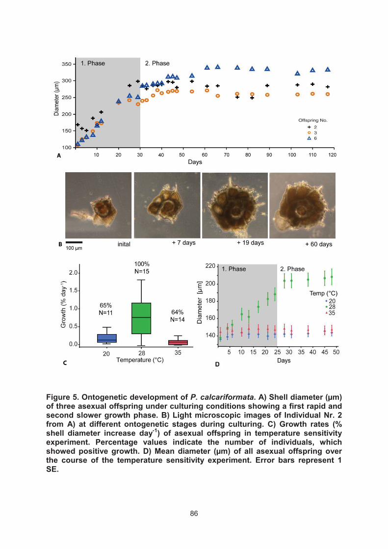
86
Figure 5. Ontogenetic development of P. calcariformata. A) Shell diameter (μm) of three asexual offspring under culturing conditions showing a first rapid and second slower growth phase. B) Light microscopic images of Individual Nr. 2 from A) at different ontogenetic stages during culturing. C) Growth rates (% shell diameter increase day-1) of asexual offspring in temperature sensitivity experiment. Percentage values indicate the number of individuals, which showed positive growth. D) Mean diameter (μm) of all asexual offspring over the course of the temperature sensitivity experiment. Error bars represent 1 SE.
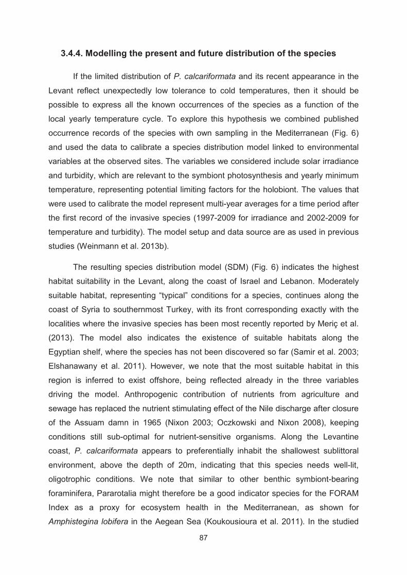
87
3.4.4. Modelling the present and future distribution of the species
If the limited distribution of P. calcariformata and its recent appearance in the
Levant reflect unexpectedly low tolerance to cold temperatures, then it should be
possible to express all the known occurrences of the species as a function of the
local yearly temperature cycle. To explore this hypothesis we combined published
occurrence records of the species with own sampling in the Mediterranean (Fig. 6)
and used the data to calibrate a species distribution model linked to environmental
variables at the observed sites. The variables we considered include solar irradiance
and turbidity, which are relevant to the symbiont photosynthesis and yearly minimum
temperature, representing potential limiting factors for the holobiont. The values that
were used to calibrate the model represent multi-year averages for a time period after
the first record of the invasive species (1997-2009 for irradiance and 2002-2009 for
temperature and turbidity). The model setup and data source are as used in previous
studies (Weinmann et al. 2013b).
The resulting species distribution model (SDM) (Fig. 6) indicates the highest
habitat suitability in the Levant, along the coast of Israel and Lebanon. Moderately
suitable habitat, representing “typical” conditions for a species, continues along the
coast of Syria to southernmost Turkey, with its front corresponding exactly with the
localities where the invasive species has been most recently reported by Meriç et al.
(2013). The model also indicates the existence of suitable habitats along the
Egyptian shelf, where the species has not been discovered so far (Samir et al. 2003;
Elshanawany et al. 2011). However, we note that the most suitable habitat in this
region is inferred to exist offshore, being reflected already in the three variables
driving the model. Anthropogenic contribution of nutrients from agriculture and
sewage has replaced the nutrient stimulating effect of the Nile discharge after closure
of the Assuam damn in 1965 (Nixon 2003; Oczkowski and Nixon 2008), keeping
conditions still sub-optimal for nutrient-sensitive organisms. Along the Levantine
coast, P. calcariformata appears to preferentially inhabit the shallowest sublittoral
environment, above the depth of 20m, indicating that this species needs well-lit,
oligotrophic conditions. We note that similar to other benthic symbiont-bearing
foraminifera, Pararotalia might therefore be a good indicator species for the FORAM
Index as a proxy for ecosystem health in the Mediterranean, as shown for
Amphistegina lobifera in the Aegean Sea (Koukousioura et al. 2011). In the studied

88
localities, P. calcariformata lives in association within turf algae or calcareous algae
that inhabit rocky substrate in the sublittoral zone on abrasion platforms and beach
rocks. As the Nile delta provides habitat lacking these conditions and because of the
elevated nutrient load (Nixon 2003), it is not surprising that it may function as a
natural barrier to a south-easterly coastal dispersal of the species.
Other species of Pararotalia have been reported to disperse with surface
ocean currents attached to gastropod larvae (Cedhagen and Middelfart 1998).
Whether or not attached to gastropod larvae, macro algae or transported as
propagules (Alve and Goldstein 2002; Alve and Goldstein 2003), the dispersal of P.
calcariformata would lead to passive northward transport along the Levantine coast
following the persistent surface currents in the Levant (Stamou and Kamizoulis 2009;
UNEP/MAP 2012). These currents move directly along the Israeli shelf and could
facilitate the transport of plant material or larvae carrying attached Pararotalia
towards the northern Levant, as it has been suggested for other benthic foraminifera
(Langer 2008). This would mean that the main direction of current transport of the
Levantine Pararotalia is further to the north, along the coast of Turkey and into the
Aegean Sea. Here, the SDM indicates poor habitat suitability, due to colder
temperatures (Fig. 6). Indeed, the species does not appear to have invaded the
Aegean Sea yet (Koukousioura et al. 2011; Frontalini et al. 2014). A recent detailed
investigation of foraminifera diversity in the Saros Bay, a region with abundant
suitable habitats for P. calcariformata, has not identified this species among the 115
species recorded (Frontalini et al. 2014).
To investigate whether the species range given by the SDM derived from its
present-day distribution is likely to expand further, we have evaluated habitat
suitability under a realistic (intermediate scenario A1B) global change projection for
the year 2100 (Jueterbock et al. 2013). Predicted habitat suitability increases
drastically along the coast of Turkey, suggesting a high probability of invasion in this
region within the next decades. In addition, the results imply a large potential for an
ongoing expansion of the species into the Aegean Sea and the Greek and Libyan
coasts of the Ionian Sea.
89
Figure 6. Occurrence records of P. calcariformata overlain with a habitat suitability values from a species distribution model based on yearly minimum temperature, mean light attenuation and mean PAR (photosynthetically active radiation) for the present day and under projected future conditions (intermediate global change scenario AB1). MESS indicates the results of the multivariate similarity surface analysis indicated by grey stripes.

90
3.5. Conclusions
A taxonomic revision of the most recently discovered invasive species of
symbiont-bearing foraminifera of the genus Pararotalia in the eastern Mediterranean
identifies the invader as P. calcariformata McCulloch 1977. Based on phylogenetic
and taxonomic evidence, the invasive population appears to have originated from
within the Pacific radiation of the genus. Pulse amplitude modulated fluorometry
measurements indicate that the species is engaged in permanent symbiosis with
photosynthesizing microalgae. Combined culturing and genotyping approach allowed
us to identify these algae as a consortium of small diatoms, including the newly
described symbiont Minutocellus polymorphus.
Pararotalia calcariformata has been observed to asexually reproduce and
grow in manipulative experiments, revealing an initial 30-day period of rapid growth
followed by a slower growth phase. Reproduction occurred at 20°C, but normal
offspring growth was only observed at 24°C and 28°C, indicating that a successful
establishment of populations of the species may be limited by the length of the
thermally defined reproductive window in their habitat. To test this hypothesis we
derived a SDM based on present day occurrence of the species and show that
together with turbidity and irradiance, yearly minimum temperature alone is sufficient
to reproduce the observed species range with remarkable fidelity. This model
indicates that the species is likely to continue expanding northwards and westwards
under realistic global change scenarios and is likely to reach the Ionian Sea by 2100.
Collectively, the evidence indicates that the symbiont-bearing foraminifera P.
calcariformata is likely a Lessepsian migrant whose invasion into the Mediterranean
has been facilitated by the recent warming in the Levant (Shaltout and Omstedt
2014). In this way, the case of P. calcariformata adds to mounting evidence for
ongoing and dramatic changes in the structure of eastern Mediterranean ecosystems
(e.g. Hiddink et al. 2012; Edelist et al. 2013). Its invasion reflects multiple aspects of
human-mediated dispersal of marine species – environmental change due to global
warming and removal of physical barriers. It shows that in the absence of limitation to
dispersal, the invasion rate is almost unlimited with ongoing range expansion (Meriç
et al. 2013) observable on sub-decadal time scale.

91
3.6. Acknowledgments
We grateful acknowledge help with fieldwork by Emri Brickner and Barak
Herut. We thank Janet Brandt for her assistance during culturing experiments and
Marco Tack for help with image analysis. We like to acknowledge Petra Witte who
took the SEM images of isolated diatoms. This research has been funded by the
BMBF through the German- Israeli cooperation project WTZ Israel 03F0639A.

92
3.7. References
Alve E (1999) Colonization of new habitats by benthic foraminifera: a review. Earth-Science Reviews 46:167-185
Alve E, Goldstein ST (2002) Resting stage in benthic foraminiferal propagules: a key feature for dispersal? Evidence from two shallow-water species. Journal of Micropalaeontology 21:95-96
Alve E, Goldstein ST (2003) Propagule transport as a key method of dispersal in benthic foraminifera (Protista). Limnology and Oceanography 48:2163-2170
Arieli RN, Almogi-Labin A, Abramovich S, Herut B (2011) The effect of thermal pollution on benthic foraminiferal assemblages in the Mediterranean shoreface adjacent to Hadera power plant (Israel). Marine Pollution Bulletin 62:1002-1012
Belanger CL, Jablonski D, Roy K, Berke SK, Krug AZ, Valentine JW (2012) Global environmental predictors of benthic marine biogeographic structure. Proceedings of the National Academy of Sciences of the United States of America 109:14046-14051
Bresler V, Yanko V (1995a) Chemical ecology - a new approach to the study of living benthic epiphytic Foraminifera. Journal of Foraminiferal Research 25:267-279
Bresler V, Yanko V (1995b) Acute toxicity of heavy-metals for benthi epiphytic foraminifera Pararotalia spinigera (LeCalvez) and influence of seaweed-derived DOC. Environmental Toxicology and Chemistry 14:1687-1695
Briguglio A, Hohenegger J (2014) Growth oscillation in larger foraminifera. Paleobiology 40:494-509
Byrne M (2012) Global change ecotoxicology: Identification of early life history bottlenecks in marine invertebrates, variable species responses and variable experimental approaches. Marine Environmental Research 76:3-15
Carlton TC (1999) The scale and ecological consequences of biological invasions in the World's oceans. In: Sandlund OT, Schei JP, Aslaug V (eds) Invasive Species and Biodiversity Management, Based on a selection of papers presented at the Norway/UN Conference on Alien Species, Trondheim, Norway. Kluwer Academic Publishers Dordrecht, The Netherlands,
Caruso A, Cosentino C (2014) The first colonization of the Genus Amphistegina and other exotic benthic foraminifera of the Pelagian Islands and south-eastern Sicily (central Mediterranean Sea). Marine Micropaleontology 111:38-52
Cedhagen T, Middelfart P (1998) Attachment to gastropod veliger shells - a possible mechanism of disperal in benthic Foraminiferans. Phuket Marine Bilological Center Special Publication 18:117-122
93
Chen IC, Hill JK, Ohlemuller R, Roy DB, Thomas CD (2011) Rapid Range Shifts of Species Associated with High Levels of Climate Warming. Science 333:1024-1026
Darriba D, Taboada GL, Doallo R, Posada D (2012) jModelTest 2: more models, new heuristics and parallel computing. Nature Methods 9:772-772
de Nooijer LJ, Toyofuku T, Kitazato H (2009) Foraminifera promote calcification by elevating their intracellular pH. Proceedings of the National Academy of Sciences 106:15374-15378
de Vargas C, Zaninetti L, Hilbrecht H, Pawlowski J (1997) Phylogeny and Rates of Molecular Evolution of Planktonic Foraminifera: SSU rDNA Sequences Compared to the Fossil Record. Journal of Molecular Evolution 45:285-294
Edelist D, Rilov G, Golani D, Carlton JT, Spanier E (2013) Restructuring the Sea: profound shifts in the world's most invaded marine ecosystem. Diversity and Distributions 19:69-77
Elith J, Phillips SJ, Hastie T, Dudik M, Chee YE, Yates CJ (2011) A statistical explanation of MaxEnt for ecologists. Diversity and Distributions 17:43-57
Elshanawany R, Ibrahim MI, Milker Y, Schmiedl G, Badr N, Kholeif SEA, Zonneveld KAF (2011) Anthropogenic impact on benthic foraminifera, Abu-Qir Bay,Alexandria, Egypt. The Journal of Foraminiferal Research 41:326-348
Frontalini F, Kaminski M, Mikellidou I, Armynot du Châtelet E (2014) Checklist of benthic foraminifera (class Foraminifera: d’Orbigny 1826; phylum Granuloreticulosa) from Saros Bay, northern Aegean Sea: a biodiversity hotspot. Marine Biodiversity:1-19
Goldstein ST (1997) Gametogenesis and the antiquity of reproductive pattern in the Foraminiferida. Journal of Foraminiferal Research 27:319-328
Goldstein ST, Alve E (2011) Experimental assembly of foraminiferal communities from coastal propagule banks. Marine Ecology Progress Series 437:1-11
Guindon S, Dufayard JF, Lefort V, Anisimova M, Hordijk W, Gascuel O (2010) New Algorithms and Methods to Estimate Maximum-Likelihood Phylogenies: Assessing the Performance of PhyML 3.0. Systematic Biology 59:307-321
Hallock P (1981) Algal symbiosis: A mathematical analysis. Marine Biology 62:249-255
Herut B, Almogi-Labin A, Jannink N, Gertman I (2000) The seasonal dynamics of nutrient and chlorophyll a concentrations on the SE Mediterranean shelf-slope. Oceanologica Acta 23:771-782
Hesemann M (2010) Concept for a foraminiferal database. In: Hesemann M (ed) Forams 2010, International Symposium on Foraminifera, Rheinische Friedrich-Wilhelms-Universität Bonn, September 5-10, 2010 Germany

94
Hesemann M (2014) Pararotalia sp. Foraminiferaeu Project Database, Accessed at http://www.foraminifera.eu/single.php?no=1003787&aktion=suche on 2014-10-2
Hiddink JG, Lasram FB, Cantrill J, Davies AJ (2012) Keeping pace with climate change: what can we learn from the spread of Lessepsian migrants? Global Change Biology 18:2161-2172
Hill R, Schreiber U, Gademann R, Larkum AWD, Kuhl M, Ralph PJ (2004) Spatial heterogeneity of photosynthesis and the effect of temperature-induced bleaching conditions in three species of corals. Marine Biology 144:633-640
Hohenegger J (2011) Large Foraminifera - Greenhouse constructions and gardeners in the oceanic microcosm. The Kagoshima University Muesum, Kagoshima, Bulletin No. 5
Hohenegger J, Briguglio A, Eder W (2014) The Natural Laboratory of Algal Symbiont-Bearing Benthic Foraminifera: Studying Individual Growth and Population Dynamics in the Sublittoral. In: Kitazato H, M. Bernhard J (eds) Approaches to Study Living Foraminifera. Springer Japan, pp13-28
Hottinger L, Halicz E, Reiss Z (1991) The Foraminiferal Genera Pararotalia, Neorotalia, and Calcarina: Taxonomic Revision. Journal of Paleontology 65:18-33
Hottinger L, Halicz E, Reiss Z (1993) Recent foraminifera from the Gulf of Aqaba, Red Sea. Slovenska Akademija Znanosti in Umetnosti, Ljubljana, Slovenia
Hyams-Kaphzan O, Almogi-Labin A, Sivan D, Benjamini C (2008) Benthic foraminifera assemblage change along the southeastern Mediterranean inner shelf due to fall-off of Nile-derived siliciclastics. Neues Jahrbuch Fur Geologie Und Palaontologie-Abhandlungen 248:315-344
Hyams-Kaphzan O, Grossowicz LP, Almogi-Labin A (2014) Characteristics of benthic foraminifera inhabiting rocky reefs in northern Israeli Mediterranean shelf. Report: ES-20-2014
Hyams O, Almogi-Labin A, Benjaminia C (2002) Larger foraminifera of the southeastern Mediterranean shallow continental shelf off Israel. Israel Journal of Earth Sciences 51:169-179
IOLR (2010) Statistical table for MedGLOSS stations and seasons, MedGLOSS sea level network., [Available at http://medgloss.ocean.org.il/statistic.asp.]
Jaynes ET (1957) Information theory and statistical mechanics Physical Review 106:620-630
Jueterbock A, Tyberghein L, Verbruggen H, Coyer JA, Olsen JL, Hoarau G (2013) Climate change impact on seaweed meadow distribution in the North Atlantic rocky intertidal. Ecology and Evolution 3:1356-1373

95
Katoh K, Standley DM (2013) MAFFT Multiple Sequence Alignment Software Version 7: Improvements in Performance and Usability. Molecular Biology and Evolution 30:772-780
Koukousioura O, Dimiza MD, Triantaphyllou MV, Hallock P (2011) Living benthic foraminifera as an environmental proxy in coastal ecosystems: A case study from the Aegean Sea (Greece, NE Mediterranean). Journal of Marine Systems 88:489-501
Langer MR (1993) Epiphytic foraminifera. Marine Micropaleontology 20:235-265
Langer MR, Hottinger L (2000) Biogeography of Selected "Larger" Foraminifera. Micropaleontology 46:105-126
Langer MR (2008) Foraminifera from the Mediterranean and the Red Sea Aqaba-Eilat, the Improbable Gulf Environment, Biodiversity and Preservation. Magnes Press, pp399-417
Langer MR, Weinmann AE, Lötters S, Rödder D (2012) "Strangers" in Paradise: Modeling the Biogeographic Range Expansion of the Foraminifera Amphistegina in the Mediterranean Sea. Journal of Foraminiferal Research 42:234-244
Langer MR, Weinmann AE, Lötters S, Bernhard JM, Rödder D (2013) Climate-Driven Range Extension of Amphistegina (Protista, Foraminiferida): Models of Current and Predicted Future Ranges. PLoS ONE 8:e54443
Lee JJ, McEnery ME, Shilo M, Reiss Z (1979) Isolation and cultivation of diatom symbionts from larger Foraminifera (Protozoa). Nature 280:57-58
Lee JJ, Hallock P (1987) Algal symbiosis as the driving force in the evolution of larger Foraminifera. Annals of the New York Academy of Sciences 503:330-347
Lee JJ, Anderson OR (1991) Symbiosis in Foraminifera. In: Lee JJ, Anderson OR (eds) Biology of Foraminifera. Academic Press Limited, San Diego, pp157-220
Lee JJ (1992) Symbiosis in foraminifera. In: W R (ed) Algae and Symbioses. Biopress Ltd., Bristol, UK, pp63-78
Lee JJ (1995) Living Sands - The symbiosis of protists and their algae can provide good models for the study of host/symbiont interactions. Bioscience 45:252-261
Lee JJ, Morales J, Symons A, Hallock P (1995) Diatom symbionts in larger foraminifera from Caribbean hosts. Marine Micropaleontology 26:99-105
Lee JJ, Reimer CW, Correia M, Morales J (2000) A revised description of the Nitzschia frustulum var. symbiotica Lee and Reimer emend. complex, the most common of the endosymbiotic diatoms in larger foraminifera. Micropaleontology 46:170-181
Lee JJ, Correia M (2005) Endosymbiotic diatoms from previously unsampled habitats. Symbiosis 38:251-260

96
Lee JJ (2006) Algal symbiosis in larger foraminifera. Symbiosis 42:63-75
Letunic I, Bork P (2011) Interactive Tree Of Life v2: online annotation and display of phylogenetic trees made easy. Nucleic Acids Research 39:W475-W478
Liu CR, Berry PM, Dawson TP, Pearson RG (2005) Selecting thresholds of occurrence in the prediction of species distributions. Ecography 28:385-393
Locarnini RA, Mishonov, A.V., Antonov, J.I., Boyer, T.P., Garcia, H.E (2006) NOAA Atlas NESDIS 61. World Ocean Atlas 2005, Volume 1: Temperature. S. Levitus. U.S. Government
Loeblich AR, Tappan H (1987) Foraminiferal Genera and their Classification. VanNostrand Reinhold, New York
Macias D, Garcia-Gorriz E, Stips A (2013) Understanding the Causes of Recent Warming of Mediterranean Waters. How Much Could Be Attributed to Climate Change? PLoS ONE 8
Madkour HA (2013) Recent benthic foraminifera of shallow marine environment from the Egyptian red sea coast
McCulloch I (1977) Qualitative observations on recent foraminiferal tests with emphasis on the eastern Pacific: Parts I–III. University of Southern California, Los Angeles, CA
McGann M, Sloan D, Cohen AN (2000) Invasion by a Japanese marine microorganism in western North America. Hydrobiologia 421:25-30
Medlin L, Elwood HJ, Stickel S, Sogin ML (1988) The characterization of enzymatically amplified eukaryotic 16s-like rRNA-coding regions. Gene 71:491-499
Meric E, Avsar N, Yokes MB (2008) Some alien foraminifers along the Aegean and southwestern coasts of Turkey. Micropaleontology 54:307-349
Meriç E, Yokes MB, Avsar KN, Kirki-Elmas E, Dinçer F, Karhan SU, Kalkan E, Demir V (2013) First report of Pararotalia calcariformata from the Hatay coastline (Turkey—north-eastern Mediterranean). Marine Biodiversity Records 6:e31
Merkado G, Holzmann M, Apotheloz-Perret-Gentil L, Pawlowski J, Abdu U, Almogi-Labin A, Hyams-Kaphzan O, Bakhrat A, Abramovich S (2013) Molecular Evidence for Lessepsian Invasion of Soritids (Larger Symbiont Bearing Benthic Foraminifera). PLoS ONE 8
Mouanga GH, Langer MR (2014) At the front of expanding ranges: Shifting community structures at amphisteginid species range margins in the Mediterranean Sea. Neues Jahrbuch für Geologie und Paläontologie -Abhandlungen 271:141-150
Nixon SW (2003) Replacing the Nile: Are Anthropogenic Nutrients Providing the Fertility Once Brought to the Mediterranean by a Great River? AMBIO: A Journal of the Human Environment 32:30-39

97
Nobes K, Uthicke S, Henderson R (2008) Is light the limiting factor for the distribution of benthic symbiont bearing foraminifera on the Great Barrier Reef? Journal of Experimental Marine Biology and Ecology 363:48-57
Oczkowski A, Nixon S (2008) Increasing nutrient concentrations and the rise and fall of a coastal fishery; a review of data from the Nile Delta, Egypt. Estuarine, Coastal and Shelf Science 77:309-319
Parker JH, Gischler E, Eisenhauer A (2012) Biodiversity of foraminifera from Late Pleistocene to Holocene coral reefs, South Sinai, Egypt. Marine Micropaleontology 86–87:59-75
Parkhill J-P, Maillet G, Cullen JJ (2001) Flourescence based maximal quantum yield for PSII as a diagnositc of nutrient stress. Journal of Phycology 37:517-529
Parmesan C (2006) Ecological and evolutionary responses to recent climate change Annual Review of Ecology Evolution and Systematics, pp637-669
Pawlowski J, Holzmann M, Tyszka J (2013) New supraordinal classification of Foraminifera: Molecules meet morphology. Marine Micropaleontology 100:1-10
Phillips SJ, Anderson RP, Schapire RE (2006) Maximum entropy modeling of species geographic distributions. Ecological Modelling 190:231-259
Phillips SJ, Dudik M (2008) Modeling of species distributions with Maxent: new extensions and a comprehensive evaluation. Ecography 31:161-175
Pruesse E, Peplies J, Glockner FO (2012) SINA: Accurate high-throughput multiple sequence alignment of ribosomal RNA genes. Bioinformatics 28:1823-1829
Ralph PJ, Gademann R (2005) Rapid light curves: A powerful tool to assessphotosynthetic activity. Aquatic Botany 82:222-237
Reinhardt EG, Patterson RT, Schroeder-Adams CJ (1994) Geoarchaeology of the ancient harbor site of Caesarea Maritima, Israel; evidence from sedimentology and paleoecology of benthic foraminifera. The Journal of Foraminiferal Research 24:37-48
Reinhardt EG, Fitton RJ, Schwarcz HP (2003) Isotopic (Sr, O, C) indicators of salinity and taphonomy in marginal marine systems. Journal of Foraminiferal Research 33:262-272
Reiss Z, Klug K, Merling P (1961) Recent foraminifera from the Mediterranean and the Red Sea coasts of Israel: Bulletin of the Geological Survey of Israel, v. 32
Reiss Z, Hottinger L (1984) Gulf of Aqaba : ecological micropaleontology. Springer, Berlin [etc.]
Rilov G, Galil B (2009) Marine Bioinvasions in the Mediterranean Sea – History, Distribution and Ecology. In: Rilov G, Crooks J (eds) Biological Invasions in Marine Ecosystems. Springer Berlin Heidelberg, pp549-575

98
Samir AM, Abdou HF, Zazou SM, El-Menhawey WH (2003) Cluster analysis of recent benthic foraminifera from the northwestern Mediterranean coast of Egypt. Revue de Micropaleontologie 46:111-130
Sarno D, Zingone A, Saggiomo V, Carrada G (1993) Phytoplankton biomass and species composition in a Mediterranean coastal lagoon. Hydrobiologia 271:27-40
SAS (2014) JMP statistics software, Version 11. SAS
Schmidt C, Heinz P, Kucera M, Uthicke S (2011) Temperature-induced stress leads to bleaching in larger benthic foraminifera hosting endosymbiotic diatoms. Limnology and Oceanography 56:1587-1602
Schmidt C, Kucera M, Uthicke S (2014) Combined effects of warming and ocean acidification on coral reef Foraminifera Marginopora vertebralis and Heterostegina depressa. Coral Reefs 33:805-818
Schweizer M, Pawlowski J, Kouwenhoven TJ, Guiard J, van der Zwaan B (2008) Molecular phylogeny of Rotaliida (Foraminifera) based on complete small subunit rDNA sequences. Marine Micropaleontology 66:233-246
Shaltout M, Omstedt A (2014) Recent sea surface temperature trends and future scenarios for the Mediterranean Sea. Oceanologia 56:411-443
Sinutok S, Hill R, Doblin MA, Ralph PJ (2013) Diurnal photosynthetic response of the motile symbiotic benthic foraminiferan Marginopora vertebralis. Marine Ecology Progress Series 478:127-138
Sisma-Ventura G, Yam R, Shemesh A (2014) Recent unprecedented warming and oligotrophy of the eastern Mediterranean Sea within the last millennium. Geophysical Research Letters 41:2014GL060393
Sorte CJB, Williams SL, Carlton JT (2010) Marine range shifts and species introductions: comparative spread rates and community impacts. Global Ecology and Biogeography 19:303-316
Stamou AI, Kamizoulis G (2009) Estimation of the effect of the degree of sewage treatment on the status of pollution along the coastline of the Mediterranean Sea using broad scale modelling. Journal of Environmental Management 90:931-939
Sunda WG, Huntsman SA (1997) Interrelated influence of iron, light and cell size on marine phytoplankton growth. Nature 390:389-392
ter Kuile B, Erez J (1984) In situ growth rate experiments on the symbiont-bearing foraminifera Amphistegina lobifera and Amphisorus hemprichii. Journal of Foraminiferal Research 14:262-276
Tsuchiya M, Takahara K, Aizawa M, Suzuki-Kanesaki H, Toyofuku T, Kitazato H (2014) How Has Foraminiferal Genetic Diversity Developed? A Case Study of Planoglabratella opercularis and the Species Concept Inferred from Its Ecology, Distribution, Genetics, and Breeding Behavior. In: Kitazato H, M.
99
Bernhard J (eds) Approaches to Study Living Foraminifera. Springer Japan, pp133-162
Tyberghein L, Verbruggen H, Pauly K, Troupin C, Mineur F, De Clerck O (2012) Bio-ORACLE: a global environmental dataset for marine species distribution modelling. Global Ecology and Biogeography 21:272-281
UNEP/MAP (2012) Initial Integrated Assessment of the Mediterranean Sea: Fulfilling Step 3 of the Ecosystem Approach Process UNEP(DEPI)/MED IG.20/Inf.28
Uthicke S, Vogel N, Doyle J, Schmidt C, Humphrey C (2012) Interactive effects of climate change and eutrophication on the dinoflagellate-bearing benthic foraminifer Marginopora vertebralis. Coral Reefs 31:401-414
Vitousek PM, Mooney HA, Lubchenco J, Melillo JM (1997) Human Domination of Earth's Ecosystems. Science 277:494-499
Weinmann AE, Rodder D, Lotters S, Langer MR (2013a) Heading for New Shores: Projecting Marine Distribution Ranges of Selected Larger Foraminifera. PLoS ONE 8
Weinmann AE, Rodder D, Lotters S, Langer MR (2013b) Traveling through time: The past, present and future biogeographic range of the invasive foraminifera Amphistegina spp. in the Mediterranean Sea. Marine Micropaleontology 105:30-39
Wolf MA, Sfriso A, Moro I (2014) Thermal pollution and settlement of new tropical alien species: The case of Grateloupia yinggehaiensis (Rhodophyta) in the Venice Lagoon. Estuarine, Coastal and Shelf Science 147:11-16
Yanko V, Kronfeld J, Flexer A (1994) Response of benthic Foraminifera to various pollution sources; implications for pollution monitoring. The Journal of Foraminiferal Research 24:1-17
Yilmaz P, Parfrey LW, Yarza P, Gerken J, Pruesse E, Quast C, Schweer T, Peplies J, Ludwig W, Glockner FO (2014) The SILVA and "All-species Living Tree Project (LTP)" taxonomic frameworks. Nucleic Acids Research 42:D643-D648
Zenetos A, Gofas S, Morri C, Rosso A, Violanti D, Raso JEG, Cinar ME, Almogi-Labin A, Ates AS, Azzurro E, Ballesteros E, Bianchi CN, Bilecenoglu M, Gambi MC, Giangrande A, Gravili C, Hyams-Kaphzan O, Karachle PK, Katsanevakis S, Lipej L, Mastrototaro F, Mineur F, Pancucci-Papadopoulou MA, Espla AR, Salas C, San Martin G, Sfriso A, Streftaris N, Verlaque M (2012) Alien species in the Mediterranean Sea by 2012. A contribution to the application of European Union's Marine Strategy Framework Directive (MSFD). Part 2. Introduction trends and pathways. Mediterranean Marine Science 13:328-352
Ziegler M, Uthicke S (2011) Photosynthetic plasticity of endosymbionts in larger benthic coral reef Foraminifera. Journal of Experimental Marine Biology and Ecology407:70-80

100
3.8. Supporting Information
S1. Species description of Pararotalia calcariformata in the Mediterranean, with a documentation of shell morphology of late ontogentic stages (Plate 1) and juveniles (early ontogenetic stages, Plate 2).
Taxonomic description and documentation of morphology of the invasive Mediterranean Pararotalia calcariformata
Superfamily ROTALIOIDEA Ehrenberg, 1839
Family ROTALIIDAE Ehrenberg, 1839
Subfamily PARAROTALIINAE Reiss, 1963
Genus Pararotalia Le Calvez, 1949
Pararotalia calcariformata McCulloch, 1977
Not Pararotalia spinigera (Le Calvez, 1949), emend. by Loeblich and Tappan (1957),
Plate 4, Figs 1-3, [middle Eocene, Lutetian, Calcarie Grossier, France]
Not Pararotalia spinigera (Le Calvez, 1949), Hottinger et al. (1991) Plate 1 Figs. 2, 5-
8 [middle Eocene, Lutetian, Grignon, Paris Basin, France]
Pararotalia (?) calcariformata McCulloch, 1977, McCulloch (1977), Plate 177, Figs
10-11, [Recent, type locality Colombo Bay, shallow waters, Station 616, off
West Nole Island, Australia, shallow waters]
Pararotalia calcariformata McCulloch 1977, Loeblich and Tappan (1994), Plate 367,
Figs. 10-13, [Hypotype, Southern Timor Sea, Figs 11-13 sample V-347 at 58.52
m [Recent, North of Bathurst Island, southeast Timor Sea, 1961 Sahul Shelf
Cruise II Stranger)
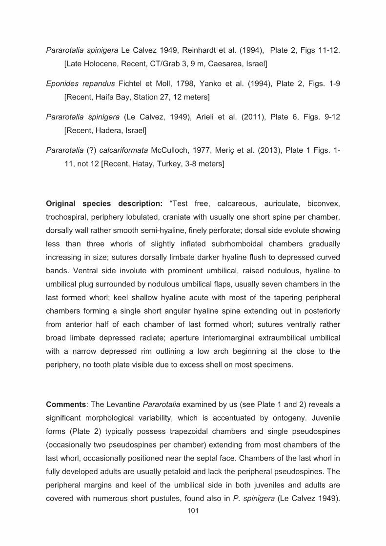
101
Pararotalia spinigera Le Calvez 1949, Reinhardt et al. (1994), Plate 2, Figs 11-12.
[Late Holocene, Recent, CT/Grab 3, 9 m, Caesarea, Israel]
Eponides repandus Fichtel et Moll, 1798, Yanko et al. (1994), Plate 2, Figs. 1-9
[Recent, Haifa Bay, Station 27, 12 meters]
Pararotalia spinigera (Le Calvez, 1949), Arieli et al. (2011), Plate 6, Figs. 9-12
[Recent, Hadera, Israel]
Pararotalia (?) calcariformata McCulloch, 1977, Meriç et al. (2013), Plate 1 Figs. 1-
11, not 12 [Recent, Hatay, Turkey, 3-8 meters]
Original species description: “Test free, calcareous, auriculate, biconvex,
trochospiral, periphery lobulated, craniate with usually one short spine per chamber,
dorsally wall rather smooth semi-hyaline, finely perforate; dorsal side evolute showing
less than three whorls of slightly inflated subrhomboidal chambers gradually
increasing in size; sutures dorsally limbate darker hyaline flush to depressed curved
bands. Ventral side involute with prominent umbilical, raised nodulous, hyaline to
umbilical plug surrounded by nodulous umbilical flaps, usually seven chambers in the
last formed whorl; keel shallow hyaline acute with most of the tapering peripheral
chambers forming a single short angular hyaline spine extending out in posteriorly
from anterior half of each chamber of last formed whorl; sutures ventrally rather
broad limbate depressed radiate; aperture interiomarginal extraumbilical umbilical
with a narrow depressed rim outlining a low arch beginning at the close to the
periphery, no tooth plate visible due to excess shell on most specimens.
Comments: The Levantine Pararotalia examined by us (see Plate 1 and 2) reveals a
significant morphological variability, which is accentuated by ontogeny. Juvenile
forms (Plate 2) typically possess trapezoidal chambers and single pseudospines
(occasionally two pseudospines per chamber) extending from most chambers of the
last whorl, occasionally positioned near the septal face. Chambers of the last whorl in
fully developed adults are usually petaloid and lack the peripheral pseudospines. The
peripheral margins and keel of the umbilical side in both juveniles and adults are
covered with numerous short pustules, found also in P. spinigera (Le Calvez 1949).
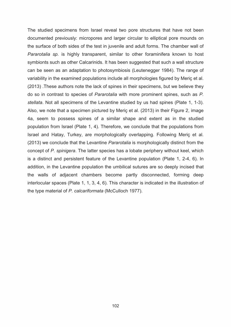
102
The studied specimens from Israel reveal two pore structures that have not been
documented previously: micropores and larger circular to elliptical pore mounds on
the surface of both sides of the test in juvenile and adult forms. The chamber wall of
Pararotalia sp. is highly transparent, similar to other foraminifera known to host
symbionts such as other Calcarinids. It has been suggested that such a wall structure
can be seen as an adaptation to photosymbiosis (Leutenegger 1984). The range of
variability in the examined populations include all morphologies figured by Meriç et al.
(2013) .These authors note the lack of spines in their specimens, but we believe they
do so in contrast to species of Pararotalia with more prominent spines, such as P.
stellata. Not all specimens of the Levantine studied by us had spines (Plate 1, 1-3).
Also, we note that a specimen pictured by Meriç et al. (2013) in their Figure 2, image
4a, seem to possess spines of a similar shape and extent as in the studied
population from Israel (Plate 1, 4). Therefore, we conclude that the populations from
Israel and Hatay, Turkey, are morphologically overlapping. Following Meriç et al.
(2013) we conclude that the Levantine Pararotalia is morphologically distinct from the
concept of P. spinigera. The latter species has a lobate periphery without keel, which
is a distinct and persistent feature of the Levantine population (Plate 1, 2-4, 6). In
addition, in the Levantine population the umbilical sutures are so deeply incised that
the walls of adjacent chambers become partly disconnected, forming deep
interlocular spaces (Plate 1, 1, 3, 4, 6). This character is indicated in the illustration of
the type material of P. calcariformata (McCulloch 1977).
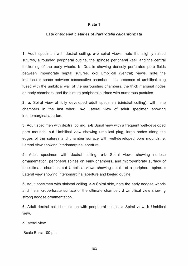
103
Plate 1
Late ontogenetic stages of Pararotalia calcariformata
1. Adult specimen with dextral coiling. a-b spiral views, note the slightly raised
sutures, a rounded peripheral outline, the spinose peripheral keel, and the central
thickening of the early whorls. b. Details showing densely perforated pore fields
between imperforate septal sutures. c-d Umbilical (ventral) views, note the
interlocular space between consecutive chambers, the presence of umbilical plug
fused with the umbilical wall of the surrounding chambers, the thick marginal nodes
on early chambers, and the hirsute peripheral surface with numerous pustules.
2. a. Spiral view of fully developed adult specimen (sinistral coiling), with nine
chambers in the last whorl. b-c Lateral view of adult specimen showing
interiomarginal aperture
3. Adult specimen with dextral coiling. a-b Spiral view with a frequent well-developed
pore mounds. c-d Umbilical view showing umbilical plug, large nodes along the
edges of the sutures and chamber surface with well-developed pore mounds. e.Lateral view showing interiomarginal aperture.
4. Adult specimen with dextral coiling. a-b Spiral views showing nodose
ornamentation, peripheral spines on early chambers, and microperforate surface of
the ultimate chamber. c-d Umbilical views showing details of a peripheral spine. eLateral view showing interiomarginal aperture and keeled outline.
5. Adult specimen with sinistral coiling. a-c Spiral side, note the early nodose whorls
and the microperforate surface of the ultimate chamber. d Umbilical view showing
strong nodose ornamentation.
6. Adult dextral coiled specimen with peripheral spines. a Spiral view. b Umblical
view.
c Lateral view.
Scale Bars: 100 μm
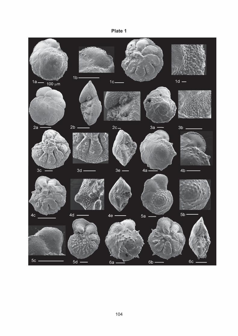
104
Plate 1

105
Plate 2
Early ontogenetic stages of Pararotalia calcariformata
1.-3. Earliest stage specimens with three and four chambers. 1 Spiral view of
specimen with incipient peripheral pseudospines. 2 Spiral view of specimen with
peripheral pseudospines. 3. Umbilical view of specimens without peripheral
pseudospines, showing incipient keel. Note the pustulate surface.
4. Juvenile specimen with peripheral pseudospine on each chamber. a. Spiral view.
b Umbilical view, note the distinct trapezoidal shape of the chambers in the spiral and
umbilical sides. c Lateral view. d Details of the interiomarginal aperture.
5. Juvenile specimen with peripheral pseudospine on each chamber. a- b Spiral
views showing well-developed pore mounds and trapezoidal shaped chambers.
6. Juvenile specimen with peripheral pseudospine on each chamber. a- c Spiral
views. d-f Umbilical views, note the relatively large pore mounds on the surface both
sides of the test. f Details of the peripheral pseudospine.
Scale Bars: 1-3: 10μm, 4-6d: 100μm and 6e: 50μm
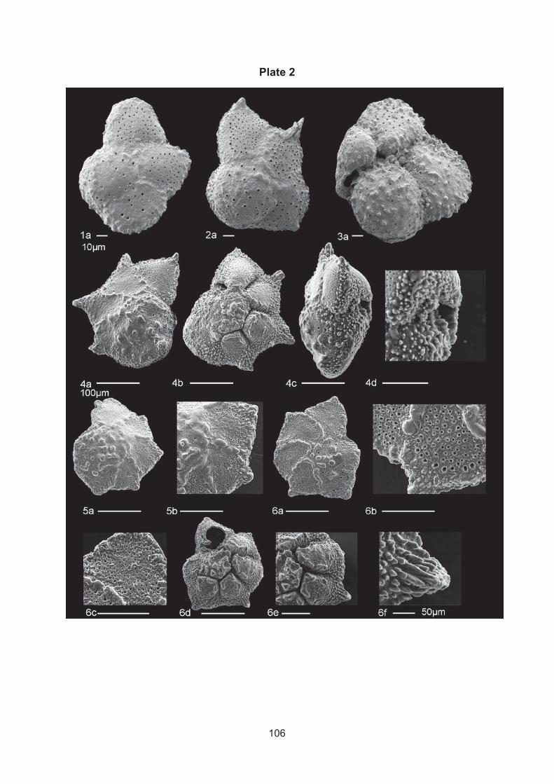
106
Plate 2

107
References
Arieli RN, Almogi-Labin A, Abramovich S, Herut B (2011) The effect of thermal pollution on benthic foraminiferal assemblages in the Mediterranean shoreface adjacent to Hadera power plant (Israel). Marine Pollution Bulletin 62:1002-1012
Hottinger L, Halicz E, Reiss Z (1991) The Foraminiferal Genera Pararotalia, Neorotalia, and Calcarina: Taxonomic Revision. Journal of Paleontology 65:18-33
Leutenegger S (1984) Symbiosis in benthic foraminifera; specificity and host adaptations. The Journal of Foraminiferal Research 14:16-35
Loeblich A, Tappan H (1957) Eleven new genera of Foraminifera. US National Museum Bulletin 215:223-232
Loeblich AR, Tappan H (1994) Foraminifera of the Sahul Shelf and Timor Sea. Journal of Foraminiferal Research Special Publications 31
McCulloch I (1977) Qualitative observations on recent foraminiferal tests with emphasis on the eastern Pacific: Parts I–III. University of Southern California, Los Angeles, CA
Meriç E, Yokes MB, Avsar KN, Kirki-Elmas E, Dinçer F, Karhan SU, Kalkan E, Demir V (2013) First report of Pararotalia calcariformata from the Hatay coastline (Turkey—north-eastern Mediterranean). Marine Biodiversity Records 6:e31
Reinhardt EG, Patterson RT, Schroeder-Adams CJ (1994) Geoarchaeology of the ancient harbor site of Caesarea Maritima, Israel; evidence from sedimentology and paleoecology of benthic foraminifera. The Journal of Foraminiferal Research 24:37-48
Yanko V, Kronfeld J, Flexer A (1994) Response of benthic Foraminifera to various pollution sources; implications for pollution monitoring. The Journal of Foraminiferal Research 24:1-17

108

109
Chapter 4
4. Publication III: Extreme heat tolerance of a foraminifera–diatom photo-symbiosis
Christiane Schmidt1, Janett Brandt1, Barak Herut2, Sigal Abramovich3
and Michal Kucera1
1MARUM, Centre of Marine Environmental Science, University of Bremen, Bremen,
Germany 2Israel Oceanographic and Limnological Research Institute, Haifa, Israel
3Ben Gurion University of the Negev, Beer-Sheva, Israel

110

111
4.1. Abstract
The loss of algal symbionts induced by thermal stress (bleaching) has been
observed in many photo-symbiotic eukaryotes, such as corals, foraminifera and
molluscs. In almost all documented cases, the bleaching threshold is close to the
highest temperatures occurring at present in the open ocean (31-32°C). It appears
that elevated temperatures universally compromise the stability of the eukaryotic
photo-symbiotic relationship and that the physiological tolerance limit may be
exceeded in many parts of the tropical oceans under realistic global change
scenarios. Here we take advantage of the recent discovery of the occurrence of the
diatom-bearing foraminifera Pararotalia calcariformata at a thermally polluted coastal
site in Israel, where water temperatures seasonally reach 36°C. To test whether this
thermal adaptation is universal in P. calcariformata we conducted manipulative
experiments exposing this species together with Amphistegina lobifera from an
unpolluted side in the eastern Mediterranean to elevated temperatures for up to three
weeks. We measured the photosynthetic activity of the photo-symbionts and
recorded survival and growth of the organisms. Reduced photosynthetic activity was
recorded for A. lobifera above 32°C, but P. calcariformata showed a higher tolerance.
Photochemical stress in P. calcariformata was first observed during exposure to 36°C
after three weeks and chronic photoinhibition (but not mortality) first occurred at 42°C
after one week. Survivorship was high in all treatments, and growth was observed at
35 and 36°C. It appears that P. calcariformata exhibits the most thermally tolerant
photosymbiosis observed to date in a eukaryote-eukaryote system. The thermal
tolerance is present in a natural environment where the thermal threshold is never
realized. Observations imply that marine eukaryote-eukaryote photosymbiosis is
more resistant to temperature than expected and this life strategy is likely to persist in
a warmer world.

112
4.2. Introduction
To increase our understanding of the impacts of global climate change on
marine ecosystems, we need to gain more specific knowledge on the thermal limits
and adaptation potential of a diverse range of marine species. Specifically, it is
essential to characterize the physiological and evolutionary traits that determine if a
species is highly climate change vulnerable or is likely able to adapt (Foden et al.
2013). Species have been shown to respond negative but variable to global change
stress (Kroeker et al. 2010). This might be due to different physiological adaptive
capacities (Füssel and Klein 2006) and different eco-physiological processes which
limit the fitness and survival of sensitive organisms (Pörtner 2002). Experimental
work has shown that under high temperatures, excessive oxygen demand in the cells
leads to falling oxygen levels in the cell fluids, reducing the metabolic capacity of
organisms (Pörtner 2002; Pörtner 2010). In addition, higher temperature causes
higher metabolic rates, which stimulate increased production of reactive oxygen
species (ROS) (Sohal and Weindruch 1996). These are damaging to protein function
and membrane structure and are thus reducing the fitness of the exposed organism
(Sohal and Orr 2012). In eukaryotes hosting an endosymbiont, the damage not only
occurs in the host but also in the endosymbiotic partner. In corals, it has been shown
that the combined physiology of the holobiont is so interlinked, that either stress in
the symbiont or the host, determines the holobiont’s thermal tolerance (Bhagooli and
Hidaka 2003; Goulet et al. 2005; Visram and Douglas 2007). If stressful conditions
increase over a threshold and the advantage of the symbiotic relationship is lost, the
zooxanthellae are expelled, a process which is known as bleaching (Brown 1997).
Under natural conditions, photosymbiosis provides an advantage in
oligotrophic conditions (Brasier 1995). It allows the host to feed on the algae-derived
‘photosynthates’ and, the host in return protects the symbionts in the nutrient
enriched cytoplasm (Hallock 1981; Lee and Hallock 1987). However, if the thermal
tolerance of the host or symbionts is exceeded, bleaching can lead to mortality of
entire reefs (Berkelmans and Oliver 1999; Wooldridge 2009). Because bleaching in
the field can be associated with more than one factor, bleaching thresholds should
not be confused with thermal thresholds, as the latter are only testable under
controlled environmental conditions. In corals, bleaching stress has been associated
with symbiont diversity. Clade C Symbiodinium has been shown to be more heat-

113
resistant than Symbiodinium clade D (Rowan 2004), but different bleaching
susceptibilities have been found even within clade C Symbiodinium (Abrego et al.
2008; Sampayo et al. 2008). Despite the existence of variable thermal tolerance,
most marine photo-symbiotic systems involving eukaryotes do not appear to tolerate
temperatures that are substantially exceeding the upper thermal limit of tropical
ocean water and bleach after exposure to 30-34°C (Fitt et al. 2001; Coles and Riegl
2013; Fine et al. 2013). The most heat-tolerant coral has been shown to be the
massive coral Porites solida, tolerating temperature of up to 34°C for 12 h (Strychar
et al. 2004; Strychar and Sammarco 2009). The most resistant reef to bleaching are
located in the Arabian Gulf, where corals experience naturally the greatest
temperature fluctuations worldwide, and have been shown to survive 36°C for a short
period of time without signs of stress in the 1980es (Coles 1988; Coles and Riegl
2013) Indeed, under normal marine conditions of the late Cenozoic (Zhang et al.
2014) organisms adapted to higher thermal tolerance would have had no adaptive
advantage and an evolution of higher tolerance would appear to be meaningless from
an evolutionary perspective.
In benthic foraminifera, upper limits of foram-algae thermal tolerance, have
been documented as tissue damage, reduced growth rates, increased mortality,
reduced photosynthetic rates and bleaching onsetting at 31°C(Hallock and Talge
1993; Schmidt et al. 2011; Uthicke et al. 2011). In comparison to corals, benthic
foraminifera establish an endosymbiosis with different algae types such as diatoms,
dinoflagellates, chrysophytes, rhodophytes, and also cyanobacteria (Pawlowski et al.
2001; Lee 2006). On the host level, their response to elevated temperatures has
been shown to be species-specific. For example, the diatom bearing Calcarina
mayorii was not affected by temperatures up to 31°C but Calcarina hispida bleached
in experimental studies at the same temperature (Schmidt et al. 2011). Thus, like
corals and other photo-symbiotic marine organisms, tropical foraminifera appear to
be vulnerable to warming levels predicted for the next century by realistic global
change scenarios (Fitt et al. 2001).
In benthic foraminifera, there appears to be one exception to this general
pattern in upper thermal thresholds. In 2011, Arieli et al. (2011) reported the
occurrence of the diatom-bearing species Pararotalia calcariformata in a thermally
polluted site on the Mediterranean coast of Israel, where this species seems to

114
tolerate long term exposure to 36°C for several month (Arieli et al. 2011). This is well
above, the bleaching threshold of most studied marine eukaryotic photo-symbiotic
systems, such as most corals species (Fitt et al. 2001; Fitt et al. 2009), except
extremely thermally tolerant corals in the Gulf of Aden, which can tolerate up to 36°C
without signs of stress (Coles and Riegl 2013), Indo-pacific foraminifera (Schmidt et
al. 2011; Uthicke et al. 2012; van Dam et al. 2012) and mollusks (Leggat et al. 2003).
However, it has not yet been shown whether the photosymbiosis in this benthic
foraminifer, P. calcariformata also takes place at these temperatures and it remains
unclear whether the resistance evolved within the unique environment of the thermal
plume or whether it is a universal property of the species.
To test these assumptions, we collected a population of this species from a
natural unpolluted location, 18 km north of the heat polluted site and exposed it to
elevated temperatures. We exposed specimens of the summer and winter
populations inside replicated sea water aquaria to temperatures up to 36°C for up to
three weeks. As a consequence of its heat resistance we then performed an extreme
test experiment, exposing to up to 42°C for three weeks in aquaria inside incubators.
We monitored weekly photosynthetic activity of the symbionts using Pulse Amplitude
Modulated (PAM) Fluorometry and recorded survivorship and growth, as an indicator
for holobiont fitness. To demonstrate the representativeness of our laboratory
conditions, the first two experiments included populations of the A. lobifera from the
same locality. The diatom-bearing genus Amphistegina has a known thermal
tolerance and species of this genus have been previously shown to bleach at 32°C
(Talge and Hallock 2003; Schmidt et al. 2011).
4.3. Methods
4.3.1. Sample collection
Living specimens of Pararotalia calcariformata and Amphistegina lobifera were
collected in the eastern Mediterranean Levantine basin at Nachsholim (32° 37.386 N,
34°55.169 E). Samples were collected by sampling filamentous coralline algae e.g.
Jania sp. from the site, a shallow coastal high-energy habitat, by snorkeling at 0.5-2
m water depth on three field campaigns 1/11/2012, 08/04/2013 and 23/10/2013.
Samples were transported in large plastic bottles filled with algae and sediment to the

115
laboratory, where the algae and sediment were rinsed with sea water and specimens
were picked from the concentrated sediment. Several specimens (up to 50) have
been put inside screw capped plastic jars (Volume 100 mL) together with some algae
and sediment material. The jars were express-shipped inside an insulation container
to Germany, experiencing minimal temperature fluctuations. During two shipments on
22/10/13 and 24/10/13 temperature was logged every 30 min (Hobo, Temperature
logger) showing mean temperatures of 22.2 -23.2°C, SD 2°C (duration of shipments
24- 36 h). The specimens were cultured inside plastic containers at in-situ water
temperature of the collection (23-24°C in November, 20-21°C in April) under a diurnal
light cycle 12 h/12 h (light conditions of <30 μmol m2 s2) in incubation chambers at
ambient salinity (38.5-40.0 ‰). Half of the sea water was replaced every week by
freshly made seawater (Tropic Marine Sea Salt, Germany).
4.3.2. Experimental design and sea water parameters
A replicated design of 12 aquaria (working Volume 18 L) was installed on a
laboratory bench top for conducting the summer (SuPE) and winter exposure (WiPE)
experiments exposing the two species to 5 consecutive temperatures. In each
aquaria temperature was manipulated separately using heating rods controlled by an
aquaria computer system (AT control, Aquamedic, Germany), which are
automatically switched on or off when temperatures in the aquaria would fall below or
be elevated from the set deviance of +/- 0.1°C from the inserted temperatures.
Seawater temperatures were recorded every 10 min by the software and saved. As
the 20°C treatment during the winter population experiment (WiPE) was below the
ambient temperatures of the room, two aquaria were externally cooled by an external
water cooler (Titan 150, Aquamedic). For this the aquaria were by put inside two
large plastic boxes (working volume XL) and were connected to a pump (Ocean
Runner 1200, Aquamedic) and the water cooler, which constantly circulated water
around the aquaria cooled to below room temperature, which was then precisely
heated by heating rods of the Aquamedic system, inside the aquaria, to the desired
20°C. Temperatures during the SuPE were between 24-35°C (duration: 2 weeks) and
during the WiPE between 20-36°C (duration: 3 weeks) (Fig. 2). We choose to slightly
adjust the temperatures and the duration of the experiments focusing during the
WiPE on the warmer end of the temperature spectrum, taking into account results of
SuPE suggesting a higher stability of thermal thresholds in P. calcariformata. Inside

116
the aquaria, water movements were mimicked by using strong flow pumps
(NanoProp 5000, Aquamedic). Manual temperature measurements were conducted
daily using a handheld thermometer and salinity meter (WTW, Germany) and showed
that there were minimal daily variations from the set temperatures (+/-0.5°C). The
aquaria were lighted from top on a 12h day /12 h night cycle by daylight fluorescent
bulbs (50:50 actinic 420nm/ 10 K trichromatic). Photosynthetic active radiation (PAR)
varied inside aquaria and was recorded at the beginning and at the end of the
experiments and varied between 35-40 μmol m2 s2 (Apogee MQ-200, USA).
For the extreme temperature experiment (ExTE), temperature controlled
incubation chambers (Pol-Eko-Aparatura, Model ST2+/ST2+, Poland) were used, as
temperatures were aimed to be set to four temperatures 24-42°C (Fig. 3) for the
duration of 3 weeks. The upper thermal temperature of 42° is not recommended for
the Aquamedic setup, therefore we used the incubation chambers for temperature
control. Each incubation chamber contained two plastic mini-aquaria (working
volume 2 L) standing on the same level to be illuminated with constant light
conditions (diurnal light cycle 12h /12 h, 19-26 μmol m2 s2, white-fluorescent light
bulbs). The aquaria were ventilated by bubbling air by small hand-held air pumps into
the seawater (duration for 30 min each). For automatic temperature monitoring
including diurnal variations, one aquaria per incubator contained a temperature
logger (Hobo, USA). Logger data was excluded from the data set when organisms
were taken outside for measurements. Temperatures varied by app. +/- 1.3°C from
the set temperatures. Exact Logger means per treatment were (N=931,
measurements ever 30 min) were for the 24°C treatment, 24.0°C (SD 0.42), for the
30°C treatment 29.9°C (SD 0.44), for the 36°C treatment 36.3°C (SD 0.53), for the
42°C treatment 42.5 (SD 0.65). Those where used to name the treatments
accordingly in Fig. 3 and 5. Manual temperature measurements inside the aquaria
were made daily using a handheld meter (WTW, Germany) for monitoring the
incubator temperatures and were slightly below the logger temperatures, as they do
not reflect higher night temperatures because lights where off: incubator 1 (24°C
treatment)= 23.6°C (SD 0.19), 2 (30°C treatment) =29.4°C (SD 0.27), incubator 3
(36°C treatment)=35.4°C (SD 0.42), incubator 4 (42°C treatment)=41.2°C (SD 0.55).
Diurnal differences (day/night) were only monitored by the loggers and were for the
24°C treatment 23.6°C/24.3°C (SD 0.28, 0.28); for the 30°C treatment 29.6°C/30.2°C

117
(SD 0.33/0.29); for the 36°C treatment 35.9°C/36.7°C (SD 0.41, 0.25); and for the
42°C treatment 42.0°C/43.0 (SD 0.53/0.32).
Sea water was made in bulk using artificial sea salt (Tropic Marin® Sea Salt,
Germany) at the beginning of the experiments. Salinity measurements were made
daily with the same instrument as temperature was checked (WTW, Germany) and
was adjusted to stay in the range between 38.5- 40.2 ppm in all experiments, by
adding deionized water when needed. These levels are generally observed in the
Levantine Basin and represent in-situ conditions for the experimental species (Herut
et al. 2000). The pH levels of the seawater were monitored weekly and stayed > 8.1
pH units.
At the beginning of the experiment and after weekly measurements,
foraminifera were fed with marine microalgae (frozen, autoclaved) by adding 15 μL of
food mixture to the glass jars. Food mixture consisted of Nannochloropsis algae
concentrate (12 x 109 cells/mL, BlueBioTech GmbH, Germany) which was diluted
with sea water (30μL concentrate: 200 mL seawater) and autoclaved.
The housings of the foraminifera were prepared to provide wall surface to
allow specimen to climb up walls using their pseudopods, and at the same time,
provide enough protection for specimens inside the water flow of the aquaria.
Previous studies also quantitatively described the motility of the specimens (Schmidt
et al. 2011), which is especially interesting in Amphistignidae, because they are very
motile, but as P. calcariformata was observed to be generally be much less motile
and is generally very small (0.3 μm), we did not examine this parameter here. 5-6
specimens were kept inside a snap-cap top vial inside the aquaria and covered by a
flow-through top which allowed passive water exchange inside the chambers.
Therefore, in the top of a standard snap-cap top vial (Volume 15 mL, Wheaton, UK) a
large hole was cut which hold in place a small piece of plankton mesh net underneath
(P. calcariformata mesh size of 100 μm and A. lobifera 300 μm). Six snap-cap vials
were put inside each aquarium on the bottom of a standard 6 well-plate to ensure
stability, containing either A. lobifera or P. calcariformata in a vial. After weekly
measurements the arrangement of the vials inside the aquaria was randomized to
reduce bias due to different positions inside the aquaria, after mesh has been
cleaned with water from algae to ensure same light conditions throughout the
experiment.

118
4.3.3. Photochemistry measurements
To carry out photochemistry measurements on PS II, a chlorophyll fluorometer
IMAGING-PAM M-Series Fluorometer (WALZ GmbH, Germany) was used. It was
equipped with MAXI-Head,1/2’’ CCD camera and zoom objective (F1.0/f=8-48 mm).
Foraminifera of each snap-cap vial were transferred by sucking with 10 mL pipette
into petri dishes containing the treatment water and dark-adapted 10-20 minutes
before measuring dark-adapted yield (=maximum quantum yield, MQY, Fv:Fm). Light-
adapted yield (effective quantum yield, EQY, Y(II)) was measured under light
adaptation similar to experimental light levels, supplied by LED lights installed in the
MAXI-Head. Measurements were conducted once per week. We elevated the Petri
dishes closer to the zoom objective on a 1.5 cm-high stand for P. calcariformata (size
0.3-0.4 μm) and used the Leaf Holder IMAG-MIN/BK to allow best possible imaging
for all specimens. Light levels of the LED light units were measured with PAR Light
Meter (Apogee, USA) and were 25 μmol photos m2 s2, similar conditions as in the
aquaria and incubator setup. Other procedure on data processing is given in previous
work on foraminifera using the IPAM (Schmidt et al. 2011; Uthicke et al. 2012; Vogel
and Uthicke 2012; Schmidt et al. 2014).
4.3.4. Survivorship & Growth measurements
Visual assessment of the foraminifera to determine survival rate was carried
out when high resolution photographs have been made at the onset, and at the end
of the experiments. Foraminifera were photographed at a resolution of 5184 x 3456
Pixel using a Canon SLR camera mounted on a Zeiss stereomicroscope. As
individual experimental units (snap-cap vials) containing the foraminifer were not
individually tracked during the SuPE and ExTE, mean growth rates per aquaria
where calculated from measuring the surface area from initial versus final images of
the specimens after the formula given in Schmidt et al. (2011). Aquaria (SuPE and
ExTE) or wells (WiPE) were excluded from the data set where more specimens than
initially inserted where recovered, to eliminate bias because of individuals which
might have moved or reproduced. Reproduction only occurred in a few specimen
during each experiment (SuPE under 32°C, and in WiPE at 34°C, and at ExTE under
36 °C), resulting in negative growth rates of the aquaria, which have been removed
from the data set. Missing and dead specimens have been identified by sorting the

119
diameters by size or by individual features and removed from the growth data set to
eliminate bias. Slightly negative growth rates in the 36°C treatment in the WiPE are a
combination between measurement error, by surrounding algae and because
mistaken identification of missing specimens and indicate a growth inhibition.
4.3.5. Statistics
Means of photochemistry data (Fv:Fm) and Y(II) per well were statistically
evaluated to avoid pseudoreplication, and consisted of 4-6 individual data points.
Photochemistry means were arc sin transformed to achieve normality of the data
because it represents proportions. PERMANOVA (Primer v6, Add-on) was performed
on transformed data. Temperature has been included as a fixed factor and Aquaria
(Temp) were included as a random factor to account for variances between the
aquaria. Monte Carlo post hoc tests were performed to test when PERMANOVA was
overall significant to establish differences between the individual temperature
treatments (Table S2). Growth data (% surface area increase per day) was not
statistically evaluated as means per aquaria (n=1-2) did yield a small sample size
which did not meet the criteria of normality assumptions.
4.4. Results
To investigate the effect of elevated temperatures on Amphistegina lobifera
and Pararotalia calcariformata, both species were initially exposed to a temperature
gradient of up to 36°C in an aquarium setup with water movement simulating natural
conditions. The experiment was replicated for a population collected in summer
(SuPE) and winter (WiPE). This was done to account for the effect of pre-adaptation
(hardening) during pre-exposure and is required because of the large seasonal
temperature cycle at the studied site (Fig. 1). In addition, a static extreme
temperature experiment (ExTE) was performed on P. calcariformata in an incubator
setup to test its upper thermal limit.

120
Figure 1. A) Monthly average SST (2011-2013) Levantine Continental Shelf (up to WD 200m), obtained by Remote Sensing MODIS Data showing an exceptionally warm year in the Levant in 2012. B) Hambonim Station temperature in the Levant (measurements at edge of tidal rocky platform), and Remote Sensing MODIS Temperatures , Source (Herut B. 2014). C) Temperature logger data from the shallow intertidal at Nachsholim National Park (32° 37.386 N, 34°55.169 E, 0.5 and 1.5 m) shows temperatures up to 32°C in August D)Mean summer sea surface temperature in the Levant Basin observed between 1955 and 2012 (July-September) extracted from the World Ocean Atlas 2013 (Locarnini 2013).
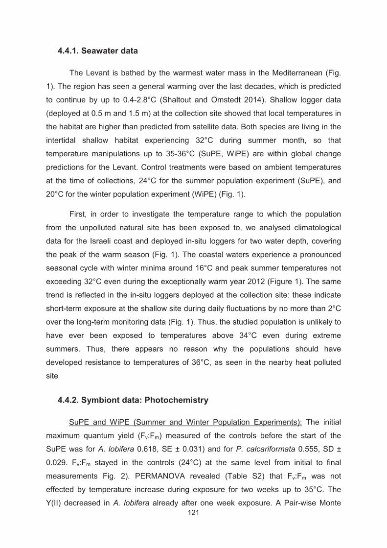
121
4.4.1. Seawater data
The Levant is bathed by the warmest water mass in the Mediterranean (Fig.
1). The region has seen a general warming over the last decades, which is predicted
to continue by up to 0.4-2.8°C (Shaltout and Omstedt 2014). Shallow logger data
(deployed at 0.5 m and 1.5 m) at the collection site showed that local temperatures in
the habitat are higher than predicted from satellite data. Both species are living in the
intertidal shallow habitat experiencing 32°C during summer month, so that
temperature manipulations up to 35-36°C (SuPE, WiPE) are within global change
predictions for the Levant. Control treatments were based on ambient temperatures
at the time of collections, 24°C for the summer population experiment (SuPE), and
20°C for the winter population experiment (WiPE) (Fig. 1).
First, in order to investigate the temperature range to which the population
from the unpolluted natural site has been exposed to, we analysed climatological
data for the Israeli coast and deployed in-situ loggers for two water depth, covering
the peak of the warm season (Fig. 1). The coastal waters experience a pronounced
seasonal cycle with winter minima around 16°C and peak summer temperatures not
exceeding 32°C even during the exceptionally warm year 2012 (Figure 1). The same
trend is reflected in the in-situ loggers deployed at the collection site: these indicate
short-term exposure at the shallow site during daily fluctuations by no more than 2°C
over the long-term monitoring data (Fig. 1). Thus, the studied population is unlikely to
have ever been exposed to temperatures above 34°C even during extreme
summers. Thus, there appears no reason why the populations should have
developed resistance to temperatures of 36°C, as seen in the nearby heat polluted
site
4.4.2. Symbiont data: Photochemistry
SuPE and WiPE (Summer and Winter Population Experiments): The initial
maximum quantum yield (Fv:Fm) measured of the controls before the start of the
SuPE was for A. lobifera 0.618, SE ± 0.031) and for P. calcariformata 0.555, SD ±
0.029. Fv:Fm stayed in the controls (24°C) at the same level from initial to final
measurements Fig. 2). PERMANOVA revealed (Table S2) that Fv:Fm was not
effected by temperature increase during exposure for two weeks up to 35°C. The
Y(II) decreased in A. lobifera already after one week exposure. A Pair-wise Monte

122
Carlo test showed that 24-30°C were significantly different from 32 and 35°C. The
initial maximum quantum yield (Fv:Fm) measured of the controls before the start of the
WiPE for A. lobifera where 0.529, SE ±0.047 and for P. calcariformata 0.451, SE
±0.049. PERMANOVA revealed that temperature had a significant effect on the Fv:Fm
in A. lobifera after one week exposure and in P. calcariformata after three weeks of
exposure (Table S2). The Y(II) was significantly reduced in A. lobifera after one week
exposure but not in P. calcariformata. However, in the Y(II) in P. calcariformata a
reduced trend from the 34°C to the 36°C treatment is shown (Fig. 2). Photoinhibition
occurred (Fv:Fm <0.01) under 36°C after one week in the WiPE (60% of A. lobifera),
and was less drastic than in Y(II) where it was 92% of specimen. After two weeks the
Fv:Fm increased again in the 36°C treatment to levels of 0.18, SD 0.08, but Y(II)
stayed <0.01 in 97% of specimens. A seasonal effect can be seen in the 30°C and
32°C treatments as those were the treatments which where exactly repeated in both
experiments. The Y(II) in A. lobifera, was higher in the SuPE than in the WiPE. In P.
calcariformata there is no visible difference in photosynthetic activity to temperatures
with season.
ExTE (Extreme Temperature experiment): This experiment was conducted
exposing P. calcariformata to four different temperatures at 24, 30, 36, and an
extreme level of 42°C to determine its upper thermal tolerance level. The Fv:Fm and
Y(II) of initial symbionts stayed on the same level in the 24°C (control) and in 30°C
treatment throughout the experiment (Fig. 3). PERMANOVA revealed that Fv:Fm and
Y(II) was significantly negatively affected by temperature after one week of exposure
(Table S2, S3). Monte Carlo Post hoc tests on Fv:Fm and Y(II) indicate that after two
weeks there are significant differences between the 24-30°C and the 36°C and 42°C
treatments. The reduction in Fv:Fm to <0.01 occurred in all specimens of the 42°C
treatment and in 14% of the specimen from the 36°C treatment after two weeks.

123
Figure 2. Pulse Amplitude Fluorometry (PAM) measurements on Amphistegina lobifera (blue) and Pararotalia calcariformata (red) after two weeks of exposure to experimental temperatures, Fv:Fm represents dark adapted yield, or maximum quantum yield, Y(II) represents light adapted yield or effective quantum yield at 25 μmol photons m2 s-1. p values indicate results of PERMANOVA, sig. level =0.05, different letters indicate sig. differences in the Monte Carlo post-hoc tests between the treatments

124
Figure 3. Pulse Amplitude Fluorometry (PAM) measurements on Pararotalia calcariformata initial, after 1, 2 and 3 weeks exposure to elevated temperatures, Fv:Fm represents dark adapted yield, Y(II) represents light adapted yield at 25μm mol photons m2 s-1. p values indicate results of PERMANOVA, sig. level
=0.05, different letters indicate sig. differences in the Monte Carlo post-hoc tests between the treatments
4.4.3. Holobiont data: Survivorship & Growth
After termination of experiments, survival of the holobiont was high, determined by
checking of cytoplasmic color. This non-terminal method was chosen, as the species
have distinct green-brownish cytoplasm color which can be distinguished from a dead
individual, which become translucent upon cell death (Bernhard 2000). Mean survival
rates were between 89-100% per treatment (Table S1). Exemplary specimens of A.
lobifera and P. calcariformata in natural habitat and after the laboratory exposure to
the respective temperatures for 2-3 weeks (Fig. 4) illustrate that cytoplasmic color
was visible in the specimen even after the highest treatments. Paling of shell took
place in A. lobifera in some specimen in the 34°C, where the expulsion of living or
dead symbiotic material is visible (Fig. 4).
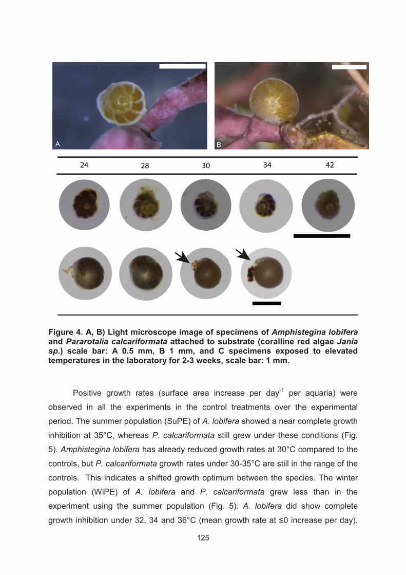
125
Figure 4. A, B) Light microscope image of specimens of Amphistegina lobiferaand Pararotalia calcariformata attached to substrate (coralline red algae Jania sp.) scale bar: A 0.5 mm, B 1 mm, and C specimens exposed to elevated temperatures in the laboratory for 2-3 weeks, scale bar: 1 mm.
Positive growth rates (surface area increase per day-1 per aquaria) were
observed in all the experiments in the control treatments over the experimental
period. The summer population (SuPE) of A. lobifera showed a near complete growth
inhibition at 35°C, whereas P. calcariformata still grew under these conditions (Fig.
5). Amphistegina lobifera has already reduced growth rates at 30°C compared to the
controls, but P. calcariformata growth rates under 30-35°C are still in the range of the
controls. This indicates a shifted growth optimum between the species. The winter
population (WiPE) of A. lobifera and P. calcariformata grew less than in the
experiment using the summer population (Fig. 5). A. lobifera did show complete
126
However, in P. calcariformata positive growth rates under 32 and 34°C are still
observed. In the extreme temperature experiment (ExTE) the summer population of
P. calcariformata did generally grew less than in the earlier experiments. Pararotalia
calcariformata showed very low growth rates in the 24° and 36°C and an inhibition of
growth in the 42°C treatment.

127
Figure 5. Growth rates (% surface area increase day-1) of Amphistegina lobiferaand Pararotalia calcariformata at the end of the experiment, after two weeks (SuPE) and after three weeks (WiPE, ExTe).
4.5. Discussion
The benthic foraminifera Pararotalia calcariformata is able to sustain an active
photosymbiosis with diatoms up to 36°C for up to 3 weeks, while Amphistegina
lobifera collected from the same habitat shows signs of stress under these
conditions. Photosynthetic activity was reduced at 34 - 35°C in A. lobifera, compared
to the controls, but was not in P. calcariformata. In P. calcariformata, chronic
photoinhibition (Fv:Fm
specimens. Reductions in the Fv:Fm represent chronic photoinhibition, defined as the
loss of photosynthetic activity due to irreversible photodamage to Photosystem II
(Brown 1997). Chronic photoinhibition (Fv:Fm
exposed to 33°C for one week (Schmidt et al. 2011). As photoinhibition represents
chronic stress in the photosystems, gradual reductions in the light adapted
photosynthetic yields Y(II), occurred sooner than reductions in dark adapted
photosynthetic yields Fv:Fm, and are associated with the onset of physiological
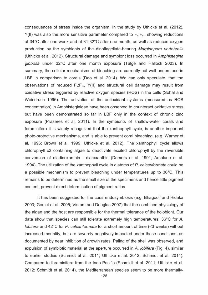
128
consequences of stress inside the organism. In the study by Uthicke et al. (2012),
Y(II) was also the more sensitive parameter compared to Fv:Fm, showing reductions
at 34°C after one week and at 31-32°C after one month, as well as reduced oxygen
production by the symbionts of the dinoflagellate-bearing Marginopora vertebralis
(Uthicke et al. 2012). Structural damage and symbiont loss occurred in Amphistegina
gibbosa under 32°C after one month exposure (Talge and Hallock 2003). In
summary, the cellular mechanisms of bleaching are currently not well understood in
LBF in comparison to corals (Doo et al. 2014). We can only speculate, that the
observations of reduced Fv:Fm, Y(II) and structural cell damage may result from
oxidative stress triggered by reactive oxygen species (ROS) in the cells (Sohal and
Weindruch 1996). The activation of the antioxidant systems (measured as ROS
concentration) in Amphisteginidae have been observed to counteract oxidative stress
but have been demonstrated so far in LBF only in the context of chronic zinc
exposure (Prazeres et al. 2011). In the symbionts of shallow-water corals and
foraminifera it is widely recognized that the xanthophyll cycle, is another important
photo-protective mechanisms, and is able to prevent coral bleaching, (e.g. Warner et
al. 1996; Brown et al. 1999; Uthicke et al. 2012). The xanthophyll cycle allows
chlorophyll c2 containing algae to deactivate excited chlorophyll by the reversible
conversion of diadinoxanthin - diatoxanthin (Demers et al. 1991; Arsalane et al.
1994). The utilization of the xanthophyll cycle in diatoms of P. calcariformata could be
a possible mechanism to prevent bleaching under temperatures up to 36°C. This
remains to be determined as the small size of the specimens and hence little pigment
content, prevent direct determination of pigment ratios.
It has been suggested for the coral endosymbiosis (e.g. Bhagooli and Hidaka
2003; Goulet et al. 2005; Visram and Douglas 2007) that the combined physiology of
the algae and the host are responsible for the thermal tolerance of the holobiont. Our
data show that species can still tolerate extremely high temperatures; 36°C for A.
lobifera and 42°C for P. calcariformata for a short amount of time (<3 weeks) without
increased mortality, but are severely negatively impacted under these conditions, as
documented by near inhibition of growth rates. Paling of the shell was observed, and
expulsion of symbiotic material at the aperture occurred in A. lobifera (Fig. 4), similar
to earlier studies (Schmidt et al. 2011; Uthicke et al. 2012; Schmidt et al. 2014).
Compared to foraminifera from the Indo-Pacific (Schmidt et al. 2011; Uthicke et al.
2012; Schmidt et al. 2014), the Mediterranean species seem to be more thermally-

129
tolerant. This is not surprising, as the seasonal natural temperature exposure the
species experience during seasonal cycles is different and likely influences their
physiological response in experiments. Background temperatures in the eastern
Mediterranean vary from 16-32°C over the yearly cycle (Fig. 1A) and can be elevated
by +2°C in the species shallow habitat (Fig. 1C). In the Great Barrier Reef 6 m water
depth at Pine Island, where hottest months (January or February 2006-2010) in each
year was 28.8°C(1SD = 0.40°C) http://data.aims.gov.au (Uthicke and Fabricius
2012). Specimens of M. vertebralis collected close to this station showed that the
health of the specimens were negatively impacted at 31°C, and most specimens at
34°C died after one week. Amphistegina radiata and Heterostegina depressa from a
similar habitat as Uthicke et al. (2012), could survive 33°C for one week but showed
disruption of the photochemistry and bleaching (Schmidt et al. 2011).
It has been shown that intertidal invertebrates contain a higher stability form of
anti-oxidative enzymes compared to sub-tidal organisms, because they are naturally
more adapted to short-term oxidative stress (Regoli et al. 1997). This finding
suggests that molecular pre-adaptations also exist between organisms originating
from different depth. The organisms from earlier studies originated from habitats in
the sub-tidal (Schmidt et al. 2011; Uthicke et al. 2012; Schmidt et al. 2014) expect M.
vertebralis in Schmidt et al. (2014) and Calcarina hispida in Schmidt et al. (2011)
whereas species in our study originate from intertidal environment. Interestingly
specimens from the shallow locations in the intertidal 1-2m have also been more
resistant towards elevated temperatures. Calcarina hispida has had no reduced
photosynthetic activity at temperatures of 31°C and M. vertebralis showed significant
decrease at this temperature but was more resistant than H. depressa. Further
studies should explore the role of pre-adaptations of depth, and/or symbiont type to in
LBF to explore this hypotehesis.
We speculate that the apparently innate resistance to high temperature of P.
calcariformata originates from the parent populations in the Indopacific. There, in tidal
pools of the tropics, it may experience temporary exposure to elevated temperatures
and may therefore better pre-adapted. We note that populations of this species
exhibiting this tolerance would have had an advantage when passing the shallow
coastal areas of the southern Red Sea on their way to invade the Red Sea and later
the Mediterranean. Such thermal filtering has been proposed to explain elevated
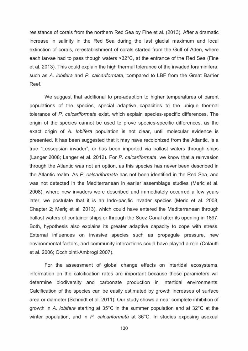
130
resistance of corals from the northern Red Sea by Fine et al. (2013). After a dramatic
increase in salinity in the Red Sea during the last glacial maximum and local
extinction of corals, re-establishment of corals started from the Gulf of Aden, where
each larvae had to pass though waters >32°C, at the entrance of the Red Sea (Fine
et al. 2013). This could explain the high thermal tolerance of the invaded foraminifera,
such as A. lobifera and P. calcariformata, compared to LBF from the Great Barrier
Reef.
We suggest that additional to pre-adaption to higher temperatures of parent
populations of the species, special adaptive capacities to the unique thermal
tolerance of P. calcariformata exist, which explain species-specific differences. The
origin of the species cannot be used to prove species-specific differences, as the
exact origin of A. lobifera population is not clear, until molecular evidence is
presented. It has been suggested that it may have recolonized from the Atlantic, is a
true “Lessepsian invader”, or has been imported via ballast waters through ships
(Langer 2008; Langer et al. 2012). For P. calcariformata, we know that a reinvasion
through the Atlantic was not an option, as this species has never been described in
the Atlantic realm. As P. calcariformata has not been identified in the Red Sea, and
was not detected in the Mediterranean in earlier assemblage studies (Meric et al.
2008), where new invaders were described and immediately occurred a few years
later, we postulate that it is an Indo-pacific invader species (Meric et al. 2008,
Chapter 2; Meriç et al. 2013), which could have entered the Mediterranean through
ballast waters of container ships or through the Suez Canal after its opening in 1897.
Both, hypothesis also explains its greater adaptive capacity to cope with stress.
External influences on invasive species such as propagule pressure, new
environmental factors, and community interactions could have played a role (Colautti
et al. 2006; Occhipinti-Ambrogi 2007).
For the assessment of global change effects on intertidal ecosystems,
information on the calcification rates are important because these parameters will
determine biodiversity and carbonate production in intertidal environments.
Calcification of the species can be easily estimated by growth increases of surface
area or diameter (Schmidt et al. 2011). Our study shows a near complete inhibition of
growth in A. lobifera starting at 35°C in the summer population and at 32°C at the
winter population, and in P. calcariformata at 36°C. In studies exposing asexual

131
juveniles of P. calcariformata for 48 days to 35°C we found, that juvenile’s
development is near inhibited under these conditions. This let us conclude that the
offspring of P. calcariformata are more susceptible to thermal stress than the adult
population and require a narrow thermal window for reproduction and ontogenetic
development in the range of >20°C and <35°C. The indo-pacific M. vertebralis
showed also decreased growth at 32°C for up to one month exposure (Doo et al.
2012; Uthicke et al. 2012), indicating that this species has a lower thermal limit than
the species in our study. We found slightly different growth optima; for A. lobifera
between 24-28°C and for P. calcariformata between 30-32°C, which are important for
judging calcification contribution of the species under global change conditions. This
indicates that the environmental envelope of P. calcariformata is slightly shifted to the
warmer end. Our growth data show that Amphistegenids will be living at the upper
limit of their environmental envelope in the Eastern Mediterranean. Their future
distribution will therefore be moving towards colder regions in the western
Mediterranean as postulated by habitat models (Langer et al. 2012; Weinmann et al.
2013). Amphistegenids, are currently the dominant symbiont-bearing species in the
Mediterranean sediments (Meric et al. 2008; Triantaphyllou et al. 2009; Mouanga and
Langer 2014), but as their distribution shifts westwards, biodiversity pattern of marine
sediments are likely to be altered. Our growth data suggests that Pararotalia
calcariformata, will be the “survivor species” of climate change in the eastern
Mediterranean, as they experience optimal conditions for calcification in waters of 30-
32°C and are thus likely to be abundant in marine sediments in the future. Benthic
foraminifera have been shown to be of large importance for coral reefs, despite their
small size (Scoffin and Tudhope 1985; Langer et al. 1997; Doo et al. 2012; Doo et al.
2014). They can be mass cultured for coastal protection of islands prone to sea-level
rise, because of their fast growth and reproduction (Hosono et al. 2014).
Debate has arisen in the coral literature to what extent the genetic variants of
Symbiodinium symbionts can influence the bleaching response of corals (Iglesias-
Prieto et al. 2004; Putnam et al. 2012). For example in the coral Pocillopora
damincornis, It has been found, that Symbiodinum Clade C are less prone to
bleaching than Symbiodinum Clade D (Glynn et al. 2001; Rowan 2004). Furthermore,
bleaching responses are likely shaped by fine-scale differences in symbiont type and
go beyond broad cladal designations (Sampayo et al. 2008). In corals the association
of symbionts is flexible and characterized by a change of strains over time (Little et

132
al. 2004) indicating that strain type might have a large influence on the physiology of
the holobiont. It has further been shown that photo-symbiont thermal adaptation can
change the coral thermal tolerance (Howells et al. 2012). The symbiosis in
foraminifera is different from corals, as they have been shown to contain multiple
different algae species (Lee and Anderson 1991; Lee 1992). Both species under
investigation host a consortium of different diatom endosymbiotic species inside their
cytoplasm, which are contained in morphological structures in their calcite shell (Lee
et al. 1995; Chai and Lee 1999; Lee and Correia 2005). Pararotalia calcariformata
host a symbiosis with at least three different diatom symbionts, but also a newly
described diatom symbiont, which has not been isolated from a host before, and
identified as Minutocellus polymorphus (<2-3 μm size) (Chapter 2). Amphistegina
lobifera has been shown to host up to seven different species of endo-symbiotic
diatoms but not M. polymorphus (Lee and Correia 2005). We propose that this
species could be the clue to its thermal resistance. Minutocellus polymorphus grown
in batch-culture showed increase anti-oxidant activity (super oxide dismutase activity,
SOD) by the factor of 3 at the beginning of its exponential growth phase and had
reduced SOD activity at the end of their growth phase, similar to other micro algae
(Sigaud-Kutner et al. 2002). This indicates that M. polymorphus might be a possible
catalyzer, when in its optimal growth phase, for inducing anti-oxidants in the
protoplasma of P. calcariformata which may reduce shell damage due to thermal-
stress. Another theory is that hosting multiple distinct species of symbionts may
provide an advantage for the host under rapidly increasing temperatures due to
climate change, because it has been shown that not all symbionts provide the same
functional benefit to the host (Douglas 1998; Loram et al. 2007). Molecular PCR-
based analysis of foraminifera suggests that multiple Symbiodinium lineages uniquely
associate with foraminifera (e.g., clades F3-F5, G1, H, I) (Garcia-Cuetos et al. 2005;
Pochon et al. 2007). Work on the symbiont-bearing Marginopora vertebralis showed
that dominant types C3 and C15, which are common in corals (e.g. LaJeunesse et al.
2003; Cooper et al. 2011; Putnam et al. 2012) are also found in foraminifera
(Momigliano and Uthicke 2013). Almost all hard coral families can host more than
one symbiont clade, and molecular work likely underestimated numerical
abundances because PCR-based identification techniques still miss cryptic diversity
(Baker and Romanski 2007).

133
Our data highlight the importance of defining thermal tolerance levels on the
basis of individual species to make predictions on the basis of species-specific traits
to best accommodate for global change. Here, we show that the photosymbiosis of
Pararotalia calcariformata is more thermally resistant than any other eukaryote-
eukaryote symbiosis in invertebrates found to date, as it can tolerate temperatures up
to 36°C sustaining a photosynthetically active photosymbiosis over three weeks and
can tolerate temperatures of 42°C with damaged photo-symbiosis but not induced
host mortality. Pararotalia calcariformata hosts at least four different endosymbiotic
diatoms, one of which is the newly described Minutocellus polymorphus (Chapter 1),
which could be a hint towards the higher thermal resistance of the species. As the
invasive history of the P. calcariformata is not entirely resolved, we can only
speculate on possible other mechanisms on its thermal tolerance beside the different
endosymbiont. Our growth data shows that P. calcariformata currently does not live
close to its physiological limits under normal summer temperatures of 32°C in the
Levant, and it is likely flourish under 1-3°C warming. Future work needs to test,
whether all species of the genus respond similar and determine what role the newly
described symbiont plays, in the extreme thermal tolerance of the photosymbiosis in
P. calcariformata. The need arises for gaining deeper understanding of the functional
benefits of endosymbiotic diversity and antioxidant capacity in eukaryotic
endosymbionts under thermal stress.

134
4.6. References
Abrego D, Ulstrup KE, Willis BL, van Oppen MJH (2008) Species-specific interactions between algal endosymbionts and coral hosts define their bleaching response to heat and light stress. Proceedings of the Royal Society B-Biological Sciences 275:2273-2282
Arieli RN, Almogi-Labin A, Abramovich S, Herut B (2011) The effect of thermal pollution on benthic foraminiferal assemblages in the Mediterranean shoreface adjacent to Hadera power plant (Israel). Marine Pollution Bulletin 62:1002-1012
Arsalane W, Rousseau B, Duval JC (1994) Influence of the pool size of the xanthophyll cycle on the effects of light stress in a diatom - competition between photoprotection and photoinhibition. Photochemistry and Photobiology 60:237-243
Baker AC, Romanski AM (2007) Multiple symbiotic partnerships are common in scleractinian corals, but not in octocorals: Comment on Goulet (2006). Marine Ecology Progress Series 335:237-242
Berkelmans R, Oliver JK (1999) Large-scale bleaching of corals on the Great Barrier Reef. Coral Reefs 18:55-60
Bernhard JM (2000) Distinguishing live from dead foraminifera: Methods review and proper applications. Micropaleontology 46:38-46
Bhagooli R, Hidaka M (2003) Comparison of stress susceptibility of in hospite and isolated zooxanthellae among five coral species. Journal of Experimental Marine Biology and Ecology 291:181-197
Brasier MD (1995) Fossil indicators of nutrient levels. 2: Evolution and extinction in relation to oligotrophy. Geological Society, London, Special Publications 83:133-150
Brown BE (1997) Coral bleaching: causes and consequences. Coral Reefs 16:S129-S138
Brown BE, Ambarsari I, Warner ME, Fitt WK, Dunne RP, Gibb SW, Cummings DG (1999) Diurnal changes in photochemical efficiency and xanthophyll concentrations in shallow water reef corals: evidence for photoinhibition and photoprotection. Coral Reefs 18:99-105
Chai JY, Lee JJ (1999) Initial recognition of endosymbiotic diatom surface antigens by the larger foraminifer Amphistegina lobifera. Symbiosis 26:39-53
Colautti RI, Grigorovich IA, MacIsaac HJ (2006) Propagule pressure: A null model for biological invasions. Biological Invasions 8:1023-1037
Coles SL, Riegl BM (2013) Thermal tolerances of reef corals in the Gulf: A review of the potential for increasing coral survival and adaptation to climate change through assisted translocation. Marine Pollution Bulletin 72:323-332

135
Cooper TF, Berkelmans R, Ulstrup KE, Weeks S, Radford B, Jones AM, Doyle J, Canto M, O'Leary RA, van Oppen MJH (2011) Environmental Factors Controlling the Distribution of Symbiodinium Harboured by the Coral Acropora millepora on the Great Barrier Reef. PLoS ONE 6
Demers S, Roy S, Gagnon R, Vignault C (1991) Rapid light-induced-changes in cell fluorescence and in xanthophyll-cycle pigments of Alexandrium excavatum (dinophyceae) and Thalassiosira Pseudonana (bacillariophyceae) - a photo-protection mechanism. Marine Ecology Progress Series 76:185-193
Doo SS, Hamylton S, Byrne M (2012) Reef-Scale Assessment of Intertidal Large Benthic Foraminifera Populations on One Tree Island, Great Barrier Reef and Their Future Carbonate Production Potential in a Warming Ocean. Zoological Studies 51:1298-1307
Doo SS, Fujita K, Byrne M, Uthicke S (2014) Fate of Calcifying Tropical Symbiont-Bearing Large Benthic Foraminifera: Living Sands in a Changing Ocean. Biological Bulletin 226:169-186
Douglas AE (1998) Host benefit and the evolution of specialization in symbiosis. Heredity 81:599-603
Fine M, Gildor H, Genin A (2013) A coral reef refuge in the Red Sea. Global Change Biology:n/a-n/a
Fitt WK, Brown BE, Warner ME, Dunne RP (2001) Coral bleaching: interpretation of thermal tolerance limits and thermal thresholds in tropical corals. Coral Reefs 20:51-65
Fitt WK, Gates RD, Hoegh-Guldberg O, Bythell JC, Jatkar A, Grottoli AG, Gomez M, Fisher P, Lajuenesse TC, Pantos O, Iglesias-Prieto R, Franklin DJ, Rodrigues LJ, Torregiani JM, van Woesik R, Lesser MP (2009) Response of two species of Indo-Pacific corals, Porites cylindrica and Stylophora pistillata, to short-term thermal stress: The host does matter in determining the tolerance of corals to bleaching. Journal of Experimental Marine Biology and Ecology 373:102-110
Foden WB, Butchart SHM, Stuart SN, Vie JC, Akcakaya HR, Angulo A, DeVantier LM, Gutsche A, Turak E, Cao L, Donner SD, Katariya V, Bernard R, Holland RA, Hughes AF, O'Hanlon SE, Garnett ST, Sekercioglu CH, Mace GM (2013) Identifying the World's Most Climate Change Vulnerable Species: A Systematic Trait-Based Assessment of all Birds, Amphibians and Corals. PLoS ONE 8
Füssel H-M, Klein RT (2006) Climate Change Vulnerability Assessments: An Evolution of Conceptual Thinking. Climatic Change 75:301-329
Garcia-Cuetos L, Pochon X, Pawlowski J (2005) Molecular evidence for host-symbiont specificity in soritid Foraminifera. Protist 156:399-412
Glynn PW, Mate JL, Baker AC, Calderon MO (2001) Coral bleaching and mortality in panama and Ecuador during the 1997-1998 El Nino-Southern oscillation

136
event: Spatial/temporal patterns and comparisons with the 1982-1983 event. Bulletin of Marine Science 69:79-109
Goulet TL, Cook CB, Goulet D (2005) Effect of short-term exposure to elevated temperatures and light levels on photosynthesis of different host-symbiont combinations in the Aiptasia pallidal Symbiodinium symbiosis. Limnology and Oceanography 50:1490-1498
Hallock P (1981) Algal symbiosis: A mathematical analysis. Marine Biology 62:249-255
Herut B, Almogi-Labin A, Jannink N, Gertman I (2000) The seasonal dynamics of nutrient and chlorophyll a concentrations on the SE Mediterranean shelf-slope. Oceanologica Acta 23:771-782
Herut B. SE, Gordon N., Galil B., Lubinevsky H., Tibor G., Tom M., Rilov G., Silverman J. and Rinkevich B. (2014) The National Monitoring Program of Israel's Mediterranean coastal waters Scientific Report for 2012 IOLR Report H62/2013
Hosono T, Lopati P, Makolo F, Kayanne H (2014) Mass culturing of living sands (Baculogypsina sphaerulata) to protect island coasts against sea-level rise. Journal of Sea Research 90:121-126
Howells EJ, Beltran VH, Larsen NW, Bay LK, Willis BL, van Oppen MJH (2012) Coral thermal tolerance shaped by local adaptation of photosymbionts. Nature Clim Change 2:116-120
Iglesias-Prieto R, Beltran VH, LaJeunesse TC, Reyes-Bonilla H, Thome PE (2004) Different algal symbionts explain the vertical distribution of dominant reef corals in the eastern Pacific. Proceedings of the Royal Society B-Biological Sciences 271:1757-1763
Kroeker KJ, Kordas RL, Crim RN, Singh GG (2010) Meta-analysis reveals negative yet variable effects of ocean acidification on marine organisms. Ecology Letters 13:1419-1434
LaJeunesse TC, Loh WKW, van Woesik R, Hoegh-Guldberg O, Schmidt GW, Fitt WK (2003) Low symbiont diversity in southern Great Barrier Reef corals, relative to those of the Caribbean. Limnology and Oceanography 48:2046-2054
Langer MR, Silk MT, Lipps JH (1997) Global ocean carbonate and carbon dioxide production; the role of reef Foraminifera. Journal of Foraminiferal Research 27:271-277
Langer MR (2008) Foraminifera from the Mediterranean and the Red Sea Aqaba-Eilat, the Improbable Gulf Environment, Biodiversity and Preservation. Magnes Press, pp399-417
Langer MR, Weinmann AE, Lötters S, Rödder D (2012) "Strangers" in Paradise: Modeling the Biogeographic Range Expansion of the Foraminifera Amphistegina in the Mediterranean Sea. Journal of Foraminiferal Research 42:234-244

137
Lee JJ, Hallock P (1987) Algal symbiosis as the driving force in the evolution of larger Foraminifera. Annals of the New York Academy of Sciences 503:330-347
Lee JJ, Anderson OR (1991) Symbiosis in Foraminifera. In: Lee JJ, Anderson OR (eds) Biology of Foraminifera. Academic Press Limited, San Diego, pp157-220
Lee JJ (1992) Taxonomy of algae symbiotic in foraminifera. In: Reisser W (ed) Algae and Symbioses. Biopress Ltd., Bristol, UK, pp79–92
Lee JJ, Morales J, Symons A, Hallock P (1995) Diatom symbionts in larger foraminifera from Caribbean hosts. Marine Micropaleontology 26:99-105
Lee JJ, Correia M (2005) Endosymbiotic diatoms from previously unsampled habitats. Symbiosis 38:251-260
Lee JJ (2006) Algal symbiosis in larger foraminifera. Symbiosis 42:63-75
Leggat W, Buck BH, Grice A, Yellowlees D (2003) The impact of bleaching on the metabolic contribution of dinoflagellate symbionts to their giant clam host. Plant, Cell & Environment 26:1951-1961
Little AF, van Oppen MJH, Willis BL (2004) Flexibility in algal endosymbioses shapes growth in reef corals. Science 304:1492-1494
Locarnini RA, A. V. Mishonov, J. I. Antonov, T. P. Boyer, H. E. Garcia, O. K. Baranova, M. M. Zweng, C. R. Paver, J. R. Reagan, D. R. Johnson, M. Hamilton, and D. Seidov (2013) World Ocean Atlas 2013 Temperature. In: 73 AMTENAN (ed) NOAA Atlas NESDIS 73, pp40
Loram JE, Trapido-Rosenthal HG, Douglas AE (2007) Functional significance of genetically different symbiotic algae Symbiodinium in a coral reef symbiosis. Molecular Ecology 16:4849-4857
Meric E, Avsar N, Yokes MB (2008) Some alien foraminifers along the Aegean and southwestern coasts of Turkey. Micropaleontology 54:307-349
Meriç E, Yokes MB, Avsar KN, Kirki-Elmas E, Dinçer F, Karhan SU, Kalkan E, Demir V (2013) First report of Pararotalia calcariformata from the Hatay coastline (Turkey—north-eastern Mediterranean). Marine Biodiversity Records 6:e31
Momigliano P, Uthicke S (2013) Symbiosis in a giant protist (Marginopora vertebralis,Soritinae): flexibility in symbiotic partnerships along a natural temperature gradient. Marine Ecology Progress Series 491:33–46
Mouanga GH, Langer MR (2014) At the front of expanding ranges: Shifting community structures at amphisteginid species range margins in the Mediterranean Sea. Neues Jahrbuch für Geologie und Paläontologie -Abhandlungen 271:141-150
Occhipinti-Ambrogi A (2007) Global change and marine communities: Alien species and climate change. Marine Pollution Bulletin 55:342-352

138
Pawlowski J, Holzmann M, Fahrni JF, Hallock P (2001) Molecular identification of algal endosymbionts in large miliolid foraminifera: 1. Chlorophytes. Journal of Eukaryotic Microbiology 48:362-367
Pochon X, Garcia-Cuetos L, Baker AC, Castella E, Pawlowski J (2007) One-year survey of a single Micronesian reef reveals extraordinarily rich diversity of Symbiodinium types in soritid foraminifera. Coral Reefs 26:867-882
Pörtner H-O (2010) Oxygen- and capacity-limitation of thermal tolerance: a matrix for integrating climate-related stressor effects in marine ecosystems. The Journal of Experimental Biology 213:881-893
Pörtner HO (2002) Climate variations and the physiological basis of temperature dependent biogeography: systemic to molecular hierarchy of thermal tolerance in animals. Comparative Biochemistry and Physiology - A Molecular and Integrative Physiology 132:739-761
Prazeres MD, Martins SE, Bianchini A (2011) Biomarkers response to zinc exposure in the symbiont-bearing foraminifer Amphistegina lessonii (Amphisteginidae, Foraminifera). Journal of Experimental Marine Biology and Ecology 407:116-121
Putnam HM, Stat M, Pochon X, Gates RD (2012) Endosymbiotic flexibility associates with environmental sensitivity in scleractinian corals. Proceedings of the Royal Society B-Biological Sciences 279:4352-4361
Regoli F, Principato GB, Bertoli E, Nigro M, Orlando E (1997) Biochemical characterization of the antioxidant system in the scallop Adamussium colbecki, a sentinel organism for monitoring the Antarctic environment. Polar Biology 17:251-258
Rowan R (2004) Coral bleaching - Thermal adaptation in reef coral symbionts. Nature 430:742-742
Sampayo EM, Ridgway T, Bongaerts P, Hoegh-Guldberg O (2008) Bleaching susceptibility and mortality of corals are determined by fine-scale differences in symbiont type. Proceedings of the National Academy of Sciences of the United States of America 105:10444-10449
Schmidt C, Heinz P, Kucera M, Uthicke S (2011) Temperature-induced stress leads to bleaching in larger benthic foraminifera hosting endosymbiotic diatoms. Limnology and Oceanography 56:1587-1602
Schmidt C, Kucera M, Uthicke S (2014) Combined effects of warming and ocean acidification on coral reef Foraminifera Marginopora vertebralis and Heterostegina depressa. Coral Reefs 33:805-818
Scoffin TP, Tudhope AW (1985) Sedimentary environments of the Central Region of the Great Barrier Reef of Australia. Coral Reefs 4:81-93
Shaltout M, Omstedt A (2014) Recent sea surface temperature trends and future scenarios for the Mediterranean Sea. Oceanologia 56:411-443

139
Sigaud-Kutner TCS, Pinto E, Okamoto OK, Latorre LR, Colepicolo P (2002) Changes in superoxide dismutase activity and photosynthetic pigment content during growth of marine phytoplankters in batch-cultures. Physiologia Plantarum 114:566-571
Sohal RS, Weindruch R (1996) Oxidative stress, caloric restriction, and aging. Science 273:59-63
Sohal RS, Orr WC (2012) The redox stress hypothesis of aging. Free Radical Biology and Medicine 52:539-555
Strychar KB, Coates M, Sammarco PW (2004) Loss of Symbiodinium from bleached Australian scleractinian corals (Acropora hyacinthus, Favites complanata and Porites solida). Mar and Freshwater Res 55:135-144
Strychar KB, Sammarco PW (2009) Exaptation in corals to high seawater temperatures: Low concentrations of apoptotic and necrotic cells in host coral tissue under bleaching conditions. Journal of Experimental Marine Biology and Ecology 369:31-42
Talge HK, Hallock P (2003) Ultrastructural responses in field-bleached and experimentally stressed Amphistegina gibbosa (Class Foraminifera). Journal of Eukaryotic Microbiology 50:324-333
Triantaphyllou MV, Koukousioura O, Dimiza MD (2009) The presence of the Indo-Pacific symbiont-bearing foraminifer Amphistegina lobifera in Greek coastal ecosystems (Aegean Sea, Eastern Mediterranean). Mediterranean Marine Science 10:73-85
Uthicke S, Vogel N, Doyle J, Schmidt C, Humphrey C (2011) Interactive effects of climate change and eutrophication on the dinoflagellate-bearing benthic foraminifer Marginopora vertebralis. Coral Reefs:1-14
Uthicke S, Fabricius KE (2012) Productivity gains do not compensate for reduced calcification under near-future ocean acidification in the photosynthetic benthic foraminifer species Marginopora vertebralis. Global Change Biology 18:2781-2791
Uthicke S, Vogel N, Doyle J, Schmidt C, Humphrey C (2012) Interactive effects of climate change and eutrophication on the dinoflagellate-bearing benthic foraminifer Marginopora vertebralis. Coral Reefs 31:401-414
van Dam JW, Negri AP, Mueller JF, Altenburger R, Uthicke S (2012) Additive pressures of elevated sea surface temperatures and herbicides on symbiont-bearing foraminifera. PLoS ONE 7:e33900
Visram S, Douglas AE (2007) Resilience and acclimation to bleaching stressors in the scleractinian coral Porites cylindrica. Journal of Experimental Marine Biology and Ecology 349:35-44
Vogel N, Uthicke S (2012) Calcification and photobiology in symbiont-bearing benthic foraminifera and responses to a high CO2 environment. Journal of Experimental Marine Biology and Ecology 424–425:15-24
140
Warner ME, Fitt WK, Schmidt GW (1996) The effects of elevated temperature on the photosynthetic efficiency of zooxanthellae in hospite from four different species of reef coral: A novel approach. Plant Cell and Environment 19:291-299
Weinmann AE, Rodder D, Lotters S, Langer MR (2013) Traveling through time: The past, present and future biogeographic range of the invasive foraminifera Amphistegina spp. in the Mediterranean Sea. Marine Micropaleontology 105:30-39
Wooldridge SA (2009) Water quality and coral bleaching thresholds: Formalising the linkage for the inshore reefs of the Great Barrier Reef, Australia. Marine Pollution Bulletin 58:745-751
Zhang YG, Pagani M, Liu Z (2014) A 12-Million-Year Temperature History of the Tropical Pacific Ocean. Science 344:84-87

141
4.7. Supporting Information
Table S1. Mean Survivorship (%) per treatment during temperature sensitivity experiments exposing Pararotalia calcariformata and Amphistegina lobifera in for 2-3 weeks to elevated temperatures, n.m.= not measured, as A. lobifera was not exposed in this experiment.
Experiment Temperature (°C) Species
Pararotalia
calcariformataAmphistegina
lobifera
1. Summer Population
(SuPE)
24 100 10028 100 10030 100 10032 95 9335 93 90
2.Winter Population
(WiPE)
20 100 10030 96 9032 90 9634 89 9836 100 98
3. Extreme Temperature
(ExTE)
24 100 n.m.30 99 n.m.36 99 n.m.
42 99 n.m.
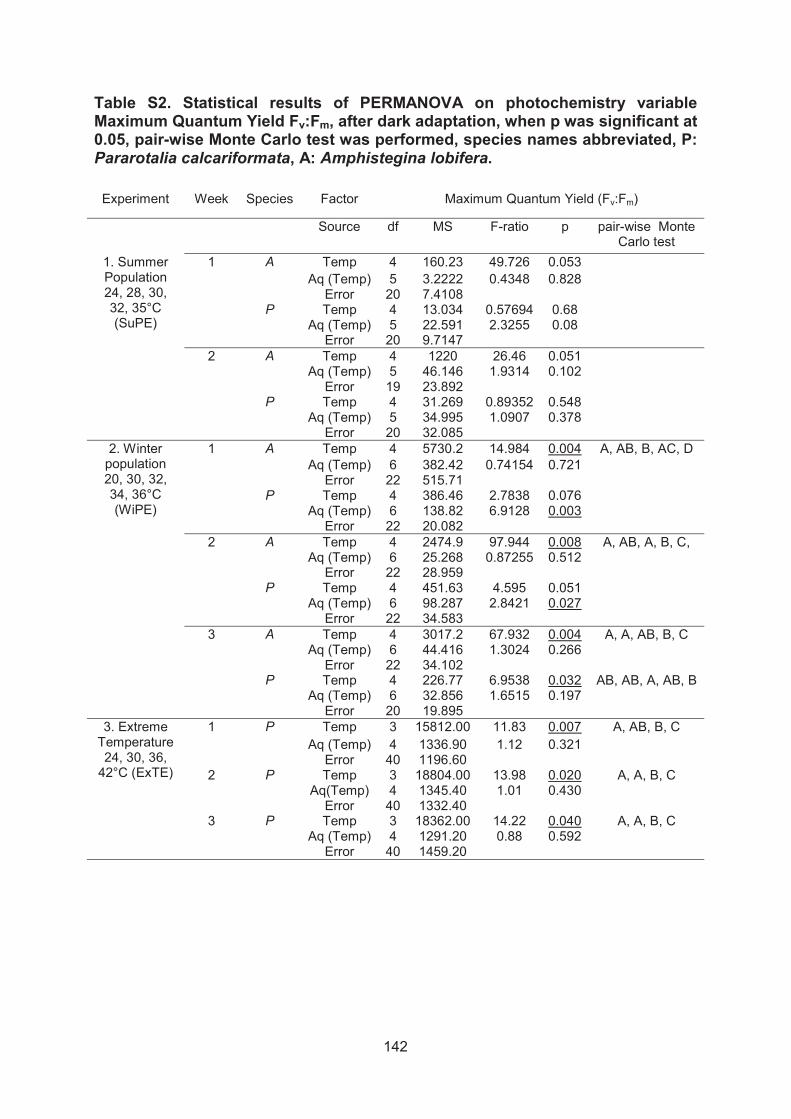
142
Table S2. Statistical results of PERMANOVA on photochemistry variable Maximum Quantum Yield Fv:Fm, after dark adaptation, when p was significant at 0.05, pair-wise Monte Carlo test was performed, species names abbreviated, P:Pararotalia calcariformata, A: Amphistegina lobifera.
Experiment Week Species Factor Maximum Quantum Yield (Fv:Fm)
Source df MS F-ratio p pair-wise Monte Carlo test
1. Summer Population 24, 28, 30, 32, 35°C (SuPE)
1 A Temp 4 160.23 49.726 0.053Aq (Temp) 5 3.2222 0.4348 0.828
Error 20 7.4108P Temp 4 13.034 0.57694 0.68
Aq (Temp) 5 22.591 2.3255 0.08Error 20 9.7147
2 A Temp 4 1220 26.46 0.051Aq (Temp) 5 46.146 1.9314 0.102
Error 19 23.892P Temp 4 31.269 0.89352 0.548
Aq (Temp) 5 34.995 1.0907 0.378Error 20 32.085
2. Winter population 20, 30, 32, 34, 36°C (WiPE)
1 A Temp 4 5730.2 14.984 0.004 A, AB, B, AC, DAq (Temp) 6 382.42 0.74154 0.721
Error 22 515.71P Temp 4 386.46 2.7838 0.076
Aq (Temp) 6 138.82 6.9128 0.003Error 22 20.082
2 A Temp 4 2474.9 97.944 0.008 A, AB, A, B, C,Aq (Temp) 6 25.268 0.87255 0.512
Error 22 28.959P Temp 4 451.63 4.595 0.051
Aq (Temp) 6 98.287 2.8421 0.027Error 22 34.583
3 A Temp 4 3017.2 67.932 0.004 A, A, AB, B, CAq (Temp) 6 44.416 1.3024 0.266
Error 22 34.102P Temp 4 226.77 6.9538 0.032 AB, AB, A, AB, B
Aq (Temp) 6 32.856 1.6515 0.197Error 20 19.895
3. Extreme Temperature 24, 30, 36,
42°C (ExTE)
1 P Temp 3 15812.00 11.83 0.007 A, AB, B, CAq (Temp) 4 1336.90 1.12 0.321
Error 40 1196.602 P Temp 3 18804.00 13.98 0.020 A, A, B, C
Aq(Temp) 4 1345.40 1.01 0.430Error 40 1332.40
3 P Temp 3 18362.00 14.22 0.040 A, A, B, CAq (Temp) 4 1291.20 0.88 0.592
Error 40 1459.20

143
Table S3. Statistical results of PERMANOVA on photochemistry variable Effective Quantum Yield Y (II), after light adaptation, under experimental conditions, when p was significant at 0.05, pair-wise Monte Carlo test was performed, species names abbreviated, P= Pararotalia calcariformata, A= Amphistegina lobifera.
Experiment Week Species Factor Effective Quantum Yield (Y(II))
Source df MS F-ratio ppair-wise Monte Carlo test
1. Summer Population 24, 28, 30, 32, 35°C (SuPE)
1
ATemp 4 452.1 44.401 0.024 A, B, B, AB, C
Aq (Temp) 5 10.182 1.1013 0.413Error 20 9.2452
PTemp 4 316.92 1.7185 0.31
Aq (Temp) 5 184.42 1.4121 0.291Error 20 130.6
2
ATemp 4 5303.8 109.39 0.005 A, A, A, B, C
Aq (Temp) 5 48.304 0.31356 0.987Error 19 154.05
PTemp 4 241.72 0.35266 0.859
Aq (Temp) 5 685.41 4.9975 0.004Error 20 137.15
2. Winter population 20, 30, 32, 34, 36°C (WiPE)
1
ATemp 4 6820.2 6.4499 0.006 AB, A, A, B, C
Aq (Temp) 6 1057.4 1.1544 0.261Error 22 915.95
PTemp 4 1186.7 3.6021 0.065
Aq (Temp) 6 329.46 1.5806 0.168Error 22 208.43
2
ATemp 4 6745.6 6.4406 0.006 A, A, A, B, C
Aq (Temp) 6 1047.4 1 0.453Error 22 1047.3
PTemp 4 918.26 1.2908 0.338
Aq (Temp) 6 711.37 3.4386 0.007Error 22 206.88
3
ATemp 4 7033.6 7.986 0.018 A, A, AB, B, C
Aq (Temp) 6 880.74 1.0274 0.426Error 22 857.25
PTemp 4 967.49 3.6015 0.056
Aq (Temp) 6 271.44 2.2034 0.067Error 20 123.19
3. Extreme Temperature 24, 30, 36,
42°C (ExTE)
1 PTemp 3 15901.00 9.57 0.020 A, AB, AB, C
Aq (Temp) 4 1662.20 1.35 0.130Error 40 1227.90
2 P.Temp 3 18197.00 13.23 0.018 A, A, B, C
Aq(Temp) 4 1375.20 0.96 0.499Error 40 1433.70
3 PTemp 3 15674.00 15.52 0.007 A, AB, B, C
Aq (Temp) 4 1009.90 0.88 0.547Error 40 1150.90

144
5. Concluding remarks and perspectives
5.1. General discussion and conclusions
Working hypothesis Chapter 2: Combined effects of warming and ocean acidification on coral reef Foraminifera Marginopora vertebralis and Heterostegina depressa.
A.) There is a difference between the individual and the combined effects of OA and warming on the physiology of Heterostegina depressa and Marginopora vertebralis
B.) The combined effects are additive, synergistic or antagonistic (cancel each other)
C.) The response to the combined effects is species-specific
Results show that the combined effects of ocean acidification and warming will
impact the physiology of benthic-symbiont bearing foraminifera Heterostegina
depressa and Marginopora vertebralis stronger than if the stressors would act
individually. Overall temperature had been the major stress parameter on
foraminiferal physiology, as shown in previous studies (Schmidt et al. 2011; Uthicke
et al. 2012; Sinutok et al. 2014), indicating that there was a significant effect of
temperature in all parameters measured. However, in many parameters combined
effects had a stronger negative effect.
In half of the investigated parameters, which were survivorship, growth,
respiration, chlorophyll a content and three parameters describing photosynthesis,
significant interactions of OA and warming were found, of which 75% were
synergistic (Chapter 1, Table 3). In these cases warming and elevated pCO2 led to a
stronger physiological response than the sum of the effects of the individual
parameters. We suggest that the energetic costs of counteracting the effect of one
stressor might reduce the ability of the holobiont to counteract the effects of an
additional stressor. For example the energetic cost for the holobiont to control its
symbiotic population under abnormal environmental scenarios such as elevated
pCO2 or elevated nutrients has been proposed to lower bleaching thresholds in
corals and larger benthic foraminifera (LBF) (Wooldridge 2009; Uthicke et al. 2012).

145
Few studies have been conducted on the interactive effects of environmental
stressors on LBF. They suggest that temperature and nutrient exposure (Uthicke et
al. 2012), temperature and OA (Reymond et al. 2013) and temperature and pesticide
exposure (van Dam et al. 2012), have interactive or additive negative effects on the
LBF. One study presented by Sinutok et al. (2011) on the combined effects of
acidification and temperature showed reduced calcification (growth, magnesium and
aragonite cystal size) in M. vertebralis in the combined treatment. However, in this
study the photosynthetic activity and chlorophyll a contents were reduced even in the
controls, indicating that LBF might have been severely stressed in this study under
culturing conditions, because of extremely high light levels. A second study on the
interactive effects of OA and temperatures showed in comparison to healthy controls
that the combined effects reduce calcification and photosynthesis in M. vertebralis
(Sinutok et al. 2014).
The results presented here, indicate that the effects of ocean acidification and
warming are likely to be underestimated when stressors are tested isolation. The
results prove that responses are species-specific, as the most important synergistic
effects were found on different parameters in H. depressa and M. vertebralis. The
latter species showed not effect of the stressor warming or OA on growth alone but
was significantly reduced in the combined treatment, where H. depressa did not
show negative synergistic effects.
Working hypotheses Chapter 3: Recent invasion of the symbiont-bearing foraminifera Pararotalia into the Eastern Mediterranean facilitated by the ongoing warming trend
A) Based on its current distribution, the recently discovered foraminifer P.calcariformata in the eastern Mediterranean Sea is an invader species
B) The foraminifer P. calcariformata contains permanent diatom endosymbionts
C) The symbionts in P. calcariformata are photosynthetically active over several month in culture
D) The foraminifer P. calcariformata will spread to currently colder regions in the Mediterranean based on global warming trend

146
E) Pararotalia calcariformata has a narrow reproductive window for asexual offspring development and is currently restricted by minimumtemperatures to spread westwards
We conclude, from current and past occurrence data, that P. calcariformata
McCulloch 1977 is likely to be an invasive species in the Mediterranean. We suggest
that the invasive population has likely originated from the Indo-pacific region based
on taxonomic and phylogenetic investigations. In summary, all evidence suggests
that P. calcariformata might be a Lessepsian migrant, whose invasion was facilitated
by the recent warming in the Levant (Shaltout and Omstedt 2014).
The identity of symbionts in P. calcariformata has never been investigated,
and the earliest observations of symbionts were described by Arieli et al. (2011). This
species harbors functional endosymbiotic diatoms inside its cytoplasm, which we
have identified by standard algae culturing and sequencing. We found three different
endosymbiotic species inside the host, which are most likely dominated by the
endosymbiont Minutocellus polymorphus. This species was not observed in any
isolated foraminifer before (Lee and Anderson 1991; Lee 2006; Lee et al. 2010) and
has been found as a pelagic free-living diatom in the Mediterranean (Sarno et al.
1993).
We measured the photosynthetic activity of symbionts of P. calcariformata and
conclude that it involves a permanent photo-symbiosis, using PAM (Pulse Amplitude
Modulated) Fluorometry, as observed in other benthic foraminifera (Nobes et al.
2008; Ziegler 2009). Rapid light curves indicated that the species might have a
stronger light sensitivity than A. lobifera, which are in line with findings of Nobes et al.
(2008) on Calcarina sp. Next, we confirmed the activity and the persistence of the
photo-symbionts after collection in culture for up to 5 month using PAM Fluorometry.
The photosynthetic activity measured by obtaining the dark adapted yield (Fv:Fm) are
comparable to other symbiont-bearing LBF (Nobes et al. 2008; Ziegler and Uthicke
2011).
Species distribution modelling (SMD) predicts that under current water
temperature conditions the Levant is the optimal habitat for P. calcariformata.
Moderately suitable habitat, representing “typical” conditions for a species, continues
along the coast of Syria to southernmost Turkey, where it was most recently reported

147
by Meriç et al. (2013). Under global warming, the species will spread from the
eastern Mediterranean region to the western and northern Mediterranean region,
possibly reaching the Ionian Sea by 2100. To construct the SDM, we used yearly
minimum temperature, turbidity and irradiance, but minimum temperature was the
single most important variable explaining the distribution.
We report asexual reproduction in P. calcariformata and growth in
manipulative experiments for the first time. The offspring contained a minimum of
three chambers when released from the parent and contained symbionts.
Subsequent experiments revealed an initial 30-day period of rapid growth, where
most of the new chamber development was occurring, followed by a phase of slower
growth, where only one or two more chambers where formed. We could prove the
hypothesis that juvenile development is restricted to a narrow thermal tolerance
window, as we observed a significant difference between growth rates of asexual
juveniles and temperature. New chamber formation and “normal” development was
only observed at 24°C and 28°C but not at 20°C or 35°C. Under the lower and upper
temperature chosen in the experiment, juveniles also survived the experiments but
did not showed strong growth. This indicates that for a successful establishment of a
population, this species might be dependent on a narrow reproductive window. We
conclude that P. calcariformata lives at its lower thermal limit in winter conditions in
the Levant and prefers warmer spring temperatures for reproduction to ensure
optimal offspring development. The Levantine basin has already experienced
significant winter warming in the past decades and will increase its yearly mean SST
by 0.5-2.3°C providing optimal conditions for the species to (Shaltout and Omstedt
2014). In the Ionian Sea (e.g. San Stefano, Corfu) the time window of temperatures
>20°C per year are shorter by two month (IOLR 2010). This mechanism would
provide a possible explanation for the restricted Levantine occurrence of the invasive
species, as well as for its apparently recent invasion, and current absence in the
Ionian Sea. The absence of the limitation to dispersal, due to the removal of physical
barriers through anthropogenic influence and even exaggerated through ship traffic,
leads to an almost unlimited ongoing range expansion (Langer et al. 2013; Meriç et
al. 2013; Weinmann et al. 2013). These results suggest dramatic changes in the
structure of Mediterranean ecosystems as a consequence of global change (e.g.
Hiddink et al. 2012; Edelist et al. 2013).

148
Working hypotheses Chapter 4: Extreme heat tolerance of a foraminifera–diatom photo-symbiosis
A) Pararotalia calcariformata shows a unique thermal tolerance, with symbionts performing photosynthesis at temperatures up to 36°C, which could be an explanation why it occurs in the heat-plume
B) The winter population reacts more sensitive than the summer population to elevated temperatures
C) Has the foraminifer P. calcariformata the same general thermal limit of bleaching of 1-3°C above observed current summer maxima, compared to corals and other eukaryotic photosymbiosis
The foraminifer P. calcariformata exhibits extreme thermal tolerance even in
populations living in natural non-heat-polluted environments. The summer population
showed active photosymbiosis under 35°C for the duration of 3 weeks which was not
significantly different from the controls and positive growth rates under 35°C. This
indicates that P. calcariformata is adapted to tolerate sustained exposure to
temperatures exceeding summer maxima in their habitat by more than 4°C.
Amphistegina lobifera, collected in the same habitat, showed already reduced
photosynthetic activity at 32°C. Some shells of A. lobifera (Fig. 4) paled and the
individuals expulsed of symbiotic material at the aperture, similar as shown in other
studies on LBF (Schmidt et al. 2011; Uthicke et al. 2012; Schmidt et al. 2014).
Thermal stress was shown to cause structural damage and symbiont loss in
Amphistegina gibbosa under 32°C after one month exposure (Talge and Hallock
2003). We can only speculate that observations of reduced photosynthetic yields and
structural cell damage may result from oxidative stress triggered in the cells under
stressful external conditions, which may lead to the production of free radicals
causing damage to membranes (Sohal and Weindruch 1996).
We repeated the experiments on a summer and winter population to account
for the effect of pre-adaptation during pre-exposure, which is required because of the
large seasonal temperature cycle at the study site which ranges from 16-32°C. In the
winter population we measured reduced dark and light adapted yields under 36°C at
exposure for 3 weeks. The results point in the direction that the summer population
was less sensitive to elevated temperatures than the winter population, and that

149
natural pre-adaptation to summer maxima, did reduce the negative effect or
postpone it until the third week of exposure.
We conducted an extreme thermal exposure experiment elevating
temperature above realistic climate change predictions on a summer population in
the following year, to test the effect of up to 42°C. The results showed chronic photo-
inhibition (dark adapted yields Fv:Fm
photochemical energy in the reaction centers of PS (Photosystem) II under these
conditions after one week of exposure. Despite the stress of the symbionts, the
survivorship was high, indicated by green-brownish cytoplasm color. Chronic
photoinhibition (Fv:Fm Amphistegina radiata and
Heterostegina depressa exposed to 33°C for one week (Schmidt et al. 2011),
resulting in bleaching. In contrast, P. calcariformata apparently survived
temperatures, above + 8°C of its thermal adaption to summer temperatures (30-
32°C) in the Levant. This indicates a remarkable thermal tolerance for P.
calcariformata, which is unique in the eukaryote-eukaryote symbiotic relationships. In
comparison corals have been shown to bleach under temperatures of up to 26-36°C
(Table 1). Bleaching thresholds in corals are dependent on regional pre-adaption (Fitt
et al. 2001; Coles and Riegl 2013; Fine et al. 2013), symbiont type (Glynn et al. 2001;
Rowan 2004) and are species-specific (Strychar et al. 2004; Strychar and Sammarco
2009). Corals seem to consistently bleach at temperatures 1-3°C above the
experienced summer maxima (Coles and Riegl 2013). Since the same appears to
apply to other studied LBF, the adaptation threshold of P. calcariformata is unique
and it remains to be answered why it acquired this adaptation (Fig. 1). Corals in the
Red Sea (Fine et al. 2013) and in the Arabian/Persian Gulf (Coles 1988; Coles and
Riegl 2013) have been shown to be the most thermally resistant corals worldwide
(Wellington et al. 2001). For the Gulf of Aqaba corals, it has been suggested that this
unique temperature resistance is due to thermal filtering of larvae, which had to pass
a corridor of >32°C in the southern Red Sea to re-colonize the Red Sea from the
Indian Ocean, following deglacial sea level rise (Fine et al. 2013).
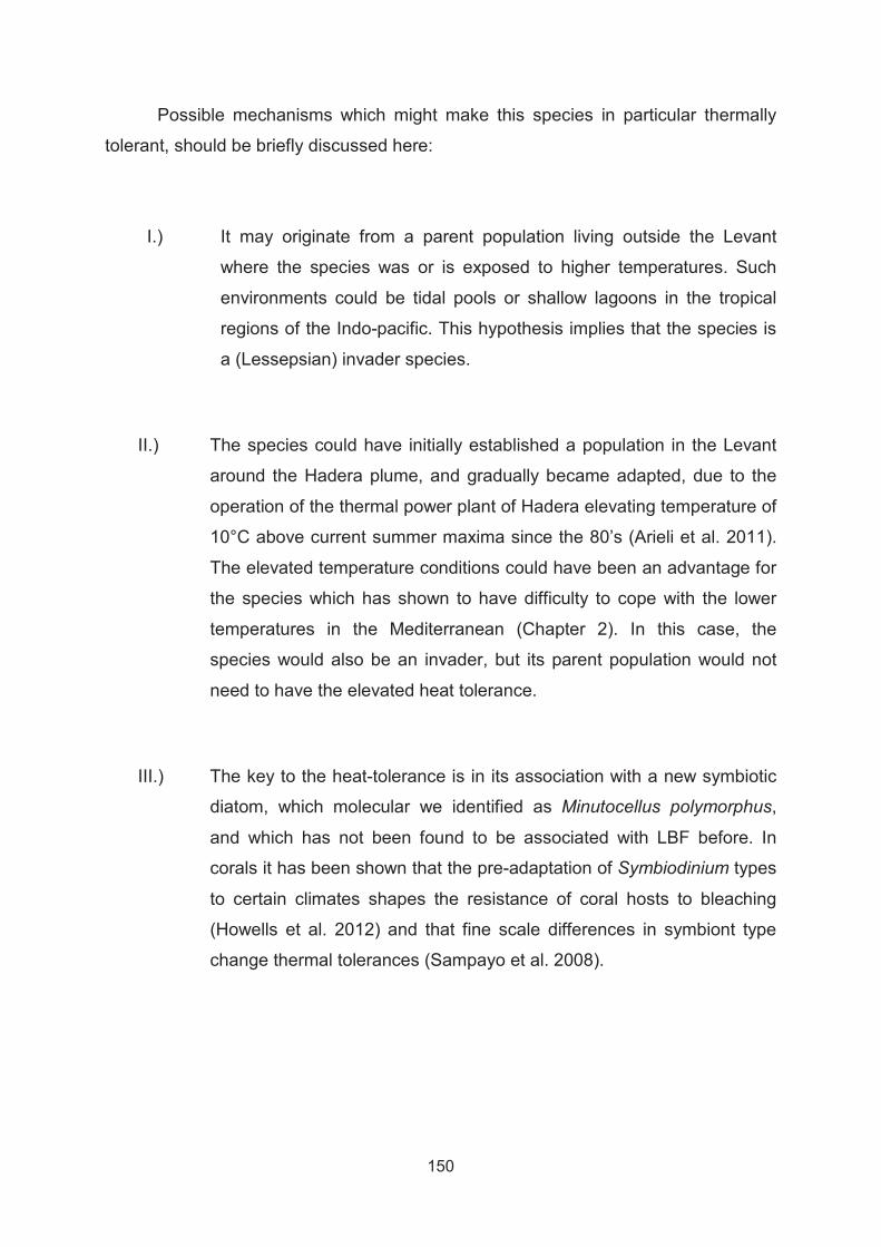
150
Possible mechanisms which might make this species in particular thermally
tolerant, should be briefly discussed here:
I.) It may originate from a parent population living outside the Levant
where the species was or is exposed to higher temperatures. Such
environments could be tidal pools or shallow lagoons in the tropical
regions of the Indo-pacific. This hypothesis implies that the species is
a (Lessepsian) invader species.
II.) The species could have initially established a population in the Levant
around the Hadera plume, and gradually became adapted, due to the
operation of the thermal power plant of Hadera elevating temperature of
10°C above current summer maxima since the 80’s (Arieli et al. 2011).
The elevated temperature conditions could have been an advantage for
the species which has shown to have difficulty to cope with the lower
temperatures in the Mediterranean (Chapter 2). In this case, the
species would also be an invader, but its parent population would not
need to have the elevated heat tolerance.
III.) The key to the heat-tolerance is in its association with a new symbiotic
diatom, which molecular we identified as Minutocellus polymorphus,
and which has not been found to be associated with LBF before. In
corals it has been shown that the pre-adaptation of Symbiodinium types
to certain climates shapes the resistance of coral hosts to bleaching
(Howells et al. 2012) and that fine scale differences in symbiont type
change thermal tolerances (Sampayo et al. 2008).

151
Figure 1. Bleaching threshold temperature exposure plots for Gulf corals compared with Indo-Pacific and Caribbean corals showing adaptation to the higher water temperatures of the Gulf (redrawn from Coles et al. (2013), to facilitate comparison to thresholds in LBF from these regions and higher adaptation potential of the foraminifera Pararotalia calcariformata, coloured dots indicate thresholds from benthic symbiont-bearing foraminifera blue dots: 1 Schmidt et al. (2011, 2014), green dots: 2 a study from Chapter 3 Amphistegina lobifera, 2 b study from Chapter 3 Pararotalia calcariformata,turquoise dots: 3 Talge & Hallock (2003) Amphistegina gibbosa, red dots: 4 Uthicke et al. (2012) Marginopora vertebralis.

152
Table 1. Marine eukaryote-eukaryote photo-symbioses and thresholds to bleaching of organisms for which symbiosis and/or bleaching was reported.
Host Type Symbiont Type Symbiont Reference
Thermal threshold
Bleaching Reference
Cnidaria
Obligate-obligate
Dinoflagellate Symbiodinium Clade C and Clade D in hermatypic corals, soft corals and sea anemones
e.g. Muscatine & Porter (1977), Baker (2003),
26-36°C +1-3°C above summer maxima, dependent on species and symbiont
e.g. Fitt et al. (2001), Strychar et al. (2004), Fine et al. (2013), Coles et al. (2013), Wellington et al. (2001)
Mollusca
Obligate-obligate, kleptoplastidy
Dinoflagellate Symbiodinium in marine bivalves and gastropods
e.g. Jones et al. (1986) e.g. Leggat et al. (2003)
>30-32°C
e.g. Addessi (2001), Leggat et al. (2003) e.g. Ziegler et al. (2014)
Rhizaria
Obligate-obligate, kleptoplastidy
benthic foraminifera host diatoms, dinoflagellates, red algae and green algae
e.g. Lee et al. (2006)
Benthic Foraminifera >30-36°C,
Hallock et al. (1992), Talge & Hallock (2003), Schmidt et al. (2011)
benthic Foraminifera Elphidium sp. retain diatom plastids (Kleptoplastidy)
Lee et al. (1990), Bernhard & Browser (1999), Pillet et al. (2011)
Not reported
Not reported
planktonic Foraminifera contain dinoflagellates
Gast et al. (1996)
Not reported
Radiolaria contain Prymensiophytes and green algae
e.g. Anderson et al. (1976)
Not repored Not reported
Ciliates
Obligate-obligate, kleptoplastiy
Coral reef ciliate Maristentor dinoferus and Euplotes unicinatus have dinoflagellates endosymbiont, Myrionecta rubra host cryptophyts
e.g. Lobban et al. (2001, 2005), Hansen & Fenchel (2006)
Not reported Not reported
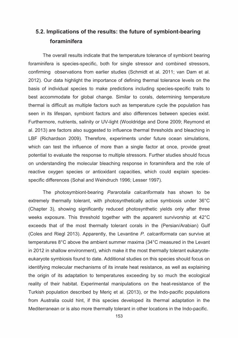
153
5.2. Implications of the results: the future of symbiont-bearing foraminifera
The overall results indicate that the temperature tolerance of symbiont bearing
foraminifera is species-specific, both for single stressor and combined stressors,
confirming observations from earlier studies (Schmidt et al. 2011; van Dam et al.
2012). Our data highlight the importance of defining thermal tolerance levels on the
basis of individual species to make predictions including species-specific traits to
best accommodate for global change. Similar to corals, determining temperature
thermal is difficult as multiple factors such as temperature cycle the population has
seen in its lifespan, symbiont factors and also differences between species exist.
Furthermore, nutrients, salinity or UV-light (Wooldridge and Done 2009; Reymond et
al. 2013) are factors also suggested to influence thermal thresholds and bleaching in
LBF (Richardson 2009). Therefore, experiments under future ocean simulations,
which can test the influence of more than a single factor at once, provide great
potential to evaluate the response to multiple stressors. Further studies should focus
on understanding the molecular bleaching response in foraminifera and the role of
reactive oxygen species or antioxidant capacities, which could explain species-
specific differences (Sohal and Weindruch 1996; Lesser 1997).
The photosymbiont-bearing Pararotalia calcariformata has shown to be
extremely thermally tolerant, with photosynthetically active symbiosis under 36°C
(Chapter 3), showing significantly reduced photosynthetic yields only after three
weeks exposure. This threshold together with the apparent survivorship at 42°C
exceeds that of the most thermally tolerant corals in the (Persian/Arabian) Gulf
(Coles and Riegl 2013). Apparently, the Levantine P. calcariformata can survive at
temperatures 8°C above the ambient summer maxima (34°C measured in the Levant
in 2012 in shallow environment), which make it the most thermally tolerant eukaryote-
eukaryote symbiosis found to date. Additional studies on this species should focus on
identifying molecular mechanisms of its innate heat resistance, as well as explaining
the origin of its adaptation to temperatures exceeding by so much the ecological
reality of their habitat. Experimental manipulations on the heat-resistance of the
Turkish population described by Meriç et al. (2013), or the Indo-pacific populations
from Australia could hint, if this species developed its thermal adaptation in the
Mediterranean or is also more thermally tolerant in other locations in the Indo-pacific.

154
Concerning changes in the microhabitat of LBF global change will lead to
changing biogeographic patterns because of invasive species, such as Pararotalia
(Chapter 2) and Amphistegina (Langer et al. 2012; Langer et al. 2013; Weinmann et
al. 2013). This together with changes in the macrohabitat influencing their algae
substrate it is likely to impact ecosystem function. Invasive species are going to
replace native fauna, because if they come from tropical regions they are likely to be
better adapted to elevated temperatures and can cope better with global change in
regions like the Mediterranean. One prominent example is the invasive macro algae
Caulerpa taxifolia, which has replaced large parts of the native sea grass Possidonia
oceanica, and turned a complex three dimensional algae substrate with a longer life-
span in a simpler substrate with a shorter life span and so indirectly influenced
relative abundance of principal foraminiferal taxa (Mateu-Vicens et al. 2010). Such
habitat changes co-occurring with global change could be potential additional
stressor to LBF, limiting the population size of already temperature sensitive species.
This is likely causing changes on the ecosystem dynamics of the Mediterranean sea,
which are going to are impact biodiversity structure (e.g. Hiddink et al. 2012; Edelist
et al. 2013) and disturb Mediterranean ecosystem services (Liquete et al. 2013;
Katsanevakis et al. 2014).
With regard to ocean acidification and temperature this thesis showed that
interactive effects of warming and pH change are going to impact the physiology of
the LBF more than the individual stressors alone. We attribute this effect to the
reduced energetic capacity of the host to cope with an additional stressor, if it already
has to counteract the negative effects of another stressor. Ocean acidification in
combination with temperature also negatively impacted growth and photosynthesis in
another study (Sinutok et al. 2014). Ocean acidification alone is predicted to have a
strong effect on calcium carbonate production, as test dissolution has been
documented in experimental manipulations on LBF (McIntyre-Wressnig et al. 2013).
Ocean acidification around volcanic pCO2 seeps also caused massive biodiversity
changes in foraminiferal communities, giving agglutinated foraminifera a selective
advantage compared to calcified taxa (Uthicke et al. 2013).
The central questions we have to ask is if LBF are likely to persist in the future
ocean, how we best can understand their adaptive capacity to global change stress
and their evolutionary potential to cope with these expected changes in their

155
environment (Munday et al. 2013). In species which are cultivable over many
generations in the laboratory for example phytoplankton, experimental evolution
approaches can test their likely response for adaptation in an altered environment
(Lohbeck et al. 2013). For benthic foraminifera, the culturing has so far been limited
to a 1-4 consecutive generations (Dettmering et al. 1998) and therefore is not an
ideal model for adaptive evolution experiments.
In foraminifera, we can try to learn from the fossil record. It has been observed
in the fossil record of planktonic foraminifera that shell morphology might have
responded to stress before extinction (Weinkauf et al. 2014). Phenotypic plasticity
and the ability of a genotype to produce different phenotypes when exposed to
different environmental conditions, could play a large role in helping foraminifera to
persist under global change. Phenotypic plasticity may also help species to adapt to
rapidly changing environmental conditions because it buys time for the slower
process of genetic adaptation (Chevin et al. 2010).
In conclusion, we confirmed species-specific differences in the thermal
tolerance of symbiont-bearing foraminifera and we identified a photo-symbiosis in
benthic foraminifera active under 36°C or higher which is more resistant than most
corals and will likely persist under global climate change.

156
5.3. References
Arieli RN, Almogi-Labin A, Abramovich S, Herut B (2011) The effect of thermal pollution on benthic foraminiferal assemblages in the Mediterranean shoreface adjacent to Hadera power plant (Israel). Marine Pollution Bulletin 62:1002-1012
Chevin L-M, Lande R, Mace GM (2010) Adaptation, Plasticity, and Extinction in a Changing Environment: Towards a Predictive Theory. PLoS Biology 8
Coles SL, Riegl BM (2013) Thermal tolerances of reef corals in the Gulf: A review of the potential for increasing coral survival and adaptation to climate change through assisted translocation. Marine Pollution Bulletin 72:323-332
Dettmering C, Rottger R, Hohenegger J, Schmaljohann R (1998) The trimorphic life cycle in foraminifera: Observations from cultures allow new evaluation. European Journal of Protistology 34:363-368
Edelist D, Rilov G, Golani D, Carlton JT, Spanier E (2013) Restructuring the Sea: profound shifts in the world's most invaded marine ecosystem. Diversity and Distributions 19:69-77
Fine M, Gildor H, Genin A (2013) A coral reef refuge in the Red Sea. Global Change Biology:n/a-n/a
Fitt WK, Brown BE, Warner ME, Dunne RP (2001) Coral bleaching: interpretation of thermal tolerance limits and thermal thresholds in tropical corals. Coral Reefs 20:51-65
Glynn PW, Mate JL, Baker AC, Calderon MO (2001) Coral bleaching and mortality in panama and Ecuador during the 1997-1998 El Nino-Southern oscillation event: Spatial/temporal patterns and comparisons with the 1982-1983 event. Bulletin of Marine Science 69:79-109
Hiddink JG, Lasram FB, Cantrill J, Davies AJ (2012) Keeping pace with climate change: what can we learn from the spread of Lessepsian migrants? Global Change Biology 18:2161-2172
Howells EJ, Beltran VH, Larsen NW, Bay LK, Willis BL, van Oppen MJH (2012) Coral thermal tolerance shaped by local adaptation of photosymbionts. Nature Clim Change 2:116-120
IOLR (2010) Statistical table for MedGLOSS stations and seasons, MedGLOSS sea level network., [Available at http://medgloss.ocean.org.il/statistic.asp.]
Katsanevakis S, Wallentinus I, Zenetos A, Leppakoski E, Cinar ME, Ozturk B, Grabowski M, Golani D, Cardoso AC (2014) Impacts of invasive alien marine species on ecosystem services and biodiversity: a pan-European review. Aquatic Invasions 9:391-423
157
Langer MR, Weinmann AE, Lötters S, Rödder D (2012) "Strangers" in Paradise: Modeling the Biogeographic Range Expansion of the Foraminifera Amphistegina in the Mediterranean Sea. Journal of Foraminiferal Research 42:234-244
Langer MR, Weinmann AE, Lötters S, Bernhard JM, Rödder D (2013) Climate-Driven Range Extension of Amphistegina (Protista, Foraminiferida): Models of Current and Predicted Future Ranges. PLoS ONE 8:e54443
Lee JJ, Anderson OR (1991) Symbiosis in Foraminifera. In: Lee JJ, Anderson OR (eds) Biology of Foraminifera. Academic Press Limited, San Diego, pp157-220
Lee JJ (2006) Algal symbiosis in larger foraminifera. Symbiosis 42:63-75
Lee JJ, Cervasco MH, Morales J, Billik M, Fine M, Levy O (2010) Symbiosis drove cellular evolution. Symbiosis 51:13-25
Lesser MP (1997) Oxidative stress causes coral bleaching during exposure to elevated temperatures. Coral Reefs 16:187-192
Liquete C, Piroddi C, Drakou EG, Gurney L, Katsanevakis S, Charef A, Egoh B (2013) Current Status and Future Prospects for the Assessment of Marine and Coastal Ecosystem Services: A Systematic Review. PLoS ONE 8
Lohbeck KT, Riebesell U, Collins S, Reusch TBH (2013) Functional genetic divergence in high co2 adapted Emiliania Huxleyi populations. Evolution 67:1892-1900
Mateu-Vicens G, Box A, Deudero S, Rodríguez B (2010) Comparative analysis of epiphytic foraminifera in sediments colonized by seagrass Posidonia oceanica and invasive macroalgae Caulerpa spp. The Journal of Foraminiferal Research 40:134-147
McIntyre-Wressnig A, Bernhard JM, McCorkle DC, Hallock P (2013) Non-lethal effects of ocean acidification on the symbiont-bearing benthic foraminifer Amphistegina gibbosa. Marine Ecology Progress Series 472:45-60
Meriç E, Yokes MB, Avsar KN, Kirki-Elmas E, Dinçer F, Karhan SU, Kalkan E, Demir V (2013) First report of Pararotalia calcariformata from the Hatay coastline (Turkey—north-eastern Mediterranean). Marine Biodiversity Records 6:e31
Munday PL, Warner RR, Monro K, Pandolfi JM, Marshall DJ (2013) Predicting evolutionary responses to climate change in the sea. Ecology Letters 16:1488-1500
Nobes K, Uthicke S, Henderson R (2008) Is light the limiting factor for the distribution of benthic symbiont bearing foraminifera on the Great Barrier Reef? Journal of Experimental Marine Biology and Ecology 363:48-57
Reymond CE, Lloyd A, Kline DI, Dove SG, Pandolfi JM (2013) Decline in growth of foraminifer Marginopora rossi under eutrophication and ocean acidification scenarios. Global Change Biology 19:291-302

158
Rowan R (2004) Coral bleaching - Thermal adaptation in reef coral symbionts. Nature 430:742-742
Sampayo EM, Ridgway T, Bongaerts P, Hoegh-Guldberg O (2008) Bleaching susceptibility and mortality of corals are determined by fine-scale differences in symbiont type. Proceedings of the National Academy of Sciences of the United States of America 105:10444-10449
Sarno D, Zingone A, Saggiomo V, Carrada G (1993) Phytoplankton biomass and species composition in a Mediterranean coastal lagoon. Hydrobiologia 271:27-40
Schmidt C, Heinz P, Kucera M, Uthicke S (2011) Temperature-induced stress leads to bleaching in larger benthic foraminifera hosting endosymbiotic diatoms. Limnology and Oceanography 56:1587-1602
Schmidt C, Kucera M, Uthicke S (2014) Combined effects of warming and ocean acidification on coral reef Foraminifera Marginopora vertebralis and Heterostegina depressa. Coral Reefs 33:805-818
Shaltout M, Omstedt A (2014) Recent sea surface temperature trends and future scenarios for the Mediterranean Sea. Oceanologia 56:411-443
Sinutok S, Hill R, Doblin MA, Wuhrer R, Ralph PJ (2011) Warmer more acidic conditions cause decreased productivity and calcification in subtropical coral reef sediment-dwelling calcifiers. Limnology and Oceanography 56:1200-1212
Sinutok S, Hill R, Kühl M, Doblin M, Ralph P (2014) Ocean acidification and warming alter photosynthesis and calcification of the symbiont-bearing foraminifera Marginopora vertebralis. Marine Biology:1-12
Sohal RS, Weindruch R (1996) Oxidative stress, caloric restriction, and aging. Science 273:59-63
Strychar KB, Coates M, Sammarco PW (2004) Loss of Symbiodinium from bleached Australian scleractinian corals (Acropora hyacinthus, Favites complanata and Porites solida). Mar and Freshwater Res 55:135-144
Strychar KB, Sammarco PW (2009) Exaptation in corals to high seawater temperatures: Low concentrations of apoptotic and necrotic cells in host coral tissue under bleaching conditions. Journal of Experimental Marine Biology and Ecology 369:31-42
Talge HK, Hallock P (2003) Ultrastructural responses in field-bleached and experimentally stressed Amphistegina gibbosa (Class Foraminifera). Journal of Eukaryotic Microbiology 50:324-333
Uthicke S, Vogel N, Doyle J, Schmidt C, Humphrey C (2012) Interactive effects of climate change and eutrophication on the dinoflagellate-bearing benthic foraminifer Marginopora vertebralis. Coral Reefs 31:401-414
Uthicke S, Momigliano P, Fabricius KE (2013) High risk of extinction of benthic foraminifera in this century due to ocean acidification. Scientific Reports 3

159
van Dam JW, Negri AP, Mueller JF, Altenburger R, Uthicke S (2012) Additive pressures of elevated sea surface temperatures and herbicides on symbiont-bearing foraminifera. PLoS ONE 7:e33900
Weinkauf MFG, Moller T, Koch MC, Kucera M (2014) Disruptive selection and bet-hedging in planktonic Foraminifera: Shell morphology as predictor of extinctions. Frontiers in Ecology and Evolution 2
Weinmann AE, Rodder D, Lotters S, Langer MR (2013) Traveling through time: The past, present and future biogeographic range of the invasive foraminifera Amphistegina spp. in the Mediterranean Sea. Marine Micropaleontology 105:30-39
Wellington GM, Glynn PW, Strong AE, Navarrete SA, Wieters E, Hubbard D (2001) Crisis on coral reefs linked to climate change. Eos, Transactions American Geophysical Union 82:1-5
Wooldridge SA (2009) Water quality and coral bleaching thresholds: Formalising the linkage for the inshore reefs of the Great Barrier Reef, Australia. Marine Pollution Bulletin 58:745-751
Wooldridge SA, Done TJ (2009) Improved water quality can ameliorate effects of climate change on corals. Ecological Applications 19:1492-1499
Ziegler M (2009) Photosynthetic plasticity of endosymbionts in larger benthic coral reef Foraminifera. Diploma Thesis, University of Veterinary Medicine Hannover,, p43
Ziegler M, Uthicke S (2011) Photosynthetic plasticity of endosymbionts in larger benthic coral reef Foraminifera. Journal of Experimental Marine Biology and Ecology 407:70-80

160

161
6. Acknowledgements I wish to thank my supervisor Professor Dr. Michal Kucera for always taking time to
discuss my research questions and providing great support throughout my PhD, and
for his curiosity about “new discoveries”. I wish to thank Professor Kai Bischoff for his
offer to be examiner of my thesis and Professor Christian Wild for his suggestions
during committee meetings.
The first chapter of this thesis would have not been possible without an invitation by
Dr. Sven Uthicke to work at AIMS for a three month research stay. I like to thank him
for enabling me the work on ocean acidification and warming on foraminifera. At
AIMS I Iike to thank Dr. Katharina Fabricius, Dr. Andrew Negri, Dr. Nicole Webber,
Florita Flores, and Sam Noonan for the good collaborative work and Michal Liddy for
her cheerful company during the stay.
For the second and third chapter of this thesis I like to thank Dr. Sigal Abramovich,
Dr. Ahuva Almogi-Labin and Dr. Barak Herut for initiating the German Israeli-
collaboration and for providing field-assistance, comments and advice during the
preparation of our manuscripts. I like to thank Gily Mercado, Emri Brickner, Danna
Titleboim, Nurit Neumark and Shai Oron for their help in Israel.
In the laboratory, I like to thank Dr. Ralph Auras, Birgit Lübben and Dr. Achim Meyer
for their help and suggestions to construct the aquaria setup in Bremen, as well as
Ramona Brunner, Janett Brandt and Mirjam Cahnbley for providing helpful
assistance with the foraminiferal cultures over three years. I like to thank Janett
Brandt for her patience to work on juvenile foraminifera and for her hard-working
assistance during the 2013 fieldtrip to Israel.
I like to thank Dr. Raphael Morard for the good collaboration on molecular work and
Dr. Anna Weinmann for introducing me to species distribution modelling and enabling
an excellent collaboration over the Atlantic.
The office at the MARUM was a great place to work with lots of cheerful people
around, thank you to all of you. I like to thank in particular Agnes Weiner and Manuel
Weinkauf for being good office-mates and Rebecca Jackson and Elizabeth Brown for
cheerful lunches and proof-reading. Furthermore, I like to thank Andrea Rebotim, Ute
Wolpmann, Lorand Silye for their friendliness around the office.

162
I like to thank the DAAD for enabling me to go to Chile for conference participation to
FORAMS 2014 presenting this work and participating in a summer school on
biogeochemical marine cycles. In Concepcion, I like to thank my friend Dharma
Reyes for her help and the unforgettable time. I like to thank my friends Verena
Kottler and Kerstin Schiele for being great PhD buddies and for nice cocktail chats.
My graduate school GLOMAR (Global Change in the Marine Realm) has provided a
great platform on the professional and the social level to engage with other students
and researchers during my PhD project. In particular I like to thank Karin Boos,
Martina Loebl and Christina Klose. I like to thank for the financial assistance to travel
to Australia and Japan for conference and workshop participation, which initiated my
post-doc plans.
I like to thank my dear boyfriend Raphael for his loving support and encouragement.
Last, but not least, I like to thank my family for always believing in me and loving me
unconditionally.
Vielen Dank, Thank you very much, , , Muchas Gracias!

163
So eine Arbeit wird eigentlich nie fertig,
man muß sie für fertig erklären,
wenn man nach Zeit und Umständen das Mögliche getan hat.
Johann Wolfgang von Goethe (1749 - 1832), deutscher Dichter der Klassik, Naturwissenschaftler und Staatsmann

164

165
7. Schriftliche Erklärung
Gem. § 6(5) Nr.1-3 PromO
Ich erkläre, dass ich
1. die Arbeit ohne unerlaubte, fremde Hilfe angefertigt habe,
2. keine anderen, als die von mir angegebenen Quellen und Hilfsmittel benutzt habe, und
3. die den benutzen Werken wörtlich und inhaltlich entnommenen Stellen als solche kenntlich
gemacht habe.
Bremen, den 30.01.2015
XChristiane Schmidt