Genome‐wide association analysis of dementia and its ...
15
Featured Article Genome-wide association analysis of dementia and its clinical endophenotypes reveal novel loci associated with Alzheimer’s disease and three causality networks: The GR@ACE project Sonia Moreno-Grau a,b , Itziar de Rojas a , Isabel Hern andez a,b , In es Quintela c , Laura Montrreal a , Montserrat Alegret a,b , Bego~ na Hern andez-Olasagarre a , Laura Madrid d , Antonio Gonz alez-Perez d , Olalla Maro~ nas c , Mait ee Rosende-Roca a , Ana Maule on a , Liliana Vargas a , Asunci on Lafuente a , Carla Abdelnour a,b , Octavio Rodr ıguez-G omez a,b , Silvia Gil a , Miguel Angel Santos-Santos a , Ana Espinosa a,b , Gemma Ortega a,b , Angela Sanabria a,b , Alba P erez-Cord on a , Pilar Ca~ nabate a,b , Mariola Moreno a , Silvia Preckler a , Susana Ruiz a,b , Nuria Aguilera a , Juan Antonio Pineda e , Juan Mac ıas e , Emilio Alarc on-Mart ın a,f , Oscar Sotolongo-Grau a , GR@ACE consortium**, DEGESCO consortium**, Alzheimer’s Disease Neuroimaging Initiative, Marta Marqui e a , Gemma Mont e-Rubio a , Sergi Valero a,b , Alba Benaque a , Jordi Clarim on b,g , Maria Jesus Bullido b,h,i , Guillermo Garc ıa-Ribas j , Pau P astor k , Pascual S anchez-Juan b,l , Victoria Alvarez m,n , Gerard Pi~ nol-Ripoll b,o , Jose Maria Garc ıa-Alberca p , Jos e Luis Royo f , Emilio Franco q , Pablo Mir b,r , Miguel Calero b,s,t , Miguel Medina b,s , Alberto R abano b,s,u , Jes us Avila b,v , Carmen Ant unez w , Luis Miguel Real e,f , Adelina Orellana a , Angel Carracedo c,x , Mar ıa Eugenia S aez d , Llu ıs T arraga a,b , Merc e Boada a,b , Agust ın Ruiz a,b, * a Research Center and Memory clinic Fundaci o ACE, Institut Catal a de Neuroci encies Aplicades, Universitat Internacional de Catalunya, Barcelona, Spain b CIBERNED, Network Center for Biomedical Research in Neurodegenerative Diseases, National Institute of Health Carlos III, Madrid, Spain c Grupo de Medicina Xen omica, Centro Nacional de Genotipado (CEGEN-PRB3-ISCIII). Universidade de Santiago de Compostela, Santiago de Compostela, Spain d CAEBI, Centro Andaluz de Estudios Bioinform aticos, Sevilla, Spain e Unidad Cl ınica de Enfermedades Infecciosas y Microbiolog ıa. Hospital Universitario de Valme, Sevilla, Spain f Department of Surgery, Biochemistry and Molecular Biology, School of Medicine, University of M alaga, M alaga, Spain g Memory Unit, Neurology Department and Sant Pau Biomedical Research Institute, Hospital de la Santa Creu i Sant Pau, Universitat Aut onoma de Barcelona, Barcelona, Spain h Centro de Biologia Molecular Severo Ochoa (C.S.I.C.-U.A.M.), Universidad Autonoma de Madrid, Madrid, Spain i Instituto de Investigacion Sanitaria “Hospital la Paz” (IdIPaz), Madrid, Spain j Hospital Universitario Ram on y Cajal, Madrid, Spain k Fundaci o per la Recerca Biom edica i Social M utua Terrassa, and Memory Disorders Unit, Department of Neurology, Hospital Universitari Mutua de Terrassa, University of Barcelona School of Medicine, Terrassa, Spain l Neurology Service ’Marqu es de Valdecilla’ University Hospital (University of Cantabria and IDIVAL), Santander, Spain m Laboratorio de Gen etica Hospital Universitario Central de Asturias, Oviedo, Spain n Instituto de Investigaci on Biosanitaria del Principado de Asturias (ISPA), Oviedo, Spain o Unitat Trastorns Cognitius, Hospital Universitari Santa Maria de Lleida, Institut de Recerca Biom edica de Lleida (IRBLLeida), Lleida, Spain The authors have declared that no conflict of interest exists. **The full list of members of the GR@ACE consortium and DEGESCO consortium were reported in the Acknowledgment section. Alzheimer’s Disease Neuroimaging Initiative: Data used in preparing this article were obtained from the Alzheimer’s Disease Neuroimaging Initiative (ADNI) database (adni.loni.usc.edu). As such, the investigators within the ADNI contributed to the design and implementation of ADNI and/or provided data but did not participate in the analysis or writing of this report. A complete listing of ADNI investigators can be found at http://adni.loni.usc.edu/wp-content/uploads/how_to_apply/ADNI_Ack- nowledgement_List.pdf *Corresponding author. Tel: 13493.444.73.18; Fax: 13493.410.17.01. E-mail address: [email protected] https://doi.org/10.1016/j.jalz.2019.06.4950 1552-5260/ Ó 2019 The Authors. Published by Elsevier Inc. on behalf of the Alzheimer’s Association. This is an open access article under the CC BY-NC-ND license (http://creativecommons.org/licenses/by-nc-nd/4.0/). Alzheimer’s & Dementia 15 (2019) 1333-1347
Transcript of Genome‐wide association analysis of dementia and its ...

Alzheimer’s & Dementia 15 (2019) 1333-1347
Featured Article
Genome-wide association analysis of dementia and its clinicalendophenotypes reveal novel loci associated with Alzheimer’s disease
and three causality networks: The GR@ACE project
Sonia Moreno-Graua,b, Itziar de Rojasa, Isabel Hern�andeza,b, In�es Quintelac, Laura Montrreala,Montserrat Alegreta,b, Bego~na Hern�andez-Olasagarrea, Laura Madridd, Antonio Gonz�alez-Perezd,Olalla Maro~nasc, Mait�ee Rosende-Rocaa, Ana Maule�ona, Liliana Vargasa, Asunci�on Lafuentea,Carla Abdelnoura,b, Octavio Rodr�ıguez-G�omeza,b, Silvia Gila, Miguel �Angel Santos-Santosa,
Ana Espinosaa,b, Gemma Ortegaa,b, �Angela Sanabriaa,b, Alba P�erez-Cord�ona, Pilar Ca~nabatea,b,Mariola Morenoa, Silvia Precklera, Susana Ruiza,b, Nuria Aguileraa, Juan Antonio Pinedae,Juan Mac�ıase, Emilio Alarc�on-Mart�ına,f, Oscar Sotolongo-Graua, GR@ACE consortium**,DEGESCO consortium**, Alzheimer’s Disease Neuroimaging Initiative, Marta Marqui�ea,
Gemma Mont�e-Rubioa, Sergi Valeroa,b, Alba Benaquea, Jordi Clarim�onb,g,Maria Jesus Bullidob,h,i, Guillermo Garc�ıa-Ribasj, Pau P�astork, Pascual S�anchez-Juanb,l,Victoria �Alvarezm,n, Gerard Pi~nol-Ripollb,o, Jose Maria Garc�ıa-Albercap, Jos�e Luis Royof,Emilio Francoq, Pablo Mirb,r, Miguel Calerob,s,t, Miguel Medinab,s, Alberto R�abanob,s,u,
Jes�us �Avilab,v, Carmen Ant�unezw, Luis Miguel Reale,f, Adelina Orellanaa, �Angel Carracedoc,x,Mar�ıa Eugenia S�aezd, Llu�ıs T�arragaa,b, Merc�e Boadaa,b, Agust�ın Ruiza,b,*
aResearch Center and Memory clinic Fundaci�o ACE, Institut Catal�a de Neuroci�encies Aplicades, Universitat Internacional de Catalunya, Barcelona, SpainbCIBERNED, Network Center for Biomedical Research in Neurodegenerative Diseases, National Institute of Health Carlos III, Madrid, Spain
cGrupo de Medicina Xen�omica, Centro Nacional de Genotipado (CEGEN-PRB3-ISCIII). Universidade de Santiago de Compostela,
Santiago de Compostela, SpaindCAEBI, Centro Andaluz de Estudios Bioinform�aticos, Sevilla, Spain
eUnidad Cl�ınica de Enfermedades Infecciosas y Microbiolog�ıa. Hospital Universitario de Valme, Sevilla, SpainfDepartment of Surgery, Biochemistry and Molecular Biology, School of Medicine, University of M�alaga, M�alaga, Spain
gMemory Unit, Neurology Department and Sant Pau Biomedical Research Institute, Hospital de la Santa Creu i Sant Pau, Universitat Aut�onoma de Barcelona,
Barcelona, SpainhCentro de Biologia Molecular Severo Ochoa (C.S.I.C.-U.A.M.), Universidad Autonoma de Madrid, Madrid, Spain
iInstituto de Investigacion Sanitaria “Hospital la Paz” (IdIPaz), Madrid, SpainjHospital Universitario Ram�on y Cajal, Madrid, Spain
kFundaci�o per la Recerca Biom�edica i Social M�utua Terrassa, andMemory Disorders Unit, Department of Neurology, Hospital Universitari Mutua de Terrassa,
University of Barcelona School of Medicine, Terrassa, SpainlNeurology Service ’Marqu�es de Valdecilla’ University Hospital (University of Cantabria and IDIVAL), Santander, Spain
mLaboratorio de Gen�etica Hospital Universitario Central de Asturias, Oviedo, SpainnInstituto de Investigaci�on Biosanitaria del Principado de Asturias (ISPA), Oviedo, Spain
oUnitat Trastorns Cognitius, Hospital Universitari Santa Maria de Lleida, Institut de Recerca Biom�edica de Lleida (IRBLLeida), Lleida, Spain
The authors have declared that no conflict of interest exists.
**The full list of members of the GR@ACE consortium and DEGESCO
consortium were reported in the Acknowledgment section.
Alzheimer’s Disease Neuroimaging Initiative: Data used in preparing
this article were obtained from the Alzheimer’s Disease Neuroimaging
Initiative (ADNI) database (adni.loni.usc.edu). As such, the investigators
within the ADNI contributed to the design and implementation of ADNI
and/or provided data but did not participate in the analysis or writing of
this report. A complete listing of ADNI investigators can be found at
http://adni.loni.usc.edu/wp-content/uploads/how_to_apply/ADNI_Ack-
nowledgement_List.pdf
*Corresponding author. Tel: 13493.444.73.18; Fax: 13493.410.17.01.
E-mail address: [email protected]
https://doi.org/10.1016/j.jalz.2019.06.4950
1552-5260/� 2019 The Authors. Published by Elsevier Inc. on behalf of the Alzheimer’s Association. This is an open access article under the CC BY-NC-ND
license (http://creativecommons.org/licenses/by-nc-nd/4.0/).

S. Moreno-Grau et al. / Alzheimer’s & Dementia 15 (2019) 1333-13471334
pAlzheimer Research Center & Memory Clinic, Andalusian Institute for Neuroscience, M�alaga, SpainqUnidad de Demencias, Servicio de Neurolog�ıa y Neurofisiolog�ıa. Instituto de Biomedicina de Sevilla (IBiS), Hospital Universitario Virgen del
Roc�ıo/CSIC/Universidad de Sevilla, Seville, SpainrUnidad de Trastornos del Movimiento, Servicio de Neurolog�ıa y Neurofisiolog�ıa. Instituto de Biomedicina de Sevilla (IBiS), Hospital Universitario Virgen del
Roc�ıo/CSIC/Universidad de Sevilla, Seville, SpainsCIEN Foundation, Queen Sofia Foundation Alzheimer Center, Madrid, Spain
tInstituto de Salud Carlos III (ISCIII), Madrid, SpainuBT-CIEN, Madrid, Spain
vDepartment of Molecular Neuropathology, Centro de Biolog�ıa Molecular “Severo Ochoa” (CBMSO), Consejo Superior de Investigaciones Cient�ıficas
(CSIC)/Universidad Aut�onoma de Madrid (UAM), Madrid, SpainwUnidad de Demencias, Hospital Cl�ınico Universitario Virgen de la Arrixaca, Murcia, Spain
xFundaci�on P�ublica Galega de Medicina Xen�omica- CIBERER-IDIS, Santiago de Compostela, Spain
Abstract Introduction: Large variability among Alzheimer’s disease (AD) cases might impact genetic dis-
coveries and complicate dissection of underlying biological pathways.Methods: Genome Research at Fundacio ACE (GR@ACE) is a genome-wide study of dementia andits clinical endophenotypes, defined based on AD’s clinical certainty and vascular burden. We as-sessed the impact of known AD loci across endophenotypes to generate loci categories. We incorpo-rated gene coexpression data and conducted pathway analysis per category. Finally, to evaluate theeffect of heterogeneity in genetic studies, GR@ACE series were meta-analyzed with additionalgenome-wide association study data sets.Results: We classified known AD loci into three categories, which might reflect the disease clinicalheterogeneity. Vascular processes were only detected as a causal mechanism in probable AD. Themeta-analysis strategy revealed the ANKRD31-rs4704171 andNDUFAF6-rs10098778 and confirmedSCIMP-rs7225151 and CD33-rs3865444.Discussion: The regulation of vasculature is a prominent causal component of probable AD.GR@ACE meta-analysis revealed novel AD genetic signals, strongly driven by the presence of clin-ical heterogeneity in the AD series.� 2019 The Authors. Published by Elsevier Inc. on behalf of the Alzheimer’s Association. This is anopen access article under the CC BY-NC-ND license (http://creativecommons.org/licenses/by-nc-nd/4.0/).Keywords: Alzheimer’s disease; Vascular pathology; Cerebral amyloid angiopathy; GWAS; Biological pathway
1. Background
Dementia is an age-related clinical syndrome that devas-tates cognitive abilities and interferes in elderly people’sdaily activities. Although its incidence is decreasing due toimprovements to public health systems and control of cardio-vascular risk factors [1], its prevalence is steadily increasingdue to rising life expectancy of human populations [2].
Dementia is linked to many underlying pathologies, withAlzheimer’s disease (AD) being the most common condi-tion. Clinical AD is a heterogeneous syndrome. Brain au-topsies have shown that roughly 80% of clinical ADpatients present with brain vascular pathology [3] in additionto the common neuropathological AD hallmarks: amyloid-osis, neurofibrillary tangles, and cerebral amyloid angiop-athy (CAA) [4]. In fact, brain vascular pathology has beenshown to be an important risk factor for AD that acceleratescognitive decline [5] and lowers the threshold for clinicaldiagnosis of AD [6]. In that context, it has been suggestedthat dementia is represented by a gradient of neurodegener-ative and vascular components [7], from pure AD forms,with a strong neurodegenerative component, to pure vasculardementia (VaD) cases and in-between mixed pathologies,
representing the coexistence of both neurodegenerativeand vascular components [7]. Despite that, whether thereare differential biological routes operating under differentlevels of vascular burden in clinical AD patients remainsmostly unknown.
In the search for the etiology of AD, genetic factors play apivotal role. Two forms of the disease can be differentiated ac-cording to individual genetic background. The Mendelianform is an uncommon disorder that mainly affects familieswith early-onset AD (,65 years), whereas the polygenicform is a complex disordermainly appearing in sporadic caseswith late-onset AD (LOAD) (.65 years). Highly penetrantmutations detected in families with early-onset AD havebeen pinpointed to three genes: APP [8], PSEN1 [9], andPSEN2 [10], leading to the establishment of the amyloidhypothesis as a potential causal mechanism for the disease[11].
LOAD heritability falls in the range of 13%‒80% [12,13].Although APOE ε4 was the first to be discovered and stillremains the strongest genetic risk factor for AD [14], almost40 additional genetic variants have been identified [15–17]using genome-wide association studies (GWAS) and large

S. Moreno-Grau et al. / Alzheimer’s & Dementia 15 (2019) 1333-1347 1335
sequencing projects. Among the biological pathways underly-ing genetic hits, the roles of the immune system, cholesterol,amyloid, and tau metabolism have been highlighted [18].Despite these, current genetic findings account for 31% ofLOAD heritability [19], and the biological picture of AD isstill poorly understood. Several reasons can explain this.Among them, the presence of clinical and neuropathologicalheterogeneity between AD cases in genetic studies, asrecently demonstrated [12], might compromise the power todetect genuine genetic associations and decrease the estimatesof risk attributed to genetic variation.
Theneuropathological variability in clinicalADcases com-prises a wide spectrum, from those with concomitant vascularbrain disease to thosewith a pureADphenotype, as previouslyproposed by Viswanathan et al. [7]. This large heterogeneitymight hamper the identification of functional categories ofgenes underlying differential biological routes to dementiaand might impact AD genetic studies. To gain insight intothe causality networks behind AD clinical subgroups and toexplore their impact in large GWAS, we conducted theGenome Research at Fundacio ACE (GR@ACE) study. Thisis a GWAS of dementia and its clinical endophenotypesdefined based on AD’s clinical certainty and the burden ofvascular comorbidity. The GR@ACE is a unique genomicresource comprising the largest number of dementia casesdiagnosed in a single memory clinic to date. First, we deter-mined whether we could identify categories of knownLOAD genes linked to clinical subgroups of AD cases. Next,we explored whether these categories suggested different bio-logical routes. Finally, to assess the impact of these clinicalsubgroups of AD cases in GWAS findings, we meta-analyzed the GR@ACE data with independent GWAS series.
2. Methods
2.1. Subjects
2.1.1. GR@ACE cohort and phenotype definitionsThe GR@ACE study comprises 4120 AD cases and 3289
control individuals (Table 1). Cases were recruited fromFundaci�o ACE, Institut Catal�a de Neuroci�encies Aplicades(Catalonia, Spain). Diagnoses were established by amultidisciplinary working group, including neurologists, neu-ropsychologists, and social workers, according to the Diag-nostic and Statistical Manual of Mental Disorders–IV criteriafor dementia and to the National Institute on Aging and Alz-heimer’sAssociation’s (NIA-AA) 2011 guidelines for definingAD[20] (seeSupplementaryMaterial). In the present study,weconsidered AD cases as dementia individuals diagnosed withprobable or possibleADat anymomentof their clinical course.
We took advantage of this wide clinical definition torefine AD cases. Considering the dementia spectrum pro-posed by Viswanathan et al. [7], we classified Gr@ACEAD patients according to the degree of clinical certaintyfor AD phenotype and the presence of vascular comorbidity,from “pure” clinical AD cases to mixed and vascular en-
riched cases. This approach was feasible due to Fundaci�oACE’s endorsement of a primary and a secondary etiologicdiagnosis as well as routine follow-up evaluations [21](Supplementary Methods). Using the entire clinical chartof each subject, we differentiated five clinical subgroupsof patients representing the GR@ACE endophenotypes:(1) the AD111 endophenotype comprises individuals witha last clinical diagnosis of probable AD in both primaryand secondary diagnoses (n5 1854); (2) the AD11 includesindividuals diagnosed with probable AD in the primary diag-nosis and probable or possible AD in the secondary diag-nosis (n 5 2611); (3) the AD1 encompasses patientsdiagnosed with probable or possible AD either in the pri-mary diagnosis or in the secondary diagnosis (n 5 3797);(4) the VaD1 includes patients diagnosed with VaD orpossible AD in the primary diagnosis (n 5 1168); and (5)the VaD11 comprises patients diagnosed with probable orpossible vascular dementia in the primary diagnosis(n 5 373) (Table 1) (Supplementary Fig. 1). Patients withVaD were defined according to the NeuroepidemiologyBranch of the National Institute of Neurological Disordersand Stroke and the Association Internationale pour la Re-cherche et l’Enseignement en Neurosciences criteria [22].
Control individuals were recruited from three centers:Fundaci�o ACE (Barcelona, Spain), Valme University Hospi-tal (Seville, Spain), and the Spanish National DNA BankCarlos III (University of Salamanca, Spain) (www.bancoadn.org). Written informed consent was obtainedfrom all participants. The Ethics and Scientific Committeeshave approved this research protocol (Acta 25/2016. EthicsCommittee. H. Clinic i Provincial, Barcelona, Spain).
2.1.2. Replication sampleWith the objective to successfully replicate novel GWAS
findings, we used an independent Spanish sample of 1943AD cases (mean age 5 79.2; standard deviation[SD] 5 7.6; 66.3% women) and 3016 controls (meanage 5 52.8; SD 5 15.2; 46% women) presenting similarcharacteristics to the GR@ACE cohort and with availablegenetic data. All AD cases were examined at a single site,Fundaci�o ACE, Institut Catal�a de Neuroci�encies Aplicades(Catalonia, Spain), and were assessed by applying thesame criteria previously explained. The sample compositionof dementia cases comprised 30.1% of AD111 (n 5 584),53.9% of AD11 (n 5 1048), 91.9% of AD1 (n 5 1783),23.0% of VaD1 (n 5 447), and 4.6% of VaD11 (n 5 89)cases. Control individuals were selected from the Spanishpopulation available at three centers, previously described.
2.2. GWAS genotyping, quality control, imputation, andstatistical analysis
Participants were genotyped using the Axiom 815KSpanish Biobank Array (Thermo Fisher). Genotyping wasperformed in the Spanish National Center for Genotyping
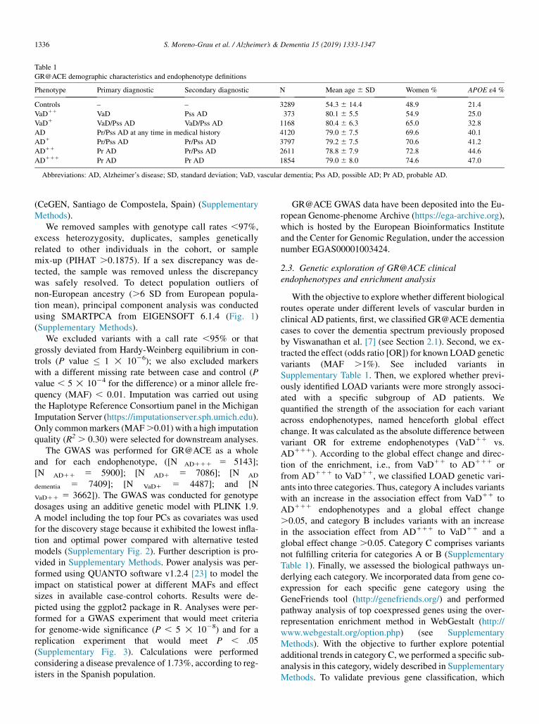
Table 1
GR@ACE demographic characteristics and endophenotype definitions
Phenotype Primary diagnostic Secondary diagnostic N Mean age 6 SD Women % APOE ε4 %
Controls – – 3289 54.3 6 14.4 48.9 21.4
VaD11 VaD Pss AD 373 80.1 6 5.5 54.9 25.0
VaD1 VaD/Pss AD VaD/Pss AD 1168 80.4 6 6.3 65.0 32.8
AD Pr/Pss AD at any time in medical history 4120 79.0 6 7.5 69.6 40.1
AD1 Pr/Pss AD Pr/Pss AD 3797 79.2 6 7.5 70.6 41.2
AD11 Pr AD Pr/Pss AD 2611 78.8 6 7.9 72.8 44.6
AD111 Pr AD Pr AD 1854 79.0 6 8.0 74.6 47.0
Abbreviations: AD, Alzheimer’s disease; SD, standard deviation; VaD, vascular dementia; Pss AD, possible AD; Pr AD, probable AD.
S. Moreno-Grau et al. / Alzheimer’s & Dementia 15 (2019) 1333-13471336
(CeGEN, Santiago de Compostela, Spain) (SupplementaryMethods).
We removed samples with genotype call rates ,97%,excess heterozygosity, duplicates, samples geneticallyrelated to other individuals in the cohort, or samplemix-up (PIHAT .0.1875). If a sex discrepancy was de-tected, the sample was removed unless the discrepancywas safely resolved. To detect population outliers ofnon-European ancestry (.6 SD from European popula-tion mean), principal component analysis was conductedusing SMARTPCA from EIGENSOFT 6.1.4 (Fig. 1)(Supplementary Methods).
We excluded variants with a call rate ,95% or thatgrossly deviated from Hardy-Weinberg equilibrium in con-trols (P value � 1 ! 1026); we also excluded markerswith a different missing rate between case and control (Pvalue , 5 ! 1024 for the difference) or a minor allele fre-quency (MAF) , 0.01. Imputation was carried out usingthe Haplotype Reference Consortium panel in the MichiganImputation Server (https://imputationserver.sph.umich.edu).Only commonmarkers (MAF.0.01) with a high imputationquality (R2 . 0.30) were selected for downstream analyses.
The GWAS was performed for GR@ACE as a wholeand for each endophenotype, ([N AD111 5 5143];[N AD11 5 5900]; [N AD1 5 7086]; [N AD
dementia 5 7409]; [N VaD1 5 4487]; and [N
VaD11 5 3662]). The GWAS was conducted for genotypedosages using an additive genetic model with PLINK 1.9.A model including the top four PCs as covariates was usedfor the discovery stage because it exhibited the lowest infla-tion and optimal power compared with alternative testedmodels (Supplementary Fig. 2). Further description is pro-vided in Supplementary Methods. Power analysis was per-formed using QUANTO software v1.2.4 [23] to model theimpact on statistical power at different MAFs and effectsizes in available case-control cohorts. Results were de-picted using the ggplot2 package in R. Analyses were per-formed for a GWAS experiment that would meet criteriafor genome-wide significance (P , 5 ! 1028) and for areplication experiment that would meet P , .05(Supplementary Fig. 3). Calculations were performedconsidering a disease prevalence of 1.73%, according to reg-isters in the Spanish population.
GR@ACE GWAS data have been deposited into the Eu-ropean Genome-phenome Archive (https://ega-archive.org),which is hosted by the European Bioinformatics Instituteand the Center for Genomic Regulation, under the accessionnumber EGAS00001003424.
2.3. Genetic exploration of GR@ACE clinicalendophenotypes and enrichment analysis
With the objective to explore whether different biologicalroutes operate under different levels of vascular burden inclinical AD patients, first, we classified GR@ACE dementiacases to cover the dementia spectrum previously proposedby Viswanathan et al. [7] (see Section 2.1). Second, we ex-tracted the effect (odds ratio [OR]) for known LOAD geneticvariants (MAF .1%). See included variants inSupplementary Table 1. Then, we explored whether previ-ously identified LOAD variants were more strongly associ-ated with a specific subgroup of AD patients. Wequantified the strength of the association for each variantacross endophenotypes, named henceforth global effectchange. It was calculated as the absolute difference betweenvariant OR for extreme endophenotypes (VaD11 vs.AD111). According to the global effect change and direc-tion of the enrichment, i.e., from VaD11 to AD111 orfrom AD111 to VaD11, we classified LOAD genetic vari-ants into three categories. Thus, category A includes variantswith an increase in the association effect from VaD11 toAD111 endophenotypes and a global effect change.0.05, and category B includes variants with an increasein the association effect from AD111 to VaD11 and aglobal effect change .0.05. Category C comprises variantsnot fulfilling criteria for categories A or B (SupplementaryTable 1). Finally, we assessed the biological pathways un-derlying each category. We incorporated data from gene co-expression for each specific gene category using theGeneFriends tool (http://genefriends.org/) and performedpathway analysis of top coexpressed genes using the over-representation enrichment method in WebGestalt (http://www.webgestalt.org/option.php) (see SupplementaryMethods). With the objective to further explore potentialadditional trends in category C, we performed a specific sub-analysis in this category, widely described in SupplementaryMethods. To validate previous gene classification, which

Fig. 1. Results of genome-wide association analysis for the GR@ACE data set (n5 7409). (A) Principal component analysis; (B) principal component analysis
centered in European population; (C) QQplot for the discovery model, adjusted for first four PCs; (D) Manhattan plot for genome-wide results. Abbreviations:
AFR, African; AMR, Admixed American; EAS, East Asian; EUR, European; SAS, South Asian.
S. Moreno-Grau et al. / Alzheimer’s & Dementia 15 (2019) 1333-1347 1337
strongly determines the pathway analysis results, we con-ducted a stringent subanalysis (see SupplementaryMethods and Supplementary Fig. 4).
2.4. Meta-analysis: Data sets and association analysis
To explore the impact of the different clinical endopheno-types in GWAS findings, we combined the whole GR@ACEGWAS data set, which represents a dementia set of samplesand its endophenotypes with (1) genotype-level data fromnine additional GWAS series (N 5 13,826), availablethrough dbGaP (https://www.ncbi.nlm.nih.gov/gap) thatwe processed by applying identical quality control andimputation procedures to those described for the GR@ACEcohort (Supplementary Table 2) and (2) aggregated sum-mary statistics available from the International GenomicsAlzheimer’s Project (IGAP) (http://web.pasteur-lille.fr/en/recherche/u744/igap/igap_download.php) [24], includingIGAP stages I (N final 5 61,571) and IGAP I and II (Nfinal 5 81,455) (Supplementary Methods). Meta-analyseswere conducted using the inverse variant method in METALsoftware (https://genome.sph.umich.edu/wiki/METAL).The linkage disequilibrium (LD) score calculations, clump-ing, and conditional analysis are described in SupplementaryMethods.
2.5. Replication of genome-wide significant findings
We then explored genome-wide significant (GWS) signalsin an independent cohort of Spanish ancestry (N5 4959).Weextracted variants of interest from GWAS data, which weregenotyped and processed applying similar methods to thoseexplained for the GR@ACE study (Section 2.3). Finally,meta-analysis including the discovery stage, named stage I,and the replica data set, stage II, was performed as previouslydescribed. Results were interpreted according to the Amer-ican Statistical Association guidelines [25,26].
2.6. Biological interpretation of meta-GWAS signals
Gene expression quantitative trait locus (eQTL) analysiswas conducted to link meta-GWAS top signals to genes.Markers with moderate-to-high LD (r2� 0.6) with the novellead markers were identified using LDlink [27] for Europeanpopulation and were included in this analysis. We used brain(n5 11) and whole-blood (n5 1) tissues from the GTEx v7repository (https://www.gtexportal.org/home) for mappingcis-eQTLs (Supplementary Table 3). As an extension ofGTEx tissue eQTL mapping, we explored brain eQTLs forGWS genomic regions using additional databases availablevia Functional Mapping and Annotation of Genome-WideAssociation Studies (FUMA) [28]. We also performed
P 1.79!
1024
1.15!
1028
2.32!
1028
1.38!
1028
9.45!
1029
IIcorresponds
@ACEstudy
S. Moreno-Grau et al. / Alzheimer’s & Dementia 15 (2019) 1333-13471338
functional annotation for GWS markers, chromatininteraction, and gene-based analysis using similar criteriato those previously described by Jansen et al. [17] (seeSupplementary Methods).
Table
2
Associationresultsforlead
single-nucleotide
polymorphismsreachinggenome-widesignificance
Marker
Nearlocus
Position
Chr:bp
A1/A2
MAF
StageI
StageII(replica)
StageI1
II
Discovery
stage
OR(95%
CI)
P
OR
(95%
CI)
P
OR
(95%
CI)
rs117834366*
CNTNAP2
7:147634891
A/G
0.011
GR@ACEVaD
11
6.03(3.22–11.2)
1.91!
1028
1.09(0.66–1.78)
.73
2.08(1.42–3.07)
rs4704171y
ANKRD31
5:74368254
C/T
0.123
GR@ACE1
dbGaP
1.19(1.12–1.27)
2.78!
1028
1.10(0.98–1.25)
.09
1.18(1.11–1.24)
rs10098778y
TP53INP1/NDUFA
F6
8:95992020
C/T
0.470
GR@ACE1
IGAPI&
II0.94(0.91–0.96)
2.54!
1028
0.96(0.89–1.05)
.40
0.94(0.92–0.96)
rs7225151y
SCIM
P17:5137047
A/G
0.126
GR@ACE1
IGAPI&
II1.10(1.06–1.14)
8.98!
1028
1.14(1.00–1.30)
.04
1.10(1.06–1.14)
rs7225151y
SCIM
P17:5137047
A/G
0.126
GR@ACE
AD1111
IGAPI&
II
1.11(1.07–1.15)
1.12!
1028
1.07(0.88–1.30)
.49
1.11(1.07–1.15)
NOTE.T
hethresholdforgenome-widesignificance
was
5!
1028.P
osition5
GRCh37/hg19coordinates
StageIcorrespondstothediscovery
stage;stageIIcorresponds
tothereplica;stageI1
tothemeta-analysisofthediscovery
andthereplica.StageI1
IIforrs117834366correspondsto
themeta-analysisofGR@ACEVaD
11andtheentire
replica(N
54959).
Abbreviations:
AD,Alzheimer’s
disease;VaD
,vasculardem
entia;
chr:bp,
chromosome:basepair;A1,minorallele;A2,mayorallele;MAF,
minorallele
frequency
obtained
from
theGR
(N5
7409);OR,oddsratio;CI,confidence
interval.
*Im
putedvariant;rs117834366r2
50.68.
y Genotyped
variant.
3. Results
3.1. GR@ACE genome-wide association study
After quality control and imputation, the GR@ACEstudy encompassed 7409 unrelated individuals from theSpanish population and 7.7 million variants(lGC 5 1.03). The APOE-rs429358 marker was the onlyone to have a GWS association (OR 5 2.27 [2.06–2.50];P 5 1.25 ! 10262) (Fig. 1). Four additional LOADvariants displayed statistically significant evidence ofreplication (BIN1-rs6733839, MAPT-rs2732703, MS4A2-rs983392, and PICALM-rs10792832) and nine additionalmarkers presented a consistent direction for the effect(Supplementary Table 1). MAPT marker associationremains significant in APOE ε4 noncarriers, but the effectsize was stable in both strata (Supplementary Table 4).GWAS of clinical endophenotypes showed thatCNTNAP2-rs117834366 was associated with the VaD11
endophenotype (Table 2). This marker is in completelinkage equilibrium with CNTNAP2-rs114360492(r2 5 0), previously reported in GWAS of AD byproxy [17]. See results in Supplementary Results(Supplementary Figs. 5 and 6; Supplementary Table 5).
3.2. Genetic exploration of GR@ACE clinicalendophenotypes and enrichment analysis
To explore whether clinical AD subgroups, representingGR@ACE endophenotypes, reflected variations in theunderlying biological pathways driving dementia, weclassified LOAD genetic variants into three categories.Category A comprised variants strongly related to the purestform of clinical AD (i.e., subjects with probable AD inprimary and secondary diagnoses). The most prominentlocus of this category was APOE-rs429358 (AD111
OR 5 2.92 [2.60–3.27], P value 5 9.26 ! 10275; VaD11
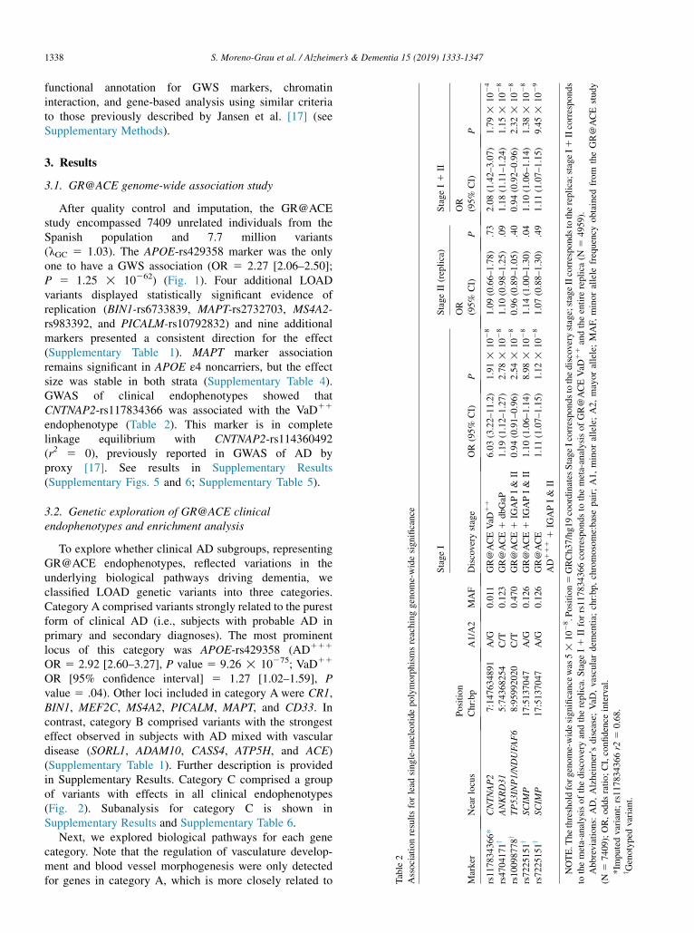
OR [95% confidence interval] 5 1.27 [1.02–1.59], Pvalue 5 .04). Other loci included in category A were CR1,BIN1, MEF2C, MS4A2, PICALM, MAPT, and CD33. Incontrast, category B comprised variants with the strongesteffect observed in subjects with AD mixed with vasculardisease (SORL1, ADAM10, CASS4, ATP5H, and ACE)(Supplementary Table 1). Further description is providedin Supplementary Results. Category C comprised a groupof variants with effects in all clinical endophenotypes(Fig. 2). Subanalysis for category C is shown inSupplementary Results and Supplementary Table 6.
Next, we explored biological pathways for each genecategory. Note that the regulation of vasculature develop-ment and blood vessel morphogenesis were only detectedfor genes in category A, which is more closely related to

Fig. 2. (A) Enrichment trend per genetic marker and gene category across GR@ACE endophenotypes. (B) Graph centered in effect change range 0–0.5. Effect
change per endophenotype5Variant Odds ratio–variant null effect; Enrichment trend per category was obtained applying a linear regression using ggplot2 in R.
Abbreviations: AD, Alzheimer’s disease; VaD, vascular dementia.
S. Moreno-Grau et al. / Alzheimer’s & Dementia 15 (2019) 1333-1347 1339
pure AD (P5 2.03! 1027, P5 1.90! 1026, respectively)(Table 3). Additional categories indicated immune systempathways (category B, P 5 2.07 ! 1027; category C,P 5 5.77 ! 10215) (Table 3). Finally, with the aim ofvalidating previous results, we conducted a subanalysis byclassifying LOAD genetic variants with more stringentclassification criteria (Supplementary Methods). Again,APOE,CR1,MEF2C,MS4A2, and PICALM loci were foundin category A; SORL1 and CASS4 were in category B; andadditional AD loci were in category C (SupplementaryFig. 4). Regulation of vasculature development wasexclusively identified as the top pathway in category A(P 5 2.14 ! 1027) when we restricted the analysis toinclude those loci coexpressing with at least 4 LOAD genes(Supplementary Table 7). Further replication in cohorts withavailable neuropathological data would be recommended.
3.3. Meta-analysis of GR@ACE study with other data sets
To assess the impact of sample composition in ADGWAS, and look for new AD loci, we first combinedthe GR@ACE data set with nine additional genomicdatabases that had genotypic level data available. Subtlegenomic inflation was detected, mainly explainedby polygenicity (lGC 5 1.10; LD scoreintercept 5 1.04). Five regions were associated withLOAD (Fig. 3); of these, four (APOE-rs429358,PICALM-rs10792832, MS4A2-rs983392, and BIN1-rs6733839) have been previously linked to AD(Supplementary Table 8), and one is a new GWASfinding (ANKDR31-rs4704171; OR 5 1.19 [1.12–1.27];P 5 2.78 ! 1028) (Table 2).
Then, we conducted a genome-wide meta-analysiscombining the GR@ACE study with IGAP stage I. We
identified 12 LOAD genomic regions reaching GWS.CD33-rs3865444, which did not reach GWS in the IGAPmeta-analysis, was significantly associated with LOAD(OR 5 0.92 [0.89–0.95]; P 5 3.61 ! 1028)(Supplementary Fig. 7). Among the top suggestive signals,we detected HBEGF-rs4150233 (OR 5 0.92 [0.90–0.95];P 5 5.10 ! 1028) previously identified by a transethnicGWAS [29].
Next, meta-analysis of the whole GR@ACE data set withIGAP I and II enabled the identification, for the first time,of NDUFAF6-rs10098778 as a GWS signal (OR 5 0.94[0.91–0.96]; P 5 2.54 ! 1028). When we combinedGR@ACE AD111 endophenotype with IGAP I and II, wealso detected SCIMP-rs7225151 (OR 5 1.11 [1.07–1.15];P 5 1.12 ! 1028) (Table 2) (Supplementary Fig. 8). Itwas previously reported as a genome-wide suggestive signalby IGAP [24]. Recently, SCIMP-rs113260531, which is incomplete LD with our lead marker (r2 5 1), was associatedwith AD [17].
3.4. Replication of genome-wide significant findings
Finally, we tested for replication of the new signals in anindependent sample of 4959 Spanish individuals. TheCNTNAP2-rs117834366, detected in the GWAS ofGR@ACE VaD11 endophenotype, had a P value of 0.79with a similar effect direction to that reported previouslybut strongly deflated in the entire replica sample(OR 5 1.09 [0.66–1.78]; P 5 .79) (Table 3). Analysis ofthe subspecific VaD phenotype in the replica (N5 89) wouldbe highly inaccurate.
In the exploration of meta-GWAS findings, we observedthat the ANKDR31-rs4704171-C marker increased the riskof AD (OR 5 1.10 [0.98–1.25]; P 5 .09; power 5 33%).
Table 3
Top ten biological pathways per gene category
Gene ontology
pathway
Top 10 coregulated pathways
for category A P value
GO:1901342 Regulation of vasculature
development
2.03 ! 1027
GO:0060326 Cell chemotaxis 2.59 ! 1027
GO:0048771 Tissue remodeling 6.77 ! 1027
GO:0050865 Regulation of cell activation 1.14 ! 1026
GO:0007159 Leukocyte cell-cell adhesion 1.21 ! 1026
GO:0048514 Blood vessel morphogenesis 1.90 ! 1026
GO:0003012 Muscle system process 2.54 ! 1026
GO:0002764 Immune response-regulating
signaling pathway
3.48 ! 1026
GO:0032103 Positive regulation of response
to external stimulus
3.91 ! 1026
GO:0010959 Regulation of metal ion transport 4.36 ! 1026
Gene ontology
pathway
Top 10 coregulated pathways
for category B P value
GO:0009620 Response to fungus 2.02 ! 1027
GO:0050886 Endocrine process 3.58 ! 1027
GO:0002443 Leukocyte mediated immunity 5.47 ! 1027
GO:0050865 Regulation of cell activation 1.52 ! 1025
GO:0031349 Positive regulation of
defense response
8.42 ! 1025
GO:0032103 Positive regulation of
response to external stimulus
1.00 ! 1024
GO:0002250 Adaptive immune response 1.30 ! 1024
GO:0098542 Defense response to
other organism
2.00 ! 1024
GO:1901568 Fatty acid derivative
metabolic process
2.24 ! 1024
GO:0050900 Leukocyte migration 2.57 ! 1024
Gene ontology
pathway
Top 10 coregulated pathways
for category C P value
GO:0007159 Leukocyte cell-cell adhesion 5.77 ! 10215
GO:0050865 Regulation of cell activation 4.37 ! 10214
GO:0002764 Immune response-regulating
signaling pathway
1.33 ! 10212
GO:0002253 Activation of immune response 3.96 ! 10212
GO:0002443 Leukocyte mediated immunity 4.34 ! 10212
GO:0002274 Myeloid leukocyte activation 7.78 ! 10212
GO:0002250 Adaptive immune response 1.24 ! 10211
GO:0002263 Cell activation involved
in immune response
7.07 ! 10211
GO:0022407 Regulation of cell-cell
adhesion
5.40 ! 1029
GO:0070661 Leukocyte proliferation 1.22 ! 1028
S. Moreno-Grau et al. / Alzheimer’s & Dementia 15 (2019) 1333-13471340
Although the expected effect is in linewith previous data, theprecision of the estimate in this replica differs, ranging froma 2% decrease (a small negative association) to a 25%increase. Of note, the result emerging from themeta-analysis of the replica with the discovery sample(n5 26,194) is compatiblewith its potential role in dementia(OR 5 1.18 [1.11–1.24]; P 5 1.15 ! 1028) (Table 3). Seeforest plot in Supplementary Fig. 9.
We observed a similar effect direction in the NDUFAF6-rs10098778 marker to that reported in the discovery stage,with interval estimates ranging from a risk decrease of
11% to a risk increase of 5% (OR 5 0.96 [0.89–1.05];P 5 .40; power 5 16%). This signal had aP 5 2.32 ! 1028 in the final meta-analysis, including thewhole GR@ACE data set, the replica, and IGAP I and II(n 5 91,373) (Supplementary Fig. 9).
SCIMP-rs7225151 showed a risk effect in the wholereplica. Limits of the interval were consistent with a positiveassociation (OR 5 1.14 [1.01–1.29]; P 5 .047;power5 39%; n cases5 1943). In the AD111 endopheno-type, the marker presented the same positive risk effectdirection (OR 5 1.07 [0.88–1.30]), although had P 5 .49,which could be mainly explained by a reduction of thesample size (power 5 19%; n cases 5 584). Our resultsfor both meta-analyses, the final meta-analysis of the wholeGR@ACE data set (n5 91,373) and the final meta-analysisof GR@ACE AD111 endophenotype, (n 5 85,055) werecompatible with a potential effect of this marker in AD(Table 3). See forest plots in Supplementary Fig. 9.
3.5. Biological interpretation of meta-GWAS signals
To identify candidate genes and potential causal variantswithin novel meta-GWAS regions, we conducted cis-eQTLmapping. The rs2335107 marker located in the ANKRD31locus (chr5:74,451,693) was associated with the corticalexpression of the long noncoding RNA (lncRNA) CTD-2235C13.3 (P 5 1.26 ! 1025). This variant is located83.4 kb from the meta-GWAS lead single nucleotidepolymorphism (rs4704171, chr5:74,368,254), and both arein complete LD (r2 5 1). The CTD-2235C13.3 gene islocated 1.6 kb from the HMGCR locus, and its function isunknown. The NDUFAF6 region mapped for NDUFAF6RNA cortical expression (P 5 5.56 ! 1026) and forTP53INP1 RNA blood expression (P 5 1.17 ! 10210).Finally, rs73976325 (chr17:5,123,227), located in theSCIMP locus, to 13.8 kb from the meta-GWAS top signal(rs7225151, chr17:5,137,047), mapped to brain cis-actingeQTL for AC012146.1 lincRNA (P 5 2.15 ! 1027). Twoadditional markers were pinpointed to blood eQTLs,SCIMP-rs6502851 (P 5 3.89 ! 10208) and RABEP-rs59277121 (P 5 3.89 ! 1028) (Supplementary Table 3).In an additional prioritization strategy, combining informa-tion from positional mapping, eQTL, chromatin interaction,and gene-based genome-wide association analysis viaFUMA, results pointed to ANKDR31 and POLK for theANKDR31 genomic region, as well as NDUFAF6 andTP53INP1 for the NDUFAF6 region (SupplementaryTable 9). Further description is provided in SupplementaryInformation and Supplementary Table 9.
4. Discussion
We present a comprehensive GWAS of AD dementiacases. This represents the first pilot study exploring thegenetics and underlying biological pathways of subgroupsof patients with AD, defined based on vascular burden. We

Fig. 3. A) Results of genome-wide association analysis for GR@ACEmeta-analysis with nine additional databases (n5 21,235). (B) QQplot. (C) Associations
of the region centered on rs4704171 located in the ANKRD31 locus and containing the HMGCR locus.
S. Moreno-Grau et al. / Alzheimer’s & Dementia 15 (2019) 1333-1347 1341
showed differential biological routes underlying clinical en-dophenotypes and demonstrated how these differential sub-groups of patients with AD impact GWAS discoveries. TheGR@ACE study represents a unique genomic resourcebecause all affected cases were diagnosed in a single mem-ory clinic using the same screening and diagnostic tech-niques. This might limit potential sources of clinicalvariation between study participants, recently demonstratedin a large meta-GWAS [12].
Based on the increase in evidence suggesting thatvascular brain pathology can act concomitantly with ADto produce a more rapid cognitive decline [5], we exploredthe effect of known LOAD loci across different levels ofvascular burden in dementia patients using only clinical def-initions. Our basic idea was to dissect, from a molecularpoint of view, the model previously proposed by Viswana-than et al. [7]. We observed three categories of loci, whichmight reflect the disease’s clinical heterogeneity, fromvascular and mixed forms to a “purer” AD phenotype.Intriguingly, we detected vascular processes to be the maincausal mechanism in clinically pure AD and found the im-mune system pervasively detected across the three cate-gories. Although both pathways have been previouslyassociated with LOAD by network analysis [30], this is thefirst study to show that the association with the vascular sys-tem is conducted by AD-specific clinical subgroup.
It should be noted that the present study used clinicalcriteria to define the AD cases [20], but recently the
classification of AD has evolved. In 2018, the NIA-AA pro-posed a novel research framework for the biological classi-fication of AD based on the presence in vivo of biomarkerevidences for amyloid (A), tau (T), and neurodegeneration(N), as surrogate of the neuropathological state of an individ-ual [31]. The AT(N) biomarker system allows the classifica-tion of individuals into three categories: those with a normalbiomarker profile, those with biomarkers compatible withAD change, and those with biomarkers compatible withnon-AD pathological changes [31]. Using the NIA-AAapproach, the generation of subgroups of AD patientsconsidering vascular pathology would be unfeasible, asnowadays the ATN classification does not integrate mea-sures of vascular dysfunction. Taking into account thatmost of dementia cases are caused by mixed pathologies,the current system seems deeply incomplete to study theprobable interaction between neurodegeneration andvascular dysfunction. This idea has also been claimed byothers [32,33]. Thus, we encourage other groups tocontrast the proposed loci classification, which was basedon the GR@ACE clinical endophenotypes, but using well-powered GWAS cohorts with available neuropathologicaldata.
Silent changes occur in brain microvasculature duringAD progression. In fact, CAA is a well-recognized AD path-ological feature characterized by the accumulation of amy-loid proteins in the walls of small cerebral vessels. CAAhas been proposed to compromise the perivascular drainage
S. Moreno-Grau et al. / Alzheimer’s & Dementia 15 (2019) 1333-13471342
of Ab from the brain to the peripheral system [34]. Almostall AD brains harbor CAA pathology to some extent,although in vivo most CAA cases remain undiagnosed,even using the validated Boston criteria [35]. Mendelian mu-tations of the APP gene have been found in CAA and AD[8,36]. APOE ε4 and CR1 have been associated with anincreased risk of CAA [37,38]. In particular, distinct ADloci have been associated with capillary and noncapillaryCAA [39]. Between them, APOE ε4 was strongly relatedto capillary CAA [39]. These links make it conceivablethat a potential genetic overlap exists between CAA andAD and suggest that CAA pathology could represents an un-derlying process for AD. In that context, we think thatintrinsic alterations to the vasculature could contribute todisease pathogenesis in more pure forms of AD, explainingour results. Conversely, in AD individuals with evident cere-brovascular lesions comprising mixed forms, the additionalrole of cardiovascular risk factors such as hypertension,atherosclerosis, or arteriosclerosis should be considered, asthese could point to a systemic pathological state leadingto vascular damage and dementia. This would agree withthe limited genetic correlation between neurodegenerativeand other neurologic disorders [12], as well as with the re-sults obtained from heterochronic parabionts in agingmodels [40].
Understanding the role of vasculature pathology in ADseems to be a pertinent step. In that scenario, CAA wouldbe a key AD hallmark. CAA represents the unique identifiedlink between the vascular and amyloid hypotheses, but it hasbeen completely neglected in the original hypothesisformulation.
From a clinical point of view, placing each patient some-where along the disease spectrum proposed by Viswanathanet al. [7] is complex. A deep understanding of heterogeneityin AD seems necessary to design better genetic studies,which must drive the discovery of novel loci and, ultimately,innovative targets for AD therapies. In this study, weexplored how clinical heterogeneity might impact GWASfindings by integrating distinct GWAS data sets with eitherthe GR@ACE cohort as a whole or its endophenotypes.We found several new GWS signals that seem stronglydependent on the sample composition. For example, aftercombining IGAP stages I and II with the entire GR@ACEdata set, we identified genetic signals in the NDUFAF6genomic region but not in the SCIMP region. When this ex-ercise was conducted using GR@ACE endophenotypes, theSCIMP signal was detected using the clinically “pure” ADGR@ACE endophenotype. It should be noted that the powerto detect SCIMP signal in the meta-analysis withGRA@ACE dementia and GR@ACE AD111 was 75 and70%, respectively.We tried to replicate this finding in a purerAD data set without clinical mixed dementia cases, but theavailable number of clinical AD111 cases might becompromising the statistical power to replicate (Ncases 5 584; power 5 19%). Despite that, we think that us-ing specific clinical subgroups of the AD population might
empower genetic studies to detect genes associated with spe-cific disease axes.
An alternative strategy is taking advantage of clinical het-erogeneity. Specifically, heterogeneity might play a dual rolein genetic studies. Although it might decrease the power todetect genes associated with more specific clinical sub-groups, incorporating detailed clinical AD definitions canalso promote identifying genes shared with other conditionsor copathologies such as small vessel disease (SVD). In fact,this was the case for the ATP5H locus, which was previouslyfound to be associated with AD [41] andmore recently foundin relation to SVD [42]. We think that the same couldapply to the ANKRD31 finding. ANKRD31 encodes aprotein containing ankyrin repeats, which is linked withneurodevelopmental disorders [43]. Of note, ANKRD31GWAS signal mapped to the brain eQTL of a lncRNA,located 1.6 kb from the HMGCR locus and residing in theCOL4A3BP gene. The HMGCR locus is one of the mostimportant coregulators of cholesterol biosynthesis, and it isthe therapeutic target of statins. The COL4A3BP gene isinvolved in lipid transport [44]. Several studies have linkedHMGCR polymorphisms and AD risk or age at onset forAD [45], and the cholesterol pathway has been identifiedto be a biological route shared between AD and SVD.Interestingly, markers in the POLK locus, associated bygene-based genome-wide association analysis on this studyand located in the same disequilibrium block of ANKDR31(Fig. 3), jointly conferred risk for AD and plasma levels ofLDL [46]. Considering prior findings, our results areconsistent with this genomic region having a role in mixeddementia. The reported genetic signal should be considereda highly probable finding, although independent replicationsare still required.
In the present work, NDUFAF6 signals reached GWS forthe first time. This finding presented the same effect direc-tion in the independent sample (power5 16%) and remainsas GWS after the final meta-analysis. Our lead GWASmarker is in high LD with NDUFAF6-rs4735340, the topsuggestive signal reported by Kunkle et al. [18] at this region(r2 5 0.95, for Utah residents of European ancestry popula-tion). Despite that, there are subtle differences in LD esti-mates for the Iberian population (r2 5 0.87), suggestingthat the genetic architecture of the Spanish population couldbe helping to pinpoint the region of interest. This region is inthe close vicinity of TP53INP1, previously associated withAD by a gene-based approach [47]. TP53INP1 is involvedin mitochondrial function. Considering these findings andresults emerging from eQTL analysis, it would be feasiblethat a regulatory element for NUDFAF6 resides upstreamof the TP53INP1 locus. We also detected that the SCIMPsignal was mainly conducted by a specific group of ADcases. This signal was reported to be a suggestive signalby IGAP [24], and a proxy of it (SCIMP-rs113260531)reached GWS recently [17]. SCIMP regions has beeninvolved in several eQTLs, from uncharacterized corticallncRNA to blood eQTLs in SCIMP and RABEP1 loci, both
S. Moreno-Grau et al. / Alzheimer’s & Dementia 15 (2019) 1333-1347 1343
associated with immune system function [48,49]. The CD33locus remains a controversial LOAD locus because largemeta-GWAS were unable to replicate this signal [24], buthere it reached GWS. We previously proposed that crypticpopulation substructure could explain the divergent observa-tions for this locus [50].
Note that the lack of definitive neuropathological data forAD cases is a severe limitation of the present study. Clinicaldefinitions have important uncertainties, and diagnosis mis-classifications sometimes occur. Hence, some AD individ-uals included in enriched AD endophenotypes may presentconcomitant vascular brain disease. The generation of largehistopathological GWAS cohorts with associated quantita-tive data on each pathological hallmark is the ultimate solu-tion to tackling the intrinsic heterogeneity in AD.Unfortunately, there are few examples of neuropathologicalcohorts: only one GWAS has investigated the genetics ofCAA, with APOE being the unique GWS signal [37]. Inthis study, a small number of AD cases evolved to vasculardementia during follow-up. Large cross-sectional clinicalGWAS cannot control diagnostic changes occurring in clin-ical practice. Clinical diagnosis is a dynamic variable, so un-derstanding the genetic profiles of subgroups of patientsevolving to other pathologies would provide powerful infor-mation.
It should be considered that there is a limitation inreducing the sample size by splitting the cohort into differentendodophenotypes, instead of combining them. Despite that,spanning the spectrum of dementia individuals to generateclinical endophenotypes provided us a versatile design,which let us explore the effect of heterogeneity in GWASand replicate the main findings of pathway analysis usingan alternative strategy. The limited number of VaD casesin subgroup analysis limits the accuracy of gene categoriza-tion and pathway analysis. Finally, the exact effector genesfor LOAD genetic findings remain unclear. This is a severelimitation to pathway analysis that can only be circumventedby isolating the causativemutations. Independent replicationwill be needed to corroborate our new GWS signals. In thatsense, the selection of specific patient groups might lead tosuccessful replication studies.
The assessment of heterogeneity has important implica-tions for gene discovery, the development of treatments,and their appropriate use in individual patients. TheGR@ACE cohort provides useful genomic information, asit accounts for potential sources of variability and containsdifferent subgroups of cases. This enabled us to analyzethe LOAD genetic landscape in terms of clinical endopheno-types. Our efforts to disentangle the mechanistic pathwaysoperating under clinical subgroups of patients revealed thatvasculature regulation may be an essential part of the caus-ative mechanism of LOAD. Finally, our exploration of ADgenetics highlights the relevance of sample composition ingenetic discoveries. Considering sample composition inthe design of genetic studies might lead to the identificationof genetic profiles, which can help clinicians distinguish
subsets of patients within the disease spectrum and promotenovel therapy targets for AD.
Acknowledgments
The authors would like to thank patients and controls whoparticipated in this project. The Genome Research @Fundaci�o ACE project (GR@ACE) is supported byFundaci�on bancaria “La Caixa”, Grifols SA, Fundaci�oACE, and ISCIII (Ministry of Health, Spain). They alsowant to thank the private sponsors who support the basicand clinical projects of our institution (Piramal AG, Labora-torios Echevarne, Araclon Biotech S.A., and Fundaci�oACE). They are indebted to the Trinitat Port-Carb�o legacyand her family for their support of Fundaci�o ACE researchprograms. Fundaci�o ACE is a participating center in theDementia Genetics Spanish Consortium (DEGESCO).A.R. and M.B. receive support from the European Union/EFPIA Innovative Medicines Initiative Joint undertakingADAPTED and MOPEAD projects (grant numbers115975 and 115985, respectively). M.B. and A.R. are alsosupported by national grants PI13/02434, PI16/01861, andPI17/01474. Acci�on Estrat�egica en Salud is integrated intothe Spanish National R 1 D 1 I Plan and funded by ISCIII(Instituto de Salud Carlos III)-Subdirecci�on General deEvaluaci�on and the Fondo Europeo de Desarrollo Regional(FEDER- “Una manera de Hacer Europa”). L.M.R. issupported by Consejer�ıa de Salud de la Junta de Andaluc�ıa(grant PI-0001/2017). Control samples and data frompatients included in this study were provided in part by theNational DNA Bank Carlos III (www.bancoadn.org,University of Salamanca, Spain) and Hospital UniversitarioVirgen de Valme (Sevilla, Spain); they were processed afterstandard operating procedures with the appropriate approvalof the Ethical and Scientific Committee. The present workwas performed as part of the Biochemistry, MolecularBiology, and Biomedicine doctoral program of S. Moreno-Grau at Universitat Aut�onoma de Barcelona (Barcelona,Spain).Data collection and sharing for this project was partiallyfunded by the Alzheimer’s Disease Neuroimaging Initiative(ADNI) (National Institutes of Health grant U01 AG024904)and DOD ADNI (Department of Defense award numberW81XWH-12-2-0012). The ADNI is funded by the NationalInstitute on Aging and the National Institute of BiomedicalImaging and Bioengineering, as well as through generouscontributions from the following: AbbVie; the Alzheimer’sAssociation; the Alzheimer’s Drug Discovery Foundation;Araclon Biotech; BioClinica, Inc.; Biogen; Bristol-MyersSquibb Company; CereSpir, Inc.; Cogstate; Eisai Inc.;Elan Pharmaceuticals, Inc.; Eli Lilly and Company; EuroIm-mun; F. Hoffmann-La Roche Ltd and its affiliated companyGenentech, Inc.; Fujirebio; GE Healthcare; IXICO Ltd.;Janssen Alzheimer Immunotherapy Research & Develop-ment, LLC.; Johnson & Johnson Pharmaceutical Research& Development LLC.; Lumosity; Lundbeck; Merck &

S. Moreno-Grau et al. / Alzheimer’s & Dementia 15 (2019) 1333-13471344
Co., Inc.; Meso Scale Diagnostics, LLC.; NeuroRxResearch; Neurotrack Technologies; Novartis Pharmaceuti-cals Corporation; Pfizer Inc.; Piramal Imaging; Servier;Takeda Pharmaceutical Company; and Transition Therapeu-tics. The Canadian Institutes of Health Research providesfunds to support ADNI clinical sites in Canada. Privatesector contributions are facilitated by the Foundation forthe National Institutes of Health (www.fnih.org). Thegrantee organization is the Northern California Institutefor Research and Education, and the study was coordinatedby the Alzheimer’s Therapeutic Research Institute at theUniversity of Southern California. ADNI data are dissemi-nated by the Laboratory for NeuroImaging at the Universityof Southern California.The AddNeuroMed data are from a public-private partner-ship supported by EFPIA companies and SMEs as part of In-noMed (Innovative Medicines in Europe), an integratedproject funded by the European Union of the Sixth Frame-work program priority FP6-2004-LIFESCIHEALTH-5.Clinical leads responsible for data collection are IwonaK1oszewska (Lodz), Simon Lovestone (London), PatriziaMecocci (Perugia), Hilkka Soininen (Kuopio), Magda Tso-laki (Thessaloniki), and Bruno Vellas (Toulouse). Imagingleads are Andy Simmons (London), Lars-Olad Wahlund(Stockholm), and Christian Spenger (Zurich). Bioinformat-ics leads are Richard Dobson (London) and Stephen New-house (London).Funding support for the Alzheimer’s Disease Genetics Con-sortium (ADGC) was provided through the NIA Division ofNeuroscience (U01-AG032984).The genotypic and associated phenotypic data usedin the study “Multi-Site Collaborative Study forGenotype-Phenotype Associations in Alzheimer’s Disease(GenADA)” were provided by GlaxoSmithKline, R&DLimited. The data sets used for the analyses describedin this manuscript were obtained from dbGaP athttp://www.ncbi.nlm.nih.gov/gap through dbGaP accessionnumber phs000219.The Mayo Clinic Alzheimer’s Disease Genetic Studies, ledby Dr. Nil€ufer Ertekin-Taner and Dr. Steven G. Younkin atthe Mayo Clinic in Jacksonville, FL, used samplesfrom the Mayo Clinic Study of Aging, the Mayo ClinicAlzheimer’s Disease Research Center, and the MayoClinic Brain Bank. Data collection was supported throughfunding by NIA grants P50 AG016574, R01 AG032990,U01 AG046139, R01 AG018023, U01 AG006576, U01AG006786, R01 AG025711, R01 AG017216, and R01AG003949, NINDS grant R01 NS080820, the CurePSPFoundation, and support from the Mayo Foundation.The Neocodex-Murcia study was funded by the Fundaci�onAlzheimur (Murcia), the Ministerio de Educaci�on y Ciencia(Gobierno de Espa~na), Corporaci�on Tecnol�ogica deAndaluc�ıa, Agencia IDEA (Consejer�ıa de Innovaci�on, Juntade Andaluc�ıa), the Diabetes Research Laboratory, and theBiomedical Research Foundation. University HospitalCl�ınico San Carlos has been supported by CIBER de
Diabetes y Enfermedades Metab�olicas Asociadas (CIBER-DEM); CIBERDEM is an ISCIII Project.The ROS/MAP study data were provided by the Rush Alz-heimer’s Disease Center, Rush University Medical Center,Chicago. Data collection was supported through fundingby NIA grants P30AG10161, R01AG15819,R01AG17917, R01AG30146, R01AG36836, U01AG32984and U01AG46152, the Illinois Department of Public Health,and the Translational Genomics Research Institute.The TGEN study was supported by Kronos Life ScienceLaboratories, the National Institute on Aging (Arizona Alz-heimer’s Disease Center grants P30 AG19610 and RO1AG023193, the Mayo Clinic Alzheimer’s Disease Centergrant P50 AG16574, and the Intramural Research Program),the National Alzheimer’s Coordinating Center (U01AG016976), and the state of Arizona.The results published here are in part based on the data ob-tained from the AMP-AD Knowledge Portal accessed athttps://doi.org/10.7303/syn2580853.The authors thank the International Genomics of Alz-heimer’s Project (IGAP) for providing summary resultdata for these analyses. The investigators within IGAPcontributed to the design and implementation of IGAPand/or provided data but did not participate in analysisor writing of this report. IGAP was made possible bythe generous participation of the control subjects, the pa-tients, and their families. The i–Select chip was funded bythe French National Foundation on Alzheimer’s diseaseand related disorders. European Alzheimer’s DiseaseInitiative (EADI) was supported by the LABEX (labora-tory of excellence program investment for the future)DISTALZ grant, Inserm, Institut Pasteur de Lille, Uni-versit�e de Lille 2, and the Lille University Hospital.GERAD was supported by the Medical Research Council(grant no. 503480), Alzheimer’s Research UK (grant no.503176), the Wellcome Trust (grant no. 082604/2/07/Z),and German Federal Ministry of Education and Research(BMBF): Competence Network Dementia (CND) grantno. 01GI0102, 01GI0711, 01GI0420. CHARGE waspartly supported by the NIH/NIA grant R01 AG033193and the NIA AG081220 and AGES contract N01–AG–12100, the NHLBI grant R01 HL105756, the IcelandicHeart Association, and the Erasmus Medical Center andErasmus University. ADGC was supported by the NIH/NIA grants: U01 AG032984, U24 AG021886, U01AG016976, and the Alzheimer’s Association grantADGC–10–196728.The GR@ACE study group: Abdelnour C1,2, Aguilera N1,Alarcon E1,3, Alegret M1,2, Benaque A1, Boada M1,2, Buen-dia M1, Ca~nabate P1,2, Carracedo A4,5, Corbat�on A6, de Ro-jas I1, Diego S1, Espinosa A1,2, Gailhajenet A1, Garc�ıaGonz�alez P1, Gil S1, Guitart M1, Gonz�alez P�erez A7,Hern�andez I1,2, Ibarria, M1, Lafuente A1, Macias J8,Maro~nas O4, Mart�ın E1, Mart�ınez MT6, Marqui�e M1,Maule�on A1, Mont�e-Rubio G1, Montrreal L1, Moreno-Grau S1,2, Moreno M1, Orellana A1, Ortega G1,2, Pancho

S. Moreno-Grau et al. / Alzheimer’s & Dementia 15 (2019) 1333-1347 1345
A1, Pelej�a E1, P�erez-Cordon A1, Pineda JA8, Preckler S1,Quintela I3, Real LM3,8, Rodr�ıguez-G�omez O1,2, Rosende-Roca M1, Ruiz A1,2, Ruiz S1,2, S�aez ME7, Sanabria A1,2,Santos-Santos MA1, Serrano-Rios M6, Sotolongo-Grau O1,T�arraga L1,2, Valero S1,2, Vargas L1 (1. Research Centerand Memory clinic Fundaci�o ACE. Institut Catal�a de Neuro-ci�encies Aplicades. Universitat Internacional de Catalunya.Barcelona, Spain; 2. CIBERNED, Center for NetworkedBiomedical Research on Neurodegenerative Diseases, Na-tional Institute of Health Carlos III, Ministry of Economyand Competitiveness, Spain; 3. Dep. of Surgery, Biochem-istry andMolecular Biology, School of Medicine. Universityof M�alaga. M�alaga, Spain; 4. Grupo de Medicina Xen�omica,Centro Nacional de Genotipado (CEGEN-PRB3-ISCIII).Universidade de Santiago de Compostela, Santiago de Com-postela, Spain; 5. Fundaci�on P�ublica Galega de MedicinaXen�omica- CIBERER-IDIS, Santiago de Compostela,Spain; 6. Centro de Investigaci�on Biom�edica en Red de Dia-betes y EnfermedadesMetab�olicas Asociadas, CIBERDEM,Spain, Hospital Cl�ınico San Carlos, Madrid, Spain; 7.CAEBI. Centro Andaluz de Estudios Bioinform�aticos., Sev-illa, Spain; 8. Unidad Cl�ınica de Enfermedades Infecciosas yMicrobiolog�ıa. Hospital Universitario de Valme, Sevilla,Spain)DEGESCO consortium: Adarmes-G�omez AD1,2, Alarc�on-Mart�ın E3,4, �Alvarez I5, �Alvarez V6,7, Amer-Ferrer G8, An-tequera M9, Ant�unez C9, Baquero M10, Bernal M11, BlesaR2,12, Boada M2,3, Buiza-Rueda D1,2, Bullido MJ2,13,14,Burguera JA10, Calero M2,15,16, Carrillo F1,2, Carri�on-ClaroM1,2, Casajeros MJ17, Clarim�on J2,12, Cruz-Gamero JM4,de Pancorbo MM18, de Rojas I3, del Ser T14, Diez-FairenM5, Fortea J2,12, Franco E11, Frank-Garc�ıa A2,14,19,Garc�ıa-Alberca JM20, Garcia Madrona S16, Garcia-RibasG16, G�omez-Garre P1,2, Hern�andez I2,3, Hevilla S20, Jes�usS1,2, Labrador Espinosa MA1,2, Lage C2,21, Legaz A9,Lle�o A2,12, L�opez de Mun�ain A22, L�opez-Garc�ıa S2,21, Ma-cias D1,2, Manzanares S9,23, Mar�ın M11, Mar�ın-Mu~noz J9,Mar�ın T20, Marqui�e M3, Mart�ın Montes A2,13,19, Mart�ınezB9, Mart�ınez C7,24, Mart�ınez V9, Mart�ınez-Lage �AlvarezP25, Medina M2,14, Mendioroz Iriarte M26, Men�endez-Gonz�alez M7,27, Mir P1,2, Molinuevo JL28, Mont�e-RubioG3, Montrreal L3, Moreno-Grau S2,3, Orellana A3, PastorAB15, Pastor P5, P�erez Tur J2,29,30, Peri~n�an-Tocino T1,2, Pi-~nol Ripoll G2,31, R�abano A2,15,32, Real de As�ua D33, Ro-drigo S11, Rodr�ıguez-Rodr�ıguez E2,21, Royo JL4, RuizA2,3, Sanchez del Valle D�ıaz R34, S�anchez-Juan P2,21,Sastre I2,13, Sotolongo-Grau O3, T�arraga L2,3, ValeroS2,3, Vicente MP9, Vivancos L9 (1. Unidad de Trastornosdel Movimiento, Servicio de Neurolog�ıa y Neurofisiolog�ıa.Instituto de Biomedicina de Sevilla (IBiS), Hospital Uni-versitario Virgen del Roc�ıo/CSIC/Universidad de Sevilla,Seville, Spain; 2. CIBERNED, Network Center forBiomedical Research in Neurodegenerative Diseases, Na-tional Institute of Health Carlos III, Spain; 3. ResearchCenter and Memory clinic Fundaci�o ACE. Institut Catal�ade Neuroci�encies Aplicades. Universitat Internacional de
Catalunya. Barcelona, Spain; 4. Dep. of Surgery, Biochem-istry and Molecular Biology, School of Medicine. Univer-sity of M�alaga. M�alaga, Spain; 5. Fundaci�o per laRecerca Biom�edica i Social M�utua Terrassa, and MemoryDisorders Unit, Department of Neurology, Hospital Uni-versitari Mutua de Terrassa, University of BarcelonaSchool of Medicine, Terrassa, Barcelona, Spain; 6. Labora-torio de Gen�etica Hospital Universitario Central de Astu-rias, Oviedo; 7. Instituto de Investigaci�on Biosanitaria delPrincipado de Asturias (ISPA); 8. Department ofNeurology, Hospital Universitario Son Espases, Palma,Spain; 9. Unidad de Demencias. Hospital Cl�ınico Universi-tario Virgen de la Arrixaca; 10. Servei de Neurologia, Hos-pital Universitari i Polit�ecnic La Fe; 11. Unidad deDemencias, Servicio de Neurolog�ıa y Neurofisiolog�ıa. In-stituto de Biomedicina de Sevilla (IBiS), Hospital Univer-sitario Virgen del Roc�ıo/CSIC/Universidad de Sevilla,Seville, Spain; 12. Memory Unit, Neurology Departmentand Sant Pau Biomedical Research Institute, Hospital dela Santa Creu i Sant Pau, Universitat Aut�onoma de Barce-lona, Barcelona, Spain; 13. Centro de Biologia MolecularSevero Ochoa (C.S.I.C.-U.A.M.), Universidad Autonomade Madrid, Madrid, Spain; 14. Instituto de InvestigacionSanitaria ‘Hospital la Paz’ (IdIPaz), Madrid, Spain; 15.CIEN Foundation, Queen Sofia Foundation AlzheimerCenter, Madrid, Spain; 16. Instituto de Salud Carlos III (IS-CIII), Madrid, Spain; 17. Hospital Universitario Ram�on yCajal; Madrid, Spain; 18. BIOMICs, Pa�ıs Vasco; Centrode Investigaci�on Lascaray. Universidad del Pa�ıs VascoUPV/EHU; 19. Neurology Service, Hospital UniversitarioLa Paz (UAM), Madrid, Spain; 20. Alzheimer ResearchCenter & Memory Clinic. Andalusian Institute for Neuro-science. M�alaga, Spain; 21. Neurology Service, Marqu�esde Valdecilla University Hospital (University of Cantabriaand IDIVAL), Santander, Spain; 22. Hospital Donostia deSan Sebast�ıan; 23. Fundaci�on para la Formaci�on e Inves-tigaci�on Sanitarias de la Regi�on de Murcia; 24. Serviciode Neurolog�ıa -Hospital de Cabue~nes-Gij�on; 25. Centrode Investigaci�on y Terapias Avanzadas. Fundaci�on CITA-alzheimer; 26. Navarrabiomed; 27. Servicio de Neurolog�ıa-Hospital Universitario Central de Asturias, Oviedo; 28.Barcelona beta Brain Research Center – Fundaci�o PasqualMaragall; 29. Unitat de Gen�etica Molecular. Institut deBiomedicina de Val�encia-CSIC; 30. Unidad Mixta de Neu-rologia Gen�etica. Instituto de Investigaci�on Sanitaria LaFe; 31. Unitat Trastorns Cognitius, Hospital UniversitariSanta Maria de Lleida, Institut de Recerca Biom�edica deLleida (IRBLLeida), Lleida, Espa~na. 32. BT-CIEN; 33.Hospital Universitario La Princesa, Madrid, Spain; 34.Hospital Cl�ınic Barcelona).
Supplementary Data
Supplementary data related to this article can be found athttps://doi.org/10.1016/j.jalz.2019.06.4950.

S. Moreno-Grau et al. / Alzheimer’s & Dementia 15 (2019) 1333-13471346
RESEARCH IN CONTEXT
1. Systematic review: Recent literature search, con-ducted using PubMed database, revealed that thepresence of Alzheimer’s disease (AD) with vascularbrain pathology is the most common form of demen-tia. Regardless, it is unclear whether subgroups ofAD patients present differential genetic profiles ordevelop dementia through differential biologicalroutes.
2. Interpretation: Our findings showed three gene cate-gories operating differently across subgroups of pa-tients with AD and highlighted the role of vascularpathways in pure forms of AD. We identified novelgenome-wide significant signals, which seemstrongly dependent on the AD subset used for meta-analysis.
3. Future directions: Our findings suggest the impor-tance of investigating the role of cerebral amyloid an-giopathy, a unique identified AD hallmark thatconnects both the vascular and amyloid hypothesesfor AD. Accounting for sample composition in thedesign of genetic studies might lead to the identifica-tion of genetic profiles, which help clinicians todistinguish subsets of patients and establish noveltherapy targets.
References
[1] Baumgart M, Snyder HM, Carrillo MC, Fazio S, Kim H, Johns H.
Summary of the evidence on modifiable risk factors for cognitive
decline and dementia: a population-based perspective. Alzheimers De-
ment 2015;11:718–26.
[2] Brookmeyer R, Johnson E, Ziegler-Graham K, Arrighi HM. Fore-
casting the global burden of Alzheimer’s disease. Alzheimers Dement
2007;3:186–91.
[3] Toledo JB, Arnold SE, Raible K, Brettschneider J, Xie SX,
Grossman M, et al. Contribution of cerebrovascular disease in autopsy
confirmed neurodegenerative disease cases in the National Alz-
heimer’s Coordinating Centre. Brain 2013;136:2697–706.
[4] Serrano-Pozo A, Frosch MP, Masliah E, Hyman BT. Neuropatholog-
ical Alterations in Alzheimer Disease. Cold Spring Harb Perspect
Med 2011;1:a006189.
[5] Regan C, Katona C, Walker Z, Hooper J, Donovan J, Livingston G.
Relationship of vascular risk to the progression of Alzheimer disease.
Neurology 2006;67:1357–62.
[6] Arvanitakis Z, Capuano AW, Leurgans SE, Bennett DA, Schneider JA.
Relation of cerebral vessel disease to Alzheimer’s disease dementia
and cognitive function in elderly people: a cross-sectional study. Lan-
cet Neurol 2016;15:934–43.
[7] Viswanathan A, Rocca WA, Tzourio C. Vascular risk factors and de-
mentia: how to move forward? Neurology 2009;72:368–74.
[8] Goate A, Chartier-Harlin MC, Mullan M, Brown J, Crawford F,
Fidani L, et al. Segregation of a missense mutation in the amyloid pre-
cursor protein gene with familial Alzheimer’s disease. Nature 1991;
349:704–6.
[9] Sherrington R, Rogaev EI, Liang Y, Rogaeva EA, Levesque G,
Ikeda M, et al. Cloning of a gene bearing missense mutations
in early-onset familial Alzheimer’s disease. Nature 1995;
375:754–60.
[10] Sherrington R, Froelich S, Sorbi S, Campion D, Chi H,
Rogaeva EA, et al. Alzheimer’s disease associated with mutations
in presenilin 2 is rare and variably penetrant. Hum Mol Genet
1996;5:985–8.
[11] Hardy JA, Higgins GA. Alzheimer’s disease: the amyloid cascade hy-
pothesis. Science 1992;256:184–5.
[12] Brainstorm Consortium V, Anttila V, Bulik-Sullivan B, Finucane HK,
Walters RK, Bras J, et al. Analysis of shared heritability in common
disorders of the brain. Science 2018;360:eaap8757.
[13] Avramopoulos D. Genetics of Alzheimer’s disease: recent advances.
Genome Med 2009;1:34.
[14] Corder E, Saunders A. Gene dose of apolipoprotein E type 4 allele and
the risk of Alzheimer’s disease in late onset families. Science 1993;
8:41–3.
[15] Kunkle BW, Grenier-Boley B, Sims R, Bis JC, Naj AC, Boland A,
et al. Meta-analysis of genetic association with diagnosed Alzheimer’s
disease identifies novel risk loci and implicates Abeta, Tau, immunity
and lipid processing. BioRxiv 2018:294629.
[16] Sims R, van der Lee SJ, Naj AC, Bellenguez C, Badarinarayan N,
Jakobsdottir J, et al. Rare coding variants in PLCG2, ABI3, and
TREM2 implicate microglial-mediated innate immunity in Alz-
heimer’s disease. Nat Genet 2017;49:1373–84.
[17] Jansen IE, Savage JE, Watanabe K, Bryois J, Williams DM,
Steinberg S, et al. Genome-wide meta-analysis identifies new loci
and functional pathways influencing Alzheimer’s disease risk. Nat
Genet 2019;51:404–13.
[18] Kunkle BW, Grenier-Boley B, Sims R, Bis JC, Damotte V, Naj AC,
et al. Genetic meta-analysis of diagnosed Alzheimer’s disease iden-
tifies new risk loci and implicates Ab, tau, immunity and lipid process-
ing. Nat Genet 2019;51:414–30.
[19] Ridge PG, Hoyt KB, Boehme K, Mukherjee S, Crane PK, Haines JL,
et al. Assessment of the genetic variance of late-onset Alzheimer’s dis-
ease. Neurobiol Aging 2016;41:200.e13–200.e20.
[20] McKhann GM, Knopman DS, Chertkow H, Hyman BT,
Jack CR, Kawas CH, et al. The diagnosis of dementia due to
Alzheimer’s disease: recommendations from the National Insti-
tute on Aging-Alzheimer’s Association workgroups on diag-
nostic guidelines for Alzheimer’s disease. Alzheimers Dement
2011;7:263–9.
[21] Boada M, T�arraga L, Hern�andez I, Valero S, Alegret M, Ruiz A, et al.
Design of a comprehensive Alzheimer’s disease clinic and research
center in Spain to meet critical patient and family needs. Alzheimers
Dement 2014;10:409–15.
[22] Rom�an GC, Tatemichi TK, Erkinjuntti T, Cummings JL, Masdeu JC,
Garcia JH, et al. Vascular dementia: diagnostic criteria for research
studies. Report of the NINDS-AIREN International Workshop.
Neurology 1993;43:250–60.
[23] Gauderman WJ. Sample size requirements for matched case-control
studies of gene-environment interaction. Stat Med 2002;21:35–50.
[24] Lambert JC, Ibrahim-Verbaas CA, Harold D, Naj AC, Sims R,
Bellenguez C, et al. Meta-analysis of 74,046 individuals identifies
11 new susceptibility loci for Alzheimer’s disease. Nat Genet 2013;
45:1452–8.
[25] Wasserstein RL, Lazar NA. The ASA’s statement on p -values: context,
process, and purpose. Am Stat 2016;70:129–33.
[26] Wasserstein RL, SchirmAL, Lazar NA.Moving to aWorld Beyond “ p
, 0.05”. Am Stat 2019;73:1–19.
[27] Machiela MJ, Chanock SJ. LDlink: a web-based application for
exploring population-specific haplotype structure and linking corre-
lated alleles of possible functional variants: Fig. 1. Bioinformatics
2015;31:3555–7.

S. Moreno-Grau et al. / Alzheimer’s & Dementia 15 (2019) 1333-1347 1347
[28] Watanabe K, Taskesen E, van Bochoven A, Posthuma D. Functional
mapping and annotation of genetic associations with FUMA. Nat
Commun 2017;8:1826.
[29] Jun GR, Chung J, Mez J, Barber R, Beecham GW, Bennett DA, et al.
Transethnic genome-wide scan identifies novel Alzheimer’s disease
loci. Alzheimers Dement 2017;13:727–38.
[30] Zhang B, Gaiteri C, Bodea L-G, Wang Z, McElwee J,
Podtelezhnikov AA, et al. Integrated systems approach identifies ge-
netic nodes and networks in late-onset Alzheimer’s disease. Cell
2013;153:707–20.
[31] Jack CR, Bennett DA, Blennow K, Carrillo MC, Dunn B,
Haeberlein SB, et al. NIA-AA Research Framework: toward a biolog-
ical definition of Alzheimer’s disease. Alzheimers Dement 2018;
14:535–62.
[32] Sweeney MD, Montagne A, Sagare AP, Nation DA, Schneider LS,
Chui HC, et al. Vascular dysfunction-The disregarded partner of Alz-
heimer’s disease. Alzheimers Dement 2019;15:158–67.
[33] Hachinski V. Dementia: paradigm shifting into high gear. Alzheimers
Dement 2019;15:985–94.
[34] Weller RO, Boche D, Nicoll JAR. Microvasculature changes and cere-
bral amyloid angiopathy in Alzheimer’s disease and their potential
impact on therapy. Acta Neuropathol 2009;118:87–102.
[35] Charidimou A, Shoamanesh A, Al-Shahi Salman R, Cordonnier C,
Perry LA, Sheth KN, et al. Cerebral amyloid angiopathy, cerebral mi-
crobleeds and implications for anticoagulation decisions: the need for
a balanced approach. Int J Stroke 2018;13:117–20.
[36] Sakai K, Yamada M. Cerebral amyloid angiopathy. Brain Nerve 2014;
66:827–35.
[37] Beecham GW, Hamilton K, Naj AC, Martin ER, Huentelman M,
Myers AJ, et al. Genome-wide associationmeta-analysis of neuropath-
ologic features of Alzheimer’s disease and related dementias. PLoS
Genet 2014;10:e1004606.
[38] Biffi A, Shulman JM, Jagiella JM, Cortellini L, Ayres AM, Schwab K,
et al. Genetic variation at CR1 increases risk of cerebral amyloid angi-
opathy. Neurology 2012;78:334–41.
[39] M€akel€a M, Kaivola K, Valori M, Paetau A, Polvikoski T,
Singleton AB, et al. Alzheimer risk loci and associated neuropa-
thology in a population-based study (Vantaa 851). Neurol Genet
2018;4:e211.
[40] Villeda SA, Plambeck KE, Middeldorp J, Castellano JM, Mosher KI,
Luo J, et al. Young blood reverses age-related impairments in cogni-
tive function and synaptic plasticity in mice. Nat Med 2014;
20:659–63.
[41] Boada M, Ant�unez C, Ram�ırez-Lorca R, DeStefano A, Gonz�alez-
P�erez A, Gay�an J, et al. ATP5H/KCTD2 locus is associated with Alz-
heimer’s disease risk. Mol Psychiatry 2014;19:682–7.
[42] Traylor M, Adib-Samii P, Harold D, Alzheimer’s Disease Neu-
roimaging Initiative the ADN, International Stroke Genetics
Consortium (ISGC), UK Young Lacunar Stroke DNA resource
TISGC, Dichgans M, et al. Shared genetic contribution to Is-
chaemic Stroke and Alzheimer’s Disease. Ann Neurol 2016;
79:739.
[43] LucarielloM, Vidal E, Vidal S, SaezM, Roa L, Huertas D, et al. Whole
exome sequencing of Rett syndrome-like patients reveals the muta-
tional diversity of the clinical phenotype. Hum Genet 2016;
135:1343–54.
[44] Hanada K, Kumagai K, Yasuda S, Miura Y, Kawano M, Fukasawa M,
et al. Molecular machinery for non-vesicular trafficking of ceramide.
Nature 2003;426:803–9.
[45] Leduc V, De Beaumont L, Th�eroux L, Dea D, Aisen P, Petersen RC,
et al. HMGCR is a genetic modifier for risk, age of onset andMCI con-
version to Alzheimer’s disease in a three cohorts study. Mol Psychiatry
2015;20:867–73.
[46] Broce IJ, Tan CH, Fan CC, Jansen I, Savage JE, Witoelar A, et al.
Dissecting the genetic relationship between cardiovascular risk
factors and Alzheimer’s disease. Acta Neuropathol 2019;
137:209–26.
[47] Escott-Price V, Bellenguez C,Wang L-S, Choi S-H, Harold D, Jones L,
et al. Gene-wide analysis detects two new susceptibility genes for Alz-
heimer’s disease. PLoS One 2014;9:e94661.
[48] Luo L, Bokil NJ,Wall AA, Kapetanovic R, Lansdaal NM,Marceline F,
et al. SCIMP is a transmembrane non-TIR TLR adaptor that promotes
proinflammatory cytokine production from macrophages. Nat Com-
mun 2017;8:14133.
[49] Delunardo F,Margutti P, Pontecorvo S, Colasanti T, Conti F, Rigan�o R,
et al. Screening of a microvascular endothelial cDNA library identifies
rabaptin 5 as a novel autoantigen in Alzheimer’s disease. J Neuroim-
munol 2007;192:105–12.
[50] Moreno-Grau S, Hern�andez I, Heilmann-Heimbach S, Ruiz S, Rose-
nde-Roca M, Maule�on A, et al. Genome-wide significant risk factors
on chromosome 19 and the APOE locus. Oncotarget 2018;
9:24590–600.

![Genome-wide association mapping of leaf mass traits in a ...horizon.documentation.ird.fr/exl-doc/pleins_textes/... · CJ06/TN1 cross [35]. A genome-wide association study (GWAS) recently](https://static.fdocument.pub/doc/165x107/5f0491ad7e708231d40e9e35/genome-wide-association-mapping-of-leaf-mass-traits-in-a-cj06tn1-cross-35.jpg)