gastrOenterOLOgÍa - Amna Shamim · Publicamos los resúmenes de los trabajos realizados en la...
13
163 Acta Gastroenterológica Latinoamericana - Vol 43 / Nº 2 / Junio 2013 ♦INVESTIGACIÓN ARGENTINA Publicamos los resúmenes de los trabajos realizados en la Argentina y aceptados para su presentación en la Digestive Disease Week 2013 realizada en Orlando (EE.UU.) del 18 al 21 de mayo. Introduction. We have recently shown that patients with un- treated celiac disease (CD) have increased psychological mor- bidity compared with the general population and that such distress is associated with the symptomatic clinical presen- tation. However, the impact produced by treatment with a gluten-free diet (GFD) on the psychological symptoms is still unclear. Aims. Our objective was to establish the long-term outcome of psychological symptoms after initiation of a GFD. Patients and methods. In this prospective, longitudinal study we enrolled 43 adult CD patients (40 females; median age: 39 yr) assessed at diagnosis of CD and re-evaluated at one-year and four-years after initiation of the GFD. We employed the Symptom Checklist-90 Revised (SCL-90-R) questionnaire which is a self-report symptom inventory to measure psychological distress in terms of nine primary symptom dimensions and three global indices. Baseline scores were compared with va- lues reported for the Buenos Aires city population. The analysis of adherence to the GFD was performed by expert nutritio- nists using a validated questionnaire and characterized as strict (n=14) or partial (n=29) compliance. Results. At diagnosis,all dimensions and indices of the SCL-90-R were significantly affected in untreated CD patients (P < 0.001) compared with the local general population. The psychological affectation was strongly associated with the symptomatic clinical presentation (P < 0.0001 for all dimensions). At the one-year follow-up, significant improvement of all dimensions and indices were observed for the overall CD population (P < 0.0001). No sig- nificant differences were observed according to the degree of compliance with the GFD at this time point. Compared with the one-year assessment, a modest but significant deterioration of 8/9 dimensions was observed at the four-year time point (P < 0.05 to P < 0.004). However, patients strictly compliant with the diet had significantly better scores in 7/9 dimensions than those partially compliant cases (P < 0.02 to P < 0.004). Strictly compliant patients had scores in the range of the control po- pulation. Conclusions. Our study shows that the impaired psychological morbidity of CD patients at diagnosis normali- zed after one year on a GFD. A further discrete deterioration of psychological parameters was shown at the four-year time point. Such impairment was related with the lack of strict ad- herence with the GFD. GASTROENTEROLOGÍA THE LONG-TERM IMPACT OF THE GLUTEN-FREE DIET ON THE PSYCHOLOGICAL DISTRESS OF CELIAC DISEASE PATIENTS.RESULTS OF A PROSPECTIVE LONGITUDINAL STUDY Nachman F, Sfoggia C, Moreno ML, Vázquez H, González A, Andrenacci P, Niveloni S, Mazure R, Smecuol E, Hwang HJ, Sugai E, Pinto-Sánchez I, Mauriño E, Bai JC. Small Bowel Section, Clinical Unit, Department of Medicine, “Dr C Bonorino Udaondo” Gastroenterology Hospital, Ciudad Autónoma de Buenos Aires, Argentina. A QUANTITATIVE ASSESSMENT OF PSYCHOLOGICAL DISTRESS IN CELIAC DISEASE PATIENTS AT THE TIME OF DIAGNOSIS Sfoggia C, Nachman F, Moreno ML, Vázquez H, González A, Andrenacci P, Niveloni S, Mazure R, Smecuol E, Hwang HJ, Sugai E, Pinto-Sánchez I, Mauriño E, Bai JC. Small Bowel Section, Clinical Unit, Department of Medicine, “Dr C Bonorino Udaondo” Gastroenterology Hospital, Ciudad Autónoma de Buenos Aires, Argentina. Introduction. There is growing interest in knowing about mood disorders of patients with celiac disease (CD). Howe- ver, little is known about the psychological distress of patients before treatment and how this perception is related with the clinical presentation of the disease. Aims. Our objectives in this cross-sectional prospective study were: 1- to determine the level of psychological morbidity in a cohort of CD patients assessed at the time of diagnosis; and 2- to establish psychological cha- racteristics of patients according to the clinical categorization at presentation. Patients and methods. We enrolled 128 con- secutive adult patients with newly diagnosed CD (110 female; median age: 35 yr). At diagnosis, 95 patients were categori- zed as having symptomatic CD (intestinal and extra-intestinal symptoms) and 33 were considered as having subclinical CD. We employed the Symptom Checklist-90 Revised (SCL-90-R) questionnaire which is a self-report symptom inventory to measure psychological distress in terms of nine primary symp- tom dimensions and three global indices; one of these, the global severity index (GSI) reflects the overall severity of all symptoms. Those with a GSI or any two primary dimensions score greater than or equal to a T score of 63 (percentile 90) were considered at high risk for a psychiatric diagnosis. Baseli- ne scores were compared with values reported for the Buenos Aires city population. Results. Compared with the local ge- neral population, 8/9 and 4/9 dimensions and the GSI were significantly impaired in female and male with untreated CD, respectively (P < 0.05 to P < 0.0001). Female patients with a symptomatic clinical presentation were the most affected po- pulation having 6/9 dimensions significantly impaired compa- red with subclinical CD cases (P < 0.01 to P < 0.0001). The most affected dimensions were: somatization and the GSI (P < 0.0001), psychoticism (P < 0.002); depression (P < 0.009); and hostility, anxiety and paranoid ideations (P < 0.01). When individual values were considered according to the normalized T-score, 35% of patients had criteria for being considered at high risk of psychiatric diagnosis. Once again, this risk was strongly associated with the symptomatic clinical presentation (P < 0.0001 for all dimensions). Furthermore, scores for all di- mensions of the SCL-90-R in subclinical CD patients did not differ from those for controls. Conclusions. Our study shows that patients with untreated CD have increased psychological morbidity compared with the general population. The psycho- logical distress was significantly associated with the symptoma- tic clinical presentation. Contrarily, patients with subclinical CD were not affected in their psychological behavior.
-
Upload
trinhtuong -
Category
Documents
-
view
212 -
download
0
Transcript of gastrOenterOLOgÍa - Amna Shamim · Publicamos los resúmenes de los trabajos realizados en la...
163Acta Gastroenterológica Latinoamericana - Vol 43 / Nº 2 / Junio 2013
♦investigación argentinaPublicamos los resúmenes de los trabajos realizados en la Argentina y aceptados para su presentaciónen la Digestive Disease Week 2013 realizada en Orlando (EE.UU.) del 18 al 21 de mayo.
introduction. We have recently shown that patients with un-treated celiac disease (CD) have increased psychological mor-bidity compared with the general population and that such distress is associated with the symptomatic clinical presen-tation. However, the impact produced by treatment with a gluten-free diet (GFD) on the psychological symptoms is still unclear. aims. Our objective was to establish the long-term outcome of psychological symptoms after initiation of a GFD. Patients and methods. In this prospective, longitudinal study we enrolled 43 adult CD patients (40 females; median age: 39 yr) assessed at diagnosis of CD and re-evaluated at one-year and four-years after initiation of the GFD. We employed the Symptom Checklist-90 Revised (SCL-90-R) questionnaire which is a self-report symptom inventory to measure psychological distress in terms of nine primary symptom dimensions and three global indices. Baseline scores were compared with va-lues reported for the Buenos Aires city population. The analysis of adherence to the GFD was performed by expert nutritio-nists using a validated questionnaire and characterized as strict (n=14) or partial (n=29) compliance. results. At diagnosis,all dimensions and indices of the SCL-90-R were significantly
affected in untreated CD patients (P < 0.001) compared with the local general population. The psychological affectation was strongly associated with the symptomatic clinical presentation (P < 0.0001 for all dimensions). At the one-year follow-up, significant improvement of all dimensions and indices were observed for the overall CD population (P < 0.0001). No sig-nificant differences were observed according to the degree of compliance with the GFD at this time point. Compared with the one-year assessment, a modest but significant deterioration of 8/9 dimensions was observed at the four-year time point (P < 0.05 to P < 0.004). However, patients strictly compliant with the diet had significantly better scores in 7/9 dimensions than those partially compliant cases (P < 0.02 to P < 0.004). Strictly compliant patients had scores in the range of the control po-pulation. conclusions. Our study shows that the impaired psychological morbidity of CD patients at diagnosis normali-zed after one year on a GFD. A further discrete deterioration of psychological parameters was shown at the four-year time point. Such impairment was related with the lack of strict ad-herence with the GFD.
gastrOenterOLOgÍathe LOng-term imPact Of the gLuten-free diet On the PsychOLOgicaL distress Of ceLiac disease Patients.resuLts Of a PrOsPective LOngitudinaL studyNachman F, Sfoggia C, Moreno ML, Vázquez H, González A, Andrenacci P, Niveloni S, Mazure R, Smecuol E, Hwang HJ, Sugai E, Pinto-Sánchez I, Mauriño E, Bai JC.Small Bowel Section, Clinical Unit, Department of Medicine, “Dr C Bonorino Udaondo” Gastroenterology Hospital, Ciudad Autónoma de Buenos Aires, Argentina.
a quantitative assessment Of PsychOLOgicaL distress in ceLiac disease Patients at the time Of diagnOsisSfoggia C, Nachman F, Moreno ML, Vázquez H, González A, Andrenacci P, Niveloni S, Mazure R, Smecuol E, Hwang HJ, Sugai E, Pinto-Sánchez I, Mauriño E, Bai JC.Small Bowel Section, Clinical Unit, Department of Medicine, “Dr C Bonorino Udaondo” Gastroenterology Hospital, Ciudad Autónoma de Buenos Aires, Argentina.
introduction. There is growing interest in knowing about mood disorders of patients with celiac disease (CD). Howe-ver, little is known about the psychological distress of patients before treatment and how this perception is related with the clinical presentation of the disease. aims. Our objectives in this cross-sectional prospective study were: 1- to determine the level of psychological morbidity in a cohort of CD patients assessed at the time of diagnosis; and 2- to establish psychological cha-racteristics of patients according to the clinical categorization at presentation. Patients and methods. We enrolled 128 con-secutive adult patients with newly diagnosed CD (110 female; median age: 35 yr). At diagnosis, 95 patients were categori-zed as having symptomatic CD (intestinal and extra-intestinal symptoms) and 33 were considered as having subclinical CD. We employed the Symptom Checklist-90 Revised (SCL-90-R) questionnaire which is a self-report symptom inventory to measure psychological distress in terms of nine primary symp-tom dimensions and three global indices; one of these, the global severity index (GSI) reflects the overall severity of all symptoms. Those with a GSI or any two primary dimensions score greater than or equal to a T score of 63 (percentile 90) were considered at high risk for a psychiatric diagnosis. Baseli-ne scores were compared with values reported for the Buenos
Aires city population. results. Compared with the local ge-neral population, 8/9 and 4/9 dimensions and the GSI were significantly impaired in female and male with untreated CD, respectively (P < 0.05 to P < 0.0001). Female patients with a symptomatic clinical presentation were the most affected po-pulation having 6/9 dimensions significantly impaired compa-red with subclinical CD cases (P < 0.01 to P < 0.0001). The most affected dimensions were: somatization and the GSI (P < 0.0001), psychoticism (P < 0.002); depression (P < 0.009); and hostility, anxiety and paranoid ideations (P < 0.01). When individual values were considered according to the normalized T-score, 35% of patients had criteria for being considered at high risk of psychiatric diagnosis. Once again, this risk was strongly associated with the symptomatic clinical presentation (P < 0.0001 for all dimensions). Furthermore, scores for all di-mensions of the SCL-90-R in subclinical CD patients did not differ from those for controls. conclusions. Our study shows that patients with untreated CD have increased psychological morbidity compared with the general population. The psycho-logical distress was significantly associated with the symptoma-tic clinical presentation. Contrarily, patients with subclinical CD were not affected in their psychological behavior.
164 Acta Gastroenterológica Latinoamericana - Vol 43 / Nº 2 / Junio 2013
Investigación Argentina a la DDW 2013
high-resOLutiOn PeriPheraL quantitative cOmPuterized tOmOgraPhy demOns-trates characteristics Of bOne fragiLity in active ceLiac disease PatientsZanchetta MB, Mazure R, Longobardi V, Longarini G, Costa F, Vázquez H, Niveloni S, Smecuol E, Moreno ML, Hwang HJ, Mauriño E, Bai JC. Small Bowel Section, Clinical Unit, Department of Medicine, “Dr C Bonorino Udaondo” Gastroenterology Hospital.Buenos Aires; IDIM; Ciudad Autónoma de Buenos Aires, Argentina.
backgrounds. Patients with active celiac disease (CD) are more likely to have osteoporosis and increased risk for bone fractu-res. Cole´s fracture is the most common event in CD patients (54%). Assessment of patients´ risk using bone mineral densi-tometry (BMD) is recommended by CD guidelines. However, BMD does not always represent a precise indicator of damage and a reliable predictor of bone fracture. High-resolution pe-ripheral quantitative computed tomography (HR-pQCT) is a new in vivo imaging technique for assessing 3D microstructure of cortical and trabecular bone and for giving inside to factors involved in bone fragility. To our knowledge, no studies have investigated microstructural quality of bones in CD patients.Aims.To determine the structure characteristics of peripheral bones in a consecutive cohort of adult pre-menopausal fema-les with active CD assessed at the time of diagnosis by using HR-pQCT. methods. We prospectively enrolled 30 consecu-tive female patients with newly diagnosed CD. Parameters of patients were compared with 30 healthy, age-matched females with normal BMD measurements (lumbar spine, femoral neck and ultradistal radius). HR-pQCT bone volumetric and struc-tural measurements were determined at the ultradistal non-dominant radius and tibia. results. CD patients and healthy controls were comparable in terms of age, height, weight and body mass index (NS). Lumbar spine, femoral neck and ul-
tradistal radius BMD of CD patients were significantly lower than those values determined for healthy controls (P < 0.01; P < 0.04, P < 0.001; respectively). However, while mean lumbar spine and femoral neck BMD values for CD patients remai-ned in the normal range, ultradistal radius BMD mean z-score value for patients (-1.9±1.2) was borderline the lower end of the range expected for age. Structural damage in patients mea-sured by using HR-pQCT was considerably higher in the ul-tradistal radius compared with tibia. Compared with controls, CD patients were significantly affected in the total volumetric density (P < 0.006), trabecular density (P < 0.001), bone volu-me/trabecular volume ratio (P < 0.001), number of trabecules/mm (P < 0.001) and trabecular thickness (P < 0.004). Cortical bone was not significantly affected in any of the areas assessed.conclusions. HR-pQCT was able to identify significant bone trabecular deterioration that may be responsible for the increa-sed fragility and fracture prevalence in active CD. This affec-ted quality of the trabecular bone was characterized by a lower number and a reduced thickness of trabecules, both producing a lower trabecular density. These changes were more pronoun-ced in the ultradistal radius. Notably, cortical bone was preser-ved. Whether these women are able to recover trabecular bone density and structure after gluten-free diet is not known and will be assess in future results of this longitudinal study.
duOdenaL histOLOgy vs. ceLiac disease-sPecific serOLOgy: Which is the best tOOL fOr assessing cOmPLiance With the gLuten-free diet at One year after diagnOsis?Hwang HJ, Nachman F, Sugai E, Cabanne A, Vázquez H, Moreno ML, Mazure RM, Niveloni S, Mauriño E, Bai JC. Small Bowel Section, Department of Medicine, “Dr C Bonorino Udaondo” Gastroenterology Hospital, Ciudad Autónoma de Buenos Aires, Argentina.
background. A gluten-free diet (GFD) is the only effective treatment for celiac disease (CD). Compliance with the diet should be monitored to reduce damage and complications, and tools to monitor adherence are still controversial. Duodenal biopsy and specific serology are considered essentials for diagno-sis but not enough reliable to assess compliance with the GFD. International consensus currently favors repeat intestinal biopsy after one year of treatment. aim. To compare the utility of duo-denal histology and the CD-specific serology in order to estimate the degree of adherence to the GFD one year after diagnosis.Patients and methods. A cohort of 26 adult patients with newly diagnosed CD was prospectively enrolled in the study. Duodenal biopsies and CD specific serology were performed at diagnosis and at one year after initiation of a GFD. Histological dama-ge assessed by a single expert pathologist (AC) was categorized according to the modified Marsh´s criteria. Improvement of histological damage was arbitrarily considered when the major degree of lesion reported at the final biopsy was reduced in ≥ 2 Marsh´s stages. The CD-specific serology was: IgA tTG, IgA EmA, IgA DGP and IgG DGP and results were expressed as absolute concentration, or positive or negative values according to the cut-off (20 U/mL). The degree of adherence to the GFD was established by expert nutritionists using a validated ques-
tionnaire and characterized as strict (n=13) or partial complian-ce (n=13). results. At the one year time point, 16/26 patients (61%) improved the histological damage and 10 of them (38%) still persisted with some degree of villous atrophy (≥ Marsh 3A). Similarly, baseline serum concentrations of antibodies decreased significantly after one year (P < 0.0001 for all tests). At one year, IgA EmA was negative in 19 of 25 (76%) of baseline positive cases (P < 0.000001). After treatment, 42%, 36% and 40% of samples were negative for IgA tTG, IgA DGP and IgG DGP, respectively. The histological improvement at the one year corre-lated significantly with the change (δ) in concentrations of IgA EmA (P < 0.0002), IgA tTG (P < 0.02), IgG DGP (P < 0.05). Neither histology nor serology were able to categorize patients strictly adherent to the GFD (n=13) from those with partially compliance. conclusions. Although both, duodenal histology and the CD specific serology performed at one year after initia-tion of the GFD may appreciate the positive outcome of CD in-duced by treatment, they were not able to discriminate patients with strict adherence from those with partial compliance at this time point. In this context, since histology and serology seem to provide similar information, we suggest that the one year duo-denal biopsy could be replaced by the less invasive CD specific serology in the assessment of the outcome of patients.

165Acta Gastroenterológica Latinoamericana - Vol 43 / Nº 2 / Junio 2013
Investigación Argentina a la DDW 2013
bifidObacterium infantis nLs suPer strain reduces exPressiOn Of aLPha-defen-sin-5, a marker Of innate immunity, in the mucOsa Of untreated ceLiac PatientsSmecuol E, Hwang HJ, Vázquez H, Bellavite F, Sugai E, Moreno ML, Pinto-Sánchez MI, Mazure RM, Niveloni S, Mauriño E, Bai JC, Verdú EF. Small Bowel Section, Department of Medicine, “Dr C Bonorino Udaondo” Gastroenterology Hospital, Ciudad Autónoma de Buenos Aires, Argentina; Farncombe Institute, McMaster University, Hamilton, Canada.
introduction. We have previously shown that oral administrationof Bifidobacterium (B.) infantis NLS super strain to patients with untrea-ted celiac disease (CD) reduced symptoms significantly compared with placebo. However, symptom improvement was not associated with significant changes in either serum levels of cytokines, chemo-kines or growth factors, or with amelioration of intestinal permeabi-lity (Smecuol et al. JCG 2012; in press). aim. To explore the effect of orally administered B. infantis NLS super strain on the mucosal expression of innate immune markers in untreated celiac patients. Patients. Twenty-two adult patients (18 female) having a positive se-rology highly suggestive of CD (two concomitantly positive tests: IgA anti-tissue transglutaminase and IgA anti-deaminated gliadin pepti-des) were enrolled in the trial. methods. The study was a prospective 3-week randomized, double blind, placebo controlled trial adminis-trating two capsules of B. infantis NLS super strain (Natren Lifestart 2®) (2 x 10⁹ CFU per capsule) or placebo T.I.D. 15 minutes before meals. Duodenal biopsies performed at the end of the trial confirmed CD in all cases. During the study period, patients consumed a gluten containing diet (at least 12 gr of gluten/day), which was weekly asses-sed by expert dietitians. Macrophage, mast cell, and alpha-defensin-5 expression was assessed by immunohistochemistry (IH) performed on 4 µm-thick paraffin sections from endoscopic duodenal biopsies obtained at the end of the trial. IH and semi-quantification were performed in a blinded fashion analyzing three fields (200X) with highest positive staining areas selected from each patient using Image J software. The results were expressed as percent of positive staining areas in the total analyzed field. results. Twelve and ten patients were
randomized to B. infantis or placebo, respectively. Compared with CD patients receiving placebo, probiotic-treated patients had a sig-nificantly reduced expression of alpha-defensin-5 (P < 0.04) in the duodenal mucosa (Table 1). There were no significant differences in the percent of staining for macrophages and mast cells between trea-ted and untreated patients.Conclusion. Our results confirm former reports of increased anti-microbial alpha-defensin-5 expression in active CD (Forsberg, Am J Gastroenterol 2004). This increase may be a consequence of the small intestinal dysbiosis also described in active CD (Sanz, BMC Microbiol 2010). The administration of B. infantis NLS super strain led to significantly lower mucosal expression of alpha-defensin-5 to similar values previously reported in patients on a gluten-free diet. We hypothesize B. infantis NLS super strain may improve symptoms in active CD by normalizing small intestinal dys-biosis and alpha-defensin-5 expression.table 1. Percent of immunohistochemistry staining for alpha-defen-sin-5, mast cells and macrophages in duodenal biopsies of patients with active celiac disease treated with B. infantis NLS super strain or placebo for three weeks and while consuming a gluten containing diet.
Median % staining
Range P
Alpha-defensin-5. Probiotic 0.84 0.27-1.92 Alpha-defensin-5. Placebo 1.35 0.89-3.89 0.04Mast cells. Probiotic 2.07 1.45-2.68 Mast cells. Placebo 2.39 1.9-3.54 0.29Macrophages. Probiotic 0.49 0.2-1.27 Macrophages. Placebo 0.67 0.26-1.62 0.43
the Pancreatitis assOciated PrOtein vmP1 reguLates autOPhagy inductiOn thrOugh the interactiOn Withthe tumOr suPPressOr PrOtein becLin 1Molejon MI, Ropolo A, Boggio V, Vaccaro MI.Pathophysiology - Institute for Biochemistry and Molecular Medicine, University ofBuenos Aires - CONICET, Ciudad Autónoma de Buenos Aires, Argentina.
The Vacuole Membrane Protein 1 -VMP1- is a pancreatitis-as-sociated protein thatfunctions as an essential autophagy-related transmembrane protein conserved from worms to mammals. Ex-pression of VMP1 triggers autophagosome formation even un-der nutrient-rich conditions and its expressionacts as a trigger for autophagy in pancreatic disorders. Moreover, VMP1 mediates the selective autophagy ofaltered zymogen granules -zymopha-gy- in acinar cells during acute pancreatitis as a cell protective response.In the current study, we unveil the mechanism through which VMP1 expression induces the autophagosomeformation. We show that VMP1 autophagy-related function requires its 20-aminoacid C-terminus hydrophilicdomain, which we named VMP1-AtgD. This function is achieved through the direct bin-ding of the VMP1-AtgDwith the BH3 motif of Beclin 1. This
interaction leads to the formation of a complex with the Class IIIphosphatidyl-3 kinase (PI3K), a key positive regulator of au-tophagy, at the site where autophagosomes aregenerated. VMP1-Beclin 1 interaction also concomitantly promotes the dissocia-tion of Bcl-2, an autophagyinhibitor, from Beclin 1. Moreover, we show that the VMP1-Beclin 1-PI3K complex promotes the association ofother autophagic core machinery proteins, such as Atg16L1 and LC3, with the autophagosomal membranes.Co-llectively, these findings reveal that VMP1 expression recruits and activates the Class III PI3K complex atthe site of autopha-gosome formation. Our findings provide further understanding of VMP1 role in the inductionof autophagy. The association of this pathway to pancreatitis-related autophagy is of significant relevance toexperimental therapeutics.

166 Acta Gastroenterológica Latinoamericana - Vol 43 / Nº 2 / Junio 2013
Investigación Argentina a la DDW 2013
the caLOrie cOntent WOuLd PLay a key rOLe in satietyBolino C, Furia M, Facio L, Adami P, Espinoza F, Delli Quadri I, Vera F, Lien Y, Vázquez H, Iantorno G.Gastroenterology Hospital Dr Carlos Bonorino Udaondo, Gedyt, Ciudad Autónoma de Buenos Aires;Sanatorio Modelo de Quilmes,Centro de Gastroenterología Quilmes, Quilmes, Buenos Aires; Argentina.
introduction. According to Rome III criteria functional dys-pepsia (FD) is classified intopostprandial distress syndrome (PDS), characterized by meal-related symptoms and epigas-tric pain syndrome (EPS) characterized by pain and burning. While 20 to 60% of patients with FD have delayed gastric emptying,there is evidence that others have accelerated sto-mach motility. Significant changes in the calorie content arein-volved in the genesis of dyspeptic symptoms. The Satiety Test (ST) is a functional examination used in thestudy of functio-nal gastric disorders. Objectives. 1. To evaluate whether there is any difference in STperformed with two formulations of a nutritional supplement with different caloric content. 2. To evaluate whether ST is different in FD subtypes with each for-mulation. materials and methods. We included consecutive adults > 18 years old with FD according to Rome III criteria between August 2011 and November 2012. Exclusion criteria were: evidence of organic disease with or without involvement of motility, use of prokinetics or antispasmodics, upper GI sur-gery, pregnancy, dementia or alcohol abuse or illicit drugs con-sumption. Design: prospective, comparative and cross sectional study. A nutritional supplement (Nutricia Bagó) was used in two formulations: ready to drink (GD-1.5 Kcal/ml) and pow-der (GP-1.0 Kcal/ml). The ST consisted on the supplement intake at a constant rate (15ml/min). Patients qualified satiety every 5 minuteswith a Likert scale (1-5 points) and intake was discontinued when they reported intolerable satiety (maximum score: 5). The ST was reported as the volume and total ca-lories ingested. Statistical analysis: Medcalc 11.21.1.0. Mann-Whitney test. results. 69 patients were included, 37 used the
ready to drink formulation(Group D) and 32 powder (Group P). The demographic data are described in Table 1. There were no statistically significant differences between groups in age, BMI and gender (NS). 1. When we evaluated ST between both samples we observed that GD patients (median: 300 Kcal, range: 90-810) tolerated higher caloric content than GP pa-tients (median: 225 Kcal, range: 75-450) (P = 0.011), but no differences wereobserved in the volume (NS). 2. There was a trend to find a difference between FD subtypes in GP patients, specifically GP-EPS patients tolerated fewer calories than those with PDS (P = 0.0992). No statistically significant differences were observed between FD subtypes in GD patients (NS).Con-clusions. The ST was different using two formulations with variations in caloric content: the GP tolerated fewer calories. The ST with powder formulation tended to be different in FD subtypes, specifically EPS patients tolerated fewer calories.This observation would have a clinical impact supporting the recommendation of keeping a low calorie diet in order to avoid the symptoms in this population.
table 1. Demographic data and results.
GD GP PPatients [n] 37 32 Age median [years (range) 39.4 (19-68) 42.9 (24-82) NSGender F/M 5/1 5.5/1 NSMean BMI [kg/m2 + SD] 24.3 + 3.55 24.9 + 2.77 NSPDS [n (%)] 22 (59.5) 13 (40.7) EPS [n (%)] 12 (32) 11 (34.3) Mixed [n (%)] 3 (8,5) 8 (25)
PrevaLence and risk factOr fOr sessiLe serrated adenOmasPereyra L, González R, Gómez EJ, Panigadi GN, Mella JM, Torres AG, Babot G, Correa L, Luna P, Mohaidle A, Bun M, Cimmino DG, Pedreira SC, Boerr LA.Hospital Alemán, Ciudad Autónoma de Buenos Aires, Argentina.
introduction. Sessile serrated adenomas (SSAs) may represent a separate and important pathway for colorectal cancer (CRC) is known about the risk factors for these lesions. Objective. to determine the prevalence of SSA and to identify risk factors for these lesions. methods. A transversal cohort study was conduc-ted. Consecutive patients referred for elective colonoscopy were asked to complete a survey with information about behavioral factors (diet, exercise and smoking), disease history (diabetes, obesity, dyslipidemia and hypertension), medications (Antili-pemic , antihypertensive and non-steroidal anti-inflammatory drugs (NSAID)) and CRC family history. Patients with pre-vious diagnosis of colorectal cancer and/or colonic polyposis syndrome were excluded. Prevalence of sessile serrated adeno-ma was determined. SSA was classified by pathologist on the basis of Snover’s diagnostic criteria. Dysplastic and large lesions (>10mm) were classified as high risk SSA. Univariate and mul-tivariate logistic regression was performed to identify risk fac-tors for these lesions. A P value < 0, 05 was consider statistical significant. results. A total of 1,318 patients were analyzed. Patients were mostly males (52%) and the average age was 58
years old (17-90). At least one SSA was present in 49/1,318 (3.7%) and at least one traditional serrated in 10/1,318 (0.7%) of patients analyzed. Only 11/49 (22.5%) presented high risk SSA. On univariate analysis: age > 65 years old (P = 0.001, OR 4.60, CI 1.99-6.34), being a current smoker (P = 0.003, OR 3.21, CI 1.60-6.70) or former smoker (P = 0.045, OR 1.78 CI 1.01-3.34), hypertension (P = 0.001 OR 2.56 CI 1.38-7.71), presence of at least one conventional adenoma (P = 0,013, OR 2.18 CI 1.15-4.10) or at least one advanced neoplastic le-sion (ANL) during same colonoscopy (P = 0.03, OR 2.65, CI 1.02-6.87) were significantly associated with SSA. Multivariate analysis revealed that: age > 65 years old (P = 0.001, OR 3.98 CI 1.82-8.70) been a current smoker (P = 0.012, OR 2.94 CI 1. 46-5, 94), and presence of at least one ANL on same colo-noscopy (2, 05 CI1, 02- 2, 87) were independent predictors of SSA. We found no independent predictors of high risk SSA.conclusion. Age, smoking status and presence of ANL on same colonoscopy seems to be risk factors of SSA. This data could have implication on CRC screening programs.

167Acta Gastroenterológica Latinoamericana - Vol 43 / Nº 2 / Junio 2013
reLatiOnshiP betWeen cOLOrectaL POLyPs and angiOtensin recePtOr bLOckers and angiOtensin-cOnverting enzime inhibitOrPanigadi GN, Pereyra L, González R, Gómez EJ, Mella JM, Fischer C, Correa L, Torres AG, Babot G, Mohaidle A, Luna P, Pedreira SC, Cimmino DG, Boerr LA.Hospital Alemán, Ciudad Autónoma de Buenos Aires, Argentina.
introduction. Angiotensin-receptor blockers (ARBs) and an-giotensin-converting enzyme inhibitors (ACEI) are widely used drugs. The renin-angiotensin system has been related with angio-genesis and tumor progression. The association of these drugs with colorectal neoplasia has not been described. aim. To determine the risk of colonic polyps in patients under ARBs or ACE inhibi-tors treatment. materials and methods. A prospective transversal cohort study was conducted in a private community hospital from August 2010 to October 2012. Patients scheduled for an outpa-tient colonoscopy during this period were included, and were as-ked to complete a survey with information about their behavioral factors (diet, exercise and smoking), disease history (diabetes, obe-sity, dyslipidemia and hypertension), medications (hypolipemiant, antihypertensive and non-steroidal anti-inflammatory drugs) and personal and family history of colorectal neoplasia. In those pa-tients under ARBs or ACEI treatment, type and treatment du-ration was also consigned. We calculated the risk of adenomas, adenomas with high grade dysplasia, advanced neoplastic lesions (ANL) (size > 1 cm, high grade dysplasia and/or > 75% of villous component) and cancer (CRC) in patients under ARBs or ACE
inhibitors treatment. Risk was expressed in OR and its 95% con-fidence intervals (CI). A P value < 0,05 was considered statistica-lly significant. results. 1,278 patients were analyzed, 280 (22%) under ARBs or ACE inhibitors treatment and 998 (78%) without treatment. There were significant differences between both groups respect to average age (63 vs. 56 (P = 0.001)), gender male 65% vs.46% (P = 0.001), diabetes 17% vs. 4% (P = 0.0001), BMI 28 vs. 25 (P = 0.001), dyslipidemia 46% vs.31% (P = 0.0001), and chronic non steroidal anti-inflammatory drugs 37% vs. 29% (P = 0,05), former smoker 26% vs.16% (P = 0,001), CRC history family 10% vs 17% (P = 0,004). The risk of adenomas, adeno-ma with high grade dysplasia, ANL and CRC was similar in both groups: OR 1,3 (CI 0.9 - 1,8), OR 0,8 (CI 0,3 - 2,2), OR 1,3 (CI 0.8 - 2,1), OR 1,6 (CI 0,6 – 3,9), respectively. We did not find statistically significant differences when analyzing separately those patients taking ACEI and those taking ARBs nor in those under different treatment durations (< 5 years, between 5 and 10 years, >10 years). Conclusion. We did not find an increased risk of colorectal neoplasia among those patients under ARB or ACEI treatment.
Investigación Argentina a la DDW 2013
effectiveness Of a theraPeutic aLgOrithm fOr functiOnaL anOrectaL Pain: exPe-rience in a Private ambuLatOry cOLOPrOctOLOgy center in buenOs aires, argentinaVázquez FD, Miravalle OR, Bolino C, Gutiérrez A, Arias J, Gualdrini U, Lumi CM, Muñoz JP, La Rosa L, Piussi S.Centro Privado de Cirugía y Coloproctología, Ciudad Autónoma de Buenos Aires, Argentina.
background. Proctalgia fugax (PF) and levator ani syndrome (LAS) are well known functional anorectal disorders. Both of them are generally misdiagnosed and have a variable inciden-ce (4 to 14%). Its pathophysiology is uncertain and there is still no single recommended treatment. Their association with psychosocial distress severely affects quality of life. Objectives. 1.To evaluate the short term response to a therapeutic algo-rithm in patients with PF and LAS. 2. To estimate the preva-lence of anxiety and depression in this population. materials and methods. We included adult patients ≥18 years old with PF and LAS according to Rome III who had completed Ha-milton survey. Patients lost in the follow up were eliminated. The study was conducted in a private ambulatory coloprocto-logy center between May 2010 and 2012. Design: Prospective, descriptive and longitudinal study. Symptoms were assessed by medical interview and patients were treated according to the institutional therapeutic algorithm. Response was assessed by Visual Analogue Scale (VAS) every 3 weeks for 6 months. Fa-vorable response was considered when patients reported symp-toms´ remission or a decrease ≥ 50% in VAS; unfavorable res-ponse was considered when patients reported a decrease < 50% in VAS. Overall response was assessed after 6 months follow up. Anxiety and depression (scores ≥ 11 in each disorder according
to Hamilton survey), psychiatric comorbidities at diagnosis, bowel habits and pain characteristics were registered. Statistical Analysis: VCCstat 1.0. Package; 95% CI were estimated; Fis-her test. results. We included 48 patients, eliminated 12 and analyzed 36 patients; 60% (22/36) were male, mean age was 52 years (range 21-83). PF was reported in 8.33% (3/36) and LAS in 92% (33/36) 1. The favorable response rate was 34/36 (94%; 95% CI 81-99). All had complete remission. 2/36 (6%; 95% CI 0.7 18) had unfavorable response. 2. The prevalence of anxiety and depression was 16/36 (44%; 95% CI 28-61) and 10/36 (28%; 95% CI 14-45) respectively. Additionally we examined whether each of these disorders were different in the absence (G1) or presence (G2) of psychiatric co morbidities. No statistically significant relationship was observed between the presence of psychiatric comorbidity and anxiety (NS), the depression was statistically more prevalent (43%; 95% CI 20-70) in G2 (P = 0.00069)). conclusions. Treatment response was favorable in most patients. One third reported anxiety or depression. This last disorder was more prevalent in the presen-ce of psychiatric comorbidity. These data evidence the impact of pain control in psychiatric symptoms/comorbidities and consequently in patient’s quality of life.
168 Acta Gastroenterológica Latinoamericana - Vol 43 / Nº 2 / Junio 2013
Investigación Argentina a la DDW 2013
resPOnse tO a theraPeutic aLgOrithm and shOrt term fOLLOW uP Of a POOrLy ma-naged symPtOm: idiOPathic Pruritus aniMiravalle OR,Vázquez FD, Bolino C, Gutiérrez A, Arias J, Gualdrini U, Lumi C, Farina P, Bruzzi S.Centro Privado de Cirugía y Coloproctología, Ciudad Autónoma de Buenos Aires, Argentina.
introduction. The pruritus ani is an unpleasant cutaneous sensation that induces the desire to scratch the skin around the anal orifice. Its incidence is variable (1-5%), it is predo-minant in men and is most common between the 4th and 6th decade of life. When there are no other obvious causes to ex-plain the symptom, it is usually called “essential, primary or idiopathic pruritus ani” (IPA). No therapeutic guidelines are currently available for this condition as diversity of treatments with varying results have been described. Objective. To eva-luate the response to a therapeutic algorithm for IPA at the end of 6 months follow up period. materials and methods. Adult patients > 18 years old referred for anal pruritus onset at least one month prior to the medical consultation. Secondary causes for Itching as coloproctological surgery, cognitive or psychia-tric disorders, immunosuppression and pregnancy were exclu-sion criteria. The study was conducted in a Colorectal Surgery centre between June 2011 and March 2012. Design: Descrip-tive, prospective, observational, longitudinal study. Patients were evaluated by interview, physical and perianal skin exami-nation (Classification of Central Washington Hospital) and laboratory tests. Pruritus was assessed by Visual Analogue Scale (VAS). Therapeutic algorithm was indicated as usual and res-ponse was assessed after 6 months follow-up period. It was re-gistered as “favorable” (complete, almost complete and partial)
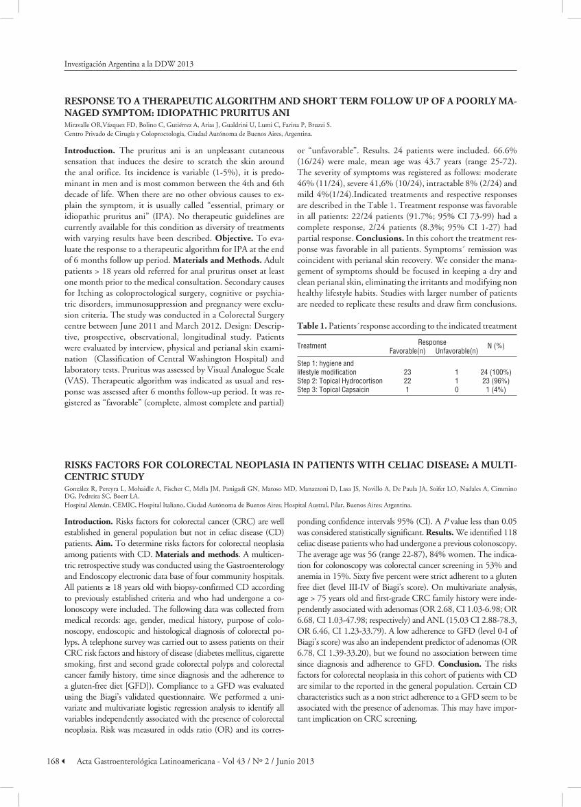
or “unfavorable”. Results. 24 patients were included. 66.6% (16/24) were male, mean age was 43.7 years (range 25-72). The severity of symptoms was registered as follows: moderate 46% (11/24), severe 41,6% (10/24), intractable 8% (2/24) and mild 4%(1/24).Indicated treatments and respective responses are described in the Table 1. Treatment response was favorable in all patients: 22/24 patients (91.7%; 95% CI 73-99) had a complete response, 2/24 patients (8.3%; 95% CI 1-27) had partial response. conclusions. In this cohort the treatment res-ponse was favorable in all patients. Symptoms´ remission was coincident with perianal skin recovery. We consider the mana-gement of symptoms should be focused in keeping a dry and clean perianal skin, eliminating the irritants and modifying non healthy lifestyle habits. Studies with larger number of patients are needed to replicate these results and draw firm conclusions.
table 1. Patients´response according to the indicated treatment
Treatment Response N (%) Favorable(n) Unfavorable(n)
Step 1: hygiene and lifestyle modification 23 1 24 (100%)Step 2: Topical Hydrocortison 22 1 23 (96%)Step 3: Topical Capsaicin 1 0 1 (4%)
risks factOrs fOr cOLOrectaL neOPLasia in Patients With ceLiac disease: a muLti-centric studyGonzález R, Pereyra L, Mohaidle A, Fischer C, Mella JM, Panigadi GN, Matoso MD, Manazzoni D, Lasa JS, Novillo A, De Paula JA, Soifer LO, Nadales A, Cimmino DG, Pedreira SC, Boerr LA.Hospital Alemán, CEMIC, Hospital Italiano, Ciudad Autónoma de Buenos Aires; Hospital Austral, Pilar, Buenos Aires; Argentina.
introduction. Risks factors for colorectal cancer (CRC) are well established in general population but not in celiac disease (CD) patients. aim. To determine risks factors for colorectal neoplasia among patients with CD. materials and methods. A multicen-tric retrospective study was conducted using the Gastroenterology and Endoscopy electronic data base of four community hospitals. All patients ≥ 18 years old with biopsy-confirmed CD according to previously established criteria and who had undergone a co-lonoscopy were included. The following data was collected from medical records: age, gender, medical history, purpose of colo-noscopy, endoscopic and histological diagnosis of colorectal po-lyps. A telephone survey was carried out to assess patients on their CRC risk factors and history of disease (diabetes mellitus, cigarette smoking, first and second grade colorectal polyps and colorectal cancer family history, time since diagnosis and the adherence to a gluten-free diet [GFD]). Compliance to a GFD was evaluated using the Biagi’s validated questionnaire. We performed a uni-variate and multivariate logistic regression analysis to identify all variables independently associated with the presence of colorectal neoplasia. Risk was measured in odds ratio (OR) and its corres-
ponding confidence intervals 95% (CI). A P value less than 0.05 was considered statistically significant. results. We identified 118 celiac disease patients who had undergone a previous colonoscopy. The average age was 56 (range 22-87), 84% women. The indica-tion for colonoscopy was colorectal cancer screening in 53% and anemia in 15%. Sixty five percent were strict adherent to a gluten free diet (level III-IV of Biagi’s score). On multivariate analysis, age > 75 years old and first-grade CRC family history were inde-pendently associated with adenomas (OR 2.68, CI 1.03-6.98; OR 6.68, CI 1.03-47.98; respectively) and ANL (15.03 CI 2.88-78.3, OR 6.46, CI 1.23-33.79). A low adherence to GFD (level 0-I of Biagi’s score) was also an independent predictor of adenomas (OR 6.78, CI 1.39-33.20), but we found no association between time since diagnosis and adherence to GFD. conclusion. The risks factors for colorectal neoplasia in this cohort of patients with CD are similar to the reported in the general population. Certain CD characteristics such as a non strict adherence to a GFD seem to be associated with the presence of adenomas. This may have impor-tant implication on CRC screening.

169Acta Gastroenterológica Latinoamericana - Vol 43 / Nº 2 / Junio 2013
Investigación Argentina a la DDW 2013
endOscOPÍaPrevaLence Of fLat POLyPs and histOLOgicaL diagnOsis in cOLOrectaL cancer screening POPuLatiOnDiehl M, Canseco S, Dalessandro M,Manzotti LN, Braner M, Bolino C, Caro LE, Cerisoli CL.Gedyt, Gastroenterology Hospital Dr Carlos Bonorino Udaondo, Ciudad Autónoma de Buenos Aires, Argentina.
introduction. Colorectal cancer (CRC) has a high mortality rate, is preventable andcurable if it is early diagnosed. Adeno-mas, preneoplastic lesions, have different morphology, being flatlesions generally more aggressive in the adenoma-carcinoma sequence. Accuracy in flat adenomasdetection during white light endoscopic examination is suboptimal and high defini-tion diagnostic techniquesare not available worldwide. For these reasons the true prevalence of these polyps is controver-sial. Objective. To estimate the prevalence and histological diagnosis of flat polyps in adults evaluated for CRC screening. materials and methods. We included consecutive adults ≥ 18 years old referred for screening.The study was conducted in a gastroenterology and endoscopy ambulatory center in Bue-nos Aires,Argentina, between April and July 2012. Poor bowel preparation (Boston scale < 6), history of colon surgeryand incomplete studies were excluded. Design: Prospective, des-criptive and cross-sectional study.Interventions: Polyethylene glycol (PEG) lavage solution or phosphates, with or without bisacodyl wereused for bowel preparation. Colonoscopies were performed under sedation with Olympus 160//180 seriesequi-pment. The resection / biopsy of lesions were performed accor-ding to endoscopists daily practice.Biopsies were evaluated by pathologists specialized in gastroenterology. Statistical analysis: VCCstat 2.0;95% CI were estimated; Chi square test. results.
2230 patients were analyzed, 1893 were included;average age was 58 years, 55.5% (1051/1893) were female. 1. The preva-lence of flat polyps was 87/1893 (4.6%; 95% CI 3.7 to 5.7). 100 flat lesions were found in 87 patients. 47% (47/100) were flat elevated polypsand 58% (58/100) were ≥ 10mm. 2. The frequency distribution of histological diagnosis was registered asfollows: tubular adenomas 39/100 (39%; 95% CI 29-49), hyperplasic polyps 28/100 (28%; 95% CI 19-38),serrated ade-nomas 22/100 (22%, 95% CI 14-39), tubulo-villous adeno-mas 10/100 (10%; 95% CI 5-18) andadenocarcinoma 1/100 (1%, 95% CI 0 - 5.4). There was no statistically significant di-fference in theprevalence of flat polyps between genders (p=ns) and individuals ≥ and < 50years old (NS). conclusions. In adults studied for CRC screening the prevalence of flat polyps was lower than 5%. No statistically significant difference was found in the prevalence between genders and individuals < and ≥50 years. The majority of flat polyps were adenomas, located in the right colon and smaller than 10 mm. Considering the misdiagnosis rate reported in the literature and lack of high definitions endoscopy methods available inmost endoscopic units, monitoring quality indicators for colonoscopy should be the main strategy to optimize diagnosis and consequently CRC prevention.
finding sessiLe serrated adenOmas: is it POssibLe tO identify them during cOn-ventiOnaL cOLOnOscOPy?Pereyra L, Gómez EJ, González R, Panigadi GN, Fischer C, Meuli F, Babot G, Torres AG, Correa L, Luna P, Pedreira SC, Cimmino DG, Boerr LA.Hospital Alemán, Ciudad Autónoma de Buenos Aires, Argentina.
background. Proximal colorectal cancer may arise from sessile serrated adenomas (SSA). Recognition of these lesions during colonoscopy can optimize the endoscopic approach. Objecti-ves. To identify specific endoscopic features of SSA with con-ventional colonoscopy. methods. Patients undergoing scree-ning colonoscopies from January 2010 to September 2012, in whom colonic polyps were found, were prospectively included in our study. Polyp morphology, location, polyp pit pattern following Kudo classification, and other previously reported features of SSA were evaluated. Histologic examination was conducted by a single blinded pathologist. Univariate and multivariate analysis were performed to identify independent predictors of SSA. Results were expressed in percentages and odds ratios (OR) with its corresponding 95% confidence inter-vals (CI). A P value < 0.05 was considered statistically signifi-cant. Sensitivity (SE), specificity (SP), positive likelihood ratio (PLR) and negative likelihood ratio (NLR) of the endoscopic features were also calculated.Results. A total of 272 patients
were included, and 440 polyps were evaluated (1.62 polyps per patient). Thirty-four polyps (8%) were SSA, 135 (31%) hyper-plastic and 249 (56%) adenomas. In the univariate analysis: flat morphology (P = 0.002; OR2.70, 95% CI 1.89–11.09), red colored surface (P = 0.000; OR 16.80,95% CI 6.39–32.09), right side location (P = 0.000; OR 16.21,95% CI 7.09–111.94), mucus cap (P = 0.000; OR 6.07,95% CI 4.76–20.01) and type II Kudo pit pattern (P = 0,001; OR 10.53, 95% CI 3,02-43.96) were associated with SSA. Multivariate analysis revealed that flat morphology (P = 0.002; OR 3.81,95% CI 1.53–9.09), red colored surface (P = 0 .000; OR 12.97, 95% CI 4.43–37.69), right side location (P = 0.000; OR 22.21, 95% CI 5.09–135.94) and mucus cap (P = 0.000; OR 8.77, 95% CI 3.76–20.44) were independent predictors of SSA. conclu-sion. We were able to identify specific features of SSA during conventional colonoscopy, which may help to identify these lesions, and therefore to optimize the endoscopic approach.

170 Acta Gastroenterológica Latinoamericana - Vol 43 / Nº 2 / Junio 2013
Investigación Argentina a la DDW 2013
risks factOrs fOr sPOradic adenOmas in Patients With uLcerative cOLitisGonzález R, Pereyra L, Gómez EJ, Panigadi GN, Mella JM, Fischer C, Hadad AR, Vizcaíno B, Pedreira SC, Cimmino DG, Boerr LA.Hospital Alemán, Ciudad Autónoma de Buenos Aires, Argentina.
introduction. Ulcerative colitis (UC) patients are at greater risk of developing colorectal dysplasia. Risks factors for spo-radic adenomas in patients without ulcerative colitis are well established but few studies have examined clinical factors for sporadic adenomas in patients with ulcerative colitis. aim: To determine risks factors for sporadic adenomas among patients with ulcerative colitis. materials and methods. A retrospective study was conducted using the Gastroenterology, Endoscopy and Pathology electronic data base of a community hospital. We included adult patients with UC colitis who had undergone a colonoscopy. The following data was collected from medical records: age, gender, endoscopic and histological diagnosis of colorectal polyps and inflammatory pseudopolyps. A telephone survey was carried out to assess patients on their CRC risk fac-tors and history of disease (diabetes mellitus, cigarette smoking, hypertension, dyslipidemia, first and second grade colorectal polyps and colorectal cancer family history, disease duration, extent of colitis, primary sclerosing cholangitis (PSC) and the use of 5-acetyl salicylic acid, inmunomodulators and biologics). We performed a univariate analysis to identify all variables asso-ciated with the presence of sporadic adenomas in patients with ulcerative colitis. Risk was measured in odds ratio (OR) and
its corresponding confidence intervals 95% (CI). A P value less than 0.05 was considered statistically significant.Results. A total of 107 patients with UC who had undergone a colonoscopy were included. The average age was 46 (range 16-81), 52% wo-men. Respect to the disease extent proctitis or ulcerative proc-tosigmoidtis was found in 32 patients (30%), left side colitis in 28 patients (26%), extensive colitis in 19 patients (18%) and pancolitis in 28 patients (26%). Five patients (5%) had PSC. On maintenance therapy, 44% received low dosis of 5-acetyl sa-licylic acid, 13% inmunomodulators drugs and 7% anti-tumor necrosis factor antibodies. We identified sporadic adenomas in 7 patients (6.5%) and colorectal cancer in 2 patients (1.8%). Disease duration in patients with sporadic adenomas was more than 10 years in 57%. Most of the polyps were sessile (86%) and 86% was < 1 cm, with predominant distribution in cecum and ascending colon (57%). Low grade dysplasia was found in all sporadic adenomas. On univariate analysis, duration of the disease more than 20 years and distal colitis were associated with sporadic adenomas (P = 0.015; OR 8.19, 95% CI 1.34-53.23 and P = 0.019; OR 0.00, 95% CI 0.10-0.00, respectively). conclusion. Disease duration and extent may be risks factors for sporadic adenomas in patients with UC.
micrOvesicuLar hyPerPLastic POLyPs: endOscOPic features and assOciatiOn With synchrOnic neOPLastic LesiOnsFischer C, Pereyra L, Gómez EJ, Panigadi GN, González R, Mella JM, Amante MF, Pedreira SC, Cimmino DG, Boerr LA.Hospital Alemán, Buenos Aires, Argentina.
background. Microvesicular hyperplastic polyps (MVHP) are thought to progress to sessile serrated adenomas (SSA), parti-cularly when located in the proximal colon. They frequently share the same molecular genetic abnormalities with these po-lyps, but their endoscopic features and association with syn-chronic neoplastic lesions (SNL) has not been well studied. aim. To determine the endoscopic features of MVHP in pa-tients who underwent colonoscopy and the association of these polyps with synchronic neoplastic lesions. methods. Reports from patients undergoing colonoscopy and polypectomy from September 2011 to September 2012, were obtained from the electronic database of a private community hospital. Micro-vesicular, goblet cell rich and mucin poor hyperplastic polyps were identified, and the clinical and endoscopic features of the patients were determined. SNL were defined by the presence of other lesions in the same colonoscopy: serrated adenomas, con-ventional adenomas, advanced neoplastic lesions (> 1cm, high grade dysplasia and/or > 75% of villous component) and can-cer. An univariate analysis was performed, looking for characte-ristics associated with MVHP and SNL. Results were expressed in odds ratio (OR) with its corresponding 95% confidence in-tervals (CI). A P value < 0.05 was considered statistically signi-ficant. results. A total of 253 patients with 324 hyperplastic
polyps were identified in the analysed period (72% MVHP, 19% goblet cell rich and 9% mucin poor hyperplastic polyps). Patients with MVHP were mainly men (58%) and had a mean age of 59 years old. Most of these polyps were sessile (85%) and small (94%), with predominant distribution in the left colon (60%). Ninety seven percent had type II Kudo’s pit pattern, 9% had red colored surface and 4% had mucus cap. SNL were present in 39% of these polyps: 72% conventional adenomas, 23% serrated adenomas (65% sessile, 35% traditional) and 5% cancer. ANL were found in 19% of conventional adenomas and 27% of serrated adenomas. In the univariate analysis we found that flat morphology was significantly associated with MVHP (P = 0.03; OR 3.78, 95% CI 1.057-16.09), whereas female gender (P = 0.000; OR 3.27, 95% CI 1.83-5.87), flat morphology (P = 0.003; OR 3.52, 95% CI 1.41-8.97) and mucus cap (P = 0.000; OR Inf.95% CI 2.76-Inf.) were sig-nificantly asso ciated with right side location of MVHP. We did not find characteristics associated withSNL. conclusion. In the present study we found specific endoscopic features of MVHP, particularly of those that are located in the proximal colon. These are similar to that observed in SSA. Further stu-dies are necessary to identify characteristics related to MVHP and their association with sessile serrated polyps and SNL.

171Acta Gastroenterológica Latinoamericana - Vol 43 / Nº 2 / Junio 2013
Investigación Argentina a la DDW 2013
PrevaLence Of POLyPs, adenOmas, advanced LesiOns and adenOcarcinOma in 45 tO 49 year-OLd PatientsCaro LE, Correa LM, Canseco S, BolinoC, CerisoliCL.Gedyt, Gastroenterology Hospital Dr Carlos Bonorino Udaondo, Ciudad Autónoma de Buenos Aires, Argentina.
introduction. Colon rectal cancer (CRC) can be prevented and cured if it is early diagnosed. Colonoscopy is the first-line procedure for screening CRC in average risk population. In 2002, Imperiale and collaborators reported that adenomas and advanced adenomas presented in 8.5% and3.5%, respec-tively, among persons 40 to 49 years of age. At this moment no recommendations for CRC screening in this population have been made. Objective. To estimate the prevalence of po-lyps, adenomas,advanced lesions and adenocarcinoma in 45 to 49 year- old patients. materials and methods. We included consecutive adults between the ages 45 and 49 years who per-formed colonoscopy because of gastrointestinal signs or symp-toms. We excluded patients at high risk for CRC, incomplete procedures and/or evidence of colonic tumor diagnosed by other methods. The study was conducted in a gastroenterolog-yand endoscopy ambulatory center in Buenos Aires, Argentina, between September 2010 and October 2011. Design: Descrip-tive, prospective and cross sectional study. Interventions: Po-lyethylene glycol (PEG)lavage solution or phosphates, with or without bisacodyl were used for bowel preparation. Colonos-copies were performed under sedation with Olympus 160/180
series equipment. Biopsies were evaluated bypathologists spe-cialized in gastroenterology. Indication for colonoscopy was registered. The protocol wasapproved by local IRB. Statistical analysis: VCCstat 2.0. 95% CI were estimated. results. 814 patientswere evaluated. 764 were included. 57% (440/764) were women; average age was 47 years.1.a) The global preva-lence of polyps was 160/764 (20%, 95% CI 18-24); 71/440 (16%, 95% CI 13-20) in women and89/324 (27%, 95% CI 22-32) in men. 1.b) The global prevalence of adenomas was 107/764 (14%, 95% CI 11-16), 59/324 (18%, 95% CI 14-22) in men and 48/440 (11%, 95% CI 8-14) in women; 1.c) The globalprevalence of advanced adenomas was 39/764 (5%, 95% CI 4-7) and of adenocarcinoma was 2/764 (0.1%, 95% CI 0-0, 7). The most common indications for colonoscopy were proctorrhagia, abdominal pain andaltered bowel habits. conclusions. The prevalence of lesions in this population is lower than average riskpopulation and it is similar to the infor-mation reported internationally. At the moment we do unders-tand thatthere is no evidence to indicate CRC screening in 45 to 49 individuals . Research on metabolic andepidemiological factors are needed to evaluate the biological behavior of CRC in young individuals.
accuracy Of kudO Pit Pattern cLasificatiOn With standard-definitiOn White Light (sd) and high definitiOn narrOW-band image (nbi) cOLOnOscOPy fOr Predic-ting adenOmas: a PrOsPective studyCimmino DG, Pereyra L, Gómez EJ, González R, Panigadi GN, Fischer C, Babot G, Torres AG, Meuli F, Correa L, Luna P, Pedreira SC, Boerr LA.Hospital Alemán, Ciudad Autónoma de Buenos Aires, Argentina.
introduction. The colonic pit pattern is recognized as an aid to the differential diagnosis between neoplastic and non-neo-plastic polyps. Accuracy of pit pattern visualization for predic-ting neoplastic polyps with non-magnification colonoscopy has not been clearly evaluated. Objective. Evaluate the precision of the Kudo pit pattern clasification to predict neoplasic le-sions of the colon with non-magnification colonoscopy and to determine accuracy according to endoscopists experience, type of colonoscope and polyp size. methods. Consecutive pa-tients referred for screening or surveillance colonoscopy were prospectively included. Colonoscopies were performed using a high-definition colonoscope with NBI capability without magnification (Olympus Exera II 180)(NBI) and a white light standard definition colonoscope (Olympus Actera 150) (SD). Kudo pit pattern classification was used to characterize colonic polyps: non-peoplastic (type I and II) and neoplastic (type III, IV and V). Morphology was evaluated using Paris classification. Endoscopist experience (> or < 5 years) was also consigned. Histological evaluation was performed by a blin-ded single pathologist. Sensitive, specificity, Positive likelihood ratio (PLR), and negative likelihood ratio (NLR) with its co-rrespondent 95 % confidence interval (CI) was calculated. Ac-curacy was compared according to: type of endoscope (NBI,
high definition or standard definition), endoscopist experience (> or < 5 years) and polyp size. Accuracy was expressed as per-centage. A X2 test was used for proportion comparison. A P value < 0.05 was considered statistically significant. results. A total of 440 polyps were found in 272 patients (1.6 polyps per patient). Patients were mostly males (54%) and mean age was 62 years old (range 30-90). The polyps average size was 8.2 mm, most frequent morphology was: 0-Is (68,6%) and 0-IIa (11,4%). Most frequent histology was: adenomas 42%, hyper-plastic 30% and serrated adenomas 10%. Polyps were assessed using: SD in 277/440 (63%) and in 163/440 (37%) with NBI colonoscopy. Overall sensitivity, specificity, PLR and NLR for predicting neoplastic lesion was: 73 % (95% CI 70-76), 88 % (85% CI 83-91), 6 and 0,3 respectively. Accuracy was signifi-cantly higher with NBI compared with SD colonoscopy (90% vs 70%, P = 0.045). Large polyps achieved also higher accuracy than small polyps: size > 5mm (84% vs 75%, P = 0.025) and size >10mm (90% vs 77%, P = 0.03). There was no difference between experienced and non-experienced colonoscopies (82% vs 76%, P = 0.3). conclusion. Visualization of pit pattern with non-magnification colonoscopy is useful for predicting neo-plastic lesions. Diagnostic accuracy is higher with NBI colo-noscopy and when evaluating larger polyps.
172 Acta Gastroenterológica Latinoamericana - Vol 43 / Nº 2 / Junio 2013
Investigación Argentina a la DDW 2013
PrevaLence Of advanced adenOma and adenOcarcinOma in Patients yOunger than 50 years Of ageWith rectaL bLeedingBraner Maribel, Canseco S, Diehl MP, Manzotti L, Bolino C, Dalessandro M, Cerisoli CL, CaroLE.Gedyt, Gastroenterology Hospital Dr Carlos Bonorino Udaondo, Ciudad Autónoma de Buenos Aires, Argentina.
introduction. Colorectal cancer (CRC) is a major cause of death from cancer inwestern world and rectal bleeding is a cli-nical presentation. In the young population with no risk fac-tors for CRC, its prevalence is lower and the etiology of rectal bleeding is often benign. For these reasons, theinvestigation of young patients who have rectal bleeding is frequently under-estimated. Objective. To estimate the prevalence of advanced adenomas and adenocarcinoma in patients < 50 years old refe-rred for rectal bleeding. materials and methods. We included consecutive adult patients 18 to 49 years of age who consulted at a gastroenterology and endoscopy ambulatory center in Bue-nos Aires, Argentina, between October 2011 and April 2012. We excluded patients at high risk for CRC, altered coagula-tion, andin complete studies except for those with stenosing carcinoma. Design: Prospective, descriptive, cross sectional stu-dy. Interventions: Polyethylene glycol (PEG) lavage solution or phosphates, with or without bisacodyl were used for bowel pre-paration. Colonoscopies were performed under sedation with Olympus 160/180 series equipment. The resection / biopsy of lesions were performed according to endoscopists’daily prac-tice. Biopsies were evaluated by pathologists specialized in gastroenterology and histology was valued as gold standard. Positive diagnosis consisted on advanced adenomas (> 1 cm,
villous component and high-grade dysplasia (HGD)) and / or adenocarcinoma. We also assessed whether there was anyre-lationship between age, gender or site of lesion and positive findings. Endoscopic and histological features were registered. The protocol was approved the local IRB. Statistical analysis: MedCalc 1,5; VCCstat 2.0 and 95% CI were estimated, Stu-dent Test, Chi square Test. Results. We analyzed 423patients, 47% (198/423) were women; average age was 37±8 years (ran-ge 19-49). 336/423 (79.4%, 95% CI 74-82) had hemorrhoi-ds. 1. The prevalence of advanced neoplasia in this population was 27/423 (6.4%, 95% CI 4.3-9.3), advanced adenoma was 17/423 (4.0%, 95% CI 2.4-6.5) and adenocarcinoma was 10/423(2.4%, 95% CI 1.2-4.4); morphologically 2 adenocar-cinoma were polyps, 2 were flat lesions (slightly elevated) and 6 were stenosing lesions. 2. Positive findings were significantly higher in patients ≥ 40 years(OR 3.29, 95% CI 1.4-7.7), equal in both genders (NS) and more prevalent in left colon. con-clusions. In our sample, 10 of 100 patients younger than 50 years with advanced adenomas and/or adenocarcinomapresent with rectal bleeding. This is lower than in older population. However, considering that CRC inyoung adults has a more aggressive biological behavior and mortality rate, diagnostic efforts should bemade when approaching these patients.
resect and discard strategy With standard-definitiOn White Light (sd) and high definitiOn White-Light With narrOW-band image (nbi) cOLOnOscOPy: a PrOsPecti-ve studyPereyra L, Gómez EJ, Panigadi GN, González R, Torres AG, Babot G, Meuli F, Correa L, Fischer C, Pedreira SC, Cimmino DG, Boerr LA.Hospital Alemán, Ciudad Autónoma de Buenos Aires, Argentina.
background. Accurate optical analysis of small colorectal polyps (<1cm) could allow a “resect and discard” strategy with survei-llance intervals determined based on these results. This strategy has been evaluated with narrow-band image (NBI), but not with standard-definition white light (SD) conventional colonoscopy. aim. To assess the feasibility and safety of the resect and discard approach using standard-definition white light (SD) and high de-finition with narrow-band image (NBI) colonoscopy. materials and methods. An observational prospective study was carried out between 2010–2012. Consecutive patients undergoing a colonos-copy with one or more polyps smaller than 10 mm were included. All procedures were performed with Olympus Actera 150 (SD) and Olympus Exera II 180 (NBI). Each polyp was categorized by two experienced endoscopist, as adenoma or non-adenoma accor-ding to Kudo pit pattern classification. Future post-polypectomy surveillance interval was suggested according to optical analysis. Histological diagnosis was evaluated by a blinded single patholo-gist. Following histopathology, post-polypectomy surveillance interval was subsequently re-assigned. Accordance between en-doscopy and histology directed surveillance strategies were cal-culated. Sensitive, specificity, Positive likelihood ratio (PLR), and negative likelihood ratio (NLR) with it is correspondent 95 % confidence interval (CI) for predicting adenomas were
also calculated. results. A total of 194 patients with 339 small colorectal polyps were included for analysis. Patients were mostly males (54%), mean age was 62 years old (range 30-90) and polyps average size was 8.2 mm. Advance histological features (high gra-de dysplasia or > 75% villous component) were present in 9/339 (3%). Sensitivity, specificity, PLR and NLR for predicting neo-plastic lesion with SD colonoscopy was 63% (95% CI 58-66), 90% (95% CI 80-94), 6,9 and 0,4 respectively. The sensitivity, specificity, PLR and NLR for predicting neoplastic lesion with NBI colonoscopy was 78 % (95% CI 71-83), 86% (95% CI 76-93), 5,7 and 0,25 respectively. The endoscopy-directed surveillan-ce strategy was in accordance with the histology-directed strategy in 102/125 (82%) patients in the NBI group and in 158/214 (74%) in the SD colonoscopy group. This difference was not statistically significant (P = 0.14). In 5% of the SD colonoscopy, and in 6% of the NBI colonoscopy group the endoscopy-directed approach would have resulted in early surveillance (P = 0.3). NBI colonosco-py was associated with smaller percentages of delayed surveillance endoscopy-directed approach (13% vs 19%, P = 0.04). conclu-sions. We found a low prevalence of advanced histological features in small colonic polyps. NBI colonoscopy seems to be safer for resect and discard strategy, been associated with smaller percentage of delayed surveillance endoscopy-directed approach.

173Acta Gastroenterológica Latinoamericana - Vol 43 / Nº 2 / Junio 2013
Investigación Argentina a la DDW 2013
derivatiOn Of the fOrcePs scOre (fundic POLyP scOre): a cLinicaL PredictiOn ruLe tO identify fundic gLand POLyPs during uPPer gi endOscOPyLuna P, Pereyra L, Zamora R, Pedreira SC, Mohaidle A, Fischer C, Boerr LA, Cimmino DG.Hospital Alemán, Ciudad Autónoma de Buenos Aires, Argentina.
background. Fundic gland polyps (FGP) represent a benign and frequent finding during routine upper GI endoscopy (UGIE). Unnecessary polypectomy is associated with a direct increase in endoscopy costs, and potentially with procedure related complications. aim. to develop a simple endoscopic score to reliably identify fundic gland polyps during UGIE. methods. A clinical prediction rule was developed from a pros-pective cohort of 95 patients in whom an UGIE was carried out, gastric polyps were found and their histology was availa-ble. Endoscopies were performed by two experienced specia-lists using Olympus GIF-H 150 and 180 endoscopes. Based on existing literature and previous information on this matter, patient’s demographic and clinical data, as well as endoscopic features were assessed. Patients with previously known gastric polyps were excluded. All analysed variables with a P value of less than 0.25 in univariate analysis were entered into the mo-del. Backwards stepwise logistic regression was used to identify factors that predict fundic gland polyps. The identified factors, and others considered biologically important by the investiga-tors, were used to develop the prediction model. Calibration of the model was evaluated with Hosmer-Lemeshow test and the discrimination power with area under the ROC curve.
results. A total of 95 patients presenting gastric polyps were included for analysis. Seventy (74%) were women, and the average age was 60 years old (22-93). Histology revealed that 57 polyps were FGP, 18 hyperplastic, 3 tubular adenomas, 2 adenocarcinomas, 1 was inflammatory in nature and in 14 pa-tients the finding was not consistent with a polyp, but with inflammatory phenomena instead. A total of 6 independent endoscopic variables were identified and selected for the model construction: polyp surface similar to the surrounding mucosa, detach in toto “like a grape” with a slight lateral movement with the biopsy forceps, visible neck, location in fundus or cor-pus, size < 1cm, number of polyps: more than1 but less than 20. The model was well calibrated (Hosmer-Lemeshow P = 0.71). The area under the ROC curve was 0.90. Sensitivity was 100% (95% CI 77-100), specificity 100% (95% CI 46-100), positive predictive value 100% and negative predictive value 100%. conclusion. A simple endoscopic score can accurately discriminate fundic gland polyps during routine UGIE, hel-ping to avoid unnecessary polipectomies. An external valida-tion of this model is still required.
increase Of neOPLasia detectiOn rate after the imPLementatiOn Of a PrOgram tO imPrOve the quaLity Of cOLOnOscOPyCassella F, Fernández JL, Wonaga A, Arnao Dellamea G, Di Paola L, Ubeira Salim R, Viola L.Centro Integral de Gastroenterología, Ciudad Autónoma de Buenos Aires, Argentina.
studies. We statistically compared dichotomous variables with chi square test and continuous variables with t student test, considering a P value < 0.05 as significant. Results. Mean age was 57.29 ± 12.01 years old in group 1 and 56.83 ±11.64 years old in group 2 (P = 0.27). Seyen hundred and twenty eight (46.3%) patients in group 1 and 780 (49.3%) in group 2 were men (P = 0.10). In 854 cases from group 1 (54.3%) and in 833 from group 2 (52.6%) the colonoscopy was due to screening (P = 0.37). Cecal intubation was documented in 1,384 cases from group 1 (88.0%) (inform of the reached site was not available in 92 cases) and in 1,534 from group 2 (96.9%) (P < 0.0001). Neoplasia detection rates in groups 1 and 2 were, respecti-vely: neoplasias 288 (18.3%) and 427 (27.0%) (P < 0.0001), high risk adenomas 76 (4.8%) and 142 (9.0%) (P < 0.0001), carcinomas 16 (1.0%) and 21 (1.3%) (P = 0.52), right colon adenomas 112 (7.1%) and 154 (9.7%) (P = 0.01), and adeno-mas in screennig 141 (16.5%) and 233 (28.0%) (P < 0.0001). conclusions. The implementation of a program to improve the quality of colonoscopy allowed to detect a significantly hig-her number of neoplasias, including high risk and right colon adenomas. Due to the low incidence, there were not significant differences in the cancer detection rates.
introduction. The measures to improve the quality of co-lonoscopy and the evaluation of the results are demanded to the endoscopy centers in the last years. Objetive. To assess if the implementation of a program to improve the quality of colonoscopy in our center allowed to achieve a better neopla-sia detection rate (adenoma and/or carcinoma). Patients and methods. From May 2009 to March 2010, we prospectively evaluated 1,573 consecutive colonoscopies (group 1). After this period we implemented a quality program including the ins-truction of patients for bowel preparation and of endoscopists for study performance (cecal intubation, withdrawal time lon-ger than 6 minutes) and for inform redaction (indication of co-lonoscopy, use of Boston scale for bowel preparation, adequate information about the extention of the procedure). Then, from February 2011 to January 2012, we prospectively evaluated another 1,583 consecutive colonoscopies (group 2). To eva-luate the homogeneity of both groups we compared mean age, sex, and number of screening colonoscopies, including high and average risk endoscopies. As outcomes, we considered cecal intubation rate correctly reported in the inform and neoplasia detection rates, including high risk adenomas (> 1 cm, villous, tubular-villous, serrated and/or high rate displasia), carcino-mas, right colon adenomas and adenomas detected in screening
174 Acta Gastroenterológica Latinoamericana - Vol 43 / Nº 2 / Junio 2013
medicatiOn, LifestyLe and diet: are they assOciated factOrs fOr adenOmas? there is stiLL a LOng Way tO gOBolinoC, Rodríguez P, Piñeyro C, Canseco S, Cádiz C, Sánchez C, Banchero I, Cerisoli CL, Caro LE.Gedyt, Ciudad Autónoma de Buenos Aires.
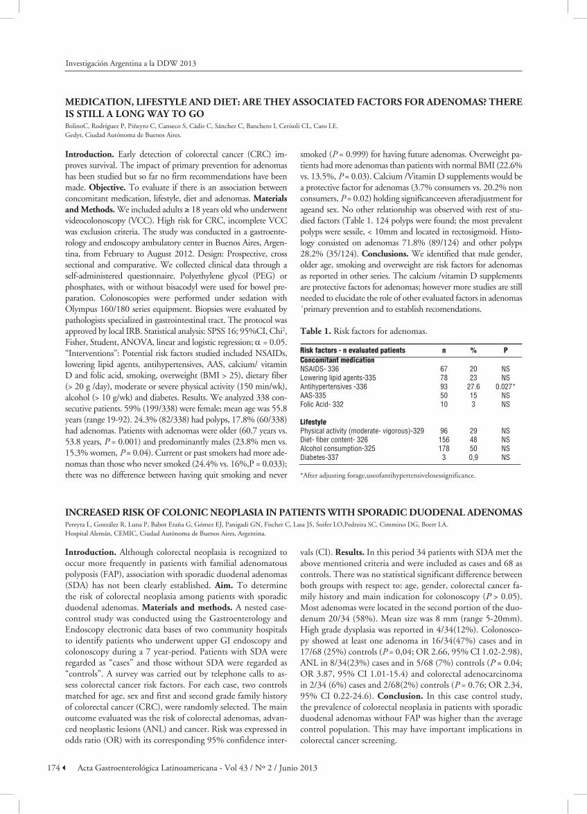
introduction. Early detection of colorectal cancer (CRC) im-proves survival. The impact of primary prevention for adenomas has been studied but so far no firm recommendations have been made. Objective. To evaluate if there is an association between concomitant medication, lifestyle, diet and adenomas. materials and methods. We included adults ≥ 18 years old who underwent videocolonoscopy (VCC). High risk for CRC, incomplete VCC was exclusion criteria. The study was conducted in a gastroente-rology and endoscopy ambulatory center in Buenos Aires, Argen-tina, from February to August 2012. Design: Prospective, cross sectional and comparative. We collected clinical data through a self-administered questionnaire. Polyethylene glycol (PEG) or phosphates, with or without bisacodyl were used for bowel pre-paration. Colonoscopies were performed under sedation with Olympus 160/180 series equipment. Biopsies were evaluated by pathologists specialized in gastrointestinal tract. The protocol was approved by local IRB. Statistical analysis: SPSS 16; 95%CI, Chi2, Fisher, Student, ANOVA, linear and logistic regression; α = 0.05. “Interventions”: Potential risk factors studied included NSAIDs, lowering lipid agents, antihypertensives, AAS, calcium/ vitamin D and folic acid, smoking, overweight (BMI > 25), dietary fiber (> 20 g /day), moderate or severe physical activity (150 min/wk), alcohol (> 10 g/wk) and diabetes. Results. We analyzed 338 con-secutive patients. 59% (199/338) were female; mean age was 55.8 years (range 19-92). 24.3% (82/338) had polyps, 17.8% (60/338) had adenomas. Patients with adenomas were older (60.7 years vs. 53.8 years, P = 0.001) and predominantly males (23.8% men vs. 15.3% women, P = 0.04). Current or past smokers had more ade-nomas than those who never smoked (24.4% vs. 16%,P = 0.033); there was no difference between having quit smoking and never
smoked (P = 0.999) for having future adenomas. Overweight pa-tients had more adenomas than patients with normal BMI (22.6% vs. 13.5%, P = 0.03). Calcium /Vitamin D supplements would be a protective factor for adenomas (3.7% consumers vs. 20.2% non consumers, P = 0.02) holding significanceeven afteradjustment for ageand sex. No other relationship was observed with rest of stu-died factors (Table 1. 124 polyps were found; the most prevalent polyps were sessile, < 10mm and located in rectosigmoid. Histo-logy consisted on adenomas 71.8% (89/124) and other polyps 28.2% (35/124). conclusions. We identified that male gender, older age, smoking and overweight are risk factors for adenomas as reported in other series. The calcium /vitamin D supplements are protective factors for adenomas; however more studies are still needed to elucidate the role of other evaluated factors in adenomas ´primary prevention and to establish recomendations.
table 1. Risk factors for adenomas.
Risk factors - n evaluated patients n % PConcomitant medication NSAIDS- 336 67 20 NSLowering lipid agents-335 78 23 NSAntihypertensives -336 93 27.6 0.027*AAS-335 50 15 NSFolic Acid- 332 10 3 NS Lifestyle Physical activity (moderate- vigorous)-329 96 29 NSDiet- fiber content- 326 156 48 NSAlcohol consumption-325 178 50 NSDiabetes-337 3 0,9 NS
*After adjusting forage,useofantihypertensivelosessignificance.
increased risk Of cOLOnic neOPLasia in Patients With sPOradic duOdenaL adenOmasPereyra L, González R, Luna P, Babot Eraña G, Gómez EJ, Panigadi GN, Fischer C, Lasa JS, Soifer LO,Pedreira SC, Cimmino DG, Boerr LA.Hospital Alemán, CEMIC, Ciudad Autónoma de Buenos Aires, Argentina.
introduction. Although colorectal neoplasia is recognized to occur more frequently in patients with familial adenomatous polyposis (FAP), association with sporadic duodenal adenomas (SDA) has not been clearly established. aim. To determine the risk of colorectal neoplasia among patients with sporadic duodenal adenomas. materials and methods. A nested case-control study was conducted using the Gastroenterology and Endoscopy electronic data bases of two community hospitals to identify patients who underwent upper GI endoscopy and colonoscopy during a 7 year-period. Patients with SDA were regarded as “cases” and those without SDA were regarded as “controls”. A survey was carried out by telephone calls to as-sess colorectal cancer risk factors. For each case, two controls matched for age, sex and first and second grade family history of colorectal cancer (CRC), were randomly selected. The main outcome evaluated was the risk of colorectal adenomas, advan-ced neoplastic lesions (ANL) and cancer. Risk was expressed in odds ratio (OR) with its corresponding 95% confidence inter-
vals (CI). results. In this period 34 patients with SDA met the above mentioned criteria and were included as cases and 68 as controls. There was no statistical significant difference between both groups with respect to: age, gender, colorectal cancer fa-mily history and main indication for colonoscopy (P > 0.05). Most adenomas were located in the second portion of the duo-denum 20/34 (58%). Mean size was 8 mm (range 5-20mm). High grade dysplasia was reported in 4/34(12%). Colonosco-py showed at least one adenoma in 16/34(47%) cases and in 17/68 (25%) controls (P = 0,04; OR 2.66, 95% CI 1.02-2.98), ANL in 8/34(23%) cases and in 5/68 (7%) controls (P = 0.04; OR 3.87, 95% CI 1.01-15.4) and colorectal adenocarcinoma in 2/34 (6%) cases and 2/68(2%) controls (P = 0.76; OR 2.34, 95% CI 0.22-24.6). conclusion. In this case control study, the prevalence of colorectal neoplasia in patients with sporadic duodenal adenomas without FAP was higher than the average control population. This may have important implications in colorectal cancer screening.
Investigación Argentina a la DDW 2013

175Acta Gastroenterológica Latinoamericana - Vol 43 / Nº 2 / Junio 2013
aPPrOPriate indicatiOns fOr cOLOnOscOPy as a quaLity indicatOr: exPerience in an OPen accessendOscOPy center in argentineBolino C, Banchero I, Sánchez C, Cerisoli CL, Caro LE.Gedyt, Gastroenterology Hospital Dr Carlos Bonorino Udaondo, Ciudad Autónoma de Buenos Aires, Argentina.
introduction. The effectiveness of colonoscopy, recommen-ded method for diagnosis and treatment of colonic disorders, is determined by the quality of the procedure. Several indica-tors have been selected for its performance, and the appropria-te indication for the procedure is a clue to avoid unnecessary studies. Objectives. 1. Estimate the prevalence of appropriate indications for colonoscopy according to the American Society for Gastrointestinal Endoscopy (ASGE). 2. Describe the fre-quency distribution of indications. materials and methods. We retrospectively reviewed videocolonoscopy (VCC)reports from adult patients ≥ 18 years who consulted at a gastroentero-logy and endoscopy ambulatory center in Buenos Aires, Argen-tine, between october, 2009 and may, 2012. Incomplete VCC, high risk forcolorectal cancer (CRC), no indication for VCC in the reports were exclusion criteria. Design: Descriptiveand cross sectional study. Interventions: Polyethylene glycol (PEG) lavage solution or phosphates, with orwithout bisacodyl were used for bowel preparation. Colonoscopies were performed un-der sedation with Olympus 160/180 series equipment. Biopsies were evaluated by pathologists specialized in gastroenterology. VCC indications were categorized into “appropriate” or “inap-propriate/ uncertain”. “Appropriate” were those recommen-ded by the ASGE in 2006 in the expert consensus on quality indicators for colonoscopy. “Inappropriate / uncertain “were those not recommended. Informed Consent was signedbefore the procedure. Statistical analysis: VCCstat 2.0; 95% CI were estimated. results. We evaluated 43,182 patients. 42,750 were included. 57% (24,57/42,750) were women. Mean age was 58.6 years (range18-94). 1. The prevalence of “appropriate
indications” for colonoscopy was 41,826/42,750 (97.8%, 95% CI 97-98) the prevalence of “inappropriate / uncertain indica-tions” was 924/42,750 (2.2%, 95% CI 2–2,3) (Table1). 2. The most prevalent indications were: CRC screening in average risk population in 13,048/42,750 (30.5%,95% CI 30-31), post po-lypectomy surveillance in 7,025/42,750 (16.5%, 95% CI 16-17), CRC screening inpatients with positive family history in 3,032/42,750 (7.1%, 95% CI 7-7.4), evaluation of unexplai-ned gastrointestinal bleeding in 6,783/42,750 (16%,95% CI 15.5-16,6), altered bowel habits in 3,684/42,750 (8.6%,95% CI 8.4-8.9), abdominal pain in 3,620/42,750 (8.5%, 95% CI 8.2 -8.7). conclusions. In this sample, most of the indications were “appropriate “according to the ASGE. Many indications categorized as” inappropriate or uncertain” are signs and/or symptoms that many gastroenterologists and endoscopists con-sider as “appropriate”. Our data suggest that more indications should be added to the “appropriate colonoscopy indications list” as re-staging patients post neoadyuvance for rectal cancer and search malignancy ofunknown primary origin.
table 1. Inappropriate Indications.
Inappropriate indications n %Diverticular disease 278 30Proctological disease 237 26Signs (wasting, malaise, etc.) 179 19.3Miscellaneous 101 10.8Rectal cancer re-staging 55 6Cancer staging 46 4.9Search malignancy of unknown primary origin 28 3
Total 924 100
Investigación Argentina a la DDW 2013