Functional consequences of ε AChR subunit truncating ...hss.ulb.uni-bonn.de/2005/0533/0533.pdf ·...
100
Functional consequences of ε AChR subunit truncating mutations linked to Congenital Myasthenic Syndrome Dissertation zur Erlangung des Doktorgrades (Dr. rer. nat.) der Mathematisch-Naturwissenschaftlichen Fakultät der Rheinischen Friedrich-Wilhelms-Universität Bonn vorgelegt von Cristian Gurgui aus Bukarest, Rumänien Bonn 2004
Transcript of Functional consequences of ε AChR subunit truncating ...hss.ulb.uni-bonn.de/2005/0533/0533.pdf ·...
Functional consequences of ε AChR subunit truncating mutations
linked to Congenital Myasthenic Syndrome
Dissertation
zur
Erlangung des Doktorgrades (Dr. rer. nat.)
der
Mathematisch-Naturwissenschaftlichen Fakultät
der
Rheinischen Friedrich-Wilhelms-Universität Bonn
vorgelegt von
Cristian Gurgui
aus
Bukarest, Rumänien
Bonn 2004

The work for this PhD thesis was performed from September 2002 to October 2004 in the
laboratory of Prof. Dr. Dieter Swandulla at the Institute of Physiology II, Bonn, Germany
Angefertigt mit Genehmigung der Mathematisch-Naturwissenschaftlichen Fakultät der
Rheinischen Friedrich-Wilhelms-Universität Bonn
1. Referent: Prof. Dr. Dieter Swandulla
2. Referent: Prof. Dr. Volker Herzog
Tag der Promotion: 06.04.05
Diese Dissertation ist auf dem Hochschulschriftenserver der ULB Bonn http://hss.ulb.uni-
bonn.de/diss_online elektronisch publiziert
Erscheinungsjahr: 2005

Acknowledgements
Thanks to Prof. Dr. Dieter Swandulla for helpful discussion, for advice, for revising the thesis
and evaluating it, and for his true friendship. Thank you very much for giving me the
opportunity to pursue my PhD thesis in Electrophysiology.
I would also like to express my gratitude to my supervisor Dr. Michael Hans for the kind
support throughout the thesis. You started by teaching me the first steps in Electrophysiology,
and made me discover the vast world of the Patch-clamp technique. Thanks for your patience
in the time you have spent next to me during recordings, analysis and writing, while
answering to my questions, making suggestions, correcting my errors, and guiding me to
achieving this goal. Without your kind support and friendship, this work would not have been
possible.
I wish to express my sincere thanks to Prof. Dr. Volker Herzog for kindly accepting to be a
co-referee of my major exam, for the support, and for evaluating the thesis.
I am greatly indebted to PD Dr. Gerhild van Echten-Deckert for being examiner of my PhD
defence, for practical and moral support, suggestions, comments, and friendship.
Special thanks also go to Prof. Dr. Gabriele König, who also kindly agreed to be the examiner
of my minor exam.
I am very grateful to Prof. Dr. Peter Propping for the kind support and true friendship
throughout the time I was member of Graduiertenkolleg GRK 246 (“Pathogenesis of Diseases
of the Central Nervous System”) in Bonn.
I acknowledge with thanks and appreciation the role of Joachim Sternberg and Hanne Bock
for their excellent technical assistance and friendship.
Thanks to my girlfriend Mihaela Dragusin for patience and moral support throughout the time
we have spent together. Thank you for proofreading the thesis and for the useful comments
and suggestions. I would not have reached here without your support either.
Special thanks go to my family in Romania who spiritually supported and trusted me in all
these years. Also, many thanks to all my friends (you all know who you are).

Scientific Work
Publications
Dragusin M., Gurgui C., Schwarzmann G., Hoernschemeyer J., and van Echten-Deckert
G. (2003) Metabolism of the unnatural anticancer lipid safingol (L-threo-dihydrosphingosine)
in cultured cells. J Lipid Res: 44(9): 1772-9.
Poster sessions
03/04: “Functional consequences of ε AChR subunit truncating mutations linked to
Congenital Myasthenic Syndrome”, at the 83rd Annual Congress of the “Deutsche
Physiologische Gesellschaft e.V.”, Leipzig, Germany. Authors: Gurgui C., Kraner S.,
Steinlein O. K., Swandulla D., and Hans M.
05/03: “Functional consequences of ε AChR subunit truncating mutations linked to
Congenital Myasthenic Syndrome”, at the 29th Göttingen Neurobiology Conference,
Göttingen, Germany. Authors: Gurgui C., Kraner S., Steinlein O. K., Swandulla D., and Hans
M.
06/03: “Metabolism of the unnatural anticancer lipid safingol (L-threo-dihydrosphingosine) in
cultured cells”, at the 2nd International Charleston Ceramide Conference, Como, Italy.
Authors: Dragusin M., Gurgui C., Schwarzmann G., Hoernschemeyer J., and van Echten-
Deckert G. (Poster Prize)
09/01: “Impedance Spectrometry in Electrodes Characterization“, at the 12th Romanian
International Congress on Chemistry and Chemical Engineering, Bucharest, Romania.
Authors: Popescu A., Lisca G., Gurgui C., Voinea C., and Vasilco R.
Presentations
12/03: Presentation “Functional consequences of ε AChR subunit truncating mutations linked
to Congenital Myasthenic Syndrome”, at the Graduiertenkolleg Joint Meeting of the
Neuroscience Research Training Groups 246 (“Pathogenesis of Diseases of the Central
Nervous System”, Bonn University) and 320 (“Pathological Processes of the Nervous System:
From Gene to Behavior”, Heinrich-Heine-Universität Düsseldorf), Bornheim-Walberberg,
Germany

5
Table of Contents Abbreviations............................................................................................................................ 8
Abstract ................................................................................................................................... 10
1 Introduction .................................................................................................................... 11
1.1 Neurological diseases. Congenital myasthenic syndromes. General remarks ......... 11
1.2 Ligand-gated ion channels of fast chemical synapses.............................................. 13
1.2.1 Nicotinic acetylcholine receptors (AChRs)...................................................... 14
1.2.1.1 Subtypes of nicotinic muscle AChR ............................................................ 15
1.2.1.2 Size and shape of the receptor molecule ...................................................... 15
1.2.1.3 The AChR topology ..................................................................................... 16
1.2.1.4 Mechanisms of ACh receptor activation...................................................... 18
1.3 The neuromuscular junction (NMJ) ......................................................................... 21
1.3.1 From nerve action potential to muscle action potential ................................... 21
1.3.2 CMS caused by defects in AChR..................................................................... 24
1.3.2.1 Slow-channel CMS ...................................................................................... 24
1.3.2.2 Fast-channel CMS........................................................................................ 25
1.3.2.3 CMS caused by low-expressor mutations .................................................... 26
1.4 The goal of the study................................................................................................ 28
2 Materials and Methods .................................................................................................. 29
2.1 Cell culture ............................................................................................................... 29
2.2 Transient transfection............................................................................................... 29
2.3 Patch-clamp recordings ............................................................................................ 30
2.3.1 Data analysis of the patch-clamp recordings.................................................... 31
2.3.1.1 Analysis of whole-cell recordings................................................................ 31
2.3.1.1.1 Dose-response curves............................................................................. 31
2.3.1.1.2 AChR desensitization kinetics and current density................................ 32
2.3.1.2 Analysis of single-channel recordings ......................................................... 32
2.4 Materials and suppliers............................................................................................. 34
6
3 Results ............................................................................................................................. 37
3.1 Pharmacological and biophysical properties of wild-type, ε-subunit lacking, and ε-
mutant AChRs inferred from whole-cell recordings............................................................ 37
3.1.1 Introduction to whole-cell recordings .............................................................. 37
3.1.2 The properties of wild-type receptor ................................................................ 38
3.1.3 The properties of ε-subunit lacking receptor.................................................... 39
3.1.4 The properties of ε-mutant receptors................................................................ 42
3.1.5 Summary of the whole-cell data....................................................................... 48
3.2 Single-channel recordings ........................................................................................ 49
3.2.1 Introduction to single-channel recordings and data kinetic analysis................ 49
3.2.2 Unitary current and slope conductance ............................................................ 51
3.2.3 Kinetic analysis ................................................................................................ 52
3.2.3.1 Gating-kinetics of wild-type receptor .......................................................... 52
3.2.3.2 Gating-kinetics of ε-subunit lacking receptor .............................................. 55
3.2.3.3 Gating-kinetics of ε-mutant receptors .......................................................... 58
3.2.3.4 The spontaneous activity of AChR .............................................................. 63
3.2.4 The correlation between the whole-cell- and the single-channel data ............. 66
3.2.5 Summary of the single-channel data ................................................................ 68
4 Discussion........................................................................................................................ 69
4.1 CMS mutations and their localization...................................................................... 69
4.2 Structure, formation and membrane insertion of the AChR .................................... 70
4.3 Mutation ε911delT in the M3 segment leads to loss of function.............................. 71
4.4 Mutations within the TM3-4 cytoplasmic loop........................................................ 72
4.4.1 Mutation ε1101insT ......................................................................................... 72
4.5 Structural elements within the TM3-4 loop that influence channel-gating.............. 73
4.5.1 Role of mutations ε1293insG and ε1206ins19................................................. 73
4.6 Mutation ε1030delC elicits switch in gating pattern................................................ 75
4.7 Outlook for treatment of CMS patients.................................................................... 76
4.8 Summary .................................................................................................................. 77

7
Appendix ................................................................................................................................. 79
The patch-clamp technique .................................................................................................. 79
i. General remarks ....................................................................................................... 79
ii. Five patch-clamp measurement configurations........................................................ 80
iii. The patch-clamp setup ............................................................................................. 82
iv. The main steps during a patch-clamp experiment.................................................... 83
References ............................................................................................................................... 84
Eidesstattliche Erklärung ...................................................................................................... 99
Curriculum Vitae ................................................................................................................. 100

8
Abbreviations
A Agonist Å Angstrom AcCoA Acetyl-Coenzyme A ACh Acetylcholine ACHE Acethylcholinesterase AChR Nicotinic Acetylcholine Receptor AD Analog-Digital AP Action Potential BCS Bovine Calf Serum bp Base pairs CAP Cell-Attached Patch cDNA Complementary DNA CHAT Choline Acetyltransferase CHRNE Cholinergic Receptor, Nicotinic, ε-polypeptide gene CMAP Compound Muscle Action Potential CMS Congenital Myasthenic Syndrome ColQ Collagenic tail subunit of Acetylcholinesterase D Desensitization 3,4-DAP 3,4-Diaminopyridine Del Deletion DMEM Dulbecco’s Modified Eagle Medium DNA Deoxyribonucleic Acid EC50 Effective Concentration 50 EGTA Ethylene Glycol-1,2-bis(aminoethylether)-Tetraacetic Acid EMG Electromyography ENaC Epithelial Na+ Channel EPC Patch-clamp Amplifier EPP Endplate Potential ER Endoplasmic Reticulum Exp Exponential FEP Fluorine Ethylene Propylene g Slope Conductance GABA γ-Aminobutyric Acid GFP Green Fluorescence Protein HA Amphipathic α-Helix HACHT High-Affinity Choline Transporter HEK Human Embryonic Kidney HEPES 2-[4-(2-Hydroxyethyl)-1-Piperazinyl]-Ethanesulfonic Acid Hz Hertz i Unitary Current Ins Insertion IOP Inside-Out Patch kbp Kilo base pairs kDa Kilo Daltons LEM Lambert-Eaton Myasthenic Syndrome LSS Leading Signal Sequence MEPP Miniature Endplate Potential

9
MG Myasthenia Gravis Min Minutes mRNA Messenger RNA MTS Methanethiosulfonate MTSET [2- (Trimethylammonium) Ethyl] MTS Bromide n Number of Cells N Number of Channels NMJ Neuromuscular Junction Norm Normalization OOP Outside-Out Patch PC Close Probability PO Open Probability ppWC Permeabilized-patch Whole-Cell R Receptor RAPSYN Receptor-Associated Protein at the Synapse Rec. time Recording time RNA Ribonucleic Acid rpm Rotations per minute SCCMS Slow-Channel Congenital Myasthenic Syndrome Sec Seconds SEM Standard Error of the Mean TauC Close Time TauO Open Time TauD Time constant of Desensitization TM Transmembrane domain VACHT Vesicular ACh Transporter Vmp Membrane Potential W Weight WC Whole-Cell WT Wild-Type

10
Abstract
Congenital myasthenic syndromes (CMS) are inherited disorders due to presynaptic, synaptic,
or postsynaptic defects of neuromuscular transmission. Some previously described kinships
with typical signs of postsynaptic CMS showed a marked deficiency of nicotinic
acetylcholine receptors (AChRs) as well as biochemical and morphological changes at the
neuromuscular junctions (Shen et al., 2003; Engel et al., 1996a,b; Ohno et al., 1997; Ohno et
al., 1998a; Abicht et al., 1999; Middleton et al., 1999; Croxen et al., 1999; Sieb et al.,
2000a,b; Shen et al., 2002). Recently, five truncating (ε911delT, ε1030delC, ε1101insT,
ε1206ins19, and ε1293insG) and one missense mutation (εV448L) in the muscular AChR ε-
subunit gene (CHRNE) were identified in patients with CMS symptoms (Engel et al., 1996a;
Ohno et al., 1998b; Sieb et al., 2000a,b). We introduced the six mutations into the human ε-
subunit and investigated their functional consequences by the means of patch-clamp technique
after co-expression with human α, β and δ-subunits in HEK 293 cells.
Whole-cell recordings revealed that with the exception of εV448L AChR all mutants
exhibited altered biophysical properties, with acceleration of the macroscopic current decay
and enhanced degree of desensitization, similar to –ε receptor. Furthermore, the receptors
carrying the truncating mutations showed a significantly reduced current density. Mutant
εV448L exhibited biophysical properties similar to those of wt receptor.
Single-channel recordings revealed that mutants ε911delT and ε1101insT opened with brief
and isolated events, similarly to –ε receptor. In contrast, mutant ε1030delC switched between
two distinct gating modes, one resembling the gating pattern of wt AChR, with bursts of
single-channel openings and a second one with very low probability of opening, as observed
with –ε receptor. Mutant εV448L exhibited gating kinetics that were similar to wt receptor.
From whole-cell- and single-channel studies, we conclude that the missense mutation εV448L
is very unlikely to alter the receptor function significantly. Only mutation ε911delT, located at
the end of the third transmembrane domain, completely prevented formation of functional
receptors at the neuromuscular junction. All the other mutations result in functional channels,
however, with altered biophysical properties and reduced current density. Furthermore, our
data suggest that the low open probability observed with the receptors carrying the ε-
truncating mutations is most probably the main factor that results in the drastic reduction of
their current density. The changes in receptor function associated with the truncating
mutations are very likely to result in the defects in neuromuscular transmission observed in
postsynaptic CMS.

INTRODUCTION
11
1 Introduction
1.1 Neurological diseases. Congenital myasthenic syndromes. General
remarks
Congenital mysthenic syndromes (CMSs) are inherited disorders that cause muscle weakness
(myasthenia) by affecting the connection between nerve cells and muscle cells, called the
neuromuscular junction (NMJ). The origins and symptoms of CMS sometimes resemble those
of two other NMJ disorders, myasthenia gravis (MG) and Lambert-Eaton myasthenic
syndrome (LEM). While these disorders occur when the immune system attacks the NMJ,
CMS is caused by defects in genes that are essential at the NMJ, at presynaptic, synaptic, or
postsynaptic level. For example, mutations in the ε-acetylcholine receptor (ε-AChR) subunit
gene cause congenital myasthenic syndromes (CMS) with postsynaptic neural transmission
defects. The myasthenic symptoms vary with the type of CMS and the specific genetic defect,
but in general, CMS has its onset after birth or during early childhood, and involves ocular,
facial, bulbar, and limb muscles. Some CMSs are sporadic or appear later in life (Croxen et
al., 2002). Clinically, the most typical symptoms are: generalized weakness (unusual fatigue
that worsen with activity), weakness in the muscles of the eyes and face, inducing partial
paralysis of eye movements (ophthalmoparesis), facial diplegia, droopy eyelids (ptosis), and
an open-mouthed expression. There also can be weakness in the mouth and throat, causing in
neonates feeble cry and feeding difficulties due to poor sucking and swallowing (dysphagia).
In some children, there can also be weakness in the respiratory muscles that may lead to
sudden respiratory insufficiency often precipitated by infections, fever, excitement or
vomiting. In contrast to autoimmune myasthenia gravis, tests for AChR antibodies are
negative. Most patients with postsynaptic transmission defects respond well to treatment with
acetylcholinesterase (ACHE) inhibitors (Engel, 1994; Engel et al., 2003a,b,c; Middleton,
1996). The clinical phenotypes of CMS are often similar; therefore, precise diagnosis requires
correlation of clinical (serologic tests, electromyographic – EMG – stimulation studies), in
vitro electrophysiological (microelectrode studies, patch-clamp recordings), morphological
(immunocytochemical localizations of ACHE, AChR at the endplate, α-bungarotoxin binding
studies), and, whenever possible, molecular genetic studies (mutation analysis, expression
studies). The corroboration of all these studies has made it possible to identifying defects in
the endplate-associated proteins (Beeson et al., 1998; Engel, 1994; Engel, 1999a).

INTRODUCTION
12
To date, genetically identified defects include mutations in a gene encoding the choline
acetyltransferase (CHAT) (Ohno et al., 2001), which were classified as ‘presynaptic CMS’
(see also Fig. 1). This type of CMS accounts for 7 % of all CMS cases, and is due to
decreased synthesis or release of ACh at the NMJ. Secondly, there are defects in the ColQ
gene that encodes for the collagenic tail subunit of acetylcholinesterase (ACHE) (Donger et
al., 1998; Ohno et al., 1998a), the enzyme that breaks down ACh molecules in the synaptic
space. This form of CMS was termed as ‘synaptic CMS’, and represents approximately 14 %
of the cases. Finally, but not the least, there are also mutations at the postsynaptic level in the
genes encoding the acetylcholine receptor (AChR) subunits (Engel et al., 1999; Kraner et al.,
2002; Sieb et al., 2000a,b), as well as in a gene encoding RAPSYN (Receptor-associated
Protein at the Synapse; Ohno et al., 2002), the protein responsible for clustering of AChRs at
the postsynaptic membrane. This so-called ‘postsynaptic CMS’ accounts for about 79 % of all
CMS cases, and is caused mainly by mutations in the AChR (Engel et al., 2003b).
Na+
K+
ACh release
ACHE
CHAT
(Modified from Bear et al., 2001)
Fig. 1 The neuromuscular endplate and the three different levels of occurrence of defects underlying CMS: presynaptic defects (in the ACh synthesis by CHAT or ACh release), synaptic defects (in the ACh cleavage by ACHE), and postsynaptic defects (in the AChR or RAPSYN).

INTRODUCTION
13
The majority of mutations underlying postsynaptic CMS have been reported for the gene
encoding the ε-subunit of the AChR (ε-AChR). Some missense mutations of the AChR ε gene
lead to kinetic abnormalities of the channel, e.g. the so-called slow-channel congenital
myasthenic syndrome (SCCMS). SCCMSs are inherited in autosomal-dominant traits.
However, most ε-AChR mutations reported to date are frameshifting, missense, nonsense,
splice site or promoter mutations that result in a deficiency of functional AChR at the
postsynaptic membrane. Severe deficiency of the adult AChR at the neuromuscular junction
as a result of decreased or absent protein expression leads to the clinical phenotype of CMS
and is usually inherited in autosomal-recessive traits (Croxen et al., 2001; Engel et al., 1999;
Vincent et al., 2000). Genotype-phenotype correlations carried out in CMS patients suggested
some peculiar clinical and electrophysiological findings as the diagnostic hints at certain
forms of CMS (Engel et al., 2003a,b,c; Middleton, 1996). For example, a selective weakness
of cervical and wrist extensor muscles has been reported in patients with SCCMS (Engel et
al., 2003b). Furthermore, a frequent mutation of the ε-subunit of AChR found in South-
Eastern European Gypsy populations (ε1267delG) usually results in ophthalmoplegia and a
relatively mild phenotype (Abicht et al., 1999; Croxen et al., 1999). However, many CMS
patients carry “private” mutations in one of the above-mentioned candidate genes, detected in
few or single kinships, only. The clinical phenotype that they produce may vary largely.
1.2 Ligand-gated ion channels of fast chemical synapses
The ligand-gated ion channels are channels specialized for mediating fast chemical synaptic
transmission. These channels gate ion movements and generate electrical signals in response
to a specific chemical neurotransmitter, such as acethylcholine, glutamate, glycine, or γ-
aminobutyric acid.
The superfamily of ligand-gated ion channels is also known as the class of Cys-loop receptors
because all family subunits contain in their amino-terminal extracellular halves a pair of
disulphide-bonded cysteines, which are separated by 13 residues (Kao and Karlin, 1986;
Karlin and Akabas, 1995; Ortells and Lunt, 1995; Tsunoyama and Gojobori, 1998). The
superfamily includes muscle-type and neuronal-type nicotinic ACh receptors, 5-
hydroxytryptamine type 3 (5-HT3) receptors, γ-aminobutyric acid type A (GABAA) and
GABAC receptors, glycine receptors, and invertebrate glutamate (Cully et al., 1994) and
histidine (Zheng et al., 2002) receptors.

INTRODUCTION
14
1.2.1 Nicotinic acetylcholine receptors (AChRs)
Nicotinic acetylcholine receptors (AChRs) are the first and the best characterized ligand-gated
ion channels and have served as an archetype for studying other ligand-gated ion channels.
Binding of the small-molecule neurotransmitter acetylcholine to two sites on the extracellular
surface of the AChR protein triggers a concerted conformational change in the AChR. This
causes a cation-specific channel through the center of the protein to flicker open repeatedly
for a millisecond or two at a time. If exposure to acetylcholine or other nicotinic agonists
persists for seconds or minutes, AChRs assume a desensitized conformation characterized by
a closed ion channel and high affinity for agonists. The passive flow through activated AChRs
of Na+ ions into the cell and K+ ions out results in a net excitatory depolarization of the cell.
This can trigger an action potential (AP) that can be conducted along a nerve cell, trigger
contraction in a muscle cell or facilitate transmitter release from a nerve ending. Some types
of AChRs are especially permeable to Ca2+ ions. Entry of Ca2+ ions to the cell may, for
example, further facilitate transmitter release at nerve endings, activate inhibitory or
excitatory Ca2+-sensitive ion channels in the postsynaptic membrane, or trigger signaling
cascades that can regulate gene transcription in the cell nucleus. The ACh receptors are
impermeable to all anions (Takeuchi and Takeuchi, 1960; Sine et al., 1990).
AChRs of the type found in skeletal muscle are the best characterized. These receptors form a
critical link in signaling between spinal motor neurons and skeletal muscles to produce all
voluntary movements. Inhibition of their function by toxins, such as curare or cobra venom
toxins, by autoantibodies in the disease myasthenia gravis (MG), or by mutations in
congenital myasthenic syndromes causes weakness or death. The homogeneity and
accessibility of AChRs at neuromuscular junctions have permitted electrophysiological,
pharmacological, and developmental studies that are difficult or impossible in the central
nervous system. The relatively huge amounts of AChRs at the modified neuromuscular
junctions that form the electric organs by which electric eels and rays produce their electrical
discharges have provided a unique source of AChRs for structural studies.
AChRs of types found in neurons are widely distributed in the nervous system. However, in
contrast to the peripheral skeletal neuromuscular system and autonomic ganglia where AChRs
are the dominant receptors, in the central nervous system of mammals, AChRs play a minor
role and receptors for glutamate function represent the major excitatory receptors. Genetic
deficits in AChRs have been found to cause rare forms of epilepsy. Autoantibodies to AChRs
have been implicated in causing rare dysautonomias. Loss of AChRs is associated with

INTRODUCTION
15
Alzheimer's disease and Parkinson disease. AChR dysregulation is associated with
schizophrenia, and nicotinic investigational drugs have been found to have beneficial effects
on Tourette syndrome and chronic pain, among other possible benefits including cognitive
enhancement and weight loss (Lindstrom, 2001).
1.2.1.1 Subtypes of nicotinic muscle AChR
The muscle-type ACh receptor is a glycoprotein complex (~ 290 kDa), which consists of five
subunits arranged around a central membrane-spanning pore. In electrolytes (Reynolds and
Karlin, 1978) and fetal muscle, the receptor composition is (α1)2 β1 γ δ, whereas in adult
muscle (Mishina et al., 1986), the γ-subunit is replaced by an ε-subunit. The subunits are
arranged in the circular order of α ε α β δ (Fig. 2), like barrel staves around a central channel
(Karlin et al., 1983; Stroud et al., 1990; Unwin, 1993; Miyazawa et al., 1999).
(Modified from Lindstrom, 2001)
Fig. 2 The muscle (embryonic and adult) subtypes of nicotinic AChR.
1.2.1.2 Size and shape of the receptor molecule
Electron-crystallographic studies of Torpedo californica electric organ AChRs have achieved
resolutions of 9 Å (Unwin, 1993), and 4.6 Å (Miyazawa et al., 1999). The AChR is about 80
Å in diameter and 120 Å long with 65 Å extending on the extracellular surface, 40 Å crossing
the lipid bilayer, and 15 Å extending beneath the bilayer into the cytoplasm. The extracellular
vestibule of the channel is about 25 Å in diameter, surrounded by walls about 25 Å thick. The
gate of the channel is thought to be at its cytoplasmic end. The acetylcholine-binding sites,
INTRODUCTION
16
which control the opening of the channel, are thought to be halfway up the sides of the
extracellular domain, about 35 Å from the bilayer (see Fig. 3 C).
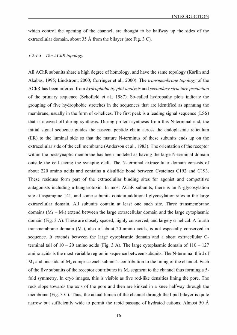
1.2.1.3 The AChR topology
All AChR subunits share a high degree of homology, and have the same topology (Karlin and
Akabas, 1995; Lindstrom, 2000; Corringer et al., 2000). The transmembrane topology of the
AChR has been inferred from hydrophobicity plot analysis and secondary structure prediction
of the primary sequence (Schofield et al., 1987). So-called hydropathy plots indicate the
grouping of five hydrophobic stretches in the sequences that are identified as spanning the
membrane, usually in the form of α-helices. The first peak is a leading signal sequence (LSS)
that is cleaved off during synthesis. During protein synthesis from this N-terminal end, the
initial signal sequence guides the nascent peptide chain across the endoplasmic reticulum
(ER) to the luminal side so that the mature N-terminus of these subunits ends up on the
extracellular side of the cell membrane (Anderson et al., 1983). The orientation of the receptor
within the postsynaptic membrane has been modeled as having the large N-terminal domain
outside the cell facing the synaptic cleft. The N-terminal extracellular domain consists of
about 220 amino acids and contains a disulfide bond between Cysteines C192 and C193.
These residues form part of the extracelullar binding sites for agonist and competitive
antagonists including α-bungarotoxin. In most AChR subunits, there is an N-glycosylation
site at asparagine 141, and some subunits contain additional glycosylation sites in the large
extracellular domain. All subunits contain at least one such site. Three transmembrane
domains (M1 – M3) extend between the large extracellular domain and the large cytoplasmic
domain (Fig. 3 A). These are closely spaced, highly conserved, and largely α-helical. A fourth
transmembrane domain (M4), also of about 20 amino acids, is not especially conserved in
sequence. It extends between the large cytoplasmic domain and a short extracellular C-
terminal tail of 10 – 20 amino acids (Fig. 3 A). The large cytoplasmic domain of 110 – 127
amino acids is the most variable region in sequence between subunits. The N-terminal third of
M1 and one side of M2 comprise each subunit’s contribution to the lining of the channel. Each
of the five subunits of the receptor contributes its M2 segment to the channel thus forming a 5-
fold symmetry. In cryo images, this is visible as five rod-like densities lining the pore. The
rods slope towards the axis of the pore and then are kinked in a knee halfway through the
membrane (Fig. 3 C). Thus, the actual lumen of the channel through the lipid bilayer is quite
narrow but sufficiently wide to permit the rapid passage of hydrated cations. Almost 50 Å
INTRODUCTION
17
away, the rode-like knees at the membrane level turn so they no longer point so directly into
the pore axis (Unwin, 1993, 1995). The inner vestibule of the channel does not open up as an
axial hole directly to the cytoplasm. Instead, it ends in a colonnade of five rods that converge
and are covered by a cupola (Miyazawa et al., 1999). This results in a lattice containing acidic
amino acids that contribute to the cation selectivity of the AChR. The rods of the colonnade
are thought to derive from the major cytoplasmic loop (M3-4 loop) of the receptor subunits,
whereas the distal part of the cupola seems to represent the receptor-aggregating protein
RAPSYN attaching to the receptor complex (Fig. 3 C). RAPSYN is a 43000 Da peripheral
membrane protein that links AChRs to the cytoskeleton and contributes to the normal tight
packing of muscle AChRs at the tips of folds in the postsynaptic membrane which are
adjacent to sites of acetylcholine release from the presynaptic terminal.
Opening of the channel occurs upon binding of ACh to both α-subunits at sites that are at, or
close to, the interfaces made with neighboring ε- (or γ- in fetal muscle) and δ-subunits (αε (γ)
and αδ; see Fig. 3 B; Karlin, 1993; Sine et al., 1995; Xie and Cohen, 2001).
(ε)
A
B
C
(Modified from Karlin, 2002)
Fig. 3 The structure of the nicotinic acetylcholine receptor (AChR); A: The threading pattern of receptor subunits through the membrane; each subunit is composed by a large extracellular N-terminus, four membrane-spanning segments, a long cytoplasmic loop, and a short extracellular C-terminus; B: Schematic representation of the pentameric structure, showing the arrangement of the subunits in the muscle-type receptor, the location of the two acetylcholine (ACh)-binding sites (between α- and ε-subunits (or γ-subunit in the embryonic muscle), and α- and δ-subunits), and the axial cation-conducting channel; C: Cross-section through the 4.6-Å structure of the receptor determined by electron microscopy of tubular crystals of Torpedo membrane embedded in ice.

INTRODUCTION
18
1.2.1.4 Mechanisms of ACh receptor activation
The structural transition from the closed to the open-channel state of the receptor has been
analyzed at 9 Å resolution (Unwin, 1995). Since the ACh-binding sites are distant from the
pore itself, ACh binding must trigger channel-opening via propagated conformational
changes. Indeed, Unwin has shown that binding of ACh initiates two interconnected events in
the extracellular domain. One is a local disturbance, involving all five subunits, in the region
of the binding sites, and the other an extended conformational change, involving
predominantly the two α-subunits, which communicates to the transmembrane portion.
However, this describes the receptor in either of the two states, in the closed and in the open
states, but does not take into account the possibility that binding to one site might affect
binding to the other before the channel opens (Hatton et al., 2003).
A simple mechanistic picture is as follows: first, ACh triggers a localized disturbance in the
region of the binding sites. Second, the effect of this disturbance is communicated by axial
rotations, involving mainly the α-subunits, to the M2 helices in the membrane. Third, the M2
helices transmit the rotations to the gate-forming side-chains, drawing them away from the
central axis; the mode of association near the middle of the membrane is thereby disfavored,
and the helices switch to the alternative side-to-side mode of association, creating an open
pore. The first attempts to investigate the mechanism of action of acetylcholine (ACh) were
made by Colquhoun and Sakmann (1981; 1985). Single-channel measurements showed that
the channel could open, though much less efficiently, with only one ACh molecule bound.
This cannot be detected from whole-cell measurements.
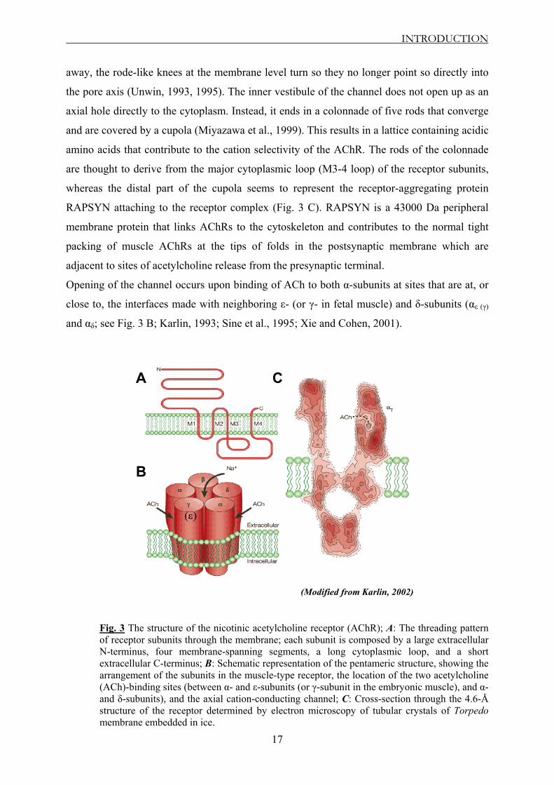
Two reaction schemes that have been widely used to represent the activation of the nicotinic
receptor are shown in Figure 4. The first proposed activation mechanism of AChR (Fig. 4 A)
assumes that the two ACh binding sites are equivalent before ACh binds, and when two
agonist molecules are bound they have equal rates of dissociation. The closed channel R can
bind ACh molecules in two steps of relatively low affinity but with high forward rate
constants almost at the diffusion limit. Agonist binding profoundly increases the probability
that the channel will become open (R*). However, there may be cooperativity in the process
of binding to the shut receptor, and binding of the second ACh molecule may not have the
same rate constants as binding of the first. The doubly occupied closed receptor (A2R) opens
with a rate of β2, and the doubly occupied open receptor (A2R*) closes with a rate of α2, while
the singly occupied closed receptor (AR) opens with a rate of β1, and AR* closes with a rate
of α1. The closed open reaction ("gating") of the diliganded AChR is much more favorable

INTRODUCTION
19
than that of the unliganded receptor (Jackson, 1986, 1988; Grosman and Auerbach, 2000).
The affinity of the AChR for ACh has to be ~ 1500 – 5000-fold higher in the open than in the
closed conformation.
A B
(B from Hatton et al., 2003)
Fig. 4 Mechanisms of AChR activation when the ACh binding sites are equivalent (A) or different (B). R represents the inactive (shut) receptor, R* the active (open) receptor, and A the agonist. The rate constants for the individual reaction steps are denoted k (for association and dissociation), or α (for shutting), and β (for opening). Agonist (A) binds to the two sites on the closed receptor, R, with association and dissociation rates of k+1, k+2, k–1 and k–2.
When ACh is bound to both α-domains, a central ion channel opens, yielding a current of ~ 2
– 5 pA (Dionne and Leibowitz, 1982; Colquhoun et al., 1990). However, the protein can then
exit the A2R state by either two routes: it can lose the agonist molecule to form AR with rate
constant k-2, or it can reopen with rate constant β2. If k-2 is much greater than β2, then usually
the agonist will dissociate before the channel reopens. However, if β2 is similar or larger than
k-2, the channel may reopen several times before losing the agonist, giving rise to a longer
composite event, a burst with several gaps (Sakmann et al., 1980; Hamill and Sakmann,
1981). For skeletal muscle AChRs, the rate constants k-2 and β2 are about equal: 30 – 60 / ms.
The open state also can give rise to the desensitized conformation (A2D). Thus, if agonist is
steadily present, most receptors will eventually end up doubly liganded and desensitized.
Rates into and out of the desensitized state (A2D) are assigned as βD and αD, respectively.
Desensitization of AChR channels is analogous to inactivation of Na+ channels. In the
desensitized state, the agonist is still bound to the channel, but the channel is closed in a
conformation distinct from that of the (unliganded) shut-channel form. The receptor is
unresponsive to added ACh and recovers its sensitivity only some milliseconds or even
minutes after the ACh is removed. Because agonist binding favors desensitization,
INTRODUCTION
20
thermodynamics requires that desensitized states will bind agonists more tightly than closed
states. The affinity increases by more than 1000-fold here. Desensitization is a complex and
ill-understood phenomenon, because it develops on many different time scales, from
milliseconds to minutes (Katz and Thesleff, 1957; Cachelin and Colquhoun, 1989; Butler and
McNamee, 1993; Franke, et al 1993), with fast- and slow-desensitized states, and therefore it
is hard to describe an adequate reaction mechanism for it. Recently, it has been shown by
single-channel methods (Elenes and Auerbach, 2002) that there are multiple desensitized
states, but there is nothing yet known about the structural differences between these states.
The second proposed activation mechanism of AChR (Fig. 4 B), which in most cases is more
realistic, assumes that the two binding sites for ACh are different from the start; so, two
distinguishable open mono-liganded states exist: ARa-Rb* and Ra-ARb*. This mechanism
allows for cooperativity of the binding reaction, because the rates for binding to site a may
depend on whether or not site b is occupied (and vice versa). Different studies (Jackson, 1986;
Grosman and Auerbach, 2000) showed that the unliganded, singly-liganded, and doubly-
liganded states all can indeed open.
When looking at the structure-function relationships of proteins, the difference between the
two ACh binding sites is of great interest. However, from the physiological point of view it is
not very important. It is clear that the two ACh binding sites differ, and this has been found to
be the case for most subtypes of the receptor. Binding of a single ACh molecule is sufficient
to produce very brief openings of the channel, though with very low efficacy (Colquhoun and
Sakmann, 1981). In the adult human receptor, the most obvious sign that the sites differ lies in
the fact that two classes of singly-liganded openings are detectable, one much briefer than the
other (Hatton et al., 2003). However, singly-liganded openings contribute next to nothing to
the endplate current that is responsible for neuromuscular transmission. From the
physiological point of view, the rates that matter most are the open and closed rate constants
for the doubly-liganded channel (α2 and β2) and the total rate at which agonist dissociates from
the doubly-liganded receptor (k-2a + k-2b; see Fig. 4 B). After exposure to the transient high
concentration of ACh released from a nerve ending, most receptor molecules will be in the
doubly-liganded states, and these three values determine the length of each individual
opening, the number of reopenings and the lengths of the short shut periods that separate each
opening. In conclusion, they are sufficient to determine the characteristics of the predominant
doubly-liganded bursts of openings (channel ‘activations’) that are responsible for
neuromuscular transmission (Colquhoun et al., 2003).

INTRODUCTION
21
Because the different conformations of the muscle AChR interconvert and the ACh-
association / dissociation rate constants are exceedingly fast, there is no prospect of being able
to measure directly (that is, with binding assays) the kinetics of the agonist-binding steps in a
conformation-specific manner. A reasonable alternative is to postulate an appropriate kinetic
model and to estimate the agonist-association / dissociation rate constants from single-channel
recordings. Both desensitization and ACh dissociation from the open state significantly
contribute to the time course of the endplate current decay in patients with congenital
myasthenic syndrome (Grosman and Auerbach, 2001).
1.3 The neuromuscular junction (NMJ)
1.3.1 From nerve action potential to muscle action potential
To understand the mechanism underlying congenital myasthenic syndrome (CMS), it is of
great importance to know how the neuromuscular junction (NMJ) functions in healthy
individuals. Thus, an action potential, having propagated along the axon of the α motor nerve,
from the ventral horn of the spinal chord to the muscle, invades the pre-junction membrane or
endplate. The depolarization (voltage changing from negative towards zero and even
becoming positive to zero) caused by the invading action potential is detected by voltage-
gated Ca2+ channels which open, admitting Ca2+ ions, raising the concentration of Ca2+ within
the endplate and causing release (Ca2+-dependent mechanism) of the neurotransmitter vesicles
(Aidley, 1989; Hille, 2001). The neurotransmitter diffuses from the pre-junction release site to
the post-junctional membrane, a distance of some 20 nm. After exocytotic release from the
nerve terminal, some acetylcholine (ACh) molecules are hydrolyzed by acethylcholinesterase
(ACHE) before they bind to the AChR and the remaining ACh molecules are hydrolyzed by
ACHE after dissociation from AChR. Choline is transported into the nerve terminal by a high-
affinity choline transporter (HACHT). ACh is resynthesized from choline and acetyl-
coenzyme A (AcCoA) by choline acetyltransferase (CHAT) and is then transported into the
synaptic vesicle by the vesicular ACh transporter (VACHT) in exchange for protons delivered
to the synaptic vesicle by a proton pump (Fig. 5).

INTRODUCTION
22
A
B
(Modified from Engel et al., 2003b)
Fig. 5 The neuromuscular junction (NMJ); A: Schematic diagram of an endplate region showing the general locations of the acetylcholinesterase (ACHE), the acetylcholine receptor (AChR) (green), and the voltage-gated Na+ channels of the Nav 1.4 type (red); B: After exocytotic release from the nerve terminal, some acetylcholine (ACh) molecules are hydrolyzed by ACHE before they bind to the AChR and the remaining ACh molecules are hydrolyzed by ACHE after dissociation from AChR. Choline is transported into the nerve terminal by a high-affinity choline transporter (HACHT). ACh is resynthesized from choline and acetyl-coenzyme A (AcCoA) by choline acetyltransferase (CHAT) and is then transported into the synaptic vesicle by the vesicular ACh transporter (VACHT) in exchange for protons delivered to the synaptic vesicle by a proton pump.
ACh binds to receptor (2 molecules of ACh per AChR), causing it to open. The channels can
pass both K+ and Na+ ions, but in reality, the ions of only one species move in any quantity.
One AChR opens and allows 1.5 x 10⁴ Na+ ions / ms of open time. The channel opens on
average 1 ms, and therefore one open channel causes a depolarization of about 0.3 µV. The
amount of neurotransmitter contained in one vesicle causes a postsynaptic potential of ~ 1
mV. Therefore, one vesicle contains enough neurotransmitter to open ~ 3000 receptors, and
because two molecules of ACh are needed to open one receptor, there must be a minimum of
~ 6000 molecules of ACh per vesicle. If the average depolarization generated at a NMJ of a
muscle fiber is 40 mV then there must be at least 40 vesicles released and in the order of
133000 receptors activated at the NMJ (Aidley, 1989; Hille, 2001). The movement of ions
tends to push the membrane potential of the post-junctional membrane towards 0 mV, and this

INTRODUCTION
23
triggers an endplate potential (EPP) that activates the voltage-gated Na+ channels of the Nav
1.4 type (Flucher and Daniels, 1989; Ruff, 1996), which are higher in density in the bottom of
the synaptic folds. The interior of a resting muscle fiber has a resting potential of about -95
mV. If the EPP reaches the threshold voltage (approximately -50 mV), Na+ ions flow in with
a rush and an action potential is generated, which then propagates away from the NMJ to
depolarize the entire muscle fiber. In the NMJ, the AChR density is highest at the tops of the
synaptic folds, ~ 7500 – 10000 / µm2 = 1 AChR / 100 – 133 nm2. This gives a spacing of ~ 10
– 12 nm between each AChR molecule. The AChR density drops off sharply as one reaches
the bottoms of the folds. A high concentration of AChRs on the crests of the synaptic folds
(Salpeter, 1987) and of Nav 1.4 in the depth of the folds (Flucher and Daniels, 1989; Ruff,
1996) ensures that excitation is propagated beyond the endplate (Martin, 1994; Wood and
Slater, 2001). The safety margin of neuromuscular transmission is a function of the difference
between the depolarization that is caused by the EPP and the depolarization that is required to
activate Nav 1.4 channels. All CMSs that have been identified so far have been traced to one
or more factors that render the EPP subthreshold for activating Nav 1.4 channels (Engel et al.,
1999b). No visible change occurs in the muscle fiber during (and immediately following) the
action potential. This period, called the latent period, lasts from 3 – 10 ms. Before the latent
period is over, the enzyme acetylcholinesterase breaks down the ACh in the neuromuscular
junction (at a speed of 25000 molecules per second), the voltage-gated Na+ channels close,
and the field is cleared for the arrival of another nerve impulse. The resting potential of the
fiber is restored by an outflow of K+ ions. The brief (1 – 2 ms) period needed to restore the
resting potential is called the refractory period.
In each CMS, the safety margin of neuromuscular transmission is impaired by one or more
specific mechanisms. Thus, the factors that govern the safety margin can be grouped into the
following main categories: factors that affect the number of acetylcholine (ACh) molecules
per synaptic vesicle, factors that affect neurotransmitter release mechanisms, and factors that
affect the efficacy of individual quanta (Wood and Slater, 2001; Katz, 1966). Therefore, a
clear understanding of the mechanisms that operate in the CMS requires an analysis of the
different ways in which these synaptic properties can be modified. They include variations in
the number of AChRs per endplate, in the synaptic localization of AChR and
acetylcholinesterase (ACHE), in the fine structure of the endplate, in the amplitude of
miniature endplate potentials (MEPPs), in the number of quanta released by nerve impulse, in
the number of readily releasable quanta, in the probability of release and in the kinetic
properties of single AChRs. Combined electrophysiological and morphological tests can

INTRODUCTION
24
probe the involvement of these different mechanisms in CMSs. If these tests point to a
candidate molecule, then genetic analysis becomes feasible. If a mutation is discovered in the
candidate gene, then expression studies with the recombinant mutant molecule can be used to
confirm its involvement in pathology and to analyze the properties of the abnormal molecule
(Engel, 1993a; Engel et al., 1993b). The candidate-gene approach has pointed to defects in
choline acetyltransferase (CHAT), ACHE, AChR and the postsynaptic molecule RAPSYN as
causes of CMS (Fig. 5). These defects lead to failure of neuromuscular transmission. In
humans, the extent to which the EPP normally exceeds the threshold, the ‘safety factor’, is
quite small and neuromuscular transmission is sensitive to pathological changes. By contrast,
in mice the safety factor appears higher, and mouse models of neuromuscular diseases do not
always demonstrate obvious weakness (Vincent et al., 1997; Bhattacharyya et al., 1997).
However, in two studies by Gomez and co-workers (1997 and 2002), transgenic mice
expressing different mutations that induce severe forms of SCCMS in humans also developed
myasthenic symptoms highly reminiscent to those of the patients. Therefore, such transgenic
animal models are actively contributing to our understanding of the complex genotype-
phenotype relationship in CMS.
1.3.2 CMS caused by defects in AChR
Our study focuses on mutations in the AChR channel, which are genetic defects that induce
postsynaptic CMS. Such mutations have now been discovered in all AChR subunits and in
different domains of the subunits (Engel et al., 2003a,b,c). The mutations fall into two groups:
mutations that alter the kinetic properties of AChR and mutations that reduce its expression
(low-expressor mutations). The kinetic mutations can be further divided into two types: slow-
channel mutations that increase the synaptic response to ACh, and fast-channel mutations that
decrease it. The slow- and fast-channel mutations represent physiological opposites in both
their phenotypic consequences and alterations of fundamental steps that underlie receptor
activation.
1.3.2.1 Slow-channel CMS
It was recognized two decades ago by its distinct phenotypic features. Five patients showed
dominant inheritance and selective weakness of cervical, scapular and finger extensor
muscles, mild ophthalmoparesis and variable weakness of other muscles. Single-nerve
INTRODUCTION
25
stimulation evoked a repetitive compound muscle action potential (CMAP), rather than the
expected single CMAP, and the normally constant amplitude of the CMAP attenuated with
repetitive nerve stimulation. Synaptic potentials decayed abnormally slowly, showing normal
or reduced amplitudes, but the evoked ACh release was normal. The disease left an
anatomical trace – termed endplate myopathy – which is associated with postsynaptic Ca2+
accumulation, as well as focal degeneration of the junctional folds with corresponding loss of
AChRs, and degenerating membranous organelles and small vacuoles in junctional fiber
regions (Engel and Biesecker, 1982; Engel et al., 2003b). The repetitive CMAP was attributed
to the EPP outlasting the absolute refractory period of the muscle fiber, and the endplate
myopathy was credited to excessive Ca2+ influx into the postsynaptic region during prolonged
episodes of synaptic activation. The slow decay of the synaptic potentials was attributed to
prolonged openings of the AChR channel (Anderson and Stevens, 1973). The presence of
normal amounts and activity of the enzyme at the synapse excluded the alternative
explanation that a defect in ACHE prolonged the lifetime of ACh in the synaptic space.
However, formal proof of a kinetic defect in AChR came only in the mid of ‘90s, with the
recording of markedly prolonged synaptic and single-channel currents from the endplate of
other patients with slow-channel CMS (Ohno et al., 1995; Engel et al., 1996b).
1.3.2.2 Fast-channel CMS
The name 'fast-channel CMS' originates from the abnormally fast decay of the synaptic
response, which is caused by abnormally brief channel-opening events. The disorder was first
recognized in 1993 in a patient with moderately severe myasthenic symptoms from birth,
which responded partially to cholinesterase inhibitors. Endplate studies showed a normal
structure, no deficiency of ACHE or AChR, a normal neurotransmitter release by nerve
impulse, but small miniature endplate potentials (MEPPs) (Uchitel et al., 1993a,b). Analysis
of the ACh-induced current noise pointed to a normal conductance of the AChR channel but
with abnormally short-lived openings. The diminished synaptic response, in face of normally
abundant AChRs, indicated a decreased ACh content of the synaptic vesicles, ACHE
overactivity or an abnormality in AChR. Morphometric analysis showed that synaptic vesicles
were of normal size, pointing away from reduced vesicular ACh content (Jones and
Kwanbunbumpen, 1970a,b), and ACHE overactivity seemed unlikely, as cholinesterase
inhibitors did not fully restore the synaptic response. The findings therefore implied a kinetic

INTRODUCTION
26
defect in AChR in which opening of the channel was impaired and its closing was enhanced
(Uchitel et al., 1993a,b; Shen et al., 1999, 2000, 2001; Maselli et al., 2002; Sine et al., 2002).
1.3.2.3 CMS caused by low-expressor mutations
In this category, most of the CMS cases that have been identified so far are caused by
homozygous or heterozygous low-expressor mutations in AChR subunit genes, and these
mutations are concentrated in the ε-subunit of the receptor (for a detailed table with low-
expressor mutations in AChR subunits, please refer to Engel et al., 2003a). Patients harboring
low-expressor or even homozygous null mutations in the ε-subunit might have mild
symptoms. Conversely, patients with low-expressor mutations in non-ε-subunits are severely
affected, and so far, no patients with null mutations in both alleles of a non-ε-subunit have
been observed. A probable explanation is that persistent low-expression level of fetal AChR
harboring γ-subunit can partially compensate for the absence of the ε-subunit (Milone et al.,
1998b; Engel et al., 1996a,b; Ohno et al., 1997). Null mutations in subunits other than ε-
subunit might be lethal, presumably owing to the lack of a substituting subunit. In patients
with null mutations of both ε-alleles, all AChR channels recorded from the endplate have the
reduced conductance and prolonged time course that are typical of γ-subunit-containing
AChRs (Ohno et al., 1997), and immunostaining confirms the presence of the γ-subunit at the
endplate (Engel et al., 1996a,b). Targeted deletion of the ε-subunit in mice showed that either
the γ- or ε-subunit is essential for survival. Thus, the AChR ε -/- mice express the γ-subunit
instead of the ε-subunit at birth (as in human fetus), but die two to three months later when the
γ-subunit mRNA is no longer transcribed (Witzemann et al., 1996). Humans, however,
continue to express low-levels of the γ-subunit at the endplate (Croxen et al., 2001), and so
this subunit rescues the ε-null phenotype.
Morphological studies of endplates with low-expressor mutations show an increased number
of endplates distributed over an increased span of individual muscle fibers. The integrity of
the junctional folds is preserved, but some endplate regions are simpler and smaller than
normal. The distribution of AChRs on the junctional folds is patchy, and the density of the
receptors is reduced. The immunocytochemical reaction for RAPSYN, the molecule that
clusters the AChRs at the postsynaptic membrane, is decreased in proportion to the decreased
expression of AChR. The quantal response at the endplate is reduced, but neurotransmitter
release by nerve impulse is frequently higher than normal. Different types of recessive
INTRODUCTION
27
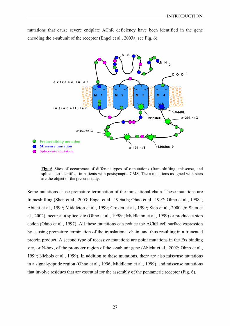
mutations that cause severe endplate AChR deficiency have been identified in the gene
encoding the ε-subunit of the receptor (Engel et al., 2003a; see Fig. 6).
M 1 M 2 M 3 M 4
N H2
C O O -
e x t r a c e l l u l a r
i n t r a c e l l u l a r
S - S
Missense mutationSplice-site mutation
Frameshifting mutation
ε911delT
ε1030delC
ε1101insT
ε1293insG
ε1206ins19
εV448L
Fig. 6 Sites of occurrence of different types of ε-mutations (frameshifting, missense, and splice-site) identified in patients with postsynaptic CMS. The ε-mutations assigned with stars are the object of the present study.
Some mutations cause premature termination of the translational chain. These mutations are
frameshifting (Shen et al., 2003; Engel et al., 1996a,b; Ohno et al., 1997; Ohno et al., 1998a;
Abicht et al., 1999; Middleton et al., 1999; Croxen et al., 1999; Sieb et al., 2000a,b; Shen et
al., 2002), occur at a splice site (Ohno et al., 1998a; Middleton et al., 1999) or produce a stop
codon (Ohno et al., 1997). All these mutations can reduce the AChR cell surface expression
by causing premature termination of the translational chain, and thus resulting in a truncated
protein product. A second type of recessive mutations are point mutations in the Ets binding
site, or N-box, of the promoter region of the ε-subunit gene (Abicht et al., 2002; Ohno et al.,
1999; Nichols et al., 1999). In addition to these mutations, there are also missense mutations
in a signal-peptide region (Ohno et al., 1996; Middleton et al., 1999), and missense mutations
that involve residues that are essential for the assembly of the pentameric receptor (Fig. 6).

INTRODUCTION
28
1.4 The goal of the study
Recently, several mutations within the ε-subunit of the human muscle AChR have been
described that impair the normal function of the neuromuscular synaptic transmission. While
there is considerable amount of information on morphological changes in the neuromuscular
synapse associated with these mutations, little is known about the functional alterations of the
mutations.
The aim of this study was to investigate the molecular mechanism underlying postsynaptic
CMS, in the presence of two frameshifting mutations, ε911delT and ε1030delC, and a
missense mutation εV448L (Sieb et al., 2000a; see Fig. 6). Additionally, three related CMS-
linked mutations all located within the ε-cytoplasmic loop of AChR, namely the insertion
mutations ε1101insT (Engel et al., 1996a), ε1206ins19 (Ohno et al., 1998b), and ε1293insG
(Engel et al., 1996a; Sieb et al., 2000b; see Fig. 6) were also included in the study to identify
the role of specific regions in the TM3-4 loop for the functionality of the receptor.
The approach we took was to express recombinant wild-type and ε-mutant AChRs in HEK
293 cells and characterize the functional properties of these receptors with
electrophysiological techniques.
We asked the following questions:
Do the mutations influence the affinity of the receptor for its natural ligand?
Do the mutations alter the kinetic properties of ACh-induced current responses, e.g. alter
speed and degree of receptor activation and desensitization?
Do the mutations impair the cell surface expression of the AChR channel?
The mutation(s) that showed in whole-cell recordings functional changes were further
investigated on the single-channel level.
There we determined the size of the ion flux through the open channel (the single-channel
conductance), the frequency of opening events (the open probability), and the open and close
lifetimes of the AChR.
The combined results from whole-cell- and single-channel recordings from these ε-mutants
should provide insight into the mechanism underlying postsynaptic CMS.

MATERIALS AND METHODS
29
2 Materials and Methods
2.1 Cell culture
Human Embryonic Kidney 293 cells (HEK 293), kindly provided by Prof. Dr. T. Schneider
(Institute of Neurophysiology, Köln), were used as expression system for the present study.
Cells were grown in Dulbecco's Modified Eagle Medium (DMEM; Invitrogen) supplemented
with 10 % bovine calf serum (BCS), 100 µg/ml of streptomycin, and 100 U/ml of penicillin.
For plating-out the HEK 293 cells, confluent cells were collected from 25 cm2 Petri dishes by
pipetting up and down, and after a 3 min centrifugation at 1000 rpm, they were resuspended
in DMEM HEK medium, and replated on 8 cm2 dishes. Cells were incubated at 37°C for ~ 48
h, and they were approximately 70 % confluent on the day of the transfection.
2.2 Transient transfection
Dr. S. Kraner (Institute of Human Genetics, Bonn) generated AChR ε-subunit mutations:
εV448L, ε911delT, ε1030delC, ε1293insG, ε1206ins19, and ε1101insT (Sieb et al., 2000a,b;
Engel et al., 1996a; Ohno et al., 1998b) by „QuikChangeTM Site-Directed Mutagenesis“
(Stratagene). The resultant ε-subunit cDNAs were sequenced to check for the presence of the
mutations and absence of any additional sequence changes (Kraner et al., 2003). The cDNAs
were subcloned into the CMV-based expression vector pCDNA 3.1 (+) (5.4 kbp) (Invitrogen).
HEK 293 cells were transiently cotransfected with cDNAs encoding the wild-type (α, β, δ,
and ε), ε-lacking (α, β, and δ), and ε-mutated AChR subunits by TransFastTM transfection
reagent (Promega) together with green fluorescence protein (GFP) using a subunit ratio of
2:1:1:1 (α: β: δ: ε). Initial experiments using Ca2+ - phosphate precipitation method had lower
transfection efficiency on our HEK 293 cell line, and therefore TransFastTM transfection was
further used. This method has some advantages being faster (it takes round about 1 h), easy-
to-use (after resuspending the reagent in water, freezing, thawing, and mixing with DNA,
everything is simply added to cells), more efficient (high-efficiency transfection – transient
and stable – in many cell lines), and, finally, more robust (it requires less optimization than
other systems). Additionally, it allows transfection of cell types such as primary cell cultures
that require continuous exposure to serum, as it can be used in the presence of serum (see
TransFastTM transfection reagent technical bulletin at www.promega.com).

MATERIALS AND METHODS
30
Transfection was started two days after plating-out the HEK 293 cells on 8 cm2 Petri dishes,
by pipetting the AChR cDNAs and the GFP cDNA together. GFP (green fluorescent protein;
Invitrogen) was used to identify transfected cells for electrophysiological studies. It followed
a dilution of the cDNAs in DMEM HEK medium, on which thawed TransFast™ reagent was
added, and this mixture was vortexed for a short time. After a 10 – 15 min incubation of the
cDNAs / TransFast™ reagent mixture, and after the removal of the old growth medium from
the 70 % - confluent HEK 293 cells on the 8 cm2 Petri dishes, the transfection mixture was
added onto the cells, and cells were incubated at 37°C for ~ 48 – 96 h. After this period, the
transfected cells were plated in fresh DMEM HEK medium on poly-D-lysine-coated glass
coverslips (Labomedic). For this purpose, cells were collected from 8 cm2 Petri dishes by
pipetting up and down, and after a 3 min centrifugation at 1000 rpm, they were resuspended
in DMEM HEK medium. A small volume (usually 20 µl) was mixed with the same amount of
Tripan Blue for counting the cells. Cell suspension was diluted in a way that 1 ml of it always
contained a density of 1 x 10⁶ cells on average. A small volume of transfected cells (30 µl)
was used for plating the cells on coverslips in 8 cm2 Petri dishes. After 30 min of incubation
at 37°C, 2 ml of DMEM HEK medium were added over the coverslips in 8 cm2 Petri dishes.
Four coverslips were used for each 8 cm2 Petri dish. Whole-cell- and cell-attached patch-
clamp recordings were started 2 – 3 h after plating the cells on coverslips.
2.3 Patch-clamp recordings
Whole cell currents were recorded using an EPC 9 patch-clamp amplifier and Pulse Software
(HEKA Elektronik). Recordings were filtered at 5 kHz (Bessel, 4-pole), digitized using an
ITC-16 AD / DA interface (HEKA Elektronik) at a rate of 25 kHz, and stored on hard disk.
Patch pipettes were pulled from borosilicate capillary tubes (Kimble Products), on a
horizontal programmable puller (Zeitz-Instrumente), and had resistance between 1.5 and 3
MΩ, when filled with internal solution. The pipette solution contained (in mM): 2.5 NaCl,
110 KCl, 0.5 CaCl2, 10 EGTA, and 10 HEPES (pH = 7.3, adjusted with KOH). The external
solution contained (in mM): 145 NaCl, 2.5 KCl, 2 CaCl2, 1.3 MgCl2, 10 HEPES, and 20
Glucose (pH = 7.3, adjusted with NaOH). ACh-induced currents were elicited by application
of ACh (a 2 sec pulse of 0.03, 0.1, 0.3, 0.5, 1, 3, 10, 30, 100 or 300 µM ACh) using a fast
pressure-application system (ALA Scientific Instruments). Flow from the pressurized
reservoirs was computer-controlled via Lee solenoid valves. The output from the valves was
connected via FEP tubing (Fluorinated Ethylene Propylene Tubing) to the micromanifold.

MATERIALS AND METHODS
31
The application system consisted of quartz glass tubes (8 or 12 x 100 µm application tubes + 1
x 200 µm flush tube) that communicated with the application tip of 100 µm. The agonist
solution was applied at intervals of 3 – 5 min. ACh solutions were prepared daily from a 100
mM stock solution. All whole-cell recordings were performed at a holding potential of –60
mV.
Cell-attached patch-clamp recordings were performed at room temperature (19 – 24°C), using
an EPC 9 patch-clamp amplifier (HEKA Elektronik). Recordings were filtered at 10 or 5 kHz
(Bessel, 4-pole), digitized using an ITC-16 AD / DA interface (HEKA Elektronik) at a rate of
25 kHz, and stored on hard disk. Patch pipettes were pulled from borosilicate capillary tubes
(Kimble Products) and coated with Sylgard 184 (Dow Corning) to reduce the noise
interference during recordings. The pipette resistance ranged from 5 to 10 MΣ. In the cell-
attached experiments, the pipette solution contained (in mM): 0.001, 0.01, 0.03 or 0.1 ACh in
142 KCl, 5.4 NaCl, 10 HEPES, 1.8 CaCl2, 1.7 MgCl2, pH = 7.3. The external solution
contained (in mM): 142 KCl, 5.4 NaCl, 10 HEPES, 1.8 CaCl2, 1.7 MgCl2, pH = 7.3. In cell-
attached recordings, the holding potential was –100 mV, unless otherwise indicated. Slope
conductance curves were determined from continuous recording protocol performed at
various membrane potentials (from –60 to –220 mV).
(For a detailed description of the patch-clamp technique, see also the Appendix section!)
2.3.1 Data analysis of the patch-clamp recordings
2.3.1.1 Analysis of whole-cell recordings
2.3.1.1.1 Dose-response curves
Analysis of the macroscopic currents was carried out using Pulse + PulseFit software (HEKA
Elektronik) and GraphPad Prism software (GraphPad Software Inc.). Using Pulse + PulseFit
software, the macroscopic currents (Imax) were manually measured at different ACh
concentrations ranging from 0.03 to 300 µM, for wt, ε-lacking, and ε-mutant AChRs. To
construct dose-response curves, the currents were normalized to the maximum response and
plotted as a function of log of the agonist concentration (in M). Making use of GraphPad
Prism software, EC50 values and Hill slopes were obtained by fitting the data points to a
logistic equation in the form: y = ymin + [(ymax - ymin) / 1+ (x / EC50)n].

MATERIALS AND METHODS
32
2.3.1.1.2 AChR desensitization kinetics and current density
Analysis of the desensitization process was carried out using PulseFit, IGOR Pro
(Wavemetrics), and GraphPad Prism software. The time course of the current decay was fitted
using the following exponential function: y = A + y0*exp (-x / TauD) in the IGOR Pro and
PulseFit software. The values obtained for the time constants of desensitization (TauD, in
seconds) were plotted as a function of the ACh concentration in the GraphPad Prism software.
The fraction of residual current (y0 = IACh residual, pA) was also obtained by this fit and
plotted vs. the ACh concentration in the GraphPad Prism software. In the text, values are
given as degree of desensitization: 100 % - X % residual current.
To correct for variation in current magnitude due to differences in cell size, we normalized
current responses to the cell surface and expressed them as current density in pA / pF. Current
density was deducted from the peak current response obtained at 30 or 100 µM ACh
concentration, by dividing the maximum current amplitude (Imax, pA) to the slow component
of the membrane capacitance (Cs, pF) in the GraphPad Prism software. Values are given as
mean ± SEM, and n represents the number of cells taken in analysis.
2.3.1.2 Analysis of single-channel recordings
Single-channel recordings were first inspected visually and corrected for baseline drift. Noisy
sections and those containing simultaneous openings of two or more channels were excluded
from analysis. Single-channel events were automatically or manually fitted using the TAC
software (Bruxton), being detected by the fixed threshold criterion and using a rise time of
0.08 ms. Amplitude histograms were built by plotting the single-channel current (i, pA) vs.
the number of events (bin counts), and mean amplitude values were obtained for wt and ε-
mutant AChRs, at 1, 10, 30, and 100 µM ACh, between –60 and –220 mV. Slope conductance
curves of single channels were constructed from linear regression of single-channel current (i,
pA) vs. membrane potential (Vmp, mV). Bursts of channel-openings from single receptors
elicited at high concentrations of agonist (Sakmann et al., 1980) were defined as a series of
openings separated by close intervals shorter than a critical duration and followed by long
closings corresponding to dwells in the desensitized state. Typically, the analysis focused on
close and open intervals within clusters, the durations of which reflect agonist binding and
channel-gating processes. The limit duration of the shut times within clusters of events was
estimated by selecting multiple stretches with bursts of openings (with > 5 events and lasting
MATERIALS AND METHODS
33
more than 100 ms) and after fitting them, a mean value of 30 ms was determined. We assume
that periods longer than 30 ms represent intervals when all of the channels in the patch were
desensitized. Therefore, two datasets called “bursts analysis” and “total recording time
analysis” were obtained by including only opening events between 0.04 and 30 ms duration or
by including all the events longer than 0.04 ms, respectively. In some experiments, for
example, at low ACh concentrations (1 µM, in the case of wt receptor), the currents were not
clustered and all open intervals in the record were measured. Open and close duration
histograms were constructed using a logarithmic abscissa of the duration (in seconds) and a
square root ordinate of the number of events, and fitted to the sum of exponentials by
maximum likelihood (Sigworth and Sine, 1987), using TACFit software (Bruxton). After a
visual inspection, we built histograms by merging similar open and close time distributions
obtained at different holding potentials (between –60 and –220 mV), and at the same ACh
concentration (1, 10, 30 or 100 µM). Mean open and close time values were extracted for wt,
ε-lacking, and ε-mutant AChRs.
Open and close probability values (PO and PC) in bursts and within the total recording time
were obtained in the TACFit software from each individual cell, and subsequently they were
averaged at each ACh concentration tested, and at –100 mV holding potential. The percentage
of cells that responded to ACh application was also estimated from whole-cell- and single-
channel recordings.

MATERIALS AND METHODS
34
2.4 Materials and suppliers
Chemicals Supplier
Acetylcholine chloride
Sigma-Aldrich Chemie GmbH, Munich,
Germany
Trypan Blue
Sigma-Aldrich Chemie GmbH, Munich,
Germany
Sylgard 184 Dow Corning Co., Midland, MI, USA
NaCl Merck KGaA, Darmstadt, Germany
KCl Merck KGaA, Darmstadt, Germany
CaCl2 Merck KGaA, Darmstadt, Germany
EGTA
Sigma-Aldrich Chemie GmbH, Munich,
Germany
HEPES
Sigma-Aldrich Chemie GmbH, Munich,
Germany
MgCl2 Merck KGaA, Darmstadt, Germany
Glucose Merck KGaA, Darmstadt, Germany
Reagents and kits Supplier
Green fluorescent protein (GFP) Invitrogen Ltd., San Diego, CA, USA
CMV-based expression vector
pCDNA3.1 (+) (5.4 kbp)
Invitrogen Ltd., San Diego, CA, USA
QuikChangeTM Site-Directed
Mutagenesis kit
Stratagene Co., La Jolla, CA, USA
TransFastTM transfection reagent Promega Co., Madison, WI, USA
Medium and Supplements Supplier
Dulbecco's Modified Eagle Medium
(DMEM)
Invitrogen Ltd., San Diego, CA, USA
Bovine Calf Serum (BCS) Invitrogen Ltd., San Diego, CA, USA
Streptomycin, Penicillin Invitrogen Ltd., San Diego, CA, USA

MATERIALS AND METHODS
35
Other materials Supplier
Poly-D-lysine-coated glass coverslips Labomedic GmbH, Bonn, Germany
Counting chamber Fuchs-rosenthal, Germany
8, 25 cm2 Petri dishes Becton, Dickinson and Co., NJ, USA
10, 50 ml Polypropylene conical tubes Becton, Dickinson and Co., NJ, USA
10, 20 ml serological pipettes Sarstedt AG and Co., Nümbrecht, Germany
10 – 1000 µl Pipettes Eppendorf AG, Hamburg, Germany
10, 200, 500 µl Pipette tips Eppendorf AG, Hamburg, Germany
0.5, 1.5 ml Eppendorf tubes Eppendorf AG, Hamburg, Germany
Pipette boy Integra Biosciences AG, Chur, Switzerland
0.2 µm Syringe filters Nalge Nunc International Co., NY, USA
1, 5, 10 ml Syringes B. Braun Melsungen AG, Melsungen, Germany
Gloves Kimberly-Clark Co., GA, USA
Parafilm M American National CanTM, Chicago, IL, USA
Apparatuses Supplier
Laminar flow hood Kojair® Tech Oy, Vilppula, Finland
MGW M20 Water bath Lauda-Königshofen, Germany
Carl Zeiss IM35 microscope Carl Zeiss, Göttingen, Germany
Refrigerator LiebHerr Premium LiebHerr-Holding GmbH, Germany
Megafuge 1.0R centrifuge Kendro Laboratory Products GmbH, Germany
HERACell CO2 incubator Kendro Laboratory Products GmbH, Germany
Patch-clamp setup component Supplier
EPC 9 patch-clamp amplifier
HEKA Elektronik Dr. Schulze GmbH,
Lambrecht, Germany
ITC-16 AD / DA interface
HEKA Elektronik Dr. Schulze GmbH,
Lambrecht, Germany
Computer system: Power Mac G4 Apple Computer Inc., CA, USA
Head-stage (or probe)
HEKA Elektronik Dr. Schulze GmbH,
Lambrecht, Germany
Axiovert 135 or 200 microscope Carl Zeiss, Göttingen, Germany
Cell chamber Custom made

MATERIALS AND METHODS
36
Micromanipulator LN mini25 Luigs and Neumann GmbH, Ratingen, Germany
Ag / AgCl recording electrode
HEKA Elektronik Dr. Schulze GmbH,
Lambrecht, Germany
Pipette holder
HEKA Elektronik Dr. Schulze GmbH,
Lambrecht, Germany
Vibration isolation table Technical Manufacturing Co., MA, USA
Perfusion pump Ismatec SA, Glattbrugg, Switzerland
Faraday cage Custom made
HBO 100 W Fluorescence lamp Carl Zeiss, Göttingen, Germany
Borosilicate capillary tubes
Kimble Products, KIMAX-51®, New Jersey,
USA
Fast pressure-application system
DAD-VM-8SP Superfusion System, ALA
Scientific Instruments, NY, USA
DMZ-Universal pipette puller
Zeitz-Instrumente Vertriebs GmbH, Augsburg,
Germany
Oscilloscope Tektronix Inc., OR, USA
Analysis Software Supplier
Pulse + PulseFit Software
HEKA Elektronik Dr. Schulze GmbH,
Lambrecht, Germany
GraphPad Prism 3.03 Software GraphPad Software Inc.
IGOR 4.01 Pro Software Wavemetrics, Lake Oswego, OR, USA
TAC X 4.1.3 + TACFit X 4.1.3 Software Bruxton Corporation

RESULTS
37
3 Results
3.1 Pharmacological and biophysical properties of wild-type, ε-subunit
lacking, and ε-mutant AChRs inferred from whole-cell recordings
3.1.1 Introduction to whole-cell recordings
Mutations of the ε-subunit (εV448L, ε911delT, ε1030delC, ε1101insT, ε1206ins19, and
ε1293insG), identified in patients with CMS symptoms were introduced in ε-subunit of adult
human muscle nicotinic acetylcholine receptor (AChR). We transiently expressed the ε-
subunits in HEK 293 cells together with α, β, and δ-subunits, and investigated functional
properties of the ε-mutated AChRs using the patch-clamp technique, with two of its
approaches: whole-cell- and single-channel recordings.
In the whole-cell recording mode, a so called “whole-cell current” (or macroscopic current;
Imax) is obtained (Fig. 7), which is a measure of the total number of functional channels (N),
the probability that a channel is open (PO), and the ionic current through each individual
channel (i), according to the formula Imax = N ·PO · i (see also section 3.2.1).
Agonist application
Desensitization
Residual current
Maximum ionic current
Activation phase
Fig. 7 Idealized representation of a whole-cell current.
The whole-cell current elicited by ACh application consists of an activation phase, until a
maximum ionic current is reached, and an exponential current decay over time, in the
continuous presence of the agonist, which represents the desensitization process. The fraction
of ionic current that remains at the end of the agonist application period is the residual current

RESULTS
38
representing the fraction of receptors that have not desensitized yet (Fig. 7). These properties
of wt and ε-mutant receptors were first addressed by whole-cell recordings.
To resolve fast ACh responses during whole-cell recordings, we used a fast pressure-
application system that allowed applications within milliseconds. ACh-induced currents were
elicited every 3 – 5 min by 2 sec-applications of different concentrations of agonist (between
0.03 and 300 µM ACh). The agonist was applied in random order. The inter-pulse interval of
3 – 5 min ensured that the receptors fully recovered from desensitization (data not shown).
3.1.2 The properties of wild-type receptor
First, we investigated the properties of wt AChR by applying ACh at concentrations between
0.3 and 100 µM. A representative set of current responses is shown in Figure 8. At lower
concentrations (0.3 – 3 µM), the onset of the current response was initially fairly slow and
became increasingly faster at higher ACh concentrations. At higher concentrations (10 – 100
µM), the desensitization of the receptor was most pronounced, and was visible as current
decay in the continuous presence of ACh (Fig. 8 and 10 A). However, the desensitization was
incomplete and after 2 sec application of ACh at 100 µM, the residual current fraction was
21.5 ± 5.17 % (n = 6). This indicates that ~ 21.5 % of the wt AChRs were still available after
2 sec application of 100 µM ACh and ~ 78.5 % (100 – 21.5%, represents the degree of
desensitization) have entered the desensitized state (Fig. 8 and 10 B).
3.02.01.0s
3.02.01.0s
3.02.01.0s
3.02.01.0s
3.02.01.0s
3.02.01.0s
1 nA
1000 ms
0.3 µΜ 1 µΜ 3 µΜ
30 µΜ10 µΜ 100 µΜ
Fig. 8 Whole-cell currents obtained from wt AChRs transiently expressed in a HEK 293 cell, at different concentrations of ACh (between 0.3 and 100 µM ACh). The bar on the top of each trace indicates the ACh application period (2 sec). The holding potential was –60 mV.

RESULTS
39
The maximum ionic current at the highest concentration tested (300 µM; not shown) was -
5445 ± 450.54 pA (n = 24).
We also studied the pharmacological properties of wt receptor and from the concentration-
dependence of the ionic current (shown in Fig. 11) we constructed dose-response relationships
from each individual cell recorded. After fitting the curves, a mean EC50 value of 1.42 ± 1.11
µM, with a Hill slope of 1.05 ± 0.098 (n = 9) was extracted.
3.1.3 The properties of ε-subunit lacking receptor
We next characterized receptors lacking the ε-subunit. In the absence of the ε-subunit (–ε
AChR), the receptor showed a significantly altered biophysical profile. The most prominent
features were the acceleration in the desensitization kinetics and the increase in the degree of
desensitization (Fig. 9).
3.02.01.0s
3.02.01.0s
3.02.01.0s
3.02.01.0s
3.02.01.0s
3.02.01.0s
100 pA
1000 ms
0.03 µΜ 0.1 µΜ 1 µΜ
30 µΜ10 µΜ 100 µΜ
Fig. 9 Whole-cell currents obtained from a single cell expressing ε-subunit lacking AChRs. Currents were elicited by the indicated ACh concentrations.
Currents elicited at ACh concentrations of 0.03 and 1 µM declined with a time course that
could be fitted to a single exponent function with a time constant (TauD) of 1.14 ± 0.04 s (n =
3), and 0.46 ± 0.02 s (n =5), respectively. At low ACh concentrations (0.03 and 1 µM), there
was a 3-fold, and, respectively, a 2-fold speed-up of the current decay compared to wt
receptor (3.4 s, n = 1, and 0.98 ± 0.06 s, n = 9, respectively). However, at larger ACh
concentration (30 to 300 µM), the time constants for desensitization were similar for wt and –
ε receptors (Fig. 10 A).

RESULTS
40
The degree of desensitization was ~ 2.5-fold increased in the absence of the ε-subunit. At
ACh concentrations of 0.03 and 1 µM, the desensitization for –ε AChR was 69.3 ± 0.64 % (n
= 3), and 80.25 ± 0.87 % (n = 7), whereas for wt receptor the desensitization was only 19.83 ±
3.88 % (n = 5) and 44.05 ± 5.61 % (n = 8), respectively (Fig. 10 B).
0.01 0.1 1 10 100 10000.0
0.5
1.0
1.5
2.0
2.5
3.0
3.5WT-ε
A
ACh [µM]
τ D[s
]
0.01 0.1 1 10 100 10000
102030405060708090
B
ACh [µM]
I AC
h re
sidu
al [%
]
Fig. 10 Desensitization kinetics of wt and –ε AChRs. The time constant for desensitization (TauD, A) and the fraction of residual current (IACh residual, B) for wt (black circles) and –ε (red squares) were plotted as a function of the ACh concentration. TauD and IACh residual were obtained by fitting the current decay in Fig. 8 and 9 to an exponential equation in the form: y = A + y0 * exp (– x / TauD).
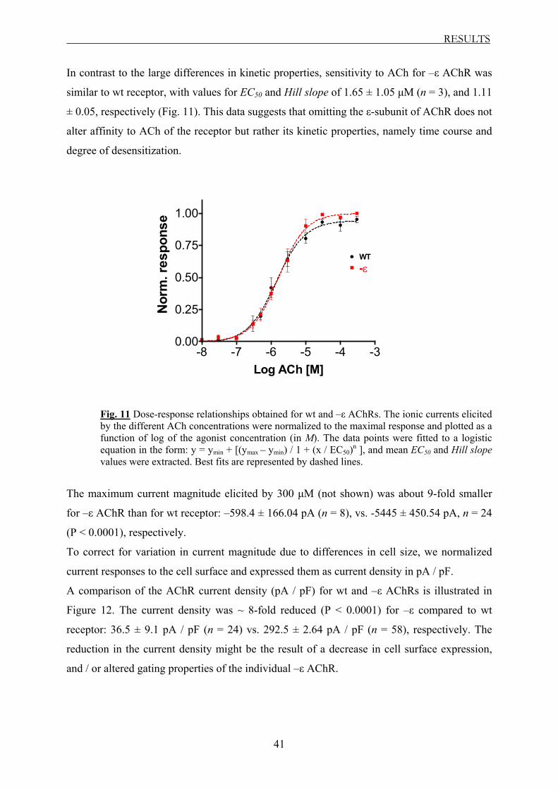
RESULTS
41
In contrast to the large differences in kinetic properties, sensitivity to ACh for –ε AChR was
similar to wt receptor, with values for EC50 and Hill slope of 1.65 ± 1.05 µM (n = 3), and 1.11
± 0.05, respectively (Fig. 11). This data suggests that omitting the ε-subunit of AChR does not
alter affinity to ACh of the receptor but rather its kinetic properties, namely time course and
degree of desensitization.
-8 -7 -6 -5 -4 -30.00
0.25
0.50
0.75
1.00
-εWT
Log ACh [M]
Nor
m. r
espo
nse
Fig. 11 Dose-response relationships obtained for wt and –ε AChRs. The ionic currents elicited by the different ACh concentrations were normalized to the maximal response and plotted as a function of log of the agonist concentration (in M). The data points were fitted to a logistic equation in the form: y = ymin + [(ymax – ymin) / 1 + (x / EC50)n ], and mean EC50 and Hill slope values were extracted. Best fits are represented by dashed lines.
The maximum current magnitude elicited by 300 µM (not shown) was about 9-fold smaller
for –ε AChR than for wt receptor: –598.4 ± 166.04 pA (n = 8), vs. -5445 ± 450.54 pA, n = 24
(P < 0.0001), respectively.
To correct for variation in current magnitude due to differences in cell size, we normalized
current responses to the cell surface and expressed them as current density in pA / pF.
A comparison of the AChR current density (pA / pF) for wt and –ε AChRs is illustrated in
Figure 12. The current density was ~ 8-fold reduced (P < 0.0001) for –ε compared to wt
receptor: 36.5 ± 9.1 pA / pF (n = 24) vs. 292.5 ± 2.64 pA / pF (n = 58), respectively. The
reduction in the current density might be the result of a decrease in cell surface expression,
and / or altered gating properties of the individual –ε AChR.
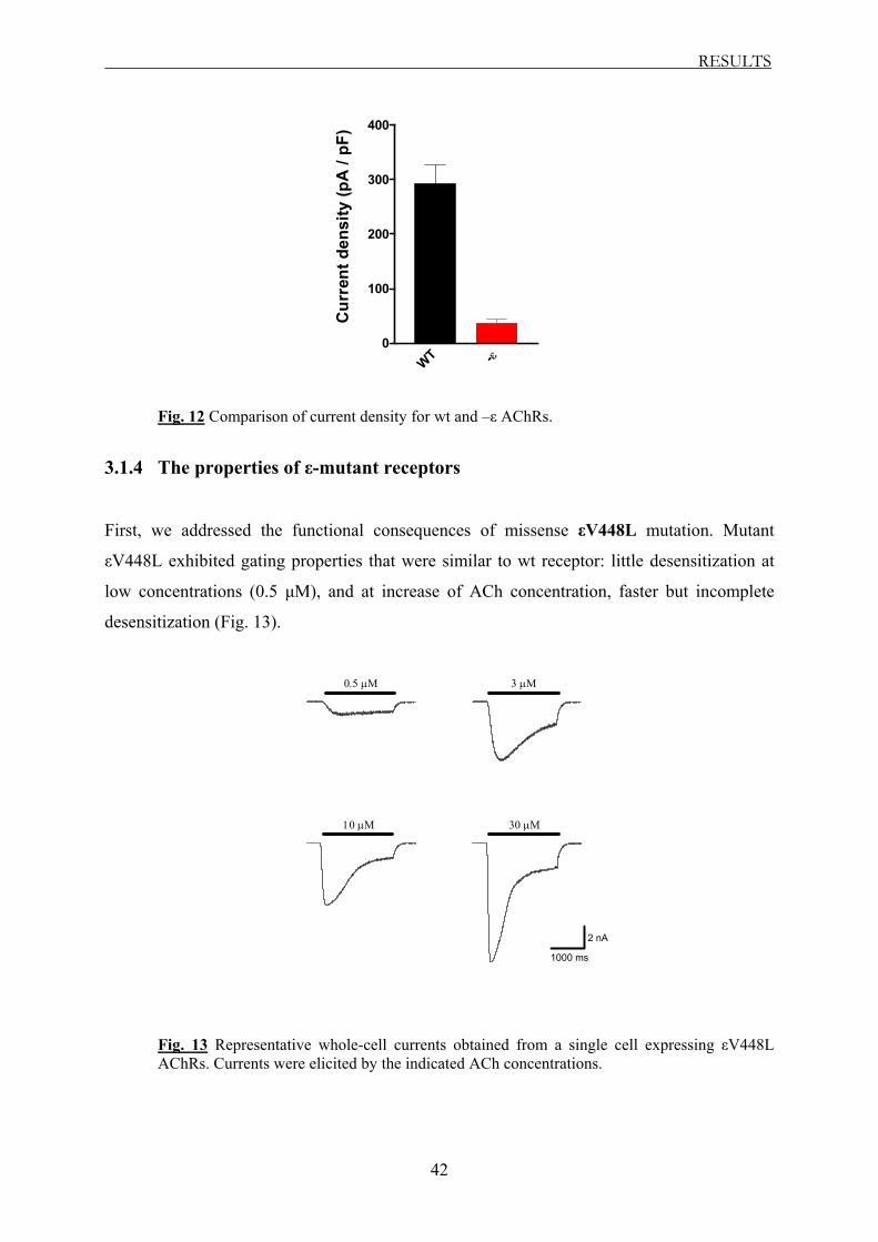
RESULTS
42
WT -ε0
100
200
300
400
Cur
rent
den
sity
(pA
/ pF
)
Fig. 12 Comparison of current density for wt and –ε AChRs.
3.1.4 The properties of ε-mutant receptors
First, we addressed the functional consequences of missense εV448L mutation. Mutant
εV448L exhibited gating properties that were similar to wt receptor: little desensitization at
low concentrations (0.5 µM), and at increase of ACh concentration, faster but incomplete
desensitization (Fig. 13).
3.02.01.0s
3.02.01.0s
3.02.01.0s
3.02.01.0s
2 nA
1000 ms
0.5 µΜ 3 µΜ
30 µΜ10 µΜ
Fig. 13 Representative whole-cell currents obtained from a single cell expressing εV448L AChRs. Currents were elicited by the indicated ACh concentrations.

RESULTS
43
The time constant for desensitization (TauD) had values that were similar to those obtained for
wt receptor (Fig. 16 A). The degree of desensitization was somewhat increased at higher ACh
concentrations (10 to 100 µM) compared to wt, with values of: 83 ± 1.4 % (n = 5), and 91 %
(n = 1) vs. 59 ± 4.47 % (n = 11), and 21.5 ± 5.17 % (n = 6), respectively (Fig. 16 B).
The maximum current density for mutant εV448L was 223.3 ± 30 pA / pF (n = 14). This
value is similar to that obtained with wt AChR (P = 0.364; Fig. 17), suggesting that εV448L
mutation is very unlikely to impair cell surface expression of the receptor.
In contrast to εV448L mutation, the properties of the receptor were altered by mutation at
position 1030 (ε1030delC). Mutation ε1030delC leads to a shift in the reading frame and
produces after 20 residues a stop codon. The resulting protein carries only 1/3 of the
cytoplasmic loop between TM3 and TM4. This may underlie the 2-fold increase in receptor
desensitization (TauD: 1.86 ± 0.11 s, n = 2, and 0.53 ± 0.01 s, n = 3) compared to wt, at ACh
concentrations between 0.03 and 1 µM, respectively (Fig. 14 and 16 A). At larger agonist
concentration (1 to 300 µM), the desensitization process became faster, with values for TauD
similar to those of –ε AChR (Fig. 16 A).
3.02.01.0s
3.02.01.0s
3.02.01.0s
3.02.01.0s
3.02.01.0s
3.02.01.0s
0.03 µΜ 1 µΜ 3 µΜ
10 µΜ 30 µΜ 100 µΜ
200 pA
1000 ms
Fig. 14 Whole-cell currents obtained from a single cell expressing ε1030delC AChRs. Currents were elicited by the indicated ACh concentrations.
Mutation ε1030delC not only altered the kinetic of the time course of the desensitization
process but also affected the degree of desensitization. Thus, between 0.03 and 1 µM there
was a 2-fold increase of desensitization in comparison to wt receptor (54.9 ± 3.5 %, n = 3, and
68.4 ± 1.1 %, n = 5, respectively). However, at higher ACh concentrations (10 to 300 µM),

RESULTS
44
the degree of desensitization was similar to –ε receptor (Fig. 16 B). Mutant ε1030delC
exhibited 3.5-fold reduced current density compared to wt receptor (82.85 ± 11.2 pA / pF, n =
25; P = 0.0003; Fig. 17), and therefore, unlike εV448L, the ε1030delC mutation is likely to
affect the cell surface expression of AChR.
The deletion mutation ε911delT, as well as the three insertion mutations ε1101insT,
ε1206ins19, and ε1293insG caused acceleration of the desensitization process (time course
and degree of desensitization). The properties of these receptors were similar in all cases to
those observed with –ε AChR (Fig. 15 and 16).
4.03.02.0s
30 µΜ10 µΜ
0.5 µΜ 3 µΜ
1000 ms
100 pA
A
4.03.02.01.0s
4.03.02.01.0s
4.03.02.01.0s
4.03.02.01.0s
4.03.02.01.0s
4.03.02.01.0s
0.03 µΜ 0.1 µΜ 0.3 µΜ
10 µΜ1 µΜ 30 µΜ
1000 ms
200 pA
B
ε911delT
ε1101insT

RESULTS
45
5.04.03.0s
5.04.03.0s
5.04.03.0s
5.04.03.0s
5.04.03.0s
5.04.03.0s
100 pA
1000 ms
0.3 µΜ 1 µΜ 10 µΜ
100 µΜ30 µΜ 300 µΜ
C
5.04.03.0s
5.04.03.0s
5.04.03.0s
-300
-200
-100
0
pA
5.04.03.0s
100 µΜ30 µΜ
3 µΜ 10 µΜ
1000 ms
100 pA
D
Fig. 15 Representative whole-cell currents in response to the indicated ACh concentrations obtained from single cells expressing ε911delT (A), ε1101insT (B), ε1206ins19 (C), and ε1293insG AChRs (D), respectively.
ε1206ins19
ε1293insG

RESULTS
46
0.01 0.1 1 10 100 10000.0
0.5
1.0
1.5
2.0
2.5
3.0
3.5
εV448L
WT
ε1030delC
-ε
ε911delT
ε1206ins19ε1101insT
ε1293insG
A
ACh [µM]
τ D[s
]
0.01 0.1 1 10 100 10000
10
2030
4050
607080
90
BWT
ε1030delC
-ε
ε911delT
ε1206ins19ε1101insT
ε1293insG
εV448L
ACh [µM]
I AC
h re
sidu
al [%
]
Fig. 16 Desensitization kinetics: current decay (A) and residual current fraction (B) of ε-mutant AChRs in comparison to wt and –ε receptors.
The maximum current amplitude (Imax) observed with these mutants was significantly reduced
compared to wt receptor, with values similar to those observed in AChR lacking the ε-subunit
(P > 0.05 vs. –ε receptor; compare Fig. 15 A-D vs. Fig. 9).
Mutant ε911delT exhibited ~ 2.5-fold reduced current density compared to –ε receptor (13.57
± 1.83 pA / pF, n = 17; P = 0.009 vs. –ε AChR), whereas all three insertion mutations
(ε1101insT, ε1206ins19, and ε1293insG) resulted in slight, but statistically not significant,
increase in current density (see Fig. 17). Thus, the very likely reduction in cell surface
expression observed with these mutants suggests a loss of function of AChR at the
neuromuscular junction.

RESULTS
47
WT -ε
εV448L
ε1030
delC
ε911d
elT
ε1101
insT
ε1206
ins19
ε1293
insG0
50
100
150
200
250
300
350
Cur
rent
den
sity
(pA
/ pF
)
Fig. 17 Comparison of current density for wt, –ε, and ε-mutant AChRs.
We also studied the affinity for ACh of these ε-mutant AChRs in comparison to wt and –ε
receptors. Except for mutant ε1293insG, which resulted in 5-fold increase in the EC50 value
(17.71 ± 1.14 µM with a Hill slope of 1.29 ± 0.20, n = 2; P < 0.05), all the other mutant
AChRs had EC50 and Hill slope values similar to wt and –ε AChRs (P > 0.05), ranging from
1.01 to 3.7 µM ACh, with Hill slopes between 0.85 and 1.07 (Fig. 18).
In summary, these findings suggest that only ε1293insG mutation shows its effects by both
decreasing sensitivity to ACh and altering desensitization kinetics of the receptor.
-8 -7 -6 -5 -4 -30.00
0.25
0.50
0.75
1.00
-ε
ε911delT
ε1206ins19
εV448Lε1030delC
ε1293insG
ε1101insT
WT
Log ACh [M]
Nor
m. r
espo
nse
Fig. 18 Dose-response relationships obtained for ε-mutant AChRs in comparison to wt and –ε
receptors.
RESULTS
48
3.1.5 Summary of the whole-cell data
With the exception of εV448L receptor, all investigated ε-mutant AChRs exhibited
acceleration of the macroscopic current decay and enhanced degree of desensitization,
compared to wt receptor. Furthermore, their current density was significantly reduced,
suggesting that these receptors might be expressed at low levels at the cell surface. Statistical
analysis also indicated that the EC50 value obtained with mutant ε1293insG was significantly
different from the value exhibited by wt AChR, and thus mutation ε1293insG is also likely to
affect the ACh affinity of the receptor. The other mutations had no effect on the sensitivity to
agonist of AChR.

RESULTS
49
3.2 Single-channel recordings
3.2.1 Introduction to single-channel recordings and data kinetic analysis
From whole-cell recordings, one can extract only average properties of large populations of
ion channels in the membrane (Hille, 2001). The whole-cell current is described by formula
Imax = N ·PO · i, where Imax represents the macroscopic current, N is the number of functional
channels, PO the probability that a channel is open, and i represents the ionic current through
one single channel. It is possible to obtain this information from one individual channel by
performing single-channel recordings.
In a single-channel recording, each opening event is a transition from the closed state (zero
current) to the open state and has defined unitary current (i, pA) and duration (time, ms). An
example of such recordings from wt AChR is illustrated in Figure 19 A.
-14
-12
-10
-8
-6
-4
-2
0
2
pA
28.5228.5128.5028.4928.4828.47kSec
28.476428.476328.476228.4761kSec
10 s
2 pA
100 ms
2 pA
A
B
Fig. 19 Single-channel bursts of openings recorded from a wt AChR at –100 mV and 30 µM ACh; A shows few clusters with channel-opening events (bursts of openings) separated by periods with no channel activity when all AChRs are desensitized. Channel-openings are presented as downward deflections of the membrane current; B represents the magnification of a cluster of opening events, when the AChR undergoes many cycles of agonist association / dissociation. The horizontal bars on the right of B represent the closed and the open states of the receptor.
Close
Open

RESULTS
50
Channel-openings and ionic flux through these channels are represented by the rapid transient
changes in the membrane current in the order of several picoampere (pA). The channel-
openings in Figure 19 are visible as downward deflections. Upon closing of the channel, the
membrane current returns to its baseline value, and these transitions are visible as upward
deflections in Figure 19. The transitions between the closed and the open states occur in the
microsecond range. In the record, channel-openings occur in clusters followed by periods
with no channel activity. This clustering of channel-openings and closings has been termed
“bursts” (Sakmann et al., 1980).
Binding of the agonist to the channel results initially in opening of the channel. However, the
channel undergoes further conformational changes in the continued presence of the agonist
that result in closing of the channel. In this state, the agonist can no longer activate the
channel. This condition has been described as desensitization of the receptor (Katz and
Thesleff, 1957). The long-lived closed intervals between the bursts of openings reflect times
when all AChRs in the patch are desensitized (Fig. 19 A). A cluster starts when one AChR
spontaneously recovers from desensitization, and continues with the protein molecule
undergoing many cycles of agonist association / dissociation (Fig. 19 B). A cluster ends when
that receptor again becomes desensitized.
The process of opening and closing of single-channels is termed as channel-gating and
reflects conformational changes of the AChR. Gating of one channel is a stochastic process,
and to derive information from such measurements requires analysis of many open-close
events using statistical methods. Major parameters that are unique to an individual ion
channel are the amplitude of the unitary current (i) and the mean lifetime (duration) of the
open or the close state. The open lifetime is the time between the opening of one channel and
its next closing, whereas the close lifetime represents the time between two opening events in
succession. The binding of the two agonists brings the receptor into an equilibrium state
between the closed and the open channel. This equilibrium is described by the open
probability (PO), a measure that an ion channel will be open under given conditions.
To understand whether for example the reduction in current density observed with the
insertion / deletion mutations (see Fig. 17) is due to changes in any of the above-described
parameters, we performed single-channel recordings in cell-attached configuration and
analyzed the properties of individual receptors.

RESULTS
51
3.2.2 Unitary current and slope conductance
The ionic current that flows through an individual channel depends on the driving force
(VEMF) for the ions passing through the channel. According to Ohm’s law: I = V / R, the size
of the unitary current (i) can be calculated as i = VEMF x g, where VEMF = Vmp – VREV (VEMF is
the electromotive force, Vmp is the membrane potential, VREV represents the reversal potential
of the permeating ion, and g, the conductance of the ion channel). The differences in whole-
cell current densities between wt and ε-mutant AChRs may be caused by alteration in levels
of expression of functional channels and / or by alteration in their single-channel properties
(e.g. changes in the conductance of the AChR and / or channel-gating).
We first evaluated the unitary current size (i) and the slope conductance (g) of these
receptors. The unitary current of wt receptor was -7.24 ± 0.22 pA (n = 22), at a membrane
potential of –100 mV. A similar value for the unitary current was obtained for the AChR
lacking the ε-subunit (P > 0.05). In contrast, mutations εV448L, ε911delT, and ε1030delC
exhibited different unitary current values, thus suggesting an alteration of the permeation
properties of the channel (Table 1). However, the changes in the unitary current size observed
with the mutants were statistically not different from that observed with wt receptor.
T yp e i (p A) g (p S ) nW T -7 .24 ± 0 .22 60 .92 ± 1 .32 22− ε -7 .73 ± 0 .31 63 ± 0 .97 12
ε V448L -6 .56 ± 0 .35 61 .94 ± 1 .88 6ε 1030de lC -8 .01 ± 0 .31 63 .13 ± 0 .9 9ε 911de lT -6 .44 ± 0 .23 63 .21 ± 0 .74 7
Table 1 Unitary current (i; pA) and slope conductance (g; pS) values for wt, –ε, and ε-mutant AChRs. The unitary current values are given at a membrane potential of –100 mV. Data represent mean ± SEM. n represents the number of cells.
We obtained the slope conductance for these receptors from linear regression fit to the
current-voltage relationship of the individual receptors (Fig. 20). The slope conductances for
wt, –ε, and ε-mutant receptors were very similar, with values ranging from 60.92 to 63.21 pS
(Table 1 and Fig. 20). The values for wt, as well as for the ε-mutant AChRs are similar to
those reported previously by Newland et al. (1995; 62 pS) or Ohno et al. (1997; 60 pS) for
human ACh receptors. These data suggest that the mutations within the ε-subunit have no
measurable effect on the size of the ion flux through the open receptors. We next investigated
the gating kinetic properties of the ε-mutant AChRs.

RESULTS
52
-130 -110 -90 -70 -50
-10
-8
-6
-4
-2
0
ε911delT
WT-ε
ε1030delCεV448L
Vmp (mV)
i (pA
)
Fig. 20 Slope conductance curves were constructed for wt, –ε, and ε-mutant AChRs by plotting the unitary current (i; pA) vs. the membrane potential (Vmp; mV).
3.2.3 Kinetic analysis
3.2.3.1 Gating-kinetics of wild-type receptor
We analyzed the gating properties of the receptors by analysis of the channel-opening events
within the bursts of openings. Bursts were defined as a series of openings with closing
intervals shorter than a critical duration of 30 ms (see Materials and Methods section 2.3.1.2).
Periods longer than 30 ms very likely represent intervals when all of the channels in the patch
are desensitized. The kinetic analysis focused on close and open intervals within clusters
(termed as “Burst analysis”). Only stretches of bursts that contained more than 5 events of
channel-openings were selected one by one and analyzed (see also Fig. 19 B). However, at
low ACh concentrations (1 µM, for wt receptor), we observed mostly isolated channel-
openings and only very rarely a burst of openings (Fig. 21 A).
Using burst analysis, we addressed the gating parameters open and close lifetime, and open
probability of wt, –ε, and ε-mutant AChRs.
The gating pattern of wt AChR, at different agonist concentrations (between 1 and 100 µM) is
shown in Figure 21. At concentration of 1 µM, the channel-opening events occurred only
infrequently and for brief periods, followed by long closing intervals (Fig. 21 A). With
increasing agonist concentration (10 – 100 µM) the frequency of channel-opening events
increased (Fig. 21). The opening events occurred mainly in bursts, and the receptor exhibited
a dose-response increase in the open probability (PO), because the time required for binding of

RESULTS
53
the agonist reduced, thus shortening the close time durations. The PO values were: 0.30 ±
0.025 (n = 3), 0.44 ± 0.026 (n = 7), 0.66 ± 0.021 (n = 9), and 0.82 ± 0.043 (n = 4), at 1, 10, 30,
and, 100 µM, respectively. In contrast, the open duration of the channel remained similar at
all agonist concentrations (Fig. 21 A-D and 22 A).
5850.55850.45850.35850.25850.15850.0Sec
1 µΜ
2pA100ms
A
474.8474.7474.6474.5474.4Sec
10 µΜ
2pA100ms
B
6399.96399.86399.76399.66399.5Sec
30 µΜ
2pA100ms
C
1590.61590.51590.41590.31590.2Sec
100 µΜ
2 pA
100 ms
D
Fig. 21 Single-channel bursts of openings of wt AChR recorded at the indicated ACh concentrations. Currents are shown at bandwidth of 10 kHz, with channel-openings as downward deflections. Holding potential: –100 mV.
The distribution of the open and close time durations from several recordings obtained at
different ACh concentrations are shown in Figure 22. The fit of open duration revealed two
major components with time constants for opening of ~ 1 and 3 ms (Tauopen1 and Tauopen2).
For example, at an ACh concentration of 30 µM, Tauopen1 and Tauopen2 had values of 1.25 ±
0.17 ms, and 2.7 ± 0.4 ms (n = 9), respectively (Fig. 22 A). The fractions of channel-openings
associated with the two open time constants were: 64 ± 2.3 %, and 36 ± 2.3 %, respectively.
Similar values were obtained at concentrations of 10 and 100 µM ACh. However, at agonist
concentration of 10 µM, the opening events occurred at about equal levels at the two open
time constants. At lower concentrations (1 µM), there was a shift towards shorter openings
(Tauopen1 : 0.52 ± 0.06 ms; Tauopen2 : 1.74 ± 0.26 ms; n = 3). However, the major open
duration was similar to those observed at higher concentrations (Fig. 22 A).
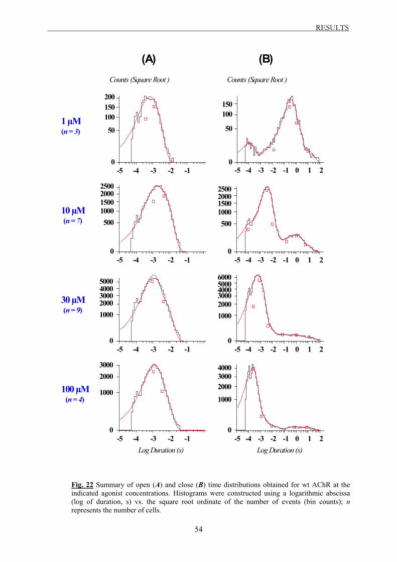
RESULTS
54
-4 -3 -2 -1-5
Log Duration (s)
-4 -3 -2 -1-5
-4 -3 -2 -1-5
-4 -3 -2 -1-5Log Duration (s)
-4 -3 -2 -1 0-5 1 2
-4 -3 -2 -1 0-5 1 2
-4 -3 -2 -1 0-5 1 2
-4 -3 -2 -1 0-5 1 2
Counts (Square Root ) Counts (Square Root )
200
100
50
0
150100
50
0
150
2000
1000500
0
1500
25002000
1000500
0
1500
2500
4000
20001000
0
3000
50004000
20001000
0
300050006000
2000
1000
0
3000 4000
2000
1000
0
3000
1 µM(n = 3)
10 µM(n = 7)
30 µM(n = 9)
100 µM(n = 4)
(A) (B)
Fig. 22 Summary of open (A) and close (B) time distributions obtained for wt AChR at the indicated agonist concentrations. Histograms were constructed using a logarithmic abscissa (log of duration, s) vs. the square root ordinate of the number of events (bin counts); n represents the number of cells.

RESULTS
55
In contrast, the distribution of the channel close times shifted in a concentration-dependent
manner towards shorter close time durations. The fastest component (Tauclose1) had an average
value of 0.2 ± 0.02 ms (n = 4), and described more than 95 ± 0.8 % of channel-closing
duration at 100 µM (Fig. 22 B). At lower concentrations (10 and 30 µM), the dominant close
time constants (Tauclose2) were 3.6 ± 0.18 ms (n = 7) and 0.75 ± 0.04 ms (n = 9), respectively.
They accounted for about 75 % of the closing intervals exhibited by wt receptor, at both
concentrations. At the lowest concentration tested (1 µM), Tauclose1 had a value of 0.12 ±
0.009 ms (n = 3), and represented only a small fraction of 8.4 ± 1.5 % of all shut times (Fig.
22 B). The majority of the closings were longer than 30 ms, reflecting low open probability at
this concentration (0.30 ± 0.025, n = 3).
3.2.3.2 Gating-kinetics of ε-subunit lacking receptor
The AChR lacking the ε-subunit exhibited a different gating kinetic pattern, consisting of
fewer and briefer opening events than wt receptor, and longer intervals with no channel
activity (Fig. 23 A-D).
-10
-8
-6
-4
-2
0
2
pA
7496.87496.77496.67496.57496.47496.3Sec
1 µΜA
2pA100ms 2855.52855.42855.32855.22855.12855.0
Sec
10 µΜ
2pA100ms
B
-10
-8
-6
-4
-2
0
2
pA
7542.57542.47542.37542.27542.17542.0Sec
30 µΜ
2pA100ms
C
-10
-8
-6
-4
-2
0
pA
8072.38072.28072.18072.08071.9Sec
100 µΜ
2 pA
100 ms
D
Fig. 23 Single-channel opening events of –ε AChR recorded at the indicated ACh concentrations. Holding potential: –100 mV.

RESULTS
56
The absence of the ε-subunit resulted in a drastic reduction of the open probability of the
receptor. For example, at a concentration of 30 µM the open probability of –ε receptor within
the total recording time was ~ 40-fold reduced compared to wt AChR (9.8 ± 3.5 · 10-4, n = 6
vs. 432 ± 52.2 · 10-4, n = 9; see Table 3 and Fig. 32). This is also illustrated by the reduction
in the overall number of channel-openings (Fig. 23).
The drastic changes in the gating kinetics are also reflected in the open and close time
distributions for the ε-subunit lacking receptor (Fig. 24). The most visible change was the
increase in the close time distribution, with a large increase in the number of long-lasting
closings (> 30 ms) compared to wt receptor (Fig. 24 B vs. 22 B).
Fitting the open time distribution revealed a fast component (Tauopen1) with a time constant of
~ 0.2 ms (Fig. 24 A). At an agonist concentration of 1 µM, this fast component had a value of
0.14 ms (n = 1) and was the only gating component observed. At higher agonist
concentrations, the values for Tauopen1 were very similar (10 µM: 0.21 ± 0.014 ms, n = 4; 30
µM: 0.18 ± 0.02 ms, n = 6; 100 µM: 0.24 ± 0.022 ms, n = 7) and were associated with ~ 85 %
of all channel-openings. In addition to the fast component, two slower gating components
(Tauopen2 and Tauopen3) were observed at agonist concentrations between 10 and 100 µM, with
values of ~ 0.8 and ~ 5 ms, respectively. The fraction of opening events associated with
Tauopen2 was ~ 10 % and that associated with Tauopen3 was ~ 5 % (Fig. 24 A).
As observed, in the AChR lacking the ε-subunit there is a 5-fold shortening of the major open
time constant (Tauopen1), in comparison to wt receptor. At all concentrations, most of the
closings were longer than 30 ms (Fig. 24 B), reflecting a low open probability for –ε receptor.
Furthermore, the fraction of channel close times that occurred during the burst of openings, at
agonist concentration of 30 µM was only 7.4 ± 2.7 %, and 12 ± 4.8 % (n = 6), for Tauclose1 and
Tauclose2, respectively (Tauclose1 : 0.45 ± 0.13 ms; Tauclose2 : 2.55 ± 0.36 ms). Similar values
were observed at agonist concentrations of 10 and 100 µM (Fig. 24 B). At the lowest
concentration tested (1 µM), Tauclose1 weighted only 3% of all close times, whereas Tauclose2
was not observed.
Together, these findings indicate that the AChR lacking the ε-subunit is still functional,
however, it is less functional than the wt receptor. This suggests that mutations that truncate
the ε-subunit are likely to generate a receptor with similarly altered gating properties as
observed with –ε AChR.

RESULTS
57
-4 -3 -2 -1-5
Log Duration (s)
-4 -3 -2 -1-5
-4 -3 -2 -1-5
-4 -3 -2 -1-5Log Duration (s)
-4 -3 -2 -1 0-5 1 2
-4 -3 -2 -1 0-5 1 2
-4 -3 -2 -1 0-5 1 2
-4 -3 -2 -1 0-5 1 2
Counts (Square Root ) Counts (Square Root )
80
4020
0
60
100 40
20
10
0
30
400
200
100
0
300 100
50
0
150
400
200
0
600 200
100
50
0
150
8
4
2
0
6
10
2
1
0
3
1 µM(n = 1)
10 µM(n = 4)
30 µM(n = 6)
100 µM(n = 7)
(A) (B)
Fig. 24 Summary of open (A) and close (B) time distributions obtained for –ε AChR at the indicated agonist concentrations.

RESULTS
58
3.2.3.3 Gating-kinetics of ε-mutant receptors
To investigate the gating properties of AChR containing mutated ε-subunit, we performed
recordings only at an agonist concentration of 30 µM. This concentration is optimal to
eliciting bursts of channel-openings from single receptors, and, at the same time, does not
induce desensitization of AChR (Sakmann et al., 1980).
As predicted by the properties observed at whole-cell level (see also section 3.1.4), mutant
εV448L exhibited open and close time distributions similar to wt receptor (Fig. 25).
-8
-6
-4
-2
0
pA
5659.05658.95658.85658.75658.6Sec
30 µΜ
2 pA
100 ms
A
Log Duration (s)
-4 -3 -2 -1-5
Log Duration (s)
-4 -3 -2 -1 0-5 1 2
Counts (Square Root ) Counts (Square Root )
2000
1000
500
0
15002000
1000500
0
1500
30 µM(n = 10)
(B) (C)
Fig. 25 Single-channel bursts of openings recorded at –100 mV from a εV448L AChR, elicited by 30 µM ACh (A), and summary of open (B) and close (C) time distributions.
The open time constants, Tauopen1 and Tauopen2, had values of 1.4 ± 0.15 ms, and 4.48 ± 0.24
ms (n = 10), respectively. The opening events occurred at about equal levels at the two open
time constants (Fig. 25 B). Furthermore, the majority of closings occurred in clusters (Fig. 25

RESULTS
59
A), as indicated by the fastest component of the close time distribution, Tauclose1 (Fig. 25 C),
which had a value of 0.71 ± 0.04 ms (n = 7). This close time constant described more than
75.3 ± 3.5 % of channel-closing duration. These values were similar in all cases to those
obtained with wt receptor. The open probability in clusters was high (PO : 0.68 ± 0.022, n =
10, at 30 µM). This value is statistically not different from that obtained for wt AChR (P >
0.05; see also Fig. 31). In conclusion, the gating kinetics of AChR were not altered by the
εV448L mutation located within the fourth transmembrane domain of the ε-subunit.
The deletion mutation ε911delT, located in the third membrane-spanning segment of the ε-
subunit, is predicted to truncate the entire M3-M4 – cytoplasmic loop of the subunit (see also
Introduction section 1.3.2.3, Fig. 6). Recordings from ε911delT-containing receptors revealed
gating kinetics similar to those observed with –ε AChRs (Fig. 26 vs. Fig. 23 and 24).
4842.94842.84842.74842.64842.5Sec
30 µΜ
100 ms2 pA
A
4842.784842.774842.764842.75Sec 10 ms
2 pA
B
Log Duration (s)
-4 -3 -2 -1-5
Log Duration (s)
-4 -3 -2 -1 0-5 1 2
Counts (Square Root ) Counts (Square Root )
80
40
20
0
60200
10050
0
150
250
30 µM(n = 12)
(C) (D)
Fig. 26 Single-channel opening events recorded from a ε911delT AChR, elicited by 30 µM ACh (A and B), and summary of open (C) and close (D) time distributions; B represents the magnification of the encircled area of A. Holding potential: –100 mV.
B

RESULTS
60
The mutant showed only rarely openings that were very brief in duration (Tauopen1 : 0.17 ±
0.01 ms; n = 12) in 78 ± 3.5 % of the apparent open time (Fig. 26 A-C). A second open time
constant (Tauopen2) was similar to that observed with –ε receptor at a concentration of 30 µM.
Additionally, ~ 9 % of the openings were longer than 10 ms (Tauopen3, Fig. 26 C).
The distribution of the channel close times exhibited a reduction in the fraction of closings
during burst gating (see Tauclose1 in Fig. 26 D), and the majority of the closings were longer
than 30 ms. The open probability within the total recording time for mutant ε911delT was 11
± 3.8 · 10-4 (n = 12, at 30 µM; see Fig. 32). Compared to wt receptors, this is almost a 40-fold
reduction in the open probability, however, this value is similar to that observed in receptors
lacking the ε-subunit (P > 0.05).
Mutant ε1030delC exhibited only few and brief channel-openings at an agonist concentration
of 1 µM (recording not shown), and spent long periods in the closed state (see Fig. 28). At
higher agonist concentrations (30 and 100 µM ACh), the frequency of opening / closing
events increased. During prolonged recordings, we observed in 3 out of 8 recordings a distinct
switch in the gating mode of the receptor, as illustrated in Figure 27.
-14
-12
-10
-8
-6
-4
-2
0
pA
374037303720371037003690Sec 10 s
2 pA
30 µΜ
Fig. 27 Single-channel bursts of openings elicited by 30 µM ACh from a ε1030delC AChR. The recording shows the switch between the two distinct gating modes during a recording time of 60 s. Holding potential: –100 mV.
The first gating mode (termed as ‘a’) resembled the gating pattern of –ε receptor with brief
channel-openings. This had a major open time constant, Tauopen1, of 0.14 ± 0.01 ms (n = 5, at
30 µM; Fig. 28 A). The second gating mode (termed as ‘b’) resembled the gating pattern
observed with wt AChR, where channel-openings occurred mainly in clusters, with a major
open time constant, Tauopen2, of 1.9 ± 0.62 ms (n = 3; Fig. 28 A). The fractions of channel-
openings associated to the two dominant open time constants were similar in all cases to those
observed in recordings with either –ε or wt AChRs, respectively.

RESULTS
61
-4 -3 -2 -1-5
Log Duration (s)
-4 -3 -2 -1-5
-4 -3 -2 -1-5
-4 -3 -2 -1-5Log Duration (s)
-4 -3 -2 -1 0-5 1 2
-4 -3 -2 -1 0-5 1 2
-4 -3 -2 -1 0-5 1 2
-4 -3 -2 -1 0-5 1 2
Counts (Square Root ) Counts (Square Root )
80
4020
0
60
100
20
10
0
30
200
100
50
0
150
-4 -3 -2 -1-5 -4 -3 -2 -1 0-5 1 2
80
40
20
0
60 20
10
0
30
80
4020
0
60
10040
2010
0
30
50
200
100
50
0
150
40
20
0
6080
80
4020
0
60
100
1 µM(n = 4)
30 µM (a)(n = 5)
30 µM (b)(n = 3)
100 µM (a)(n = 3)
100 µM (b)(n = 2)
(A) (B)
Fig. 28 Summary of open (A) and close (B) time distributions obtained for the ε1030delC AChR at the indicated ACh concentrations; (a) stands for gating pattern with brief events, whereas (b) represents the gating mode with bursts of openings.
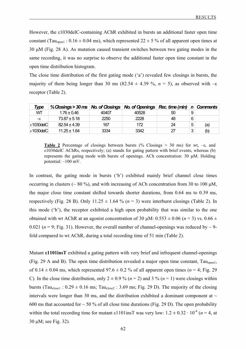
RESULTS
62
However, the ε1030delC-containing AChR exhibited in bursts an additional faster open time
constant (Tauopen1 : 0.16 ± 0.04 ms), which represented 22 ± 5 % of all apparent open times at
30 µM (Fig. 28 A). As mutation caused transient switches between two gating modes in the
same recording, it was no surprise to observe the additional faster open time constant in the
open time distribution histogram.
The close time distribution of the first gating mode (‘a’) revealed few closings in bursts, the
majority of them being longer than 30 ms (82.54 ± 4.39 %, n = 5), as observed with –ε
receptor (Table 2).
Type % Closings > 30 ms No. of Closings No. of Openings Rec. time (min) n CommentsWT 1.76 ± 0.46 40407 40528 50 9−ε 73.87 ± 5.18 2250 2228 48 6
ε1030delC 82.54 ± 4.39 167 172 24 5 (a)ε1030delC 11.25 ± 1.64 3334 3342 27 3 (b)
Table 2 Percentage of closings between bursts (% Closings > 30 ms) for wt, –ε, and ε1030delC AChRs, respectively; (a) stands for gating pattern with brief events, whereas (b) represents the gating mode with bursts of openings. ACh concentration: 30 µM. Holding potential: –100 mV.
In contrast, the gating mode in bursts (‘b’) exhibited mainly brief channel close times
occurring in clusters (~ 80 %), and with increasing of ACh concentration from 30 to 100 µM,
the major close time constant shifted towards shorter durations, from 0.64 ms to 0.39 ms,
respectively (Fig. 28 B). Only 11.25 ± 1.64 % (n = 3) were interburst closings (Table 2). In
this mode (‘b’), the receptor exhibited a high open probability that was similar to the one
obtained with wt AChR at an agonist concentration of 30 µM: 0.553 ± 0.06 (n = 3) vs. 0.66 ±
0.021 (n = 9; Fig. 31). However, the overall number of channel-openings was reduced by ~ 9-
fold compared to wt AChR, during a total recording time of 51 min (Table 2).
Mutant ε1101insT exhibited a gating pattern with very brief and infrequent channel-openings
(Fig. 29 A and B). The open time distribution revealed a major open time constant, Tauopen1,
of 0.14 ± 0.04 ms, which represented 97.6 ± 0.2 % of all apparent open times (n = 4; Fig. 29
C). In the close time distribution, only 2 ± 0.9 % (n = 2) and 3 % (n = 1) were closings within
bursts (Tauclose1 : 0.29 ± 0.16 ms; Tauclose2 : 3.69 ms; Fig. 29 D). The majority of the closing
intervals were longer than 30 ms, and the distribution exhibited a dominant component at ~
600 ms that accounted for ~ 50 % of all close time durations (Fig. 29 D). The open probability
within the total recording time for mutant ε1101insT was very low: 1.2 ± 0.32 · 10-4 (n = 4, at
30 µM; see Fig. 32).

RESULTS
63
-8
-6
-4
-2
0pA
3060.63060.53060.43060.33060.2Sec
30 µΜ
2 pA100 ms
A
-8
-6
-4
-2
0
pA
3060.623060.613060.603060.593060.58Sec
30 µΜ
2 pA
10 ms
B
Log Duration (s)
-4 -3 -2 -1-5
Log Duration (s)
-4 -3 -2 -1 0-5 1 2
Counts (Square Root ) Counts (Square Root )
10
5
0
1580
40
20
0
60
30 µM(n = 4)
(C) (D)
Fig. 29 Single-channel opening events recorded from a ε1101insT AChR (A and B), and summary of open (C) and close (D) time distributions, obtained at an agonist concentration of 30 µM; B represents the magnification of the encircled area of A. Holding potential: –100 mV.
3.2.3.4 The spontaneous activity of AChR
Spontaneous openings of AChR in the absence of ACh have been reported in the literature for
mouse, rat, and bovine AChRs (Jackson, 1984, 1986; Jaramillo et al., 1988; Jackson et al.,
1990; Zhou et al., 1999a,b; Grosman et al., 2000). To test whether these spontaneous channel-
openings also occur in human AChR and to estimate their contribution in our recordings, we
performed recordings in the absence of ACh, for wt and ε-subunit lacking AChRs.
In the absence of the agonist and within the total recording time, wt and –ε AChRs exhibited
very low open probability values of 1.6 ± 0.6 · 10-4 (n = 5) and 1.3 ± 0.6 · 10-4 (n = 5),
respectively (see Table 3 and Fig. 32). These values were in any case smaller than the values
obtained at low ACh concentrations (1 – 10 µM) for both types of receptors (Table 3).
B
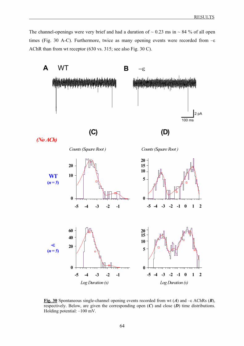
RESULTS
64
The channel-openings were very brief and had a duration of ~ 0.23 ms in ~ 84 % of all open
times (Fig. 30 A-C). Furthermore, twice as many opening events were recorded from –ε
AChR than from wt receptor (630 vs. 315; see also Fig. 30 C).
-8
-6
-4
-2
0
pA
2414.72414.62414.52414.42414.3Sec
WT
2pA
100ms
A
-8
-6
-4
-2
0
pA
713.5713.4713.3713.2713.1Sec
−ε
2 pA
100 ms
B
-4 -3 -2 -1-5 -4 -3 -2 -1 0-5 1 2
Counts (Square Root ) Counts (Square Root )
20
10
0
WT(n = 5)
-4 -3 -2 -1-5 -4 -3 -2 -1 0-5 1 2
-ε(n = 5)
Log Duration (s) Log Duration (s)
40
20
0
60
10
5
0
1520
10
5
0
1520
(No ACh)(C) (D)
Fig. 30 Spontaneous single-channel opening events recorded from wt (A) and –ε AChRs (B), respectively. Below, are given the corresponding open (C) and close (D) time distributions. Holding potential: –100 mV.

RESULTS
65
Interestingly, in the absence of ACh, ~ 32 % of the total close times were short closings with
a duration of ~ 0.7 ms, in the case of wt receptor (Fig. 30 D). Some 68 % were closing
intervals longer than 1 s. Similar values were obtained for –ε AChR as well (P > 0.05; see Fig.
30 D).
Taken together, these findings suggest that the spontaneous opening events in wt and –ε
receptors occur even in the absence of ACh, however these openings occur very rarely, and
therefore disturb very little our observations at very low ACh concentrations.
Type [ACh; µM] Po total rec. time SEM n Total rec. time (min)WT 0 1.60E-04 6.00E-05 5 17WT 1 7.31E-03 4.46E-03 3 30WT 10 2.93E-02 4.20E-03 7 24WT 30 4.32E-02 5.22E-03 9 50WT 100 2.28E-01 1.06E-01 4 30−ε 0 1.30E-04 6.00E-05 5 20−ε 10 4.56E-04 1.65E-04 4 16−ε 30 9.80E-04 3.50E-04 6 48−ε 100 5.09E-03 1.71E-03 7 39
Table 3 Open probability (PO) values within the total recording time obtained for wt, and –ε AChRs, in the absence or presence (at the indicated concentrations) of agonist. Holding potential: –100 mV.
WT
εV448L
ε103
0delC
(b)
0.0
0.1
0.2
0.3
0.4
0.5
0.6
0.7
0.8
0.9
1.0WTε V448Lε 1030delC (b)
P O b
urst
s
Fig. 31 Open probability (PO) within bursts (between 0.04 and 30 ms) exhibited by wt, εV448L, and ε1030delC AChRs at an ACh concentration of 30 µM. Holding potential: –100 mV. In the case of mutant ε1030delC AChR, the PO value is given for the gating mode with bursts of openings (‘b’; n = 3 cells).

RESULTS
66
WT -ε
εV44
8L
ε103
0delC
ε911
delT
WT (no A
Ch)
-ε (no A
Ch)0.0001
0.001
0.01
0.1 WT-εε V448Lε 1030delC (a + b)ε 911delTWT (no ACh)-ε (no ACh)
P O to
tal r
ecor
ding
Fig. 32 Open probability (PO) within the total recording time exhibited by wt, –ε, and ε-mutant AChRs at an agonist concentration of 30 µM ACh. Holding potential: –100 mV. The PO values were plotted on a logarithmic ordinate. In the case of ε1030delC AChR, the PO value represents the overall open probability when the receptor switched between the two distinct gating modes (‘a’ + ‘b’; n = 8 cells), during a total recording time of 51 min.
3.2.4 The correlation between the whole-cell- and the single-channel data
As mentioned in Materials and Methods section 2.3.1.1.2, we introduced the current density
(pA / pF) parameter, as a normalization of current responses to the cell surface. We did this,
to correct for variation in current magnitude due to differences in cell size. On examining the
data, we observed that the current density values obtained for –ε, and ε-mutant AChRs were
significantly reduced in comparison to wt receptor (Table 4 A and Fig. 17).
In whole-cell configuration, the recordings are taken from large populations of channels (Imax
= N ·PO · i, see section 3.1.1). It was therefore difficult to tell if the reduction in the current
density was a contribution of one or a combination of the parameters, number of functional
channels (N), open probability (PO) and / or unitary current (i).
To address this question, we have further investigated the ε-mutant receptors at single-channel
level. A comparison of the data obtained in whole-cell- and single-channel mode is shown in
Table 4.
The number of cells transfected with wt and ε-mutant AChRs, which responded to ACh
application in whole-cell configuration was very similar to the values calculated from single-
channel recordings (compare Table 4 A with 4 B). These data indicate that the ε-mutations
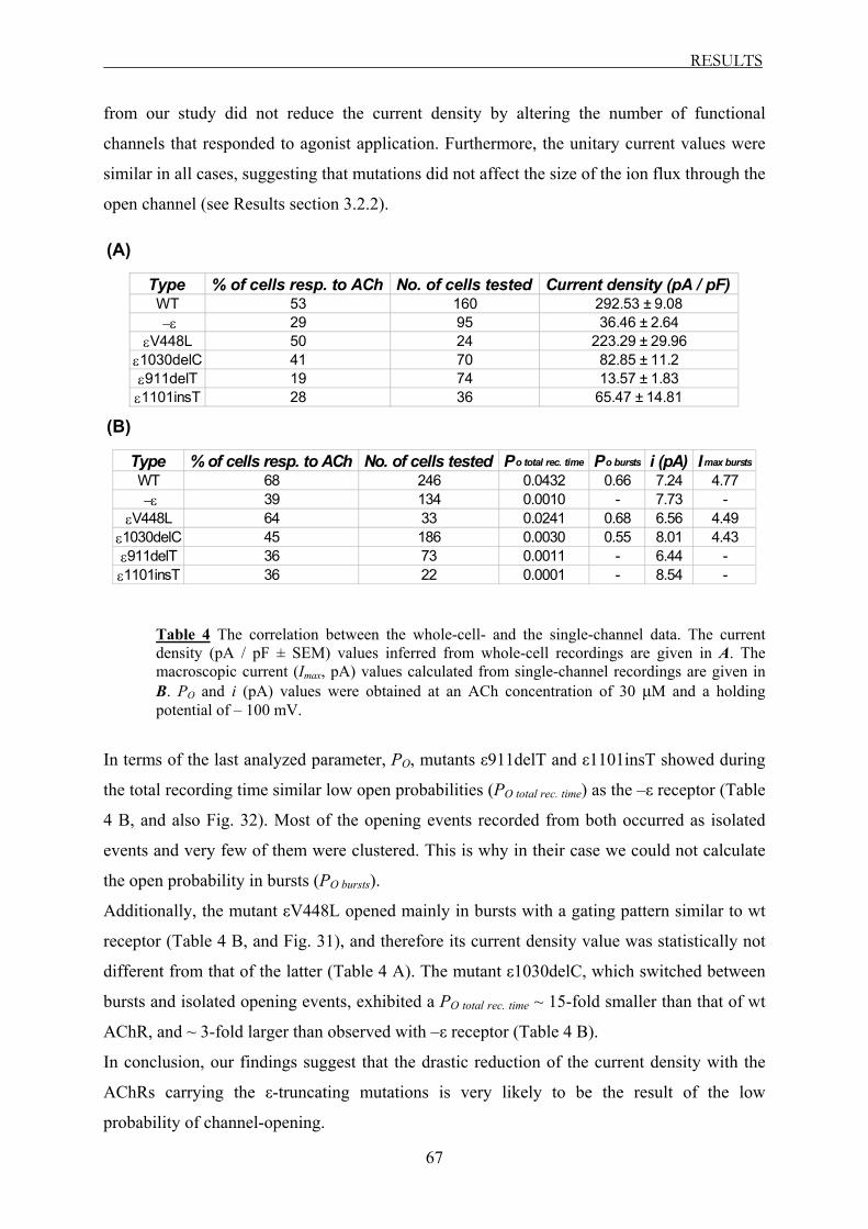
RESULTS
67
from our study did not reduce the current density by altering the number of functional
channels that responded to agonist application. Furthermore, the unitary current values were
similar in all cases, suggesting that mutations did not affect the size of the ion flux through the
open channel (see Results section 3.2.2).
Type % of cells resp. to ACh No. of cells tested Current density (pA / pF)WT 53 160 292.53 ± 9.08 −ε 29 95 36.46 ± 2.64
εV448L 50 24 223.29 ± 29.96ε1030delC 41 70 82.85 ± 11.2ε911delT 19 74 13.57 ± 1.83ε1101insT 28 36 65.47 ± 14.81
Type % of cells resp. to ACh No. of cells tested Po total rec. time Po bursts i (pA) Imax bursts
WT 68 246 0.0432 0.66 7.24 4.77 −ε 39 134 0.0010 - 7.73 -
εV448L 64 33 0.0241 0.68 6.56 4.49ε1030delC 45 186 0.0030 0.55 8.01 4.43ε911delT 36 73 0.0011 - 6.44 -ε1101insT 36 22 0.0001 - 8.54 -
Table 4 The correlation between the whole-cell- and the single-channel data. The current density (pA / pF ± SEM) values inferred from whole-cell recordings are given in A. The macroscopic current (Imax, pA) values calculated from single-channel recordings are given in B. PO and i (pA) values were obtained at an ACh concentration of 30 µM and a holding potential of – 100 mV.
In terms of the last analyzed parameter, PO, mutants ε911delT and ε1101insT showed during
the total recording time similar low open probabilities (PO total rec. time) as the –ε receptor (Table
4 B, and also Fig. 32). Most of the opening events recorded from both occurred as isolated
events and very few of them were clustered. This is why in their case we could not calculate
the open probability in bursts (PO bursts).
Additionally, the mutant εV448L opened mainly in bursts with a gating pattern similar to wt
receptor (Table 4 B, and Fig. 31), and therefore its current density value was statistically not
different from that of the latter (Table 4 A). The mutant ε1030delC, which switched between
bursts and isolated opening events, exhibited a PO total rec. time ~ 15-fold smaller than that of wt
AChR, and ~ 3-fold larger than observed with –ε receptor (Table 4 B).
In conclusion, our findings suggest that the drastic reduction of the current density with the
AChRs carrying the ε-truncating mutations is very likely to be the result of the low
probability of channel-opening.
(A)
(B)

RESULTS
68
3.2.5 Summary of the single-channel data
As observed, the mutants expanded to some extent the distributions for channel-opening and
closing time constants. Thus, inspection of the open and close time distributions revealed
briefer opening events and prolonged closings for ε-mutant receptors compared to wild-type
receptors. Only mutant εV448L showed gating kinetics similar to wt receptor. Mutant
ε1030delC exhibited kinetic components of both, the individual wt and the ε-subunit lacking
AChRs. Mutants ε911delT and ε1101insT showed gating kinetics similar to –ε AChR.
The low open probability exhibited by the individual receptors carrying the ε-truncating
mutations is most probably responsible for the significant reduction of their current density
observed in whole-cell recordings.

DISCUSSION
69
4 Discussion
4.1 CMS mutations and their localization
Mutations in the AChR ε-subunit gene are the most common cause of congenital myasthenic
syndrome (CMS) characterized by deficiency of AChR at the motor endplate (Engel et al.,
1996a; Ohno et al., 1997; Ohno et al., 1998a,b; Middleton et al., 1999; Croxen et al., 1999;
Abicht et al., 1999; Abicht et al., 2000; Sieb et al., 2000a,b; Croxen et al., 2001). The
mutations are found along the length of the ε-subunit gene, including the promoter region
(Nichols et al., 1999; Ohno et al., 1999; Abicht et al., 2002), and result in frameshifts or
nonsense codons that truncate the subunit polypeptide chain. This results in loss of residues
essential for AChR assembly or function.
In the present study, we investigated two recently by Sieb and colleagues (2000a) identified
mutations in the ε-subunit gene, ε911delT, and ε1030delC. These mutations are located at the
end of the M3 transmembrane domain and in the cytoplasmic loop between TM3-4,
respectively. We were interested in investigating the molecular mechanism underlying AChR
deficiency in these cases since ε-subunits expressed from these mutant alleles contain
domains thought to be essential for AChR function.
The functional consequences of these mutations were investigated using patch-clamp
recordings in whole-cell- and cell-attached configuration. Three related CMS-linked
mutations all located in the TM3-4 cytoplasmic region, ε1101insT, ε1206ins19 and
ε1293insG, were included in the study to identify the role of specific regions in TM3-4 loop
in AChR function. All mutations are frameshifting and are predicted to truncate the ε-subunit
of AChR, generating a protein product with impaired functionality.
A common finding for all mutations located within the TM3-4 loop was a functional
reduction of the underlying receptor via two mechanisms: reduction in number of receptors
expressed on the cell surface and interference with channel-gating. In addition, mutant
ε1030delC introduced a switch in the gating mode of the receptor with characteristics
resembling those of wt, as well as ε-subunit lacking AChR, while mutation ε911delT, located
at the end of TM3, completely prevented formation of functional receptors.

DISCUSSION
70
4.2 Structure, formation and membrane insertion of the AChR
The AChR belongs to the ligand-gated ion channel superfamily that shares many structural
features. These pentameric receptors are usually comprised of several different subunits
arranged in a circle like structure to form a central pore, the ion channel (Karlin, 2002; see
Introduction section 1.2.1.1). Within the endoplasmic reticulum (ER), the different subunits of
the receptor are assembled and processed to the mature protein. The ability to distinguish
between correctly assembled receptors and intermediate-building steps is of utmost
importance to ensure reliable and stable signal transduction processes. Incorrectly folded
proteins cannot be assembled and are retained and degraded within the ER, and only
physiological active complexes with the correct stoichiometry are transported to the cell
surface (Smith et al., 1987; Verrall et al., 1992; Blount et al., 1990; Gu et al., 1991). The
AChR is comprised of four different subunits with a stoichiometry of 2α1,β1,γ,δ for the fetal
form, and 2α1,β1,ε,δ for the adult form of the receptor. Within the ER, the receptor is
assembled in a step-like fashion where the α1-δ, α1-γ and α1-ε subunits form heterodimers that
interact with the β1 subunit to form the final pentameric structure. In skeletal muscle,
assembly of AChR is an inefficient process and more than 90 % of newly synthesized AChR
subunits are incorrectly folded (Smith et al., 1987; Gu et al., 1991). Single or incomplete
associated subunits are retained within the ER and degraded (Smith et al., 1987; Merlie et al.,
1983; Blount et al., 1990). Further, transport of all subunits to the cell surface is prevented if
transcript for the α-subunit is erroneous (Black et al., 1987; Gu et al., 1989). A vital point for
regulation of cell surface expression is the control of exit from the ER, however, currently
little is known about the regulation of these steps. Recently, Wang et al. (2002) identified a
conserved signal sequence (PLYFxxN motif) within the M1 domain of all AChR subunits that
controls incorporation of the mature functional protein into the cell membrane. In fully
assembled pentamers, the signal sequence is hidden inside the protein (close to the pore-
forming region) and allows transport to the cell surface. In contrast, if the signal sequence is
accessible to the lumen, these subunits will not be assembled and are subject to degradation.
The signal sequence therefore serves as quality control in the process of receptor formation
and membrane insertion.
Several structural elements within the ε-subunit have been identified that may influence
function of the receptor by controlling interaction with other subunits or translocation to the
cell membrane. Recent studies on the AChR ε-subunit resulted in the proposal that the N-
terminal domain mediates the initial subunit associations, whereas signals in its C-terminus
DISCUSSION
71
half are required for subsequent subunit interactions (Eertmoed and Green, 1999). In addition,
studies of a three amino acid deletion in the β-subunit have identified a region in the
cytoplasmic loop between M3 and M4 where the secondary structure is crucial for interaction
between the β- and δ-subunits (Quiram et al., 1999).
A single cysteine residue within the carboxyl-terminal region at position 470 was recently
identified by Ealing and co-workers (2002) and has been suggested to play a crucial role in
surface expression of adult AChR. Robust surface expression was seen only when the ε-
subunit contained the C470 residue but was completely lost when C470 was deleted or
mutated to serine (S470). Fluorescent labeling of the ε-subunit showed that the mutant ε-
subunits 1369delG or C470S are retained within the ER. The interaction partner(s) of the C-
terminus εC470 are currently not known. The authors suggested that the loss of εC470
partially reduces the efficiency of the steps involved in the initial α–ε subunit association and
affect subsequent steps in pentamer assembly, transfer from the ER and incorporation into the
plasma membrane. It is further possible that C470 might be involved in formation of a
disulphide bond in addition to that of the N-terminal extracellular cysteine-loop structure that
is common to all members of this ion channel superfamily. Ealing et al. (2002) concluded that
all truncating mutations that occur before position C470 would lead to a complete loss of the
AChR protein. The findings from the present study do not support this hypothesis.
4.3 Mutation ε911delT in the M3 segment leads to loss of function
Aim of the work was to gain insights into the molecular mechanism underlying AChR
deficiency in postsynaptic CMS due to truncating mutations of the ε-subunit. The initial
characterization of human wild-type muscle AChR (2α1,β1,ε,δ) and the ε-subunit lacking
AChR (2α1,β1,δ) set the framework that allowed the interpretation of functional effects of the
CMS mutations.
AChRs lacking the ε-subunit could be easily distinguished from wt receptors based on their
biophysical properties (largely reduced current density, rapid desensitization kinetics,
complete desensitization). On the single-channel level, the AChR lacking the ε-subunit
exhibited a different gating pattern compared to wt receptor. The number of opening events
was about 40-fold reduced and the open channel duration was only 1/5 of that observed with
wt AChR (see Results section 3.2.3.2). In contrast, the single-channel conductances for wt
and –ε AChRs were very similar, with a value of 61 pS. Similar values have been previously
reported for wt human muscle AChR (Newland et al., 1995; Ohno et al., 1997). A larger
DISCUSSION
72
conductance of 78 pS has been reported for mouse AChR (Ohno et al., 1995) and it is very
likely to be due to species differences (but see below section 4.4.1).
The AChR that contained the frameshifting mutation in transmembrane domain 3 - ε911delT -
exhibited biophysical properties that resembled those observed with AChR lacking the ε-
subunit. The current density of ε911delT-containing receptors was below that seen with ε-
lacking AChR, suggesting a severe interference with cell surface expression of the receptor.
Single-channel recordings further revealed biophysical properties for ε911delT similar to
those observed with ε-lacking AChR, indicating a loss of function of the receptor containing
the mutated ε-subunit. The deletion ε911delT leads to a frameshift in the predicted amino acid
sequence, causing a premature stop codon after 60 missense codons, and would preserve only
the first 15 amino acids of the large intracellular loop. This would indicate that critical
elements controlling receptor formation / expression are located downstream of position 911.
The data suggest that homozygous expression of ε911delT is very likely to result in a loss of
function. To date, no case report of a homozygous presence of mutation ε911delT has been
published.
Patients that are heterozygous for mutation ε911delT show marked reduction in the number of
secondary synaptic clefts in association with reduced AChR density at the neuromuscular
junctions with signs of a myasthenic syndrome (Sieb et al., 1998). Affected family members
were heteroallelic for the ε911delT and εIVS4 + 1G → A, a splice-site mutation at the
junction between exon 4 and intron 4 (Sieb et al., 2000a). The latter produces a stop codon at
the N-terminal loop that results in loss of all four transmembrane segments of the ε-subunit.
These findings would support the idea that homozygous expression of ε911delT is very likely
to result in a severe myasthenic phenotype.
4.4 Mutations within the TM3-4 cytoplasmic loop
4.4.1 Mutation ε1101insT
Mutation ε1101insT leads to a shift in the reading frame and the production of a stop codon
that truncates 2/3 of the cytoplasmic loop between TM3 and TM4, as well as the remaining C-
terminus part. The AChRs containing the ε1101insT mutation were functional but showed a
reduction in current density to ~ 22 % of wt receptor level. This finding is in line with
previous studies, showing an approximately 4-fold reduction in α-bungarotoxin binding to
AChR carrying the ε1101insT mutation compared to wt receptor (Engel et al., 1996a; Sieb et
DISCUSSION
73
al., 2000b). In single-channel recordings, ε1101insT AChR exhibited very short open
lifetimes, and long closing intervals, as well as reduced open probability compared to wt
AChR. Our findings appear to be in contrast to a previous study by Engel et al. (1996a) on the
same mutation ε1101insT, which had apparently no influence on the kinetic properties of the
receptor. The discrepancy between the results of the two studies of mutant ε1101insT could
arise from species differences, as in the study by Engel et al. (1996a) the mutation was
investigated in mouse AChR. In fact, the same group showed that human and mouse AChR
containing εL296F exhibit significant differences in single-channel gating properties (Engel et
al., 1996b). The ε-subunit mutation L296F, when introduced in mouse AChR, resulted in an
approximately 3-fold reduction in channel open duration compared to that obtained from
human AChR. Similarly, species-related functional differences have been described for
epithelial Na+ channels. In a structure-function study of human epithelial Na+ channels
(ENaC), Snyder and co-workers (1999) performed a cysteine screen of the pore-lining regions
of α, β and γ-subunits. For position βG520C, they reported a strong inhibition by a
thioreactive agent (MTSET) that resulted from covalent modification of the introduced
cysteine. In contrast, mouse epithelial Na+ channels carrying the corresponding cysteine
modification (βG525C) showed no modulation by MTSET (Li et al., 2003).
The findings with ε1101insT AChR provide evidence that the C-terminus, that includes
residue C470, is not crucial for maturation and functional expression of the AChR as
proposed by Ealing et al. (2002). In the study by the latter, truncation of the C-terminus up to
amino acid 470 resulted in retention of the protein within the ER. These truncated proteins
still contained the full TM3-4 loop and the complete TM4 segment. As ε1101insT-containing
AChRs form functional channels, it is possible that parts of the TM4 segment or the TM3-4
loop, that are still present in the C470 mutant, allow interaction with structural elements
within the ER that case the retention of the receptor.
4.5 Structural elements within the TM3-4 loop that influence channel-gating
4.5.1 Role of mutations ε1293insG and ε1206ins19
The M3-M4 cytoplasmic loop of the ε-subunit is the least conserved structure of all AChR
subunits. Several studies have investigated the role of an amphipathic α-helix (HA) that is
located at the C-terminus of the M3-M4 cytoplasmic loop (Finer-Moore and Stroud, 1984;
Bouzat et al., 1994; Milone et al., 1998a; Akk and Steinbach, 2000; Wang et al., 2000). The

DISCUSSION
74
HA region extends from residues 376 to 435 of the homologous human ε-subunit, and
displays stripes of hydrophobic and positively and negatively charged residues along its
length. The actual location of the HA region with respect to the membrane or the rest of the
cytoplasmic loop is not known. Recently, a projection from the cytoplasmic side of each
subunit has been identified that has helical content, so it may be that the HA region projects
into the cytoplasm and interacts with the intracellular loops from other subunits as well
(Miyazawa et al., 1999).
This region has significant impact on the gating properties of the associated receptor. First
evidence was presented by Bouzat et al. (1994) showing that the gating properties of AChR
could be modulated by the HA region of the ε-subunit. Gating of AChR could be switched
from “adult” (short opening, fast channel-closing) to “fetal” (long opening, slow channel-
closing) gating pattern, when the HA region within the ε-subunit was replaced by that of the
γ-subunit. Two additional fast-channel mutations that have been identified in CMS patients
are located in HA region. One of these is an in-frame duplication of the six residues,
STRNQE (ε1154ins18; Milone et al., 1998a), and the other is a point mutation, εA411P
(Wang et al., 2000). Both mutations corrupt the fidelity of gating of the receptor channels so
that they open and close with irregular kinetics. The tandem duplication causes abrupt
switches between efficient and inefficient channel-gating with individual activation modes.
Such changes in gating modes are very uncommon and usually not observed in normal
receptors. In contrast, mutation εA411P does not cause mode switching within individual
activation episodes, but instead results in activation episodes with a large range of open
probabilities. These mutants show, in addition to the loss in gating fidelity, a marked
reduction in receptor expression (Milone et al., 1998a; Wang et al., 2000), and both features
jeopardize the safety margin of neuromuscular transmission.
Two of the truncating mutations investigated in the present study, ε1206ins19 and ε1293insG,
are located within the HA region of the long cytoplasmic loop. Similar to the changes in
gating properties described above, the two mutations showed in whole-cell recordings altered
biophysical properties with speed-up in the time course, as well as enhanced degree of
receptor desensitization. As neither one of the mutations has been characterized on the single-
channel level, it remains speculative what the underlying mechanism for the observed kinetic
changes might be.
In addition to the alterations in channel-gating, the two mutants also caused a marked
reduction in current density, however, significantly above the level observed with ε-subunit
lacking AChR. If the reduction in current density is the result of alteration in channel-gating

DISCUSSION
75
or reduction in the number of receptors expressed on the cell surface is unclear. Previous
studies showed an approximately 4-fold reduction in α-bungarotoxin binding of ε1206ins19
and ε1293insG AChR, respectively (Ohno et al., 1998b; Engel et al., 1996a; Croxen et al.,
2001), suggesting a reduction in surface expression. This might be the reason why no single-
channel activity could be recorded from cells expressing ε1293insG AChRs (Engel et al.,
1996a).
In addition to changes in channel-gating and surface expression, mutant ε1293insG also
reduced the receptor sensitivity to ACh by approximately 5-fold. It is likely that the observed
change in ACh affinity is due to a change in ACh association or dissociation constant.
Previous studies from CMS patients with slow-channel syndrome identified two mutations
within the α-subunit that enhance ACh affinity of the receptor (Engel et al., 1996b; Milone et
al., 1997). The mutations N217K and V249F are located within TM1 and TM2 of the α-
subunit, respectively, quite distant to the ACh binding site. The increase in ACh affinity by
these mutations is mainly due to a reduction in the ACh dissociation rate constant (Engel et
al., 1996b; Milone et al., 1997). The location of ε1293insG within the TM3-4 loop is even
further away from the ACh binding site and would suggest that these mutations may exert
their effect through structures that link the channel gate to the binding site.
4.6 Mutation ε1030delC elicits switch in gating pattern
Mutation ε1030delC leads to a shift in the reading frame and produces after 20 residues a stop
codon. The resulting protein carries only 1/3 of the cytoplasmic loop between TM3 and TM4.
Similar to the above-described mutants, ε1030delC also affected gating properties and current
density of the underlying receptors. The latter was reduced to ~ 1/3 of wt receptor level.
In the whole-cell mode, the mutant exhibited gating kinetics with two patterns. At low agonist
concentrations (< 1 µM), the time course of desensitized receptors was substantially slower
than that of ε-lacking receptors (see Fig. 16 A), whereas at higher agonist concentrations the
time course was indistinguishable from that of the latter. This change in speed of
desensitization was even more pronounced for wt AChR, whereas for –ε receptor only
marginal. In single-channel recordings, mutant ε1030delC exhibited switches in the gating
kinetics between two distinct modes within individual activation episodes. The two modes
resembled properties observed with wild-type (channel-openings that occurred in bursts with
high open probability) and ε-lacking AChRs (brief and isolated opening events), respectively.
This switch in the gating mode might explain the different kinetic pattern in desensitization

DISCUSSION
76
kinetics. It is very likely that at higher agonist concentrations, the gating mode corresponding
to the –ε receptor is prevailing, whereas at lower concentrations a mixture between wt and –ε
receptor gating is prevailing.
Switches in gating mode have so far only been reported for mutations located within the HA
motif of the TM3-4 cytoplasmic loop (see above). The findings with ε1030delC suggest that
also other regions within the TM3-4 loop may influence the gating properties of the receptor.
The overall changes in TM3-4 structure that are caused by these mutations are presently
unclear. Taken together, the findings suggest that part(s) of the cytoplasmic loop are able to
interact with the gating machinery of the receptor and influence the gating process. A
systematic study of the individual residues within the TM3-4 loop may enhance our
understanding in the contribution of individual TM3-4 regions in channel-gating.
To date, there is no report of a patient found to express ε1030delC homozygous. The
functional consequences of ε1030delC would predict loss of function in the underlying
AChR. The identified affected patient was heterozygous for ε1030delC and a mutation
εR64X, a truncating mutation within the first third of the N-terminus (Ohno et al., 1997).
Muscle biopsies from this patient showed reduction in ACh binding sites, paucity of the
secondary synaptic clefts, as well as reduced endplate size (Sieb et al., 2000a). The parents,
each of them carrying one mutation, neither were affected nor exhibited morphological
changes. This would suggest that in a heterozygous situation the predicted loss of function by
either ε1030delC or εR64X could be compensated by the healthy allele. However, when both
alleles carry predicted loss of function mutations, the inherent rescue by the fetal γ-subunit is
not sufficient to compensate for the functional loss.
4.7 Outlook for treatment of CMS patients
In patients carrying such CMS truncating mutations, the number of AChRs available at the
postsynaptic regions of the motor endplate drastically decreases upon sustained muscular
activity, as the mutant receptors desensitize more rapidly and in greater numbers, during
prolonged exposure to ACh. Additionally, because of the low open probability, the markedly
attenuated postsynaptic response to ACh compromises the safety margin of the neuromuscular
transmission. All this finally leads to generalized weakness and fatigability of voluntary
muscles including those controlling movement, eye movement, swallowing, and breathing.
Increasing the postsynaptic response is therefore a suitable therapy. Indeed, these CMS
patients generally respond well to the combined use of 3,4-diaminopyridine (3,4-DAP), which
DISCUSSION
77
increases the number of quanta released by nerve impulse, and to anticholinesterases, which
increase the availability of ACh in the synaptic cleft. The synaptic transmission is enhanced as
follows: 3,4-DAP selectively blocks the voltage-gated K+ channels, and that prolongs the
nerve action potentials. As a result, the voltage-gated Ca2+ channels remain activated for
longer periods, and that allows greater influx of Ca2+ at the presynaptic terminal.
Theoretically, this chain of physiological events results in additional ACh release.
Pyridostigmine (Mestinon) is the most commonly used acethylcholinesterase inhibitor. It
reversibly binds to acethylcholinesterase enzyme and slows the breakdown of ACh, raising its
concentration at the junctional folds and increasing its probability of remaining attached to
functional receptors. This leads to endplate potentials (EPPs) with a more rapid rise time,
higher amplitude and thus a greater likelihood of generating action potentials in previously
blocked muscle fibers. Patients with a normal density of AChRs on the junctional folds
respond best, as a decreased density of receptors on the folds entails a proportionate reduction
in the number of AChRs that can be activated by any given quantum (Engel et al., 2003b). All
these demonstrate how understanding of fundamental mechanistic steps that are altered in the
disease can lead to rational clinical therapies.
4.8 Summary
In summary, with one exception, namely εV448L AChR, all the investigated mutants
exhibited acceleration of the desensitization decay and increase in the degree of
desensitization, as well as reduced current density compared to wt receptor. Furthermore, the
mutations did not affect the size of the ion flux through the open receptors (the slope
conductance), and only slightly reduced the percentage of cells expressing individual AChRs.
Receptors carrying the ε-truncating mutations opened mainly with brief and isolated events,
similar to ε-lacking receptor, and exhibited very low probability of opening in clusters. Only
mutant ε1030delC induced switches between the kinetic modes of wt and –ε AChRs, however
the majority of the events were brief and isolated. The reduction in the overall number of
opening events is very likely to be the main factor that accounts for the reduced current
density observed with the ε-mutant receptors from the present study.
From all mutations, only deletion mutation ε911delT, located at the end of TM3, completely
prevented formation of functional receptors, whereas in the presence of the other truncating
mutations, the AChRs were still functional but exhibited altered kinetic properties as
described above.

DISCUSSION
78
The present findings are consistent with the notion that mutations causing postsynaptic CMS
due to AChR deficiency are concentrated in the ε-subunit of the receptor. There are two
possible reasons for that: one likely reason is that persistent low-expression level of the
embryonic γ-subunit-containing AChR can partially compensate for the absence of the ε-
subunit (Engel et al., 1996a,b; Ohno et al., 1997; Milone et al., 1998b; Croxen et al., 2001).
Patients harboring null mutations in subunits other than ε might not survive due to the lack of
a substituting subunit. In addition, the gene encoding the ε-subunit, and especially exons
encoding the long cytoplasmic loop, has a high GC content (61.2 %) that could predispose to
DNA rearrangements (Middleton et al., 1999).

APPENDIX
79
Appendix
The patch-clamp technique
i. General remarks
The patch-clamp technique is an extremely powerful and versatile method that allows the
recording of macroscopic whole-cell- or microscopic single-channel currents flowing across
biological membranes through ion channels. With no other technique, it is possible to observe
the functioning of a single protein molecule in a lipid bilayer. Neher and Sakmann applied
this technique (starting with 1976) to record for the first time ion currents through single
channels in cell membranes, in the pico-Ampere range (1 pA = 10-12 A). Since then, several
developments improved its applicability to virtually all biological preparations such as animal
and plant cells, bacteria, yeast and cell organelles. Others had measured similar single-channel
events in reconstituted lipid bilayers. For their discovery and development of this technique
and its vast applicability, Neher and Sakmann were awarded the 1991 Nobel Prize in
Physiology or Medicine (see the Nobel Lectures: Neher, 1992; Sakmann, 1992). In patch-
clamp experiments, a small patch of membrane is tightly sealed to a 1 µm diameter glass
micropipette tip, allowing the measurement of membrane potential or membrane current
across the patch of membrane (Fig. I).
Fig. I A simple scheme of a patch-clamp experiment.

APPENDIX
80
If the piece of membrane contains a single channel, then the currents through that individual
channel dominate the measured current across the membrane patch. Observations of single-
channel recordings give insight into the mechanisms responsible for transmembrane currents.
Paired with cell and molecular biological approaches, the patch-clamp technique constitutes
an indispensable pillar of modern cell biology. There are two approaches of this technique:
one is the voltage-clamp procedure, that applies a voltage across the cell membrane and the
resulting current is a direct measure of ion movements across a known membrane (Fig. I). The
second approach is the current-clamp procedure, in which a current is applied as a stimulus
and the ensuing changes in membrane potential are measured.
ii. Five patch-clamp measurement configurations
Neher and Sakmann and their co-workers soon discovered a simple way to improve the patch-
clamp recording technique. They used glass pipettes with super-clean ("fire-polished") tips in
filtered solutions and applied slight under-pressure in the pipette. This procedure caused tight
sealing of the membrane against the pipette tip measured in terms of resistance: GΩ sealing,
(giga = 109). This measurement configuration is called cell-attached patch (CAP) (Fig. II),
which allowed the recording of single-channel currents from the sealed patch with the intact
cell still attached. The cell-attached measurement leaves the cell largely intact, and allows one
to observe channels open and close, or to record action potentials extracellularly.
(Modified from Hamill et al., 1981)
Fig. II Patch-clamp configurations.
This giga-seal procedure allowed Neher and Sakmann and their co-workers to obtain three
other measurement configurations, including one for intracellular voltage- and current-clamp:

APPENDIX
81
while maintaining the tight seal, a short pulse of suction or voltage breaks the membrane
patch between the pipette solution and the cytoplasm (Hamill et al., 1981), without loss of the
glass-to-membrane seal. Thus, an electrical connection is established between measuring
pipette and cell, with the pipette-cell assembly well insulated against the outside bath. In this
so-called whole-cell (WC) configuration (Fig. II), the applied pipette potential extends into
the cell to voltage-clamp the plasma membrane. There is rapid diffusional exchange and
equilibration between patch pipette and cell (Neher et al., 1983; Pusch and Neher, 1988). This
provides control over the composition of the medium inside the cell. A cell can easily be
loaded with ions, chelators, second messengers, fluorescent probes, etc., simply by including
these substances in the measuring pipette (Neher, 1992). However, this exchange also implies
that the internal milieu is disturbed, and that signaling cascades may be disrupted, because the
contents of the recording pipette solution will begin to alter the composition of the cell
cytoplasm (Hille, 2001). Alternatively, the amplifier could be used to inject current into the
cell to current-clamp the cell membrane and to record voltage, for example to study action
potentials of small excitable cells. Another achievement of the WC-configuration was the
possibility to perfuse the intracellular compartment with the defined pipette solution.
Although the WC-clamp configuration is no longer a clamp of a small membrane patch,
electrophysiologists continued to refer to the WC-clamp configuration as a variant of the
patch-clamp technique, probably because the WC-clamp starts with giga-sealing a small
membrane patch. The giga-seal cell-attached patch, sometimes called an "on-cell" patch, can
be excised from the cell by suddenly pulling the pipette away from the cell. Often the cell
survives this hole-punching procedure by resealing of the damaged membrane, so that the
excision can be repeated on the same cell. The excised patch is called an inside-out patch
(IOP) (Fig. II), because the inside of the plasma membrane is now exposed to the external salt
solution. In the excised patches, membrane patches are removed from their natural
environment for optimal control of solution composition on both sides of the membrane. For
example, this allows one to expose the cytoplasmic side to defined solutions in order, for
example, to test for intracellular factors that control membrane channel activity. Another type
of excised patch can be obtained, but now from the WC-configuration rather than the cell-
attached configuration. It is the outside-out patch (OOP), which is excised from the WC-
configuration by slowly pulling the pipette away from the WC (Fig. II). This maneuver first
defines a thin fiber that eventually breaks to form a vesicle at the tip of the pipette. The
configuration obtained is a micro-WC configuration, which allows one to study small
populations of channels or single channels and to readily manipulate the “tiny cell” to
APPENDIX
82
different bathing solutions for rapid perfusion. The fifth configuration is the permeabilized-
patch WC-configuration (ppWC) (not shown), in which the CAP is not actually ruptured
for direct access to the inside of the cell, but made permeable by adding artificial ion channels
(monovalent cation channel-forming antibiotics) via the pipette solution (Horn and Marty,
1988). Examples of such antibiotics are amphotericin and nystatin, both produced by
microorganisms. The great advantage of this configuration is that it allows intracellular
voltage- and current-clamp measurements on relatively intact cells, i.e. cells with a near
normal cytoplasmic composition. This is in contrast to the perfused WC-configuration.
iii. The patch-clamp setup
A patch-clamp setup consists of the amplifier main unit (1, in Fig. III), with the integrated
interface board, the computer system (2), the head-stage (or probe; 3), and the microscope (4)
with the cell chamber (5). The amplifier main unit contains the power supply, the signal
processing electronics, the analog / digital and digital / analog converters and the connectors
for analog and digital input / output. The probe is contained in a small enclosure, designed to
be mounted on a micromanipulator (6) and directly attached to the recording electrode
through a pipette holder (7). The recording electrode, which will advance inside a glass-
pipette (that contains the intracellular recording solution), is an Ag / AgCl electrode allowing
ionic currents flowing in the pipette solution to be seen as electrical currents by the amplifier
head-stage. A second Ag / AgCl electrode in the bath solution is connected to the head-stage
ground terminal that is directly connected to the signal ground of the main unit. The
micromanipulator and the micromanipulator unit – consisting of the keypad (8) and the
control system (9) – control the movement of the pipette holder in the submicrometer range.
The microscope is mounted on an appropriate vibration isolation table (10), to dump out
undesired microscopic movements and vibrations. The cell chamber (or cell bath) is mounted
onto the stage of the microscope, and will contain the extracellular solution that will feed the
cells patched during experiments. A perfusion pump (not with this setup) may be used for
continuously running the extracellular solution through the cell chamber during recordings.
The microscope and the vibration isolation table are placed into a Faraday cage (11), used as a
shield against the electromagnetic radiation present in the environment. Because of the
extreme sensitivity of the setup components, all surfaces that will be near the probe input are
grounded, in order to minimize the noise interference during recordings. The microscope and
its stage are also grounded, because they constitute typical conducting surfaces nearest the
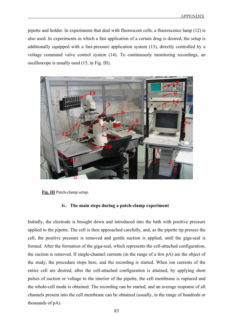
APPENDIX
83
pipette and holder. In experiments that deal with fluorescent cells, a fluorescence lamp (12) is
also used. In experiments in which a fast application of a certain drug is desired, the setup is
additionally equipped with a fast-pressure application system (13), directly controlled by a
voltage command valve control system (14). To continuously monitoring recordings, an
oscilloscope is usually used (15, in Fig. III).
Fig. III Patch-clamp setup.
iv. The main steps during a patch-clamp experiment
Initially, the electrode is brought down and introduced into the bath with positive pressure
applied to the pipette. The cell is then approached carefully, and, as the pipette tip presses the
cell, the positive pressure is removed and gentle suction is applied, until the giga-seal is
formed. After the formation of the giga-seal, which represents the cell-attached configuration,
the suction is removed. If single-channel currents (in the range of a few pA) are the object of
the study, the procedure stops here, and the recording is started. When ion currents of the
entire cell are desired, after the cell-attached configuration is attained, by applying short
pulses of suction or voltage to the interior of the pipette, the cell membrane is ruptured and
the whole-cell mode is obtained. The recording can be started, and an average response of all
channels present into the cell membrane can be obtained (usually, in the range of hundreds or
thousands of pA).
REFERENCES
84
References
Abicht A., Stucka R., Karcagi V., Herczegfalvi A., Horvath R., Mortier W., Schara U.,
Ramaekers V., Jost W., Brunner J., Janssen G., Seidel U., Schlotter B., Müller-Felber W.,
Pongratz D., Rüdel R., and Lochmüller H. (1999) A common mutation (ε1267delG) in
congenital myasthenic patients of Gypsy ethnic origin. Neurology: 53(7): 1564-9.
Abicht A., Stucka R., Song I. H., Karcagi V., Kugler K., Baumgarten-Walczak A., Stier
C., Pongratz D., Mortier W., Müller-Ferber W., and Lochmüller H. (2000) Genetic
analysis of the entire AChR ε-subunit gene in 52 congenital myasthenic families. Acta Myol:
19: 23–27.
Abicht A., Stucka R., Schmidt C., Briguet A., Hopfner S., Song I. H., Pongratz D.,
Müller-Felber W., Ruegg M. A., and Lochmüller H. (2002) A newly identified
chromosomal microdeletion and an N-box mutation of the AChR ε gene cause a congenital
myasthenic syndrome. Brain: 125(Pt 5): 1005-13.
Aidley D. J. (1989) The physiology of excitable cells. 3rd Ed. University Press, Cambridge,
England.
Akk G., and Steinbach J. H. (2000) Structural elements near the C-terminus are responsible
for changes in nicotinic receptor gating kinetics following patch excision. J Physiol: 527 Pt 3:
405-17.
Anderson C. R., and Stevens C. F. (1973) Voltage clamp analysis of acetylcholine produced
endplate current fluctuations at frog neuromuscular junction. J Physiol: 235(3): 655-91.
Anderson D. J., Blobel G., Tzartos S., Gullick W., and Lindstrom J. (1983)
Transmembrane orientation of an early biosynthetic form of acetylcholine receptor δ-subunit
determined by proteolytic dissection in conjunction with monoclonal antibodies. J Neurosci:
3(9): 1773-84.
Bear M. F., Connors B. W., and Pardiso M. A. (2001) Neuroscience: Exploring the Brain.
2nd Ed. Lippincott Williams and Wilkins, Philadelphia, USA.

REFERENCES
85
Beeson D., Newland C., Croxen R., Buckel A., Li F. Y., Larsson C., Tariq M., Vincent A.,
and Newsom-Davis J. (1998) Congenital myasthenic syndromes. Studies of the AChR and
other candidate genes. Ann N Y Acad Sci: 841: 181-3.
Bhattacharyya B. J., Day J. W., Gundeck J. E., Leonard S., Wollmann R. L., and Gomez
C. M. (1997) Desensitization of mutant acetylcholine receptors in transgenic mice reduces the
amplitude of neuromuscular synaptic currents. Synapse: 27(4): 367-77.
Black R., Goldman D., Hochschwender S., Lindstrom J., and Hall Z. W. (1987) Genetic
variants of C2 muscle cells that are defective in synthesis of the α-subunit of the acetylcholine
receptor. J Cell Biol: 105(3): 1329-36.
Blount P., and Merlie J. P. (1990) Mutational analysis of muscle nicotinic acetylcholine
receptor subunit assembly. J Cell Biol: 111(6 Pt 1): 2613-22.
Bouzat C., Bren N., and Sine S. M. (1994) Structural basis of the different gating kinetics of
fetal and adult acetylcholine receptors. Neuron: 13(6): 1395-402.
Butler D. H., and McNamee M. G. (1993) FTIR analysis of nicotinic acetylcholine receptor
secondary structure in reconstituted membranes. Biochim Biophys Acta: 1150(1): 17-24.
Cachelin A. B., and Colquhoun D. (1989) Desensitization of the acetylcholine receptor of
frog endplates measured in a Vaseline-gap voltage clamp. J Physiol: 415: 159-88.
Colquhoun D., and Sakmann B. (1981) Fluctuations in the microsecond time range of the
current through single acetylcholine receptor ion channels. Nature: 294(5840): 464-6.
Colquhoun D., and Sakmann B. (1985) Fast events in single-channel currents activated by
acetylcholine and its analogues at the frog muscle endplate. J Physiol: 369: 501-57.
Colquhoun D., Cachelin A. B., Marshall C. G., Mathie A., and Ogden D. C. (1990)
Function of nicotinic synapses. Prog Brain Res: 84: 43-50.
REFERENCES
86
Colquhoun D., Unwin N., Shelley C., Hatton C. J., and Sivilotti L. (2003) In Burger’s
Medical Chemistry, Vol II “Drug Discovery and Drug Development”, Chapter 11 ("Nicotinic
Acetylcholine Receptors”: 357 - 405). Ed. Abraham, D., J. Wiley, New York.
Corringer P. J., Le Novere N., and Changeux J. P. (2000) Nicotinic receptors at the amino
acid level. Annu Rev Pharmacol Toxicol: 40: 431-58.
Croxen R., Newland C., Betty M., Vincent A., Newsom-Davis J., and Beeson D. (1999)
Novel functional ε-subunit polypeptide generated by a single nucleotide deletion in
acetylcholine receptor deficiency congenital myasthenic syndrome. Ann Neurol: 46(4): 639-47.
Croxen R., Young C., Slater C., Haslam S., Brydson M., Vincent A., and Beeson D. (2001)
Endplate γ- and ε-subunit mRNA levels in AChR deficiency syndrome due to ε-subunit null
mutations. Brain: 124(Pt 7): 1362-72.
Croxen R., Vincent A., Newsom-Davis J., and Beeson D. (2002) Myasthenia gravis in a
woman with congenital AChR deficiency due to ε-subunit mutations. Neurology: 58(10):
1563-5.
Cully D. F., Vassilatis D. K., Liu K. K., Paress P. S., Van der Ploeg L. H., Schaeffer J. M.,
and Arena J. P. (1994) Cloning of an avermectin-sensitive glutamate-gated chloride channel
from Caenorhabditis elegans. Nature: 371(6499): 707-11.
Dionne V. E., and Leibowitz M. D. (1982) Acetylcholine receptor kinetics. A description
from single-channel currents at snake neuromuscular junctions. Biophys J: 39(3): 253-61.
Donger C., Krejci E., Serradell A. P., Eymard B., Bon S., Nicole S., Chateau D., Gary F.,
Fardeau M., Massoulie J., and Guicheney P. (1998) Mutation in the human
acetylcholinesterase-associated collagen gene, COLQ, is responsible for congenital myasthenic
syndrome with endplate acetylcholinesterase deficiency (Type Ic). Am J Hum Genet: 63(4):
967-75.
Ealing J., Webster R., Brownlow S., Abdelgany A., Oosterhuis H., Muntoni F., Vaux D.
J., Vincent A., and Beeson D. (2002) Mutations in congenital myasthenic syndromes reveal

REFERENCES
87
an ε-subunit C-terminal cysteine, C470, crucial for maturation and surface expression of adult
AChR. Hum Mol Genet: 11(24): 3087-96.
Eertmoed A. L., and Green W. N. (1999) Nicotinic receptor assembly requires multiple
regions throughout the γ-subunit. J Neurosci: 19(15): 6298-308.
Elenes S., and Auerbach A. (2002) Desensitization of diliganded mouse muscle nicotinic
acetylcholine receptor channels. J Physiol: 541(Pt 2): 367-83.
Engel A. G., and Biesecker G. (1982) Complement activation in muscle fiber necrosis:
demonstration of the membrane attack complex of complement in necrotic fibers. Ann Neurol:
12(3): 289-96.
Engel A. G. (1993a) The investigation of congenital myasthenic syndromes. Ann N Y Acad
Sci: 681: 425-34.
Engel A. G., Hutchinson D. O., Nakano S., Murphy L., Griggs R. C., Gu Y., Hall Z. W.,
and Lindstrom J. (1993b) Myasthenic syndromes attributed to mutations affecting the ε-
subunit of the acetylcholine receptor. Ann N Y Acad Sci: 681: 496-508.
Engel A. G. (1994) Congenital myasthenic syndromes. Neurol Clin: 12(2): 401-37.
Engel A. G., Ohno K., Bouzat C., Sine S. M., and Griggs R. G. (1996a) Endplate
acetylcholine receptor deficiency due to nonsense mutations in the ε-subunit. Ann Neurol: 40:
810-17.
Engel A. G., Ohno K., Milone M., Wang H. L., Nakano S., Bouzat C., Pruitt J. N.,
Hutchinson D. O., Brengman J. M., Bren N., Sieb J. P., and Sine S. M. (1996b) New
mutations in acetylcholine receptor subunit genes reveal heterogeneity in the slow-channel
congenital myasthenic syndrome. Hum Mol Genet: 5(9): 1217-27.
Engel A. G. (1999a) Congenital myasthenic syndromes. J Child Neurol: 14(1): 38-41.
REFERENCES
88
Engel A. G., Ohno K., and Sine S. M. (1999b) Congenital myasthenic syndromes: recent
advances. Arch Neurol: 56(2): 163-7.
Engel A. G., Ohno K., and Sine S. M. (2003a) Congenital myasthenic syndromes: progress
over the past decade. Muscle Nerve: 27(1): 4-25.
Engel A. G., Ohno K., and Sine S. M. (2003b) Sleuthing molecular targets for neurological
diseases at the neuromuscular junction. Nat Rev Neurosci: 4(5): 339-52.
Engel A. G., Ohno K., and Sine S. M. (2003c) Congenital myasthenic syndromes: A diverse
array of molecular targets. J Neurocytol: 32(5-8): 1017-37.
Finer-Moore J., and Stroud R. M. (1984) Amphipathic analysis and possible formation of
the ion channel in an acetylcholine receptor. Proc Natl Acad Sci U S A: 81(1): 155-9.
Flucher B. E., and Daniels M. P. (1989) Distribution of Na+ channels and ankyrin in
neuromuscular junctions is complementary to that of acetylcholine receptors and the 43 kDa
protein. Neuron: 3(2): 163-75.
Franke C., Parnas H., Hovav G., and Dudel J. (1993) A molecular scheme for the reaction
between acetylcholine and nicotinic channels. Biophys J: 64(2): 339-56.
Gomez C. M., Maselli R., Gundeck J. E., Chao M., Day J. W., Tamamizu S., Lasalde J.
A., McNamee M., and Wollmann R. L. (1997) Slow-channel transgenic mice: a model of
postsynaptic organellar degeneration at the neuromuscular junction. J Neurosci: 17(11): 4170-
9.
Gomez C. M., Maselli R. A., Groshong J., Zayas R., Wollmann R. L., Cens T., and
Charnet P. (2002) Active calcium accumulation underlies severe weakness in a panel of mice
with slow-channel syndrome. J Neurosci: 22(15): 6447-57.
Grosman C., and Auerbach A. (2000) Kinetic, mechanistic, and structural aspects of
unliganded gating of acetylcholine receptor channels: a single-channel study of second
transmembrane segment 12' mutants. J Gen Physiol: 115(5): 621-35.
REFERENCES
89
Grosman C., and Auerbach A. (2001) The dissociation of acetylcholine from open nicotinic
receptor channels. Proc Natl Acad Sci U S A: 98(24): 14102-7.
Gu Y., Ralston E., Murphy-Erdosh C., Black R. A., and Hall Z. W. (1989) Acetylcholine
receptor in a C2 muscle cell variant is retained in the endoplasmic reticulum. J Cell Biol:
109(2): 729-38.
Gu Y., Forsayeth J. R., Verrall S., Yu X. M., and Hall Z. W. (1991) Assembly of the
mammalian muscle acetylcholine receptor in transfected COS cells. J Cell Biol: 114(4): 799-
807.
Hamill O. P., Marty A., Neher E., Sakmann B., and Sigworth F. J. (1981) Improved patch-
clamp techniques for high-resolution current recording from cells and cell-free membrane
patches. Pflugers Arch: 391(2): 85-100.
Hatton C. J., Shelley C., Brydson M., Beeson D., and Colquhoun D. (2003) Properties of
the human muscle nicotinic receptor, and of the slow-channel myasthenic syndrome mutant
εL221F, inferred from maximum likelihood fits. J Physiol: 547(Pt 3): 729-60.
Hille B. (2001) Ionic channels of excitable membranes. 3rd Ed., Sinauer Associates, Inc.,
Sunderland, Massachusetts, USA.
Horn R., and Marty A. (1988) Muscarinic activation of ionic currents measured by a new
whole-cell recording method. J Gen Physiol: 92(2): 145-59.
Jackson M. B. (1984) Spontaneous openings of the acetylcholine receptor channel. Proc Natl
Acad Sci U S A: 81(12): 3901-4.
Jackson M. B. (1986) Kinetics of unliganded acetylcholine receptor channel-gating. Biophys
J: 49(3): 663-72.
Jackson M. B. (1988) Dependence of acetylcholine receptor channel kinetics on agonist
concentration in cultured mouse muscle fibers. J Physiol: 397: 555-83.
REFERENCES
90
Jackson M. B., Imoto K., Mishina M., Konno T., Numa S., and Sakmann B. (1990)
Spontaneous and agonist-induced openings of an acetylcholine receptor channel composed of
bovine muscle α-, β-, and δ-subunits. Pflugers Arch: 417(2): 129-35.
Jaramillo F., Vicini S., and Schuetze S. M. (1988) Embryonic acetylcholine receptors
guarantee spontaneous contractions in rat developing muscle. Nature: 335(6185): 66-8.
Jones S. F., and Kwanbunbumpen S. (1970a) The effects of nerve stimulation and
hemicholinium on synaptic vesicles at the mammalian neuromuscular junction. J Physiol:
207(1): 31-50.
Jones S. F., and Kwanbunbumpen S. (1970b) Some effects of nerve stimulation and
hemicholinium on quantal transmitter release at the mammalian neuromuscular junction. J
Physiol: 207(1): 51-61.
Kao P. N., and Karlin A. (1986) Acetylcholine receptor binding site contains a disulfide
cross-link between adjacent half-cystinyl residues. J Biol Chem: 261(18): 8085-8.
Karlin A., Holtzman E., Yodh N., Lobel P., Wall J., and Hainfeld J. (1983) The
arrangement of the subunits of the acetylcholine receptor of Torpedo californica. J Biol Chem:
258(11): 6678-81.
Karlin A. (1993) Structure of nicotinic acetylcholine receptors. Curr Opin Neurobiol: 3(3):
299-309.
Karlin A., and Akabas M. H. (1995) Toward a structural basis for the function of nicotinic
acetylcholine receptors and their cousins. Neuron: 15(6): 1231-44.
Karlin A. (2002) Emerging structure of the nicotinic acetylcholine receptors. Nat Rev
Neurosci: 3(2): 102-14.
Katz B., and Thesleff S. (1957) A study of the desensitization produced by acetylcholine at
the motor endplate. J Physiol: 138(1): 63-80.

REFERENCES
91
Katz B. (1966) Nerve, Muscle, and Synapse. Ed. McGraw-Hill, New York.
Kraner S., Laufenberg I., Strassburg H. M., Sieb J. P., and Steinlein O. K. (2003)
Congenital myasthenic syndrome with episodic apnea in patients homozygous for a CHAT
missense mutation. Arch Neurol: 60(5): 761-3.
Li J., Sheng S., Perry C. J., and Kleyman T. R. (2003) Asymmetric organization of the pore
region of the epithelial sodium channel. J Biol Chem: 278(16): 13867-74.
Lindstrom J. (2000) The structures of neuronal nicotinic receptors. In Handbook of
Experimental Pharmacology (Clementi F., Fornasari D., Gotti C., eds), vol. 144: 101- 162.
Springer-Verlag, Berlin-Heidelberg.
Lindstrom J. (2001) Nicotinic Acethylcholine Receptors. Encyclopedia of Life Sciences /
Macmillan Publishers Ltd., Nature Publishing Group, London, England.
Lindstrom J. M. (2000) Acetylcholine receptors and myasthenia. Muscle Nerve: 23(4): 453-
77.
Martin A. R. (1994) Amplification of neuromuscular transmission by postjunctional folds.
Proc. R. Soc. Lond: B 258: 321-26.
Maselli R., Chen M., Chen T. Y., Kong D. Z., Agius M. A., Bowe C., Wollman R. L., and
Gomez C. M. (2002) Severe low-affinity fast-channel syndrome due to a heteroallelic
nonsense/missense mutation in the acetylcholine receptor ε-subunit. Neurology: 58(Suppl 3):
A228.
Merlie J. P., Sebbane R., Gardner S., Olson E., and Lindstrom J. (1983) The regulation of
acetylcholine receptor expression in mammalian muscle. Cold Spring Harb Symp Quant Biol:
48 Pt 1: 135-46.
Middleton L. T. (1996) Congenital myasthenic syndromes. 34th ENMC International
Workshop, 10-11 June 1995. Neuromuscul Disord: 6(2): 133-6.
REFERENCES
92
Middleton L., Ohno K., Christodoulou K., Brengman J., Milone M., Neocleous V.,
Serdaroglu P., Deymeer F., Ozdemir C., Mubaidin A., Horany K., Al-Shehab A.,
Mavromatis I., Mylonas I., Tsingis M., Zamba E., Pantzaris M., Kyriallis K., and Engel
A. G. (1999) Chromosome 17p-linked myasthenias stem from defects in the acetylcholine
receptor ε-subunit gene. Neurology: 53(5): 1076-82.
Milone M., Wang H. L., Ohno K., Fukudome T., Pruitt J. N., Bren N., Sine S. M., and
Engel A. G. (1997) Slow-channel myasthenic syndrome caused by enhanced activation,
desensitization, and agonist binding affinity attributable to mutation in the M2 domain of the
acetylcholine receptor alpha subunit. J Neurosci: 17(15): 5651-65.
Milone M., Wang H. L., Ohno K., Prince R., Fukudome T., Shen X. M., Brengman J. M.,
Griggs R. C., Sine S. M., and Engel A. G. (1998a) Mode switching kinetics produced by a
naturally occurring mutation in the cytoplasmic loop of the human acetylcholine receptor ε-
subunit. Neuron: 20(3): 575-88.
Milone M., Ohno K., Fukudome T., Shen X. M., Brengman J., Griggs R. C., and Engel A.
G. (1998b) Congenital myasthenic syndrome caused by novel loss-of-function mutations in the
human AChR ε-subunit gene. Ann N Y Acad Sci: 841: 184-8.
Mishina M., Takai T., Imoto K., Noda M., Takahashi T., Numa S., Methfessel C., and
Sakmann B. (1986) Molecular distinction between fetal and adult forms of muscle
acetylcholine receptor. Nature: 321(6068): 406-11.
Miyazawa A., Fujiyoshi Y., Stowell M., and Unwin N. (1999) Nicotinic acetylcholine
receptor at 4.6 Å resolution: transverse tunnels in the channel wall. J Mol Biol: 288(4): 765-86.
Neher E., and Sakmann B. (1976) Single-channel currents recorded from membrane of
denervated frog muscle fibers. Nature: 260(5554): 799-802.
Neher E., Marty A., and Fenwick E. (1983) Ionic channels for signal transmission and
propagation. Prog Brain Res: 58: 39-48.

REFERENCES
93
Neher E. (1992) Nobel Lecture. Ion channels for communication between and within cells.
EMBO J: 11(5): 1672-9.
Newland C. F., Beeson D., Vincent A., and Newsom-Davis J. (1995) Functional and non-
functional isoforms of the human muscle acetylcholine receptor. J Physiol: 489(Pt 3): 767-78.
Nichols P., Croxen R., Vincent A., Rutter R., Hutchinson M., Newsom-Davis J., and
Beeson D. (1999) Mutation of the acetylcholine receptor ε-subunit promoter in congenital
myasthenic syndrome. Ann Neurol: 45(4): 439-43.
Ohno K., Hutchinson D. O., Milone M., Brengman J. M., Bouzat C., Sine S. M., and
Engel A. G. (1995) Congenital myasthenic syndrome caused by prolonged acetylcholine
receptor channel-openings due to a mutation in the M2 domain of the ε-subunit. Proc Natl Acad
Sci U S A: 92(3): 758-62.
Ohno K., Wang H. L., Milone M., Bren N., Brengman J. M., Nakano S., Quiram P.,
Pruitt J. N., Sine S. M., and Engel A. G. (1996) Congenital myasthenic syndrome caused by
decreased agonist binding affinity due to a mutation in the acetylcholine receptor ε-subunit.
Neuron: 17(1): 157-70.
Ohno K., Quiram P. A., Milone M., Wang H. L., Harper M. C., Pruitt J. N., Brengman J.
M., Pao L., Fischbeck K. H., Crawford T. O., Sine S. M., and Engel A. G. (1997)
Congenital myasthenic syndromes due to heteroallelic nonsense/missense mutations in the
acetylcholine receptor ε-subunit gene: identification and functional characterization of six new
mutations. Hum Mol Genet: 6(5): 753-66.
Ohno K., Anlar B., Özdirim E., Brengman J. M., and Engel A. G. (1998a) Frameshifting
and splice-site mutations in the acetylcholine receptor ε-subunit gene in three Turkish kinships
with congenital myasthenic syndromes. Ann N Y Acad Sci: 841: 189-94.
Ohno K., Anlar B., Özdirim E., Brengman J. M., DeBleecker J. L., and Engel A. G.
(1998b) Myasthenic syndromes in Turkish kinships due to mutations in the acetylcholine
receptor. Ann Neurol: 44(2): 234-41.
REFERENCES
94
Ohno K., Anlar B., and Engel A. G. (1999) Congenital myasthenic syndrome caused by a
mutation in the Ets-binding site of the promoter region of the acetylcholine receptor ε-subunit
gene. Neuromuscul Disord: 9(3): 131-5.
Ohno K., Tsujino A., Brengman J. M., Harper C. M., Bajzer Z., Udd B., Beyring R.,
Robb S., Kirkham F. J., and Engel A. G. (2001) Choline acetyltransferase mutations cause
myasthenic syndrome associated with episodic apnea in humans. Proc Natl Acad Sci U S A:
98(4): 2017-22.
Ohno K., Engel A. G., Shen X. M., Selcen D., Brengman J., Harper C. M., Tsujino A.,
and Milone M. (2002) RAPSYN mutations in humans cause endplate acetylcholine-receptor
deficiency and myasthenic syndrome. Am J Hum Genet: 70(4): 875-85.
Ortells M. O., and Lunt G. G. (1995) Evolutionary history of the ligand-gated ion channel
superfamily of receptors. Trends Neurosci: 18(3): 121-7.
Pusch M., and Neher E. (1988) Rates of diffusional exchange between small cells and a
measuring patch pipette. Pflugers Arch: 411(2): 204-11.
Quiram P., Ohno K., Milone M., Patterson M. C., Pruitt N. J., Brengman J. M., Sine S.
M., and Engel A. G. (1999) Mutation causing congenital myasthenia reveals acetylcholine
receptor β/δ subunit interaction essential for assembly. J Clin Invest: 104: 1403–1410.
Reynolds J. A., and Karlin A. (1978) Molecular weight in detergent solution of acetylcholine
receptor from Torpedo californica. Biochemistry: 17(11): 2035-8.
Ruff R. L. (1996) Sodium channel slow inactivation and the distribution of sodium channels
on skeletal muscle fibers enable the performance properties of different skeletal muscle fiber
types. Acta Physiol Scand: 156(3): 159-68.
Sakmann B., Patlak J., and Neher E. (1980) Single acetylcholine-activated channels show
burst-kinetics in presence of desensitizing concentrations of agonist. Nature: 286(5768): 71-3.

REFERENCES
95
Sakmann B. (1992) Nobel Lecture. Elementary steps in synaptic transmission revealed by
currents through single ion channels. EMBO J: 11(6): 2002-16.
Salpeter M. M. (1987) In the Vertebrate Neuromuscular Junction. Ed. John Wiley & Sons Inc.,
New York.
Schofield P. R., Darlison M. G., Fujita N., Burt D. R., Stephenson F. A., Rodriguez H.,
Rhee L. M., Ramachandran J., Reale V., and Glencorse T. A. (1987) Sequence and
functional expression of the GABAA receptor shows a ligand-gated receptor super-family.
Nature: 328(6127): 221-7.
Shen X. M., Ohno K., Fukudome T., Brengman J. M., and Engel A. G. (1999) Deletion of
a single codon from the long cytoplasmic loop of the AChR subunit gene causes brief single-
channel currents. Soc Neurosci Abstr: 25: 1721.
Shen X. M., Tsujino A., Ohno K., Brengman J. M., Gingold M., and Engel A. G. (2000) A
novel fast-channel congenital myasthenic syndrome caused by a mutation in the Cys-loop
domain of the acetylcholine receptor ε-subunit. Neurology: 54(Suppl3): A138.
Shen X. M., Ohno K., Milone M., Brengman J. M., Spilsbury P. R., and Engel A. G.
(2001) Fast-channel syndrome. Neurology: 56(Suppl. 3): A60.
Shen X. M., Ohno K., Fukudome T., Tsujino A., Brengman J. M., De Vivo D. C., Packer
R. J., and Engel A. G. (2002) Congenital myasthenic syndrome caused by low-expressor fast-
channel AChR δ-subunit mutation. Neurology: 59(12): 1881-8.
Shen X. M., Ohno K., Tsujino A., Brengman J. M., Gingold M., Sine S. M., and Engel A.
G. (2003) Mutation causing severe myasthenia reveals functional asymmetry of AChR
signature cystine loops in agonist binding and gating. J Clin Invest: 111(4): 497-505.
Sieb J. P., Dorfler P., Tzartos S., Wewer U. M., Ruegg M. A., Meyer D., Baumann I.,
Lindemuth R., Jakschik J., and Ries F. (1998) Congenital myasthenic syndromes in two
kinships with endplate acetylcholine receptor and utrophin deficiency. Neurology: 50(1): 54-
61.

REFERENCES
96
Sieb J. P., Kraner S., Rauch M., and Steinlein O. K. (2000a) Immature endplates and
utrophin deficiency in congenital myasthenic syndrome caused by ε-AChR subunit truncating
mutations. Hum Genet: 107(2): 160-4.
Sieb J. P., Kraner S., Schrank B., Reitter B., Goebel T. H., Tzartos S. J., and Steinlein O.
K. (2000b) Severe congenital myasthenic syndrome due to homozygosity of the 1293insG ε-
acetylcholine receptor subunit mutation. Ann Neurol: 48(3): 379-83.
Sigworth F. J., and Sine S. M. (1987) Data transformations for improved display and fitting
of single-channel dwell time histograms. Biophys J: 52(6): 1047-54.
Sine S. M., Claudio T., and Sigworth F. J. (1990) Activation of Torpedo acetylcholine
receptors expressed in mouse fibroblasts. Single-channel current kinetics reveal distinct agonist
binding affinities. J Gen Physiol: 96(2): 395-437.
Sine S. M., Ohno K., Bouzat C., Auerbach A., Milone M., Pruitt J. N., and Engel A. G.
(1995) Mutation of the acetylcholine receptor α-subunit causes a slow-channel myasthenic
syndrome by enhancing agonist-binding affinity. Neuron: 15(1): 229-39.
Sine S. M., Shen X. M., Wang H. L., Ohno K., Lee W. Y., Tsujino A., Brengmann J., Bren
N., Vajsar J., and Engel A. G. (2002) Naturally occurring mutations at the acetylcholine
receptor binding site independently alter ACh binding and channel-gating. J Gen Physiol:
120(4): 483-96.
Smith M. M., Lindstrom J., and Merlie J. P. (1987) Formation of the α-bungarotoxin
binding site and assembly of the nicotinic acetylcholine receptor subunits occur in the
endoplasmic reticulum. J Biol Chem: 262(9): 4367-76.
Snyder P. M., Olson D. R., and Bucher D. B. (1999) A pore segment in DEG / ENaC Na+
channels. J Biol Chem: 274(40): 28484-90.
Stroud R. M., McCarthy M. P., and Shuster M. (1990) Nicotinic acetylcholine receptor
superfamily of ligand-gated ion channels. Biochemistry: 29(50): 11009-23.

REFERENCES
97
Takeuchi A., and Takeuchi N. (1960) On the permeability of endplate membrane during the
action of transmitter. J Physiol (Paris): 154: 52-67.
Tsunoyama K., and Gojobori T. (1998) Evolution of nicotinic acetylcholine receptor
subunits. Mol Biol Evol: 15(5): 518-27.
Uchitel O., Engel A. G., Walls T. J., Nagel A., Bril V., and Trastek V. F. (1993a)
Congenital myasthenic syndrome attributed to an abnormal interaction of acetylcholine with its
receptor. Ann N Y Acad Sci: 681: 487-95.
Uchitel O., Engel A. G., Walls T. J., Nagel A., Atassi M. Z., and Bril V. (1993b) Congenital
myasthenic syndromes: II. Syndrome attributed to abnormal interaction of acetylcholine with
its receptor. Muscle Nerve: 16(12): 1293-301.
Unwin N. (1993) Nicotinic acetylcholine receptor at 9 Å resolution. J Mol Biol: 229(4): 1101-
24.
Unwin N. (1995) Acetylcholine receptor channel imaged in the open state. Nature: 373(6509):
37-43.
Verrall S., and Hall Z. W. (1992) The N-terminal domains of acetylcholine receptor subunits
contain recognition signals for the initial steps of receptor assembly. Cell: 68(1): 23-31.
Vincent A., Newland C., Croxen R., and Beeson D. (1997) Genes at the junction-candidates
for congenital myasthenic syndromes. Trends Neurosci: 20(1): 15-22.
Vincent A., Beeson D., and Lang B. (2000) Molecular targets for autoimmune and genetic
disorders of neuromuscular transmission. Eur J Biochem: 267(23): 6717-28.
Wang H. L., Ohno K., Milone M., Brengman J. M., Evoli A., Batocchi A. P., Middleton L.
T., Christodoulou K., Engel A. G., and Sine S. M. (2000) Fundamental gating mechanism of
nicotinic receptor channel revealed by mutation causing a congenital myasthenic syndrome. J
Gen Physiol: 116(3): 449-62.

REFERENCES
98
Wang J. M., Zhang L., Yao Y., Viroonchatapan N., Rothe E., and Wang Z. Z. (2002) A
transmembrane motif governs the surface trafficking of nicotinic acetylcholine receptors. Nat
Neurosci: 5(10): 963-70.
Witzemann V., Schwarz H., Koenen M., Berberich C., Villarroel A., Wernig A., Brenner
H. R., and Sakmann B. (1996) Acetylcholine receptor ε-subunit deletion causes muscle
weakness and atrophy in juvenile and adult mice. Proc Natl Acad Sci U S A: 93(23): 13286-91.
Wood S. J., and Slater C. R. (2001) Safety factor at the neuromuscular junction. Prog
Neurobiol: 64(4): 393-429.
Xie Y., and Cohen J. B. (2001) Contributions of Torpedo nicotinic acetylcholine receptor γ
Trp-55 and δ Trp-57 to agonist and competitive antagonist function. J Biol Chem: 276(4):
2417-26.
Zheng Y., Hirschberg B., Yuan J., Wang A. P., Hunt D. C., Ludmerer S. W., Schmatz D.
M., and Cully D. F. (2002) Identification of two novel Drosophila melanogaster histamine-
gated chloride channel subunits expressed in the eye. J Biol Chem: 277(3): 2000-5.
Zhou M., Engel A. G., and Auerbach A. (1999a) Serum choline activates mutant
acetylcholine receptors that cause slow-channel congenital myasthenic syndromes. Proc Natl
Acad Sci USA: 96(18): 10466-71.
Zhou M., Grosman C., and Auerbach A. (1999b) Single-channel properties of the ε-less
acetylcholine receptor channel. Biophys J: 76: A371 (Abstr.).
99
Eidesstattliche Erklärung
An Eides Statt versichere ich, dass ich diese Arbeit selbst und ohne jede unerlaubte Hilfe
angefertigt habe, und dass diese oder eine ähnliche Arbeit noch keiner anderen Stelle zur
Prüfung vorgelegen hat.
Bonn, November 2004
Cristian Gurgui

100
Curriculum Vitae
CRISTIAN GURGUI Phone: +49 (0) 1632963758
Personal Data
Name, First Name GURGUI, CRISTIAN
Born December 31st, 1977
In Bucharest, Romania
Family status Single
Nationality Romanian
Education
09/02 – 10/04 University of Bonn, Institute of Physiology II, Germany
PhD thesis: “Functional consequences of ε AChR subunit truncating
mutations linked to Congenital Myasthenic Syndrome”
10/01 – 08/02 University of Bonn, Kekulé Institute of Organic Chemistry and
Biochemistry, Germany
Master thesis: “The physiological significance of intracellular
sphingosine-1-phosphate. Knock out of sphingosine-1-phosphate lyase
by RNA interference in Swiss 3T3 fibroblasts”
09/00 – 06/02 University of Bucharest, Faculty of Biology, Romania
Master of Biochemistry and Molecular Biology
09/99 – 06/00 Physical Chemistry Department, Faculty of Chemistry, Romania
Diploma thesis: “Characterization through Cyclic Voltammetry of
Some New Synthesized Coumarin Derivatives, biologically active”
09/96 – 06/00 University of Bucharest, Faculty of Chemistry, Romania
Head subject: Technological Biochemistry
09/92 – 06/96 High School “Dimitrie Leonida“, Bucharest, Romania
Baccalaureate: June 1996
09/84 – 06/92 Primary School No. 82, Bucharest, Romania
Bonn, Nov 2004