Fluids & Electrolytes
71
FLUID & FLUID & ELECTROLYTES ELECTROLYTES Fundamnetal of nursing 2
description
Fluids & Electrolytes
Transcript of Fluids & Electrolytes

FLUID & FLUID & ELECTROLYTESELECTROLYTES
Fundamnetal of nursing 2
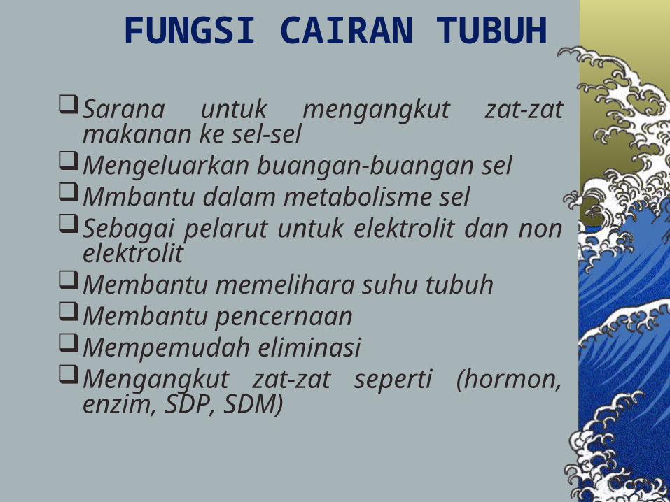
FUNGSI CAIRAN TUBUH
Sarana untuk mengangkut zat-zat makanan ke sel-sel
Mengeluarkan buangan-buangan selMmbantu dalam metabolisme selSebagai pelarut untuk elektrolit dan non
elektrolitMembantu memelihara suhu tubuhMembantu pencernaanMempemudah eliminasiMengangkut zat-zat seperti (hormon, enzim,
SDP, SDM)
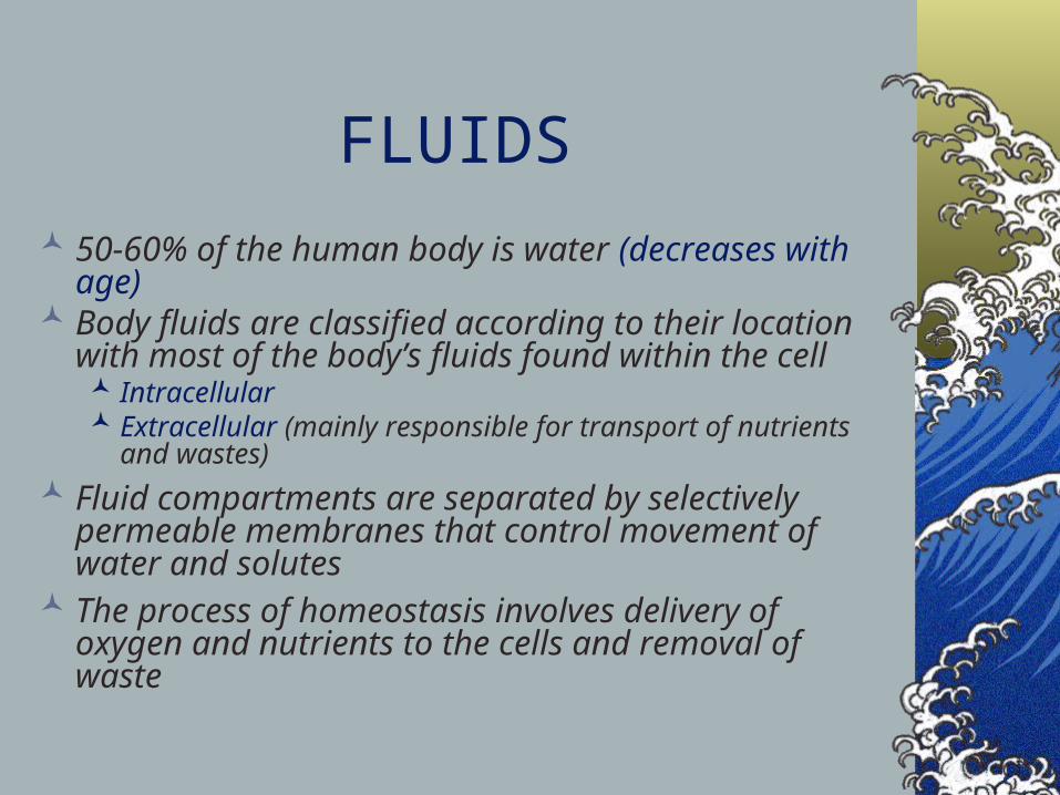
FLUIDS
50-60% of the human body is water (decreases with age)
Body fluids are classified according to their location with most of the body’s fluids found within the cell Intracellular Extracellular (mainly responsible for transport of nutrients
and wastes)
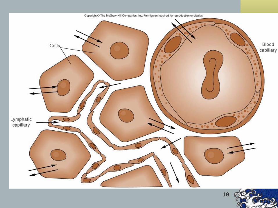
Fluid compartments are separated by selectively permeable membranes that control movement of water and solutes
The process of homeostasis involves delivery of oxygen and nutrients to the cells and removal of waste
4

6

7
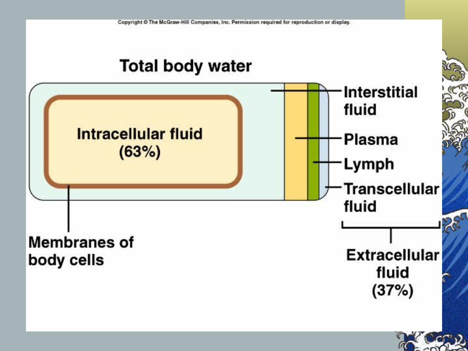
Body Fluid Compartments2/3 (65%) of TBW is intracellular (ICF)1/3 extracellular water
25 % interstitial fluid (ISF) 5- 8 % in plasma (IVF intravascular fluid)1- 2 % in transcellular fluids – CSF,
intraocular fluids, serous membranes, and in GI, respiratory and urinary tracts (third space)
9
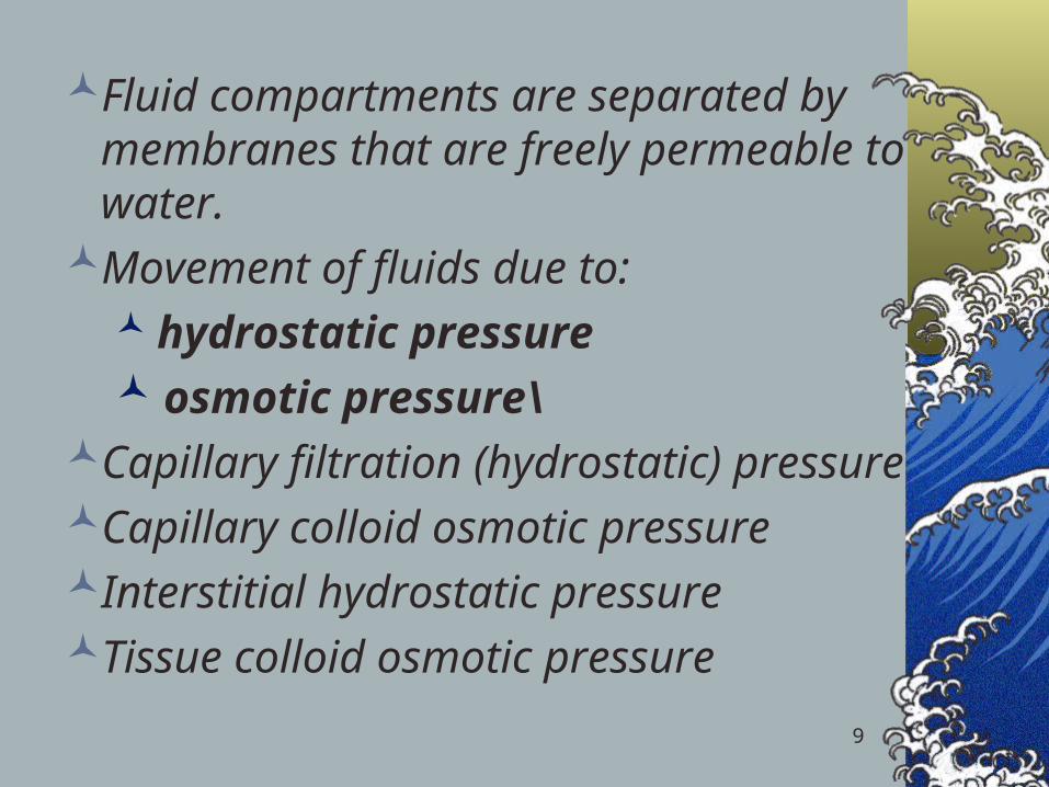
Fluid compartments are separated by membranes that are freely permeable to water.
Movement of fluids due to: hydrostatic pressure osmotic pressure\
Capillary filtration (hydrostatic) pressureCapillary colloid osmotic pressureInterstitial hydrostatic pressureTissue colloid osmotic pressure
10

11
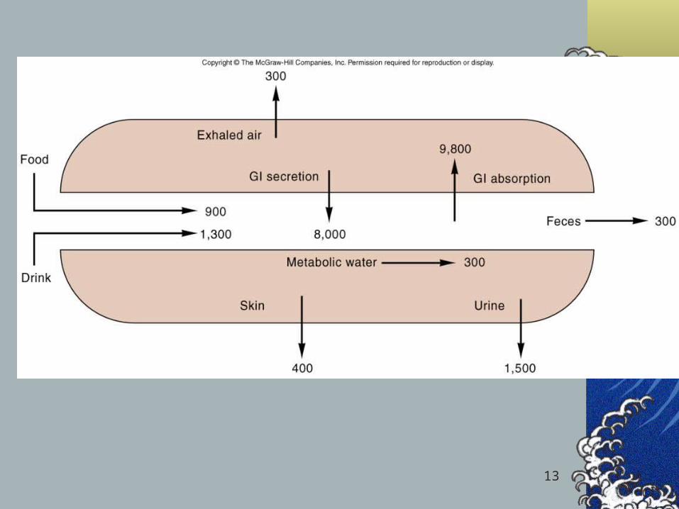
BalanceFluid and electrolyte homeostasis is
maintained in the bodyNeutral balance: input = outputPositive balance: input > outputNegative balance: input < output

12
13

14
Solutes – dissolved particles
Electrolytes – charged particlesCations – positively charged ions
Na+, K+ , Ca++, H+
Anions – negatively charged ionsCl-, HCO3
- , PO43-
Non-electrolytes - Uncharged Proteins, urea, glucose, O2, CO2
15
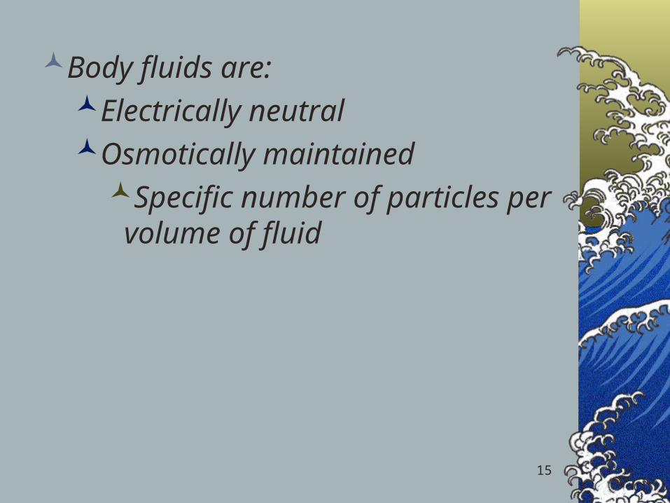
Body fluids are:Electrically neutralOsmotically maintained
Specific number of particles per volume of fluid
16
Homeostasis maintained by:
Ion transportWater movement Kidney function
17
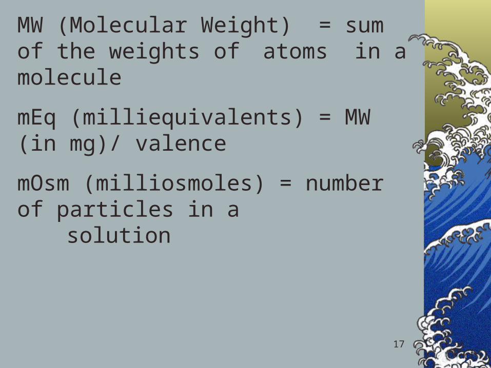
MW (Molecular Weight) = sum of the weights of atoms in a molecule
mEq (milliequivalents) = MW (in mg)/ valence
mOsm (milliosmoles) = number of particles in a solution
18
Tonicity
Isotonic
Hypertonic
Hypotonic

19

20
Cell in a hypertonic solution

21
Cell in a hypotonic solution

22
Movement of body fluids “ Where sodium goes, water follows.”
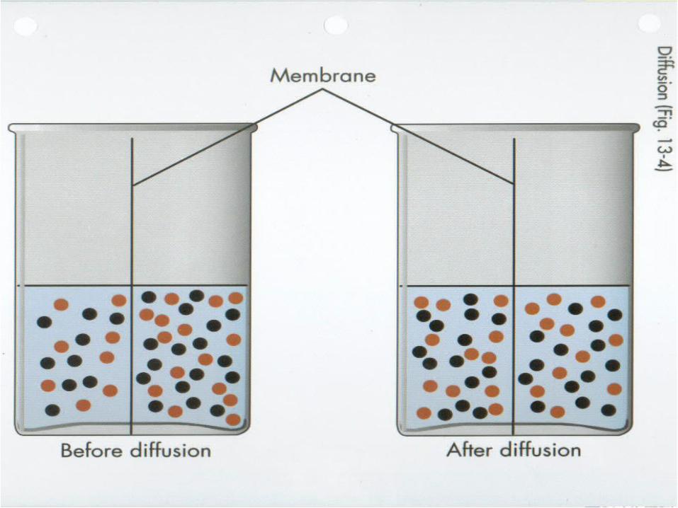
Diffusion – movement of particles down a concentration gradient.
Osmosis – diffusion of water across a selectively permeable membrane
Active transport – movement of particles up a concentration gradient ; requires energy

25
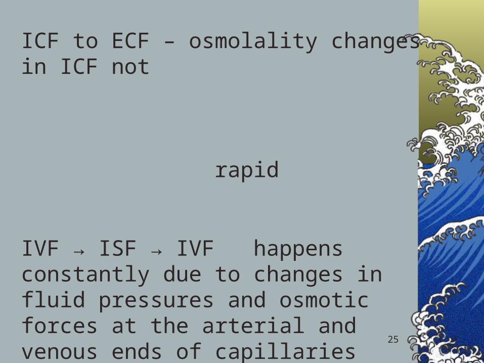
ICF to ECF – osmolality changes in ICF not rapid
IVF → ISF → IVF happens constantly due to changes in fluid pressures and osmotic forces at the arterial and venous ends of capillaries

26

27
Regulation of body water
ADH – antidiuretic hormone + thirstDecreased amount of water in bodyIncreased amount of Na+ in the bodyIncreased blood osmolalityDecreased circulating blood volume
Stimulate osmoreceptors in hypothalamusADH released from posterior pituitaryIncreased thirst

28

Regulation of Fluid Volume
Kidneys Capillary pressure forces fluid through the
walls and into the tubuleAt this point H2O or electrolytes are then
either retained or excretedThe urine becomes more dilute or more
concentrated based on the needs of the body

Regulation of Fluid Volume, cont.
Antidiuretic hormone (ADH)Produced by the hypothalamusStored in the pituitary glandRestores blood volume by increasing or decreasing
excretion of water Increased osmolality or decreased blood volume
stimulates the release of ADHThen the kidneys reabsorb waterAlso may be released by stress, pain, surgery, and
some meds
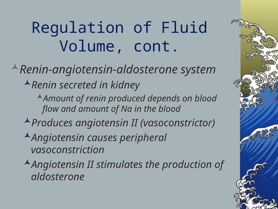
Regulation of Fluid Volume, cont.
Renin-angiotensin-aldosterone systemRenin secreted in kidney
Amount of renin produced depends on blood flow and amount of Na in the blood
Produces angiotensin II (vasoconstrictor)Angiotensin causes peripheral
vasoconstrictionAngiotensin II stimulates the production of
aldosterone
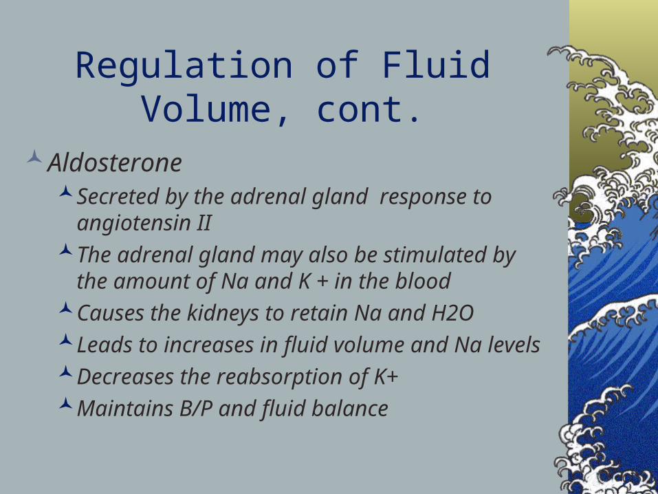
Regulation of Fluid Volume, cont.
AldosteroneSecreted by the adrenal gland response to
angiotensin IIThe adrenal gland may also be stimulated by the
amount of Na and K + in the bloodCauses the kidneys to retain Na and H2OLeads to increases in fluid volume and Na levelsDecreases the reabsorption of K+Maintains B/P and fluid balance
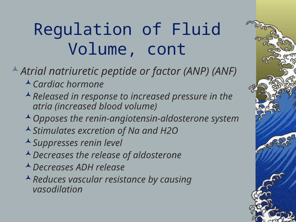
Regulation of Fluid Volume, cont
Atrial natriuretic peptide or factor (ANP) (ANF)Cardiac hormoneReleased in response to increased pressure in the
atria (increased blood volume)Opposes the renin-angiotensin-aldosterone systemStimulates excretion of Na and H2OSuppresses renin levelDecreases the release of aldosteroneDecreases ADH releaseReduces vascular resistance by causing vasodilation

REGULATION OF FLUID VOLUME

35
Result:increased water consumptionincreased water conservation
Increased water in body, increased volume and decreased Na+ concentration
36
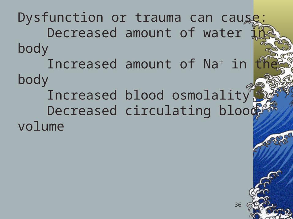
Dysfunction or trauma can cause:Decreased amount of water in bodyIncreased amount of Na+ in the bodyIncreased blood osmolalityDecreased circulating blood volume

37
Edema is the accumulation of fluid within the interstitial spaces.
Causes:increased hydrostatic pressure
lowered plasma osmotic pressure
increased capillary membrane permeability lymphatic channel obstruction
38
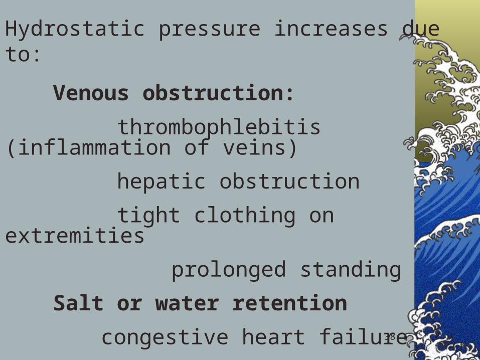
Hydrostatic pressure increases due to:
Venous obstruction:
thrombophlebitis (inflammation of veins)
hepatic obstruction
tight clothing on extremities
prolonged standing
Salt or water retention
congestive heart failure
renal failure

39
Decreased plasma osmotic pressure:
↓ plasma albumin (liver disease or protein malnutrition)
plasma proteins lost in :
glomerular diseases of kidney
hemorrhage, burns, open wounds and cirrhosis of liver
40
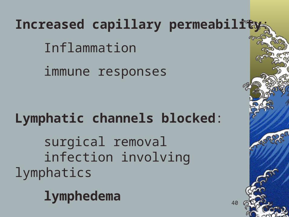
Increased capillary permeability:
Inflammation
immune responses
Lymphatic channels blocked:
surgical removalinfection involving lymphatics
lymphedema

41
Volume Abnormalities
Edema the accumulation of fluid within the interstitial
space
Causes:
•increased hydrostatic pressure
• venous obstruction, lymphedema, CHF, renal failure
•lowered plasma osmotic pressure (protein loss)
• liver failure, malnutrition, burns
•increased capillary membrane permeability
• Inflammation, SIRS, sepsis

42
Volume Abnormalities
Edema the accumulation of fluid within the interstitial space
Results in:• increased distance for diffusion
• impaired blood flow
• slower healing
• increased risk of infection
• pressure sores over bony prominences
• impaired organ function (brain, liver, gut, kidney)
43
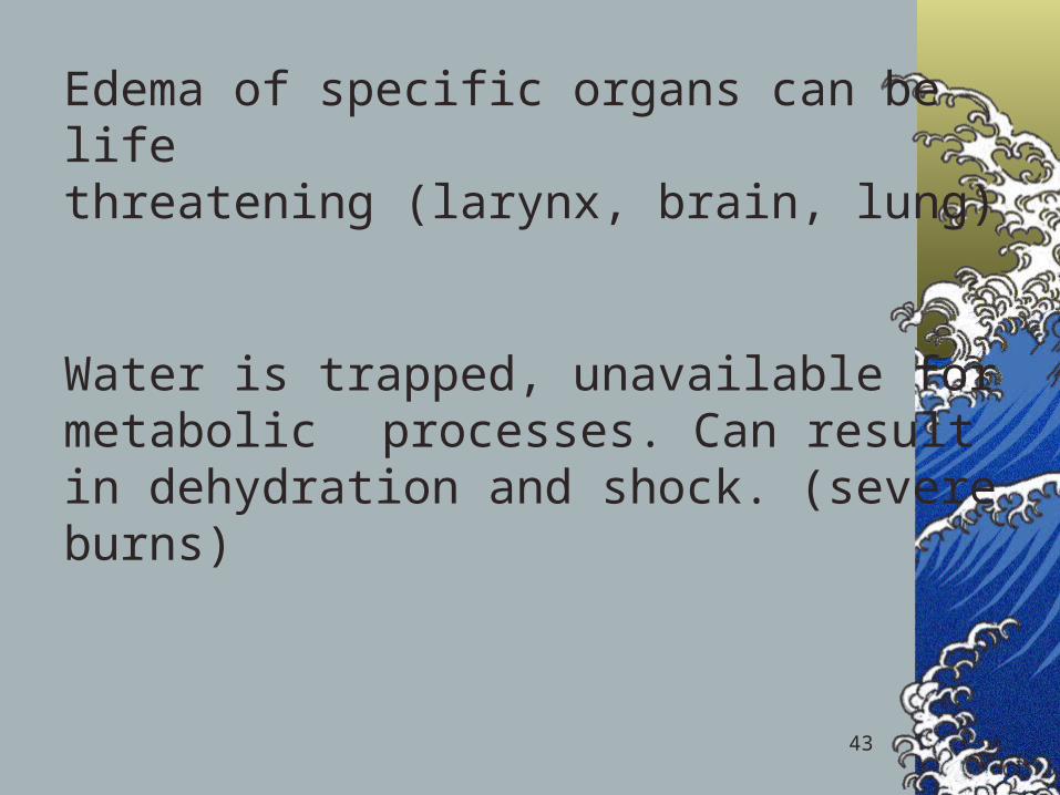
Edema of specific organs can be life threatening (larynx, brain, lung)
Water is trapped, unavailable for metabolic processes. Can result in dehydration and shock. (severe burns)
44
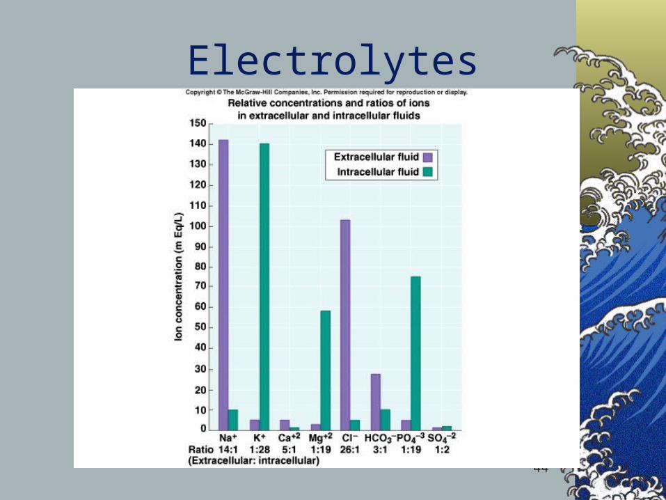
Electrolytes

45
Electrolyte balance
Na + (Sodium) 90 % of total ECF cations 136 -145 mEq / LPairs with Cl- , HCO3
- to neutralize charge
Low in ICF Most important ion in regulating water
balanceImportant in nerve and muscle function

46
Regulation of Sodium
Renal tubule reabsorption affected by hormones:AldosteroneRenin/angiotensinAtrial Natriuretic Peptide (ANP)

47
Potassium
Major intracellular cationICF conc. = 150- 160 mEq/ LResting membrane potentialRegulates fluid, ion balance inside cellpH balance

48
Regulation of Potassium
Through kidneyAldosteroneInsulin

49
Isotonic alterations in water balance
Occur when TBW changes are accompanied by = changes in electrolytesLoses plasma or ECFIsotonic fluid loss
↓ECF volume, weight loss, dry skin and mucous membranes, ↓ urine output, and hypovolemia ( rapid heart rate, flattened neck veins, and normal or ↓ B.P. – shock)

50
Isotonic fluid excessExcess IV fluidsHypersecretion of aldosteroneEffect of drugs – cortisone
Get hypervolemia – weight gain, decreased hematocrit, diluted plasma proteins, distended neck veins, ↑ B.P.
Can lead to edema (↑ capillary hydrostatic pressure) pulmonary edema and heart failure

51
Electrolyte imbalances: Sodium
Hypernatremia (high levels of sodium)Plasma Na+ > 145 mEq / LDue to ↑ Na + or ↓ waterWater moves from ICF → ECFCells dehydrate

52

53
Hypernatremia Due to:Hypertonic IV soln.Oversecretion of aldosteroneLoss of pure water
Long term sweating with chronic fever
Respiratory infection → water vapor loss
Diabetes – polyuriaInsufficient intake of water (hypodipsia)
54
Clinical manifestationsof Hypernatremia
ThirstLethargyNeurological dysfunction due to
dehydration of brain cellsDecreased vascular volume
55
Treatment of Hypernatremia
Lower serum Na+Isotonic salt-free IV fluidOral solutions preferable
56
HyponatremiaOverall decrease in Na+ in ECFTwo types: depletional and dilutionalDepletional Hyponatremia
Na+ loss:diuretics, chronic vomitingChronic diarrheaDecreased aldosteroneDecreased Na+ intake
57
Dilutional Hyponatremia:Renal dysfunction with ↑ intake of hypotonic
fluidsExcessive sweating→ increased thirst →
intake of excessive amounts of pure waterSyndrome of Inappropriate ADH (SIADH) or
oliguric renal failure, severe congestive heart failure, cirrhosis all lead to:Impaired renal excretion of water
Hyperglycemia – attracts water

58
Clinical manifestations of Hyponatremia
Neurological symptomsLethargy, headache, confusion, apprehension,
depressed reflexes, seizures and coma
Muscle symptoms Cramps, weakness, fatigue
Gastrointestinal symptomsNausea, vomiting, abdominal cramps, and
diarrhea
Tx – limit water intake or discontinue meds
59
Hypokalemia
Serum K+ < 3.5 mEq /LBeware if diabetic
Insulin gets K+ into cellKetoacidosis – H+ replaces K+, which
is lost in urine
β – adrenergic drugs or epinephrine
60
Causes of Hypokalemia
Decreased intake of K+
Increased K+ lossChronic diureticsAcid/base imbalanceTrauma and stressIncreased aldosteroneRedistribution between ICF and ECF

61
Clinical manifestations of Hypokalemia
Neuromuscular disordersWeakness, flaccid paralysis, respiratory
arrest, constipationDysrhythmias, appearance of U wavePostural hypotensionCardiac arrestOthers – table 6-5Treatment-
Increase K+ intake, but slowly, preferably by foods

62
Hyperkalemia
Serum K+ > 5.5 mEq / LCheck for renal diseaseMassive cellular traumaInsulin deficiencyAddison’s disease Potassium sparing diureticsDecreased blood pHExercise causes K+ to move out of cells
63
Clinical manifestations of Hyperkalemia
Early – hyperactive muscles , paresthesiaLate - Muscle weakness, flaccid paralysisChange in ECG patternDysrhythmiasBradycardia , heart block, cardiac arrest
64
Treatment of Hyperkalemia
If time, decrease intake and increase renal excretion
Insulin + glucoseBicarbonateCa++ counters effect on heart
65
Calcium ImbalancesMost in ECFRegulated by:
Parathyroid hormone↑Blood Ca++ by stimulating osteoclasts↑GI absorption and renal retention
Calcitonin from the thyroid glandPromotes bone formation↑ renal excretion

66
HypercalcemiaResults from:
Hyperparathyroidism Hypothyroid statesRenal diseaseExcessive intake of vitamin DMilk-alkali syndromeCertain drugsMalignant tumors – hypercalcemia of malignancy
Tumor products promote bone breakdownTumor growth in bone causing Ca++ release
67
HypercalcemiaUsually also see hypophosphatemiaEffects:
Many nonspecific – fatigue, weakness, lethargyIncreases formation of kidney stones and
pancreatic stonesMuscle crampsBradycardia, cardiac arrestPainGI activity also common
Nausea, abdominal crampsDiarrhea / constipation
Metastatic calcification
68
HypocalcemiaHyperactive neuromuscular reflexes and
tetany differentiate it from hypercalcemiaConvulsions in severe casesCaused by:
Renal failureLack of vitamin DSuppression of parathyroid functionHypersecretion of calcitoninMalabsorption statesAbnormal intestinal acidity and acid/ base bal.Widespread infection or peritoneal inflammation
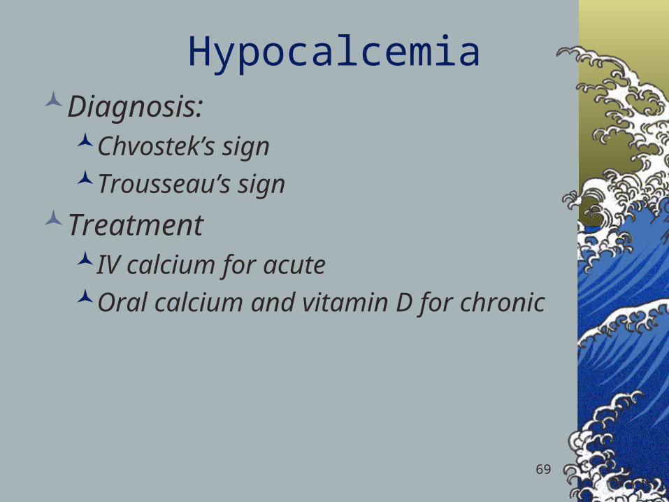
HypocalcemiaDiagnosis:
Chvostek’s signTrousseau’s sign
TreatmentIV calcium for acuteOral calcium and vitamin D for chronic
69
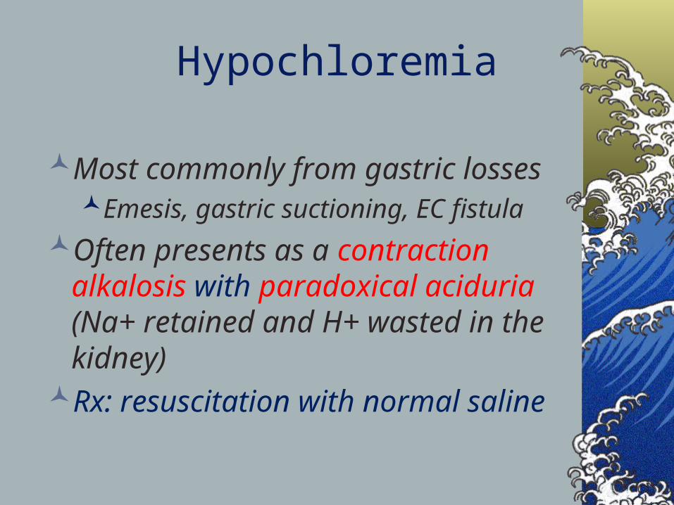
Most commonly from gastric lossesEmesis, gastric suctioning, EC fistula
Often presents as a contraction alkalosis with paradoxical aciduria (Na+ retained and H+ wasted in the kidney)
Rx: resuscitation with normal saline
Hypochloremia
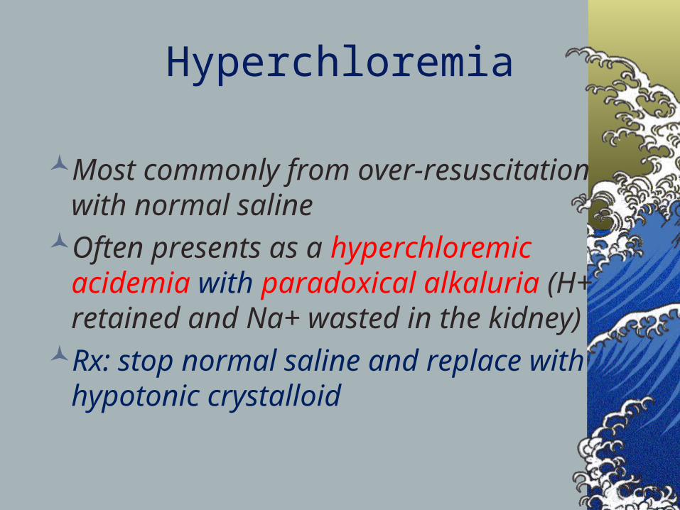
Most commonly from over-resuscitation with normal saline
Often presents as a hyperchloremic acidemia with paradoxical alkaluria (H+ retained and Na+ wasted in the kidney)
Rx: stop normal saline and replace with hypotonic crystalloid
Hyperchloremia