Fertőző betegségek Szervátültetés Tumorok eliminálása Gatekeeper funkció Kórokozók...
24
Fertőző betegségek Szervátültetés Tumorok eliminálása Gatekeeper funkció Kórokozók „érzékelése” Adaptív immunválasz elindítása Saját struktúrákkal szembeni tolerancia fenntartása THE ROLE OF PROFESSIONAL ANTIGEN PRESENTING CELLS IN THE IMMUNE RESPONSE
-
Upload
nigel-blair -
Category
Documents
-
view
215 -
download
0
Transcript of Fertőző betegségek Szervátültetés Tumorok eliminálása Gatekeeper funkció Kórokozók...
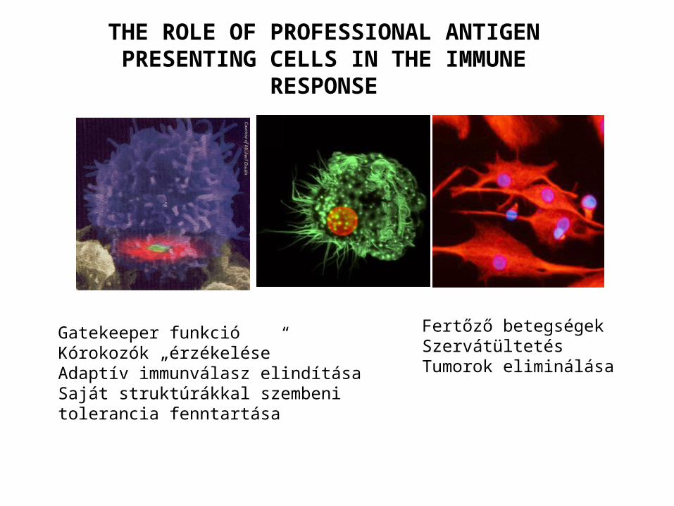
Fertőző betegségekSzervátültetésTumorok eliminálása
Gatekeeper funkcióKórokozók „érzékelése”Adaptív immunválasz elindításaSaját struktúrákkal szembeni tolerancia fenntartása
THE ROLE OF PROFESSIONAL ANTIGEN PRESENTING CELLS IN THE IMMUNE
RESPONSE
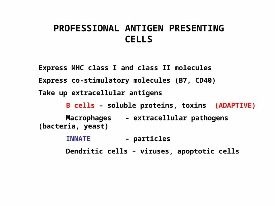
PROFESSIONAL ANTIGEN PRESENTING CELLS
Express MHC class I and class II molecules
Express co-stimulatory molecules (B7, CD40)
Take up extracellular antigens
B cells – soluble proteins, toxins (ADAPTIVE)
Macrophages – extracellular pathogens (bacteria, yeast)
INNATE – particles
Dendritic cells – viruses, apoptotic cells
PROFESSIONAL ANTIGEN PRESENTING CELLS
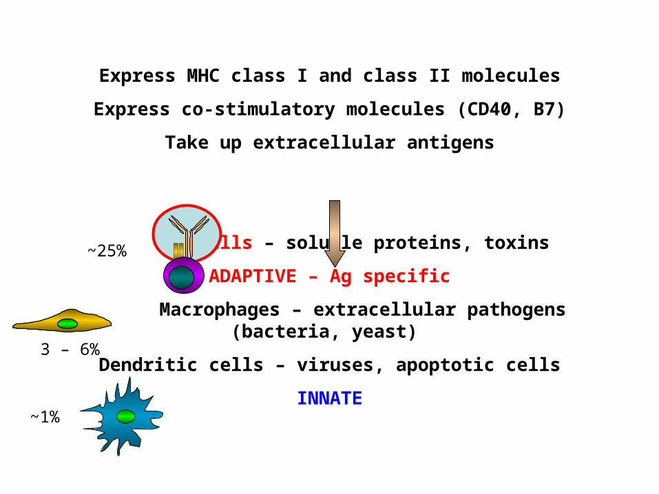
Express MHC class I and class II molecules
Express co-stimulatory molecules (CD40, B7)
Take up extracellular antigens
B cells – soluble proteins, toxins
ADAPTIVE – Ag specific
Macrophages – extracellular pathogens (bacteria, yeast)
Dendritic cells – viruses, apoptotic cells
INNATE
3 – 6%
~1%
~25%
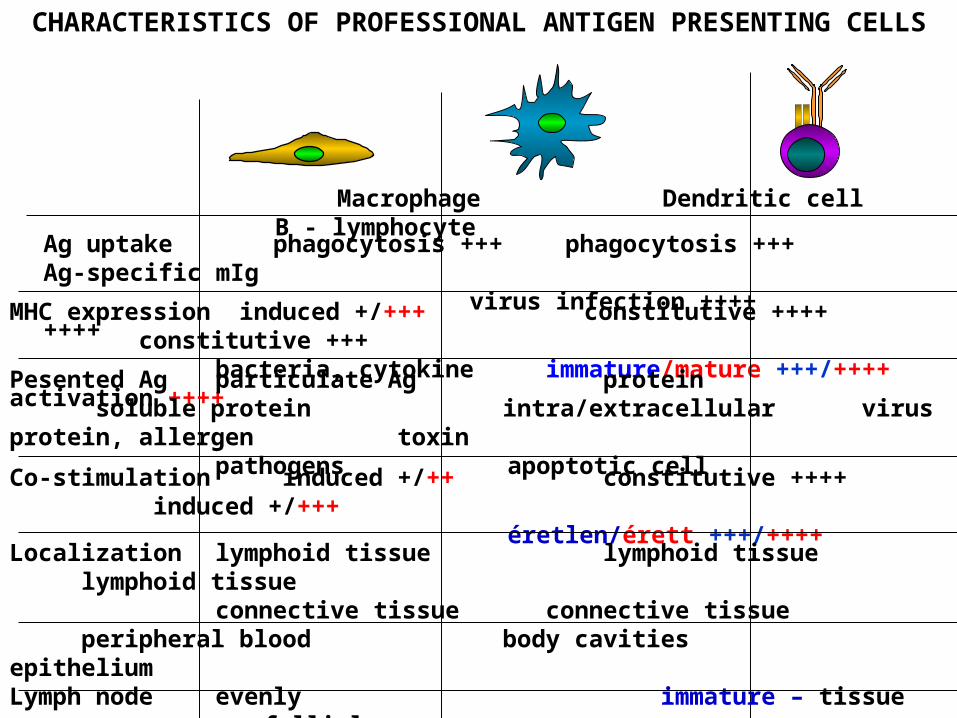
CHARACTERISTICS OF PROFESSIONAL ANTIGEN PRESENTING CELLS
Macrophage Dendritic cell B - lymphocyte
Ag uptake phagocytosis +++ phagocytosis +++ Ag-specific mIg virus infection ++++ ++++
MHC expression induced +/+++ constitutive ++++ constitutive +++ bacteria, cytokine immature/mature +++/++++ activation ++++
Pesented Ag particulate Ag protein soluble protein intra/extracellular virus protein, allergen toxin pathogens apoptotic cell
Co-stimulation induced +/++ constitutive ++++ induced +/+++ éretlen/érett +++/++++
Localization lymphoid tissue lymphoid tissue lymphoid tissue connective tissue connective tissue peripheral blood body cavities epithelium
Lymph node evenly immature – tissue follicles mature – T cell area
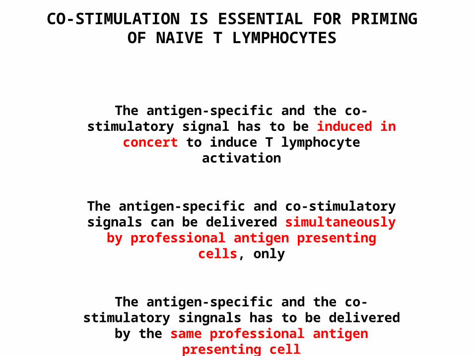
CO-STIMULATION IS ESSENTIAL FOR PRIMING OF NAIVE T LYMPHOCYTES
The antigen-specific and the co-stimulatory signal has to be induced in concert to induce T
lymphocyte activation
The antigen-specific and co-stimulatory signals can be delivered simultaneously by professional
antigen presenting cells, only
The antigen-specific and the co-stimulatory singnals has to be delivered by the same
professional antigen presenting cell

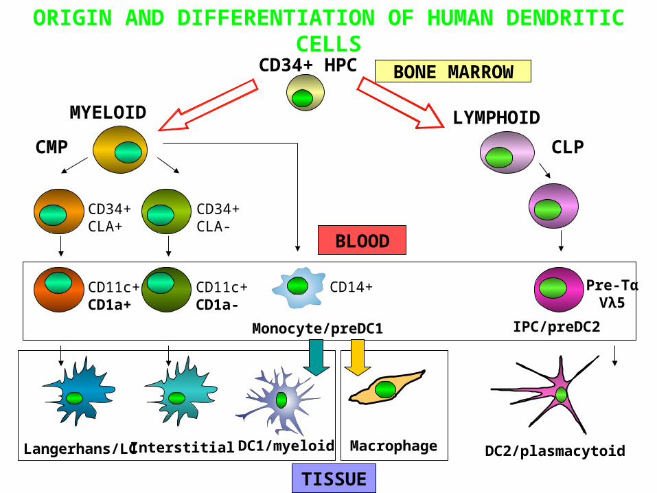
MYELOID LYMPHOID
BONE MARROW
CMP CLP
DC2/plasmacytoid
IPC/preDC2
Pre-TαVλ5
CD34+CLA+
CD11c+CD1a+
Langerhans/LC
CD34+CLA-
CD11c+CD1a-
Interstitial
BLOOD
TISSUE
Macrophage
CD14+
Monocyte/preDC1
DC1/myeloid
CD34+ HPC
ORIGIN AND DIFFERENTIATION OF HUMAN DENDRITIC CELLS
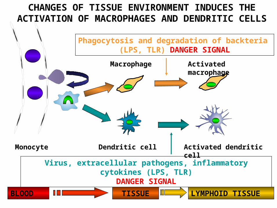
Macrophage
Dendritic cell
Activated macrophage
Phagocytosis and degradation of backteria(LPS, TLR) DANGER SIGNAL
Activated dendritic cell
Virus, extracellular pathogens, inflammatory cytokines (LPS, TLR)DANGER SIGNAL
Monocyte
CHANGES OF TISSUE ENVIRONMENT INDUCES THE ACTIVATION OF MACROPHAGES AND DENDRITIC
CELLS
LYMPHOID TISSUEBLOOD TISSUE
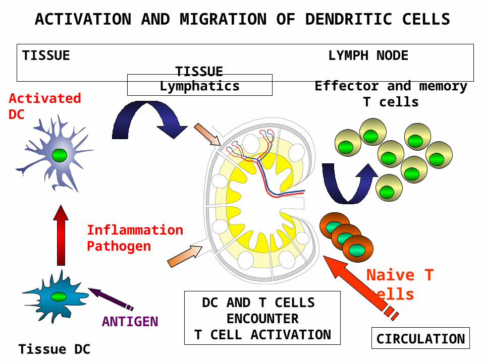
Tissue DC
Activated DC
DC AND T CELLS ENCOUNTER
T CELL ACTIVATION CIRCULATION
Naive T cells
Effector and memory T cells
TISSUE LYMPH NODE TISSUE
Lymphatics
InflammationPathogen
ACTIVATION AND MIGRATION OF DENDRITIC CELLS
ANTIGEN
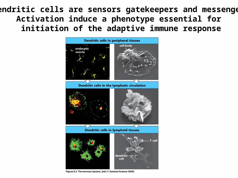
Dendritic cells are sensors gatekeepers and messengersActivation induce a phenotype essential for initiation of the adaptive immune response
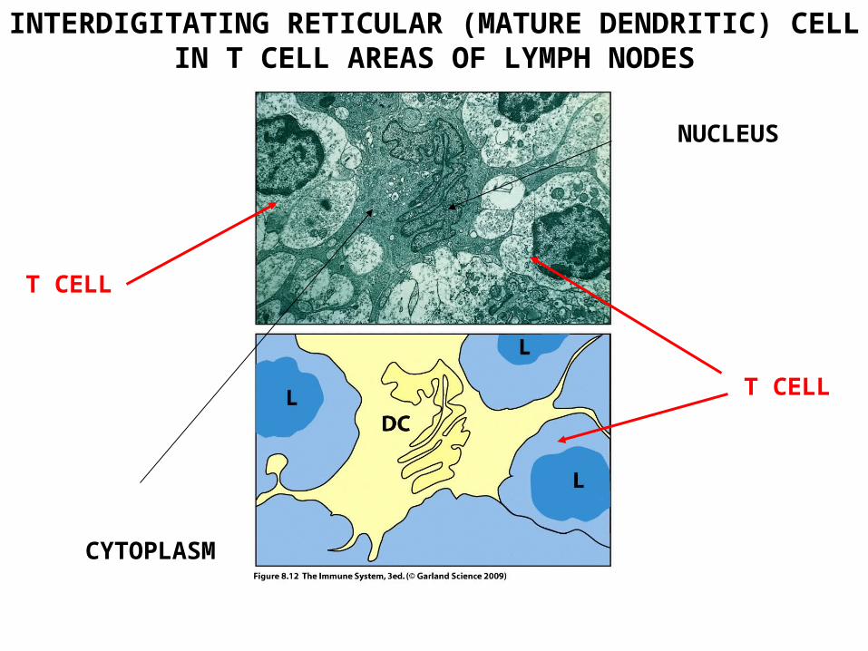
INTERDIGITATING RETICULAR (MATURE DENDRITIC) CELL IN T CELL AREAS OF LYMPH NODES
NUCLEUS
CYTOPLASM
T CELL
T CELL
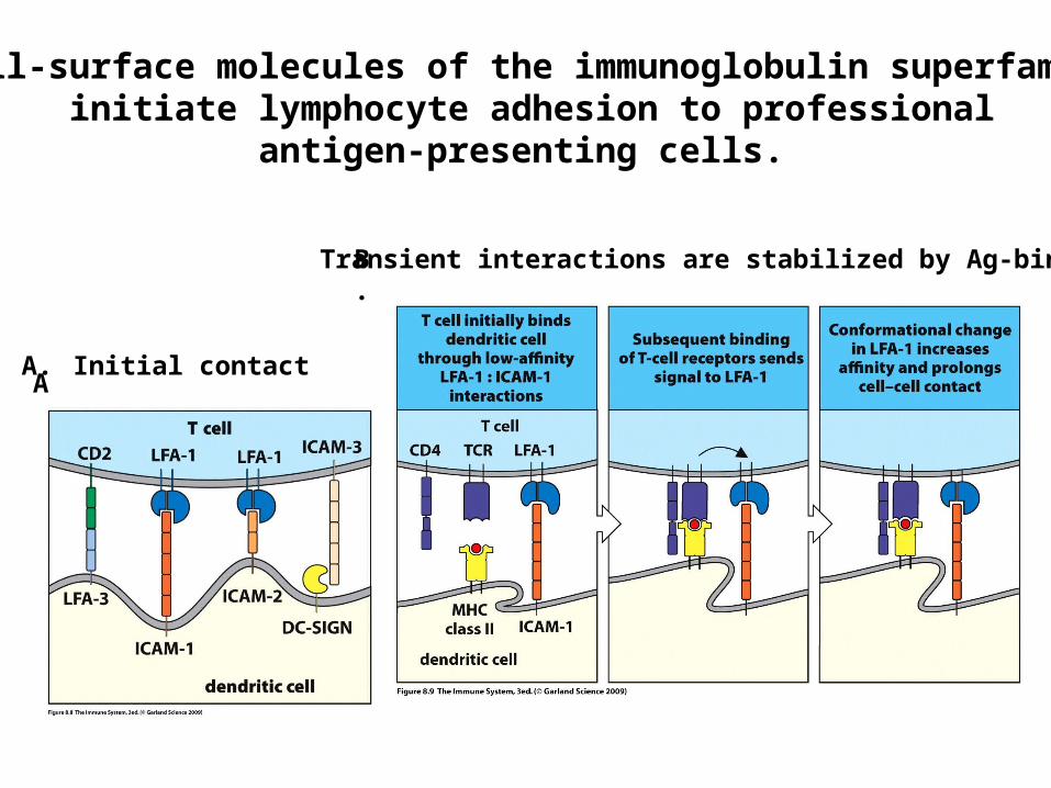
Cell-surface molecules of the immunoglobulin superfamily initiate lymphocyte adhesion to professional
antigen-presenting cells.
Initial contactA.
Transient interactions are stabilized by Ag-bindingB.
A
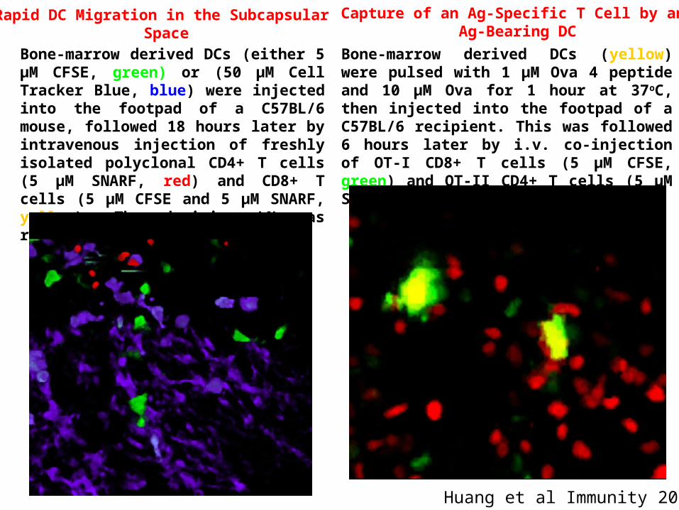
Huang et al Immunity 2004
Bone-marrow derived DCs (yellow) were pulsed with 1 µM Ova 4 peptide and 10 µM Ova for 1 hour at 37oC, then injected into the footpad of a C57BL/6 recipient. This was followed 6 hours later by i.v. co-injection of OT-I CD8+ T cells (5 µM CFSE, green) and OT-II CD4+ T cells (5 µM SNARF, red).
Rapid DC Migration in the Subcapsular Space
Capture of an Ag-Specific T Cell by an Ag-Bearing DC
Bone-marrow derived DCs (either 5 µM CFSE, green) or (50 µM Cell Tracker Blue, blue) were injected into the footpad of a C57BL/6 mouse, followed 18 hours later by intravenous injection of freshly isolated polyclonal CD4+ T cells (5 µM SNARF, red) and CD8+ T cells (5 µM CFSE and 5 µM SNARF, yellow). The draining LN was removed 6 hours after injection
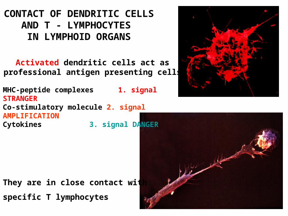
Activated dendritic cells act as professional antigen presenting cells
MHC-peptide complexes 1. signal STRANGERCo-stimulatory molecule 2. signal AMPLIFICATIONCytokines 3. signal DANGER
They are in close contact with
specific T lymphocytes
CONTACT OF DENDRITIC CELLS AND T - LYMPHOCYTES IN LYMPHOID ORGANS
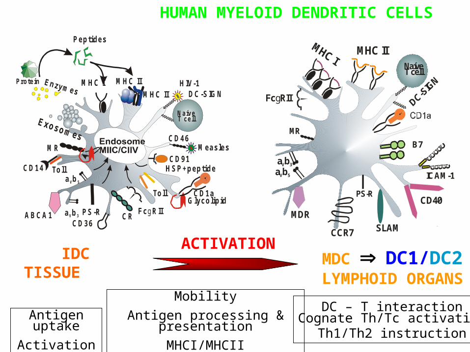
IDCTISSUE
ACTIVATION
Antigen uptake
Activation
Mobility
Antigen processing & presentation
MHCI/MHCII
MHC II
PS-R
MR
Fc RIIg
a bV 5
a bV 3
NaíveT cell
B7
ICAM-1
CD40
SLAMCCR7
MDR
MDC DC1/DC2 LYMPHOID ORGANS
DC – T interactionCognate Th/Tc activation
Th1/Th2 instruction
HUMAN MYELOID DENDRITIC CELLS
CD14
DC-SIGNM HC I M HC II
CD36PS-R
M R
HSP+peptide
Fc RIIg
a bV 5
a bV 3
HIV-1
N aíveT ce ll
CD46M easles
CD1aGlycolipid
Protein
Peptides
Toll
Toll
M HC II
C 91D
CRABCA1
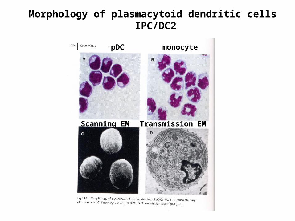
Morphology of plasmacytoid dendritic cells IPC/DC2
monocytepDC
Scanning EM Transmission EM
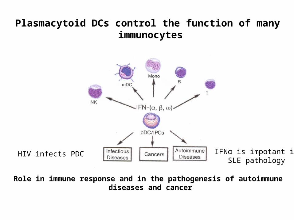
Plasmacytoid DCs control the function of many immunocytes
Role in immune response and in the pathogenesis of autoimmune diseases and cancer
HIV infects PDC IFNα is impotant inSLE pathology
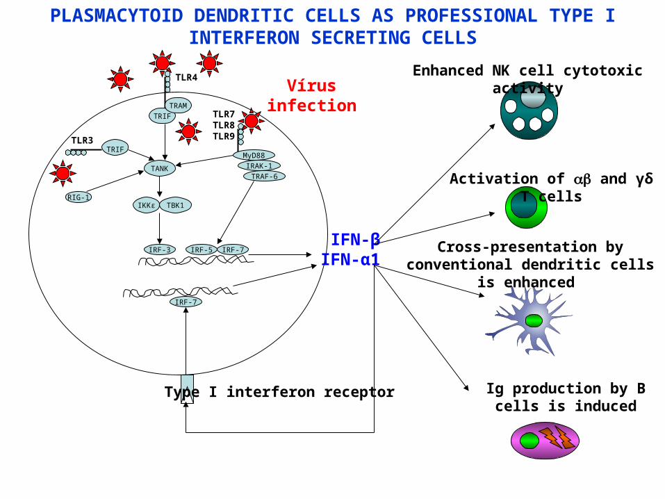
TRIF
TANK
IKKε TBK1
IRF-3
TRIF
TRAM
TLR3
TLR4
MyD88
IRF-5
TLR7TLR8TLR9
IFN-βIFN-α1
RIG-1
Ig production by B cells is induced
Type I interferon receptor
IRF-7
Enhanced NK cell cytotoxic activity
Activation of and γδ T cells
Cross-presentation by conventional dendritic cells is enhanced
IRAK-1
TRAF-6
IRF-7
PLASMACYTOID DENDRITIC CELLS AS PROFESSIONAL TYPE I INTERFERON SECRETING CELLS
Vírus infection
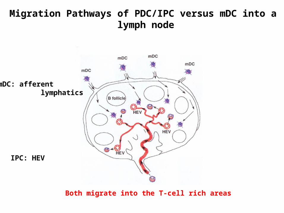
Migration Pathways of PDC/IPC versus mDC into a lymph node
IPC: HEV
mDC: afferent lymphatics
Both migrate into the T-cell rich areas
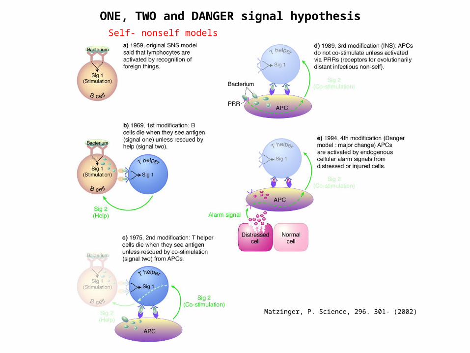
ONE, TWO and DANGER signal hypothesis
Matzinger, P. Science, 296. 301- (2002)
Self- nonself models
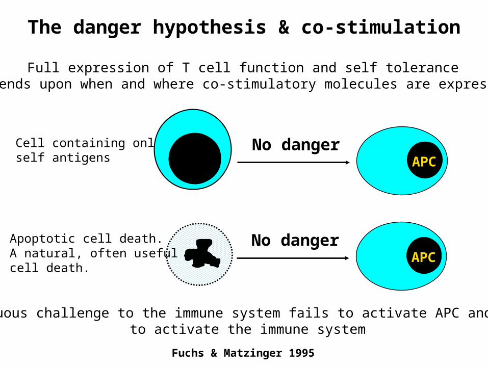
The danger hypothesis & co-stimulation
Fuchs & Matzinger 1995
Full expression of T cell function and self tolerance depends upon when and where co-stimulatory molecules are expressed.
Apoptotic cell death.A natural, often usefulcell death.
APC
APC
No danger
No dangerCell containing onlyself antigens
Innocuous challenge to the immune system fails to activate APC and failsto activate the immune system
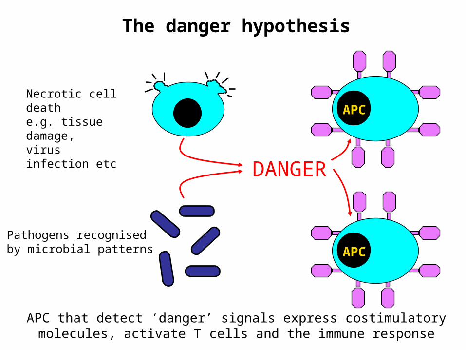
The danger hypothesis
APC
APC
Necrotic cell deathe.g. tissue damage,virus infection etc
Pathogens recognisedby microbial patterns
DANGER
APC that detect ‘danger’ signals express costimulatorymolecules, activate T cells and the immune response
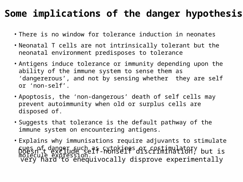
• There is no window for tolerance induction in neonates
• Neonatal T cells are not intrinsically tolerant but the neonatal environment predisposes to tolerance
• Antigens induce tolerance or immunity depending upon the ability of the immune system to sense them as ‘dangererous’, and not by sensing whether they are self or ‘non-self’.
• Apoptosis, the ‘non-dangerous’ death of self cells may prevent autoimmunity when old or surplus cells are disposed of.
• Suggests that tolerance is the default pathway of the immune system on encountering antigens.
• Explains why immunisations require adjuvants to stimulate cues of danger such as cytokines or costimulatory molecule expression.
Some implications of the danger hypothesis
Doesn’t exclude self-nonself discrimination, but is very hard to enequivocally disprove experimentally
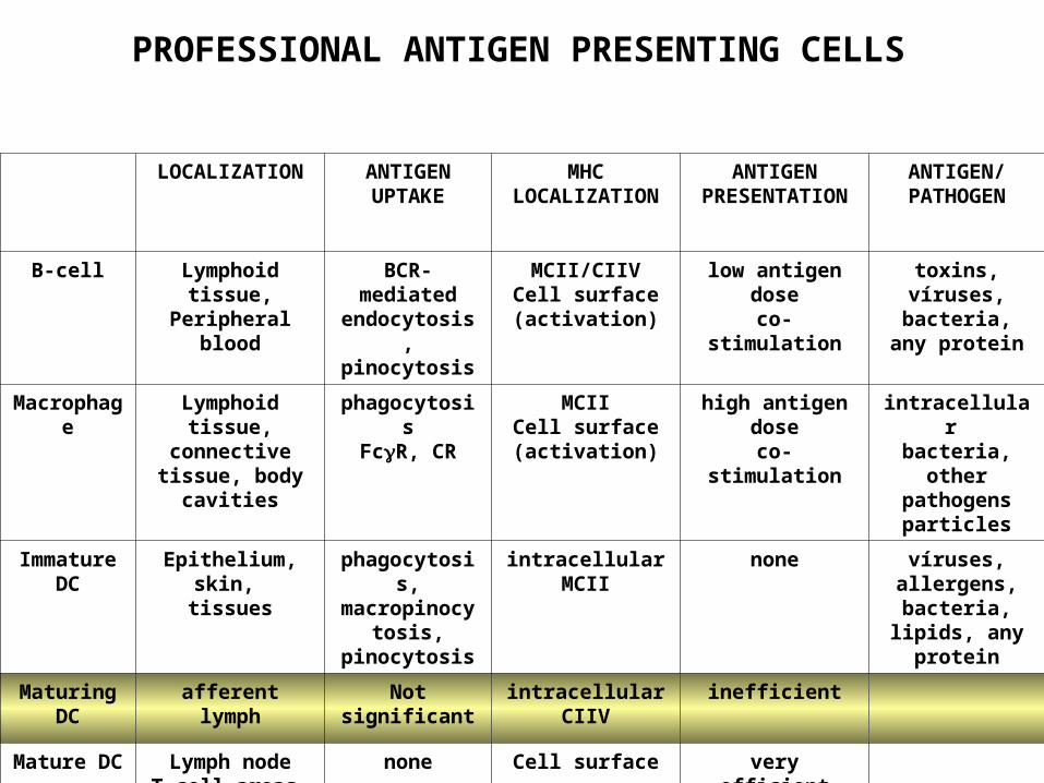
LOCALIZATION ANTIGEN UPTAKE
MHCLOCALIZATION
ANTIGEN PRESENTATION
ANTIGEN/PATHOGEN
B-cell Lymphoid tissue,Peripheral blood
BCR-mediated endocytosis,pinocytosis
MCII/CIIVCell surface (activation)
low antigen doseco-stimulation
toxins, víruses, bacteria,
any protein
Macrophage Lymphoid tissue, connective tissue,
body cavities
phagocytosisFcR, CR
MCIICell surface (activation)
high antigen doseco-stimulation
intracellular bacteria,
other pathogensparticles
Immature DC
Epithelium,skin,
tissues
phagocytosis,macropinocytosi
s, pinocytosis
intracellularMCII
none víruses, allergens,
bacteria, lipids, any protein
Maturing DC
afferent lymph Not significant intracellularCIIV
inefficient
Mature DC Lymph nodeT-cell areas
none Cell surface very efficientco-stimulation
PROFESSIONAL ANTIGEN PRESENTING CELLS