Expression, purification, crystallization, and preliminary ...Expression, purification,...
13
Expression, purification, crystallization, and preliminary X-ray crystallographic studies of the human adiponectin receptors, AdipoR1 and AdipoR2 Hiroaki Tanabe • Kanna Motoyama • Mariko Ikeda • Motoaki Wakiyama • Takaho Terada • Noboru Ohsawa • Toshiaki Hosaka • Masakatsu Hato • Yoshifumi Fujii • Yoshihiro Nakamura • Satoshi Ogasawara • Tomoya Hino • Takeshi Murata • So Iwata • Miki Okada-Iwabu • Masato Iwabu • Kunio Hirata • Yoshiaki Kawano • Masaki Yamamoto • Tomomi Kimura-Someya • Mikako Shirouzu • Toshimasa Yamauchi • Takashi Kadowaki • Shigeyuki Yokoyama Received: 18 September 2014 / Accepted: 19 November 2014 / Published online: 10 January 2015 Ó The Author(s) 2015. This article is published with open access at Springerlink.com Abstract The adiponectin receptors (AdipoR1 and Adi- poR2) are membrane proteins with seven transmembrane helices. These receptors regulate glucose and fatty acid metabolism, thereby ameliorating type 2 diabetes. The full- length human AdipoR1 and a series of N-terminally trun- cated mutants of human AdipoR1 and AdipoR2 were expressed in insect cells. In small-scale size exclusion chromatography, the truncated mutants AdipoR1D88 (res- idues 89–375) and AdipoR2D99 (residues 100–386) eluted mostly in the intact monodisperse state, while the others eluted primarily as aggregates. However, gel filtration chromatography of the large-scale preparation of the tag- affinity-purified AdipoR1D88 revealed the presence of an excessive amount of the aggregated state over the intact state. Since aggregation due to contaminating nucleic acids may have occurred during the sample concentration step, anion-exchange column chromatography was performed immediately after affinity chromatography, to separate the intact AdipoR1D88 from the aggregating species. The H. Tanabe K. Motoyama M. Ikeda M. Wakiyama T. Terada N. Ohsawa T. Hosaka M. Hato Y. Fujii Y. Nakamura T. Murata S. Iwata T. Kimura-Someya M. Shirouzu S. Yokoyama (&) RIKEN Systems and Structural Biology Center, 1-7-22 Suehiro- cho, Tsurumi-ku, Yokohama 230-0045, Japan e-mail: [email protected] H. Tanabe S. Yokoyama Department of Biophysics and Biochemistry and Laboratory of Structural Biology, Graduate School of Science, The University of Tokyo, Hongo, Bunkyo-ku, Tokyo 113-0033, Japan H. Tanabe M. Ikeda M. Wakiyama N. Ohsawa T. Hosaka M. Hato Y. Nakamura T. Kimura-Someya M. Shirouzu Division of Structural and Synthetic Biology, RIKEN Center for Life Science Technologies, 1-7-22 Suehiro-cho, Tsurumi-ku, Yokohama 230-0045, Japan T. Terada Y. Fujii S. Yokoyama RIKEN Structural Biology Laboratory, 1-7-22 Suehiro-cho, Tsurumi-ku, Yokohama 230-0045, Japan S. Ogasawara T. Hino T. Murata S. Iwata Department of Cell Biology, Graduate School of Medicine, Kyoto University, Yoshida-Konoe-cho, Sakyo-ku, Kyoto 606-8501, Japan T. Hino T. Murata S. Iwata JST, Research Acceleration Program, Membrane Protein Crystallography Project, Yoshida-Konoe-cho, Sakyo-ku, Kyoto 606-8501, Japan T. Murata Department of Chemistry, Graduate School of Science, Chiba University, Yayoi-cho, Inage, Chiba 263-8522, Japan S. Iwata Division of Molecular Biosciences, Membrane Protein Crystallography Group, Imperial College, London SW7 2AZ, UK S. Iwata Diamond Light Source, Harwell Science and Innovation Campus, Chilton, Didcot, Oxfordshire OX11 0DE, UK S. Iwata K. Hirata Y. Kawano M. Yamamoto RIKEN SPring-8 Center, Harima Institute, Kouto, Sayo, Hyogo 679-5148, Japan M. Okada-Iwabu M. Iwabu T. Yamauchi (&) T. Kadowaki (&) Department of Diabetes and Metabolic Diseases, Graduate School of Medicine, The University of Tokyo, Hongo, Bunkyo-ku, Tokyo 113-0033, Japan e-mail: [email protected] 123 J Struct Funct Genomics (2015) 16:11–23 DOI 10.1007/s10969-014-9192-z
Transcript of Expression, purification, crystallization, and preliminary ...Expression, purification,...

Expression, purification, crystallization, and preliminary X-raycrystallographic studies of the human adiponectin receptors,AdipoR1 and AdipoR2
Hiroaki Tanabe • Kanna Motoyama • Mariko Ikeda • Motoaki Wakiyama •
Takaho Terada • Noboru Ohsawa • Toshiaki Hosaka • Masakatsu Hato •
Yoshifumi Fujii • Yoshihiro Nakamura • Satoshi Ogasawara • Tomoya Hino •
Takeshi Murata • So Iwata • Miki Okada-Iwabu • Masato Iwabu •
Kunio Hirata • Yoshiaki Kawano • Masaki Yamamoto • Tomomi Kimura-Someya •
Mikako Shirouzu • Toshimasa Yamauchi • Takashi Kadowaki • Shigeyuki Yokoyama
Received: 18 September 2014 / Accepted: 19 November 2014 / Published online: 10 January 2015
� The Author(s) 2015. This article is published with open access at Springerlink.com
Abstract The adiponectin receptors (AdipoR1 and Adi-
poR2) are membrane proteins with seven transmembrane
helices. These receptors regulate glucose and fatty acid
metabolism, thereby ameliorating type 2 diabetes. The full-
length human AdipoR1 and a series of N-terminally trun-
cated mutants of human AdipoR1 and AdipoR2 were
expressed in insect cells. In small-scale size exclusion
chromatography, the truncated mutants AdipoR1D88 (res-
idues 89–375) and AdipoR2D99 (residues 100–386) eluted
mostly in the intact monodisperse state, while the others
eluted primarily as aggregates. However, gel filtration
chromatography of the large-scale preparation of the tag-
affinity-purified AdipoR1D88 revealed the presence of an
excessive amount of the aggregated state over the intact
state. Since aggregation due to contaminating nucleic acids
may have occurred during the sample concentration step,
anion-exchange column chromatography was performed
immediately after affinity chromatography, to separate the
intact AdipoR1D88 from the aggregating species. The
H. Tanabe � K. Motoyama � M. Ikeda � M. Wakiyama �T. Terada � N. Ohsawa � T. Hosaka � M. Hato � Y. Fujii �Y. Nakamura � T. Murata � S. Iwata � T. Kimura-Someya �M. Shirouzu � S. Yokoyama (&)
RIKEN Systems and Structural Biology Center, 1-7-22 Suehiro-
cho, Tsurumi-ku, Yokohama 230-0045, Japan
e-mail: [email protected]
H. Tanabe � S. Yokoyama
Department of Biophysics and Biochemistry and Laboratory of
Structural Biology, Graduate School of Science, The University
of Tokyo, Hongo, Bunkyo-ku, Tokyo 113-0033, Japan
H. Tanabe � M. Ikeda � M. Wakiyama � N. Ohsawa � T. Hosaka �M. Hato � Y. Nakamura � T. Kimura-Someya � M. Shirouzu
Division of Structural and Synthetic Biology, RIKEN Center for
Life Science Technologies, 1-7-22 Suehiro-cho, Tsurumi-ku,
Yokohama 230-0045, Japan
T. Terada � Y. Fujii � S. Yokoyama
RIKEN Structural Biology Laboratory, 1-7-22 Suehiro-cho,
Tsurumi-ku, Yokohama 230-0045, Japan
S. Ogasawara � T. Hino � T. Murata � S. Iwata
Department of Cell Biology, Graduate School of Medicine,
Kyoto University, Yoshida-Konoe-cho, Sakyo-ku,
Kyoto 606-8501, Japan
T. Hino � T. Murata � S. Iwata
JST, Research Acceleration Program, Membrane Protein
Crystallography Project, Yoshida-Konoe-cho, Sakyo-ku,
Kyoto 606-8501, Japan
T. Murata
Department of Chemistry, Graduate School of Science, Chiba
University, Yayoi-cho, Inage, Chiba 263-8522, Japan
S. Iwata
Division of Molecular Biosciences, Membrane Protein
Crystallography Group, Imperial College, London SW7 2AZ, UK
S. Iwata
Diamond Light Source, Harwell Science and Innovation
Campus, Chilton, Didcot, Oxfordshire OX11 0DE, UK
S. Iwata � K. Hirata � Y. Kawano � M. Yamamoto
RIKEN SPring-8 Center, Harima Institute, Kouto, Sayo,
Hyogo 679-5148, Japan
M. Okada-Iwabu � M. Iwabu � T. Yamauchi (&) �T. Kadowaki (&)
Department of Diabetes and Metabolic Diseases, Graduate
School of Medicine, The University of Tokyo, Hongo,
Bunkyo-ku, Tokyo 113-0033, Japan
e-mail: [email protected]
123
J Struct Funct Genomics (2015) 16:11–23
DOI 10.1007/s10969-014-9192-z
separated intact AdipoR1D88 did not undergo further
aggregation, and was successfully purified to homogeneity
by gel filtration chromatography. The purified Adi-
poR1D88 and AdipoR2D99 proteins were characterized by
thermostability assays with 7-diethylamino-3-(4-malei-
midophenyl)-4-methyl coumarin, thin layer chromatogra-
phy of bound lipids, and surface plasmon resonance
analysis of ligand binding, demonstrating their structural
integrities. The AdipoR1D88 and AdipoR2D99 proteins
were crystallized with the anti-AdipoR1 monoclonal anti-
body Fv fragment, by the lipidic mesophase method. X-ray
diffraction data sets were obtained at resolutions of 2.8 and
2.4 A, respectively.
Keywords Membrane protein � Adiponectin receptors
AdipoR1 and AdipoR2 � Purification � Antibody �Crystallization � Lipidic mesophase
Introduction
Adiponectin is an anti-diabetic and anti-atherogenic
adipokine, and is exclusively expressed in adipose tissue
[1–4]. Serum adiponectin levels are significantly reduced
in patients with obesity, metabolic syndrome, and type 2
diabetes [5]. We previously reported the expression
cloning of complementary DNAs encoding adiponectin
receptors (Adipor) 1 and 2 [6]. These adiponectin
receptors, AdipoR1 and AdipoR2, are key membrane
proteins that exert anti-metabolic syndrome effects.
Adiponectin accomplishes its biological effects by bind-
ing to the AdipoR1 and AdipoR2 receptors. In the liver,
both adiponectin receptors mediate the major part of the
insulin-sensitizing actions of adiponectin, while AdipoR1
primarily does so in skeletal muscle. AdipoR1 and
AdipoR2 regulate glucose and fatty acid metabolism
partly via the activation of the AMPK [7–9], Ca2? [10],
and PPARa [11, 12] signaling pathways. Interestingly,
AdipoR1 and AdipoR2 are predicted to contain seven-
transmembrane domains [6], but they are structurally
distinct from G-protein coupled receptors (GPCRs) [13].
The adiponectin receptors possess an internal N-terminus
and an external C-terminus, which is opposite to the
topology of GPCRs. Therefore, AdipoRs are predicted
to have unique structures, as compared to those of
GPCRs.
Here, we report the expression and purification of the
N-terminally truncated human AdipoR1 and AdipoR2
proteins, crystallization of the truncated AdipoR1 and
AdipoR2 in complexes with the Fv fragment of an anti-
AdipoR1 monoclonal antibody, and their preliminary
X-ray crystallographic studies.
Materials and methods
Plasmid construction
The BglII-FLAG-TEV-BamHI-EcoRI DNA (50-GGAAGA
TCTATGGATTACAAGGACGACGACGATAAGGAAA
ACCTGTATTTTCAGGGCGGATCCGAATTCCCG-30)and its complementary DNA were synthesized, annealed
together, digested with BglII and EcoRI, and subcloned
into the BamHI and EcoRI sites of pFastBac1. The
resulting plasmid encodes a Flag tag followed by a TEV
cleavage site at the N-terminus, and is referred to as
pFastBac1-FT hereafter. The cDNAs encoding the full-
length human AdipoR1 (residues 1–375) and N-termi-
nally-truncated mutants of AdipoR1 and AdipoR2 (Adi-
poR1D46, residues 47–375; D76, 77–375; D88, 89–375;
D101, 102–375; and D119, 120–375; AdipoR2D58, resi-
dues 59–386; D87, 88–386; D99, 100–386; D112,
113–386; and D130, 131–386) were amplified by PCR.
The PCR products were digested with BamHI and XhoI
for AdipoR1 and EcoRI and XhoI for AdipoR2, and then
inserted into the pFastBac1-FT vector.
Protein expression in insect cells
High-titer recombinant baculoviruses were obtained with
the Bac-to-Bac Baculovirus Expression System (Invitro-
gen), according to the manufacturer’s protocol. For large-
scale and small-scale production of the recombinant pro-
teins, Trichoplusia ni (High Five) cells, at densities of
2 9 106 and 2–5 9 106 cell/ml, respectively, were infec-
ted with the high-titer viral stock at a multiplicity of
infection (m.o.i.) of 0.5. Cells were harvested by centri-
fugation at 42-h post infection, and were washed once with
phosphate buffer saline (PBS). Cells were flash-frozen in
liquid nitrogen, and stored at -80 �C until use.
T. Kadowaki
e-mail: [email protected]
M. Okada-Iwabu � M. Iwabu � T. Yamauchi � T. Kadowaki
Department of Integrated Molecular Science on Metabolic
Diseases, 22nd Century Medical and Research Center, The
University of Tokyo, Hongo, Bunkyo-ku, Tokyo 113-0033,
Japan
M. Iwabu
PRESTO, Japan Science and Technology Agency, Kawaguchi,
Saitama 332-0012, Japan
T. Yamauchi
CREST, Japan Science and Technology Agency, Kawaguchi,
Saitama 332-0012, Japan
12 H. Tanabe et al.
123

Large-scale membrane preparation
For large-scale preparations of the full-length AdipoR1,
AdipoR1D88, and AdipoR2D99 proteins, frozen cells were
thawed in high osmotic buffer [10 mM HEPES–NaOH
buffer (pH 7.4) containing 1.0 M NaCl, 10 mM MgCl2,
20 mM KCl, and EDTA-free Complete Protease Inhibitor
Cocktail (Roche)], and disrupted by Dounce homogeniza-
tion. The raw membranes were collected by ultracentrifu-
gation at 100,0009g for 30 min, and were resuspended in
high osmotic buffer. These ultracentrifugation and resus-
pension steps were repeated four times, to remove the
peripheral membrane proteins. Finally, the washed mem-
branes were resuspended in 20 mM HEPES–NaOH buffer
(pH 7.4) containing 100 mM NaCl and 10 % (v/v) glyc-
erol, flash-frozen with liquid nitrogen, and stored at
-80 �C until use. The membrane proteins were quantified
with the DC Protein Assay (Bio-Rad), using bovine serum
albumin (BSA) as the standard.
Large-scale protein purification
The purified membranes (20 mg/ml of total membrane
proteins) were solubilized with 20 mM HEPES–NaOH
buffer (pH 7.4) containing 100 mM NaCl, 10 % (v/v)
glycerol, and 1 % (w/v) n-dodecyl-b-D-maltoside (DDM,
Anatrace), for 1–2 h at 4 �C. The insoluble materials were
removed by ultracentrifugation at 100,0009g for 1 h. The
supernatant was filtered (0.45 lm) and incubated with
Anti DYKDDDDK Tag Antibody Beads (Wako) in
20 mM HEPES–NaOH buffer (pH 7.4) containing
300 mM NaCl, 10 % (v/v) glycerol, and 0.5 % (w/v)
DDM, at 4 �C with gentle agitation. The beads were
washed with thirty column volumes of buffer A [20 mM
HEPES–NaOH buffer (pH 7.4), containing 10 % (v/v)
glycerol, 0.025 % (w/v) DDM, and 0.0001 % (w/v) cho-
lesteryl-hemi-succinate (CHS, Anatrace)], containing
200 mM NaCl. Then, the adsorbed AdipoR1/AdipoR2
proteins were eluted with five column volumes of buffer
A containing 100 or 200 mM NaCl and 0.1 mg/ml DY-
KDDDDK peptide (Wako). Further purification was per-
formed by the following three methods.
First, the affinity-purified sample was concentrated by
ultrafiltration with an Ultra-15 30 K-MWCO filter (Mil-
lipore), and then loaded on a HiLoad 16/600 Superdex
200 (GE Healthcare) column equilibrated in buffer A
containing 200 mM NaCl. Second, the affinity-purified
sample, in 100 mM NaCl, was loaded on a 1-ml HiTrap
Q column (GE Healthcare) equilibrated with buffer A
containing 100 mM NaCl, and was eluted by a
100–1,000 mM linear NaCl gradient in buffer A. The
sample was further purified by SEC on a Superdex 200
10/300 (GE Healthcare) column, in buffer A containing
200 mM NaCl. Third, the affinity-purified sample, in
200 mM NaCl, was loaded on a 1-ml HiTrap Q column
equilibrated with buffer A containing 200 mM NaCl, and
the flow-through fraction was collected. The polyhisti-
dine-tagged TEV protease was added to the anion-
exchange-purified fraction, and incubated with the
receptor overnight at 4 �C. The receptor was separated
from TEV by adsorption to TALON resin (Clontech), and
was further purified by SEC on a Superdex 200 10/300
column in buffer A containing 200 mM NaCl. The puri-
ties of the AdipoR1D88 and AdipoR2D99 proteins were
assessed by SDS-PAGE.
Small-scale SEC analysis
Frozen cells were thawed in the high osmotic buffer, and
then sonicated to disrupt the cells. The membranes were
collected by ultracentrifugation at 100,0009g for 15 min,
and were resuspended in 20 mM HEPES–NaOH buffer
(pH 7.4) containing 150 mM NaCl, 1 mM EDTA, and
5 mM MgCl2. The purified membranes (10 mg/ml of
membrane protein) were solubilized with 20 mM
HEPES–NaOH buffer (pH 7.4) containing 1 % (w/v)
DDM, 150 mM NaCl, 1 mM EDTA, and 5 mM MgCl2,
for 1 h at 4 �C. The insoluble material was removed by
ultracentrifugation at 100,0009g for 30 min. The super-
natant was incubated with 100 ll anti-FLAG M2 affinity
gel (Sigma) in 20 mM HEPES–NaOH buffer (pH 7.4)
containing 150 mM NaCl, 1 mM EDTA, 5 mM MgCl2,
5 % (v/v) glycerol, and 0.5 % (w/v) DDM, at 4 �C with
gentle agitation. The beads were washed with five col-
umn volumes of 20 mM HEPES–NaOH buffer (pH 7.4)
containing 150 mM NaCl, 1 mM EDTA, 5 mM MgCl2,
5 % (v/v) glycerol, and 0.04 % (w/v) DDM, and the
adsorbed receptor was eluted with five column volumes
of the same buffer containing 0.1 mg/ml FLAG peptide
(Sigma). The affinity purified sample was then loaded on
a Superdex 200 10/300 column in 20 mM HEPES–NaOH
buffer (pH 7.4) containing 200 mM NaCl, 10 % (v/v)
glycerol, and 0.04 % (w/v) DDM. Fractions (1 ml) were
collected. Portions (5 ll) of the eluates were separated
by SDS-PAGE, and transferred to a PVDF membrane.
The membranes were blocked in 5 % (w/v) dry milk in
TBS-T buffer [Tris-buffered saline, 0.1 % (v/v) Tween-
20] at room temperature for 1 h. The blocked membranes
were detected with the anti-FLAG M2 monoclonal anti-
body (Sigma) and the anti-mouse IgG, HRP-linked whole
antibody from sheep (GE Healthcare) in TBS-T buffer.
The membranes were visualized using Immobilon Wes-
tern Chemiluminescent HRP Substrate (Millipore), and
detected with an LAS3000 imager (Fuji).
The human adiponectin receptors AdipoR1 and AdipoR2 13
123

Characterization of the purified N-terminally truncated
mutants of AdipoR1 and AdipoR2
The purified AdipoR1D88 and AdipoR2D99 proteins were
analyzed by the following three methods. First, the thermal
stabilities of AdipoR1D88 and AdipoR2D99 were analyzed
by the 7-diethylamino-3-(4-maleimidophenyl)-4-methyl
coumarin (CPM) assay method [14, 15]. The fluorescence
of the CPM dye was measured with a 340-nm excitation
filter with a 10-nm bandpass and a 460-nm emission filter
with a 35-nm bandpass at 40 �C, on a FUSION a Micro-
plate Reader PerkinElmer. Second, the lipids that co-puri-
fied with the AdipoR1D88 and AdipoR2D99 proteins were
analyzed by thin layer chromatography (TLC). The full-
length AdipoR1 and AdipoR2, prepared by FLAG affinity
and anion-exchange chromatography, were also analyzed
for comparison. The proteins were dissolved in chloroform/
methanol [2:1 (v/v)], and the bound lipids were extracted
from the proteins. The extracted samples were applied to a
silica gel 60 TLC plate (Merck Millipore), which was then
developed by a solvent system composed of chloroform/
methanol/water [65:25:4 (v/v)]. The lipids were visualized
with acetic acid/sulfuric acid [1:1 (v/v)], the phosphomo-
lybdic reagent (Pierce), and the ninhydrin reagent (Wako).
Third, the ligand-binding activity of AdipoR1D88 was
measured by surface plasmon resonance (SPR) measure-
ments. The purified AdipoR1D88 was reconstituted into
liposomes [5 mg/ml egg yolk phosphatidylcholine (PC)
(Avanti Polar Lipids Inc.) and 0.05 mg/ml biotinyl-phos-
phatidylethanolamine (biotinyl-PE) (Avanti Polar Lipids
Inc.)], and the reconstituted liposomes were immobilized
onto a sensor chip SA. Binding analyses were performed
with a range of osmotin concentrations (0.5–8 lM) on a
Biacore T200 (GE Healthcare).
Production of the anti-AdipoR1 monoclonal antibody
All animal experiments described here were approved by
the Institutional Animal Care and Use Committee of Kyoto
University Graduate School of Medicine. The purified
untagged AdipoR1D88 was reconstituted into liposomes
[5 mg/ml egg yolk PC and 1 mg/ml lipid A (Sigma)].
Female BALB/c mice were immunized five times with
0.1 mg doses of the reconstituted AdipoR1D88, at intervals
of 10 days. Single-cell suspensions were prepared from the
spleens of the immunized mice, and the cells were fused
with P3U1 myeloma cells, using the conventional poly-
ethylene glycol (PEG) method [16]. Screening of anti-
bodies was performed by three methods, enzyme-linked
immunosorbent assay (ELISA), fluorescence-detection
SEC (FSEC), and denatured dot blot assays [17, Ogasawara
et al., manuscript in preparation]. For ELISA, the purified
AdipoR1D88 was reconstituted into liposomes containing
biotinyl-PE, and was immobilized on Immobilizer Strep-
tavidin plates (Nunc). High-affinity antibodies that formed
stable complexes with the purified AdipoR1D88 were
selected by FSEC, using the fluorescein-conjugated Fab
fragment of an anti-mouse IgG (Jackson), on a Superdex
200 5/150 column (GE Healthcare). Antibodies that rec-
ognized the native conformation of AdipoR1D88 were
selected by dot blot assays with SDS-denatured Adi-
poR1D88. Each selected clone was isolated by the limiting
dilution-culture method, and monoclonal hybridoma cell
lines producing anti-AdipoR1D88 antibodies were
established.
Recombinant production of the Fv fragment of the anti-
AdipoR1 antibody
The sequences of the VH and VL regions were determined
by the standard method, with total RNA isolated from the
hybridoma cells [18]. The cloned VH and VL cDNA frag-
ments were subcloned into the TA-cloning vector, pCR2.1
TOPO (Invitrogen) [19], encoding a fusion protein with an
N-terminal His-tag, a SUMO tag, and a SUMO protease
cleavage site. The VH and VL fragments were co-synthe-
sized by the E. coli cell-free protein synthesis method [20],
supplemented with DsbC and the reduced and oxidized
forms of glutathione (GSH and GSSG, respectively) to
form disulfide bonds [21]. The reaction solution was cen-
trifuged at 20,0009g and 4 �C for 10 min. The supernatant
was loaded on a 1-ml HisTrap column (GE Healthcare)
equilibrated with 20 mM Tris–HCl buffer (pH 8.0) con-
taining 500 mM NaCl and 20 mM imidazole, and was
eluted by a 20–500 mM linear gradient of imidazole in
20 mM Tris–HCl buffer (pH 8.0) containing 500 mM
NaCl. The His- and SUMO-tags were cleaved by SUMO
protease for 2–3 days at room temperature, and were
removed by a second passage through the HisTrap column.
The protein sample was then loaded on a HiLoad 16/600
Superdex 200 column, equilibrated in 20 mM HEPES–
NaOH buffer (pH 7.4) containing 200 mM NaCl. The
purified Fv fragment (Fv43) was concentrated to approxi-
mately 20 mg/ml, by ultrafiltration with an Ultra-15 10 K-
MWCO filter (Millipore).
Preparation of the AdipoR1�Fv43 and AdipoR2�Fv43
complexes
The Fv43 (24.8 kDa) was mixed with the purified FLAG-
tagged AdipoR1D88 (35.0 kDa) or untagged AdipoR2D99
(32.9 kDa), and incubated on ice for 30 min. The mixture
was loaded onto a Superdex 200 10/300 column equili-
brated with 20 mM HEPES–NaOH buffer (pH 7.4) con-
taining 200 mM NaCl, 0.025 % (w/v) DDM, and 0.0001 %
(w/v) CHS, and was eluted using the same buffer. Fractions
14 H. Tanabe et al.
123
containing the complex were collected and concentrated to
approximately 15 mg/ml by ultrafiltration (Ultra-4 30 K-
MWCO, Millipore). The purities of the AdipoR1D88�Fv43
and AdipoR2D99�Fv43 complexes were assessed by SDS-
PAGE.
Crystallization and X-ray data collection
The purified AdipoR1D88�Fv43 and AdipoR2D99�Fv43
complexes were reconstituted into a lipidic mesophase, by
mixing with molten lipid in a mechanical syringe mixer [22].
The protein-LCP mixture contained 40 % (w/w) protein
solution, 54 % (w/w) monoolein (Sigma) and 6 % (w/w)
cholesterol (Sigma). Forty nanoliter drops of the resulting
lipidic mesophase sample were dispensed into 96 well glass
plates, overlaid with 0.8 ll precipitant solution, and covered
with thin cover glasses, by the use of laboratory-constructed
manual and robotic micro-dispensers [23, Hato et al., man-
uscript in preparation]. Crystallization setups were per-
formed at room temperature, and the plates were incubated at
20 �C. Crystals were harvested directly from the lipidic
mesophase using MiTeGen micromounts, and flash cooled in
liquid nitrogen. Data collection was performed on beamline
BL32XU at SPring-8, using an MX225HE CCD detector
[24–26]. X-ray diffraction data with a micro beam of
1 lm 9 10 lm (horizontal 9 vertical) were collected at
a b
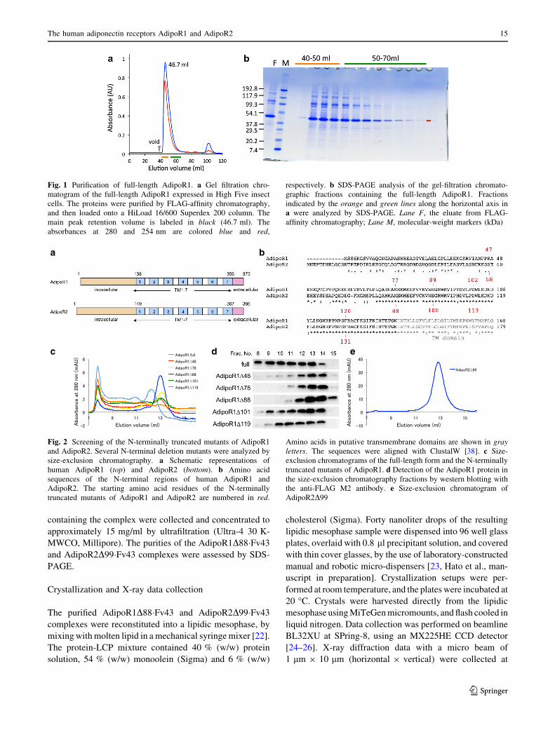
c d e
Fig. 2 Screening of the N-terminally truncated mutants of AdipoR1
and AdipoR2. Several N-terminal deletion mutants were analyzed by
size-exclusion chromatography. a Schematic representations of
human AdipoR1 (top) and AdipoR2 (bottom). b Amino acid
sequences of the N-terminal regions of human AdipoR1 and
AdipoR2. The starting amino acid residues of the N-terminally
truncated mutants of AdipoR1 and AdipoR2 are numbered in red.
Amino acids in putative transmembrane domains are shown in gray
letters. The sequences were aligned with ClustalW [38]. c Size-
exclusion chromatograms of the full-length form and the N-terminally
truncated mutants of AdipoR1. d Detection of the AdipoR1 protein in
the size-exclusion chromatography fractions by western blotting with
the anti-FLAG M2 antibody. e Size-exclusion chromatogram of
AdipoR2D99
Fig. 1 Purification of full-length AdipoR1. a Gel filtration chro-
matogram of the full-length AdipoR1 expressed in High Five insect
cells. The proteins were purified by FLAG-affinity chromatography,
and then loaded onto a HiLoad 16/600 Superdex 200 column. The
main peak retention volume is labeled in black (46.7 ml). The
absorbances at 280 and 254 nm are colored blue and red,
respectively. b SDS-PAGE analysis of the gel-filtration chromato-
graphic fractions containing the full-length AdipoR1. Fractions
indicated by the orange and green lines along the horizontal axis in
a were analyzed by SDS-PAGE. Lane F, the eluate from FLAG-
affinity chromatography; Lane M, molecular-weight markers (kDa)
The human adiponectin receptors AdipoR1 and AdipoR2 15
123

100 K, by the helical scan method with 1� oscillation. The
data from the AdipoR1D88�Fv43 and AdipoR2D99�Fv43
crystals were indexed, scaled, and merged with the
HKL2000 program suite [27] and the XDS package [28],
respectively.
Results and discussion
Screening of deletion mutants of AdipoR1
and AdipoR2
The full-length AdipoR1 (residues 1–375) was expressed in
baculovirus-infected High Five insect cells, and the mem-
brane fractions were prepared. Upon gel filtration chro-
matography (size-exclusion chromatography, SEC) on a
HiLoad 16/600 Superdex 200 column, the detergent-solu-
bilized full-length AdipoR1 mainly eluted just after the
void volume (40 ml) (Fig. 1). If the full-length AdipoR1
was monomeric and monodisperse, then the proteomicelle
should exhibit an estimated molecular mass of ca. 125 kDa
(the full-length AdipoR1 monomer, 44.7 kDa; DDM
micelle, ca. 80 kDa), and elute after 55 ml (the elution
volume of ferritin, 440 kDa) on the gel filtration column.
However, most of the full-length AdipoR1 eluted before
55 ml (Fig. 1), indicating that this sample was highly
polydisperse and not suitable for crystallization.
We therefore tried to modify the AdipoR1 construct.
Prediction servers of protein secondary structure, PSIPRED
v3.3 [29], and transmembrane domains, HMMTOP [30],
suggested that human AdipoR1 and AdipoR2 have a long
N-terminal region, seven transmembrane (TM) helices with
short loops connecting the TM helices, and a short C-ter-
minal region (Fig. 2a). Since more than 50 % of the
N-terminal region was predicted to be flexible, we specu-
lated that this long N-terminal tail is related to the observed
polydispersity of AdipoR1 (Fig. 1).
Therefore, we constructed a series of N-terminally-
deleted mutants of AdipoR1 (Fig. 2b). They were expres-
sed on a small scale, and were analyzed by SEC without
concentration. In this small-scale SEC analysis of AdipoR1
(Fig. 2c, d), the non-concentrated sample of the full-length
AdipoR1 was less aggregated, as compared with the con-
centrated sample in the large-scale preparation, because it
exhibited a small peak eluting at ca. 13 ml (Fig. 1). This
ca. 13-ml elution volume in the small-scale SEC analysis
should correspond to ca. 65 ml in the large-scale gel fil-
tration chromatography. Deletions of residues 1–46 (D46)
and 1–76 (D76) of AdipoR1 significantly increased the
fraction eluting at ca. 13 ml, as compared with the full-
length AdipoR1. However, the total amounts of these
deletion mutants were much lower than that of the full-
length AdipoR1 obtained from the same amount of cells.
By contrast, the deletion of residues 1–88 (D88) of Adi-
poR1 further improved the monodispersity in the SEC
analysis, and the total amount of this deletion mutant
protein was as high as that of the full-length AdipoR1
(Fig. 2c, d). On the other hand, further deletion mutants
(AdipoR1D101 and AdipoR1D119) were less aggregated
than the full-length protein, but appreciably more aggre-
gated than AdipoR1D88. Thus, we concluded that the
AdipoR1D88 mutant was the best among the tested dele-
tion mutants. The screening of AdipoR2D58, D87, D99,
D112, and D130 was performed in the same manner, and
AdipoR2D99 was found to be the best (Fig. 2e). Conse-
quently, the AdipoR1D88 and AdipoR2D99 mutants were
selected and used for further crystallization trials.
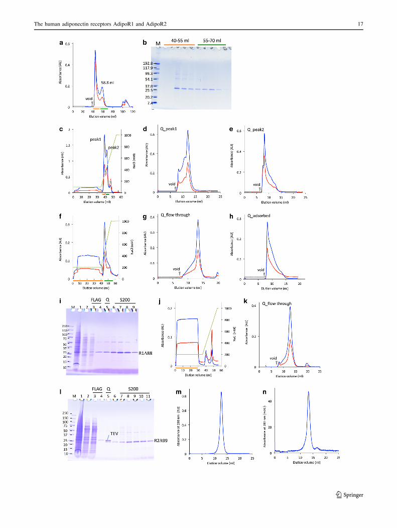
Fig. 3 Large-scale preparation of FLAG-tagged AdipoR1D88 and
AdipoR2D99 expressed in High Five cells. In the chromatograms (a,
c–h, j, k, m, n), the absorbances at 280 and 254 nm, and the NaCl
concentration are shown in blue, red, and light green, respectively.
a Gel filtration chromatogram of FLAG-tagged AdipoR1D88
expressed in High Five insect cells. The proteins were purified by
FLAG-affinity chromatography, and then chromatographed on a
HiLoad 16/600 Superdex 200 column. The main peak elution volume
is labeled in black (58.8 ml). b SDS-PAGE analysis of the gel-
filtration chromatographic fractions containing the FLAG-tagged
AdipoR1D88. Fractions indicated by the orange and green lines along
the horizontal axis in a were analyzed by SDS-PAGE. Lane M,
molecular-weight markers (kDa). c Anion-exchange chromatogram of
the FLAG-tagged AdipoR1D88, with isocratic elution by 100 mM
NaCl in buffer A, and subsequently with gradient elution by
100–1,000 mM NaCl in buffer A. d, e Gel filtration chromatograms
of the peak 1 (d) and peak 2 (e) fractions, indicated by the orange and
green lines in c, respectively. f Anion-exchange chromatogram of the
FLAG-tagged AdipoR1D88, with isocratic elution by 200 mM NaCl
in buffer A, and subsequently with gradient elution by 200–1,000 mM
NaCl in buffer A. g, h Gel filtration chromatograms of the flow-
through (g) and adsorbed (h) fractions, indicated by the orange and
green lines in f, respectively. i SDS-PAGE analysis of the FLAG-
tagged AdipoR1D88. Lane M, molecular-weight markers (kDa); lane
1, the membrane fraction; lane 2, DDM-solubilized membrane
proteins in the supernatant after ultracentrifugation; lane 3, the
flow-through fraction from FLAG-affinity chromatography; lane 4,
the eluate from FLAG-affinity chromatography; lane 5, the flow-
through fraction from anion-exchange chromatography (f); lanes 6–9,
the peak fractions of the FLAG-tagged AdipoR1D88 from gel
filtration chromatography (g). j Anion-exchange chromatogram of
AdipoR2D99, with isocratic elution by 200 mM NaCl in buffer A and
subsequently with gradient elution by 200–1,000 mM NaCl in buffer
A. k Gel filtration chromatogram of the flow-through fractions
indicated by the orange line in j. l SDS-PAGE analysis of
AdipoR2D99. Lane M, molecular-weight markers (kDa); lane 1, the
membrane fraction; lane 2, DDM-solubilized membrane proteins in
the ultracentrifugation supernatant; lane 3, the flow-through fraction
from FLAG-affinity chromatography; lane 4, the eluate from FLAG-
affinity chromatography; lane 5, the flow-through fraction from
anion-exchange chromatography after the TEV protease digestion;
lane 6, the flow-through fraction from TALON chromatography;
lanes 7–11, the peak fractions of AdipoR2D99 from gel filtration
chromatography. m, n Gel filtration chromatographic analysis of the
purified FLAG-tagged AdipoR1D88 (m) and the purified Adi-
poR2D99 (n)
c
16 H. Tanabe et al.
123
The human adiponectin receptors AdipoR1 and AdipoR2 17
123

Large-scale preparations of N-terminally truncated
AdipoR1 and AdipoR2
The FLAG-tagged AdipoR1D88 and AdipoR2D99 were
overexpressed in baculovirus-infected High Five insect
cells. In general, membrane proteins are purified by
minimal steps of chromatography, such as one-step
affinity chromatography or two-step (affinity and size-
exclusion) chromatography, to avoid deterioration caused
by delipidation due to excessive washing with detergents.
Therefore, the FLAG-tagged AdipoR1D88 was partially
purified by stepwise FLAG affinity chromatography, and
then fractionated by gel filtration chromatography, in
which the AdipoR1D88 eluted from the void volume
(40 ml) to 70 ml, and formed two peaks (Fig. 3a, b). In
this large-scale preparation, the affinity-purified Adi-
poR1D88 behaved differently in the gel filtration (Fig. 3a,
b), as compared to the small-scale SEC analysis (Fig. 2c,
d). The low molecular mass fraction eluting at 58.8 ml
(Fig. 3a) was considered to correspond to the monodis-
perse fraction of AdipoR1D88 eluting at 13 ml in the
small-scale SEC analysis. On the other hand, the high
molecular mass, aggregated fraction was drastically larger
in the large-scale preparation, as compared to the small-
scale preparation (Figs. 2c, 3a). Therefore, the affinity-
purified AdipoR1D88 still aggregated during the sample
concentration for gel filtration chromatography, as in the
case of the full-length AdipoR1 (Fig. 1), although the
properties of AdipoR1 were greatly improved by the
N-terminal deletion (D88).
Accordingly, we explored the cause of the aggregation.
As compared with the monodisperse fraction, the aggre-
gated fraction exhibited a stronger absorbance at 254 nm
relative to that at 280 nm (Fig. 3a). Therefore, we
hypothesized that nucleic acids contained in the FLAG-
affinity-purified AdipoR1D88 preparation promoted pro-
tein aggregation. To quickly remove the putative nucleic
acid contamination, we included an anion-exchange
column chromatography step immediately after the FLAG
affinity chromatography. The FLAG-tagged AdipoR1D88,
in buffer A containing 100 or 200 mM NaCl, was applied
to a 1-ml HiTrap Q column. The FLAG-tagged Adi-
poR1D88, in buffer A containing 100 mM NaCl, was
adsorbed on the column and eluted at low salt concentra-
tions (150–250 mM NaCl) (Fig. 3c–e), whereas that in
buffer A containing 200 mM NaCl was eluted in the flow-
through fraction (Fig. 3f–i). On the other hand, the aggre-
gated AdipoR1D88 was adsorbed on the column with either
100 mM or 200 mM NaCl, and was eluted at high salt
concentrations (250–400 mM NaCl) (Fig. 3c, e, f, h). Thus,
we could quickly separate the ‘‘intact’’ FLAG-tagged
AdipoR1D88 from the aggregated AdipoR1D88, in buffer
A containing 200 mM NaCl, by anion-exchange chroma-
tography (Fig. 3f, g). Once it was purified in this manner,
the ‘‘intact’’ FLAG-tagged AdipoR1D88 did not undergo
the fast aggregation, and was eluted as a symmetrical peak
in gel filtration chromatography. In other words, the ‘‘rot-
ten apple’’ (the nucleic acid-aggregated AdipoR1) can be
removed quickly by the anion-exchange chromatography,
before it ‘‘spoils the barrel’’. As the cause of the aggrega-
tion had been found, we tried another method to reduce it:
the sonication of the membrane preparation was also useful
for breaking up nucleic acids, and the aggregation of the
receptors was significantly reduced during the purification
step.
In the same manner, the affinity-purified FLAG-tagged
AdipoR2D99, in buffer A containing 200 mM NaCl, was
eluted in the flow-through fraction in the HiTrap Q column
chromatography (Fig. 3j–l). The FLAG tag of Adi-
poR2D99 was removed after the HiTrap Q column chro-
matography. Thus, the FLAG-tagged AdipoR1D88 and the
untagged AdipoR2D99 were purified to near homogeneity
(Fig. 3m, n). Removing the nucleic acids during the
membrane preparation and the early stages of purification
was essential to obtain larger amounts of the highly
homogeneous AdipoR1 and AdipoR2 proteins.
Fig. 4 Characterization of the purified AdipoR1D88 and Adi-
poR2D99 proteins. a, b CPM assay of AdipoR1D88 (a) and
AdipoR2D99 (b). c–e TLC analysis of AdipoR1D88 (D88), the
full-length AdipoR1 (FL1), AdipoR2D99 (D99), and the full-length
AdipoR2 (FL2). The lipids were visualized with acetic acid/sulfuric
acid [1:1 (v/v)] (c), the phosphomolybdic reagent (d), and the
ninhydrin reagent (e). Lane M, polar lipid mixture (Matreya)
18 H. Tanabe et al.
123
Characterization of the purified AdipoR1D88
and AdipoR2D99 proteins
First, the stabilities of the purified AdipoR1D88 and Adi-
poR2D99 proteins were analyzed by the CPM assay
method [14, 15] (Fig. 4a, b). When the t1/2 value of thermal
denaturation at 40 �C is 17 min or longer, the membrane
protein is considered to be sufficiently stable [31]. The
corresponding t1/2 values of our AdipoR1D88 and Adi-
poR2D99 proteins are 74 and 20 min, respectively. The
CPM profile of AdipoR2D99 revealed two phases, fast and
slow, probably corresponding to the exposed and trans-
membrane cysteine residues, respectively, among which
the latter reflect the stability of the transmembrane struc-
ture. In fact, AdipoR2D99 has more exposed cysteine
residues than AdipoR1D88. Therefore, the t1/2 value of
20 min for AdipoR2D99 may be an underestimate. Con-
sequently, we concluded that both of the present prepara-
tions of AdipoR1D88 and AdipoR2D99 are sufficiently
stable. Second, the lipids that co-purified with the Adi-
poR1D88 and AdipoR2D99 in these preparations were
analyzed by TLC [32] (Fig. 4c–e). Several lipid species co-
purified with AdipoR1D88 and AdipoR2D99, as well as the
full-length proteins, indicating that the ‘‘bound lipids’’,
which are important for the native folding of the membrane
proteins, are probably retained in these preparations. Third,
the purified AdipoR1D88 protein was reconstituted into
liposomes by the reported method [33]. The reconstituted
proteoliposomes exhibited the binding activity for osmotin
(KD = 0.7 lM; Rmax = 49.2 RU) in the SPR analysis.
Together, these results confirmed that we have successfully
prepared structurally and functionally intact samples of
AdipoR1 and AdipoR2 that are suitable for crystallization.
Thus, crystallization trials of AdipoR1D88 and Adi-
poR2D99 were performed with commercially available
kits, such as MemStart, MemSys, and MemGold (Molec-
ular Dimensions), by the vapor diffusion method. First,
AdipoR2D99 crystals were obtained in one of the Mem-
Gold kit conditions. Despite extensive attempts to optimize
the crystallization conditions to improve the crystal quality,
no diffraction was obtained from these crystals. Further-
more, crystallization trials by the lipidic mesophase
method were performed for the AdipoR1D88 and Adi-
poR2D99 preparations. However, crystals were obtained
only in a limited number of conditions, and their diffrac-
tions were all poor (data not shown). Consequently, these
results indicated that improvement of the crystal packing
was necessary.
Antibody generation
Antibody fragments are useful tools to improve the reso-
lution in membrane protein crystallography [34]. In fact,
GPCRs, such as the b2 adrenergic receptor and the A2A
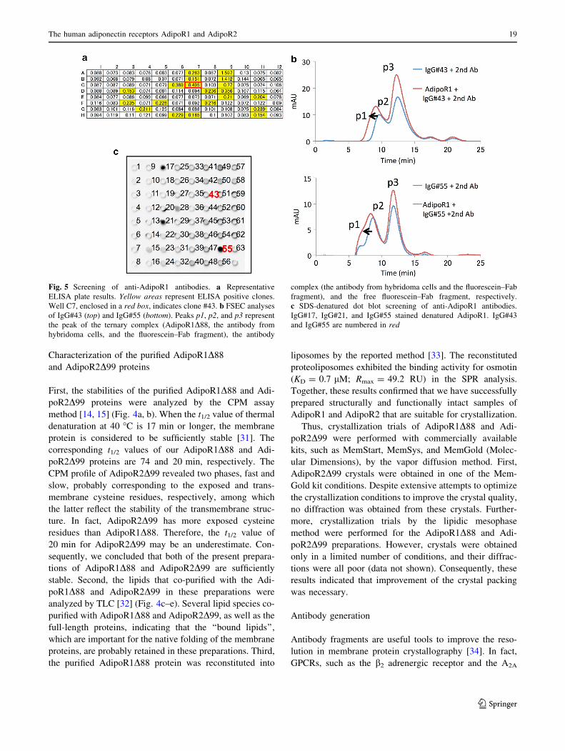
Fig. 5 Screening of anti-AdipoR1 antibodies. a Representative
ELISA plate results. Yellow areas represent ELISA positive clones.
Well C7, enclosed in a red box, indicates clone #43. b FSEC analyses
of IgG#43 (top) and IgG#55 (bottom). Peaks p1, p2, and p3 represent
the peak of the ternary complex (AdipoR1D88, the antibody from
hybridoma cells, and the fluorescein–Fab fragment), the antibody
complex (the antibody from hybridoma cells and the fluorescein–Fab
fragment), and the free fluorescein–Fab fragment, respectively.
c SDS-denatured dot blot screening of anti-AdipoR1 antibodies.
IgG#17, IgG#21, and IgG#55 stained denatured AdipoR1. IgG#43
and IgG#55 are numbered in red
The human adiponectin receptors AdipoR1 and AdipoR2 19
123
adenosine receptor, were co-crystallized with antibody
fragments [35, 36]. Therefore, we planned to produce a
high affinity and conformational epitope-recognizing anti-
AdipoR1 antibody, to improve the crystal packing of
AdipoR1. The purified untagged AdipoR1D88 was recon-
stituted into liposomes, and the resultant proteoliposomes
were used as the immunogen. Mouse anti-AdipoR1 anti-
bodies were produced by a conventional hybridoma sys-
tem. Proteoliposomes containing the purified AdipoR1D88
and biotinyl-PE were used for screening the antibodies by
ELISA (liposome-ELISA). In the first round of liposome-
ELISA from 960 wells of hybridoma cultures, 72 positive
wells were selected (Fig. 5a). Subsequently, 11 positive
clones were selected in the second round of liposome-
ELISA and FSEC (Fig. 5b). Finally, 2 stable hybridoma
cell lines (clone #43 and clone #55) were established. The
denatured dot blot analysis showed that IgG#43 recognized
the native conformation of AdipoR1, whereas IgG#55
recognized the linear epitope (Fig. 5c). In addition, ELISA
and FSEC analyses revealed that both IgG#43 and IgG#55
cross-reacted with AdipoR2D99 (data not shown). The
cDNAs encoding the VH and VL regions of IgG#43 were
cloned from hybridoma cells, according to the standard
method [18]. The very N-terminal amino acid residues of
the VH and VL fragments were determined by Edman
degradation, and the 15 residue sequences of the N-termini
of the VH and VL regions from clone #43 were determined
as EVLLQQSGPELVKPG and DIQMTQSPASLSASV,
respectively. The base sequences of the cloned cDNAs
were corrected accordingly. The variable region of IgG#43
(Fv43) was synthesized by the E. coli cell-free protein
synthesis method, and purified to homogeneity by column
chromatography. The yield of the purified Fv43 was
0.3 mg per 1 ml cell-free synthesis reaction.
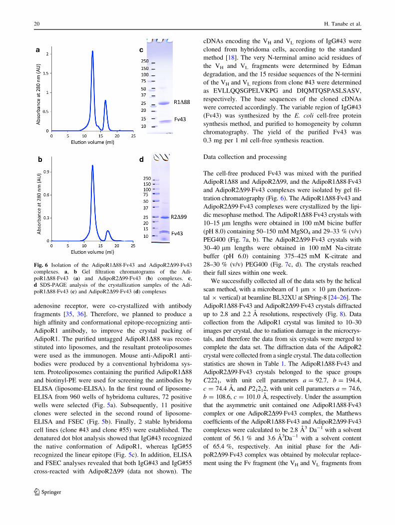
Data collection and processing
The cell-free produced Fv43 was mixed with the purified
AdipoR1D88 and AdipoR2D99, and the AdipoR1D88�Fv43
and AdipoR2D99�Fv43 complexes were isolated by gel fil-
tration chromatography (Fig. 6). The AdipoR1D88�Fv43 and
AdipoR2D99�Fv43 complexes were crystallized by the lipi-
dic mesophase method. The AdipoR1D88�Fv43 crystals with
10–15 lm lengths were obtained in 100 mM bicine buffer
(pH 8.0) containing 50–150 mM MgSO4 and 29–33 % (v/v)
PEG400 (Fig. 7a, b). The AdipoR2D99�Fv43 crystals with
30–40 lm lengths were obtained in 100 mM Na-citrate
buffer (pH 6.0) containing 375–425 mM K-citrate and
28–30 % (v/v) PEG400 (Fig. 7c, d). The crystals reached
their full sizes within one week.
We successfully collected all of the data sets by the helical
scan method, with a microbeam of 1 lm 9 10 lm (horizon-
tal 9 vertical) at beamline BL32XU at SPring-8 [24–26]. The
AdipoR1D88�Fv43 and AdipoR2D99�Fv43 crystals diffracted
up to 2.8 and 2.2 A resolutions, respectively (Fig. 8). Data
collection from the AdipoR1 crystal was limited to 10–30
images per crystal, due to radiation damage in the microcrys-
tals, and therefore the data from six crystals were merged to
complete the data set. The diffraction data of the AdipoR2
crystal were collected from a single crystal. The data collection
statistics are shown in Table 1. The AdipoR1D88�Fv43 and
AdipoR2D99�Fv43 crystals belonged to the space groups
C2221, with unit cell parameters a = 92.7, b = 194.4,
c = 74.4 A, and P21212, with unit cell parameters a = 74.6,
b = 108.6, c = 101.0 A, respectively. Under the assumption
that the asymmetric unit contained one AdipoR1D88�Fv43
complex or one AdipoR2D99�Fv43 complex, the Matthews
coefficients of the AdipoR1D88�Fv43 and AdipoR2D99�Fv43
complexes were calculated to be 2.8 A3 Da-1 with a solvent
content of 56.1 % and 3.6 A3Da-1 with a solvent content
of 65.4 %, respectively. An initial phase for the Adi-
poR2D99�Fv43 complex was obtained by molecular replace-
ment using the Fv fragment (the VH and VL fragments from
Fig. 6 Isolation of the AdipoR1D88�Fv43 and AdipoR2D99�Fv43
complexes. a, b Gel filtration chromatograms of the Adi-
poR1D88�Fv43 (a) and AdipoR2D99�Fv43 (b) complexes. c,
d SDS-PAGE analysis of the crystallization samples of the Adi-
poR1D88�Fv43 (c) and AdipoR2D99�Fv43 (d) complexes
20 H. Tanabe et al.
123
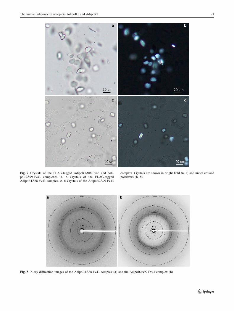
Fig. 7 Crystals of the FLAG-tagged AdipoR1D88�Fv43 and Adi-
poR2D99�Fv43 complexes. a, b Crystals of the FLAG-tagged
AdipoR1D88�Fv43 complex. c, d Crystals of the AdipoR2D99�Fv43
complex. Crystals are shown in bright field (a, c) and under crossed
polarizers (b, d)
Fig. 8 X-ray diffraction images of the AdipoR1D88�Fv43 complex (a) and the AdipoR2D99�Fv43 complex (b)
The human adiponectin receptors AdipoR1 and AdipoR2 21
123

PDB IDs 1E6J and 1FDL, respectively) in Phaser [37] as search
models. The refinement is in progress.
Acknowledgments This work was supported by grants from the
Targeted Proteins Research Program (S.Y., T.K., S.I., and M.Y.) and
the Platform for Drug Discovery, Informatics, and Structural Life
Science (S.Y. and M.Y.) from the Ministry of Education, Culture,
Sports, Science and Technology of Japan, and by the research
acceleration program of the Japan Science and Technology Agency
(S.I.). We thank M. Toyama, M. Inoue, M. Goto, M. Aoki, and K.
Ishii for expression plasmid preparation, M. Nishimoto, Y. Tomabe-
chi, and Y. Terazawa for technical assistance with protein expression
and purification, and R. Akasaka for protein analysis. The synchrotron
radiation experiments were performed on BL32XU at SPring-8
(Proposal Nos. 2012A1332, 2012B1453, 2013B1034, 2014A1008,
and 2014A1186) with the approval of RIKEN. We are also grateful to
the staffs of I24 (Diamond Light Source) and X06SA (Swiss Light
Source) for assistance with data collection.
Open Access This article is distributed under the terms of the
Creative Commons Attribution License which permits any use, dis-
tribution, and reproduction in any medium, provided the original
author(s) and the source are credited.
References
1. Scherer PE, Williams S, Fogliano M, Baldini G, Lodish HF
(1995) A novel serum protein similar to C1q, produced exclu-
sively in adipocytes. J Biol Chem 270(45):26746–26749
2. Hu E, Liang P, Spiegelman BM (1996) AdipoQ is a novel adi-
pose-specific gene dysregulated in obesity. J Biol Chem 271
(18):10697–10703
3. Maeda K, Okubo K, Shimomura I, Funahashi T, Matsuzawa Y,
Matsubara K (1996) cDNA cloning and expression of a novel
adipose specific collagen-like factor, apM1 (AdiPose Most
abundant Gene transcript 1). Biochem Biophys Res Commun
221(2):286–289
4. Nakano Y, Tobe T, Choi-Miura NH, Mazda T, Tomita M (1996)
Isolation and characterization of GBP28, a novel gelatin-binding
protein purified from human plasma. J Biochem 120(4):803–812
5. Hotta K, Funahashi T, Arita Y, Takahashi M, Matsuda M,
Okamoto Y, Iwahashi H, Kuriyama H, Ouchi N, Maeda K,
Nishida M, Kihara S, Sakai N, Nakajima T, Hasegawa K, Mur-
aguchi M, Ohmoto Y, Nakamura T, Yamashita S, Hanafusa T,
Matsuzawa Y (2000) Plasma concentrations of a novel, adipose-
specific protein, adiponectin, in type 2 diabetic patients. Arte-
rioscler Thromb Vasc Biol 20(6):1595–1599
6. Yamauchi T, Kamon J, Ito Y, Tsuchida A, Yokomizo T, Kita S,
Sugiyama T, Miyagishi M, Hara K, Tsunoda M, Murakami K,
Ohteki T, Uchida S, Takekawa S, Waki H, Tsuno NH, Shibata Y,
Terauchi Y, Froguel P, Tobe K, Koyasu S, Taira K, Kitamura T,
Shimizu T, Nagai R, Kadowaki T (2003) Cloning of adiponectin
receptors that mediate antidiabetic metabolic effects. Nature
423(6941):762–769
7. Tomas E, Tsao TS, Saha AK, Murrey HE, Zhang CC, Itani SI,
Lodish HF, Ruderman NB (2002) Enhanced muscle fat oxidation
and glucose transport by ACRP30 globular domain: acetyl-CoA
carboxylase inhibition and AMP-activated protein kinase acti-
vation. Proc Natl Acad Sci USA 99(25):16309–16313
8. Yamauchi T, Kamon J, Minokoshi Y, Ito Y, Waki H, Uchida S,
Yamashita S, Noda M, Kita S, Ueki K, Eto K, Akanuma Y,
Froguel P, Foufelle F, Ferre P, Carling D, Kimura S, Nagai R,
Kahn BB, Kadowaki T (2002) Adiponectin stimulates glucose
utilization and fatty-acid oxidation by activating AMP-activated
protein kinase. Nat Med 8(11):1288–1295
9. Kahn BB, Alquier T, Carling D, Hardie DG (2005) AMP-acti-
vated protein kinase: ancient energy gauge provides clues to
modern understanding of metabolism. Cell Metab 1(1):15–25
10. Iwabu M, Yamauchi T, Okada-Iwabu M, Sato K, Nakagawa T,
Funata M, Yamaguchi M, Namiki S, Nakayama R, Tabata M,
Ogata H, Kubota N, Takamoto I, Hayashi YK, Yamauchi N,
Waki H, Fukayama M, Nishino I, Tokuyama K, Ueki K, Oike Y,
Ishii S, Hirose K, Shimizu T, Touhara K, Kadowaki T (2010)
Adiponectin and AdipoR1 regulate PGC-1a and mitochondria by
Ca2? and AMPK/SIRT1. Nature 464(7293):1313–1319
11. Kersten S, Desvergne B, Wahli W (2000) Roles of PPARs in
health and disease. Nature 405(6785):421–424
12. Yamauchi T, Kamon J, Waki H, Imai Y, Shimozawa N, Hioki K,
Uchida S, Ito Y, Takakuwa K, Matsui J, Takata M, Eto K, Ter-
auchi Y, Komeda K, Tsunoda M, Murakami K, Ohnishi Y, Na-
itoh T, Yamamura K, Ueyama Y, Froguel P, Kimura S, Nagai R,
Kadowaki T (2003) Globular adiponectin protected ob/ob mice
from diabetes and ApoE-deficient mice from atherosclerosis.
J Biol Chem 278(4):2461–2468
13. Wess J (1997) G-protein-coupled receptors: molecular mecha-
nisms involved in receptor activation and selectivity of G-protein
recognition. FASEB J 11(5):346–354
14. Alexandrov AI, Mileni M, Chien EY, Hanson MA, Stevens RC
(2008) Microscale fluorescent thermal stability assay for mem-
brane proteins. Structure 16(3):351–359
15. Hanson MA, Cherezov V, Griffith MT, Roth CB, Jaakola VP,
Chien EY, Velasquez J, Kuhn P, Stevens RC (2008) A specific
cholesterol binding site is established by the 2.8 A structure of
the human b2-adrenergic receptor. Structure 16(6):897–905
16. Kohler G, Milstein C (1975) Continuous cultures of fused cells
secreting antibody of predefined specificity. Nature 256(5517):
495–497
Table 1 Data collection statistics
Structure AdipoR1D88�Fv43
complex
AdipoR2D99�Fv43
complex
No. of crystals 6 1
X-ray source BL32XU, SPring-8 BL32XU, SPring-8
Wavelength (A) 1 1
Space group C2221 P21212
Cell dimensions
a, b, c (A) 92.7, 194.4, 74.4 74.6, 108.6, 101.0
a, b, c (�) 90.0, 90.0, 90.0 90.0, 90.0, 90.0
No. of reflections
measured
135111 145165
No. of unique
reflections
16509 32174
Resolution (A) 20.00–2.80
(2.90–2.80)
19.52–2.40
(2.49–2.40)
Rmerge 0.176 ([1) 0.115 (1.297)
Mean I/r(I) 8.5 (1.3) 8.6 (1.2)
Completeness (%) 97.2 (99.1) 98.3 (99.4)
Redundancy 8.2 (8.0) 4.5 (4.5)
VM (A3 Da-1) 2.8 3.6
Solvent content (%) 56.1 65.4
Values in parentheses are for the outer shell
22 H. Tanabe et al.
123

17. Hino T, Iwata S, Murata T (2013) Generation of functional
antibodies for mammalian membrane protein crystallography.
Curr Opin Struct Biol 23(4):563–568
18. Toleikis L, Broders O, Dubel S (2004) Cloning single-chain
antibody fragments (scFv) from hybridoma cells. Methods Mol
Med 94:447–458
19. Yabuki T, Motoda Y, Hanada K, Nunokawa E, Saito M, Seki E,
Inoue M, Kigawa T, Yokoyama S (2007) A robust two-step PCR
method of template DNA production for high-throughput cell-
free protein synthesis. J Struct Funct Genomics 8(4):173–191
20. Kigawa T, Yabuki T, Matsuda N, Matsuda T, Nakajima R, Ta-
naka A, Yokoyama S (2004) Preparation of Escherichia coli cell
extract for highly productive cell-free protein expression. J Struct
Funct Genomics 5(1–2):63–68
21. Matsuda T, Watanabe S, Kigawa T (2013) Cell-free synthesis
system suitable for disulfide-containing proteins. Biochem Bio-
phys Res Commun 431(2):296–301
22. Hato M, Yamashita J, Shiono M (2009) Aqueous phase behavior
of lipids with isoprenoid type hydrophobic chains. J Phys Chem
B 113(30):10196–10209
23. Hato M, Hosaka T, Tanabe H, Kitsunai T, Yokoyama S (2014) A
new manual dispensing system for in meso membrane protein
crystallization with using a stepping motor-based dispenser.
J Struct Funct Genomics 15(3):165–171
24. Ueno G, Kanda H, Kumasaka T, Yamamoto M (2005) Beamline
Scheduling Software: administration software for automatic
operation of the RIKEN structural genomics beamlines at SPring-
8. J Synchrotron Radiat 12(Pt 3):380–384
25. Murakami I, Fujii T, Kameyama K, Iwata T, Saito M, Kubushiro
K, Aoki D (2012) Tumor volume and lymphovascular space
invasion as a prognostic factor in early invasive adenocarcinoma
of the cervix. J Gynecol Oncol 23(3):153–158
26. Hirata K, Kawano Y, Ueno G, Hashimoto K, Murakami H,
Hasegawa K, Hikima T, Kumasaka T, Yamamoto M (2013)
Achievement of protein micro-crystallography at SPring-8
beamline BL32XU. J Phys Conf Ser 425:012002
27. Otwinowski Z, Minor W (1997) Processing of X-ray diffraction data
collected in oscillation mode. Methods Enzymol 276:307–327
28. Kabsch W (2010) Xds. Acta Crystallogr D Biol Crystallogr 66(Pt
2):125–132
29. Buchan DW, Minneci F, Nugent TC, Bryson K, Jones DT (2013)
Scalable web services for the PSIPRED protein analysis work-
bench. Nucleic Acids Res 41(Web Server issue):W349–W357
30. Tusnady GE, Simon I (2001) The HMMTOP transmembrane
topology prediction server. Bioinformatics 17(9):849–850
31. Sonoda Y, Newstead S, Hu NJ, Alguel Y, Nji E, Beis K, Yashiro
S, Lee C, Leung J, Cameron AD, Byrne B, Iwata S, Drew D
(2011) Benchmarking membrane protein detergent stability for
improving throughput of high-resolution X-ray structures.
Structure 19(1):17–25
32. Han X, Cheng H (2005) Characterization and direct quantitation
of cerebroside molecular species from lipid extracts by shotgun
lipidomics. J Lipid Res 46(1):163–175
33. Rigaud JL, Levy D (2003) Reconstitution of membrane proteins
into liposomes. Methods Enzymol 372:65–86
34. Iwata S, Ostermeier C, Ludwig B, Michel H (1995) Structure at
2.8 A resolution of cytochrome c oxidase from Paracoccus
denitrificans. Nature 376(6542):660–669
35. Rasmussen SG, DeVree BT, Zou Y, Kruse AC, Chung KY,
Kobilka TS, Thian FS, Chae PS, Pardon E, Calinski D, Mathiesen
JM, Shah ST, Lyons JA, Caffrey M, Gellman SH, Steyaert J,
Skiniotis G, Weis WI, Sunahara RK, Kobilka BK (2011) Crystal
structure of the b2 adrenergic receptor-Gs protein complex.
Nature 477(7366):549–555
36. Hino T, Arakawa T, Iwanari H, Yurugi-Kobayashi T, Ikeda-Suno
C, Nakada-Nakura Y, Kusano-Arai O, Weyand S, Shimamura T,
Nomura N, Cameron AD, Kobayashi T, Hamakubo T, Iwata S,
Murata T (2012) G-protein-coupled receptor inactivation by an
allosteric inverse-agonist antibody. Nature 482(7384):237–240
37. McCoy AJ, Grosse-Kunstleve RW, Adams PD, Winn MD, Sto-
roni LC, Read RJ (2007) Phaser crystallographic software. J Appl
Crystallogr 40(Pt 4):658–674
38. Larkin MA, Blackshields G, Brown NP, Chenna R, McGettigan
PA, McWilliam H, Valentin F, Wallace IM, Wilm A, Lopez R,
Thompson JD, Gibson TJ, Higgins DG (2007) Clustal W and
Clustal X version 2.0. Bioinformatics 23(21):2947–2948
The human adiponectin receptors AdipoR1 and AdipoR2 23
123