Rab5-mediated endocytosis of activin is not required for gene ...
Upload
takashi-yamamotoCategory
view
212download
0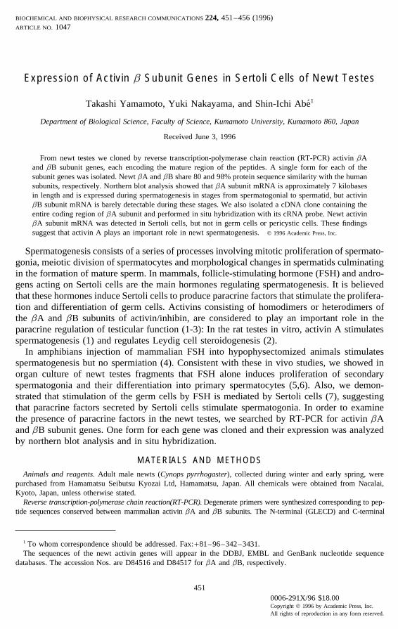
BIOCHEMICAL AND BIOPHYSICAL RESEARCH COMMUNICATIONS 224, 451–456 (1996)ARTICLE NO. 1047
Expression of Activin b Subunit Genes in Sertoli Cells of Newt Testes
Takashi Yamamoto, Yuki Nakayama, and Shin-Ichi Abe1
Department of Biological Science, Faculty of Science, Kumamoto University, Kumamoto 860, Japan
Received June 3, 1996
From newt testes we cloned by reverse transcription-polymerase chain reaction (RT-PCR) activin bAand bB subunit genes, each encoding the mature region of the peptides. A single form for each of thesubunit genes was isolated. Newt bA and bB share 80 and 98% protein sequence similarity with the humansubunits, respectively. Northern blot analysis showed that bA subunit mRNA is approximately 7 kilobasesin length and is expressed during spermatogenesis in stages from spermatogonial to spermatid, but activinbB subunit mRNA is barely detectable during these stages. We also isolated a cDNA clone containing theentire coding region of bA subunit and performed in situ hybridization with its cRNA probe. Newt activinbA subunit mRNA was detected in Sertoli cells, but not in germ cells or pericystic cells. These findingssuggest that activin A plays an important role in newt spermatogenesis. q 1996 Academic Press, Inc.
Spermatogenesis consists of a series of processes involving mitotic proliferation of spermato-gonia, meiotic division of spermatocytes and morphological changes in spermatids culminatingin the formation of mature sperm. In mammals, follicle-stimulating hormone (FSH) and andro-gens acting on Sertoli cells are the main hormones regulating spermatogenesis. It is believedthat these hormones induce Sertoli cells to produce paracrine factors that stimulate the prolifera-tion and differentiation of germ cells. Activins consisting of homodimers or heterodimers ofthe bA and bB subunits of activin/inhibin, are considered to play an important role in theparacrine regulation of testicular function (1-3): In the rat testes in vitro, activin A stimulatesspermatogenesis (1) and regulates Leydig cell steroidogenesis (2).
In amphibians injection of mammalian FSH into hypophysectomized animals stimulatesspermatogenesis but no spermiation (4). Consistent with these in vivo studies, we showed inorgan culture of newt testes fragments that FSH alone induces proliferation of secondaryspermatogonia and their differentiation into primary spermatocytes (5,6). Also, we demon-strated that stimulation of the germ cells by FSH is mediated by Sertoli cells (7), suggestingthat paracrine factors secreted by Sertoli cells stimulate spermatogonia. In order to examinethe presence of paracrine factors in the newt testes, we searched by RT-PCR for activin bAand bB subunit genes. One form for each gene was cloned and their expression was analyzedby northern blot analysis and in situ hybridization.
MATERIALS AND METHODSAnimals and reagents. Adult male newts (Cynops pyrrhogaster), collected during winter and early spring, were
purchased from Hamamatsu Seibutsu Kyozai Ltd, Hamamatsu, Japan. All chemicals were obtained from Nacalai,Kyoto, Japan, unless otherwise stated.
Reverse transcription-polymerase chain reaction(RT-PCR). Degenerate primers were synthesized corresponding to pep-tide sequences conserved between mammalian activin bA and bB subunits. The N-terminal (GLECD) and C-terminal
1 To whom correspondence should be addressed. Fax:/81–96–342–3431.The sequences of the newt activin genes will appear in the DDBJ, EMBL and GenBank nucleotide sequence
databases. The accession Nos. are D84516 and D84517 for bA and bB, respectively.
0006-291X/96 $18.00Copyright q 1996 by Academic Press, Inc.All rights of reproduction in any form reserved.
451
AID BBRC 5013 / 6903$$$321 06-25-96 12:16:26 bbrcas AP: BBRC
Vol. 224, No. 2, 1996 BIOCHEMICAL AND BIOPHYSICAL RESEARCH COMMUNICATIONS
(EECGC) region of mature subunits were selected according to the procedure of Thomsen et al.(8). A BamHI restrictionsite was introduced at the 5* end of N-terminal sense strand primer (5*-CGGATCCGG(AGCT)(CT)T(AGCT)GA(AG)TG-(CT) GA-3*) and an EcoRI site was introduced at the 5* end of the C-terminal antisense strand primer(5*-CGAATTC(AG)-CA(AGCT)CC(AG)CA(CT)TC (CT)TC-3*). Using oligo(dT)-primed cDNAs prepared from testes total RNA as a template,PCR amplification was performed (40 cycles at 947C for 30 sec, at 557C for 30 sec, and at 707C for 5min), and the PCRproducts were analyzed on 2% agarose gel. PCR products were treated with both BamHI and EcoRI, and then subclonedinto pBluescript II SK (0) (U.S. and Biochemical Corp.).
DNA sequencing. Cloned DNA was sequenced by ABI PRISM dye primer cycle sequencing ready reaction kit(PERKIN ELMER) using DNA sequencer (model ABI 373S, Applied Biosystems, USA). All sequence data wereobtained for both strands.
Northern blot analysis. Total RNA was prepared from testes fragments derived from various stages of spermatogene-sis. Northern blot analysis was done using 20 mg of total RNA. PCR products for bA and bB subunits representingthe mature region of the peptides were used as probes.
cDNA cloning. A newt activin bA cDNA was obtained by screening spermatogonial cDNA library (9) with the360bp PCR product of bA cDNA labeled with [a-32P]dCTP by Megaprime DNA labeling system (Amersham). Onepositive clone was isolated and insert DNA was subcloned into the EcoRI site of pBluescript II SK (0).
In situ hybridization. Testes fragments containing spermatogonia and primary spermatocytes were fixed in Bouin’ssolution at 47C for 12 h. Then, fragments were dehydrated in methanol at 0207C before use. After hydrating, fragmentswere treated with 10 mg/ml proteinase K in PBS and then postfixed in 4% paraformaldehyde in PBS. After treatmentwith triethanolamine with acetic acid, fragments were hybridized with a digoxigenin-labeled probe. Digoxigenin-labeled sense and antisense RNA probes were prepared with the T3 and T7 MEGAscript kit (Ambion) with digoxigenin-UTP (Boehringer, Mannheim) from the activin bA cDNA fragment (position 965-1411) subcloned into pBluescriptII SK(0) (U.S. and Biochemical Corp.). Hybridization was performed in 50% formamide, 5 1 SSC, 1 1 Denhardt’s,1mg/ml digoxigenin-labeled RNA probe, 20 mg/ml tRNA, 0.25% SDS, 10% dextran sulfate at 557C for 72 h in ahumid chamber. After washing twice with 2 1 SSC, 50% formamide at 607C for 1 h, the fragments were washedtwice in 1 1 SSC at 457C for 1 h. The fragments were dehydrated, embedded in paraffin and sectioned at 8 mm.After deparaffinizing with xylene, the sections were blocked by 1% BSA in PBS. The hybridized RNA probes in thesection were detected using digoxigenin nucleic acid detection kit (Boehringer, Mannheim).
FIG. 1. Comparison of amino acid sequence of activin b subunits. (A) A comparison of amino acid sequence ofactivin bA subunits deduced from the nucleotide sequences. The amino acid sequences of human (11), rat (12),Xenopus (8) and newt bA subunits are aligned. (B) A comparison of amino acid sequences of activin bB subunits.The amino acid sequences of human, rat, Xenopus (13) and newt bB subunits are aligned. The pluses (/) denotesequences derived from PCR primers.
452
AID BBRC 5013 / 6903$$$321 06-25-96 12:16:26 bbrcas AP: BBRC

Vol. 224, No. 2, 1996 BIOCHEMICAL AND BIOPHYSICAL RESEARCH COMMUNICATIONS
FIG. 2. Northern blot analysis of activin b subunits during spermatogenesis. Expression of (A) bA and (B) bBsubunit mRNAs during spermatogenesis. Total RNA (20mg) from testes fragments rich in secondary spermatogonia(SG), primary spermatocytes (PC) and round spermatids(RT) were loaded in each lane. The arrow indicates theposition of 7 kb newt activin bA subunit mRNA. The ethidium bromide-stained patterns of rRNAs indicate that thetotal RNA loaded in each lane was approximately equal.
RESULTS
In order to search for activin b subunit mRNAs in the newt testis, we performed RT-PCRwith degenerate primers based on sequences in the conserved N-terminal and C-terminal regionof mature activin proteins. Using newt testes cDNA as a template, we isolated a single productof approximately 360bp (data not shown). Ten clones of PCR products were sequenced, anda single form corresponding to the mature region of bA and bB subunits was isolated. Exclud-ing the N- and C-terminal regions used as PCR primers, the deduced amino acid sequence ofnewt activin bA subunit shares 80% and 85% sequence similarity to human and Xenopus bAsubunit, respectively (Fig.1A). The newt bB sequence shows even greater similarity to thehuman and Xenopus bB subunits - 97% and 99%, respectively (Fig.1B). Also, the distributionof Cys residues is conserved in the newt b subunits as has been observed in the TGF-bsuperfamily. These results indicate that both activin bA and bB subunit mRNAs exist inthe newt testes and share a high level of similarity with both the mammalian and Xenopuscounterparts.
To examine the expression of activin b subunit mRNAs during newt spermatogenesis,Northern blot analysis was performed with total RNA derived from various spermatogenicstages. A single species of bA subunit mRNA of approximately 7 kilobases was detected instages from spermatogonia to round spermatid (Fig.2A), but bB subunit mRNA was barelydetectable(Fig.2B).
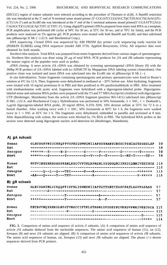
Using a PCR product as a hybridization probe, we isolated from a testes cDNA library acDNA clone containing the entire coding region of activin bA subunit. This clone was 2129bpin length and encoded 413-amino acid residues; there was a 15-amino acid signal peptide atthe N-terminal region (Fig.3). The putative cleavage site of newt activin bA is pentabasic andgenerates a mature protein of 116 amino acids. These findings indicate that the newt activin
453
AID BBRC 5013 / 6903$$$321 06-25-96 12:16:26 bbrcas AP: BBRC
Vol. 224, No. 2, 1996 BIOCHEMICAL AND BIOPHYSICAL RESEARCH COMMUNICATIONS
454
AID BBRC 5013 / 6903$$5013 06-25-96 12:16:26 bbrcas AP: BBRC
Vol. 224, No. 2, 1996 BIOCHEMICAL AND BIOPHYSICAL RESEARCH COMMUNICATIONS
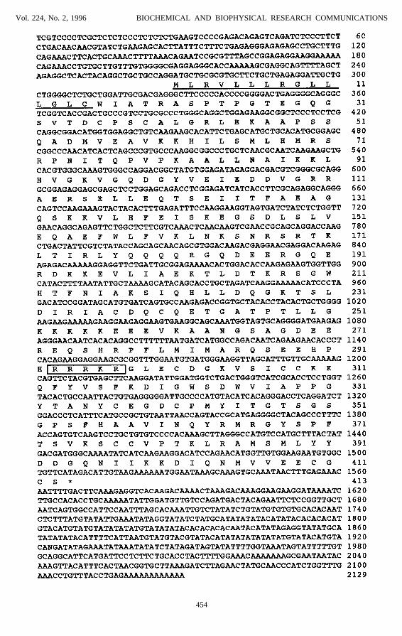
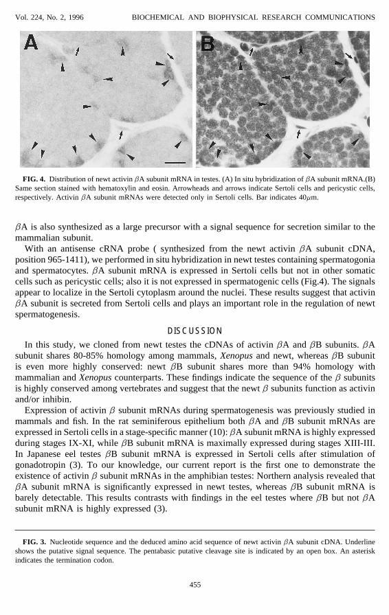
FIG. 4. Distribution of newt activin bA subunit mRNA in testes. (A) In situ hybridization of bA subunit mRNA.(B)Same section stained with hematoxylin and eosin. Arrowheads and arrows indicate Sertoli cells and pericystic cells,respectively. Activin bA subunit mRNAs were detected only in Sertoli cells. Bar indicates 40mm.
bA is also synthesized as a large precursor with a signal sequence for secretion similar to themammalian subunit.
With an antisense cRNA probe ( synthesized from the newt activin bA subunit cDNA,position 965-1411), we performed in situ hybridization in newt testes containing spermatogoniaand spermatocytes. bA subunit mRNA is expressed in Sertoli cells but not in other somaticcells such as pericystic cells; also it is not expressed in spermatogenic cells (Fig.4). The signalsappear to localize in the Sertoli cytoplasm around the nuclei. These results suggest that activinbA subunit is secreted from Sertoli cells and plays an important role in the regulation of newtspermatogenesis.
DISCUSSION
In this study, we cloned from newt testes the cDNAs of activin bA and bB subunits. bAsubunit shares 80-85% homology among mammals, Xenopus and newt, whereas bB subunitis even more highly conserved: newt bB subunit shares more than 94% homology withmammalian and Xenopus counterparts. These findings indicate the sequence of the b subunitsis highly conserved among vertebrates and suggest that the newt b subunits function as activinand/or inhibin.
Expression of activin b subunit mRNAs during spermatogenesis was previously studied inmammals and fish. In the rat seminiferous epithelium both bA and bB subunit mRNAs areexpressed in Sertoli cells in a stage-specific manner (10): bA subunit mRNA is highly expressedduring stages IX-XI, while bB subunit mRNA is maximally expressed during stages XIII-III.In Japanese eel testes bB subunit mRNA is expressed in Sertoli cells after stimulation ofgonadotropin (3). To our knowledge, our current report is the first one to demonstrate theexistence of activin b subunit mRNAs in the amphibian testes: Northern analysis revealed thatbA subunit mRNA is significantly expressed in newt testes, whereas bB subunit mRNA isbarely detectable. This results contrasts with findings in the eel testes where bB but not bAsubunit mRNA is highly expressed (3).
FIG. 3. Nucleotide sequence and the deduced amino acid sequence of newt activin bA subunit cDNA. Underlineshows the putative signal sequence. The pentabasic putative cleavage site is indicated by an open box. An asteriskindicates the termination codon.
455
AID BBRC 5013 / 6903$$$321 06-25-96 12:16:26 bbrcas AP: BBRC

Vol. 224, No. 2, 1996 BIOCHEMICAL AND BIOPHYSICAL RESEARCH COMMUNICATIONS
Our in situ hybridization studies showed that bA subunit mRNA is synthesized in Sertolicells, but not in other somatic cells nor in germ cells, an observation similar to that reportedfor rat and eel testes(3,10). Because the newt bA subunit is synthesized only in Sertoli cellsand as a precursor with a signal peptide, we suggest that the bA subunit is secreted fromSertoli cells and then is involved in the regulation of newt spermatogenesis. For example inthe Japanese eel testes, the expression of activin bB mRNA is accompanied by spermatogonialproliferation (3). Currently, we are examining in vitro the function of activin A in the newttestes in order to understand its role during newt spermatogenesis.
ACKNOWLEDGMENTSWe thank Professor Marie A. DiBerardino for editing the manuscript. We also express our appreciation to Dr.
Nobuhiko Mizuno for technical advice. This work was supported by Grants-in-Aid for scientific research (05454654)and priority areas (07262213) from the Ministry of Education, Science, Sports and Culture of Japan and a grant fromMitsubishi Science Foundation.
REFERENCES1. Lin, T., Calkins, H., Morris, P. L., Vale, W. W., and Bardin, C. W. (1989) Endocrinology 125, 2134–2140.2. Mather, J. P., Attie, K. M., Woodruff, T. K., Rice, G. C., and Phillips, D. M. (1990) Endocrinology 127, 3206–
3214.3. Nagahama, Y., Miura, T., and Kobayashi, T. (1994) Ciba Found. Symp. 182, 255–267.4. Callard, I. P., Callard, G. V., Lance, V., Bolaffi, J. L., and Rosset, J. S. (1978) Biol.Reprod. 18, 16–43.5. Ji, Z.-S., Kubokawa, K., Ishii, S., and Abe, S.-I. (1992) Dev. Growth Differ. 34, 649–660.6. Abe, S.-I., and Ji, Z.-S. (1994) Int. J. Dev. Biol. 38, 201–208.7. Maekawa, K., Ji, Z.-S., K., and Abe, S.-I. (1995) J. Exp. Zool. 272, 363–373.8. Thomsen, G., Woolf, T., Whitman, M., Sokol, S., Vaughan, J., Vale, W., and Melton, D. A. (1990) Cell 63, 485–
493.9. Yamamoto, T., Hikino, T., and Abe, S.-I. (1996) Roux’s Arch. Dev. Biol., in press.
10. Kaipia, A., Penttila, T. L., Shimasaki, S., Ling, N., Parvinen, M., and Toppari, J. (1992) Endocrinology 131,2703–2710.
11. Mason, A. J., Niall, H. D., and Seeburg, P. H. (1986) Biochem. Biophys. Res. Commun. 135, 957–964.12. Esch, F. S., Shimasaki, S., Cooksey, K., Mercado, M., Mason, A. J., Ying, S-Y., Ueno, N., and Ling, N. (1987)
Mol. Endocrinol. 1, 387–396.13. Dohrmann, C. E., Hemmati-Brivanlou, A., Thomsen, G. H., Fields, A., Woolf, T., and Melton, D. A. (1993) Dev.
Biol. 157, 474–483.
456
AID BBRC 5013 / 6903$$$321 06-25-96 12:16:26 bbrcas AP: BBRC