Evaluation of antioxidant, anti-hemolytic and anticancer ...
16
RESEARCH ARTICLE Open Access Evaluation of antioxidant, anti-hemolytic and anticancer activity of various solvent extracts of Acacia hydaspica R. Parker aerial parts Tayyaba Afsar 1 , Suhail Razak 2,3* , Muhammad Rashid Khan 1 , Saadia Mawash 1 , Ali Almajwal 3 , Maria Shabir 1 and Ihsan Ul Haq 4 Abstract Background: Acacia hydaspica R. Parker, family leguminosae, is a medicinally important plant. Different plant parts are used in various ailments in folk medicine. The current study aimed at investigating the in vitro antioxidant, anti-hemolytic and anticancer activity of A. hydaspica. Methods: Antioxidant potential was assessed using DPPH, ABTS and •OH, scavenging of H 2 O 2 , inhibition of lipid peroxidation and β-carotene bleaching inhibition assays. Anti-hemolytic activity was assessed using H 2 O 2 induced hemolysis of RBCs. Anticancer potential was assessed using MTT assay. Spectrometric methods and HPLC-DAD analysis was performed for phytochemical screening. Results: EC 50 values based on reduction of DPPH, ABTS and •OH, scavenging of H 2 O 2 , inhibition of lipid peroxidation and β-carotene bleaching for AHB, AHE and AHM were generally lower manifesting potential antiradical capacities. The fractions also exhibited significant (P <0.001) anti-hemolytic potential. Regarding IC 50 values for anticancer activity against HCC-38 and MDA-MB-361 cancer cell lines; AHB, AHE and AHM exhibited significant (P <0.001) cyto-selection indices. Plant extracts showed no cytotoxicity against normal Vero cells (IC 50 > 250 μg/ml). While significant (P <0.001) cytotoxicity was elicited by these extract/fractions against cancer cell lines. AHE was the most effective and IC 50 was found to be 29.9 ± 0.909 μg/ml (SI = 9.83) and 39.5 ± 0.872 μg/ml (SI = 7.44) against MDA-MB-361 and HCC-38 cancer cells respectively. Higher amounts of TPC and TFC were exhibited by AHE and AHB as compared to other fractions. Gallic acid, catechin and myricetin were identified in AHE whereas gallic acid and catechin were identified in AHB by HPLC. Conclusion: The presence of bioactive constituents in AHE and AHB might be responsible for antioxidant, anti-hemolytic and anticancer activities. Keywords: Cytotoxic activity, Antioxidant activity, Phenolic, Flavonoids, Anti-lipid peroxidation * Correspondence: [email protected] 2 Department of Animal Sciences, Faculty of Biological Sciences, Quaid-i-Azam University, Islamabad, Pakistan 3 Department of Community Health Sciences, College of Applied Medical Sciences, King Saud University, Riyadh, Kingdom of Saudi Arabia Full list of author information is available at the end of the article © 2016 The Author(s). Open Access This article is distributed under the terms of the Creative Commons Attribution 4.0 International License (http://creativecommons.org/licenses/by/4.0/), which permits unrestricted use, distribution, and reproduction in any medium, provided you give appropriate credit to the original author(s) and the source, provide a link to the Creative Commons license, and indicate if changes were made. The Creative Commons Public Domain Dedication waiver (http://creativecommons.org/publicdomain/zero/1.0/) applies to the data made available in this article, unless otherwise stated. Afsar et al. BMC Complementary and Alternative Medicine (2016) 16:258 DOI 10.1186/s12906-016-1240-8
Transcript of Evaluation of antioxidant, anti-hemolytic and anticancer ...
RESEARCH ARTICLE Open Access
Evaluation of antioxidant, anti-hemolyticand anticancer activity of various solventextracts of Acacia hydaspica R. Parker aerialpartsTayyaba Afsar1, Suhail Razak2,3*, Muhammad Rashid Khan1, Saadia Mawash1, Ali Almajwal3, Maria Shabir1
and Ihsan Ul Haq4
Abstract
Background: Acacia hydaspica R. Parker, family leguminosae, is a medicinally important plant. Different plantparts are used in various ailments in folk medicine. The current study aimed at investigating the in vitroantioxidant, anti-hemolytic and anticancer activity of A. hydaspica.
Methods: Antioxidant potential was assessed using DPPH, ABTS and •OH, scavenging of H2O2, inhibition oflipid peroxidation and β-carotene bleaching inhibition assays. Anti-hemolytic activity was assessed using H2O2
induced hemolysis of RBCs. Anticancer potential was assessed using MTT assay. Spectrometric methods andHPLC-DAD analysis was performed for phytochemical screening.
Results: EC50 values based on reduction of DPPH, ABTS and •OH, scavenging of H2O2, inhibition of lipidperoxidation and β-carotene bleaching for AHB, AHE and AHM were generally lower manifesting potentialantiradical capacities. The fractions also exhibited significant (P <0.001) anti-hemolytic potential. RegardingIC50 values for anticancer activity against HCC-38 and MDA-MB-361 cancer cell lines; AHB, AHE and AHMexhibited significant (P <0.001) cyto-selection indices. Plant extracts showed no cytotoxicity against normalVero cells (IC50 > 250 μg/ml). While significant (P <0.001) cytotoxicity was elicited by these extract/fractionsagainst cancer cell lines. AHE was the most effective and IC50 was found to be 29.9 ± 0.909 μg/ml (SI = 9.83)and 39.5 ± 0.872 μg/ml (SI = 7.44) against MDA-MB-361 and HCC-38 cancer cells respectively. Higher amountsof TPC and TFC were exhibited by AHE and AHB as compared to other fractions. Gallic acid, catechin andmyricetin were identified in AHE whereas gallic acid and catechin were identified in AHB by HPLC.
Conclusion: The presence of bioactive constituents in AHE and AHB might be responsible for antioxidant,anti-hemolytic and anticancer activities.
Keywords: Cytotoxic activity, Antioxidant activity, Phenolic, Flavonoids, Anti-lipid peroxidation
* Correspondence: [email protected] of Animal Sciences, Faculty of Biological Sciences, Quaid-i-AzamUniversity, Islamabad, Pakistan3Department of Community Health Sciences, College of Applied MedicalSciences, King Saud University, Riyadh, Kingdom of Saudi ArabiaFull list of author information is available at the end of the article
© 2016 The Author(s). Open Access This article is distributed under the terms of the Creative Commons Attribution 4.0International License (http://creativecommons.org/licenses/by/4.0/), which permits unrestricted use, distribution, andreproduction in any medium, provided you give appropriate credit to the original author(s) and the source, provide a link tothe Creative Commons license, and indicate if changes were made. The Creative Commons Public Domain Dedication waiver(http://creativecommons.org/publicdomain/zero/1.0/) applies to the data made available in this article, unless otherwise stated.
Afsar et al. BMC Complementary and Alternative Medicine (2016) 16:258 DOI 10.1186/s12906-016-1240-8
BackgroundCancer is a prominent reason of death in many devel-oped and developing countries. Although the etiologiesof cancer are varied, oxidative stress plays a major rolefor the pathophysiological developments. Oxidativestress forced by free radicals such as singlet oxygenspecies, superoxide, hydroxyl and ferrous may causelipid peroxidation, protein damages, inflammation,autoimmune pathologies, DNA damage, altering cell-signaling pathways and modulating gene expression,induction and promotion of tumor [1, 2]. Syntheticantioxidants, such as butylated hydroxyanisole (BHA)and butylated hydroxytoluene (BHT) are widely usedin the food industry because they are effective andless expensive than natural antioxidants. Syntheticantioxidants gained safety concerns and have beenrestricted due to their DNA damaging and other toxiceffects [3]. Complementary and alternative medicineis one of the emerging fields in health care today,especially as supportive medicine in treating diseaseslike cancer [4]. Plant secondary metabolites such asflavonoids, terpenes, alkaloids, α-tocopherol andcarotenoids have received considerable attention inrecent years due to their diverse pharmacologicalproperties, including cytotoxic and chemo-preventiveeffects. The possible health benefits of polyphenolconsumption have been suggested to derive from theirantioxidant properties [4, 5]. Therefore it is interest-ing to identify selectivity of plant extracts possessingantioxidant potential against cancer and normal cells.In fact, literature has verified an association betweenintake of diet rich in fruits and vegetables with adecline in oxidative stress induced disorders [6].Acacia hydaspica R. Parker synonym A. eburnea, com-
monly known as ‘kikar’ family leguminosae is an eco-nomically important plant. It is a slender deciduousshrub, 1.2–1.8 m tall, twigs glabrous slightly zigzag; barksmooth, dark grey and 1.2–2.5 cm long stipular spines,seeds 1–8, areole not well marked. The bark and seedsare the source of tannin. Leaves serves as fodder forgoats [7, 8]. The bark and seeds are the source of tannins.The plant is locally used as antiseptic. The traditionalhealers of India use various parts of the plant for the treat-ment of diarrhea; the leaves and the bark are useful inarresting secretion or bleeding. The pods are helpful in re-moving catarrhal matter and phlegm from the bronchialtubes. The gum dispels irascibility of the skin and soothesthe inflamed membranes of the pharynx, alimentary canaland genito-urinary organs (http://trade.indiamart.com/details.mp?offer=6763150691). Gallic acid, catechin, rutinand caffeic acid have been identified in A. hydaspica byHPLC-DAD screening of crude methanol extract, while 7-O-galloyl catechin, +catechin and methyl gallate have beenisolated from ethyl acetate fraction of A. hydaspica (AHE).
The A. hydaspica possess anti-inflammatory, antipyreticand analgesic potentials [9]. Polyphenolic compoundsisolated from A. hydaspica induce apoptosis and inhibitcancer cell growth in vitro in breast and prostate cancercells by modulating various signal transduction pathways[10]. Various species of Acacia were investigated for theirantioxidant and anticancer potentials in various animalmodels [11]. The extracts from the bark and heartwood ofAcacia confusa showed significant antioxidant activity invarious antioxidant assays, including free radical andsuperoxide radical scavenging assays and lipid peroxida-tion assay as well as hydroxyl radical-induced DNA strandscission assay [12]. A. mangium and A. auriculiformisheartwood extracts showed excellent quenching abilityagainst DPPH free radicles [13]. The antioxidant activitiesof bark extract of Acacia confusa and some of the isolatedconstituents from its ethyl acetate (EtOAc) fraction invarious in vitro systems together with authentic antioxi-dant standards revealed that EtOAc fraction showedstrong superoxide radical scavenging activity, reducingpower, and ferrous ion-chelating ability. Results obtainedindicated that the bark extracts from A. confusa have agreat potential to prevent disease caused by the overpro-duction of radicals and also it might be used as a potentialsource of natural antioxidant agent [14]. Heartwood ex-tract of Acacia catechu induces apoptosis in human breastcancer cell [15]. However to the best of our knowledgethere is no single scientific report demonstrating the anti-oxidant and cyto-selective anticancer potential of A.hydaspica.In this study qualitative phytochemical screening, total
phenolic content (TPC), total flavonoid content (TFC),antioxidant and anti-hemolytic activities of extract/frac-tions were evaluated. Extracts with potent antioxidantactivity were tested against normal (VERO) and cancercell lines (HCC-38 and MDA-MB-361) in order toevaluate the cyto-selective potential against cancer andnormal cells. Furthermore polyphenol constituents inactive fractions were determined by HPLC-DAD chro-matography using standard reference compounds.
MethodsPlant collectionThe plant was collected from Kirpa village Islamabad,Pakistan. After identification with the help of relevantliterature a voucher specimen was deposited (0642531)at the Herbarium of Pakistan Museum of NaturalHistory, Islamabad.
Preparation of extract/fractionsThe aerial parts (twigs and leaves) of the plant weredried in an aerated but shaded area. Dried material wasground by an electrical grinder to obtain 60 μm powder.The methanol extract was obtained by allowing 3 kg of
Afsar et al. BMC Complementary and Alternative Medicine (2016) 16:258 Page 2 of 16
powder to macerate three times in 95 % methanol (3 ×4000 ml) for five consecutive days. The supernatantswere mixed and filtered. Solvent was evaporated byrotary vacuum evaporator (Buchi, R114, Switzerland).The residue was taken to dryness to obtain a viscousmass as the crude methanol extract (AHM). An amountof 12 g of AHM was suspended in water (250 ml) withcontinuous stirring then successively added (3 × 200 ml)following solvents; n-hexane, ethyl acetate, chloroformand n-butanol respectively, shake well and each layerwas allowed to separate for 3 h in a separating funneland at last water soluble fraction was obtained (AHA).Each of the fractions obtained were dried using a rotaryevaporator. AHM and its five subsequent fractions:AHH, AHE, AHC, AHB and AHA were weighed andexpressed in terms of percentage of air dried weight ofplant material.
ChemicalsAscorbic acid, aluminum chloride, 2,2-azino-bis-(3-ethylbenzothiazoline-6-sulphonic acid) (ABTS), ferricchloride (FeCl3), Tween 80, β-carotene, (+)-catechin,gallic acid, rutin, quercitin, potassium persulphate,Folin-Ciocalteu’s reagent, ferrozine, gallic acid, rutin,linoleic acid, 2,2-diphenyl-1-picrylhydrazyl (DPPH), nitroblue tetrazolium (NBT), linoleic acid, phenazine metho-sulphate (PMS), thiobarbituric acid (TBA) andtrichloroacetic acid (TCA) were purchased from SigmaAldrich (Germany). Deoxyribose, riboflavin, sodium car-bonate (Na2CO3), sodium hydroxide (NaOH), disodiumhydrogen phosphate (Na2HPO4) and hydrogen peroxide(H2O2) were obtained from Wako Co. (Osaka, Japan).Potassium ferricyanide (K3Fe (CN) 6), triflouroaceticacid, sodium dihydrogen phosphate (NaH2PO4) and allsolvents used were of analytical grade and were pur-chased from Sigma Aldrich (Germany).
Preliminary phytochemical screeningThe methanol extract and its soluble fractions were sub-jected to phytochemical analysis by using the methodsdescribed previously for the detection of terpenoids,alkaloids [16], saponins, tannins, flavonoids [17], cardiacglycoside [18], reducing sugars [19], pholobatannins,coumarins and anthraquinones [20] by qualitativemethods.
Estimation of total phenolic content (TPC)The total phenols of AHM and its derived fractions werequantified by previously described spectrophotometricmethod [21]. TPC was calculated from the calibrationcurve of gallic acid. Estimation of TPC was recorded intriplicate and expressed as mg of gallic acid equivalent/gof dry sample.
Estimation of total flavonoid content (TFC)Aluminium chloride colorimetric technique with slightmodifications was used for the estimation of TFC [22].Quantity of TFC was recorded in triplicate from the cali-bration curve of rutin and expressed as mg of rutinequivalent/g of dry sample.
Antioxidant assaysSample preparationEach sample was dissolved in 95 % methanol at a con-centration of 1 mg/ml and diluted to prepare the serial-ized dilutions (10–500 μg/ml) for various antioxidantassays. Reference standard chemicals were used for com-parison in all assays.
Antioxidant activity assessment assaysDPPH radical scavenging activity assayThe DPPH assay was executed following previouslyestablished protocol with slight modifications [23]. Onehundred milliliter methanol (80 %) was added to 24 mgof DPPH to make the stock solution and the stock wasstored at 20 °C till used. For the assay the working solu-tion of DPPH was prepared by diluting the stock withmethanol until an absorbance of 0.751 ± 0.02 at 517 nmwas achieved. An aliquot of 1 ml DPPH solution wasdispensed in 100 μl of the test samples of different con-centrations (0–250 μg/ml). The mixture was shaken andplaced in the dark for 10 min at room temperature. Theabsorbance of the mixture was recorded using a UV-1601 spectrophotometer (Shimadzu, Kyoto, Japan) at517 nm. The decrease in absorbance was correlated withthe radical scavenging potential of test samples. Thepercentage of inhibition was assessed as follow
% DPPH scavenging ¼ Ad− Asd−Asað ÞAd
� �� 100:
Where Ad is the DPPH solution absorbance, Asd isthe absorbance of solution containing test sample andDPPH solution, and Asa is the absorbance sample solu-tion without DPPH. Each sample was analyzed in thrice.As a standard reference compound ascorbic acid was
employed.
Superoxide anion radical quenching assayQuenching potential for superoxide anion was assessedvia riboflavin light-NBT system [24]. 1 ml sample solu-tion (25–250 μg/ml) was poured to the solution com-prised of 50 mM phosphate buffer (500 μl, pH 7.6),50 mM riboflavin (300 μl), 20 mM PMS (250 μl) and0.5 mM NBT (100 μl). Illumination of the solution wasdone by using a fluorescent lamp for initiating the reac-tion. The absorbance of samples was recorded at560 nm after 20 min of illumination. Restraint of
Afsar et al. BMC Complementary and Alternative Medicine (2016) 16:258 Page 3 of 16
superoxide anion liberation was assessed using thefollowing formula:
Inhibitory potentieal %ð Þ ¼ Control abs−sample abscontrol abs
� �� 100
Gallic acid was used as a standard compound.
Hydroxyl radical quenching activityScavenging potential of test samples for the hydroxylradicals was examined using 2-deoxyribose method [25].0.2 M Phosphate buffer saline (PH 7.4) was consumed asa solvent in this test. Sample solution (0–100 μM) wasmixed with test mixture containing 2-deoxyribose(2.8 mM), ferrous ammonium sulphate solution(20 mM), EDTA (100 μM). Total volume of test mixturewas made up to 1 ml with 0.2 M Phosphate buffer saline(PH 7.4). Ferrous ion solution and EDTA were premixedbefore adding to the assay mixture. The reaction wasinitiated by the addition of 100 μl of 20 mM H2O2
and 100 μl of 2 mM ascorbic acid and incubated at37 °C for 15 min. Then, thiobarbituric acid solution(1 ml, 1 %, w/v) and trichloroacetic acid solution(1 ml, 2 %, w/v) were added. The mixture was boiledin water bath for 15 min and cooled in ice, and itsabsorbance was measured at 532 nm. All experimentsinvolving these samples were triplicated. The scaven-ging activity were calculated by following formula.
Radical quenching capacity %ð Þ
¼ Control absorbance−sample absorbancecontrol absorbance
� �� 100
Gallic acid was employed as a reference standard.
Hydrogen peroxide radical quenching assayHydrogen peroxide solution (200 mM) was prepared inphosphate buffer (50 mM, pH 7.4). 100 μl of test sample(0.1–0.5 mg/ml) mixed with 400 μl of 50 mM phosphatebuffer (pH 7.4), then add 600 μl hydrogen peroxidesolution and vortex the sample tubes. Note the ab-sorbance of the solution at 230 nm after 10 minagainst a blank [26].Hydrogen peroxide scavenging ability is estimated as
follow:
Quenching capacity %ð Þ ¼ Control abs−sample absControl abs
� �� 100
Ascorbic acid was used as a standard reference.
ABTS radical scavenging activityABTS test was employed to evaluate the antioxidantprospective of biological fluids, tissues, natural and syn-thetic complexes. The ABTS+ radical cation formationinduced by metmyoglobin and hydrogen peroxide is
measured by previously establish protocol [27]. ABTS(7 mM) was allowed to react in dark with potassiumpersulfate (2.45 mM) for 12 h to get a dark shadedABTS radical cations solution. The ABTS solution usedfor the assay was prepared by diluting it with methanol(50 %) to achieve an absorbance of around 0.70 at745 nm. ABTS radical quenching potential was judgedby adding 1.0 ml of ABTS working solution in 100 μl oftest sample. The drop in absorbance was recorded pre-cisely after one minute, then at 3rd min and last readingrecorded at 6th min. The following formula was appliedto calculate percentage inhibition:
Quenching ability %ð Þ ¼ Control abs−sample abscontrol abs
� �� 100
Ascorbic acid was used as a standard control.
Iron chelating powerThe extract potency to chelate iron (II) was estimated byprevious described procedure [26]. Two hundred micro-liter each sample (50–250 μg/ml) was mixed with 0.1 mlof FeCl2.2H2O (2.0 mM) and 0.9 ml of MeOH. The reac-tion was started by the addition of 0.4 ml of ferrozine(5.0 mM) after 5 min of incubation. The absorbance ofthe solution was noted at 562 nm after incubation of10 min. The percent chelating potential (%) was assessedby employing the following equation:
Chelating action %ð Þ ¼ Control abs−sample absControl abs
� �� 100
Catechin was used as a reference compound.
β- carotene bleaching testThe test was accomplished as per previously describedprotocol with slight modifications [28]. Twenty-fivemicroliter of linoleic acid and 400 μl of Tween 80 werepoured in 500 μg of β-carotene (dissolved in 1 ml ofchloroform). Next step was the removal of chloroformunder vacuum. After evaporation of chloroform 100 mlof distilled water was added to the residue and shakenwell to make β- carotene linoleate suspension. 1 ml ofsuspension was mixed with test sample (0.1 ml) and theabsorbance of the mixture was noted instantaneouslyagainst the blank at 470 nm. Next the samples were po-sitioned for 2 h in water bath set at 45 °C. Subsequentlythe absorbance is recorded again at 470 nm. The anti-oxidant potency was assessed as percent impediment ofoxidation by employing the subsequent equation.
Bleaching inhibition %ð Þ ¼ 1−At0−At120Ac0−Ac120
� �� 100
At0 is the initial absorbanceAt120 is the absorbance of solution after 120 min
Afsar et al. BMC Complementary and Alternative Medicine (2016) 16:258 Page 4 of 16
Catechin and BHT were employed as a standardreference.
Anti-lipid peroxidation analysisThis test was performed in accord with scheme de-scribed earlier [29]. The extract/fractions were dissolvedin methanol to prepare varying concentrations of samplesolutions (50–1000 μg/ml). An aliquot of 300 μl ofCuCl2 solution (0.05 mM) was added to each test tubebefore adding sample (50 μl) and linoleic acid (100 μl).Mixture was vortexes for 5 s and kept for 20 h for incu-bation in shaking water bath set at 37 °C. Twenty micro-liter of BHT (prepared in 10 mM in ethanol) was pouredto each test tube to stop the reaction. Solution of TBAwas prepared by dissolving 0.67 % TBA in 0.1 M HCl bysonication and momentary heating. Afterward, 3 ml ofthis freshly prepared solution of TBA (thiobarbituricacid) was added to each sample tube and mixture wasvortexed for 5 s. The sample tubes were kept in hotwater bath for 10 min. After cooling the sample tubes,the pink aqueous layer was transferred to new test tubescontaining 2.5 ml of 100 % n-butanol. Mixture wasvortexed for 5 s and allowed to settle. Absorbance of pinksolution was noticed at 532 nm using spectrophotometer.Percentage inhibition was measured according to
following formula:
Lipid peroxidation impediment %ð Þ
¼ Control absorbance−sample absorbancecontrol absorbance
� �� 100
BHA was used as a reference standard.
Total antioxidant capacity (TAC) (Phosphomolybdate assay)Phosphomolybdate method was used to determine theantioxidant capacity of compounds [30]. One thousandmicroliter of assay mixture comprising H2SO4 (0.6 M),sodium phosphate (0.028 M) and ammonium molybdate(0.004 M) was poured to the sample tubes containing100 μl of test sample. Incubation of mixture was donefor 90 min in hot water bath set at 95 °C. The absorb-ance of reaction mixture was noted at 765 nm after thesamples were cooled.Ascorbic acid employed as reference standard.
Reducing power assayThe protocol of Kumaran was followed for assessing thereducing ability of the extract/fractions [31]. An amountof 0.5 ml of phosphate buffer (0.2 M, pH 6.6) and 0.5 mlof potassium ferricyanide was mixed with 0.5 ml of theextract/fractions (50–250 μg/ml) and incubated at 50 °Cfor duration of 20 min. 10 % TCA solution (0.5 ml) wasadded to the reaction mixture in order to stop the reac-tion. Afterwards, 0.5 ml solution was pipetted out from
each reaction mixture tube and permitted to mix withferric chloride (100 μl) and of distilled water (0.5 ml).Optical density of the chromogen made was note downat 700 nm after incubation of sample for 10 min. Higherabsorbance values were proportionated to higher redu-cing potency.Values obtained for gallic acid were used as reference
standard.
Anti-hemolytic activityAnti-hemolytic potential of extract/fractions was inspectedby spectrophotometric procedure as described previously[32]. Five milliliter of blood from a healthy person was col-lected in EDTA vials and centrifuged for 5 min at 1000 × g.Supernatant was removed and pellet was washed thricewith PBS (0.2 M, pH 7.4) before re-suspending in salinesolution (0.5 %). 0.5 ml of the extract/fractions (100–1000 μg/ml in PBS) was dispensed to 1 ml of erythrocytesuspension and incubated at room temperature for 20 min.Next add 0.5 ml of H2O2 solution made in buffered salineto the reaction mixture for provoking oxidative degrad-ation of the membrane lipids. Subsequently, the sampleswere centrifuged at 1000 × g for 10 min and the absorb-ance of supernatant was noted spectrophotometrically at540 nm. The relative hemolysis was assessed in compari-son with the hemolysis in the H2O2 treated (negative con-trol), which was set as 100 %. For positive controlphosphate buffer saline was used. Each set of experimentswas performed in triplicate and inhibitory activity of differ-ent fractions was calculated and expressed as percentinhibition of hemolysis. Quercetin (100–500 μg/ml) treatedin the similar manner was employed as a reference com-pound. The study protocol was in agreement with HelsinkiDeclaration. Study approval (Bch#0256) was obtained fromthe Ethical Review Committee, Quaid-i-Azam University.Islamabad. Informed consent was obtained from personswho participated in the study.
Cytotoxicity screeningCell lines and cell cultureHCC-38 (CRL-2314™, homosapien, mammary carcinomaepithelial cells, estrogen receptor negative), MDA-MB-361 (HTB-27™, homosapien, mammary gland/breast; de-rived from metastatic site: brain) and Vero (CCL-81™,normal kidney cells) cell lines were obtained from ATCC(Manassas, VA, USA). MDA-MB-361 and HCC-38 cellswere routinely cultured in DMEM/F12, whereas Verocells were grown in MEM media (Invitrogen), supple-mented with 10 % FBS (Invitrogen 16000–044) and 1 %Penicillin/Streptomycin (Invitrogen 15140–122). Thecells were incubated at 37 °C in a humidified atmospherecontaining 5 % CO2 and 95 % oxygen at all times.HCC38 and Vero cells were seeded in 96-well microtiterplates at density 5 × 10⁴ cells/well, whereas MDA361
Afsar et al. BMC Complementary and Alternative Medicine (2016) 16:258 Page 5 of 16
cells were plated at 1.25 × 10⁴ cells/well and incubatedovernight with respective medium described above toobtain a 70 % confluent layer. The monolayer wastreated with different concentrations (3.125–25 μg/ml)of the plant extract/fractions and incubated for 48 h at37 °C. In all experiments a negative control and a posi-tive control were maintained. Negative control containedonly growth media while the positive control contained50 % DMSO.
MTT assayThe principle of MTT is based on cellular reduction ofsoluble yellow MTT tetrazolium salt (3, 4, 5-(dimethyl-thiazol-2-yl)-2, 5-diphenyl-tetrazolium bromide) to itspurple color formazan product by the mitochondrialdehydrogenase in viable cells. MTT assay was used todetermine cytotoxicity of A. hydaspica crude extract/fractions. After the end of treatment as described abovethe culture medium was replaced with fresh mediumand MTT assay was performed [33]. Absorbance wasrecorded using a plate reader (Spe 5 M) on 570 nm, withreference wavelength at 690 nm.
Estimation of cytotoxicity and IC50Cell cytotoxicity was calculated as a percentage of corre-sponding control value (non-treated cells) obtained in aminimum of three independent experiments. The half-maximal inhibitory concentration values (IC50), definedas the concentration that inhibits 50 % of cell growth,were calculated from concentration-response curves.Cytotoxicity was measured using following formula:
Cell survival %ð Þ ¼ At−AbAc−Ab
� �� 100
Where, At = Absorbance value of test compound,Ab = Absorbance value of blank, Ac = Absorbancevalue of control,
Cell death %ð Þ ¼ 100−% Cell survival
IC50 values were calculated using Graph pad prism 5.
HPLC-DAD analysis
Preparation of standard for HPLC-DAD Stock solu-tions of rutin, kaempherol, myricetin, gallic acid,catechin, caffeic acid and quercetin were prepared inmethanol at concentration of 1 mg/ml and diluted withmethanol to get 10, 20, 50, 100 and 200 μg/ml for thestandard calibration curve. Calibration curves for stand-ard analytes at 10, 20, 50, 100 and 200 μg/ml concentra-tions were found to be linear.
Preparation of samples for HPLC-DAD Variousanalytes and plant extract/fractions stock solutions wereprepared in methanol, at a concentration of 10–100 μg/ml.Samples were filtered through 0.45 μm membrane filter(Sortolon polymide; Sortorious). All samples were preparedfreshly and used immediately for analysis or stored at 4 °Cif not analyzed for more than 1 h.
Chromatographic condition Chromatographic analysiswas carried out by using HPLC-DAD (Agilent Germany)attached with Sorbex RX-C8 (Agilent USA) analytical col-umn. Briefly, mobile phase A was acetonitrile-methanol-water acetic acid (5: 10: 85: 1) and mobile phase B wasacetonitrile methanol- acetic acid (40: 60: 1). A gradient oftime 0–20 min for 0 to 50 % B, 20–25 min for 50 to 100 %B, and then isocratic 100 % B till 40 min was used. Theflow rate was 1 ml/min and injection volume was 20 μl.Rutin and gallic acid were analyzed at 257 nm, catechin at279 nm, caffeic acid at 325 nm and quercetin, myricetin,kaempferol were analyzed at 368 nm. Every time columnwas reconditioned for 10 min before the next analysis. Allthe samples were assayed in triplicate at ambienttemperature. Quantification was carried out by the inte-gration of peak using the external standard method usingfollowing formula:
Conc: of SC in sample ¼ Peak area mAU � sð Þof SC in samplePeak area mAU � sð Þof SC
� �
� Conc: Of SC
SC is for standard compoundThe concentration of standard compound in each frac-
tion was expressed as μg/100 of dry plant powder.
Statistical analysisAll assays were performed in triplicates and results areexpressed as mean ± SEM. Data of in vitro antioxidantand anticancer assays was analyzed with help of comput-erized Graph pad prism software to determine the EC50
and IC50 values. For analyzing the differences amongEC50 values of different fractions in different antioxidantassays, a Completely Randomized AOV followed byTukey HSD All-Pairwise Comparison Test was used,alpha set at 0.05 as a level of significance using Statistix8.1 software. Correlation analysis was performed todetermine the correlation between EC50 of various anti-oxidant assays and total phenolic and total flavonoidcontent.
Results and discussionExtraction yieldAcacia hydaspica crude methanol extract (AHM) yieldwas 15 % of the dry powder, while AHH, AHE, AHC,AHB and AHA yielded 5.27, 27.77, 1.94, 41.66 and
Afsar et al. BMC Complementary and Alternative Medicine (2016) 16:258 Page 6 of 16
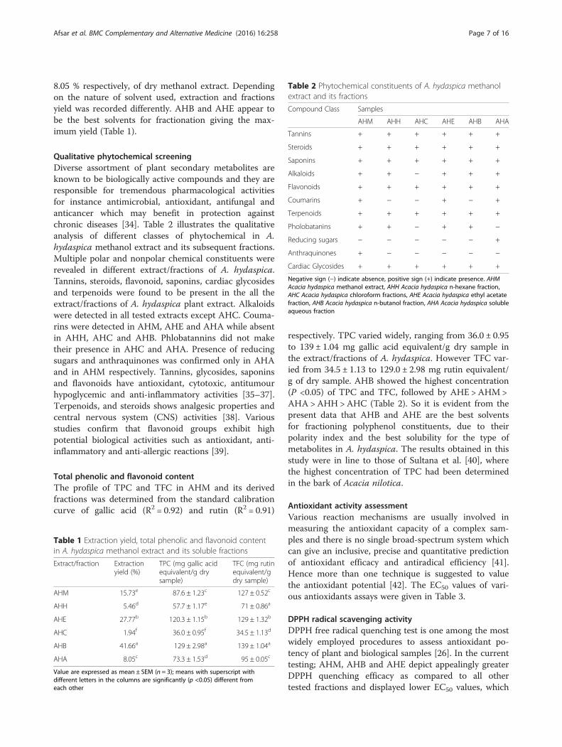
8.05 % respectively, of dry methanol extract. Dependingon the nature of solvent used, extraction and fractionsyield was recorded differently. AHB and AHE appear tobe the best solvents for fractionation giving the max-imum yield (Table 1).
Qualitative phytochemical screeningDiverse assortment of plant secondary metabolites areknown to be biologically active compounds and they areresponsible for tremendous pharmacological activitiesfor instance antimicrobial, antioxidant, antifungal andanticancer which may benefit in protection againstchronic diseases [34]. Table 2 illustrates the qualitativeanalysis of different classes of phytochemical in A.hydaspica methanol extract and its subsequent fractions.Multiple polar and nonpolar chemical constituents wererevealed in different extract/fractions of A. hydaspica.Tannins, steroids, flavonoid, saponins, cardiac glycosidesand terpenoids were found to be present in the all theextract/fractions of A. hydaspica plant extract. Alkaloidswere detected in all tested extracts except AHC. Couma-rins were detected in AHM, AHE and AHA while absentin AHH, AHC and AHB. Phlobatannins did not maketheir presence in AHC and AHA. Presence of reducingsugars and anthraquinones was confirmed only in AHAand in AHM respectively. Tannins, glycosides, saponinsand flavonoids have antioxidant, cytotoxic, antitumourhypoglycemic and anti-inflammatory activities [35–37].Terpenoids, and steroids shows analgesic properties andcentral nervous system (CNS) activities [38]. Variousstudies confirm that flavonoid groups exhibit highpotential biological activities such as antioxidant, anti-inflammatory and anti-allergic reactions [39].
Total phenolic and flavonoid contentThe profile of TPC and TFC in AHM and its derivedfractions was determined from the standard calibrationcurve of gallic acid (R2 = 0.92) and rutin (R2 = 0.91)
respectively. TPC varied widely, ranging from 36.0 ± 0.95to 139 ± 1.04 mg gallic acid equivalent/g dry sample inthe extract/fractions of A. hydaspica. However TFC var-ied from 34.5 ± 1.13 to 129.0 ± 2.98 mg rutin equivalent/g of dry sample. AHB showed the highest concentration(P <0.05) of TPC and TFC, followed by AHE > AHM >AHA >AHH >AHC (Table 2). So it is evident from thepresent data that AHB and AHE are the best solventsfor fractioning polyphenol constituents, due to theirpolarity index and the best solubility for the type ofmetabolites in A. hydaspica. The results obtained in thisstudy were in line to those of Sultana et al. [40], wherethe highest concentration of TPC had been determinedin the bark of Acacia nilotica.
Antioxidant activity assessmentVarious reaction mechanisms are usually involved inmeasuring the antioxidant capacity of a complex sam-ples and there is no single broad-spectrum system whichcan give an inclusive, precise and quantitative predictionof antioxidant efficacy and antiradical efficiency [41].Hence more than one technique is suggested to valuethe antioxidant potential [42]. The EC50 values of vari-ous antioxidants assays were given in Table 3.
DPPH radical scavenging activityDPPH free radical quenching test is one among the mostwidely employed procedures to assess antioxidant po-tency of plant and biological samples [26]. In the currenttesting; AHM, AHB and AHE depict appealingly greaterDPPH quenching efficacy as compared to all othertested fractions and displayed lower EC50 values, which
Table 1 Extraction yield, total phenolic and flavonoid contentin A. hydaspica methanol extract and its soluble fractions
Extract/fraction Extractionyield (%)
TPC (mg gallic acidequivalent/g drysample)
TFC (mg rutinequivalent/gdry sample)
AHM 15.73e 87.6 ± 1.23c 127 ± 0.52c
AHH 5.46d 57.7 ± 1.17e 71 ± 0.86a
AHE 27.77b 120.3 ± 1.15b 129 ± 1.32b
AHC 1.94f 36.0 ± 0.95f 34.5 ± 1.13d
AHB 41.66a 129 ± 2.98a 139 ± 1.04a
AHA 8.05c 73.3 ± 1.53d 95 ± 0.05c
Value are expressed as mean ± SEM (n = 3); means with superscript withdifferent letters in the columns are significantly (p <0.05) different fromeach other
Table 2 Phytochemical constituents of A. hydaspica methanolextract and its fractions
Compound Class Samples
AHM AHH AHC AHE AHB AHA
Tannins + + + + + +
Steroids + + + + + +
Saponins + + + + + +
Alkaloids + + − + + +
Flavonoids + + + + + +
Coumarins + − − + − +
Terpenoids + + + + + +
Pholobatanins + + − + + −
Reducing sugars − − − − − +
Anthraquinones + − − − − −
Cardiac Glycosides + + + + + +
Negative sign (−) indicate absence, positive sign (+) indicate presence. AHMAcacia hydaspica methanol extract, AHH Acacia hydaspica n-hexane fraction,AHC Acacia hydaspica chloroform fractions, AHE Acacia hydaspica ethyl acetatefraction, AHB Acacia hydaspica n-butanol fraction, AHA Acacia hydaspica solubleaqueous fraction
Afsar et al. BMC Complementary and Alternative Medicine (2016) 16:258 Page 7 of 16
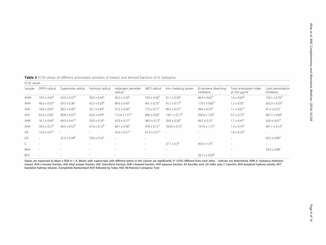
Table 3 EC50 values of different antioxidant activities of extract and derived fractions of A. hydaspica
EC50 values
Sample DPPH radical Superoxide radical Hydroxyl radical Hydrogen peroxideradical
ABTS radical Iron chelating power β-carotene bleachinginhibition
Total antioxidant indexat 250 μg/ml
Lipid peroxidationinhibition
AHM 19.5 ± 0.43a 43.0 ± 0.37b 39.0 ± 0.44a 40.3 ± 0.26a 193 ± 0.26d 41.7 ± 0.34a 86.4 ± 0.62d 1.6 ± 0.69b 156.1 ± 0.76d
AHH 46.5 ± 0.55b 59.3 ± 0.36c 42.3 ± 0.29b 80.6 ± 0.43c 401 ± 0.23e 91.7 ± 0.17b 173.5 ± 0.85d 1.2 ± 0.35c 562.3 ± 0.55g
AHE 18.0 ± 0.45a 38.3 ± 0.40a 35.7 ± 0.40a 37.2 ± 0.36a 173 ± 0.11c 40.3 ± 0.21a 48.4 ± 0.55b 1.7 ± 0.61b 61.3 ± 0.52c
AHC 65.0 ± 0.36c 90.8 ± 0.55d 63.0 ± 0.09c 115.6 ± 1.51d 600 ± 0.26e 130.1 ± 0.17d 260.6 ± 1.05f 0.7 ± 0.73e 467.3 ± 0.60f
AHB 16.7 ± 0.43a 49.0 ± 0.41b 33.0 ± 0.16a 42.0 ± 0.11a 98.0 ± 0.12a 39.0 ± 0.26a 66.2 ± 0.72c 1.7 ± 0.41a 50.0 ± 0.61b
AHA 39.5 ± 0.51b 46.3 ± 0.52b 47.0 ± 0.12b 68.1 ± 0.36b 478 ± 0.12e 103.0 ± 0.12c 131.6 ± 1.15e 1.3 ± 0.14e 401.1 ± 0.12e
AA 15.0 ± 0.41a - - 35.0 ± 0.21a 61.0 ± 0.21b - - 1.8 ± 0.24b -
GA - 32.3 ± 0.28a 29.0 ± 0.16a - - - - - 43.5 ± 0.86a
C - - - - - 37.7 ± 0.3a 40.4 ± 1.01a - -
BHA - - - - - - - 59.3 ± 0.98c
BHT - - - - - - 50.11 ± 0.59b -
Values are expressed as Mean ± SEM (n = 3). Means with superscripts with different letters in the column are significantly (P <0.05) different from each other. - indicate not determined, AHM A. hydaspica methanolextract, AHH n-hexane fraction, AHE ethyl acetate fraction, AHC chloroform fraction, AHB n-butanol fraction, AHA aqueous fraction, AA Ascorbic acid, GA Gallic acid, C Catechin, BHA butylated hydroxy anisole, BHTbutylated hydroxy toluene. (Completely Randomized AOV followed by Tukey HSD All-Pairwise Compariso Test)
Afsar
etal.BM
CCom
plementary
andAlternative
Medicine
(2016) 16:258 Page
8of
16
were non-significantly different from one another. TheEC50 values in DPPH assay range from 16.7 ± 0.43–65 ±0.36 μg/ml. AHB and AHE showed the lowest EC50
values (16.7 ± 0.43 and 18 ± 0.45 μg/ml), which werecomparable to EC50 of reference compound (Ascorbicacid). DPPH radical scavenging activity of A. hydaspicaextract and its various fractions showed good correlationwith TPC (R2 = 0.9879) and TFC (R2 = 0.8477). Resultsof present investigation imply that A. hydaspica containphyto-constituents that are proficient of donating hydro-gen to a free radical in order to rescue the potentialimpairment. Most specifically, phenolic and flavonoid ex-hibit antioxidant activity due to the hydroxyl group attachto the aromatic ring which is capable donating electronand stabilizing the free radicals. The research conductedby Sultana et al. [40] in A. nilotica and Singh et al. [43] onAcacia auriculiformis revealed similar results.
Superoxide radical scavenging activityAlthough superoxide anion is a weak oxidant, but itlead to the generation of powerful and hazardous hy-droxyl radicals as well as singlet oxygen, both ofwhich contribute to oxidative stress. Therefore, it isvery important to study the scavenging of superoxideanion [44]. The EC50 values in superoxide scavengingactivities were in the order of AHE < AHM < AHB <AHA < AHH < AHC. When compared to ascorbicacid, the superoxide scavenging activity of the AHEwas found to be statistically (P >0.05) similar. Thepotent electron scavenging ability of the methanolextract and its various fractions might be due itsbioactive phytoconstituents like that are able tominimize the oxidation of biological macromolecules[26, 37]. A significant correlation was detected withTPC (R2 = 0.7909, P <0.05) while nonsignificant cor-relation (R2 = 0.641, P >0.05) was observed with TFC.This strong superoxide radical neutralizing capacity ofA. hydaspica might be functional therapeuticallyagainst oxidative stress induced ailments.
Hydroxyl radical scavenging activityThe evidence of OH radical scavenging activity by A.hydaspica extract and its fractions was determined bymeasuring the inhibition of 2-deoxyribose degradationby the free radicals generated during Fenton reaction.Crude methanol extract and derived fractions markedlyscavenged OH radicals and prevented the degradation of2-deoxyribose. A dose dependent mode was observedfor hydroxyl radical scavenging activity. The lowest EC50
values were shown by AHB (36.0 ± 0.16 μg/ml) followedby AHE and AHM (37.7 ± 0.40 and 40.0 ± 0.44 μg/mlrespectively). However EC50 values were significantly dif-ferent from standard gallic acid. A significant correlationwas observed with TPC (R2 = 0.844, P <0.01) and TFC
(R2 = 0.776, P <0.05). The strong antioxidant activity ofAHB and AHE might be utilized as a source of naturalantioxidant in oxidative stress for minimizing the detri-mental effects of hydroxyl radical in the body. A highscavenging activity of AHE for OH radical was reportedin one of previous studies done in our lab.
Hydrogen peroxide radical scavenging activityIn the body, H2O2 is rapidly decomposed into oxygenand water and this may produce hydroxyl radicals (•OH)that can initiate lipid peroxidation and cause DNA dam-age [6]. Therefore, the ability of plant extracts to scav-enge hydrogen peroxide was also determined in order toget the idea that whether samples have same pattern ofactivity as OH radical reducing ability. Methanolextract/fractions of A. hydaspica possess significantability to quench the hydrogen peroxide radicals,demonstrating the antioxidant potential of the plant.AHE proved to be efficient fraction against hydrogenperoxide (EC50 = 37.2 ± 0.36 μg/ml). EC50 valuesshowed significant correlation with both TPC (R 2 =0.844, P <0.01) and TFC (R2 = 0.776, P <0.05), attrib-uting the activity to the occurrence of polyphenoliccompounds that give electrons to hydrogen peroxide,thus neutralizing it into water. These findings are inline with the previous study.
ABTS radical scavenging assayIn this assay, the reaction of ABTS with potassium per-sulfate in the existence of hydrogen-donating antioxi-dants outcomes in the formation of ABTS+ blue/greenchromophores, this reduction reaction is noted spectro-photometrically at an absorbance of 745 nm. Thisscheme of determination of antioxidant action is equallypertinent to hydrophilic and lipophilic classes of antioxi-dants like; flavonoids, hydroxycinnamates, carotenoidsand antioxidants in the plasma [45]. The result obtainedindicated that AHM and its derived fractions scavengethe ABTS radicals in a dose dependent pattern. Amongthe extract/fractions lowest EC50 values for ABTS radicalscavenging were determined for AHB (98.0 ± 0.1 μg/ml)while highest EC50 values were recorded for AHC (>500± 0.26 μg/ml) as shown in Table 1. However EC50 valuesof AHB were significantly lower than ascorbic acid (61 ±0.2 μg/ml). The ABTS scavenging activity of the presentstudy suggests that the phyto-constituents within the ex-tract and various fractions of A. hydaspica might donateelectron/hydrogen while minimizing the oxidative stress.Furthermore correlation analysis indicated significantcorrelation between ABTS radical scavenging activityand TPC (R2 = 0.881, P <0.001) as well as TFC (R2 =0.857, P <0.001). The results are in line with the study ofkhan et al. [46].
Afsar et al. BMC Complementary and Alternative Medicine (2016) 16:258 Page 9 of 16
Chelating activity on Fe2+An imperative mechanism of antioxidant activity is theability to chelate/counteract transition metals, whichhave the ability to demolish hydro peroxides andFenton-type reactions. Therefore it was considered im-portant to screen the iron (II) chelating ability ofextract/fractions. The sequence for chelating power wasAHB ~ AHE ≥AHM>AHH >AHA >AHC. The EC50
values of AHB, AHE and AHM were close to the EC50
value of standard catechin. Correlation analysis sug-gested that the iron (II) chelating possessions of A.hydaspica may be accredited to its endogenous chelatingagents like polyphenolic compounds [47]. As somephenolic compounds have properly oriented functionalgroups, which can chelate metal ions to protect againstoxidative damage [48]. Chelating activity was correlatedwell with the phenolic (R2 = 0.8971, P <0.001) and fla-vonoid content (R2 = 0.8177, p <0.05) Our results are ingood agreement with previous report where ethyl acetatefraction possessing polyphenolic constituents of A. auri-culiformis showed highest metal chelating power [43].
β-carotene bleaching inhibitionIn this test, antioxidant competence was evaluated byquantifying the inhibition of the conjugated diene-hydroperoxides and the volatile organic compounds cre-ation as an outcome of linoleic acid oxidation. Hence,the antioxidant occurrence can impede the magnitude ofβ-carotene bleaching by counterbalancing the linoleateand other free radicals formed in the process. The colorof reaction solution was retained for a long time in thepresence of an antioxidant compound while a rapid de-crease in absorbance was noticed in the absence of anti-oxidant [49]. The antioxidant activity of A. hydaspicaextract/fractions with regard to the β-carotene bleachingmethod could be ranked as AHE > AHB > AHM >AHA >AHH >AHC. The EC50 values of AHE (48.4 ±0.55 μg/ml) were significantly different yet comparablewith standard catechin (EC50 = 40.4 ± 1.01 μg/ml).However AHE showed better efficacy as compared tostandard BHT. The differential efficacy of A. hydaspicaextract/fractions to inhibit oxidation of linoleic acidemulsion is an indication of the complexity of the ex-tract/fractions as well as potential interaction betweenthe extract and emulsion components. Correlation ana-lysis indicate significant correlation with phenolic (R2 =0.9670, P <0.0001) and flavonoid (R2 = 0.8831, P <0.001)content. The correlation of phenolic and flavonoids withβ-carotene bleaching inhibition potential was reportedby other researchers as well [26, 50].
Anti-lipid peroxidation potentialLipid peroxidation is involved in a number of pathologicalconditions so evaluation of antioxidant potential of natural
and synthetic compounds requires an assay in lipid systemtoo. The EC50 values in lipid peroxidation inhibition werein the order of AHB <AHE <AHM<AHA <AHC <AHH. EC50 values showed by AHB (50 ± 0.61 μg/ml) weresignificantly lower as compared to BHA values (EC50 =59.3 ± 0.98 μg/ml). In this study the in vitro ability of plantextract/fractions to prevent the production of TBARSdepicts the potential of samples to inhibit oxidationin lipid system. Significant correlation was observedwith TPC (R2 = 0.779, P <0.05) and TFC (R2 = 0.836, P<0.05). Phenolic compounds are very important plantconstituents because they exhibit antioxidant activitiesby inactivating lipid free radicals, or by preventingthe decomposition of hydro-peroxides into free radi-cals. Results of lipid peroxidation were in line withthe previous study of Singh et al. [43].
Total antioxidant capacity assayThe total antioxidant capacity of the extract/fractionswas measured by phosphomolybdenum method basedon the reduction of molybnedum (VI) to molybnedum(V) by the antioxidant action of extract and the subse-quent formation of a green phosphate Mo (V) complexat acid pH of the medium with maximum absorbance at695 nm [51]. Total antioxidant capacity of the extract/fractions recorded at the highest dose of 250 μg/ml wasin order of AHB (1.7 ± 0.015) ~ AHE (1.7 ± 0.08) > AHM(1.5 ± 0.016) > AHA (1.3 ± 0.05) > AHH (1.2 ± 0.02) >AHC (0.738 ± 0.012). The current analysis reveals thatAHB and AHE displayed the uppermost antioxidantcapacity. Latest researches proved that flavonoids andrelated polyphenols contributes substantially to thephosphomolybdate quenching capability of medicinalplants [46, 52]. AHB and AHE exhibited the highestantioxidant index comparable with ascorbic acid. Phos-phomolybdenum assay in general detects antioxidantssuch as carotenoids, ascorbic acid, α -tocopherol, andsome phenolic, cysteine, and aromatic amines due tohydrogen and electron donating ability. The antioxidantcapacities of the extract/fractions have a strong relation-ship with the solvent employed, mainly due to the differ-ent antioxidant potential of compounds with differentpolarities. Phytochemical analysis reveals the presence ofvarious boiactive phytochemicals that might be attrib-uted to the antioxidant capacity of A. hydaspica. Our re-sult correlate well with the research of Tung at al.reporting gallic acid, catechin, myricetin along withother polyphenols in A. confusa leaves extract wereresponsible for the significant antioxidant potential [53].
Reducing powerSamples owning more reducing efficacy displayed highabsorbance values. From the literature, it’s evident thatreducing power is attributed to the existence of
Afsar et al. BMC Complementary and Alternative Medicine (2016) 16:258 Page 10 of 16
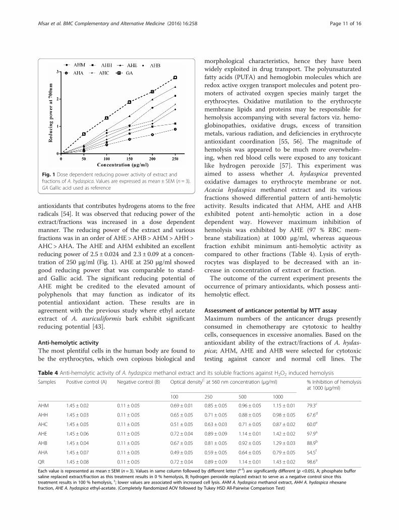
antioxidants that contributes hydrogens atoms to the freeradicals [54]. It was observed that reducing power of theextract/fractions was increased in a dose dependentmanner. The reducing power of the extract and variousfractions was in an order of AHE >AHB >AHM>AHH>AHC >AHA. The AHE and AHM exhibited an excellentreducing power of 2.5 ± 0.024 and 2.3 ± 0.09 at a concen-tration of 250 μg/ml (Fig. 1). AHE at 250 μg/ml showedgood reducing power that was comparable to stand-ard Gallic acid. The significant reducing potential ofAHE might be credited to the elevated amount ofpolyphenols that may function as indicator of itspotential antioxidant action. These results are inagreement with the previous study where ethyl acetateextract of A. auriculiformis bark exhibit significantreducing potential [43].
Anti-hemolytic activityThe most plentiful cells in the human body are found tobe the erythrocytes, which own copious biological and
morphological characteristics, hence they have beenwidely exploited in drug transport. The polyunsaturatedfatty acids (PUFA) and hemoglobin molecules which areredox active oxygen transport molecules and potent pro-moters of activated oxygen species mainly target theerythrocytes. Oxidative mutilation to the erythrocytemembrane lipids and proteins may be responsible forhemolysis accompanying with several factors viz. hemo-globinopathies, oxidative drugs, excess of transitionmetals, various radiation, and deficiencies in erythrocyteantioxidant coordination [55, 56]. The magnitude ofhemolysis was appeared to be much more overwhelm-ing, when red blood cells were exposed to any toxicantlike hydrogen peroxide [57]. This experiment wasaimed to assess whether A. hydaspica preventedoxidative damages to erythrocyte membrane or not.Acacia hydaspica methanol extract and its variousfractions showed differential pattern of anti-hemolyticactivity. Results indicated that AHM, AHE and AHBexhibited potent anti-hemolytic action in a dosedependent way. However maximum inhibition ofhemolysis was exhibited by AHE (97 % RBC mem-brane stabilization) at 1000 μg/ml, whereas aqueousfraction exhibit minimum anti-hemolytic activity ascompared to other fractions (Table 4). Lysis of eryth-rocytes was displayed to be decreased with an in-crease in concentration of extract or fraction.The outcome of the current experiment presents the
occurrence of primary antioxidants, which possess anti-hemolytic effect.
Assessment of anticancer potential by MTT assayMaximum numbers of the anticancer drugs presentlyconsumed in chemotherapy are cytotoxic to healthycells, consequences in excessive anomalies. Based on theantioxidant ability of the extract/fractions of A. hydas-pica; AHM, AHE and AHB were selected for cytotoxictesting against cancer and normal cell lines. The
Fig. 1 Dose dependent reducing power activity of extract andfractions of A. hydaspica. Values are expressed as mean ± SEM (n = 3).GA Gallic acid used as reference
Table 4 Anti-hemolytic activity of A. hydaspica methanol extract and its soluble fractions against H2O2 induced hemolysis
Samples Positive control (A) Negative control (B) Optical densityC at 560 nm concentration (μg/ml) % Inhibition of hemolysisat 1000 (μg/ml)
100 250 500 1000
AHM 1.45 ± 0.02 0.11 ± 0.05 0.69 ± 0.01 0.85 ± 0.05 0.96 ± 0.05 1.15 ± 0.01 79.3c
AHH 1.45 ± 0.03 0.11 ± 0.05 0.65 ± 0.05 0.71 ± 0.05 0.88 ± 0.05 0.98 ± 0.05 67.6d
AHC 1.45 ± 0.05 0.11 ± 0.05 0.51 ± 0.05 0.63 ± 0.03 0.71 ± 0.05 0.87 ± 0.02 60.0e
AHE 1.45 ± 0.06 0.11 ± 0.05 0.72 ± 0.04 0.89 ± 0.09 1.14 ± 0.01 1.42 ± 0.02 97.9a
AHB 1.45 ± 0.04 0.11 ± 0.05 0.67 ± 0.05 0.81 ± 0.05 0.92 ± 0.05 1.29 ± 0.03 88.9b
AHA 1.45 ± 0.07 0.11 ± 0.05 0.49 ± 0.05 0.59 ± 0.05 0.64 ± 0.05 0.79 ± 0.05 54.5f
QR 1.45 ± 0.08 0.11 ± 0.05 0.72 ± 0.04 0.89 ± 0.09 1.14 ± 0.01 1.43 ± 0.02 98.6a
Each value is represented as mean ± SEM (n = 3). Values in same column followed by different letter (a–f) are significantly different (p <0.05), A; phosphate buffersaline replaced extract/fraction as this treatment results in 0 % hemolysis, B; hydrogen peroxide replaced extract to serve as a negative control since thistreatment results in 100 % hemolysis, c; lower values are associated with increased cell lysis. AHM A. hydaspica methanol extract, AHH A. hydaspica nhexanefraction, AHE A. hydaspica ethyl-acetate. (Completely Randomized AOV followed by Tukey HSD All-Pairwise Comparison Test)
Afsar et al. BMC Complementary and Alternative Medicine (2016) 16:258 Page 11 of 16
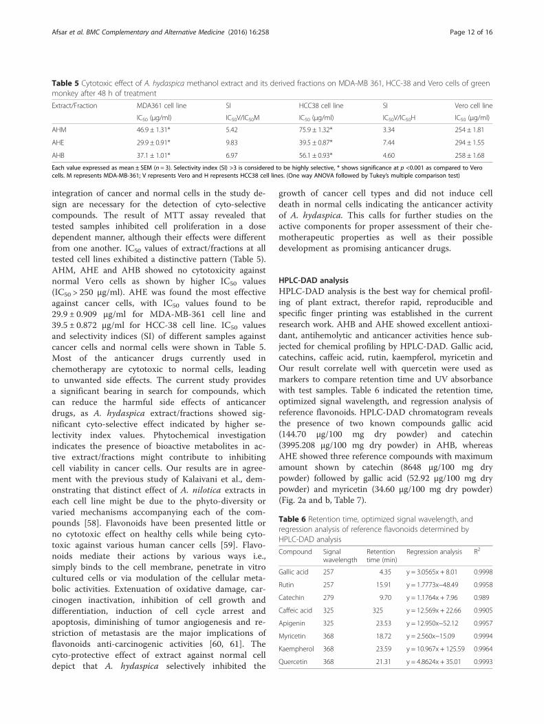
integration of cancer and normal cells in the study de-sign are necessary for the detection of cyto-selectivecompounds. The result of MTT assay revealed thattested samples inhibited cell proliferation in a dosedependent manner, although their effects were differentfrom one another. IC50 values of extract/fractions at alltested cell lines exhibited a distinctive pattern (Table 5).AHM, AHE and AHB showed no cytotoxicity againstnormal Vero cells as shown by higher IC50 values(IC50 > 250 μg/ml). AHE was found the most effectiveagainst cancer cells, with IC50 values found to be29.9 ± 0.909 μg/ml for MDA-MB-361 cell line and39.5 ± 0.872 μg/ml for HCC-38 cell line. IC50 valuesand selectivity indices (SI) of different samples againstcancer cells and normal cells were shown in Table 5.Most of the anticancer drugs currently used inchemotherapy are cytotoxic to normal cells, leadingto unwanted side effects. The current study providesa significant bearing in search for compounds, whichcan reduce the harmful side effects of anticancerdrugs, as A. hydaspica extract/fractions showed sig-nificant cyto-selective effect indicated by higher se-lectivity index values. Phytochemical investigationindicates the presence of bioactive metabolites in ac-tive extract/fractions might contribute to inhibitingcell viability in cancer cells. Our results are in agree-ment with the previous study of Kalaivani et al., dem-onstrating that distinct effect of A. nilotica extracts ineach cell line might be due to the phyto-diversity orvaried mechanisms accompanying each of the com-pounds [58]. Flavonoids have been presented little orno cytotoxic effect on healthy cells while being cyto-toxic against various human cancer cells [59]. Flavo-noids mediate their actions by various ways i.e.,simply binds to the cell membrane, penetrate in vitrocultured cells or via modulation of the cellular meta-bolic activities. Extenuation of oxidative damage, car-cinogen inactivation, inhibition of cell growth anddifferentiation, induction of cell cycle arrest andapoptosis, diminishing of tumor angiogenesis and re-striction of metastasis are the major implications offlavonoids anti-carcinogenic activities [60, 61]. Thecyto-protective effect of extract against normal celldepict that A. hydaspica selectively inhibited the
growth of cancer cell types and did not induce celldeath in normal cells indicating the anticancer activityof A. hydaspica. This calls for further studies on theactive components for proper assessment of their che-motherapeutic properties as well as their possibledevelopment as promising anticancer drugs.
HPLC-DAD analysisHPLC-DAD analysis is the best way for chemical profil-ing of plant extract, therefor rapid, reproducible andspecific finger printing was established in the currentresearch work. AHB and AHE showed excellent antioxi-dant, antihemolytic and anticancer activities hence sub-jected for chemical profiling by HPLC-DAD. Gallic acid,catechins, caffeic acid, rutin, kaempferol, myricetin andOur result correlate well with quercetin were used asmarkers to compare retention time and UV absorbancewith test samples. Table 6 indicated the retention time,optimized signal wavelength, and regression analysis ofreference flavonoids. HPLC-DAD chromatogram revealsthe presence of two known compounds gallic acid(144.70 μg/100 mg dry powder) and catechin(3995.208 μg/100 mg dry powder) in AHB, whereasAHE showed three reference compounds with maximumamount shown by catechin (8648 μg/100 mg drypowder) followed by gallic acid (52.92 μg/100 mg drypowder) and myricetin (34.60 μg/100 mg dry powder)(Fig. 2a and b, Table 7).
Table 5 Cytotoxic effect of A. hydaspica methanol extract and its derived fractions on MDA-MB 361, HCC-38 and Vero cells of greenmonkey after 48 h of treatment
Extract/Fraction MDA361 cell line SI HCC38 cell line SI Vero cell line
IC50 (μg/ml) IC50V/IC50M IC50 (μg/ml) IC50V/IC50H IC50 (μg/ml)
AHM 46.9 ± 1.31* 5.42 75.9 ± 1.32* 3.34 254 ± 1.81
AHE 29.9 ± 0.91* 9.83 39.5 ± 0.87* 7.44 294 ± 1.55
AHB 37.1 ± 1.01* 6.97 56.1 ± 0.93* 4.60 258 ± 1.68
Each value expressed as mean ± SEM (n = 3). Selectivity index (SI) >3 is considered to be highly selective, * shows significance at p <0.001 as compared to Verocells. M represents MDA-MB-361; V represents Vero and H represents HCC38 cell lines. (One way ANOVA followed by Tukey’s multiple comparison test)
Table 6 Retention time, optimized signal wavelength, andregression analysis of reference flavonoids determined byHPLC-DAD analysis
Compound Signalwavelength
Retentiontime (min)
Regression analysis R2
Gallic acid 257 4.35 y = 3.0565x + 8.01 0.9998
Rutin 257 15.91 y = 1.7773x−48.49 0.9958
Catechin 279 9.70 y = 1.1764x + 7.96 0.989
Caffeic acid 325 325 y = 12.569x + 22.66 0.9905
Apigenin 325 23.53 y = 12.950x−52.12 0.9957
Myricetin 368 18.72 y = 2.560x−15.09 0.9994
Kaempherol 368 23.59 y = 10.967x + 125.59 0.9964
Quercetin 368 21.31 y = 4.8624x + 35.01 0.9993
Afsar et al. BMC Complementary and Alternative Medicine (2016) 16:258 Page 12 of 16
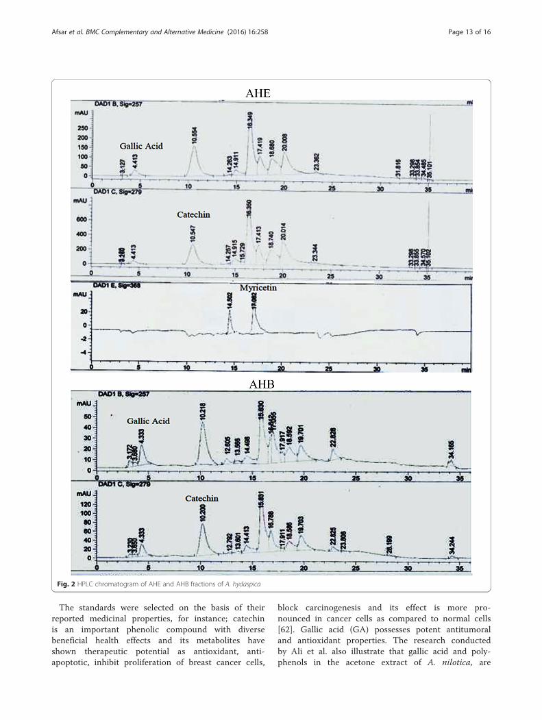
The standards were selected on the basis of theirreported medicinal properties, for instance; catechinis an important phenolic compound with diversebeneficial health effects and its metabolites haveshown therapeutic potential as antioxidant, anti-apoptotic, inhibit proliferation of breast cancer cells,
block carcinogenesis and its effect is more pro-nounced in cancer cells as compared to normal cells[62]. Gallic acid (GA) possesses potent antitumoraland antioxidant properties. The research conductedby Ali et al. also illustrate that gallic acid and poly-phenols in the acetone extract of A. nilotica, are
Fig. 2 HPLC chromatogram of AHE and AHB fractions of A. hydaspica
Afsar et al. BMC Complementary and Alternative Medicine (2016) 16:258 Page 13 of 16
responsible for cytotoxic activity [63]. Myricetin isalso able to induce apoptosis of pancreatic cancercells, human bladder carcinoma cell line, triggerapoptosis, regression of tumor growth, decrease me-tastasis and it increase bioavailability of tamoxifen, adrug used to treat breast cancer [64].Our result correlate well with the research of Tung at
al., reporting gallic acid, catechin, myricetin along withother polyphenols in ethyl acetate fraction of A. confusaleaves extract were responsible for the significant antioxi-dant and anticancer potential [53]. This calls for furtherstudies on the active components for proper assessmentof their chemotherapeutic properties as well as their pos-sible development as promising anticancer drugs.
ConclusionThe present study demonstrates the phytochemicalprofiling, in vitro antioxidant, anti-hemolytic andcyto-selective anticancer activity of A. hydaspica aerialparts extracts. Extracts with higher antioxidant cap-acity also had higher polyphenol content. It can beconcluded that the extract obtained using higherpolarity solvents were more effective radical scaven-gers then those obtained using less polar solvents.Ethyl acetate and n-butanol showed better character-istics as solvent for phenolic compounds. Furthermorethese fractions tended to possess superior activity inlipid peroxidation inhibition and β-carotene bleachingassay as compared to BHA and BHT. Therefore, theymight be used as preservative ingredients in the foodand/or pharmaceutical industry. Moreover safety pro-file and chemotherapeutic potential of active fractionsand methanol extract was determined by assessingthe anti-hemolytic activity and in vitro testing againstboth cancer and normal cell lines. Bioactive com-pounds present in A. hydaspica active fractions mightwork synergistically and specifically in inhibitingproliferation of breast cancer cells with high SI value,suggesting that they might be used as a naturaladditive in human diets for cancer chemoprevention.However the evaluation and the discovery of newanticancer agents is long-term process that encompasses
many steps by step approaches with the screening foranticancer properties, followed by the isolation andidentification of bioactive compounds and finally invivo anticancer activity testing in order to verify theaptitude of the compounds. Therefore further re-search would be required before such uses could beproposed with confidence.
AbbreviationsABTS, 2,2′-azino-bis (3-ethylbenzothiazoline-6-sulphonic acid; AHA, Acaciahydaspica residual aqueous fraction of methanol extract of aerial parts; AHB,Acacia hydaspica n-butanol fraction of methanol extract of aerial parts; AHC,Acacia hydaspica chloroform fraction of methanol extract of aerial parts; AHE,Acacia hydaspica ethyl acetate fraction of methanol extract of aerial parts;AHH, Acacia hydaspica n-hexane fraction of methanol extract of aerial parts;AHM, Acacia hydaspica methanol extract of aerial parts; BHA, butylatedhydroxyanisole; BHT, butylated hydroxytoluene; DMEM/F12, Dulbecco’smodified eagle medium: nutrient mixture F-12; DPPH, 2,2-Diphenyl-1-Picrylhydrazyl; EDTA, ethylene diamine tetra acetic acid; PBS, phosphatebuffer saline; PMS, phenazine methosulphate; TBA, thiobarbituric acid;TBARS, thiobarbituric acid reactive substances; TFC, total flavonoidcontent; TPC, total phenolic content
AcknowledgementsWe acknowledge Higher Education Commission (HEC) of Pakistan forawarding indigenous scholarship and IRSP scholarship for University ofMinnesota, USA to the first author. Furthermore we acknowledge Deanshipof Scientific Research at King Saud University for its technical assistant in thisresearch project.
FundingThe project was partially funded by the Higher Education Commission (HEC)of Pakistan by awarding indigenous scholarship to the first author. We aregrateful to the Deanship of Scientific Research at King Saud University for itsfunding of this research through Research Group Project number 193.
Availability of data and materialsAll the data is contained in the manuscript.
Authors’ contributionsTA made significant contributions to conception, design, experimentation,acquisition and interpretation of data and writing of manuscript. SR, MRK,SM, AA and MS made substantial contribution in interpretation of data andrevising the manuscript for intellectual content. IUH made a contribution tothe HPLC experimentation and analysis. All authors read and approved thefinal manuscript.
Authors’ informationTA did PhD in Biochemistry/Pharmacological biology from Department ofBiochemistry, Quaid-i-Azam University, Islamabad, Pakistan. SR is PhD Scholarfrom Department of Animal Sciences, Faculty of Biological Sciences, Quaid-i-Azam University, Islamabad and Researcher at the Department of CommunityHealth Sciences, College of Applied Medical Sciences, Clinical nutrition program,King Saud University, Riyadh KSA. MRK did his Diploma in Unani Medicine andSurgery (DUMS) and is a registered practitioner of the National Councilfor Tibb of Pakistan. He is working as Associate Professor at the Departmentof Biochemistry, Quaid-i-Azam University, Islamabad, Pakistan. SM did MPhil inbiochemistry/Pharmacological biology from Department of Biochemistry,Quaid-i-Azam University, Islamabad, Pakistan. AA is associate professor atDepartment of Community Health Sciences, College of Applied MedicalSciences, Clinical nutrition program, King Saud University, Riyadh KSA.MS did PhD in Biochemistry/Pharmacological biology from Departmentof Biochemistry, Quaid-i-Azam University, Islamabad, Pakistan. IUH isassistant professor at the Department of Pharmacy, Faculty of BiologicalSciences, Quaid-i-Azam University, Islamabad, Pakistan.
Competing interestsThe authors declare that they have no competing interests.
Table 7 HPLC-DAD profile of A. hydaspica ethyl acetate andn-butanol fractions
Extract/fraction Compounds Signalwavelength
Retentiontime
Quantity(μg/100 mgdry powder)
AHE Gallic acid 275 nm 4.52 52.92
Catechin 279 nm 11.43 8648.0
Myricetin 368 nm 17.08 34.60
AHB Gallic acid 257 nm 4.41 144.70
Catechin 279 nm 10.55 3995.21
Afsar et al. BMC Complementary and Alternative Medicine (2016) 16:258 Page 14 of 16
Consent for publicationNot applicable.
Ethics approval and consent to participateThis study makes use of rats and human blood, and the experimentalprotocol for the use of animal and human blood was approved (Bch#0256)by the ethical board of Quaid-i-Azam University, Islamabad Pakistan.
Author details1Department of Biochemistry, Faculty of Biological Sciences, Quaid-i-AzamUniversity, Islamabad, Pakistan. 2Department of Animal Sciences, Faculty ofBiological Sciences, Quaid-i-Azam University, Islamabad, Pakistan.3Department of Community Health Sciences, College of Applied MedicalSciences, King Saud University, Riyadh, Kingdom of Saudi Arabia.4Department of Pharmacy, Faculty of Biological Sciences, Quaid-i-AzamUniversity, Islamabad, Pakistan.
Received: 4 December 2015 Accepted: 23 July 2016
References1. Mena S, Ortega A, Estrela J. Oxidative stress in environmental-induced
carcinogenesis. Mutat Res Genet Toxicol Environ Mutagen. 2009;674(1):36–44.
2. Waris G, Ahsan H. Reactive oxygen species: role in the development ofcancer and various chronic conditions. J Carcinog. 2006;5(1):14.
3. Sasaki Y, Kawaguchi S, Kamaya A, Ohshita M, Kabasawa K, Iwama K,Taniguchi K, Tsuda S. The comet assay with 8 mouse organs: results with39 currently used food additives. Mutat Res Genet Toxicol Environ Mutagen.2002;519(1):103–19.
4. Soobrattee M, Bahorun T, Aruoma O. Chemopreventive actions ofpolyphenolic compounds in cancer. Biofactors. 2006;27(1–4):19–35.
5. Dai J, Mumper R. Plant phenolics: extraction, analysis and their antioxidantand anticancer properties. Molecules. 2010;15(10):7313–52.
6. Rj R, Cheng S-J, Je K. Prevention of cytotoxicity and inhibition ofintercellular communication by antioxidant catechins isolated from ChineseGreen Tea. Carcinogenesis. 1989;10(6):1003–8.
7. Chakrabarty T, Gangopadhyay M. The genus Acacia P. Miller (Leguminosae:Mimosoideae) in India. J Econ Taxon Bot. 1996;20(3):599–633.
8. Jabeen A, Khan Ma, Ahmad M, Zafar M, Ahmad F. Indigenous uses ofeconomically important Flora of Margallah Hills National Park, Islamabad,Pakistan. African J Biotechnol. 2009;8(5):763–784.
9. Afsar T, Khan M, Razak S, Ullah S, Mirza B. Antipyretic, anti-inflammatoryand analgesic activity of Acacia Hydaspica R. Parker and its phytochemicalanalysis. BMC Complement Altern Med. 2015;15:136.
10. Afsar T, Trembley J, Salomon C, Razak S, Khan M, Ahmed K. Growthinhibition and apoptosis in cancer cells induced by polyphenoliccompounds of Acacia Hydaspica: involvement of multiple signaltransduction pathways. Sci Rep. 2016;6:23077.
11. Malviya S, Rawat S, Kharia A, Verma M. International Journal of Pharmacy &Life Sciences. Int J Pharm Life Sci(Ijpls). 2011;2(6):830–7.
12. Chang S-T, Wu J-H, Wang S-Y, Kang P-L, Yang N-S, Shyur L-F. Antioxidantactivity of extracts from Acacia Confusa bark and heartwood. J Agric FoodChem. 2001;49(7):3420–4.
13. Mihara R, Barry K, Mohammed C, Mitsunaga T. Comparison of antifungaland antioxidant activities of Acacia Mangium and A. Auriculiformisheartwood extracts. J Chem Ecol. 2005;31(4):789–804.
14. Tung Y-T, Wu J-H, Huang C-Y, Kuo Y-H, Chang S-T. Antioxidant activities andphytochemical characteristics of extracts from Acacia Confusa Bark.Bioresour Technol. 2009;100(1):509–14.
15. Ghate N, Hazra B, Sarkar R, Mandal N. Heartwood extract of Acacia Catechuinduces apoptosis in human breast carcinoma by altering Bax/Bcl-2 ratio.Pharmacogn Mag. 2014;10(37):27.
16. Siddiqui A, Ali M. Practical pharmaceutical chemistry. Cbs Publishers DistribNew Delhi. 1997;126:131.
17. Sofowora A. Recent trends in research into African medicinal plants.J Ethnopharmacol. 1993;38(2):197–208.
18. Trease G. Trease and Evans’ pharmacognosy. London: Bailliere Tindal; 1989.19. Talukdar A, Choudhury M, Chakraborty M, Dutta B. Phytochemical screening
and tlc profiling of plant extracts of Cyathea Gigantea (Wall. Ex. Hook.) Haltt.
and Cyathea Brunoniana. Wall. Ex. Hook (Cl. & Bak.). Assam Univ J SciTechnol. 2010;5(1):70–4.
20. Harborne Jb. Phytochemical Methods. Springer. 1984;278:37-99.21. Mcdonald J, O’dwyer S, Rout S, Chakrabarty B, Sikand K, Fulford P,
Wilson M, Renehan A. Classification of and Cytoreductive surgeryfor low-grade Appendiceal Mucinous Neoplasms. Br J Surg. 2012;99(7):987–92.
22. Chang C-C, Yang M, Wen H, Chern J. Estimation of total flavonoid contentin propolis by two complementary colorimetric methods. J Food Drug Anal.2002;10(3):178–82.
23. Brand-Williams W, Cuvelier M, Berset C. Use of a free radical method toevaluate antioxidant activity. LWT-Food Scie Technol. 1995;28(1):25–30.
24. Nishikimi M, Rao N, Yagi K. The occurrence of superoxide anion in thereaction of reduced phenazine methosulfate and molecular oxygen.Biochem Biophys Res Commun. 1972;46(2):849–54.
25. Gutteridge J, Halliwell B. Free radicals and antioxidants in the year 2000:a historical look to the future. Ann N Y Acad Sci. 2000;899(1):136–47.
26. Sahreen S, Khan M, Khan R. Evaluation of antioxidant activities of varioussolvent extracts of Carissa Opaca fruits. Food Chem. 2010;122(4):1205–11.
27. Re R, Pellegrini N, Proteggente A, Pannala A, Yang M, Rice-Evans C.Antioxidant activity applying an improved Abts radical cation decolorizationassay. Free Radic Biol Med. 1999;26(9):1231–7.
28. Beauchamp C, Fridovich I. Superoxide dismutase: improved assays and anassay applicable to acrylamide gels. Anal Biochem. 1971;44(1):276–87.
29. Kishida E, Tokumaru S, Ishitani Y, Yamamoto M, Oribe M, Iguchi H, Kojo S.Comparison of the formation of malondialdehyde and thiobarbituricacid-reactive substances from autoxidized fatty acids based on oxygenconsumption. J Agric Food Chem. 1993;41(10):1598–600.
30. Umamaheswari M, Chatterjee T. In vitro antioxidant activities of the fractionsof Coccinia Grandis L. Leaf extract. Afr J Tradit Complement Altern Med.2008;5(1):61–73.
31. Kumaran A. Antioxidant and free radical scavenging activity of an aqueousextract of Coleus Aromaticus. Food Chem. 2006;97(1):109–14.
32. Yang Z-G, Sun H-X, Fang W-H. Haemolytic activities and adjuvant effect ofAstragalus Membranaceus Saponins (Ams) on the immune responses toovalbumin in mice. Vaccine. 2005;23(44):5196–203.
33. Mosmann T. Rapid colorimetric assay for cellular growth and survival:application to proliferation and cytotoxicity assays. J Immunol Methods.1983;65(1):55–63.
34. Akhtar N, Mirza B. Phytochemical analysis and comprehensive evaluation ofantimicrobial and antioxidant properties of 61 medicinal plant species. ArabJ Chem. 2015. doi:10.1016/j.arabjc.2015.01.013.
35. Lu X-L, Qiu S-S, Sun X-X, Li Z-J. Preliminary study on the capability ofantioxidation and scavenging free radicals of sasanquasaponins [J]. FoodSci. 2005;11:016.
36. Beninger C, Hosfield G. Antioxidant activity of extracts, condensed tanninfractions, and pure flavonoids from Phaseolus Vulgaris L. Seed Coat colorgenotypes. J Agric Food Chem. 2003;51(27):7879–83.
37. Cai Y, Luo Q, Sun M, Corke H. Antioxidant activity and phenolic compoundsof 112 traditional Chinese medicinal plants associated with anticancer.Life Sci. 2004;74(17):2157–84.
38. Sangeetha S, Deepa M, Sugitha N, Mythili S, Sathiavelu A. Antioxidantactivity and phytochemical analysis of Datura Metel. Int J Drug Dev Res.2014;6(4):46–53.
39. Korkina L, Ib Afanas’ E. Antioxidant and chelating properties of flavonoids.Adv Pharmacol. 1996;38:151–63.
40. Sultana B, Anwar F, Przybylski R. Antioxidant activity of phenolic componentspresent in barks of Azadirachta Indica Terminalia Arjuna Acacia Nilotica andEugenia Jambolana Lam. Trees Food Chem. 2007;104(3):1106–14.
41. Shah Na, Khan Mr, Naz K, Khan Ma. Antioxidant potential, Dna protection,and Hplc-dad analysis of neglected Medicinal Jurinea Dolomiaea roots.Biomed Res Int. 2014;2014:726241.
42. Oi A. Methodological considerations for characterizing potential antioxidantactions of bioactive components in plant foods. Mutat Res Fundam MolMech Mutagen. 2003;523:9–20.
43. Singh R, Singh S, Kumar S, Arora S. Evaluation of antioxidant potential ofEthyl Acetate Extract/Fractions of Acacia Auriculiformis A. Cunn Food ChemToxicol. 2007;45(7):1216–23.
44. Hochstein P, Atallah A. The nature of oxidants and antioxidant systems inthe inhibition of mutation and cancer. Mutat Res Fundam Mol MechMutagen. 1988;202(2):363–75.
Afsar et al. BMC Complementary and Alternative Medicine (2016) 16:258 Page 15 of 16
45. Gülçin İ, Oktay M, Küfrevioğlu Ö, Aslan A. Determination of antioxidantactivity of Lichen Cetraria Islandica (L) ach. J Ethnopharmacol. 2002;79(3):325–9.
46. Khan R, Khan M, Sahreen S, Ahmed M. Assessment of flavonoids contentsand in vitro antioxidant activity of Launaea Procumbens. Chem Cent J. 2012;6(1):43.
47. Chew Y-L, Goh J-K, Lim Y-Y. Assessment of in vitro antioxidant capacity andpolyphenolic composition of selected medicinal herbs from Leguminosaefamily in Peninsular Malaysia. Food Chem. 2009;116(1):13–8.
48. Manian R, Anusuya N, Siddhuraju P, Manian S. The antioxidant activity andfree radical scavenging potential of two different solvent extracts ofCamellia Sinensis (L.) O. Kuntz, Ficus Bengalensis L. and Ficus Racemosa L.Food Chem. 2008;107(3):1000–7.
49. Kartal N, Sokmen M, Tepe B, Daferera D, Polissiou M, Sokmen A.Investigation of the antioxidant properties of Ferula Orientalis L. using asuitable extraction procedure. Food Chem. 2007;100(2):584–9.
50. Barros L, Ferreira M-J, Queiros B, Ferreira I, Baptista P. Total phenols, ascorbicacid, B-Carotene and lycopene in Portuguese wild edible mushrooms andtheir antioxidant activities. Food Chem. 2007;103(2):413–9.
51. Khan R, Khan M, Sahreen S, Ahmed M. Evaluation of phenolic contents andantioxidant activity of various solvent extracts of Sonchus Asper (L.) hill.Chem Cent J. 2012;6(12):1–7.
52. Sharififar F, Dehghn-Nudeh G, Mirtajaldini M. Major flavonoids withantioxidant activity from Teucrium Polium L. Food Chem. 2009;112(4):885–8.
53. Tung Y-T, Wu J-H, Hsieh C-Y, Chen P-S, Chang S-T. Free radical-scavengingphytochemicals of hot water extracts of Acacia Confusa leaves detected byan on-line screening method. Food Chem. 2009;115(3):1019–24.
54. Fejes S, Blázovics A, Lugasi A, Lemberkovics É, Petri G, Kéry Á. In vitroantioxidant activity of Anthriscus Cerefolium L. (Hoffm.) extracts.J Ethnopharmacol. 2000;69(3):259–65.
55. Ebrahimzadeh M, Nabavi S, Nabavi S. Antioxidant activities of methanolextract of Sambucus Ebulus L. Flower Pak J Biol Sci. 2009;12(5):447.
56. Hamidi M, Tajerzadeh H. Carrier erythrocytes: an overview. Drug Deliv.2003;10(1):9–20.
57. Naim M, Gestetner B, Bondi A, Birk Y. Antioxidative and antihemolyticactivities of soybean isoflavones. J Agric Food Chem. 1976;24(6):1174–7.
58. Kalaivani T, Rajasekaran C, Suthindhiran K, Mathew L. Free radicalscavenging, cytotoxic and hemolytic activities from leaves of Acacia Nilotica(L.) Wild. Ex. Delile Subsp. Indica (Benth.) Brenan. Evid Based ComplementAlternat Med. 2011;2011.
59. Sghaier M, Skandrani I, Nasr N, Franca M-G, Chekir-Ghedira L, Ghedira K.Flavonoids and sesquiterpenes from Tecurium Ramosissimum promoteantiproliferation of human cancer cells and enhance antioxidant activity: astructure-activity relationship study. Environ Toxicol Pharmacol. 2011;32(3):336–48.
60. Li N, Liu J-H, Zhang J, Yu B-Y. Comparative evaluation of cytotoxicity andantioxidative activity of 20 flavonoids. J Agric Food Chem. 2008;56(10):3876–83.
61. Sak K. Cytotoxicity of dietary flavonoids on different human cancer types.Pharmacogn Rev. 2014;8(16):122.
62. Obrenovich M, Nair N, Beyaz A, Aliev G, Reddy V. The role of polyphenolicantioxidants in health, disease, and aging. Rejuvenation Res. 2010;13(6):631–43.
63. Ali A, Akhtar N, Khan B, Khan M, Rasul A, Zaman S, Khalid N, Waseem K,Mahmood T, Ali L. Acacia Nilotica: a plant of multipurpose medicinal uses.J Med Plant Res. 2012;6:1492–6.
64. Gaascht F, Dicato M, Diederich M. Venus Flytrap (Dionaea MuscipulaSolander Ex Ellis) contains powerful compounds that prevent and curecancer. Front Oncol 2013;3.
• We accept pre-submission inquiries
• Our selector tool helps you to find the most relevant journal
• We provide round the clock customer support
• Convenient online submission
• Thorough peer review
• Inclusion in PubMed and all major indexing services
• Maximum visibility for your research
Submit your manuscript atwww.biomedcentral.com/submit
Submit your next manuscript to BioMed Central and we will help you at every step:
Afsar et al. BMC Complementary and Alternative Medicine (2016) 16:258 Page 16 of 16


![Evaluation of antioxidant and anticancer activity of ...verse effects like uterine cancer, thromboembolism, cataracts, and perimenopausal symptoms [1-3]. On the other hand, the more](https://static.fdocument.pub/doc/165x107/5f3dd775de28b74623550ec0/evaluation-of-antioxidant-and-anticancer-activity-of-verse-effects-like-uterine.jpg)