Establishment of Organogenesis Protocol for Genetic ...
7
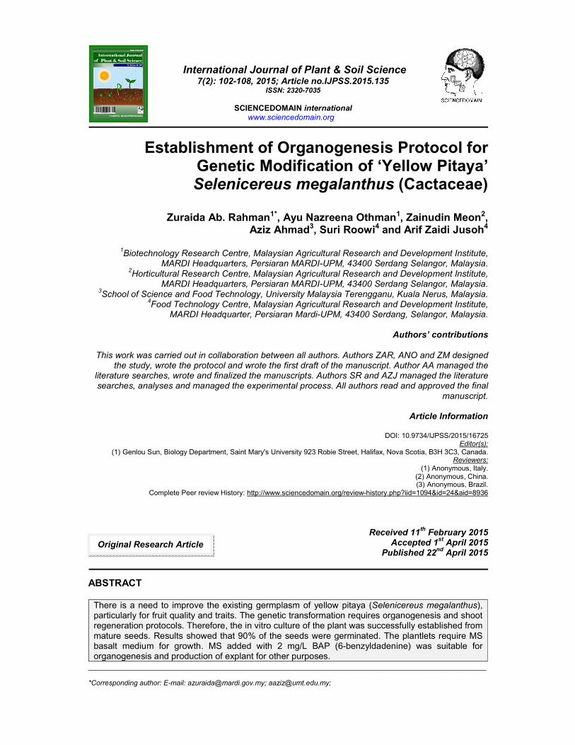
_____________________________________________________________________________________________________ *Corresponding author: E-mail: [email protected]; [email protected]; International Journal of Plant & Soil Science 7(2): 102-108, 2015; Article no.IJPSS.2015.135 ISSN: 2320-7035 SCIENCEDOMAIN international www.sciencedomain.org Establishment of Organogenesis Protocol for Genetic Modification of ‘Yellow Pitaya’ Selenicereus megalanthus (Cactaceae) Zuraida Ab. Rahman 1* , Ayu Nazreena Othman 1 , Zainudin Meon 2 , Aziz Ahmad 3 , Suri Roowi 4 and Arif Zaidi Jusoh 4 1 Biotechnology Research Centre, Malaysian Agricultural Research and Development Institute, MARDI Headquarters, Persiaran MARDI-UPM, 43400 Serdang Selangor, Malaysia. 2 Horticultural Research Centre, Malaysian Agricultural Research and Development Institute, MARDI Headquarters, Persiaran MARDI-UPM, 43400 Serdang Selangor, Malaysia. 3 School of Science and Food Technology, University Malaysia Terengganu, Kuala Nerus, Malaysia. 4 Food Technology Centre, Malaysian Agricultural Research and Development Institute, MARDI Headquarter, Persiaran Mardi-UPM, 43400 Serdang, Selangor, Malaysia. Authors’ contributions This work was carried out in collaboration between all authors. Authors ZAR, ANO and ZM designed the study, wrote the protocol and wrote the first draft of the manuscript. Author AA managed the literature searches, wrote and finalized the manuscripts. Authors SR and AZJ managed the literature searches, analyses and managed the experimental process. All authors read and approved the final manuscript. Article Information DOI: 10.9734/IJPSS/2015/16725 Editor(s): (1) Genlou Sun, Biology Department, Saint Mary's University 923 Robie Street, Halifax, Nova Scotia, B3H 3C3, Canada. Reviewers: (1) Anonymous, Italy. (2) Anonymous, China. (3) Anonymous, Brazil. Complete Peer review History: http://www.sciencedomain.org/review-history.php?iid=1094&id=24&aid=8936 Received 11 th February 2015 Accepted 1 st April 2015 Published 22 nd April 2015 ABSTRACT There is a need to improve the existing germplasm of yellow pitaya (Selenicereus megalanthus), particularly for fruit quality and traits. The genetic transformation requires organogenesis and shoot regeneration protocols. Therefore, the in vitro culture of the plant was successfully established from mature seeds. Results showed that 90% of the seeds were germinated. The plantlets require MS basalt medium for growth. MS added with 2 mg/L BAP (6-benzyldadenine) was suitable for organogenesis and production of explant for other purposes. Original Research Article
Transcript of Establishment of Organogenesis Protocol for Genetic ...
_____________________________________________________________________________________________________ *Corresponding author: E-mail: [email protected]; [email protected];
International Journal of Plant & Soil Science 7(2): 102-108, 2015; Article no.IJPSS.2015.135
ISSN: 2320-7035
SCIENCEDOMAIN international www.sciencedomain.org
Establishment of Organogenesis Protocol for Genetic Modification of ‘Yellow Pitaya’ Selenicereus megalanthus (Cactaceae)
Zuraida Ab. Rahman1*, Ayu Nazreena Othman1, Zainudin Meon2,
Aziz Ahmad3, Suri Roowi4 and Arif Zaidi Jusoh4
1Biotechnology Research Centre, Malaysian Agricultural Research and Development Institute,
MARDI Headquarters, Persiaran MARDI-UPM, 43400 Serdang Selangor, Malaysia. 2Horticultural Research Centre, Malaysian Agricultural Research and Development Institute,
MARDI Headquarters, Persiaran MARDI-UPM, 43400 Serdang Selangor, Malaysia. 3School of Science and Food Technology, University Malaysia Terengganu, Kuala Nerus, Malaysia.
4Food Technology Centre, Malaysian Agricultural Research and Development Institute,
MARDI Headquarter, Persiaran Mardi-UPM, 43400 Serdang, Selangor, Malaysia.
Authors’ contributions
This work was carried out in collaboration between all authors. Authors ZAR, ANO and ZM designed the study, wrote the protocol and wrote the first draft of the manuscript. Author AA managed the
literature searches, wrote and finalized the manuscripts. Authors SR and AZJ managed the literature searches, analyses and managed the experimental process. All authors read and approved the final
manuscript.
Article Information
DOI: 10.9734/IJPSS/2015/16725 Editor(s):
(1) Genlou Sun, Biology Department, Saint Mary's University 923 Robie Street, Halifax, Nova Scotia, B3H 3C3, Canada. Reviewers:
(1) Anonymous, Italy. (2) Anonymous, China. (3) Anonymous, Brazil.
Complete Peer review History: http://www.sciencedomain.org/review-history.php?iid=1094&id=24&aid=8936
Received 11th
February 2015 Accepted 1
st April 2015
Published 22nd April 2015
ABSTRACT
There is a need to improve the existing germplasm of yellow pitaya (Selenicereus megalanthus), particularly for fruit quality and traits. The genetic transformation requires organogenesis and shoot regeneration protocols. Therefore, the in vitro culture of the plant was successfully established from mature seeds. Results showed that 90% of the seeds were germinated. The plantlets require MS basalt medium for growth. MS added with 2 mg/L BAP (6-benzyldadenine) was suitable for organogenesis and production of explant for other purposes.
Original Research Article

Rahman et al.; IJPSS, 7(2): 102-108, 2015; Article no.IJPSS.2015.135
103
Keywords: Selenicereus megalanthus; micropropagation; yellow pitaya; cactaceae.
ABBREVIATION
MS0- phytohormone-free MS basal medium; MSA- MS medium added with 100 mg/l of asparagines; arginine or glutamine; MS2B -MS medium added with 2mg/L BAP; MS5B- MS medium added with 5 mg/L BAP; MS2N- MS medium added with 2 mg/L NAA; MS5N- MS medium added with 5 mg/L NAA; WPM- phtohormone-free woody plant medium.
1. INTRODUCTION Pitaya is a common name applied to a broad variety of warm-climate cacti fruit from different species and genera. It represents an interesting group of under-exploited crops with potential for human consumption. Pitaya is a member of the Cactaceae includes several edible fruit species that known rich in micronutrients [1]. It is native to the tropical forest regions of Mexico and Central and South America [2]. To date, the cultivated species are the white (Hylocereus undatus), red (H. polyrhizus) and yellow (Selenicereus megalanthus) varieties. These varieties are commercially grown in Taiwan, Nicaragua, Colombia, Vietnam, Israel, Australia and the USA. The red pitaya produces fruit with reddish-purple colour due to betacyanin contains high antioxidant properties [3]. Comparatively, yellow pitaya have smaller size than the red pitaya, but its fleshy pulp is pleasantly sweet. The yellow pitaya is a perennial climbing segmented cactus with triangular fleshy stems [4]. It is an epiphyte, and its aerial roots attach themselves to various types of supports such as wood and trees. The fruit is ovoid, spiny and yellow, with numerous small black seed embedded in a white pulp.
Pitaya is usually propagated through seeds or cuttings [5]. However, there is a need to improve the existing germplasm, particularly for fruit quality traits. Genetic transformation may be used to introduce desirable genes but it is necessary first to develop protocols for the initiation of organogenesis and for regeneration of whole plants. The tissue culture technique also is promising for commercial cultivation as the demand for the planting materials increase in the near future [6].
The shoot proliferation and somatic embryogenesis in yellow pitaya have been previously reported [7,6]. Infante [7], used yellow pitaya epicotyls as explants in a culture medium containing various combinations of naphthaleneacetic acid (NAA) and 6-benzyldadenine (BAP). With this method, certain
difficulties were observed by us such as a lack of development or death of calli. Pelah and co-workers [6] used the thidiazuron (TDZ) on in vitro culture of yellow pitaya. In the present study we develop the organogenesis in yellow pitaya by means for application in genetic modification of the germplasm.
2. MATERIALS AND METHODS 2.1 Source of Plant Materials Seeds of Selenicereus megalanthus (K. Schum. ex Vaupel) Moran were obtained from mature fruits bought at Harrods Market that sold them as products of Colombia (Figs. 4a-c). The seeds were thoroughly washed in 10% (v/v) Decon™ solution for 10 min and followed sterile double-distilled water. In aseptic condition, the seeds were surface sterilized by immersion in 20% (v/v) of commercial bleach for 30 min and followed by rinsed three times with sterile double-distilled water.
2.2 Establishment of Culture A total of 200 seeds were randomly divided into two groups. The seed coats were excised and removed from the first group, while second group remained intact. Each group was 100 seeds. Seeds were separately germinated on sterile petri-dishes containing solid basal MS Murashige and Skoog [8] medium. After 2 weeks, the number of germinated seed were recorded. Subsequently, the two-week old seedling were randomly selected and transferred into 100mL Erlenmeyer flasks containing 30mL growth medium. Five seedling per flask and three flask per treatment medium. The medium used were MSO, MSA, MS2B, MS5B and WPM. Media were supplemented with 3% (w/v) sucrose and solidified with 7 g/L agar. All cultures were incubated in culture room at 24ºC under 16h/8h photoperiod provided by cool-white fluorescent lamps. After 4 weeks, the fresh weight of the plantlets was recorded for calculation of the percentage growth rate.

Rahman et al.; IJPSS, 7(2): 102-108, 2015; Article no.IJPSS.2015.135
104
2.3 Organogenesis
The well-developed plantlets were randomly selected and transferred onto MS0, MS2B MS5B, MS2N or MS5N for 4 weeks and subsequently sub-cultured onto organogenesis medium (Table 1). After 4 weeks the number of branches or any changes on the plantlet was recorded. The data (25 replicates per treatment) were subjected to one way analysis of variance (ANOVA) to assess treatment differences and interaction using the SPSS version 11.0.
3. RESULTS AND DISCUSSION Results showed that hundred percent of successful in contaminant-free explants were obtained in Clorox concentrations at 80% (v/v) and above. The highest survival rate of explants was also obtained in 20 and 40% (v/v) of Clorox. The survival rate was declined in as the Clorox concentration used increases (Fig. 1). The right concentration of detergent is crucial to obtain high successful rate of surface sterilization and at the same time the number of survival explants also high. In most cases, high free-contamination is achieved in high concentration of detergent, but lower survival rate. Bleaching of tissue was the major phenomenon that contributed the lower survival rate. Comparatively, using seed as explants is more promising than the fragile
explant such as young leaves, shoot tip or wounded explants. The germination rate of seeds was varied between the two groups of explants. The excised seeds exhibited faster germination than the intact seeds (Fig. 2). Results showed, 70% percent of the excised seeds were successfully germinated after 2 weeks on the growth medium. The percentage of germinated seeds was increased to 90% after the 16
th week for both types of
explant used. These results indicated that removing of seed coat was facilitated the germination (Figs. 4d-f). The embryos was directly exposed to the culture medium and allowed the water and nutrient uptake by embryos [9].
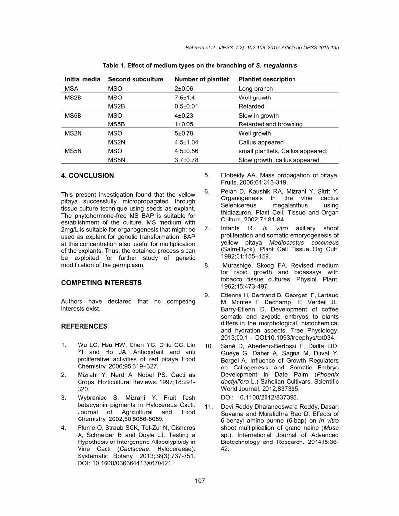
3.1 Growth and Organogenesis The growth of plantlets was significantly influenced by the type of medium used (Fig. 3). The highest growth rate was in MSO basal medium followed with MSA and WPM. BAP added medium exhibited the lowest growth rate. The results indicated that basal MS medium was the most suitable medium for the growth seedling of yellow pitaya. On the other hand, the formation of organ on yellow pitaya was influence by the hormone, BAP added into the culture medium (Table 1).
Fig. 1. Survival rate of S. megalanthus seeds cultured on MS medium treated with different
concentrations Clorox
0
20
40
60
80
100
120
5 10 20 40 60 80 100
Surv
ival ra
te (%
)
Clorox concentration (%)

Rahman et al.; IJPSS, 7(2): 102-108, 2015; Article no.IJPSS.2015.135
105
The highest number of branches was on explants cultures in 2 mg/L BAP for 4 weeks and in basal MS for another 4 weeks (Fig. 4i). Prolong the culture period on basal medium; for 8 weeks was enhanced the height of plantlet (Fig. 4h). Callogenesis was observed on explant culture in NAA containing media for 8 weeks (Table 1). The capacity of callogenesis depends on genotype
and plant growth regulator in the medium. Auxin, such NAA is normally applied for initiation of callus [10]. According to Devi et al. [11], BAP at concentration of 2.0mg/L showed the best shoot induction of Grand naine plantlets. Neha et al. [12], in their finding reported that 4.0mg/L BAP was the best response for increasing the number of shoots of Curcuma caesia.
Fig. 2. Germination rate of the excised and non-excised seeds of S. Megalanthus
Fig. 3. Effect of medium modifications on the growth of embryos into plantlets
0
20
40
60
80
100
120
1 2 4 8 16 32
Germ
ination
rate
s (
%)
Weeks
excised seeds
0
50
100
150
200
250
300
350
400
450
MSA WPM MSO MSO+2BAP MSO+ 5BAP
Gro
wth
rate
(%
)
Media types

Fig. 4. ripen-fruit of yellow pitaya (a), pitaya 10x magnification (c, d), emergence of shoot tips on culture medium (e, f), germinated
seeds (g), plantlets after four plantlets in phytohormone-free MS medium (j),
acclimatization of the seedling (k) and plantlets on chamber (l) with 100% survival rate. The Jiffy pellets increased the growth rate of plantlets,
compa
a
d
g
j
Rahman et al.; IJPSS, 7(2): 102-108, 2015; Article no.
106
fruit of yellow pitaya (a), Cross-section of the ripe yellow pitaya fruitx magnification (c, d), emergence of shoot tips on culture medium (e, f), germinated
month (h), organ proliferation (i), rooting on the individual free MS medium (j), plantlets that directly transplanted into Jiffy for
acclimatization of the seedling (k) and plantlets on pellets in a simple high humidity mist survival rate. The Jiffy pellets increased the growth rate of plantlets,
compared to the traditional sand mix
b c
e f
h i
k l
; Article no.IJPSS.2015.135
section of the ripe yellow pitaya fruit (b), seed of x magnification (c, d), emergence of shoot tips on culture medium (e, f), germinated
(i), rooting on the individual that directly transplanted into Jiffy for
pellets in a simple high humidity mist survival rate. The Jiffy pellets increased the growth rate of plantlets,
Rahman et al.; IJPSS, 7(2): 102-108, 2015; Article no.IJPSS.2015.135
107
Table 1. Effect of medium types on the branching of S. megalantus
Initial media Second subculture Number of plantlet Plantlet description
MSA MSO 2±0.06 Long branch
MS2B MSO 7.5±1.4 Well growth
MS2B 0.5±0.01 Retarded
MS5B MSO 4±0.23 Slow in growth
MS5B 1±0.05 Retarded and browning
MS2N MSO 5±0.78 Well growth
MS2N 4.5±1.04 Callus appeared
MS5N MSO 4.5±0.56 small plantlets, Callus appeared,
MS5N 3.7±0.78 Slow growth, callus appeared
4. CONCLUSION
This present investigation found that the yellow pitaya successfully micropropagated through tissue culture technique using seeds as explant. The phytohormone-free MS BAP is suitable for establishment of the culture. MS medium with 2mg/L is suitable for organogenesis that might be used as explant for genetic transformation. BAP at this concentration also useful for multiplication of the explants. Thus, the obtained process s can be exploited for further study of genetic modification of the germplasm.
COMPETING INTERESTS
Authors have declared that no competing interests exist.
REFERENCES
1. Wu LC, Hsu HW, Chen YC, Chiu CC, Lin YI and Ho JA. Antioxidant and anti proliferative activities of red pitaya. Food Chemistry. 2006;95:319–327.
2. Mizrahi Y, Nerd A, Nobel PS. Cacti as Crops. Horticultural Reviews. 1997;18:291-320.
3. Wybraniec S, Mizrahi Y. Fruit flesh betacyanin pigments in Hylocereus Cacti. Journal of Agricultural and Food Chemistry. 2002;50:6086-6089.
4. Plume O, Straub SCK, Tel-Zur N, Cisneros A, Schneider B and Doyle JJ. Testing a Hypothesis of Intergeneric Allopolyploidy in Vine Cacti (Cactaceae: Hylocereeae). Systematic Botany. 2013;38(3):737-751. DOI: 10.1600/036364413X670421.
5. Elobeidy AA. Mass propagation of pitaya. Fruits. 2006;61:313-319.
6. Pelah D, Kaushik RA, Mizrahi Y, Sitrit Y. Organogenesis in the vine cactus Selenicereus megalanthus using thidiazuron. Plant Cell, Tissue and Organ Culture. 2002;71:81-84.
7. Infante R. In vitro axillary shoot proliferation and somatic embryogenesis of yellow pitaya Mediocactus coccineus (Salm-Dyck). Plant Cell Tissue Org Cult. 1992;31:155–159.
8. Murashige, Skoog FA. Revised medium for rapid growth and bioassays with tobacco tissue cultures. Physiol. Plant. 1962;15:473-497.
9. Etienne H, Bertrand B, Georget F, Lartaud M, Montes F, Dechamp E, Verdeil JL, Barry-Etienn D. Development of coffee somatic and zygotic embryos to plants differs in the morphological, histochemical and hydration aspects. Tree Physiology. 2013;00,1 – DOI:10.1093/treephys/tpt034.
10. Sané D, Aberlenc-Bertossi F, Diatta LID, Guèye G, Daher A, Sagna M, Duval Y, Borgel A. Influence of Growth Regulators on Callogenesis and Somatic Embryo Development in Date Palm (Phoenix dactylifera L.) Sahelian Cultivars. Scientific World Journal. 2012;837395.
DOI: 10.1100/2012/837395.
11. Devi Reddy Dharaneeswara Reddy, Dasari Suvarna and Muralidhra Rao D. Effects of 6-benzyl amino purine (6-bap) on In vitro shoot multiplication of grand naine (Musa sp.). International Journal of Advanced Biotechnology and Research. 2014;l5:36-42.
Rahman et al.; IJPSS, 7(2): 102-108, 2015; Article no.IJPSS.2015.135
108
12. Neha Behar, Tiwari KL, Jadhav SK. Effect of explants type in development of In vitro micropropagation protocol of an
endangered medicinal plant: Curcuma caesia Roxb. Biotechnology; 2014:13(1): 22-27.
_________________________________________________________________________________ © 2015 Rahman et al.; This is an Open Access article distributed under the terms of the Creative Commons Attribution License (http://creativecommons.org/licenses/by/4.0), which permits unrestricted use, distribution, and reproduction in any medium, provided the original work is properly cited.
Peer-review history: The peer review history for this paper can be accessed here:
http://www.sciencedomain.org/review-history.php?iid=1094&id=24&aid=8936