Effect of α-thalassemia and β-globin gene cluster haplotypes on the hematological and clinical...
5
American Journal of Hematology 53:72-76 (1996) Effect of a-Thalassemia and p-Globin Gene Cluster Haplotypes on the Hematological and Clinical Features of Sickle-Cell Anemia in Brazil M.S. Figueiredo, J. Kerbauy, M.S. Gonqalves, V.R. Arruda, S.T.O. Saad, M.F. Sonati, T. Stoming, and F.F. Costa Federal University of SBo Paulo, SBo Paulo, SP (M.S.F., J.K.), Hematology-Hemotherapy Center, Department of Clinical Medicine, State University of Campinas-UNICAMP, Campinas, SP (M.S.G., V.R.A., S.T.O.S., M.F.S., F.F.C.), Brazil; Department of Biochemistry, Medical College of Georgia, Augusta, Georgia (T.S.) To compare the features of sickle-cell anemia in Brazil with those in other locales, we studied the effects of the p-globin-like gene cluster haplotype and a-thalassemia upon the clinical and hematological features in 85 patients. The distribution of haplotypes differed from that in the United States and Jamaica. The Central African Republic (CAR) haplotype predominated; 34% of patients were CAR haplotype homozygotes, 45% CAW Benin homozygotes, and 11 % Benin homozygotes. No Senegal haplotype chromosomes were observed. a-thalassemia was present in 17.5% of patients. HbF levels were higher in Benin homozygotes, compared with the other two groups (P < 0.05). Nearly half the patients with a CAR haplotypehad leg ulcers, compared to 12.5% of the Benin homozygote group; stroke did not occur in a-thalassemia carriers, but neither result was statistically significant. As in other studies, our results indicate that the CAR haplotype may be associated with more severe disease. Key words: sickle-cell disease, clinical features, hemoglobin S, haplotypes, d h a - lassemia o 1996 Wiley-Liss, Inc. INTRODUCTION Clinical manifestations of sickle-cell anemia, the ho- mozygous state for hemoglobin S (HbS), vary according the geographical locations of the populations studied [ 1- 31. Several lines of evidence suggest that the clinical and hematological expression of sickle-cell anemia may be modified by other genetic determinants. These include a-thalessemia and modulators of fetal hemoglobin con- centrations [4-61. An array of polymorphisms linked to the ps mutation, called haplotypes, has provided important anthropologi- cal information regarding the multiple origins of the Hb S gene [7,8]. There are at least four different major ps haplotypes, named according to the regions of their high- est frequency: Central African Republic (CAR or Bantu), Benin, Senegal, and Saudi-Arabian types. The Ps-globin gene cluster haplotype may be associated with special hematological and clinical features of sickle-cell anemia [9-121. The report of Powars [lo] suggested that the CAR haplotype was a major risk factor associated with clinically severe sickle-cell anemia and organ damage. However, the possible role of ps haplotypes is controver- sial, since other work suggsted that in adults, haplotype had little bearing on the clinical and hematological picture of sickle-cell anemia [ 1 1,121. The ps haplotype distribution in Brazil is distinct from that of the USA and Jamaica, where most studies of the association of haplotypes and clinical expression have been done [ 13,141. To compare the features of sickle-cell anemia in Brazil with those in other locales, we studied the effects of P-globin gene cluster haplotype and a - thalassemia upon the clinical and hematological features of 85 patients with sickle-cell anemia from Brazil. Received for publication July 25, 1995; accepted April 10, 1996. Address reprint requests to Fernando F. Costa, M.D., Hematology- Hemotherapy Center, State University of Campinas (UNICAMP), CP 6198 CEP 13.081-970, Campinas, SP, Brazil. 0 1996 Wiley-Liss, Inc.
Transcript of Effect of α-thalassemia and β-globin gene cluster haplotypes on the hematological and clinical...
American Journal of Hematology 53:72-76 (1996)
Effect of a-Thalassemia and p-Globin Gene Cluster Haplotypes on the Hematological and Clinical Features of
Sickle-Cell Anemia in Brazil
M.S. Figueiredo, J. Kerbauy, M.S. Gonqalves, V.R. Arruda, S.T.O. Saad, M.F. Sonati, T. Stoming, and F.F. Costa
Federal University of SBo Paulo, SBo Paulo, SP (M.S.F., J.K.), Hematology-Hemotherapy Center, Department of Clinical Medicine, State University of Campinas-UNICAMP, Campinas, SP (M.S.G., V.R.A., S.T.O.S., M.F.S., F.F.C.), Brazil; Department of Biochemistry, Medical
College of Georgia, Augusta, Georgia (T.S.)
To compare the features of sickle-cell anemia in Brazil with those in other locales, we studied the effects of the p-globin-like gene cluster haplotype and a-thalassemia upon the clinical and hematological features in 85 patients. The distribution of haplotypes differed from that in the United States and Jamaica. The Central African Republic (CAR) haplotype predominated; 34% of patients were CAR haplotype homozygotes, 45% CAW Benin homozygotes, and 11 % Benin homozygotes. No Senegal haplotype chromosomes were observed. a-thalassemia was present in 17.5% of patients. HbF levels were higher in Benin homozygotes, compared with the other two groups (P < 0.05). Nearly half the patients with a CAR haplotype had leg ulcers, compared to 12.5% of the Benin homozygote group; stroke did not occur in a-thalassemia carriers, but neither result was statistically significant. As in other studies, our results indicate that the CAR haplotype may be associated with more severe disease.
Key words: sickle-cell disease, clinical features, hemoglobin S, haplotypes, d h a - lassemia
o 1996 Wiley-Liss, Inc.
INTRODUCTION
Clinical manifestations of sickle-cell anemia, the ho- mozygous state for hemoglobin S (HbS), vary according the geographical locations of the populations studied [ 1- 31. Several lines of evidence suggest that the clinical and hematological expression of sickle-cell anemia may be modified by other genetic determinants. These include a-thalessemia and modulators of fetal hemoglobin con- centrations [4-61.
An array of polymorphisms linked to the ps mutation, called haplotypes, has provided important anthropologi- cal information regarding the multiple origins of the Hb S gene [7,8]. There are at least four different major ps haplotypes, named according to the regions of their high- est frequency: Central African Republic (CAR or Bantu), Benin, Senegal, and Saudi-Arabian types. The Ps-globin gene cluster haplotype may be associated with special hematological and clinical features of sickle-cell anemia [9-121. The report of Powars [ lo] suggested that the
CAR haplotype was a major risk factor associated with clinically severe sickle-cell anemia and organ damage. However, the possible role of ps haplotypes is controver- sial, since other work suggsted that in adults, haplotype had little bearing on the clinical and hematological picture of sickle-cell anemia [ 1 1,121.
The ps haplotype distribution in Brazil is distinct from that of the USA and Jamaica, where most studies of the association of haplotypes and clinical expression have been done [ 13,141. To compare the features of sickle-cell anemia in Brazil with those in other locales, we studied the effects of P-globin gene cluster haplotype and a - thalassemia upon the clinical and hematological features of 85 patients with sickle-cell anemia from Brazil.
Received for publication July 25, 1995; accepted April 10, 1996.
Address reprint requests to Fernando F. Costa, M.D., Hematology- Hemotherapy Center, State University of Campinas (UNICAMP), CP 6198 CEP 13.081-970, Campinas, SP, Brazil.
0 1996 Wiley-Liss, Inc.
a-Thalassemia and ps Haplotypes on Sickle-Cell Anemia in Brazil 73
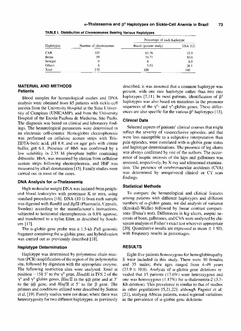
TABLE 1. Distribution of Chromosomes Bearina Various HaDlotvDes
Percentage of each haplotype
Haplotypes Number of chromosomes Brazil (present study) USA [I21
CAR Benin Senegal Others Total
I05 59 0 6
170
61 .I6 15.9 34.71 50.0 0 8.0 3.53 26.1
100 100
MATERIAL AND METHODS Patients
Blood samples for hematological studies and DNA analysis were obtained from 85 patients with sickle-cell anemia from the University Hospital at the State Univer- sity of Campinas (UNICAMP), and from the University Hospital of the Escola Paulista de Medicina, Siio Paulo. The diagnosis was based on clinical and laboratory find- ings. The hematological parameters were determined in an electronic cell-counter. Hemoglobin electrophoresis was performed on cellulose acetate strips with Tris- EDTA-boric acid, pH 8.9, and on agar gels with citrate buffer, pH 6.1. Presence of HbS was confirmed by a low solubility in 2.35 M phosphate buffer containing dithionite. HbA, was measured by elution from cellulose acetate strips following electrophoresis, and HbF was measured by alkali denaturation [ 151. Family studies were carried out in most of the cases.
DNA Analysis for a-Thalassemia
High molecular weight DNA was isolated from periph- eral blood leukocytes with proteinase K or urea, using standard procedures [ 161. DNA (10 1) from each sample was digested with BamHI and BglII (Pharmacia, Uppsala, Sweden) according to the manufacturer’s instructions, subjected to horizontal electrophoresis in 0.8% agarose, and transferred to a nylon filter, as described by South- ern 1171.
The a-globin gene probe was a 1.5-kb Pstl genomic fragment containing the a-globin gene, and hybridization was carried out as previously described [ 181.
Haplotype Determination
Haplotype was determined by polymerase chain reac- tion (PCR) amplification of the region of the polymorphic site, followed by digestion with the appropriate enzyme. The following restriction sites were analysed: XmnI at position - 158 5 ’ to the yG gene, Hind11 in IVS 2 of the yG and yA globin genes, HincII in the $6 gene and at 3‘ to the $p gene, and HinflI at 5’ to the p gene. The primers and conditions utilized were described by Sutton et al. [ 191. Family studies were not done; where there was heterozygosity for two different haplotypes, as previously
described, it was assumed that a common haplotype was present, with one rare haplotype rather than two rare haplotypes [7,11]. In most patients, identification of ps haplotypes was also based on mutations in the promoter sequences of the yc- and yA-globin genes. These differ- ences are also specific for the various ps haplotypes [13].
Clinical Data
Selected aspects of patients’ clinical courses that might reflect the severity of vasoocclusive episodes, and that were less susceptible to a subjective interpretation than pain episodes, were correlated with a-globin gene status and haplotype determinations. The presence of leg ulcers was always confirmed by one of the authors. The occur- rence of aseptic necrosis of the hips and gallstones was assessed, respectively, by X-ray and ultrasound examina- tion. The presence of cerebrovascular accidents (CVA) was determined by unequivocal clinical or CT scan findings.
Statistical Methods
To compare the hematological and clinical features among patients with different haplotypes and different numbers of a-globin genes, we did analysis of variance (Kruskall-Wallis) followed by linear contrast compari- sons (Dunn’s test). Differences in leg ulcers, aseptic ne- crosis of bone, gallstones, and CVA were analyzed by chi- square analysis or Fisher’s exact test wherever appropriate 1201. Quantitative results are expressed as mean 5 I SD, with frequency results in percentages.
RESULTS
Eight-five patients homozygous for hemoglobinopathy S were included in this study. There were 50 females and 35 males; their ages ranged from 4 4 9 years (21.9 5 10.0). Analysis of a-globin gene deletions re- vealed that 15 patients (17.6%) were heterozygous and one was homozygous (1.17%) for a-thalassemia-2 (3.7- Kb deletion). This prevalence is similar to that of studies in other populations [5,21,22], although Pagnier et al. [2 11, studying African patients, noted regional variations in the prevalence of a-globin gene deletions.

74 Figueiredo et at.
TABLE II. Prevalence of a-Thalassemia Among Different HaDlOtVDeS*
Haplotypes acxlaa (n ) a - / w (n) a-h- (n) Total
CARICAR 22 (70.9%) 8 (25.8%) I (3.3%) 31 CARIBenin 32 (84.3%) 6 (15.7%) 0 (0%) 38 BendBenin 10 (100%) 0 (0%) 0 (0%) 10 CAR/? 4 (80%) 1 (20%) 0 (0%) 5 Benin/? 1(100%) 0 (0%) 0 (0%) 1 Total 69 (81.1%) 15 (17.6%) 1 (1.170) 85
*n, number of patients
There were 31 subjects with CAWCAR (36.5%), 38 with CAWBenin (44.7%), 10 with BenidBenin (1 1.7%), 5 with CAWatypical (5.8%), and 1 with Benin/atypical (1.2%) haplotypes. The Senegal haplotype was not ob- served; nor was the Saudi-Arabian haplotype. Percentages of chromosomes bearing each haplotype are shown in Table I . The prevalence of haplotypes varies in patients resident in different regions of Brazil, reflecting differ- ences in the origins of slaves brought to this country [ 141.
The a-thalassemia distribution among the various hap- lotypes is shown in Table 11. There were no patients with a Benin haplotype and a-thalassemia. This may be indicative of a higher prevalence of thalassemia in CAR haplotype patients, as shown by Castillo et al. [22] and de Montalembert et al. [12]. However, this difference was not statistically significant.
Some of the hematological data for these groups are shown in Table 111. We observed a significant difference ( P < 0.05) in hemoglobin concentration between patients with and without a-gene deletion, although no difference was found when we compared the hemoglobin concentra- tion among the various haplotypes. The same effect was observed in the distribution of mean corpuscular volume (MCV) values. No differences in HbF were noted in patients with and without a-thalassemia. There was a statistically significant difference ( P < 0.05) in HbF lev- els in CAR and Benin haplotypes. The HbA2 was similar in all groups (data not shown).
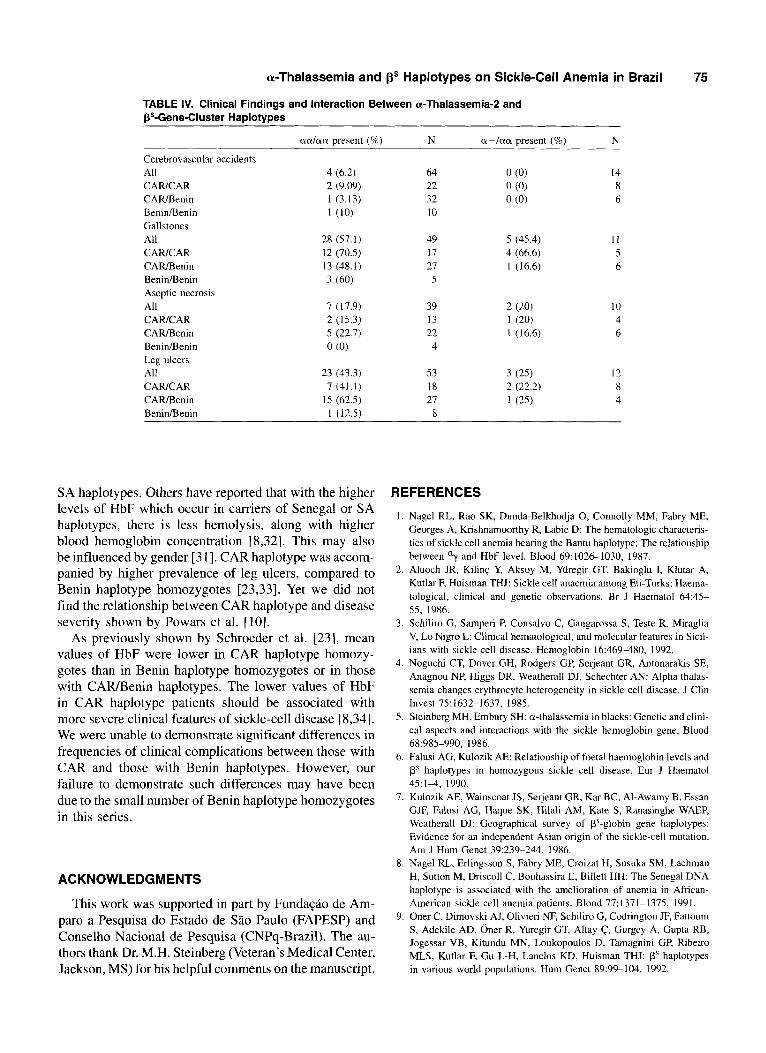
Clinical observations, which were available for all pa- tients, are shown in Table IV. There were no statistically significant differences among the groups in the clinical features examined.
DISCUSSION
Brazil, a large continental country, has an interesting distribution of hemoglobinopathies. This follows from the influx of many different immigrant groups, and from the slave trade of the eighteenth and nineteenth centuries I1 31. This varied genetic background makes the study of sickle-cell anemia in Brazil of special interest, and raises the possibility of achieving additional insights into the genetic modulation of this disease.
Consonant with other work, analysis of our laboratory
TABLE 111. Hematologic Values and Fetal Hemoglobin: Interaction of ~Thalassemia-2 With PS-Gene-Cluster Haplotypes
Age (years) All 21.4 t 9.6 24.2 i 11.8 CAWCAR 22.9 -c 9.3 CARlBenin 20.3 -C 8.8 22 t 13.7 BeninlBenin 21.1 t 11.9 Hh (gldl) All 7.6 2 1.2 8.6 t 1.07* CAR/C AR 7.6 ? 1.2 8.7 5 1.2" CAR/Benin 7.7 2 1.3 8.3 i 0.7 BeninlBenin 7.7 5 1.1 HbF (%) All 6.6 f 4.1 5.1 2 3.8 CAWCAR 4.9 -C 2.9 2.8 f 2.3 CAWBenin 7.3 5 4.7* 6.5 i 3.9 BeninlBenin 8.3 5 3.0* HbF (gldl) All 0.52 f 0.37 0.43 -C 0.30 CAWCAR 0.37 t 0.22 0.32 t 0.28 CAR/Benin 0.60 i 0.47* 0.53 i 0.29 BeniniBenin 0.64 t 0.2 I*
*P < 0.05
27.5 i 9.8
data showed that patients with a-thalassemia-2 trait had a significant increase in hemoglobin concentration and reduction in MCV [5,22,23]. Also concordant with prior studies, there was no significant difference in HbF levels among the a-globin genotypes [24-261. Previous work has shown a relationship between a-thalassemia and the hematological and some clinical features of sickle-cell disease [27,28]. The prevalence of CVA was higher in patients with sickle-cell anemia without a-thalassemia. As suggested by Piomelli et a]. [29] and Adams et al. [30], a-thalassemia may exert a protective effect against stroke in patients with sickle-cell anemia. While there was no effect of concurrent a-thalassemia on the occurrence of gallstones or osteonecrosis, leg ulcers were less frequent in those with an a-thalassemia gene. Other investigators have also made this observation [5,23,31].
P-globin gene haplotypes did not appear to influence hemoglobin concentration or selected clinical manifesta- tions of sickle-cell anemia. Perhaps this was due to the absence in this series of patients of either the Senegal or
a-Thalassemia and gs Haplotypes on Sickle-Cell Anemia in Brazil 75
TABLE IV. Clinical Findings and Interaction Between a-Thalassemia-2 and BS-Gene-Cluster Haplotypes
aa/aa present (%) N a-/aa present (%) N
Cerebrovascular accidents All CAWCAR CARIBenin BeninlBenin Gallstones All CARICAR CARlSenin BeninBenin Aseptic necrosis All CAWCAR CARlSenin BenidBenin Leg ulcers All CARICAR CARlSenin BeninlBenin
4 (6.2) 2 (9.09) 1 (3.13) 1 (10)
28 (57.1) 12 (70.5) 13 (48.1) 3 (60)
7 ( I 7.9) 2 (15.3) 5 (22.7) 0 (0)
23 (43.3) 7 (41.1)
15 (62.5) 1 (12.5)
64 22 32 10
49 17 27
5
39 13 22 4
53 18 27 8
14 8 6
I 1 5 6
10 4 6
12 8 4
SA haplotypes. Others have reported that with the higher levels of HbF which occur in carriers of Senegal or SA haplotypes, there is less hemolysis, along with higher blood hemoglobin concentration [8,32]. This may also be influenced by gender [3 I]. CAR haplotype was accom- panied by higher prevalence of leg ulcers, compared to Benin haplotype homozygotes [23,33]. Yet we did not find the relationship between CAR haplotype and disease severity shown by Powars et al. [lo].
As previously shown by Schroeder et al. [23], mean values of HbF were lower in CAR haplotype homozy- gotes than in Benin haplotype homozygotes or in those with C A m e n i n haplotypes. The lower values of HbF in CAR haplotype patients should be associated with more severe clinical features of sickle-cell disease [8,34]. We were unable to demonstrate significant differences in frequencies of clinical complications between those with CAR and those with Benin haplotypes. However, our failure to demonstrate such differences may have been due to the small number of Benin haplotype homozygotes in this series.
ACKNOWLEDGMENTS
This work was supported in part by FundaSAo de Am- par0 a Pesyuisa do Estado de S2o Paulo (FAPESP) and Conselho Nacional de Pesquisa (CNPy-Brazil). The au- thors thank Dr. M.H. Steinberg (Veteran’s Medical Center, Jackson, MS) for his helpful comments on the manuscript.
REFERENCES
1. Nagel RL, Rao SK, Dunda-Belkhodja 0, Connolly MM, Fabry ME, Georges A, Krishnamoorthy R, Labie D: The hematologic characteris- tics of sickle cell anemia bearing the Bantu haplotype: The relationship between ‘-y and HbF level. Blood 69:102&1030, 1987.
2. Aluoch JR, KilinG Y, Aksoy M, Yuregir GT, Bakioglu I, Klutar A, Kutlar F, Huisman THJ: Sickle cell anaemia among Eti-Turks: Haema- tological, clinical and genetic observations. Br J Haematol 64:45- 55, 1986.
3. Schiliro G, Samperi P, Consalvo C, Gangarossa S, Teste R, Miraglia V, Lo Nigro L: Clinical hematological, and molecular features in Sicil- ians with sickle cell disease. Hemoglobin 16:46948O, 1992.
4. Noguchi CT, Dover GH, Rodgers GP, Serjeant GR, Antonarakis SE, Anagnou NP, Higgs DR, Weatherall DJ, Schechter AN: Alpha thalas- seinia changes erythrocyte heterogeneity in sickle cell disease. J Clin Invest 75:1632-1637. 1985.
5. Steinberg MH, Embury SH: a-thalassemia in blacks: Genetic and clini- cal aspects and interactions with the sickle hemoglobin gene. Blood 68:985-990, 1986.
6. Falusi AG, Kulozik AE: Relationship of foetal haemoglobin levels and ps haplotypes in homozygous sickle cell disease. Eur J Haematol 45:14, 1990.
7. Kulozik AE, Wainscoat JS, Serjeant GR, Kar BC, Al-Awamy B, Essan GJF, Falusi AG, Haque SK, Hilali AM, Kate S, Ranasinghe WAEP, Weatherall DJ: Geographical survey of Ps-globin gene haplotypes: Evidence for an independent Asian origin of the sickle-cell mutation. Am J Hum Genet 39:239-244, 1986.
8. Nagel RL, Erlingsson S, Fabry ME, Croizat H, Susuka SM, Lachman H, Sutton M, Driscoll C, Bouhassira E, Billett HH: The Senegal DNA haplotype is associated with the amelioration of anemia in African- American sickle cell anemia patients. Blood 77:1371-1375, 1991.
9. Oner C, Dimovski AJ, Olivieri NF, Schiliro G, Codrington JF, Fattoum S, Adekile AD, Oner R, Yiiregir GT, Altay c, Gurgey A, Gupta RB, Jogessar VB, Kitundu MN, Loukopoulos D, Tamagnini GP, Ribeiro MLS, Kutlar F, Gu L-H, Lanclos KD, Huisman THJ: ps haplotypes in various world populations. Hum Genet 89:99-104, 1992.
76 Figueiredo et al. 10. Powars DR: Sickle cell anemia: @5-gene-cluster haplotypes as prognos-
tic indicators of vital organ failure. Semin Hematol28:202-208, 1991. 11. De Montalembert M, Maier-Redelsperger M, Girot R, Belloy M, Vilmer
E, Ducrocp R, Guidal C, Elion J: P-globin gene cluster and a-thalas- semia do not correlate with the acute clinical manifestations of sickle cell disease in children. Blood 82:2595-2596, 1993.
12. Rieder RF, Safaya S, Gillette P, Fryd S, Hsu H, Adams JG 111, Steinberg MH: Effect of P-globin gene cluster haplotype on the hematological and clinical featuresofsicklecell anemia. AmJHematol36:184-189,1991.
13. Gonfalves MS, Nechman JF, Figueiredo MS, Kerbauy J, Amtda VR, Sonati MF, Saad STO, Costa FF, Stoming TA: Sickle cell anemia in a Brazilian population from Slo Paulo. A study of the p” haplotypes. Hum Hered 44:322-327, 1994.
14. Costa FF, Armda VR, Gonfalves MS, Miranda SRP, Carvalho MH, Sonati MF, Saad STO, Gesteira F, Fernandes D, Nascimento ML, Queiroz IL: Bs-gene-cluster haplotypes in sickle cell anemia patients from two regions of Brazil. Am J Hematol 45:96-97, 1994.
15. Pembrey ME, McWade P, Weatherall DJ: Reliable routine estimation of small amounts of foetal haemoglobin by alkali denaturation. J Clin Pathol 25:738-740, 1972.
16. Poncz M, Solwiejcyk D, Harpel B, Mory Y, Schwartz E, Surrey S: Construction of human gene libraries from small amounts of peripheral human blood: Analysis of P-like globin genes. Hemoglobin 6:27- 36, 1982.
17. Southern EM: Detection of specific sequences among DNA fragments separated by gel electrophoresis. J Mol Biol 98:503-517, 1975.
18. Sonati MF, Farah SB, Ramalho AS, Costa FF: High prevalence of deletional type of alpha thalassemia among a black population of Brazil. Hemoglobin 15:309-311, 1991.
19. Sutton M, Bouhassira EE, Nagel RL: Polymerase chain reaction ampli- fication applied to the determination of P-like globin gene cluster haplotypes. Am J Hematol 32:66-69, 1989.
20. Agresti A, Finlay B, eds. Statistical Methods for the Social Sciences. San Francisco: Dellen Publishing Co., 1986.
21. Pagnier J, Dunda-Belkhodja 0, Zohoun I, Teyssier J, Baya H, Jaeger G, Nagel RL, Labie D: a-thalassemia among sickle cell anemia patients in various African populations. Hum Genet 68:318-319, 1984.
22. Castillo R, Gay RN, Ballas SK: Homozygosity for the CAR P-haplotype in sickle in sickle cell anemia (SS) is associated with increased preva- lence of a-gene deletion and leg ulcers. Blood 8 0 (99 (abstract), 1992.
23. Higgs DR, Aldridge BE, Lamb J, Clegg JB, Weatherall DJ, Hayes RJ,
Grandison Y, Lowrie Y, Mason W, Serjeant BE, Serjeant GR: The interaction of alpha-thalassemia and homozygous sickle-cell disease. N Engl J Med 306:1441-1446, 1982.
24. Schroeder WA, Powards DR, Kay LM, Chan LS, Huynh V, Shelton JB, Shelton JR: p-cluster haplotypes, a-gene status, and hematological data from SS, SC, and S- P-thalasemia patients in Southern California. Hemoglobin 13:325-353, 1989.
25. Felice AE, Wehber B, Miller A, Mayson SM, Hams HF, Henson JB, Gravely ME, Huisman THJ: The association of sickle cell anemia with heterozygous and homozygous a-thalassemia-2: In vitro Hb chain synthesis. Am J Hematol 6:91-106, 1979.
26. Felice AE, McKie KM, Cleek MP, Marino EM, Kutlar A, McKie VC: Effects of a-thalassemia-2 on the developmental changes of hematolog- ical values in children with sickle cell disease from Georgia. Am J Hematol 25:389400, 1987.
27. McKie VC, McKie KM, Felice AE: a-thalassemia and cholelithiasis in children with SS disease. Clin Res 35:64 (abstract), 1987.
28. Ballas SK, Talacki CA, Rao VM, Steiner RM: The prevalence of avascular necrosis in sickle cell anemia: Correlation with a-thalas- semia. Hemoglobin 13:649-655, 1989.
29. Piomelli S, Seaman C, Cirella B, Ince C, Meyer P, Pavlakis S, Schaefer- Rego K, Bank A, Mears G: Does alpha-thalassemia protect from early stroke sickle-cell anemia? Pediatr Res 20:285 (abstract), 1986.
30. Adams RJ, Kutlar A, McKie V, Carl E, Nichols FT, Liu JC, McKie K, Clary A: Alpha thalassemia and stroke risk in sickle cell anemia. Am J Hematol 45:279-282, 1994.
31. Adams JG, Benjamin L, Fryd S, Gillette P, Gilman J, Hellman-Erlings- son S, Hsu H, Milner PF, Nagel RL, Rieder RF, Safaya S, Steinberg MH, Wrightstone R: Gender and haplotype effects upon hematological and clinical manifestations of sickle cell anemia. Clin Res 40:378 (abstract), 1992.
32. Powars DR, Chan L, Schroeder WA: ps-,aene-cluster haplotypes in sickle cell anemia: Clinical implications. Am J Pediatr Hematol Oncol 12:367-374, 1990.
33. Koshy M, Entsuah R, Koranda A, Kraus AP, Johnson R, Bellvue R, Floumoy-Gill Z, Levy P: Leg ulcers in patients with sickle cell disease. Blood 74:1403-1408, 1989.
34. Powars DR, Chan L, Schroeder WA: The influence of fetal hemoglobin on the clinical expression of sickle cell anemia. Ann NY Acad Sci 565:262-278, 1989.