Dzīvnieki. Veselība. Pārtikas higiēna. Konferences...
121
1
Transcript of Dzīvnieki. Veselība. Pārtikas higiēna. Konferences...
1
2
ISSN 1407 - 1754 E-ISBN 978-9984-48-157-9
LATVIJAS LAUKSAIMNIECĪBAS UNIVERSITĀTE
VETERINĀRMEDICĪNAS FAKULTĀTE
LATVIA UNIVERSITY OF AGRICULTURE
FACULTY OF VETERINARY MEDICINE
Dzīvnieki. Veselība.
Pārtikas higiēna.
Animals. Health.
Food Hygiene.
Konferences „Veterinārmedicīnas zinātnes un prakses aktualitātes - 2014” RAKSTI
Jelgava, 2014. gada 27. - 28. novembrī
PROCEEDINGS of
Conference „Research and Practice in Veterinary Medicine - 2014„
27th – 28th November 2014, Jelgava, Latvia
JELGAVA
2014
3
Scientific Committee
Edīte Birģele, Dr.habil.biol., Professor emeritus, corresponding member of Latvian Academy
of Sciences, Latvia University of Agriculture (Latvia)
Vita Antāne, Dr.med.vet., Professor, Latvia University of Agriculture (Latvia)
Arnis Mugurēvičs, Dr.med.vet., Professor, Latvia University of Agriculture (Latvia)
Anda Valdovska, Dr.med.vet., Professor, Latvia University of Agriculture (Latvia)
Mati Roasto, Dr.med.vet., Professor, Estonian University of Life Sciences (Estonia)
Mario Giorgi, ChemD, SpecPharmacol, Aggregate professor, University of Pisa (Italy) Ilmārs Dūrītis, Dr.med.vet., Assoc. professor, Latvia University of Agriculture (Latvia)
Laima Liepa, Dr.med.vet., Assoc. professor, Latvia University of Agriculture (Latvia)
Kaspars Kovalenko, Dr.med.vet., Assist. professor, Latvia University of Agriculture (Latvia)
Līga Kovaļčuka, Dr.med.vet., Assist. professor, Latvia University of Agriculture (Latvia)
Margarita Terentjeva, Dr.med.vet., Assist. professor, Latvia University of Agriculture
(Latvia)
Andrea Zatelli, DMV, Pharmacross Co. Ltd (Malta)
Paola D’Ippolito, DMV, Msc, Pharmacross Co. Ltd. (Malta)
Xavier Roura, DMV, PhD, Dipl ECVIM-CA, Hospital Clínic Veterinari, Universitat
Autònoma de Barcelona (Spain)
Paolo Bogoni, DMV, Amb Vet Ass Bogoni-Pasotti (Italy)
Ilze Pētersone, Mag.med.vet., LFHom(Vet) (Latvia)
Ligita Zorgevica-Pockeviča, Mag.med.vet., Lithuanian University of Health Sciences
(Lithuania)
Atbildīgais par izdevumu un maketēšanu / Responsible for edition and layout design
Anda Valdovska
Visi krājumā ievietotie raksti ir recenzēti.
All articles are reviewed.
4
SATURS TABLE OF CONTENTS
ZINĀTNISKIE RAKSTI / SCIENTIFIC PAPERS
PREBIOTIKU UN PROBIOTIKU IETEKME UZ TEĻU AUGŠANU UN GREMOŠANAS
KANĀLA ATTĪSTĪBU PIRMAJOS ČETROS POSTNATĀLĀS ONTOĢENĒZES
MĒNEŠOS
PREBIOTIC AND PROBIOTIC FEEDING EFFECTS ON CALF GROWTH AND
DIGESTIVE CANAL DEVELOPMENT IN THE FIRST FOUR MONTHS OF LIFE
Astra Ārne, Aija Ilgaža…………………………………………………………………….…9
ALĀRIJU MEZOCERKĀRIJU INVĀZIJA MEŽA CŪKĀM LATVIJĀ
INVASION OF ALARIA MESOCERCARIAE IN WILD BOAR IN LATVIA
Veronika Berģe, Dace Keidāne, Anna Krūklīte...................................................................18
BRIEŽU, DAMBRIEŽU, MUFLONU UN MEŽA CŪKU IZPLATĪTĀKĀS PARAZITOZES
BRIEŽU DĀRZOS
DEER, FALLOW DEER, MOUFLON AND WILD BOARS PREVALENT PARASITOSES
IN DEER PARKS
Dace Keidāne, Anna Krūklīte, Kristīne Ganola…………………………………………...22
LAKTOFERĪNA DINAMIKA PIENĀ ATKARĪBĀ NO GOVJU SEZONĀLĀS
TURĒŠANAS UN MASTĪTU IEROSINĀTĀJU KLĀTBŪTNES TESMENĪ
THE DYNAMICS OF LACTOFERRIN IN
MILK IN RELATION TO COW SEASONAL KEEPING AND PATHOGENS PRESENCE
IN THE UDDER
Iveta Kociņa, Vita Antāne, Ivars Lūsis…………………………………………………….26
STEROID HEPATOPATHY IN DOGS
STEROĪDĀ HEPATOPĀTIJA SUŅIEM
Jevgēnija Kondratjeva, Edīte Birģele……………………………………………………...37
THE INFLUENCE OF NEW MINERAL- VITAMIN BLOCKS ON COW PRODUCTIVITY
AND REPRODUCTIVE ABILITIES
Kristaps Krapauskis, Aiga Trūpa, Uldis Osītis....................................................................42
USE OF 24-Hour AMBULATORY ELECTROCARDIOGRAPHY (HOLTER
MONITORING) FOR ASSESSMENT OF HEART RATE VARIABILITY IN HEALTHY
DOBERMAN PINSCHERS IN COMPARISION WITH A ROUTINE ECG AND
PHYSICAL EXAMINATION
Dana Laizāne, Līga Nātiņa, Ilmārs Dūrītis………………………………………………..47
SYNOVIAL FLUID ANALYSIS, BIOMARKERS CRP (C-REACTIVE PROTEIN) AND
COMP (CARTILAGE OLIGOMERIC MATRIX PROTEIN) IMPORTANCE IN
DIAGNOSTIC OF CANINE JOINT DISEASES
Ruta Noreikaite-Bulotiene, Vidmantas Bizokas…………………………………………...54

5
SLAUCAMO GOVJU ĶERMEŅA KONDĪCIJAS, DZĪVMASAS UN PIENA
PRODUKTIVITĀTES SAKARĪBU ANALĪZE
DAIRY COWS BODY CONDITION SCORE, LIVE WEIGHT AND MILK YIELD
RELATIONSHIPS
Solvita Petrovska, Daina Jonkus…………………………………………………………...64
AR KALCIJU BAGĀTAS BARĪBAS PIEDEVAS PIELIETOŠANA SUŅIEM
KAULAUDU VIELMAIŅAI
CALCIUM RICH ADDITIVES USE IN DOGS FOR BONE METABOLISM
Ilga Šematoviča, Arturs Ivanovs............................................................................................72
MYCOPLASMA SYNOVIAE SEROPREVALENCE DĒJĒJVISTU GANĀMPULKĀ
SEROPREVALENCE OF MYCOPLASMA SYNOVIAE IN THE COMMERCIAL LAYER
FLOCK
Inita Zute, Anda Valdovska………………………………………………………………...78
PĀRSKATA RAKSTI / REVIEW PAPERS VETERINĀRĀRSTU PROFESIJAS IZAICINĀJUMI UN RISINĀJUMI
Gundega Mičule……………………………………………………………………………..82
ELKOŅA LOCĪTAVAS DISPLĀZIJA SUŅIEM
CANINE ELBOW DYSPLASIA
Armands Vekšins, Oskars Kozinda, Kerstin Amort Heike………………………………88
KOPSAVILKUMI / ABSTRACTS
MEDĪJAMO DZĪVNIEKU TIESU VETERINĀRMEDICĪNAS EKSPERTĪZES LLU
VETERINĀRMEDICĪNAS FAKULTĀTĒ
VETERINARY FORENSIC INVESTIGATIONS OF THE GAME ANIMALS IN THE
FACULTY OF VETERINARY MEDICINE, LUA
Dace Bērziņa…………………………………………………………………………………91
TRICHINELLA SPECIES VARIETY IN PINE MARTEN (MARTES MARTES) AND STONE
MARTEN (MARTES FOINA) IN LATVIA AND LITHUANIA (KAUNAS REGION)
Zanda Bērziņa, Inese Jahundoviča, Muza Kirjušina……………………………………..93
CYSTICERCOSIS: CURRENT SITUATION IN LATVIA
Gunita Deksne, Zanda Esīte, Bettija Ligere, Evita Leitāne………………………………94
STAPHYLOCOCCUS AUREUS IZPLATĪBA MĀJĀS RAŽOTOS PIENA PRODUKTOS
THE PREVALENCE OF STAPHYLOCOCCUS AUREUS IN HOME MADE MILK
PRODUCTS
Daiga Gāliņa, Anda Valdovska……………………………………………………………..95
6
MEANING OF CLINICAL SIGNS AND BLOOD HORMONE MEASUREMENTS, TO
DETERMINE HYPERADRENOCORTICISM IN FERRETS (Mustela putorius furo)
Silva Grīnblate, Aija Ilgaža…………………………………………………………………96
COMPARATIVE STUDY OF GLUCOSE TRANSPORTERS GLUT-2 AND GLUT-5 IN
OSTRICHES GASTROINTESTINAL TRACT
Piret Hussar, Aleksandra Rotmistrova, Ilmārs Dūrītis, Martin Kärner, Tõnu Järveots, Arnis Mugurevičs……………………………………………………………………………97
STAPHYLOCOCCUS AUREUS SASTOPAMĪBA CŪKU KAUTUVĒS
OCCURRENCE OF STAPHYLOCOCCUS AUREUS IN PIG SLAUGHTERHOUSES
Meldra Ivbule, Anda Valdovska……………………………………………………………98
ANALYSIS OF DOGS’ HIP DISEASES
Dalia Juodzente, Ligita Zorgevica – Pockevica, Vita Riskeviciene………………………99
DETERMINANTS OF CLINICAL OUTCOME OF CUTANEOUS MAST CELL TUMOR
IN 15 DOGS (2012-2014)
Linda Kokoreviča, Ilze Matīse-Van Houtana…………………………………………….100
CLINICAL OUTCOMES OF 17 LATVIAN CATS WITH INJECTION SITE SARCOMA
TREATED WITH SURGERY ALONE
Linda Kokoreviča, Ilze Matīse-Van Houtana…………………………………………….101
PHENOL RED THREAD TEST READINGS AND INTRAOCULAR PRESSURE VALUES
IN CLINICALLY NORMAL EYES OF WHOOPER SWANS (CYGNUS CYGNUS)
Liga Kovalcuka , Dmitrij Boiko…………………………………………………………...102
COMPARISON OF HEALTH INDICES OF NEWBORN CALVES FED DIFFERENT
DOSES OF COLOSTRUM USING NIPPLE OR OESOPHAGEAL FEEDER
Laima Liepa, Evita Zolnere, Marija Kulakova, Ivars Lūsis.............................................103
ACUTE ABDOMEN IN DOG: TORSION OF THE SPLEEN
Evija Liepina.........................................................................................................................104
DETECTION OF POSSIBLE MASTITIC COWS BY USING MILKING SYSTEM WITH
ON-LINE SOMATIC CELL COUNTER
Ivars Lusis, Vita Antane, Armins Laurs………………………………………………….105
NEURONAL CEROID LIPOFUSCINOSIS IN A BEAR FROM KALVENE ZOO
Ilze Matise-VanHoutana, Anibal Armien...........................................................................106
HEPATOCUTANEOUS SYNDROME IN ADULT DOG
Ilze Matise-VanHoutana, Olga Ponomarjova....................................................................107
PREVALENCE AND ANTIMICROBIAL SUSCEPTIBILITY OF CAMPYLOBACTER SPP.
IN ESTONIA
Mihkel Mäesaar, Mati Roasto……………………………………………………………..108
7
GOAT KIDS GROWTH AND MORPHOLOGICAL DEVELOPMENT OF STOMACH IN
FIRST 60 DAYS OF LIFE
Laura Otzule, Aija Ilgaža………………………………………………………………….109
HOMEOPĀTISKO LĪDZEKĻU IEGŪŠANA, SAGATAVOŠANA, IEDARBĪBA
Ilze Pētersone……………………………………………………………………………….110
RAW MILK SAFETY PILOT STUDY IN ESTONIA
Mati Roasto, Piret Kalmus, Toomas Kramarenko, Kadrin Meremäe, Arvo Viltrop…111
TEHNOLOĢISKO PROCESU IETEKME UZ KARTUPEĻU PRODUKTU
MIKROBIOLOĢISKO DROŠĪBU UZGLABĀŠANAS LAIKĀ
EFFECT OF TECHNOLOGICAL PROCESSES ON THE POTATO PRODUCT
MICROBIOLOGICAL SAFETY DURING STORAGE
Aija Ruzaiķe, Sandra Muižniece-Brasava………………………………………………..112
SHIATSU FOR HORSES
Dmitry Sharafutdinov……………………………………………………………………...114
ANALYSIS OF PREVALENCE OF THE MOST COMMON CANINE SKIN AND
MAMMARY TUMOURS
Donatas Šimkus, Alius Pockevičius, Petras Mačiulskis,Virginija Šimkienė, Ligita Zorgevica-Pockeviča…………………………………………………………………….....115
EVALUATION OF BACTERIAL MICROFLORA OF EUROPEAN EEL (ANGUILLA
ANGUILLA) SKIN SAMPLES FROM LAKES IN LATVIA
Margarita Terentjeva, Inga Eizenberga, Olga Valciņa, Aleksandr Novoslavskij, Jevgēnija Ošmjana, Aivars Bērziņš……………………………………………………….116
KOORDINĀCIJAS TRAUCĒJUMI ZIRGIEM. KLĪNISKO GADĪJUMU ANALĪZE
Anna Vainute.........................................................................................................................117
DIROFILARIA REPENS SUŅIEM LATVIJAS PATVERSMĒS 2013. GADĀ
DIROFILARIA REPENS INFECTION AMONG DOGS IN LATVIAN ANIMAL
SHELTERS DURING 2013
Armands Vekšins, Anna Krūklīte, Dace Keidāne, Ilze Matīse-Van Houtana……….…118
DIAGNOSIS OF ANTERIOR CRUCIATE LIGAMENT RUPTURE
OF THE CANINE STIFLE
Ligita Zorgevica-Pockeviča, Alius Pockevičius, Dalia Juodžentė, Benas Noreikis….…119
8
ZINĀTNISKIE RAKSTI
SCIENTIFIC PAPERS
9
PREBIOTIKU UN PROBIOTIKU IETEKME UZ TEĻU AUGŠANU UN GREMOŠANAS KANĀLA ATTĪSTĪBU PIRMAJOS ČETROS
POSTNATĀLĀS ONTOĢENĒZES MĒNEŠOS
PREBIOTIC AND PROBIOTIC FEEDING EFFECTS ON CALF GROWTH AND DIGESTIVE CANAL DEVELOPMENT IN THE FIRST
FOUR MONTHS OF LIFE Astra Ārne, Aija Ilgaža LLU Veterinārmedicīnas fakultāte, Preklīniskais institūts, Latvija
Faculty of Veterinary Medicine LUA, Preclinical institute, Latvia
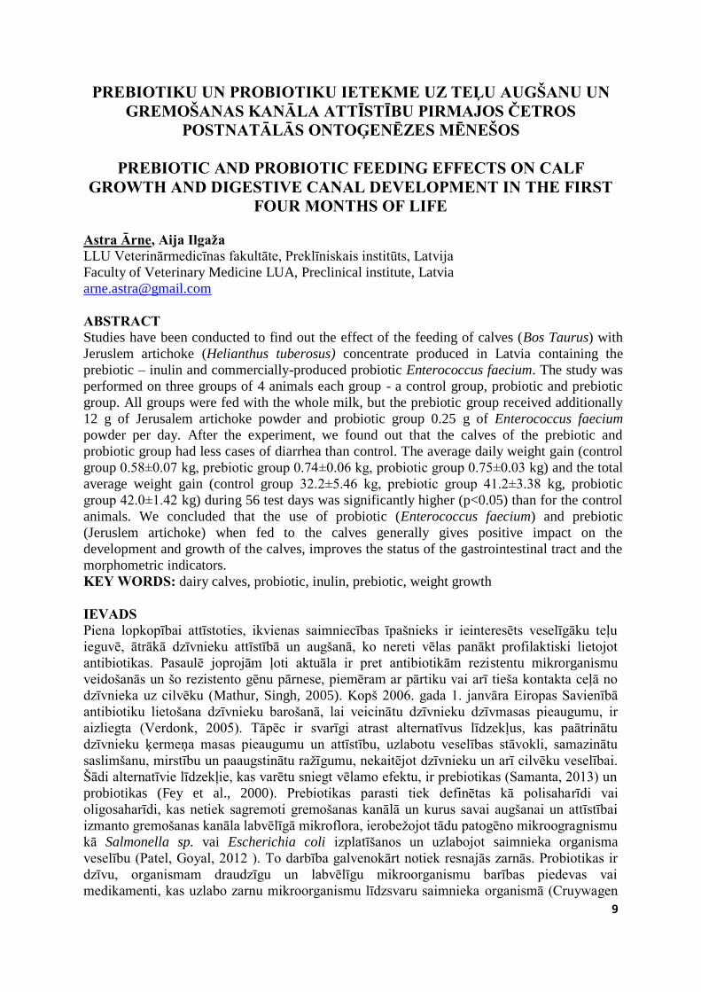
ABSTRACT Studies have been conducted to find out the effect of the feeding of calves (Bos Taurus) with
Jeruslem artichoke (Helianthus tuberosus) concentrate produced in Latvia containing the
prebiotic – inulin and commercially-produced probiotic Enterococcus faecium. The study was
performed on three groups of 4 animals each group - a control group, probiotic and prebiotic
group. All groups were fed with the whole milk, but the prebiotic group received additionally
12 g of Jerusalem artichoke powder and probiotic group 0.25 g of Enterococcus faecium
powder per day. After the experiment, we found out that the calves of the prebiotic and
probiotic group had less cases of diarrhea than control. The average daily weight gain (control
group 0.58±0.07 kg, prebiotic group 0.74±0.06 kg, probiotic group 0.75±0.03 kg) and the total
average weight gain (control group 32.2±5.46 kg, prebiotic group 41.2±3.38 kg, probiotic
group 42.0±1.42 kg) during 56 test days was significantly higher (p<0.05) than for the control
animals. We concluded that the use of probiotic (Enterococcus faecium) and prebiotic
(Jeruslem artichoke) when fed to the calves generally gives positive impact on the
development and growth of the calves, improves the status of the gastrointestinal tract and the
morphometric indicators.
KEY WORDS: dairy calves, probiotic, inulin, prebiotic, weight growth IEVADS Piena lopkopībai attīstoties, ikvienas saimniecības īpašnieks ir ieinteresēts veselīgāku teļu
ieguvē, ātrākā dzīvnieku attīstībā un augšanā, ko nereti vēlas panākt profilaktiski lietojot
antibiotikas. Pasaulē joprojām ļoti aktuāla ir pret antibiotikām rezistentu mikrorganismu
veidošanās un šo rezistento gēnu pārnese, piemēram ar pārtiku vai arī tieša kontakta ceļā no
dzīvnieka uz cilvēku (Mathur, Singh, 2005). Kopš 2006. gada 1. janvāra Eiropas Savienībā
antibiotiku lietošana dzīvnieku barošanā, lai veicinātu dzīvnieku dzīvmasas pieaugumu, ir
aizliegta (Verdonk, 2005). Tāpēc ir svarīgi atrast alternatīvus līdzekļus, kas paātrinātu
dzīvnieku ķermeņa masas pieaugumu un attīstību, uzlabotu veselības stāvokli, samazinātu
saslimšanu, mirstību un paaugstinātu ražīgumu, nekaitējot dzīvnieku un arī cilvēku veselībai.
Šādi alternatīvie līdzekļie, kas varētu sniegt vēlamo efektu, ir prebiotikas (Samanta, 2013) un
probiotikas (Fey et al., 2000). Prebiotikas parasti tiek definētas kā polisaharīdi vai
oligosaharīdi, kas netiek sagremoti gremošanas kanālā un kurus savai augšanai un attīstībai
izmanto gremošanas kanāla labvēlīgā mikroflora, ierobežojot tādu patogēno mikroogragnismu
kā Salmonella sp. vai Escherichia coli izplatīšanos un uzlabojot saimnieka organisma
veselību (Patel, Goyal, 2012 ). To darbība galvenokārt notiek resnajās zarnās. Probiotikas ir
dzīvu, organismam draudzīgu un labvēlīgu mikroorganismu barības piedevas vai
medikamenti, kas uzlabo zarnu mikroorganismu līdzsvaru saimnieka organismā (Cruywagen
10
et al., 1995), kuru darbība noris tievajās zarnās. Vairāki autori atzīmē probiotiku pozitīvo
ietekmi uz dzīvnieku imūnsistēmu, paaugstinot makrofāgu darbību, palielinot gan vispārējo
(parasti IgG un IgM), gan specifisko zarnu antivielu veidošanos (IgA), kā arī palielinot
gamma interferonu līmeni (Cruywagen et al., 1995; Krehbiel et al., 2003; Jatkauskas,
Vrotniekiene, 2010).
Izpētīts, ka prebiotika inulīns ir brīvi sastopams dažādos dārzeņos un labībā (Van Loo et al.,
1995). Viens no ar inulīnu bagātākajiem augiem ir topinambūrs (Helianthus tuberosus), tāpēc
to jau sen izmanto rūpnieciskai inulīna ieguvei (Fleming et al., 1979). Ir vairāki pētījumi,
kuros parādīta no topinambūra iegūta inulīna izēdināšanas ietekme uz vienkameras kuņģa
dzīvnieku un putnu augšanu un veselības stāvokli (Farnworth, 1992; Kleessen, 2003;
Valdovska et al., 2012), taču trūkst kompleksu pētījumu par tā izēdināšanas rezultātiem
daudzkameru kuņģa dzīvniekiem.
Tāpēc mūsu pētījuma mērķis bija noskaidrot Latvijā ražota topinambūra koncentrāta (inulīns
48.5 – 50.1%) un komerciāli ražotā prebiotiķa Enterecoccus faecium izēdināšanas ietekmi uz
teļu vispārējo veselības stāvokli, dzīvmasas pieaugumu un gremošanas kanāla morfoloģisko
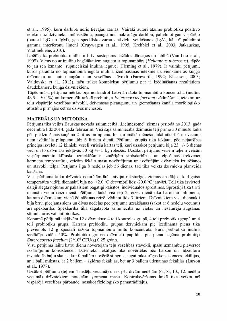
attīstību pirmajos četros dzīves mēnešos. MATERĀLS UN METODIKA Pētījums tika veikts Bauskas novada saimniecībā „Lielmežotne” ziemas periodā no 2013. gada
decembra līdz 2014. gada februārim. Visi šajā saimniecībā dzimušie teļi pirmo 30 minūšu laikā
pēc piedzimšanas saņēma 2 litrus pirmpienu, bet turpmākā mēneša laikā atkarībā no vecuma
tiem izēdināja pilnpienu līdz 6 litriem dienā. Pētījuma grupās tika iekļauti pēc nejaušības
principa izvēlēti 12 klīniski veseli vīriešu kārtas teļi, kuri uzsākot pētījumu bija 23 +/- 5 dienas
veci un to dzīvmasa iekļāvās 50 kg +/- 5 kg robežās. Uzsākot pētījumu visiem teļiem veicām
vispārpieņemto klīnisko izmeklēšanu: izmērījām sirdsdarbības un elpošanas frekvenci,
ķermeņa temperatūru, veicām fekālo masu novērtējumu un izvērtējām dzīvnieka izturēšanos
un stāvokli telpā. Pētījums ilga 8 nedēļas jeb 56 dienas, tad tika veikta dzīvnieku plānveida
kaušana.
Visu pētījuma laiku dzīvniekus turējām ārā Latvijai raksturīgos ziemas apstākļos, kad gaisa
temperatūra vidēji diennaktī bija no +2.0 0C decembrī līdz -20.0 0C janvārī. Teļi tika izvietoti
daļēji slēgtā nojumē ar pakaišiem bagātīgi kaisītos, individuālos sprostiņos. Sprostiņi tika tīrīti
manuāli vienu reizi dienā. Pētījuma laikā visi teļi 2 reizes dienā tika baroti ar pilnpienu,
katram dzīvniekam vienā ēdināšanas reizē izēdinot līdz 3 litriem. Dzīvniekiem visu diennakti
bija brīvi pieejams siens un divas nedēļas pēc pētījuma uzsākšanas (sākot ar 6 nedēļu vecumu)
arī spēkbarība. Spēkbarība tika sagatavota saimniecībā uz vietas un nesaturēja augšanas
stimulatorus vai antibiotikas.
Kopumā pētījumā iekļāvām 12 dzīvniekus: 4 teļi kontroles grupā, 4 teļi prebiotiku grupā un 4
teļi probiotiku grupā. Katram prebiotiku grupas dzīvniekam pie izēdināmā piena tika
pievienots 12 g speciāli ražota topinambūra miltu koncentrāta, kurā prebiotika inulīns
sastādīja vidēji 50%. Probiotiku grupas dzīvnieki papildus pie piena saņēma probiotiķi
Enterococcus faecium (2*109 CFU/g) 0.25 g/dnn.
Visu pētījuma laiku katru dienu novērtējām teļu veselības stāvokli, īpašu uzmanību pievēršot
izkārnījumu konsistencei. Dzīvnieku fekālijas tika novērtētas pēc Larson un līdzautoru
izveidotās baļļu skalas, kur 0 ballēm novērtē stingras, sugai raksturīgas konsistences fekālijas,
ar 1 balli mīkstas, ar 2 ballēm – šķidras fekālijas, bet ar 3 ballēm ūdeņainas fekālijas (Larson
et al., 1977).
Uzsākot pētījumu (teļiem 4 nedēļu vecumā) un ik pēc divām nedēļām (6., 8., 10., 12. nedēļu
vecumā) dzīvniekiem noteicām ķermeņa masu. Kontrolsvēršanas laikā tika veikta arī
vispārējā veselības pārbaude, nosakot fizioloģisko pamatrādītājus.
11
Pēc 56 dienām no pētījuma pirmās dienas, kad dzīvnieki bija sasnieguši 12 nedēļu vecumu,
tika veikta teļu plānveida kaušana. Tūliņ pēc teļu kaušanas atdalījām šādas gremošanas kanāla
daļas: spurekli, acekni, grāmatnieku un glumienieku, tievās un resnās zarnas līdz anālajai
atverei un noteicām: kopējo gremošanas kanāla svaru, spurekļa un glumenieka svaru bez
barības masām.
Lai veiktu datu analizēšanu izmantojām MS Excel un R-Studio programmas. Aprēķinājām
visu grupu dzīvniekiem noteikto rādītāju vidējo aritmētisko un standartnovirzi, kā arī relatīvo
dzīvmasas pieaugumu. Šo rādītāju atšķirību būtiskumu novērtējām ar T-testa palīdzību, kur p
vērtības zem 0.05 tika vērtēta kā zemākā statistiski būtiskā atķširība.
REZULTĀTI UN DISKUSIJA Šajā pētījumā vēlējāmies noskaidrot Latvijā ražota topinambūra koncentrāta un probiotiķa
Enterococcus faecium izēdināšanas ietekmi uz teļu organismu. Parasti topinambūra milti
inulīnu satur vidēji 10 %, bet speciāli izstrādāta tehnoloģija ļauj paaugstināt inulīna daudzumu
līdz 48.5% – 50.1%, kas atvieglo tā pievienošanu barības līdzekļiem (Fleming et al., 1979;
Valdovska et al., 2012). Rezultāti liecina, ka visu grupu dzīvniekiem elpošanas, sirdsdarbības
un temperatūras rādītāji visumā iekļāvās normas robežās. Jāuzsver, ka pētījums notika ziemas
apstākļos un dzīvnieki tika turēti laukā sprostiņos, tātad apstākļos, kad sals reizēm sasniedza -
20 0C, taču bez atsevišķām sala radītām veselības problēmām (lokāli, nelieli 2. pakāpes
apsaldējuma laukumi) būtiski veselības traucējumi netika novēroti un dzīvnieki bija šķietami
klīniski veseli.
Savstarpēji salīdzinot visu grupu rādītājus dažādu grupu dzīvniekiem, konstatējām, ka
ķermeņa temperatūras rādītāji gan prebiotiku (p<0.0017) grupas, gan probiotiku (p<0.0023)
grupas dzīvniekiem ir būtiski zemāki nekā kontroles grupas dzīvniekiem. Savukārt
sirdsdarbības un elpošana rādītāju vērtības starp šo grupu dzīvniekiem būtiski neatšķīrās.
Vienīgā reize, kad visiem dzīvniekiem konstatējām sirdsdarbības frekvences paātrināšanos
virs normas augstākajām robežām, bija teļiem 12 nedēļu vecumā, veicot mērījumus kautuvē.
Arī citi autori ir konstatējuši būtisku sirdsdarbības paātrināšanos teļiem dažādās stresa
situācijās (Westervelt et al., 1979; Mohr et al., 2002).
Mūs interesēja prebiotiķa inulīna un probitiķa Enterococcus faecium izēdināšanas ietekme uz
teļu gremošanas kanāla funkcionālo stāvokli un veselību. Kā jau minējām, fekālo masu
konsistenci visiem dzīvniekiem novērtējām katru dienu, izmantojot 1-3 baļļu sistēmu (Larson
et al., 1977). Lai rezultāti būtu uzskatāmāki, iegūtos rezultātus apkopojām, izrēķinot vidējo
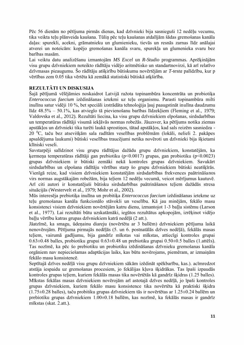
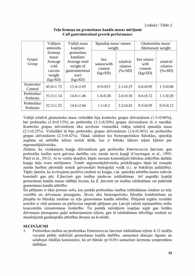
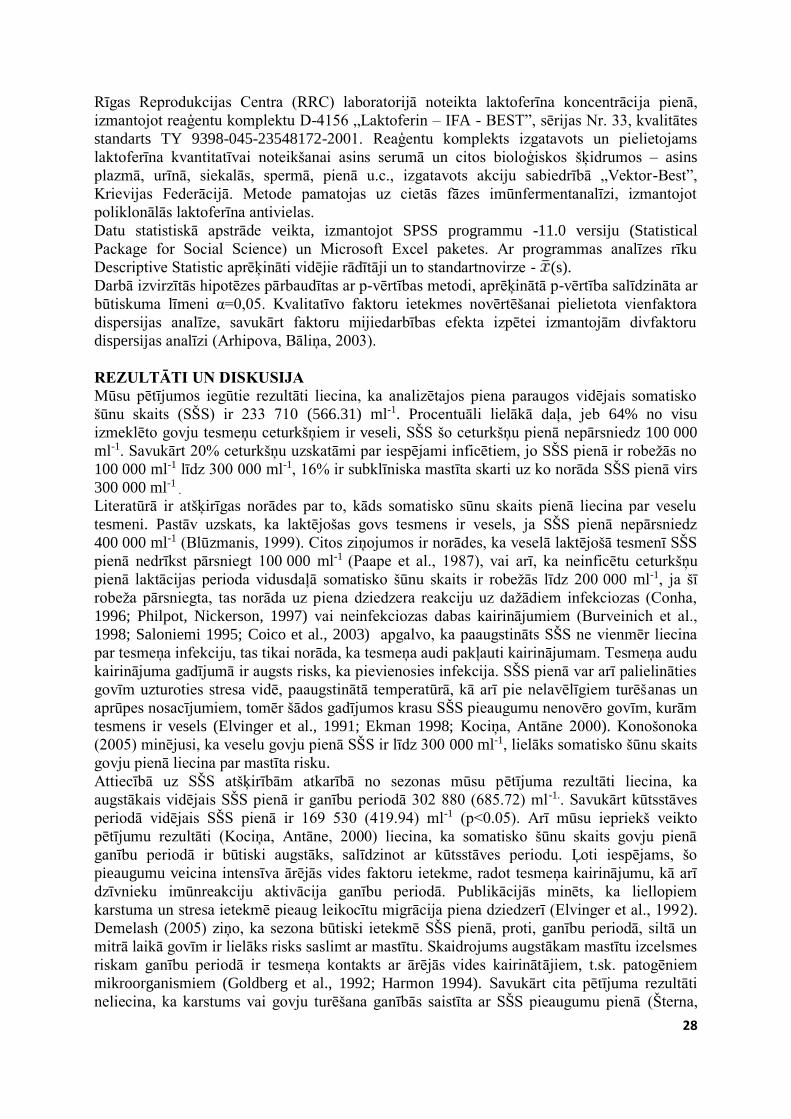
baļļu vērtību katras grupas dzīvniekiem katrā nedēļā (2 att.).
Jāatzīmē, ka smagu, ūdeņainu diareju (novērtētu ar 3 ballēm) dzīvniekiem pētījuma laikā
nenovērojām. Pētījuma pirmajās nedēļās (5. un 6. postnatālās dzīves nedēļā), fekālās masas
teļiem, vairumā gadījumu, bija gandrīz mīkstas vai mīkstas, attiecīgi kontroles grupai
0.63±0.48 balles, probiotiku grupai 0.63±0.48 un prebiotiku grupai 0.50±0.5 balles (1.attēls).
Tas nozīmē, ka pēc šo prebiotiku un probiotiku izēdināšanas dzīvnieku gremošanas kanāla
orgāniem nav nepieciešamas adaptācijas laiks, kas būtu novērojams, piemēram, ar izmaiņām
fekālo masu konsistencē.
Septītajā dzīves nedēļā visu grupu dzīvniekiem sākām izēdināt spēkbarību, kas.). acīmredzot
atstāja iespaidu uz gremošanas procesiem, jo fekālijas kļuva šķidrākas. Tas īpaši izpaudās
kontroles grupas teļiem, kuriem fekālās masas tika novērtētās kā gandrīz šķidras (1.25 balles).
Mīkstas fekālas masas dzīvniekiem novērojām arī astotajā dzīves nedēļā, jo īpaši kontroles
grupas dzīvniekiem, kuriem fekālo masu konsistence tika novērtēta kā praktiski šķidra
(1.75±0.28 balles), taču probitiku grupas dzīvniekiem tās ir novērtētas ar 1.25±0.24 ballēm un
prebiotiku grupas dzīvniekiem 1.00±0.18 ballēm, kas nozīmē, ka fekālās masas ir gandrīz
mīkstas (skat. 2.att.).
12
00.2
0.40.60.8
1
1.21.41.6
1.82
5 6 7 8 9 10 11 12teļu vecums(nedēļās)/
calf age (weeks)
Fekā
liju
kons
isten
ce (b
allē
s)/
Feca
l sco
re (m
arks
)
1.attēls. Fekāliju konsistence pētījuma grupu teļiem: ■-kontroles grupa,
■-probiotiku grupa, ■-prebiotiku grupa Figure 1. Fecal score among group of calves; ■-control group, ■-probiotic group,
■-prebiotic group
Devītās nedēļas laikā situācija visās grupās bija stabilizējusies un bažas par iespējamiem
diarejas gadījumiem bija mazinājusies. Vislabākie rezultāti bija probiotiku grupas
dzīvniekiem, kur 9. nedēļas fekālo masu konsistnce bija vidēji 0.88±0.25 balles (kontroles
grupai 1.25±0.28 un prebiotiku grupai 1.0±0.00 balles). Sākot ar 10 dzīves nedēļu fekāliju
konsistence visiem pētījumā iekļautajiem dzīvniekiem kļūst stingrāka un ir stabilizējusies: 12
nedēļā kontroles grupas teļiem 0.63±0.48 balles, probiotku grupas dzīvniekiem 0.5±0.40
balles, bet prebiotiku grupas dzīvniekiem 0.38±0.28 balles.
Kopumā varam teikt, ka, pētījuma vidus posmā (7. dzīves nedēļā), kad dzīvniekiem tiek
piedāvāti jauni barības līdzekļi (spēkbarība) fekālajām masām mainās konsistence un
dzīvniekiem biežāk novērotas šķidras fekālijas (novērtētas ar 1.75 ballēm un zemāk).
Salīdzinoši stabilāku gremošanas kanāla orgānu darbību šajā vecumā uzrādīja teļi, kuriem
papildus tika izēdinātas probiotikas un sevišķi prebiotikas. Tas sakrīt ar pētījumiem par
probiotiku un prebiotiku ietekmi ar vienkameru kuņģa dzīvnieku gremošanas orgānu darbības
stabilizējošo ietekmi, mazinot diarejas gadījumu skaitu, kā arī slimošanas ilgumu un
simptomu intensitāti (Cruywagen et al., 1995; Fey et al., 2000; Krehbiel et al., 2003;
Jatkauskas, Vrotniekiene, 2010; Patel, Goyal, 2012; Samanta, 2013).
Kaut arī siens teļiem bija pieejams visu pētījuma laiku, mūsu novērojumi liecina, ka sākot ar
astoņu nedēļu vecumu, teļi to sāk uzņemt daudz intensīvāk, kas acīmredzot palīdz stabilizēt
gremošanas procesus. Sākot ar 10 dzīves nedēļu fekāliju konsistence no šķidras kļūst mīksta
vai stingra, ko varētu skaidrot, ka šajā vecumā teļš izveidojas par stabilu atgremotājdzīvnieku
un gremošanas sistēmas darbība līdz 11 dzīves nedēļai pilnībā nostabilizējas. Līdzīgus
rezultātus novērojuši arī citi autori, kuri pēc jaunu barības līdzekļu ieviešanas barības devā
konstatējuši gremošanas kanāla darbības stabilizēšanos vienas līdz divu nedēļu laikā teļiem,
kuriem tika papildus izēdināti līdzīgi bioterapeitiskie līdzekļi. (Flickinger et al., 2003;
Heinrichs et al., 2009; Król, 2011; Grand et al., 2013).
Viens no šī pētījuma uzdevumiem bija noskaidrot pētījumā iekļauto bioterapeitisko līdzekļu
ietekmi uz dzīvnieku dzīvmasas pieaugumu un gremošanas kanāla orgānu attīstību. Pirmajā
tabulā parādīts ik pēc divām nedēļām noteiktais katras grupas dzīvnieku dzīvmasa (vidējie
13
rādītāji un standartnovirze), kā arī vidējais dzīvmasas pieaugums, salīdzinot ar pirmo pētījuma
dienu.
1.tabula / Table 1 Vidējais dzīvsvars un tā pieauguma dinamika teļiem 56 dienas izēdinot prebiotiku
inulīnu un probiotiku Enterococcus faecium
Calves average total live weight growth dynamics 56 days feeding prebiotic inulin and probiotic Enterococcus faecium
Grupa/
Group
Vidējais dzīvnieka svars attiecīgajā
pētījuma dienā/ Average weight of the
animal at the research day
(kg±SD)
Dzīvmasas pieaugums
attiecīgajā laika
periodā/ Live weight
gains at the time
period
(kg/d±SD)
Vidējais
diennakts
pieaugums/
Average
daily body
weight gain
(kg±SD) 1 14 28 56 1-14 1-28 1-56
Kontroles/
Control
53.6
±5.72
60.0±5.52 71.2
±7.76
84.6
±7.36
7.2
±3.50
19.0
±1.00
32.2
±5.46 0.58±0.07
Prebiotiku/
Prebiotic
54.4
±3.28
69.0±1.42 79.2
±7.82
98.0
±7.54
13.6
±3.05
19.2
±3.65
41.2
±3.38 0.74±0.06
Probiotiku/
Probiotic
56.4
±5.62
70.0±6.12 80.5
±4.94
98.5
±3.54
12.5
±0.72
24.0
±2.82
42.0
±1.42 0.75±0.03
Uzsākot pētījumu vidējais dzīvnieku svars būtiski neatšķīrās, bet jau divas nedēļas pēc
prebiotiku (69.0±1.42) un probiotiku (70.0±6.12) papildus izēdināšanas vidējais teļu svars
šajās grupās bija būtiski augstāks (gandrīz 10 kg) nekā kontroles grupas (60.0±5.5)
dzīvniekiem. Pētījuma noslēgumā dzīvniekiem, kuriem tika izēdināti bioterapeitiskie līdzekļis
svars ir statistiski būtiski lielāks (p>0.05) (par gandrīz 14 kg) kā kontroles grupas
dzīvniekiem.
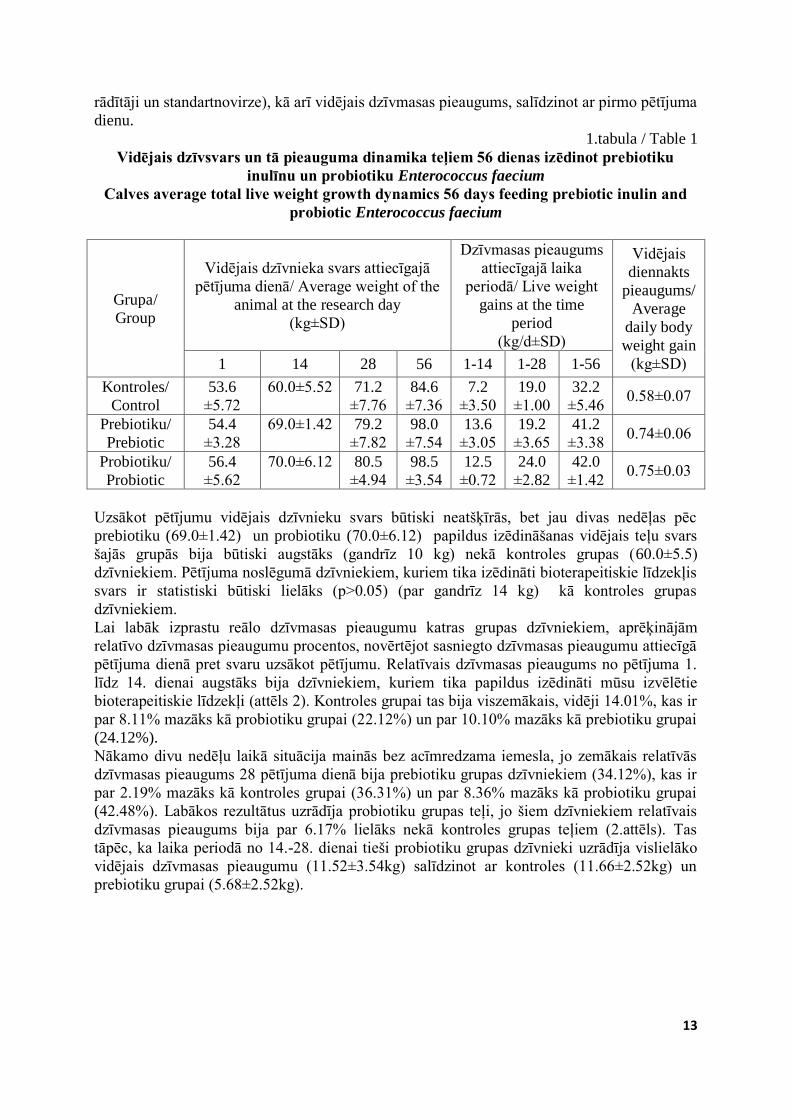
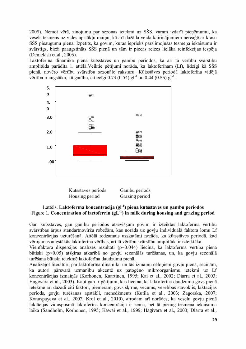
Lai labāk izprastu reālo dzīvmasas pieaugumu katras grupas dzīvniekiem, aprēķinājām
relatīvo dzīvmasas pieaugumu procentos, novērtējot sasniegto dzīvmasas pieaugumu attiecīgā
pētījuma dienā pret svaru uzsākot pētījumu. Relatīvais dzīvmasas pieaugums no pētījuma 1.
līdz 14. dienai augstāks bija dzīvniekiem, kuriem tika papildus izēdināti mūsu izvēlētie
bioterapeitiskie līdzekļi (attēls 2). Kontroles grupai tas bija viszemākais, vidēji 14.01%, kas ir
par 8.11% mazāks kā probiotiku grupai (22.12%) un par 10.10% mazāks kā prebiotiku grupai
(24.12%).
Nākamo divu nedēļu laikā situācija mainās bez acīmredzama iemesla, jo zemākais relatīvās
dzīvmasas pieaugums 28 pētījuma dienā bija prebiotiku grupas dzīvniekiem (34.12%), kas ir
par 2.19% mazāks kā kontroles grupai (36.31%) un par 8.36% mazāks kā probiotiku grupai
(42.48%). Labākos rezultātus uzrādīja probiotiku grupas teļi, jo šiem dzīvniekiem relatīvais
dzīvmasas pieaugums bija par 6.17% lielāks nekā kontroles grupas teļiem (2.attēls). Tas
tāpēc, ka laika periodā no 14.-28. dienai tieši probiotiku grupas dzīvnieki uzrādīja vislielāko
vidējais dzīvmasas pieaugumu (11.52±3.54kg) salīdzinot ar kontroles (11.66±2.52kg) un
prebiotiku grupai (5.68±2.52kg).
14
14.01
36.31
61.78
74.34
24.12
34.12
72.94
42.48
22.12
0.00
10.00
20.00
30.00
40.00
50.00
60.00
70.00
80.00
14 diena/day 28 diena/day 56 diena/day
%
2.attēls. Relatīvais dzīvmasas pieaugums (%) no pētījuma 1. līdz 14., 28. un līdz 56.
dienai, kontroles (■), probiotiku (■) un prebiotiku grupas (■) teļiem Figure 2. Relative weight gain from 1th day till 14th, 28 th, and till 56 th day, control group
(■), probiotic group (■), prebiotic group (■) calves
Laika posmā no 1. līdz 56. dienai relatīvās dzīvmasas pieaugums augstākais joprojām ir
probiotiku grupas teļiem (74.34%). Prebiotiku grupas dzīvniekiem tas ir pieaudzis un
sasniedzis 72.94%, kas ir par 11.2% lielāks kā kontorles grupai (61.78%) un vairs tikai par
1.4% mazāks kā probiotikas grupas dzīvniekiem (74.34%).
Arī citi autori savos pētījumos par prebiotikas mannaoligosaharīda ietekmi uz teļu organismu
atzīmē tā pozitīvo ietekmi dzīvmasas pieaugumu (Stolić et al., 2012). Savukārt J. Jatkauskas,
V.Vrotniekiene (2010) pētījumā par Enterococcus faecium ietekmi uz teļu attīstību izpētījuši,
ka vidējais svara pieaugums dzīvniekiem, kuriem 56 dienas izēdināja šo probiotiku bija par
9.4% lielāks (p<0.01) kā kontroles grupas teļiem.
Papildus aprēķinājām arī vidējo dzīvmasas pieaugumu diennaktī katras grupas dzīvniekiem
visa pētījuma (56 dienu) laikā, t.i. teļiem no 4 līdz 12 nedēļu vecumam. Konstatējām, ka
prebiotikas grupas un probitokas grupas teļiem tas ir gandrīz vienāds (attiecīgi
0.74±0.03kg/dnn un 0.75±0.06kg/dnn) un ir statistiski būtiski lielāks (p<0.05) kā kontroles
grupas dzīvniekiem (0.58±0.07kg/dnn). Tātad mūsu pētījumā iekļauto bioterapeitisko līdzekļu
izēdināšana 4-12 nedēļu vecumā teļiem būtiski palielina dzīvmasas pieaugumu.
Kā minējām, 56 dienā no pētījuma sākuma veicām dzīvnieku plānveida kaušanu, pēc kuras
noteicām atdzesēta liemeņa masu, gremošanas kanāla kopējo masu un atsevišķi masu gan
spureklim, gan glumeniekam. Izmantojot iegūtos datus, aprēķinājām relatīvo spurekļa un
glumenieka masu (2.tabula).
Atdzesēta liemeņa, tāpat kā visa gremošanas kanāla vidējā masa vislielākā izrādījās prebiotiku
grupas teļiem (attiecīgi 53.3±1.14 kg un 14.82±1.46 kg), probiotiku grupas teļiem (52.3±1.52
kg un 14.62±2.66 kg) un vismazākā kontroles grupai (45.0±1.72 un 13.42±2.05 kg), taču šī
atšķirība nav statistiski būtiska.
Vislielākā tukša spurekļa masa tika konstatēta prebiotikas grupas dzīvniekiem - vidēji
1.40±0.36 kg, lai gan atšķirība ar kontroles grupas (0.95±015kg) un probiotiku (1.18±0.2 kg)
grupas dzīvniekiem bija ievērojama, tomēr tā neizrādījās statistiski būtiska. Kontroles grupas
dzīvniekiem tukša glumenieka masa bija 0.6±0.05 kg, kas ir statistiski būtiski lielāka (p<0.05)
kā probiotku grupas dzīvniekiem (0.5±0.05 kg).
15
2.tabula / Table 2
Teļu liemeņa un gremošanas kanāla masas mērījumi Calf gastrointestinal growth performance
Grupa/
Group
Vidējais
atdzesēta
liemeņa
masa/
Average
cold
carcass
weight
(kg±SD)
Vidējā masa
kopējam
gremošnas
kanālam/
Average total
weight of
gastrointestinal
tract
(kg±SD)
Spurekļa masa/ rumen
weight
Glumenieka masa/
Abomasum weight
bez
satura/with
content
(kg±SD)
relatīvā/
ralative
(%±SD)
bez satura/
with
content
(kg±SD)
relatīvā/
relative
(%±SD)
Kontroles/
Control 45.0±1.72 13.4±2.05 0.9±015 2.1±0.25 0.6±0.05 1.3±0.06
Prebiotiku/
Prebiotic 53.3±1.14 14.8±1.46 1.4±0.36 2.6±0.36 0.6±0.12 1.1±0.20
Probiotiku/
Probiotic 52.3±1.52 14.6±2.66 1.1±0.2 2.2±0.42 0.5±0.05 0.9±0.12
Vidējā relatīvā glumenieka masa vislielākā bija kontroles grupas dzīvniekiem (1.3±0.06%),
bet probiotiku (1.0±0.12%) un prebiotiku (1.1±0.20%) grupas dzīvniekiem tā ir mazākā.
Kontroles grupas dzīvniekiem tika novērota vismazākā vidēja relatīvā spurekļa masa
(2.1±0.25%). Vislielākā tā bija prebiotiku grupas dzīvniekiem (2.6±0.36%) un probiotiku
grupas dzīvniekiem (2.3±0.42%). Tātad, izēdinot šos bioterapeitiskos līdzekļus, spurekļa
augšana un attīstība teļiem notiek ātrāk, kas ir būtisks faktors teļam kļūstot par
atgremotājdzīvnieku.
Zināms, ka vienkameru kuņģa dzīvniekiem gan probiotika Enterococcus faecium, gan
prebiotika inulīns savu galveno darbību veic zarnās nevis kuņģī (Cruywagen et al., 1995;
Patel et al., 2012). Ar to varētu skaidrot, kāpēc neesam konstatējuši būtiskas atšķirības dažādu
kuņģa daļu svara mērījumos. Tomēr atgremotājdzīvnieku priekškuņģos tāpat kā resnajās
zarnās barības pārstrāde notiek galvenokārt bioloģiskā veidā (t.i. ar baktēriju palīdzību).
Tāpēc jāatzīst, ka ievērojamu pozitīvu ietekmi uz kuņģu, t.sk. spurekļa attīstību mums izdevās
konstatēt gan pēc E.faecium gan inulīna piedevas izēdināšanas. Arī augstāki kopējā
gremošanas kanāla masas rādītāji liecina, ka E. faecium un inulīna izēdināšana var paātrināt
gremošanas kanāla attīstību.
Šis pētījums ir tikai pirmais solis, kas parāda prebiotikas inulīna izēdināšanas ietekmi uz teļu
veselību un dzīvmasas pieaugums. Jāveic abu bioterapeitisko līdzekļu kombinēšana un
jāizpēta šo līdzekļu ietekme uz teļu gremošanas kanāla attīstību. Pētījumā iegūtie rezultāti
noteikti ir vērā ņemami un pārliecina turpināt pētījums par Latvijā ražotā topinambūru miltu
koncentrāta izmantošanas lietderību. Tie parāda ražotājiem iespējas iegūt gan lielāku
dzīvmasas pieaugumu gaļai nobarojamiem teļiem, gan tā izēdināšanas labvēlīgo ietekmi uz
ataudzējamā ganāmpulka attīstības ātrumu un kvalitāti.
SECINĀJUMI 1. Prebiotikas inulīna un probiotikas Enterococcus faecium izēdināšana teļiem 4-12 nedēļu
vecumā palīdz stabilizēt gremošanas kanāla darbību, samazinot diarejas ilgumu un
uzlabojot fekāliju konsistenci, kā arī būtiski (p<0.05) samazinot ķermeņa temperatūras
rādītājus.
16
2. Izēdinot bioterapeitiskos līdzekļus vidējais diennakts dzīvmasas pieaugums ir statistiski
būtiski lielāks (p>0.028) probiotiku un (p>0.011) prebiotiku grupu teļiem salīdzinot ar
kontroles grupas dzīvniekiem, par ko liecina arī augstāki vidējie atdzesēta liemeņa svara
rādītāji.
3. Bioterapeitiskie līdzekļi paātrina gremošanas kanāla, tai skaitā spurekļa augšanu, jo
kopējā grmemošanas kanāla masa 12 nedēļu vecumā prebiotiku (14.8±1.46 kg) un
probiotiku (14.6±2.66 kg) grupas teļiem ir augstāka nekā kontroles grupai (13.4±2.05
kg). Arī spurekļa masas mērījumi augstāki ir prebiotiku 1.4±0.36 kg, probiotiku 1.1±0.2
kg grupu dzīvniekiem, salīdzinot ar kontroles grupu 0.9±015 kg.
4. Prebiotikas inulīna un prebiotikas Enterococcus faecium izēdināšana teļiem 4-12 nedēļu
vecumam kopumā uzlabo gremošanas kanāla darbību un attīstību, kas liecina par šo
bioterapeitisko līdzekļu pozitīvo ietekmi uz teļu attīstību un augšanu.
LITERATŪRA 1. Cruywagen, C.W., Jordan I., Venter L. (1995) Effect of Lactobacillus acidophilus
supplementation of milk replacer on preweaning performance of calves. Journal of
Dairy Science. (79), 483-486.
2. Fey P. D., Safranek T. J., Rupp M. E., Dunne E. F., Ribot E., Iwen P. C., Bradford P.
A., Angulo F. J., Hinrichs S. H. (2000). Ceftriaxone-Resistant Salmonella Infection
Acquired by a Child from Cattle. New England Journal of Medicine. (17), 1242.
3. Fleming S., Groot Wassink J. (1979) Preparation of high-fructose syrup from the tubers
of the Jerusalem artichoke (Helianthus tuberosus). CRC Critical reviews in Food
Science and Nutrition. (12), 1-28.
4. Flickinger E., Van Loo J., Fahey G. (2003) Nutritional responses to the presence of
inulin and oligofructose in the diets of domesticated animals: a review. Critical Reviews
in Food Science and Nutrition. (43), 19-60.
5. Gaggìa F., Mattarelli P., Biavati B. (2010) Probiotics and prebiotics in animal feeding
for safe food production. International Journal of Food Microbiology. (141), 15–28.
6. Gibson G.R., Roberfroid M.B. (1995) Dietary modulation of the human colonic
microbiota: introducing the concept of prebiotics. The Journal of Nutrition. (125),
1401–1412.
7. Grand E., Respondek F., Martineau C., Detilleux J., Bertrand G. (2013) Effects of short-
chain fructooligosaccharides on growth performance of preruminant veal calves.
Journal of Dairy Science. (96), 1094–1101.
8. Heinrichs J., Jones C.M., Elizondo-Salazar J., Terrill S.J. (2009) Effects of a prebiotic
supplement on health of neonatal dairy calves. Livestock Science. (125), 149–154.
9. Hill T., Bateman H., Aldrich J., Schlotterbeck R.L. (2008) Oligosaccharides for Dairy
Calves. The Professional Animal Scientist. (24), 460–464.
10. Houdijk J.G.M., Bosch M.W., Verstegen M.W.A., Berenpas H.J. (1998) Effects of
dietary oligosaccharides on the growth performance and faecal characteristics of young
growing pigs. Animal Feed Science and Technology. (71), 35-48.
11. Jatkauskas J., Vrotniakiene V. (2010) Effects of probiotic dietary supplementation on
diarrhea patterns, faecal microbiota and performance of early weaned calves.
Veterinarni Medicina (55), 494–503.
12. Kleessen B., Elsayed N., Loehren U., Schroedl W., Krueger M. (2003) Jerusalem
artichokes stimulate growth of broiler chickens and protect them against cecal
endotoxins and potential pathogens. Journal of Food Protection. (11), 2171-2175.
13. Krehbiel C.R., Rust S.R., Zhang G., Gilliland S.E. (2003) Bacterial direct-fed
microbials in ruminant diets: Perfomance response and mode of action.- Journal of
Dairy Science.(81), E120-E132.
17
14. Król B., (2011) Mannanoligosaccharides, inulin and yeast nucleotides added to calf
milk replacers on rumen mikroflora, level of serum immunoglobulin and health
condition of calves. Electronic Journal of Polish Agricultural Universitas. (2), 1-18.
15. Larson L., Owen F.G., Albright J.L., Appleman R.D., Lamb R.C., Muller L.D. (1977)
Guidelines Toward More Uniformity in Measuring and Reporting Calf Experimental
Data. Journal of Dairy Science. (60), 989-991.
16. Mathur S., Singh R. (2005) Antibiotic resistance in food lactic acid bacteria- a review.
International Journal of Food Microbiology. (105) 281–295.
17. Masanetz S., Preißinger W., Meyer H.H.D., Pfaffl M.W. (2011) Effects of the prebiotics
inulin and lactulose on intestinal immunology and hematology of preruminant calves.
Animal. (5), 1099–1106.
18. Mohr E., Langbein J., Nürnberg G. (2002) Heart rate variability: A noninvasive
approach to measure stress in calves and cows. Physiology and Behavior. (75), 251–
259.
19. Patel S., Goyal A. (2012) The current trends and future perspectives of prebiotics
research: A review. 3 Biotech. (2), 115–125.
20. Quezada-Mendoza V., Heinrichs J., Jones C.M. (2011) The effects of a prebiotic
supplement (Prebio Support) on fecal and salivary IgA in neonatal dairy calves.
Livestock Science. (142), 222–228.
21. Samanta K., Jayapal N., Senani S., Kolte A., Sridhar M. (2013) Prebiotic inulin: Useful
dietary adjuncts to manipulate the livestock gut microflora. Brazilian Journal of
Microbiology. (44), 1–14.
22. Stolić N., Milošević B., Spasić Z., Ilić Z. (2012) Effects of prebiotic inclusion in the
diet of weaned calves. Macedonian Journal of Animal Science. (2), 53-57.
23. Valdovska A., Jemeļjanovs A., Zītare I., Krastiņa V., Pilmane M., Proškina L. (2012)
Impact of prebiotic on chicken digestive tract morphofunctional status. In: Conference
on Current events in veterinary research and practice, LLU, Jelgava. 63-67.
24. Van Loo J. (2007) How Chicory Fructans Contribute to Zootechnical Performance and
Well-Being in Livestock and Companion Animals. The Journal of Nutrition (137),
2594–2597.
25. Verdonk J.M., Shim S.B., Van Leeuwen P., Verstegen M.W. (2005) Application of
inulin-type fructans in animal feed and pet food. British Journal of Nutrition. (93), 125–
138.
26. Westervelt R. G., Kinsman D. M., Prince R. P., Giger W. (1979) Physiological Stress
Measurement during Slaughter in Calves. Journal of Animal Science. (42), 831-837.
18
ALĀRIJU MEZOCERKĀRIJU INVĀZIJA MEŽA CŪKĀM LATVIJĀ
INVASION OF ALARIA MESOCERCARIAE IN WILD BOAR IN LATVIA
Veronika Berģe1, Dace Keidāne2, Anna Krūklīte2 1 Pārtikas un veterinārais dienests, Latvija; Food and Veterinary Service, Latvia 2 LLU Veterinārmedicīnas fakultāte, Latvija; Faculty of Veterinary Medicine LUA, Latvia
ABSTRACT Alariosis is a parasitological disease caused by parasitic trematode from the family
Diplostomatidae and the genus Alaria. During the examination of wild boar (Sus scrofa) meat
for presence of Trichinella, frequently Alaria mesocercariae are detected. The aim of the
present work was to explore the invasion of Alaria mesocercariae in wild boar in Latvia. The
study was performed in Latvia University of Agriculture Faculty of Veterinary Medicine in
laboratory of Parasitology. For the study we used meat samples from hunted wild boars of
different age and gender, from different regions of Latvia. We took meat samples from the
pillars of diaphragm and the sinewy part of diaphragm. Examined sample weight was 100 g.
Samples were examined using digestion method during one to seven days. To estimate about
the invasion, we calculated extensity of invasion. Our study showed that Alaria mesocercariae
were found in wild boar meat during all seasons. Since a year 2010 to 2013 the highest
invasion was detected during winter months, with extensity 4,8% to 11,7%. The lowest
extensity was detected during summer months in years 2010, 2011 and 2012.
KEY WORDS: wild boar, alariosis, mesocercariae
IEVADS Alarioze, suņu dzimtas dzīvnieku zarnu trematodoze ir plaši izplatīta invāzijas slimība visā
pasaulē. Šobrīd pasaulē pastāv aktuāls jautājums par iespējamo cilvēku saslimšanu lietojot
uzturā ar alāriju mezocerkārijiem invadētu gaļu. To ka alarioze tiek uzskatīta par potenciālu
zoonozi parāda 2011. gadā konstatētā cilvēka nāve, kurš uzturā bija lietojis ar alāriju
mezocerkārijiem invadētu gaļu (Portier et al., 2011; Wasiluk, 2013). Tomēr trūkst pētījumu
par to tieši kādas alāriju sugas ierosina saslimšanu cilvēkam. Tāpat neskaidrs ir parazīta
attīstības cikls saistībā ar cilvēku invadēšanos (Möhl et al., 2009; Paulsen et al., 2012; Riehn
et al., 2010; Urosevic et al., 2012).
Latvijas Lauksaimniecības universitātes Veterinārmedicīnas fakultātes parazitoloģijas
laboratorijā suņveidīgajiem alārijas diagnosticētas kopš 1989. gada. Jāatzīmē, ka nereti veicot
izmeklējumus uz trihinelozi meža cūku gaļā tiek diagnosticēti arī alāriju mezocerkāriji. Mūsu
darba mērķis bija pētīt alāriju mezocerkāriju invāziju meža cūkām.
MATERIĀLS UN METODIKA Pētījums veikts LLU Veterinārmedicīnas fakultātes Pārtikas un vides higiēnas institūta
Parazitoloģijas laboratorijā. Pētījumā izmantoti dažāda vecuma un dzimuma nomedīto meža
cūku (Sus scrofa) gaļas paraugi no dažādiem reģioniem Latvijā. Paraugiem izmantoti 100
grami diafragmas muskuļaudu.
Meža cūku gaļas paraugi tika iegūti visa gada garumā no dažādiem medību reģioniem.
Paraugi, pēc iegūšanas medību reģionā un nogādāšanas Parazitoloģijas laboratorijā, tika
izmeklēts svaigs materiāls pēc hidrolīzes metodes. Iegūtos rezultātus analizējot aprēķinājām
alāriju mezocerkāriju invāzijas ekstensitāti un pētījām sezonalitāti.
19
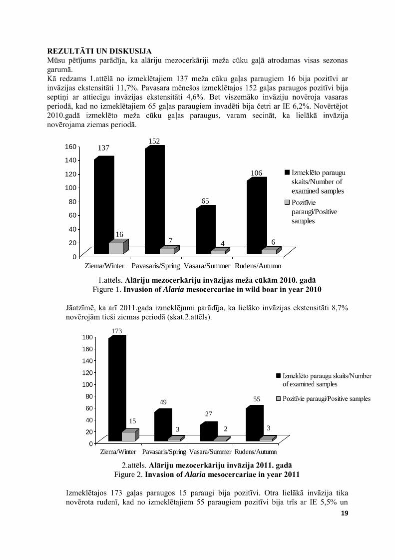
REZULTĀTI UN DISKUSIJA Mūsu pētījums parādīja, ka alāriju mezocerkāriji meža cūku gaļā atrodamas visas sezonas
garumā.
Kā redzams 1.attēlā no izmeklētajiem 137 meža cūku gaļas paraugiem 16 bija pozitīvi ar
invāzijas ekstensitāti 11,7%. Pavasara mēnešos izmeklētajos 152 gaļas paraugos pozitīvi bija
septiņi ar attiecīgu invāzijas ekstensitāti 4,6%. Bet viszemāko invāziju novēroja vasaras
periodā, kad no izmeklētajiem 65 gaļas paraugiem invadēti bija četri ar IE 6,2%. Novērtējot
2010.gadā izmeklēto meža cūku gaļas paraugus, varam secināt, ka lielākā invāzija
novērojama ziemas periodā.
137
16
152
7
65
4
106
6
0
20
40
60
80
100
120
140
160
Ziema/Winter Pavasaris/Spring Vasara/Summer Rudens/Autumn
Izmeklēto paraugu
skaits/Number of
examined samples
Pozitīvie
paraugi/Positive
samples
1.attēls. Alāriju mezocerkāriju invāzijas meža cūkām 2010. gadā
Figure 1. Invasion of Alaria mesocercariae in wild boar in year 2010
Jāatzīmē, ka arī 2011.gada izmeklējumi parādīja, ka lielāko invāzijas ekstensitāti 8,7%
novērojām tieši ziemas periodā (skat.2.attēls).
173
15
49
3
27
2
55
3
0
20
40
60
80
100
120
140
160
180
Ziema/Winter Pavasaris/Spring Vasara/Summer Rudens/Autumn
Izmeklēto paraugu skaits/Number
of examined samples
Pozitīvie paraugi/Positive samples
2.attēls. Alāriju mezocerkāriju invāzija 2011. gadā
Figure 2. Invasion of Alaria mesocercariae in year 2011 Izmeklētajos 173 gaļas paraugos 15 paraugi bija pozitīvi. Otra lielākā invāzija tika
novērota rudenī, kad no izmeklētajiem 55 paraugiem pozitīvi bija trīs ar IE 5,5% un
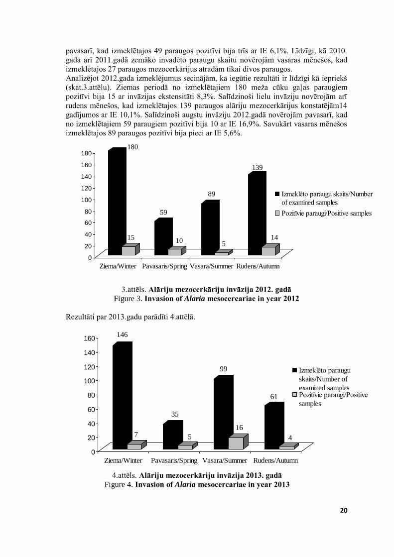
20
pavasarī, kad izmeklētajos 49 paraugos pozitīvi bija trīs ar IE 6,1%. Līdzīgi, kā 2010.
gada arī 2011.gadā zemāko invadēto paraugu skaitu novērojām vasaras mēnešos, kad
izmeklētajos 27 paraugos mezocerkārijus atradām tikai divos paraugos.
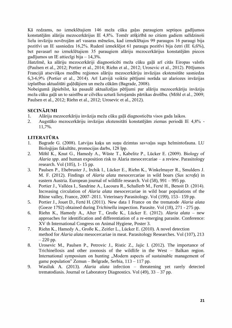
Analizējot 2012.gada izmeklējumus secinājām, ka iegūtie rezultāti ir līdzīgi kā iepriekš
(skat.3.attēlu). Ziemas periodā no izmeklētajiem 180 meža cūku gaļas paraugiem
pozitīvi bija 15 ar invāzijas ekstensitāti 8,3%. Salīdzinoši lielu invāziju novērojām arī
rudens mēnešos, kad izmeklētajos 139 paraugos alāriju mezocerkārijus konstatējām14
gadījumos ar IE 10,1%. Salīdzinoši augstu invāziju 2012.gadā novērojām pavasarī, kad
no izmeklētajiem 59 paraugiem pozitīvi bija 10 ar IE 16,9%. Savukārt vasaras mēnešos
izmeklētajos 89 paraugos pozitīvi bija pieci ar IE 5,6%.
180
15
59
10
89
5
139
14
0
20
40
60
80
100
120
140
160
180
Ziema/Winter Pavasaris/Spring Vasara/Summer Rudens/Autumn
Izmeklēto paraugu skaits/Number
of examined samples
Pozitīvie paraugi/Positive samples
3.attēls. Alāriju mezocerkāriju invāzija 2012. gadā
Figure 3. Invasion of Alaria mesocercariae in year 2012
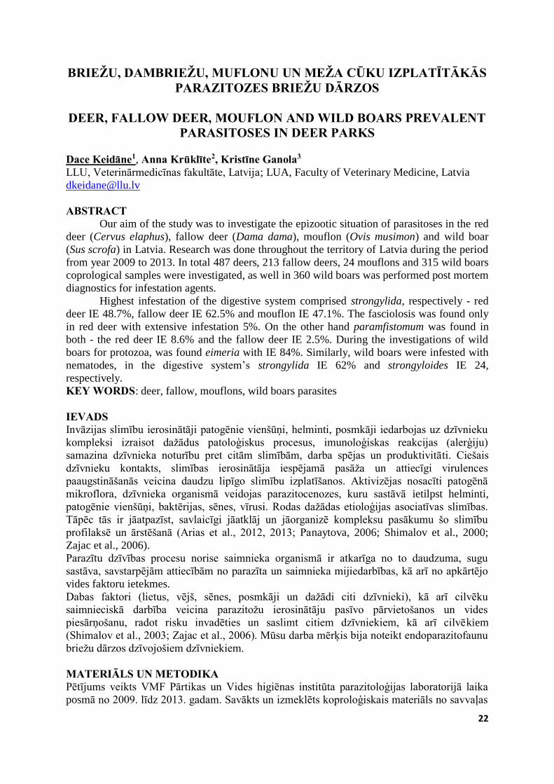
Rezultāti par 2013.gadu parādīti 4.attēlā.
146
7
35
5
99
16
61
4
0
20
40
60
80
100
120
140
160
Ziema/Winter Pavasaris/Spring Vasara/Summer Rudens/Autumn
Izmeklēto paraugu
skaits/Number of
examined samplesPozitīvie paraugi/Positive
samples
4.attēls. Alāriju mezocerkāriju invāzija 2013. gadā
Figure 4. Invasion of Alaria mesocercariae in year 2013
21
Kā redzams, no izmeklētajiem 146 meža cūku gaļas paraugiem septiņos gadījumos
konstatējām alāriju mezocerkārijus IE 4,8%. Tomēr atšķirībā no citiem gadiem salīdzinoši
lielu invāziju novērojām arī vasaras mēnešos, kad izmeklētajos 99 paraugos 16 paraugi bija
pozitīvi un IE sasniedza 16,2%. Rudenī izmeklējot 61 paraugu pozitīvi bija četri (IE 6,6%),
bet pavasarī no izmeklētajiem 35 paraugiem alāriju mezocerkārijus konstatējām piecos
gadījumos un IE attiecīgi bija – 14,3%.
Jāatzīmē, ka alāriju mezocerkāriji diagnosticēti meža cūku gaļā arī citās Eiropas valstīs
(Paulsen et al., 2012; Portier et al., 2014; Riehn et al., 2012; Urosevic et al., 2012). Pētījumos
Francijā atsevišķos medību reģionos alāriju mezocerkāriju invāzijas ekstensitāte sasniedza
6,3-6,9% (Portier et al., 2014). Arī Latvijā veiktie pētījumi norāda uz alariozes invāzijas
izplatības aktualitāti gaļēdājiem un meža cūkām (Bagrade, 2008).
Nobeigumā jāpiebilst, ka pasaulē aktualizējas pētījumi par alāriju mezocerkāriju invāziju
meža cūku gaļā un to saistību ar cilvēku uzturā lietojamās pārtikas drošību. (Möhl et al., 2009;
Paulsen et al., 2012; Riehn et al., 2012; Urosevic et al., 2012).
SECINĀJUMI 1. Alāriju mezocerkāriju invāzija meža cūku gaļā diagnosticēta visos gada laikos.
2. Augstāko mezocerkāriju invāzijas ekstensitāti konstatējām ziemas periodā IE 4,8% -
11,7%.
LITERATŪRA 1. Bagrade G. (2008). Latvijas kaķu un suņu dzimtas savvaļas sugu helmintofauna. LU
Bioloģijas fakultāte, promocijas darbs, 128 lpp.
2. Möhl K., Knut G., Hamedy A., Wüste T., Kabelitz P., Lücker E. (2009). Biology of
Alaria spp. and human exposition risk to Alaria mesocercariae – a review. Parasitology
research. Vol (105), 1- 15 pp.
3. Paulsen P., Ehebruster J., Irchik I., Lücker E., Riehn K., Winkelmayer R., Smulders J.
M. F. (2012). Findings of Alaria alata mesocercariae in wild boars (Sus scrofa) in
eastern Austria. European journal of wildlife research. Vol (58), 991 – 995 pp.
4. Portier J., Valléea I., Sandrine A., Lacoura R., Schallerb M., Ferté H., Benoit D. (2014).
Increasing circulation of Alaria alata mesocercariae in wild boar populations of the
Rhine valley, France, 2007–2011. Veterinary Parasitology. Vol (199), 153– 159 pp.
5. Portier J., Jouet D., Ferté H. (2011). New data I France on the trematode Alaria alata
(Goeze 1792) obtained during Trichinella inspection. Parasite. Vol (18), 271 - 275 pp.
6. Riehn K., Hamedy A., Alter T., Große K., Lücker E. (2012). Alaria alata – new
approaches for identification and differentiation of a re-emerging parasite. Conference:
XV th International Congress on Animal Hygiene, Poster 3.
7. Riehn K., Hamedy A., Große K., Zeitler L., Lücker E. (2010). A novel detection
method for Alaria alata mesocercariae in meat. Parasitology Researches. Vol (107), 213
– 220 pp.
8. Urosevic M., Paulsen P., Petrovic J., Ristic Z., Jajic I. (2012). The importance of
Trichinellosis and other zoonosis of the wildlife in the West – Balkan region.
International symposium on hunting „Modern aspects of sustainable management of
game population” Zemun – Belgrade, Serbia, 113 – 117 pp.
9. Wasiluk A. (2013). Alaria alata infection – threatening yet rarely detected
trematodiasis. Journal or Laboratory Diagnostics. Vol (49), 33 – 37 pp.
22
BRIEŽU, DAMBRIEŽU, MUFLONU UN MEŽA CŪKU IZPLATĪTĀKĀS PARAZITOZES BRIEŽU DĀRZOS
DEER, FALLOW DEER, MOUFLON AND WILD BOARS PREVALENT
PARASITOSES IN DEER PARKS
Dace Keidāne1, Anna Krūklīte2, Kristīne Ganola3
LLU, Veterinārmedicīnas fakultāte, Latvija; LUA, Faculty of Veterinary Medicine, Latvia
ABSTRACT Our aim of the study was to investigate the epizootic situation of parasitoses in the red
deer (Cervus elaphus), fallow deer (Dama dama), mouflon (Ovis musimon) and wild boar
(Sus scrofa) in Latvia. Research was done throughout the territory of Latvia during the period
from year 2009 to 2013. In total 487 deers, 213 fallow deers, 24 mouflons and 315 wild boars
coprological samples were investigated, as well in 360 wild boars was performed post mortem
diagnostics for infestation agents.
Highest infestation of the digestive system comprised strongylida, respectively - red
deer IE 48.7%, fallow deer IE 62.5% and mouflon IE 47.1%. The fasciolosis was found only
in red deer with extensive infestation 5%. On the other hand paramfistomum was found in
both - the red deer IE 8.6% and the fallow deer IE 2.5%. During the investigations of wild
boars for protozoa, was found eimeria with IE 84%. Similarly, wild boars were infested with
nematodes, in the digestive system’s strongylida IE 62% and strongyloides IE 24,
respectively.
KEY WORDS: deer, fallow, mouflons, wild boars parasites
IEVADS Invāzijas slimību ierosinātāji patogēnie vienšūņi, helminti, posmkāji iedarbojas uz dzīvnieku
kompleksi izraisot dažādus patoloģiskus procesus, imunoloģiskas reakcijas (alerģiju)
samazina dzīvnieka noturību pret citām slimībām, darba spējas un produktivitāti. Ciešais
dzīvnieku kontakts, slimības ierosinātāja iespējamā pasāža un attiecīgi virulences
paaugstināšanās veicina daudzu lipīgo slimību izplatīšanos. Aktivizējas nosacīti patogēnā
mikroflora, dzīvnieka organismā veidojas parazitocenozes, kuru sastāvā ietilpst helminti,
patogēnie vienšūņi, baktērijas, sēnes, vīrusi. Rodas dažādas etioloģijas asociatīvas slimības.
Tāpēc tās ir jāatpazīst, savlaicīgi jāatklāj un jāorganizē kompleksu pasākumu šo slimību
profilaksē un ārstēšanā (Arias et al., 2012, 2013; Panaytova, 2006; Shimalov et al., 2000;
Zajac et al., 2006).
Parazītu dzīvības procesu norise saimnieka organismā ir atkarīga no to daudzuma, sugu
sastāva, savstarpējām attiecībām no parazīta un saimnieka mijiedarbības, kā arī no apkārtējo
vides faktoru ietekmes.
Dabas faktori (lietus, vējš, sēnes, posmkāji un dažādi citi dzīvnieki), kā arī cilvēku
saimnieciskā darbība veicina parazitožu ierosinātāju pasīvo pārvietošanos un vides
piesārņošanu, radot risku invadēties un saslimt citiem dzīvniekiem, kā arī cilvēkiem
(Shimalov et al., 2003; Zajac et al., 2006). Mūsu darba mērķis bija noteikt endoparazitofaunu
briežu dārzos dzīvojošiem dzīvniekiem.
MATERIĀLS UN METODIKA Pētījums veikts VMF Pārtikas un Vides higiēnas institūta parazitoloģijas laboratorijā laika
posmā no 2009. līdz 2013. gadam. Savākts un izmeklēts koproloģiskais materiāls no savvaļas
23
dārzos dzīvojošiem dzīvniekiem visā Latvijas teritorijā. Pavisam kopā izmeklēti 487
staltbriežu, 213 dambriežu, 24 muflonu un meža cūkām izmeklēti 315 koproloģiskie paraugi,
kā arī 360 meža cūkām veikta pēcnāves parazitožu ierosinātāju diagnostika. Helmintožu
diagnostikai izmantotas standartizētās ovoskopiskās un larvoskopiskās metodes. Meža cūku
gaļas izmeklēšanai izmantota hidrolīzes metode. Aprēķināta invāzijas ekstensitāte (IE).
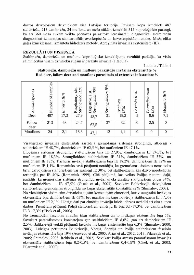
REZULTĀTI UN DISKUSIJA Staltbriežu, dambriežu un muflonu koproloģisko izmeklējumu rezultāti parādīja, ka visās
saimniecībās visām dzīvnieku sugām ir parazītu invāzija (1.tabula).
1.tabula / Table 1
Staltbriežu, dambriežu un muflonu parazitožu invāzijas ekstensitāte % Red deer, fallow deer and mouflons parasitosis of extensive infestations%
Anim
al s
pec
ies
Num
ber
of
sam
ple
s
Eim
eria
IE
%
Res
pir
atory
. si
st.
Str
ongyl
ida I
E%
Dig
esti
ve.
sis
t.
Str
ongyl
ida I
E%
Str
ongyl
oid
es I
E%
Tri
chyr
is
IE%
Fasc
iola
IE
%
Para
mfi
stom
um
IE%
Monie
zia I
E%
Deer 487 17,3 27,9 48,7 31 18,2 5 8,6 7,1
Fallow
deer
213 63 24,7 62,5
37 32 0 2,5 0
Mouflons 24 2,1 18,3 47,1 12 1,1 0 0 0
Visaugstāko invāzijas ekstensitāti sastādīja gremošanas sistēmas strongilīdi, attiecīgi -
staltbriežiem IE 48,7%, dambriežiem IE 62,5 %, bet mufloniem IE 47,1%.
Elpošanas sistēmas strongilīdi staltbriežiem bija IE 27,9%, dambriežiem IE 24,7%, bet
mufloniem IE 18,3%. Strongiloidoze staltbriežiem IE 31%, dambriežiem IE 37%, un
mufloniem IE 12%. Trichuris invāzija staltbriežiem bija IE 18,2%, dambriežiem IE 32% un
mufloniem IE 1,1%. Romaniuks savā pētījumā norādījis, ka gremošanas sistēmas nematodes
brīvi dzīvojošiem staltbriežiem var sasniegt IE 30%, bet staltbriežiem, kas dzīvo norobežotās
teritorijās pat IE 40% (Romaniuk 1999). Citā pētījumā, kas veikts Polijas rietumu daļā,
parādīts, ka gremošanas sistēmas strongilīdu invāzijas ekstensitāte staltbriežiem bijusi 84%,
bet dambriežiem - IE 47,5% (Cisek et al., 2003). Savukārt Baltkrievijā dzīvojošiem
staltbriežiem gremošanas strongilīdu invāzijas ekstensitāte konstatēta 92% (Shimalov, 2003).
No vienšūņiem visām trim dzīvnieku sugām konstatējām eimeriozi, kur visaugstākā invāzijas
ekstensitāte bija dambriežiem IE 63%, bet mazāku invāziju novēroja staltbriežiem IE 17,3%
un mufloniem IE 2,1%. Līdzīgi dati par eimēriju invāziju briežu dārzos uzrādīti arī citu autoru
darbos. Piemēram pētījumā Polijā staltbriežiem eimēriju IE bija 3,1-17,3%, bet dambriežiem
IE 3-17,3% (Cisek et al., 2003).
No trematodēm fasciolas atradām tikai staltbriežiem un to invāzijas ekstensitāte bija 5%.
Savukārt paramfistomas konstatējām gan staltbriežiem IE 8,6%, gan arī dambriežiem IE
2,5%. Baltkrievijā veiktā pētījumā fasciolu invāzijas ekstensitāte bija 6,3% (Shimalov, 2000,
2003). Līdzīgos pētījumos Baltkrievijā, Vācijā, Spānijā un Polijā staltbriežiem fasciolu
invāzijas ekstensitāte bija 19% (Acevedo et al., 2005; Arias et al., 2012, 2013; Pilarczyk et al.,
2005; Shimalov, 2003; Rehbein et al., 2002). Savukārt Polijā atrasto paramfistomu invāzijas
ekstensitāte staltbriežiem bija 0,2-0,5%, bet dambriežiem 0,4-0,9% (Cisek et al., 2003;
Pilarczyk et al., 2005).
24
No cestodēm konstatējām moniēzijas staltbriežiem un to invāzijas ekstensitāte bija 7,1%.
Līdzīgi arī Polijā veiktā pētījumā moniēziju invāzija konstatēta tikai staltbriežiem ar IE 0,5-
0,9% (Cisek et al., 2003; Pilarczyk et al., 2005).
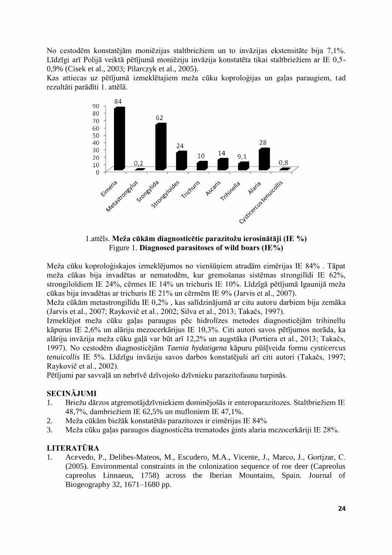
Kas attiecas uz pētījumā izmeklētajiem meža cūku koproloģijas un gaļas paraugiem, tad
rezultāti parādīti 1. attēlā.
1.attēls. Meža cūkām diagnosticētie parazitožu ierosinātāji (IE %)
Figure 1. Diagnosed parasitoses of wild boars (IE%) Meža cūku koproloģiskajos izmeklējumos no vienšūņiem atradām eimērijas IE 84% . Tāpat
meža cūkas bija invadētas ar nematodēm, kur gremošanas sistēmas strongilīdi IE 62%,
strongiloīdiem IE 24%, cērmes IE 14% un trichuris IE 10%. Līdzīgā pētījumā Igaunijā meža
cūkas bija invadētas ar trichuris IE 21% un cērmēm IE 9% (Jarvis et al., 2007).
Meža cūkām metastrongilīdu IE 0,2% , kas salīdzinājumā ar citu autoru darbiem bija zemāka
(Jarvis et al., 2007; Raykovič et al., 2002; Silva et al., 2013; Takačs, 1997).
Izmeklējot meža cūku gaļas paraugus pēc hidrolīzes metodes diagnosticējām trihinellu
kāpurus IE 2,6% un alāriju mezocerkārijus IE 10,3%. Citi autori savos pētījumos norāda, ka
alāriju invāzija meža cūku gaļā var būt arī 12,2% un augstāka (Portiera et al., 2013; Takačs,
1997). No cestodēm diagnosticējām Taenia hydatigena kāpuru pūšļveida formu cysticercus
tenuicollis IE 5%. Līdzīgu invāziju savos darbos konstatējuši arī citi autori (Takačs, 1997;
Raykovič et al., 2002).
Pētījumi par savvaļā un nebrīvē dzīvojošo dzīvnieku parazitofaunu turpinās.
SECINĀJUMI 1. Briežu dārzos atgremotājdzīvniekiem dominējošās ir enteroparazitozes. Staltbriežiem IE
48,7%, dambriežiem IE 62,5% un mufloniem IE 47,1%.
2. Meža cūkām biežāk konstatētās parazitozes ir eimērijas IE 84%
3. Meža cūku gaļas paraugos diagnosticēta trematodes ģints alaria mezocerkāriji IE 28%.
LITERATŪRA 1. Acevedo, P., Delibes-Mateos, M., Escudero, M.A., Vicente, J., Marco, J., Gortįzar, C.
(2005). Environmental constraints in the colonization sequence of roe deer (Capreolus
capreolus Linnaeus, 1758) across the Iberian Mountains, Spain. Journal of
Biogeography 32, 1671–1680 pp.
25
2. Arias, M.S., Martķnez-Carrasco, C., León-Vizcaķno, L., Paz-Silva, A., Dķez-Bańos, P.,
Morrondo, P., Alonso, F. (2012). Detection of antibodies in wild ruminants to evaluate
exposure to liver trematodes. Journal of Parasitology 98, 754–759 pp.
3. Arias M.S., Pińeiro P., Sįnchez-Andrade R., Suįrez J.L., Hillyer G.V., Dķez-Bańos P.,
Paz-Silva A. (2013). Relationship between exposure to Fasciola hepatica in roe deer
(Capreolus capreolus) and cattle extensively reared in an endemic area. Research in
Veterinary Science 95 1031–1035 pp.
4. Cisek A., Balicka-Ramisz A., Ramisz A., Pilarczyk B.(2003). Occurrence of gastro-
intestinal nematodes in cervids (Cervidae) of north-western Poland, Elec J P Agric
Univ, 6, 2.
5. García-González Á.M., Pérez-Martín J.E, Gamito-Santos J.A, Calero-Bernal R.,
Alcaide Alonso M., Frontera Carrión E.M. (2013). Epidemiologic study of lung
parasites (Metastrongylus spp.) in wild boar (Sus scrofa) in southwestern Spain. J Wildl
Dis. Jan;49(1):157-62. doi: 10.7589/2011-07-217.
6. Järvis, T.A., Kapel, Moks, C.H., Talvik, E., Mägi, H. E. (2007). Helminths of wild boar
in the isolated population close to the northern border of its habitat area. In Veterinary
Parasitology 150(4):366-369 pp.
7. Panayotova-Pencheva, M.S., (2006). New records of protostrongylid lungworms from
wild ruminants in Bulgaria. Veterinarni Medicina 51, 477–484 pp.
8. Pilarczyk B., Balicka-Ramisz A., Ramisz A., Lachowska S. (2005). The occurrence of
intestinal parasites of roe deer and red deer in the western Pomerania voivodeship. (In
Polish) Wiad Parazytol, 51,4, 307-10 pp.
9. Portiera P., Valléea I., Lacoura S.A., Schallerb R. M., Fertéc H., Durandd B. (2014).
Increasing circulation of Alaria alata mesocercaria in wild boar populations of the
Rhine valley, France, 2007–2011. Veterinary Parasitology, Volume 199, Issues 3–4, 31
January , 153–159 pp.
10. Rajković J., Bosnić R., Rimac S., Dragičević D., Vinković P., Bara (2002).
Prevalence of helminths in wild boar from hunting grounds in eastern Croatia.
Zeitschrift für Jagdwissenschaft; Dec, Vol. 48 Issue 4, p261-270, 10p
11. Rehbein Von S., Lutz W., Visser M., Winter W.( 2002). Investigation of the parasite
fauna of game animals in Northrhine –Westfalia. 3. Endoparasites of red deer (In
German), Z Jagdwiss, 48, 69- 93 pp.
12. Romaniuk K, 1999, Evaluation of parasitic invasion of stags on farms and in free range
conditions (In Polish), Medycyna Wet, 55, 1, 46-7.
13. Shimalov, V.V., Shimalov, V.T. (2003). Helminth fauna of cervids in Belorussian
Polesie. Parasitology Research 89, 75–76 pp.
14. Shimalov, V.V., Shimalov, V.T. (2000). Findings of Fasciola hepatica Linnaeus 1758,
in wild animals in Belorussian Polesye. Parasitology Research 86, 342 p.
15. Silva D., Müller G. (2013). Parasites of the respiratory tract of Sus scrofa scrofa (wild
boar) from commercial breeder in southern Brazil and its relationship with Ascaris
suum. Parasitol Res. Mar;112(3):1353-6. doi: 10.1007/s00436-012-3214-1
16. Takács, A. (1997). Contribution to the helminth fauna of wild boar in Hungary. Wiener
Tierärztliche Monatsschrift 84 (11), 314-316 pp.
17. Zajac A.M., Conboy G.A.,Wragg S. M (2006). Veterinary clinical parasitology,
Blackwell Publishing.
26
LAKTOFERĪNA DINAMIKA PIENĀ ATKARĪBĀ NO GOVJU SEZONĀLĀS TURĒŠANAS UN MASTĪTU IEROSINĀTĀJU
KLĀTBŪTNES TESMENĪ
THE DYNAMICS OF LACTOFERRIN IN MILK IN RELATION TO COW SEASONAL KEEPING AND
PATHOGENS PRESENCE IN THE UDDER
Iveta Kociņa1, Vita Antāne2, Ivars Lūsis2 1Pārtikas drošības, dzīvnieku veselības un vides zinātniskais institūts BIOR, Latvija, Institute
of Food Safety, Animal Health and Environment BIOR, Latvia 2Veterinārmedicīnas fakultāte LLU, Latvija; Faculty of Veterinary Medicine LUA, Latvia
ABSTRACT Recent studies show that lactoferrin contribute significantly to the maintenance of udder
health. Sadly the concentration of lactoferrin in cow’s milk during the middle period of
lactation is low. That is an urgent issue - how to stimulate and maintain a sufficient level of
lactoferrin in the udder at all stages of lactation. The aim of the present study was to evaluate
the dynamics of lactoferrin level in cow’s milk in relation to the cow seasonal keeping and
presence of pathogens in the udder.
The experimental part of the study was carried out on the dairy farm “Pērles”, Vidzeme
region. Cows were kept in a cold loose housing system, grouped and fed differently
depending on cow productivity and lactation period.Two times in the housing period and two
times in the grazing period milk were sampled from 16 dairy cows and examined for the
concentration of lactoferrin and for the presence of pathogens. Cows for the study were
selected to analyse the milk obtained from clinically healthy udder quarters of cows of similar
age and productivity at the middle stage of lactation.
It was discovered, that during grazing period compared with the housing period the lactoferrin
concentration in milk increases significantly (p<0.05). Some pathogenic bacteria species
infecting the udder quarters had significantly increased the concentration of lactoferrin
(p<0.05) in milk. Wide variation amplitude of lactoferrin concentration in milk was observed,
which indicates the important role of the individual factor of an animal in the formation of
self-defence response.
KEY WORDS: lactoferrin, mammary gland, seasonal keeping IEVADS Neskatoties uz ilggadējiem zinātnieku centieniem ierobežot mastītu izplatību, tā joprojām tiek
uzskatīta par dārgāko un izplatītāko slimību piena lopkopībā. Mastītu radītos zaudējumus
sastāda, gan izslaukuma samazināšanās un piena sliktā kvalitāte, gan izdevumi, kas saistīti ar
slimo govju ārstēšanu vai izbrāķēšanu, bet 70% zaudējumu cēlonis ir tieši govju subklīniskie
mastīti (Schukken et al., 2003; Fourichon et al., 2001; Schukken et al., 1992; Philpot,
Nickerson,1997). Mastītu dažādību un attīstību nosaka organisma aizsargspēju potenciāls un
ierosinātāju virulences faktori. Dzīvnieka dabīgais aizsargmehānisms var veiksmīgi ierobežot
un likvidēt infekciju, vai arī infekcija, daļēji likvidēta, var turpināties ilgāku periodu un
parasti šis process izpaužas subklīniska mastīta formā (Ali-Vehmas, Sandholm, 1995; Baker
et al., 2002). Pētījumi apliecina laktoferīna nozīmīgo lomu tesmeņa veselības saglabāšanā
(Marnila, Korhonen, 2002; Hagiwara et al., 2003; Kutila et al., 2003). Laktoferīna
koncentrācija govju pienā laktācijas vidus posmā ir zema, tāpēc aktuāls ir jautājums, kādā
27
veidā tesmenī, mastīta profilakses nolūkos, stimulēt un uzturēt pietiekami augstu laktoferīna
koncentrāciju tesmeņa audos (Kai et al., 2002). Ir virkne faktoru, kas stimulē organisma
imūnsistēmas aktivitāti. Aktivācijai tiek izmantotas dažādas specifiskas vakcīnas (S.aureus,
Coli u.c.), bakterofāgi, citokīni (IL-1, IL-2, GM-CSF u.c.), kā arī citas organisma aizsargspēju
aktivizējošas bioloģiski aktīvas vielas. Tomēr nepārvērtējama loma organisma aizsardzības un
pretestības spēju stiprināšanā ir pareizai un pilnvērtīgai dzīvnieku ēdināšanai un aprūpei,
ērtām, komfortablām mītnēm, kurās līdz minimumam samazināta stresfaktoru un dažādu citu
nelabvēlīgu faktoru iedarbība (Barkema et al.,1999; Blūzmanis, 1999).
Ar nodomu izprast govju tesmeņa dabīgā aizsargkomponenta laktoferīna nozīmi tesmeņa
veselības saglabāšanā, šajā darbā pētīta laktoferīna dinamika pienā dažādos turēšanas
apstākļos, kā arī izvērtēta tā koncentrācija saistībā ar patogēno mikroorganismu klātbūtni
tesmenī.
Pētījumu mērķis bija izvērtēt laktoferīna nozīmi tesmeņa veselības saglabāšanā saistībā ar
govju sezonālo turēšanu un mastītu ierosinātāju klātbūtni tesmenī.
MATERIĀLS UN METODIKA Pētījuma eksperimentālā daļa veikta Vidzemes reģiona slaucamo govju novietnē „Pērles”.
Pētījuma veikšanas laikā bija 75 slaucamas govis, vidējais izslaukums 7156 kg gadā, vidējais
somatisko šūnu skaits ganāmpulka koppienā 255 000 šūnu mililitrā (tūkst.ml-1). Ganāmpulku
veido divdesmit četras Latvijas Brūnās šķirnes govis, trīsdesmit divas Holšteinas Melnraibās
šķirnes govis, kā arī deviņpadsmit govis, kas ir iepriekšminēto govju šķirņu hibrīdi.
Ganāmpulka govju vecums ir no 2 līdz 8 gadi, bet vidējais laktāciju skaits – 3.5 laktācijas.
Govis tiek turētas nepiesieti grupās nesiltinātā, jeb atvieglota tipa mītnē un barotas atkarībā no
to produktivitātes un laktācija perioda. Govju barības racionā iekļauts skābsiens, siens,
spēkbarības maisījums, sakņaugi, kā arī barības piedevas. Kā pakaišu materiālu govju
turēšanas telpā izmanto skujkoku skaidas. Vasaras – rudens periodā govis atrodas dienas
ganībās, turpretim ziemā un pavasarī tiek turētas mītnē. Govis slauc divas reizes dienā,
slaukšanas zālē ar „skujiņas” tipa slaukšanas iekārtu, plkst. 6.00 un plkst.18.00. Pirms
slaukšanas govju pupus tīra ar vienreizējas lietošanas salvetēm un katra ceturkšņa pirmās
piena strūklas ieslauc krūzītē ar melnu pamatni un izvērtē piena kvalitāti. Katru reizi pēc
govju izslaukšanas notiek pupu dezinfekcija.
Sešpadsmit klīniski veselas līdzīga vecuma (2. un 3. laktācija) un produktivitātes (22-25kg
piena dienā) ganāmpulka govis laktācijas vidusposmā izvēlējāmies kā ganāmpulka govis
pārstāvošu paraugkopu
Visām govīm izmeklējumi veikti divas reizes ganību periodā un divas reizes kūtsstāves
periodā. Piena paraugus ieguvām no tesmeņa katra ceturkšņa. Kopā izmeklēti 214 piena
paraugi.
Govīm, kurām kaut viena ceturkšņa piena paraugā Kalifornijas mastīta testa (KMT) rezultāts
uzrādīja neskaidru vai pozitīvu reakciju, visu ceturkšņu piena paraugus izmeklējām
bakterioloģiski. Kopumā bakterioloģiski izmeklēti 111 piena paraugi.
Piena bakterioloģiskie izmeklējumi veikti Latvijas Lauksaimniecības universitātes
Veterinārmedicīnas fakultātes Klīniskā institūta ganāmpulka veselības un reprodukcijas
problēmu laboratorijā pēc ISO 707(¹) standartā rekomendētās metodes un ierosinātāju
identificēšana veikta pēc Quinn et al., (2000) aprakstītās procedūras.
Somatisko šūnu skaitu pienā noteicām Akciju sabiedrībā „Rīgas piena kombināts” piena
kvalitātes laboratorijā, saskaņā ar LVS EN ISO 13366-3:1997 "Piens. Somatisko šūnu skaita
noteikšana”, Fluora-opto-elektroniskā metode". Tā kā SŠS pienā govīm svārstās no dažiem
simtiem līdz vairāk kā četriem miljoniem šūnu mililitrā, tad aprēķinos, lai veidotu
uzskatāmākus grafikus, lietotas naturālās logaritmiskās vērtības ln(somat), savukārt teksta
daļā un diskusijā izmantots laboratoriski noteiktais somatisko šūnu skaits pienā.
28
Rīgas Reprodukcijas Centra (RRC) laboratorijā noteikta laktoferīna koncentrācija pienā,
izmantojot reaģentu komplektu D-4156 „Laktoferin – IFA - BEST”, sērijas Nr. 33, kvalitātes
standarts TY 9398-045-23548172-2001. Reaģentu komplekts izgatavots un pielietojams
laktoferīna kvantitatīvai noteikšanai asins serumā un citos bioloģiskos šķidrumos – asins
plazmā, urīnā, siekalās, spermā, pienā u.c., izgatavots akciju sabiedrībā „Vektor-Best”,
Krievijas Federācijā. Metode pamatojas uz cietās fāzes imūnfermentanalīzi, izmantojot
poliklonālās laktoferīna antivielas.
Datu statistiskā apstrāde veikta, izmantojot SPSS programmu -11.0 versiju (Statistical
Package for Social Science) un Microsoft Excel paketes. Ar programmas analīzes rīku
Descriptive Statistic aprēķināti vidējie rādītāji un to standartnovirze - (s).
Darbā izvirzītās hipotēzes pārbaudītas ar p-vērtības metodi, aprēķinātā p-vērtība salīdzināta ar
būtiskuma līmeni α=0,05. Kvalitatīvo faktoru ietekmes novērtēšanai pielietota vienfaktora
dispersijas analīze, savukārt faktoru mijiedarbības efekta izpētei izmantojām divfaktoru
dispersijas analīzi (Arhipova, Bāliņa, 2003).
REZULTĀTI UN DISKUSIJA Mūsu pētījumos iegūtie rezultāti liecina, ka analizētajos piena paraugos vidējais somatisko
šūnu skaits (SŠS) ir 233 710 (566.31) ml-1. Procentuāli lielākā daļa, jeb 64% no visu
izmeklēto govju tesmeņu ceturkšņiem ir veseli, SŠS šo ceturkšņu pienā nepārsniedz 100 000
ml-1. Savukārt 20% ceturkšņu uzskatāmi par iespējami inficētiem, jo SŠS pienā ir robežās no
100 000 ml-1 līdz 300 000 ml-1, 16% ir subklīniska mastīta skarti uz ko norāda SŠS pienā virs
300 000 ml-1 .
Literatūrā ir atšķirīgas norādes par to, kāds somatisko sūnu skaits pienā liecina par veselu
tesmeni. Pastāv uzskats, ka laktējošas govs tesmens ir vesels, ja SŠS pienā nepārsniedz
400 000 ml-1 (Blūzmanis, 1999). Citos ziņojumos ir norādes, ka veselā laktējošā tesmenī SŠS
pienā nedrīkst pārsniegt 100 000 ml-1 (Paape et al., 1987), vai arī, ka neinficētu ceturkšņu
pienā laktācijas perioda vidusdaļā somatisko šūnu skaits ir robežās līdz 200 000 ml-1, ja šī
robeža pārsniegta, tas norāda uz piena dziedzera reakciju uz dažādiem infekciozas (Conha,
1996; Philpot, Nickerson, 1997) vai neinfekciozas dabas kairinājumiem (Burveinich et al.,
1998; Saloniemi 1995; Coico et al., 2003) apgalvo, ka paaugstināts SŠS ne vienmēr liecina
par tesmeņa infekciju, tas tikai norāda, ka tesmeņa audi pakļauti kairinājumam. Tesmeņa audu
kairinājuma gadījumā ir augsts risks, ka pievienosies infekcija. SŠS pienā var arī palielināties
govīm uzturoties stresa vidē, paaugstinātā temperatūrā, kā arī pie nelavēlīgiem turēšanas un
aprūpes nosacījumiem, tomēr šādos gadījumos krasu SŠS pieaugumu nenovēro govīm, kurām
tesmens ir vesels (Elvinger et al., 1991; Ekman 1998; Kociņa, Antāne 2000). Konošonoka
(2005) minējusi, ka veselu govju pienā SŠS ir līdz 300 000 ml-1, lielāks somatisko šūnu skaits
govju pienā liecina par mastīta risku.
Attiecībā uz SŠS atšķirībām atkarībā no sezonas mūsu pētījuma rezultāti liecina, ka
augstākais vidējais SŠS pienā ir ganību periodā 302 880 (685.72) ml-1.. Savukārt kūtsstāves
periodā vidējais SŠS pienā ir 169 530 (419.94) ml-1 (p<0.05). Arī mūsu iepriekš veikto
pētījumu rezultāti (Kociņa, Antāne, 2000) liecina, ka somatisko šūnu skaits govju pienā
ganību periodā ir būtiski augstāks, salīdzinot ar kūtsstāves periodu. Ļoti iespējams, šo
pieaugumu veicina intensīva ārējās vides faktoru ietekme, radot tesmeņa kairinājumu, kā arī
dzīvnieku imūnreakciju aktivācija ganību periodā. Publikācijās minēts, ka liellopiem
karstuma un stresa ietekmē pieaug leikocītu migrācija piena dziedzerī (Elvinger et al., 1992).
Demelash (2005) ziņo, ka sezona būtiski ietekmē SŠS pienā, proti, ganību periodā, siltā un
mitrā laikā govīm ir lielāks risks saslimt ar mastītu. Skaidrojums augstākam mastītu izcelsmes
riskam ganību periodā ir tesmeņa kontakts ar ārējās vides kairinātājiem, t.sk. patogēniem
mikroorganismiem (Goldberg et al., 1992; Harmon 1994). Savukārt cita pētījuma rezultāti
neliecina, ka karstums vai govju turēšana ganībās saistīta ar SŠS pieaugumu pienā (Šterna,
29
2005). Ņemot vērā, ziņojumu par sezonas ietekmi uz SŠS, varam izdarīt pieņēmumu, ka
vesels tesmens uz vides apstākļu maiņu, kā arī dažāda veida kairinājumiem nereaģē ar krasu
SŠS pieaugumu pienā. Izpētīts, ka govīm, kuras iepriekš pārslimojušas tesmeņa iekaisumu ir
svārstīgs, bieži paaugstināts SŠS pienā un tām ir piecas reizes lielāka reinfekcijas iespēja
(Demelash et.al., 2005).
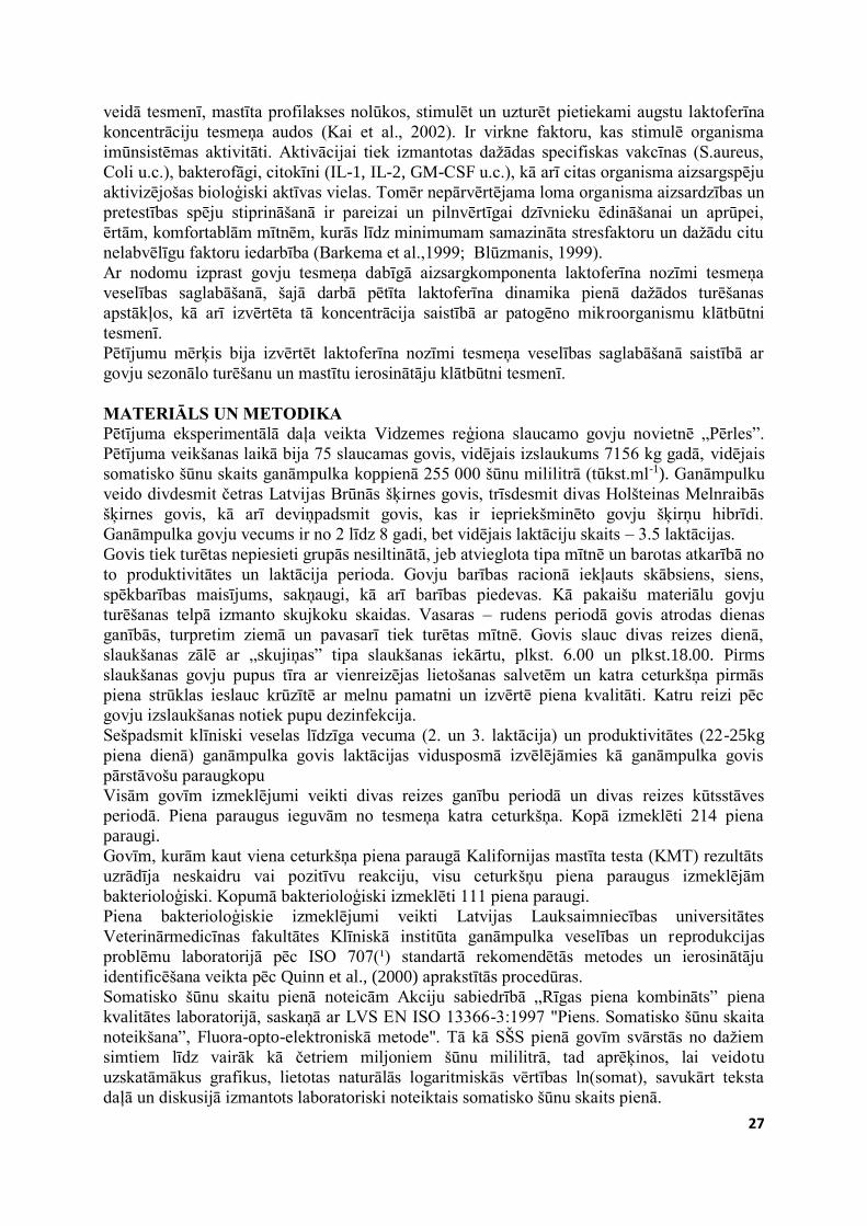
Laktoferīna dinamika pienā kūtsstāves un ganību periodos, kā arī tā vērtību svārstību
amplitūda parādīta 1. attēlā.Veiktie pētījumi norāda, ka laktoferīnam (Lf), līdzīgi kā SŠS
pienā, novēro vērtību svārstību sezonālo raksturu. Kūtsstāves periodā laktoferīna vidējā
vērtība ir augstāka, kā ganību, attiecīgi 0.73 (0.54) gl-1 un 0.44 (0.55) gl-1.
1.attēls. Laktoferīna koncentrācija (gl-1) pienā kūtsstāves un ganību periodos
Figure 1. Concentration of lactoferrin (gL-1) in milk during housing and grazing period Gan kūtsstāves, gan ganību periodos atsevišķām govīm ir izteiktas laktoferīna vērtību
svārstības ārpus standartnoviržu robežām, kas norāda uz govju individuālā faktora lomu Lf
koncentrācijas uzturēšanā. Attēlā redzamais uzskatāmi norāda, ka kūtsstāves periodā, kad
vērojamas augstākās laktoferīna vērības, arī tā vērtību svārstību amplitūda ir izteiktāka.
Vienfaktora dispersijas analīzes rezultāti (p=0.044) liecina, ka laktoferīna vērtība pienā
būtiski (p<0.05) atšķiras atkarībā no govju sezonālās turēšanas, un, ka govju sezonālā
turēšana būtiski ietekmē laktoferīna daudzumu pienā.
Analizējot literatūru par laktoferīna dinamiku un tās izmaiņu cēloņiem govju pienā, secinām,
ka autori pārsvarā uzmanību akcentē uz patogēno mikroorganismu ietekmi uz Lf
koncentrācijas izmaiņās (Korhonen, Kaartinen, 1995; Kai et al., 2002; Diarra et al., 2003;
Hagiwara et al., 2003). Kaut gan ir pētījumi, kas liecina, ka laktoferīna daudzumu govs pienā
ietekmē arī dažādi citi faktori, piemēram, govs šķirne, vecums, veselības stāvoklis, laktācijas
periods, govju turēšanas apstākļi, menedžments (Kutila et al., 2003; Zagorska, 2007;
Konuspayeva et al., 2007; Krol et al., 2010), atrodam arī norādes, ka veselu govju pienā
laktācijas vidusposmā laktoferīna koncentrācija ir zema, bet tā pieaug tesmeņa iekaisuma
laikā (Sandholm, Korhonen, 1995; Kawai et al., 1999; Hagivara et al., 2003; Diarra et al.,
Ganību periods
Grazing period
Kūtsstāves periods
Housing period
5.
0
4.
0
3.0
2.0
1.0
.00
141 163 158 195 147 156 161 152 121
135
69 104 76 94 98 62 41 96
72
30
2003). Pastāv viedoklis, ka Lf vērtības govju pienā svārstās plašās robežās un visi, to
koncentrāciju ietekmējošie faktori, nemaz vēl nav apzināti (Kutila, 2003). Arī cits, Latvijā
veiktais pētījums liecina, ka laktoferīna saturs govju piena paraugos svārstās ļoti plašās
robežās (Zagorska, 2007).
Mūsu veiktajā pētījumā laktoferīna saturs pienā svārstās plašās robežās un ievērojami atšķiras
gan individuālu paraugu starpā, gan atkarībā no govju sezonālās turēšanas. Mūsu iegūtie
rezultāti liecina, ka laktoferīna vidējā vērtība analizētajos piena paraugos ir 0.61 (0.60) gl-1, un
tā saturs svārstās ievērojamā amplitūdā, no 0.00 gl-1 līdz 4.40 gl-1. Publikācijās norādīts, ka laktoferīna daudzums pienā nozīmīgi variē atkarībā no sezonas
(Konuspayeva et al., 2007), praktizētās govju turēšanas sistēmas un turēšanas apstākļiem un
imunitātes stiprināšanas pasākumiem (Kutila, 2003; Zagorska, 2007). Lf koncentrācijas
pieaugums pienā ne vienmēr saistāms ar tesmeņa iekaisumu. Ir pētījumi, kuru rezultāti
apgalvo, ka Lf līmeni pienā galvenokārt ietekmē govju laktācijas periods un tesmeņa infekcija
(Kawai et al., 1999; Eckersall et al., 2001; Hagivara et al., 2003; Korhonen, Kaartinen, 2005).
Mūsu iegūtie dati liecina, ka govju sezonālā turēšana būtiski ietekmē laktoferīna
koncentrāciju pienā un tā vērtības būtiski atšķiras (p<0.05) kūtsstāves un ganību periodā.
Laktoferīna vidējā koncentrācija pienā kūtsstāves periodā ir 0.739 (0.54) gl-1, kas ir būtiski
augstāka par Lf koncentrāciju ganību periodā, proti, 0.448 (0.55) gl-1. Novērojām, ka
kūtsstāves periodā ir augstāka gan vidējā, gan maksimālā, 4.40 gl-1 Lf vērtība pienā, kā arī ir
vislielākās Lf vērtību svārstības. Minētais iespējams liecina par individuālā faktora nozīmīgo
lomu Lf koncentrācijas uzturēšanā, un norāda, ka kūtsstāves periodā tesmenī kāda iemesla
pēc, iespējams salīdzinoši zemākas šūnu imunitātes dēļ, nepieciešama augstāka Lf
koncentrācija.
Ja laktoferīna koncentrācijas dinamiku pienā vērtējam, pamatojoties uz jebkuru citu, ar
tesmeņa infekciju nesaistītu faktoru iedarbību, rodas jautājums - kāpēc Lf koncentrācija savas
maksimālās vērtības nesasniedz ganību periodā, kad aktivizējas imūnreakcijas un pastāv
lielāks tesmeņa audu kairinājuma risks, bet gan kūtsstāves periodā. Respektējot laktoferīna
nozīmīgās, antibakteriālās spējas, varam pieņemt, ka ganību periodā salīdzinoši zemākie Lf
vērtību rādītāji līdz ar paaugstinātu SŠS pienā, liecina par sekojošo: ganību periodā SŠS govju
pienā palielinās galvenokārt tesmeņa neinfekciozu kairinājumu rezultātā, par ko liecina arī
salīdzinoši zemā Lf koncentrācija pienā šai periodā; fagocitārās šūnas (t.sk. somatiskās šūnas)
tesmenī, kam ganību periodā ir būtiski augstāka aktivitāte, spēj ierobežot baktērijas un
nepieļaut iekaisuma procesa attīstību, tādējādi imunoloģiskajās aizsargreakcijās laktoferīns
neiesaistas tik lielā koncentrācijā, kā kūtsstāves periodā.
1. tabula / Table 1
Laktoferīna koncentrācija (gl-1) pienā saistībā ar tesmeņu ceturkšņu inficētību Lactoferrin concentration in milk (gL-1) in relation to presence of infection in the udder
quarters
Ceturkšņu piens /
Quarter milk
Laktoferīns / Lactoferrin (gl-1)
Paraugu
Skaits
(n)
Vidējā
vērtība /
Average
Standart-
novirze /
Standart-
deviation
Minimālā
vērtība
Min
Maksimālā
vērtība
Max
Bez mastīta ierosinātāja
Without pathogens
69 0.405 0.47 0.00 2.25
Ar mastīta ierosinātāju
With pathogens
42 0.400 0.40 0.02 1.60
Kopā / Total 111 0.403 0.44 0.00 2.25
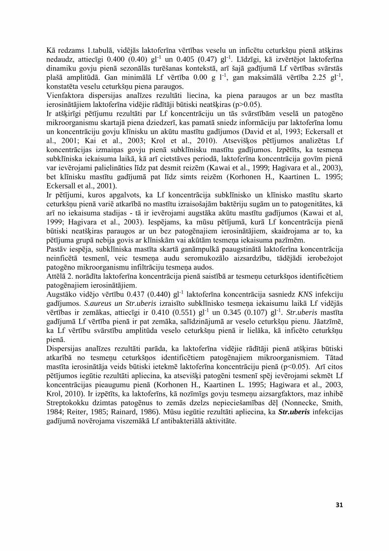
31
Kā redzams 1.tabulā, vidējās laktoferīna vērtības veselu un inficētu ceturkšņu pienā atšķiras
nedaudz, attiecīgi 0.400 (0.40) gl-1 un 0.405 (0.47) gl-1. Līdzīgi, kā izvērtējot laktoferīna
dinamiku govju pienā sezonālās turēšanas kontekstā, arī šajā gadījumā Lf vērtības svārstās
plašā amplitūdā. Gan minimālā Lf vērtība 0.00 g l-1, gan maksimālā vērtība 2.25 gl-1,
konstatēta veselu ceturkšņu piena paraugos.
Vienfaktora dispersijas analīzes rezultāti liecina, ka piena paraugos ar un bez mastīta
ierosinātājiem laktoferīna vidējie rādītāji būtiski neatšķiras (p>0.05).
Ir atšķirīgi pētījumu rezultāti par Lf koncentrāciju un tās svārstībām veselā un patogēno
mikroorganismu skartajā piena dziedzerī, kas pamatā sniedz informāciju par laktoferīna lomu
un koncentrāciju govju klīnisku un akūtu mastītu gadījumos (David et al, 1993; Eckersall et
al., 2001; Kai et al., 2003; Krol et al., 2010). Atsevišķos pētījumos analizētas Lf
koncentrācijas izmaiņas govju pienā subklīnisku mastītu gadījumos. Izpētīts, ka tesmeņa
subklīniska iekaisuma laikā, kā arī cietstāves periodā, laktoferīna koncentrācija govīm pienā
var ievērojami palielināties līdz pat desmit reizēm (Kawai et al., 1999; Hagivara et al., 2003),
bet klīnisku mastītu gadījumā pat līdz simts reizēm (Korhonen H., Kaartinen L. 1995;
Eckersall et al., 2001).
Ir pētījumi, kuros apgalvots, ka Lf koncentrācija subklīnisko un klīnisko mastītu skarto
ceturkšņu pienā variē atkarībā no mastītu izraisošajām baktēriju sugām un to patogenitātes, kā
arī no iekaisuma stadijas - tā ir ievērojami augstāka akūtu mastītu gadījumos (Kawai et al,
1999; Hagivara et al., 2003). Iespējams, ka mūsu pētījumā, kurā Lf koncentrācija pienā
būtiski neatšķiras paraugos ar un bez patogēnajiem ierosinātājiem, skaidrojama ar to, ka
pētījuma grupā nebija govis ar klīniskām vai akūtām tesmeņa iekaisuma pazīmēm.
Pastāv iespēja, subklīniska mastīta skartā ganāmpulkā paaugstinātā laktoferīna koncentrācija
neinficētā tesmenī, veic tesmeņa audu seromukozālo aizsardzību, tādējādi ierobežojot
patogēno mikroorganismu infiltrāciju tesmeņa audos.
Attēlā 2. norādīta laktoferīna koncentrācija pienā saistībā ar tesmeņu ceturkšņos identificētiem
patogēnajiem ierosinātājiem.
Augstāko vidējo vērtību 0.437 (0.440) gl-1 laktoferīna koncentrācija sasniedz KNS infekciju
gadījumos. S.aureus un Str.uberis izraisīto subklīnisko tesmeņa iekaisumu laikā Lf vidējās
vērtības ir zemākas, attiecīgi ir 0.410 (0.551) gl-1 un 0.345 (0.107) gl-1. Str.uberis mastīta
gadījumā Lf vērtība pienā ir pat zemāka, salīdzinājumā ar veselo ceturkšņu pienu. Jāatzīmē,
ka Lf vērtību svārstību amplitūda veselo ceturkšņu pienā ir lielāka, kā inficēto ceturkšņu
pienā.
Dispersijas analīzes rezultāti parāda, ka laktoferīna vidējie rādītāji pienā atšķiras būtiski
atkarībā no tesmeņu ceturkšņos identificētiem patogēnajiem mikroorganismiem. Tātad
mastīta ierosinātāja veids būtiski ietekmē laktoferīna koncentrāciju pienā (p<0.05). Arī citos
pētījumos iegūtie rezultāti apliecina, ka atsevišķi patogēni tesmenī spēj ievērojami sekmēt Lf
koncentrācijas pieaugumu pienā (Korhonen H., Kaartinen L. 1995; Hagiwara et al., 2003,
Krol, 2010). Ir izpētīts, ka laktoferīns, kā nozīmīgs govju tesmeņu aizsargfaktors, maz inhibē
Streptokokku dzimtas patogēnus to zemās dzelzs nepieciešamības dēļ (Nonnecke, Smith,
1984; Reiter, 1985; Rainard, 1986). Mūsu iegūtie rezultāti apliecina, ka Str.uberis infekcijas
gadījumā novērojama viszemākā Lf antibakteriālā aktivitāte.
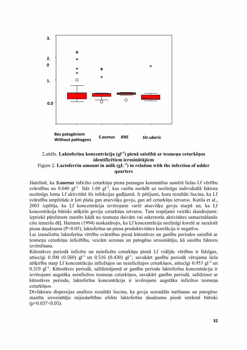
32
2.attēls. Laktoferīna koncentrācija (gl-1) pienā saistībā ar tesmeņu ceturkšņos
identificētiem ierosinātājiem Figure 2. Lactoferrin amount in milk (gL-1) in relation with the infection of udder
quarters Jāatzīmē, ka S.aureus inficēto ceturkšņu piena paraugos konstatētas samērā lielas Lf vērtību
svārstības no 0.040 gl-1 līdz 1.60 gl-1, kas varētu norādīt uz nozīmīgu individuālā faktora
nozīmīgo lomu Lf aktivitātē šīs infekcijas gadījumā. Ir pētījumi, kuru rezultāti liecina, ka Lf
svārstību amplitūda ir ļoti plaša gan atsevišķu govju, gan arī ceturkšņu ietvaros. Kutila et al.,
2003 izpētīja, ka Lf koncentrācija ievērojami variē atsevišķu govju starpā un, ka Lf
koncentrācija būtiski atšķirās goviju ceturkšņu ietvaros. Tam iespējami vairāki skaidrojumi:
iepriekš pārslimots mastīts kādā no tesmeņa daivām vai sekretorās aktivitātes samazināšanās
citu iemeslu dēļ. Harmon (1994) noskaidrojis, ka Lf koncentrācija nozīmīgi korelē ar saražotā
piena daudzumu (P<0.05), laktoferīna un piena produktivitātes korelācija ir negatīva.
Lai izanalizētu laktoferīna vērtību svārstības pienā kūtsstāves un ganību periodos saistībā ar
tesmeņu ceturkšņu inficētību, veicām sezonas un patogēno ierosinātāju, kā saistītu faktoru
izvērtēšanu.
Kūtsstāves periodā inficēto un neinficēto ceturkšņu pienā Lf vidējās vērtības ir līdzīgas,
attiecīgi 0.500 (0.560) gl-1 un 0.516 (0.430) gl-1, savukārt ganību periodā vērojama liela
atšķirība starp Lf koncentrāciju inficētajos un neinficētajos ceturkšņos, attiecīgi 0.953 gl-1 un
0.319 gl-1. Kūtsstāves periodā, salīdzinājumā ar ganību periodu laktoferīna koncentrācija ir
ievērojami augstāka neinficētos tesmeņa ceturkšņos, savukārt ganību periodā, salīdzinot ar
kūtsstāves periodu, laktoferīna koncentrācija ir ievērojami augstāka inficētos tesmeņa
ceturkšņos.
Divfaktoru dispersijas analīzes rezultāti liecina, ka govju sezonālās turēšanas un patogēno
mastīta ierosinātāju mijiedarbības efekts laktoferīna daudzumu pienā ietekmē būtiski
(p=0.037<0.05).
Str.uberis KNS S.aureus Bez patogēniem Without pathogens
3.
0
2.
0
1.
0
0.0
.0
33
2. tabula / Table 2
Laktoferīna koncentrācija pienā (gl-1) kūtsstāves un ganību periodos, atkarībā no patogēnā ierosinātāju klātbūtnes ceturksnī
The dynamic of Lactoferrin (gL-1) in milk during the housing and grazing period in relation with the udder infection
Laktoferīns
Lactoferrin
Kūtsstāves periods
Houzing period
Ganību periods
Grazing period
bez patogēniem
without
pathogens
ar patogēniem
with pathogens
bez
patogēniem
without
pathogens
ar
patogēniem
with
pathogens
Vidējā vērtība
Average
Standartnovirze
Standartdeviation
Minimālā vērtība
Minimum
Maksimālā vērtība
Maximum
0.500
0.560
0.035
2.250
0.516
0.430
0.152
1.400
0.319
0.353
0.00
0.915
0.953
0.924
0.00
1.600
Kā iepriekš norādījām, ganību periodā SŠS ievērojami pieaug piena paraugos bez patogēniem
mikroorganismiem, savukārt laktoferīna koncentrācija neinficēto ceturkšņu pienā augstāka ir
kūtsstāves laikā. Iespējams tas apstiprina apgalvojumu, ka laktoferīns tāpat kā somatiskās
šūnas tesmenī aktīvi reaģē arī uz tesmeņa audu neinfekciozu kairinājumu (Hagiwara et al.,
2003). Mūsu pētījuma rezultāti liecina, ka sezonālās turēšanas un patogēno ierosinātāju
mijiedarbība būtiski (p<0.05) ietekmē laktoferīna koncentrāciju pienā.
Iespējams, ka mūsu pētījumā konstatētās paaugstinātās Lf vērtības norāda uz to, ka laktoferīna
bakteriostatiskas un baktericīdas iedarbības rezultātā tiek ierobežota tesmeņu infekciju
izplatība gan tām govīm, kurām tesmenī identificēti patogēnie mikroorganismi, gan tām,
kurām patogēni nav identificēti (Kai et al., 2002; Hagiwara et al., 2003, Krol et al., 2010).
Konstatētais mijiedarbības efekts subklīnisko mastītu skartā ganāmpulkā iespējams norāda
gan uz laktoferīna dinamikas sezonālām atšķirībām, saistībā ar govju turēšanu (Zagorska u.c.,
2007), gan uz Lf antibakteriālo aktivitāti atsevišķu tesmeņa infekciju gadījumā (Kawai et al.,
1999; Hagivara et al., 2003; Korhonen, Kaartinen, 2005), gan to, ka šie faktori (govju
sezonālā turēšana un mastītu izraisošie patogēni) savstarpēji mijiedarbojieties nozīmīgi
ietekmē govju tesmeņa veselību.
SECINĀJUMI 1. Govju sezonālā turēšana būtiski ietekmē somatisko šūnu skaitu (p<0.05) un laktoferīna
koncentrāciju (p<0.05) govju pienā: ganību periodā, salīdzinājumā ar kūtsstāves periodu
būtiski pieaug somatisko šūnu skaits govju pienā, savukārt kūtsstāves periodā būtiski
palielinās laktoferīna daudzums pienā.
2. Laktoferīna koncentrācija subklīniski inficēto un veselo ceturkšņu pienā būtiski
neatšķiras (p>0.05), tomēr atsevišķas patogēnās baktērijas būtiski paaugstina laktoferīna
koncentrāciju pienā (p<0.05).
3. Ganību periodā, salīdzinājumā ar kūtsstāves periodu, būtiski palielinās laktoferīna
koncentrācija inficēto ceturkšņu pienā (p<0.001) un somatisko šūnu skaits neinficēto
ceturkšņu pienā (p<0.001).
34
4. Laktoferīnam pienā novēro plašu vērtību svārstību amplitūdu, tas iespējams norāda uz
dzīvnieka individuālā faktora nozīmīgo lomu imunoloģisko aizsargreakciju formēšanā.
LITERATŪRA 1. Ali-Vehmas T., Sandholm M. (1995) Balance between bacteria and host – the bacterias
point of view. In: The Bovine Udder and Mastitis. Editors: M.Sndholm., T.Honkanen,
T.Honkanen-Buzalski, L.Kaartinen, S.Pyorala. Iyvaskyla, Finland: Gummerus
Kirjapaino Oy, p 49-58.
2. Arhipova I., Bāliņa S. (2003) Statistika Ekonomikā. Risinājumi ar SPSS un Microsoft
Exel. Rīga: Datorzinību centrs. 352 lpp.
3. Baker E.N., Baker H.M., Kidd R.D. (2002) Lactoferrin and transferrin: Functional
variations on a common structural framework. Biochemistry and Cell and Molecular
Biology, No.80, p.27-34.
4. Barkema H.W., Schukken Y.H., Lam T.J., Beiboer M.L., Benedictus G., Brand A.
(1999) Management practices associated with the incidence rate of clinical mastitis.
Journal of Dairy Science, Vol. 82, p.1643-1654.
5. Blūzmanis J. (1999) Dažādu faktoru ietekme uz somātisko šūnu daudzumu govs pienā
un to samazināšanas iespējas. No: Latvijas lauksaimniecības zinātniskie pamati. Galv.
red. V.Strīķis. Jelgava: LLU, 7.136 -7.143 lpp.
6. Burveinich C., Guidry A.J., Paape M.J. (1998) Natural defence mechanism of the
lacting and dry mammary gland. Journal of Dairy Science, Vol.74, p.3-8.
7. Coico R., Sunshine G., Benjamini E. (2003). Immunology: a shourt course. 5th edition.
New Jersey: John Wiley & Sons.
8. Conha C. (1996) Cell types and their immunological functions in bovine mammary
tissues and secretion. National Veterinary Institute Swedish University of Agricultural
Sciences, Uppsala, p.257-264.
9. David A.D., Paul A.G., Joanne M..M. (1993) Forms of lactoferrin: Their antibacterial
effect on enterotoxigenic. Escherichia coli. Journal of Dairy Science, Vol.76, p.2597-
2606.
10. Demelash B., Etana D., Fekadu B. (2005). Prevalence and risk factors of mastitis in
lacting dairy cows in Southern Ethiopia. The International Journal of Applied Research
in Veterinary Medicine, Vol.3., No.3, p.189-198.
11. Eckersall P., Young F.J., McComb C., Hogarth C.J., Safi S., Weber A., McDonald T.,
Nolan A.M., Fitzpatrick J.L. (2001) Acute phase proteins in serum and milk from dairy
cows with clinical mastitis. Veterinary Record, Vol.148, p.35–41.
12. Elvinger F., Hansen P.J., Natzke R.P. (1991) Modulation of function of bovine
polymorphonuclear leukocytes and lymphocytes by high temperature in vitro and in
vivo. American Journal of Veterinary Research, Vol.52, p.1692-1698.
13. Elvinger F., Natzke R.P., Hansen P.J. (1992) Interactions of heat stress and bovine
somatotropin affecting physiology and immunology of lacting cows. Journal of Dairy
Science, Vol.75, p.449-462.
14. Ekman T. (1998) A study of dairy herds with constantly low or high bulk milk somatic
cell count with emphasis on management. PhD thesis. Swedish University of
Agricultural Sciences. Uppsala, Sweden. 170 p.
15. Fourichon C., Seegers H., Beaudeau F., Bareille N. (2001) Economic losses consecutive
to health disorders in dairy farms. In: 52nd Meeting of the European Association of
Animal Production, Budapest, p.26-29.
16. Goldberg J.J, Wildman, Pankey J.W. (1992). The influence of intensively managed
rotational grazing, and confinement housing on bulk tank milk quality and udder health.
Journal of Dairy Science, Vol.75, p.96-104.
35
17. Hagiwara S., Kawai K., Anr A., Nagahat H. (2003) Lactoferrin Concentration in milk
from normal and subclinical mastitic cows. The Journal of Veterinary Medical Science,
No.65, p.319-325.
18. Harmon R.J. (1994) Physiology of mastitis and factors affecting somatic cell count.
Journal of Dairy Science, Vol.77, p.2103-2112.
19. Kai K., Komine K-i., Komine Y., Kuroishi T., Kozutsum I.T., Kobayashi J., Ohta M.,
Kitamur H., Kumagai K. (2002) Lactoferrin stimulates a S.aureus killing activity of
bovine phagocytes in the mammary gland. Microbiology and Immunology, Vol.46,
No.3, p.187-194.
20. Kawai K., Hagiwara S., Anri A., Nagahata, H. (1999) Lactoferrin concentration in milk
of bovine clinical mastitis. Veterinary Research Communications, Vol.23, p.391-398.
21. Kociņa I, Antāne V. (2000) Praktiskie novērojumi par somātisko šūnu skaita izmaiņām
govju pienā. Veterinārmedicīnas Raksti, 2000, 84.-89.lpp.
22. Konošonoka I.H. (2005) Govs piena mikrobiālā kontaminācija un izolētās
mikroorganismu asociācijas: promocijas darbs Dr. sc. ing. zin. grāda iegūšanai pārtikas
zinātnes nozarē pārtikas kvalitātes apakšnozarē.; Latvijas Lauksaimniecības
universitāte. Biotehnoloģijas un veterinārmedicīnas Zinātniskais institūts "Sigra".
Sigulda: LLU Biotehnoloģijas un veterinārmedicīnas Zinātniskais institūts "Sigra". 120
lpp.
23. Konuspayeva G, Faye B, Loiseau G, Levieux D. (2007) Lactoferrin and
Immunoglobulin contents in camels milk from Kazahstan. Journal of Dairy Science,
Vol.90, p.38-46.
24. Korhonen H., Kaartinen L. (1995) Changes in the composition of milk induced by
mastitis. In: The Bovine Udder and Mastitis. Editors M.Sndholm, T.Honkanen,
T.Honkanen-Buzalski, L.Kaartinen, S.Pyorala. Finland: Gummerus Kirjapaino Oy,
Iyvaskyla, p.76–82.
25. Krol J, Litvinczuk Z., Brodziak A., Barlowska J. (2010) Lactoferrin, lysozyme and
immunoglobulin G content in milk of four breeds of cow’s managed under intensive
production system. Polish Journal of Veterinary Science, Vol.13, No.2, p.357-361.
26. Kutila T., Pyörälä S., Saloniemi H., Kaartinen L. (2003) Antibacterial effect of bovine
lactoferrin against udder pathogens. Acta Veterinaria Scandinavica, Vol.44, No.1, p.35-
42.
27. Marnila P., Korhonen H. (2002) Immunoglobulins. In: Encyclopedia of Dairy Science.
H.Roginski, J.W.Fuquay, P.F.Fox (eds). Vol.3. Amsterdam: Academic Press, p.1950-
1956.
28. Nonnecke B.J., Smith K.L. (1984) Inhibition of mastitic bacteria by bovine milk apo-
lactoferrin evaluated by in vitro microassay of bacterial growth. Journal of Dairy
Science, Vol.67, p.606-613.
29. Quinn P.J., Carter M.E., Markey B.K., Carter G.R. (2000) Clinical veterinary
microbiology. London: Mosby-year book Europe limited, p.120-121.
30. Paape M.J., Wergin W.P., Guidry A.J. (1981) Phagocytic defense of the ruminant
mammary gland. Advances in Experimental Medicine and Biology, Vol.137, p.555.
31. Philpot W.N., Nickerson C.S. (1997) Mastitis: Counter attack. Louisiana State
University Agricultural Center. Louisiana: Bobson Bross Co. 150 p.
32. Rainard P. (1986) Bacteriostatic activity of bovine milk lactoferrin against mastitic
bacteria. Veterinary Microbiology, Vol.11, p.387-392.
33. Reiter B. (1985) Protective proteins in milk – biological significance and exploitation.
Bulletin of the International Dairy Federation, No.191, p.1-35.
36
34. Saloniemi H. 1995. Use of Somatic Cell Count in udder Health Work. In: The Bovine
Udder and Mastitis. Editors: M.Sandholm, T.Honkanen, T.Honkanen-Buzalski, L
Kaartinen, S. Pyorala. Iyvaskyla, Finland: Gummerus Kirjapaino Oy, p.105-110.
35. Sandohm M., Korhonen H. (1995) Antibacterial defence mechanisms of the udder. In:
The Bovine Udder and Mastitis. Editors: M.Sandholm, T.Honkanen, T.Honkanen-
Buzalski, L Kaartinen, S. Pyorala. Iyvaskyla, Finland: Gummerus Kirjapaino Oy, p. 37-
48.
36. Schukken, 1992. Ontario bulk milk somatic cell count reduction programmm. Journal
of Dairy Science, Vol.75, No.12, p. 3352-3358.
37. Schukken Y H; Wilson D J; Welcome F; Garrison-Tikofsky L; Gonzalez R N (2003).
Monitoring udder health and milk quality using somatic cell counts. Veterinary
Research, 34, 579–596.
38. Šterna V. (2005) Holesterīna dinamikas likumsakarības Latvijas govju pienā saistībā ar
šķirnēm un labturību: promocijas darbs inženierzinātņu doktora zin. grāda iegūšanai
pārtikas zinātnē, pārtikas produktu kvalitātes apakšnozarē. Promocijas darbs. Latvijas
Lauksaimniecības universitāte. Pārtikas tehnoloģijas fakultāte, Biotehnoloģijas un
veterinārmedicīnas zinātniskais institūts "Sigra". Jelgava: LLU. 114 lp.
39. Zagorska J, Ciproviča I, Miķelsone V. (2007) Baktericīdo vielu un antivielu satura
izvērtējums dažādās lauksaimniecības sistēmās turēto govju pienā, Latvijas
Lauksaimniecības Universitātes Raksti, Nr.18 (313), 45.-50.lpp.
37
STEROID HEPATOPATHY IN DOGS
STEROĪDĀ HEPATOPĀTIJA SUŅIEM
Jevgēnija Kondratjeva, Edīte Birģele Latvia University of Agriculture
ABSTRACT The use of glucocorticoids induces a specific pathology in dogs called steroid hepatopathy.
The objective of this study was to determine the possibility of the hepatoprotectants to
decrease the corticosteroid-induced alteration in dogs’ liver functional condition. To reach the
aim such blood serum enzymes as alaninaminotransferase (ALAT), gammaglutamyltranferase
(GGT), alkaline phosphatase (AP) and corticosteroid-induced thermostable alkaline
phosphatase (cAP) were analysed. The study took place in private veterinary clinics in Latvia,
during 2013 – 2014, with the permission of dogs’ owners. Twenty animals, which received
glucocorticoids due to present diagnosis, were divided into two groups. In the first group long-
lasting glucocorticoid methylprednisolone acetate injection was used once, while in the second
group, the hepatoprotectants were used after the injection of glucocorticoids. It was discovered
that after 45 days of hepatoprotectants use, blood enzymes were significantly lower (p<0.05)
than in dogs that did not receive hepatoprotectants. In the group where the hepatoprotectants
were used the enzyme values reached the reference limits.
KEY WORDS: dogs, glucocorticoids, liver, hepatoprotectants INTRODUCTION Glucocorticoids are widely used in veterinary medicine. Glucocorticoids are anti-inflammatory
and immunosuppressive group of drugs, which are used for animals with allergic and
anaphylactic reactions, in shock, with autoimmune diseases or in other conditions (Dillon et
al., 1980; Lucena et al., 1999; Abraham et al., 2006). Together with desirable effects they can
cause undesirable as well: osteoporosis, diabetes mellitus, hypertension, iatrogenic
hyperadrenocorticisms and others. The telic use because of these factors is extremely
important. Local and systemic corticosteroids are used in veterinary medicine, both causing
changes in different organ systems of the animal, including the liver morphofunctional
condition, which is called steroid hepatopathy (Dillon et al., 1980; Abraham et al., 2006). This
is a specific pathology only in dogs (Fittschen and Bellamy, 1984). Some blood serum
enzymes indirectly reflect the liver morphofunctional condition, but histological findings of
the liver biopsy reflect it directly. Increased values of such blood serum enzymes as
alaninaminotransferase (ALAT), aspartataminotransferase (ASAT), gammaglutamyltranferase
(GGT), alkaline phosphatase (AP) and corticosteroid-induced thermostable alkaline
phosphatase (cAP) are specific for the steroid hepatopathy. The alkaline phosphatase value
increases in the serum because of glycogen raised deposit in the liver and the vacuolization of
hepatocytes. This process can be caused by corticosteroids. Hepatocytes of dog influenced by
corticosteroids produce an isoenzyme of alkaline phosphatase (Dillon et al., 1980). Increased
AP value is one of the most used biochemical indicators in the diagnostics of the liver disease
(Center et al., 1992). The possibility to distinguish corticosteroid-induced AP from other liver
pathologies induced AP has greater differential diagnostic value. The safe differential method
is thermic processing of AP isoenzymes – corticosteroid-induced AP isoenzyme is
thermostable (Teske, 1999; Feldman and Nelson, 2004).

38
Different solutions have proved capable of protecting the dog's body and its liver against the
negative glucocorticoid alteration. There exists an evidence of the evaluation of the aminoacid
S-adenosylmethionine’s influence on systemic and hepatic effects on prednisolone in dogs
(Center et al., 2005), the efficiency of a butafosfan and vitamin B12 on biochemical and
hematological blood parameters in dogs treated with dexamethasone (Deniz et al., 2009) and
others. Even though there are a lot of different hepatoprotectants, their efficiency towards
protecting or reversing corticosteroid-induced changes in dog liver are insufficiently
investigated. Therefore the aim of the study is to investigate the corticosteroid-induced alteration in the liver
function in the dogs and the possibility to decrease liver alterations by the use of
hepatoprotectants.
MATERIALS AND METHODS The study took place in private veterinary clinics in Latvia, during 2013 - 2014, with the
permission of dogs’ owners. Twenty dogs of various age, weight, breed and gender were used
in the present study. All dogs had a confirmed disease and were treated with glucocorticoids.
The animals were divided into two groups conditionally, ten animals in each: in the first group
dogs received only an injection of glucocorticoid, in the second group dogs received
hepatoprotectants after the injection. For this study we selected a long-acting glucocorticoid,
methylprednisolone acetate, 40 mg/ml, in intramuscular route once on the first day of the study
in a dose of 0.1 mg/kg, but as hepatoprotectective agent – “GlutaMax” in a recommended dose
1 pill for each 15 kilograms, which contains the extracts of silymarin (Silybum marianum),
curcuma (Curcuma longa), artichoke (Cynara scolymus), choline and B group vitamins. These
were used once per day per os from the first day of study for 45 days.
To estimate the hepatoprotectant influence on dog’s condition and the possibility to protect the
liver against glucocorticoid impact, the following blood serum enzymes were determined –
alaninaminotransferase (ALAT), gammaglutamyltransferase (GGT), alkaline phosphatase
(AP) and corticosteroid-induced thermostable alkaline phosphatase (cAP) values.
The hepatoprotectant „GlutaMax” was used because of its unique composition. The extract of
silymarin has antioxidant, hepatoprotective, antifibrotic, and anti-inflammatory effects
(Flatland, 2003; Johnson, 2008). It was used in the treatment of experimentally induced
mushroom hepatotoxicity in dogs (Vogel, 1984). The extracts of curcuma and artichoke have
antioxidant and anti-inflammatory effects and are also used for liver detoxication. Choline
regulates the metabolism of fats, works against fatty degeneration of liver (Johnson, 2008).
A day before glucocorticoid usage, blood samples were collected from each animal v.
cephalica. Next samples were taken on the days 15, 30 and 45. To separate the serum we
centrifuged the blood on 1300 rounds per minute for 10 minutes (Gulbis, 2011). We analyzed
the serum not more than 15 minutes after the separation. GGT, ALAT and AP were
determined in serum by biochemical analyzer ‘MINDRAY BS-120’. Corticosteroid-induced
enzyme thermostable alkaline phosphatase was determined by Teske method (Teske, 1999),
the blood serum was handled for 15 minutes in temperature 60 ºC, therefore only thermostable
isoform will be presented into blood serum and then it was determined by the same
biochemical analyzer.
To analyze the data the programs MS Excel and ‘RStudio’ were applied. P-values less than
0.05 were considered to be statistically significant. For the comparison of blood serum
enzymes values T-test was used.
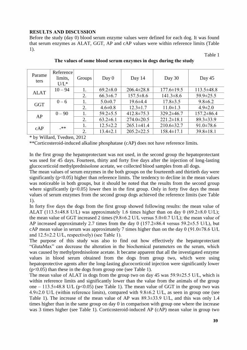
39
RESULTS AND DISCUSSION Before the study (day 0) blood serum enzyme values were defined for each dog. It was found
that serum enzymes as ALAT, GGT, AP and cAP values were within reference limits (Table
1).
Table 1 The values of some blood serum enzymes in dogs during the study
Parame
ters
Reference
limits,
U/L*
Groups Day 0 Day 14 Day 30 Day 45
ALAT 10 – 94 1. 69.2±8.0 206.4±28.8 177.6±19.5 113.5±48.8
2. 66.3±6.7 157.5±8.6 141.3±8.6 59.9±25.5
GGT 0 – 6 1. 5.0±0.7 19.6±4.4 17.8±3.5 9.8±6.2
2. 4.6±0.8 12.3±1.7 11.0±1.3 4.9±2.0
AP 0 – 90 1. 59.2±5.5 412.8±75.3 329.2±46.7 157.2±86.4
2. 63.2±6.1 274.0±20.5 221.2±18.1 89.3±33.9
cAP -** 1. 12.5±2.2 265.1±41.4 210.6±32.7 91.0±78.6
2. 13.4±2.1 205.2±22.5 158.4±17.1 39.8±18.1
* by Willard, Tvedten, 2012
**Corticosteroid-induced alkaline phosphatase (cAP) does not have reference limits.
In the first group the hepatoprotectant was not used, in the second group the hepatoprotectant
was used for 45 days. Fourteen, thirty and forty five days after the injection of long-lasting
glucocorticoid methylprednisolone acetate, we collected blood samples from all dogs.
The mean values of serum enzymes in the both groups on the fourteenth and thirtieth day were
significantly (p<0.05) higher than reference limits. The tendency to decline in the mean values
was noticeable in both groups, but it should be noted that the results from the second group
where significantly (p<0.05) lower then in the first group. Only in forty five days the mean
values of serum enzymes from the second group dogs achieved the reference limits (see Table
1).
In forty five days the dogs from the first group showed following results: the mean value of
ALAT (113.5±48.8 U/L) was approximately 1.6 times higher than on day 0 (69.2±8.0 U/L);
the mean value of GGT increased 2 times (9.8±6.2 U/L versus 5.0±0.7 U/L); the mean value of
AP increased approximately 2.7 times from the day 0 (157.2±86.4 versus 59.2±5.5 U/L), but
cAP mean value in serum was approximately 7 times higher than on the day 0 (91.0±78.6 U/L
and 12.5±2.2 U/L, respectively) (see Table 1).
The purpose of this study was also to find out how effectively the hepatoprotectant
“GlutaMax” can decrease the alteration in the biochemical parameters on the serum, which
was caused by methylprednisolone acetate. It became apparent that all the investigated enzyme
values in blood serum obtained from the dogs from group two, which were using
hepatoprotective agents after the long-lasting glucocorticoid injection were significantly lower
(p<0.05) than these in the dogs from group one (see Table 1).
The mean value of ALAT in dogs from the group two on day 45 was 59.9±25.5 U/L, which is
within reference limits and significantly lower than the value from the animals of the group
one – 113.5±48.8 U/L (p<0.05) (see Table 1). The mean value of GGT in the group two was
4.9±2.0 U/L (within reference limits), compared with 9.8±6.2 U/L, as seen in group one (see
Table 1). The increase of the mean value of AP was 89.3±33.9 U/L, and this was only 1.4
times higher than in the same group on day 0 in comparison with group one where the increase
was 3 times higher (see Table 1). Corticosteroid-induced AP (cAP) mean value in group two
40
on the forty fifth day was high – 39.8±18.1 U/L, that was 3 times higher than on day 0, but in
group one the same value was approximately 3 times higher than on day 0 (see Table 1).
It should be noted that corticosteroid-induced thermostable AP in serum was found in small
amounts (12.5±2.2 U/L and 13.4±2.1 U/L) even on the day 0. This can be explained by the
stress of the dogs because of the veterinarian presence and blood collecting. It is acknowledged
that it is higher glucocorticoid, e.g. cortisol level, in blood when animal is in stress (Feldmann
et al., 1994). It is experimentally proven that there are small amounts of thermostable alkaline
phosphatase even in healthy dogs’ blood as the authors describe it with being in stress
condition (Fukui et al., 2006).
It should be noted that the mean values of ALAT, GGT, AP and cAP in the serum of the dogs
from the second group on day 45 influenced by hepatoprotectant “GlutaMax” were
significantly lower. That indicates that the negative effect of long-acting methylprednisolone
acetate is decreased. Every enzyme mean value was lower compared to the same mean values
from the animals of the first group, and these achieved the reference limits (see Table 1).
The results of our study prove the fact of glucocorticoid-induced negative effects on liver
functional condition (Badylak and van Vleet., 1981; Lucena et al., 1999; Abraham et al.,
2006). These negative effects have been reflected by enzymes ALAT, GGT, AP and especially
cAP significant increase in blood serum. The hepatoprotectant “GlutaMax” could completely
prevent these functional failures of liver during this study.
CONCLUSIONS 1. The corticosteroid methylprednisolone acetate statistically significantly increases the
values of alaninaminotransferase (ALAT), gammaglutamyltranferase (GGT), alkaline
phosphatase (AP) and corticosteroid-induced thermostable alkaline phosphatase (cAP) in
dogs’ blood serum.
2. The hepatoprotectant ‘GlutaMax’ in dosage of 1 pill for 15 kg of bodyweight, used 45
days after the one injection of long-lasting methylprednisolone acetate for dogs,
significantly decreases the values of alaninaminotransferase (ALAT),
gammaglutamyltranferase (GGT), alkaline phosphatase (AP) and corticosteroid-induced
thermostable alkaline phosphatase (cAP) in dogs’ blood serum.
REFERENCES 1. Abraham G., Demirai F., Ungemach F.R. (2006) Comparison of the hypothalamic-
pituitary-adrenal axis susceptibility upon single dose i.m. depot versus long-acting i.v.
triamcinolone acetonide therapy: a direct pharmacokinetic correlation. Journal of
Endocrinology, 191, pp. 491-496.
2. Badylak S.F., Van Vleet J.R. (1981) Sequential morphologic and clinicopathologic
alterations in dogs with experimentally induced glucocorticoid hepatopathy. American
Journal of Veterinary Research, 42, pp. 1310-1318.
3. Center S.A., Warner K.L., McCabe J., Foureman P., Hoffmann W.E., Erb H.N. (2005)
Evaluation of the influence of S-adenosylmethionine on systemic and hepatic effects of
prednisolone in dogs. American Journal of Veterinary Research, 66, pp. 330-341.
4. Center S.A., Slater M.R., Manwarren T., Prymak K. (1992) Diagnostic efficacy of
serum alkaline phosphatase and gamma-glutamyltransferase in dogs with histologically
confirmed hepatobiliary disease: 270 cases (1980-1990). Journal of the American
Veterinary Medical Association, 201, pp. 1258-1264.
5. Deniz A., Spiecker-Hauser U., Rehagen M. (2009) Efficacy of a Butafosfan and
Vitamin B12 Combination („Catosal”) on Biochemical and Hematological Blood
Parameters in Dogs Treated with Dexamethasone. Journal of Applied Research in
Veterinary Medicine, 7, pp. 116-129.
41
6. Dillon A.R., Spano J.S., Powers R.D. (1980) Prednisolone induced haematological,
biochemical and histological changes in the dog. Journal of the American Animal
Hospital Association, 16, pp. 831-837.
7. Feldman E.C., Nelson R.W. (2004) Canine and Feline Endocrinology and
Reproduction. Elsevier Science, Sounders, USA, 1104 p.
8. Fittschen C. and Bellamy J.E. (1984) Prednisone-induced morphologic and chemical
changes in liver of dogs. Veterinary Pathology, 21, pp. 399-406.
9. Flatland B. (2003) Botanicals, vitamins, and minerals and the liver: Therapeutic
applications and potential toxicities. Compendium on Continuing Education for the
Practising Veterinarian, 25, pp. 514-524.
10. Fukui Y., Sato J., Sato R., Yasuda J., Naito Y. (2006) Canine Serum Thermostable
Alkaline Phosphatase Isoenzyme From a Dog With Hepatocellular Carcinoma. The
Journal of Veterinary Medical Science, 68, pp. 1129-1132.
11. Gulbis E. (2011) Klīnisko analīžu rokasgrāmata. E. Gulbja laboratorija, Rīga, 399 lpp.
12. Johnson S.E. (2008) Hepatoprotective Therapy. Available at:
www.cincyvma.com/files/, 03 March 2014.
13. Lucena R., Ginel P.J., Novales M., Molleda J.M. (1999) Effects of dexamethasone
administration on serum trypsin-like immunoreactivity in healthy dogs. American
Journal of Veterinary Research, 60, pp. 1357-1359.
14. Teske E. (1999) Corticosteroid-induced alkaline phosphatase isoenzyme in the
diagnosis of canine hypercorticism. Journal of the American Veterinary Medical
Association. 215, pp. 323-327.
15. Vogel G. (1984) Protection by silibinin against Amanita phalloides intoxication in
beagles. Toxicology and Applied Pharmacology, 73, pp. 355-362.
16. Willard M.D., Tvedten H.T. (2012) Small animal clinical diagnosis by laboratory
methods. Elsevier. Sounders. p.432.
42
THE INFLUENCE OF NEW MINERAL - VITAMIN BLOCKS ON COW PRODUCTIVITY AND REPRODUCTIVE ABILITIES
Kristaps Krapauskis, Aiga Trūpa, Uldis Osītis Institute of Agrobiotechnology, LUA, Latvia
ABSTRACT For the research Latvian Brown breed (LB) cows were selected and assigned by the analogy
principle to two treatments groups (n=2x12). During the trial, the dairy cows received the
total feed mix (TMR) which consisted, calculating per cow per day of: 20 kg maize silage
(45.15% by mass), 16 kg cereal grass - clover silage (36.12% by mass), 2 kg hay (cereal grass
- clover) (4.51% by mass), 4 kg concentrated feed mixture (rape oil - cake-barley meal)
(9.03% by mass), 2 kg protein concentrate (4.53% by mass), 0.1 kg mineralpremix ViloMin
9/6 (0.22% by mass), 0.1 kg sodium chloride (0.22% by mass) and 0.1 kg sodium carbonate
(0.22% by mass). The analysis of the productivity indicators suggests that the milk yield in
the experimental group of cows was approximately 0.17 to 1.02 kg ECM per day higher than
that in the control group of cows. Feeding cows with the mineral and vitamin blocks has
contributed to the improvement of indicators of the reproductive abilities: shortening of the
service period by 9 days, the insemination index by 0.1 and the number of pregnant cows
from the first insemination by 16%, as compared with the control group. Feeding of the
supplementary blocks left a positive effect on the process of parturition of cows.
KEY WORDS: mineral substances and vitamin blocks, cow, milk, reproductive abilities
INTRODUCTION The need of mineral substances for the animal is known and the difference between the
existing in the feed and the required quantity in the cow is reduced by appropriate mineral
supplements. However, various factors (many of them have not even been established) affect
the need and availability of mineral substances and vitamins. The concentration of mineral
substances and vitamins in feed is highly variable and often their quantity is not even
determined. In order to resolve this situation, in addition to the staple feed, animals are fed
with additives of different types and composition (Lean et al., 2006; Hackbart et al., 2010).
Unlike various imported mineral additives used in Latvia, in the block the local raw materials
are used at maximum such as feed lime, sapropel, red clay, gypsum, etc., supplemented with
minerals that are not found in Latvia in sufficient quantity. The block comprises, in addition
to the above mentioned raw materials, the quantity and content of Zn, Mn, Cu, I, Co, Se in
both organic and inorganic forms correspond to the quantities of the latest scientific
knowledge. The aim of the study: to examine the effect of the new mineral-vitamin blocks on
the productivity, milk quality and reproductive abilities of cows.
MATERIALS AND METHODS Trials were carried out on the peasant facility “Druvas”, Vitinu Parish, Dobeles Municipality.
For the research Latvian Brown breed (LB) cows were selected and assigned by the analogy
principle to two treatments groups (n=2x12). The trial was carried out from January to May
2013, i.e., 151 days.
The average live weight of cows was 600 kg, the cows of high productivity in the end phase
of lactation were included in the experiment with the average yield of 20.33 kg per day, fat
content 4.03 % and protein content 3.64 %. During the trial, the dairy cows received the total
feed mix (TMR) which consisted, calculating per cow per day of: 20 kg maize silage (45.15%
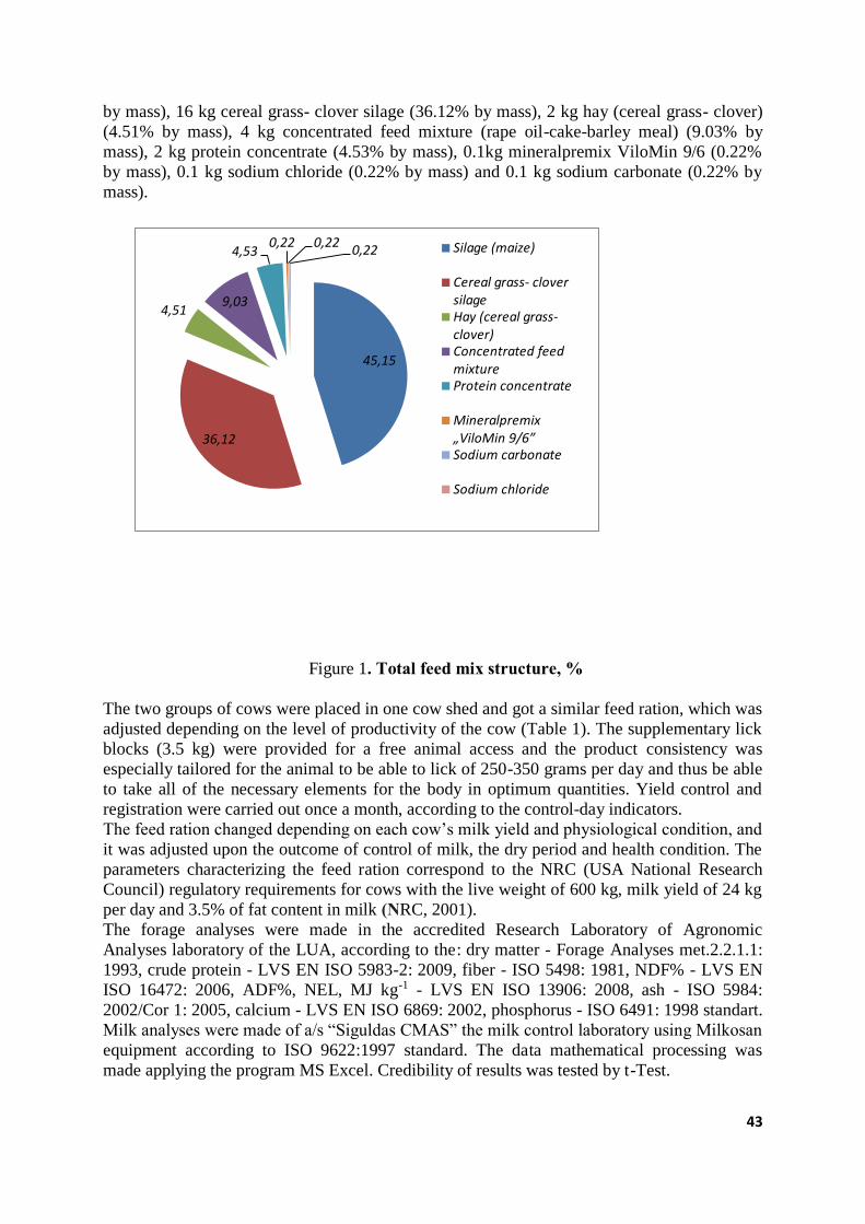
43
by mass), 16 kg cereal grass- clover silage (36.12% by mass), 2 kg hay (cereal grass- clover)
(4.51% by mass), 4 kg concentrated feed mixture (rape oil-cake-barley meal) (9.03% by
mass), 2 kg protein concentrate (4.53% by mass), 0.1kg mineralpremix ViloMin 9/6 (0.22%
by mass), 0.1 kg sodium chloride (0.22% by mass) and 0.1 kg sodium carbonate (0.22% by
mass).
45,15
36,12
4,519,03
4,530,22 0,22
0,22 Silage (maize)
Cereal grass- cloversilageHay (cereal grass-clover)Concentrated feedmixtureProtein concentrate
Mineralpremix „ViloMin 9/6”Sodium carbonate
Sodium chloride
Figure 1. Total feed mix structure, %
The two groups of cows were placed in one cow shed and got a similar feed ration, which was
adjusted depending on the level of productivity of the cow (Table 1). The supplementary lick
blocks (3.5 kg) were provided for a free animal access and the product consistency was
especially tailored for the animal to be able to lick of 250-350 grams per day and thus be able
to take all of the necessary elements for the body in optimum quantities. Yield control and
registration were carried out once a month, according to the control-day indicators.
The feed ration changed depending on each cow’s milk yield and physiological condition, and
it was adjusted upon the outcome of control of milk, the dry period and health condition. The
parameters characterizing the feed ration correspond to the NRC (USA National Research
Council) regulatory requirements for cows with the live weight of 600 kg, milk yield of 24 kg
per day and 3.5% of fat content in milk (NRC, 2001).
The forage analyses were made in the accredited Research Laboratory of Agronomic
Analyses laboratory of the LUA, according to the: dry matter - Forage Analyses met.2.2.1.1:
1993, crude protein - LVS EN ISO 5983-2: 2009, fiber - ISO 5498: 1981, NDF% - LVS EN
ISO 16472: 2006, ADF%, NEL, MJ kg-1 - LVS EN ISO 13906: 2008, ash - ISO 5984:
2002/Cor 1: 2005, calcium - LVS EN ISO 6869: 2002, phosphorus - ISO 6491: 1998 standart.
Milk analyses were made of a/s “Siguldas CMAS” the milk control laboratory using Milkosan
equipment according to ISO 9622:1997 standard. The data mathematical processing was
made applying the program MS Excel. Credibility of results was tested by t-Test.
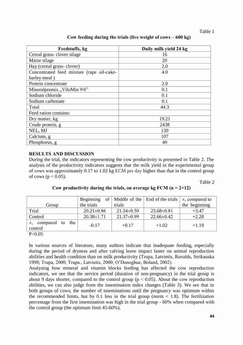
44
Table 1
Cow feeding during the trials (live weight of cows – 600 kg)
Feedstuffs, kg Daily milk yield 24 kg Cereal grass- clover silage 16
Maize silage 20
Hay (cereal grass- clover) 2.0
Concentrated feed mixture (rape oil-cake-
barley meal )
4.0
Protein concentrate 2.0
Mineralpremix „ViloMin 9/6” 0.1
Sodium chloride 0.1
Sodium carbonate 0.1
Total 44.3
Feed ration contains:
Dry matter, kg 19.21
Crude protein, g 2438
NEL, MJ 130
Calcium, g 107
Phosphorus, g 49
RESULTS AND DISCUSSION During the trial, the indicators representing the cow productivity is presented in Table 2. The
analysis of the productivity indicators suggests that the milk yield in the experimental group
of cows was approximately 0.17 to 1.02 kg ECM per day higher than that in the control group
of cows (p < 0.05).
Table 2
Cow productivity during the trials, on average kg FCM (n = 2×12)
Group
Beginning of
the trials
Middle of the
trials
End of the trials ±, compared to
the beginning
Trial 20.21±0.86 21.54±0.50 23.68±0.81 +3.47
Control 20.38±1.71 21.37±0.99 22.66±0.42 +2.28
±, compared to the
control -0.17 +0.17 +1.02 +1.19
P<0.05
In various sources of literature, many authors indicate that inadequate feeding, especially
during the period of dryness and after calving leave impact faster on animal reproduction
abilities and health condition than on milk productivity (Trupa, Latvietis, Ruvalds, Strikauska
1999; Trupa, 2000; Trupa., Latvietis, 2000; O’Donoghue, Boland, 2002).
Analysing how mineral and vitamin blocks feeding has affected the cow reproduction
indicators, we see that the service period (duration of non-pregnancy) in the trial group is
about 9 days shorter, compared to the control group (p < 0.05). About the cow reproduction
abilities, we can also judge from the insemination index changes (Table 3). We see that in
both groups of cows, the number of inseminations until the pregnancy was optimum within
the recommended limits, but by 0.1 less in the trial group (norm < 1.8). The fertilization
percentage from the first insemination was high in the trial group - 60% when compared with
the control group (the optimum limit 45-60%).
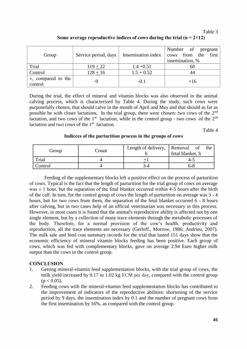
45
Table 3
Some average reproductive indices of cows during the trial (n = 2×12)
Group Service period, days Insemination index
Number of pregnant
cows from the first
insemination, %
Trial 119 + 22 1.4 +0.51 60
Control 128 + 16 1.5 + 0.52 44
±, compared to the
control -9 -0.1 +16
During the trial, the effect of mineral and vitamin blocks was also observed in the animal
calving process, which is characterized by Table 4. During the study, such cows were
purposefully chosen, that should calve in the month of April and May and that should as far as
possible be with closer lactations. In the trial group, there were chosen: two cows of the 2nd
lactation, and two cows of the 1st lactation, while in the control group - two cows of the 2nd
lactation and two cows of the 1st lactation.
Table 4
Indicces of the parturition process in the groups of cows
Group Count Length of delivery,
h
Removal of the
fetal blanket, h
Trial 4 +1 4-5
Control 4 3-4 6-8
Feeding of the supplementary blocks left a positive effect on the process of parturition
of cows. Typical is the fact that the length of parturition for the trial group of cows on average
was ± 1 hour, but the separation of the fetal blanket occurred within 4-5 hours after the birth
of the calf. In turn, for the control group of cows the length of parturition on average was 3 - 4
hours, but for two cows from them, the separation of the fetal blanket occurred 6 - 8 hours
after calving, but in two cases help of an official veterinarian was necessary in this process.
However, in most cases it is found that the animal's reproductive ability is affected not by one
single element, but by a collection of many trace elements through the metabolic processes of
the body. Therefore, for a normal provision of the cow’s health, productivity and
reproduction, all the trace elements are necessary (Gerloff., Morrow, 1986; Andrieu, 2007).
The milk sale and feed cost summary records for the trial that lasted 151 days show that the
economic efficiency of mineral vitamin blocks feeding has been positive. Each group of
cows, which was fed with complementary blocks, gave on average 2.94 Euro higher milk
output than the cows in the control group.
CONCLUSION 1. Getting mineral-vitamin feed supplementation blocks, with the trial group of cows, the
milk yield increased by 0.17 to 1.02 kg ECM per day, compared with the control group
(p < 0.05).
2. Feeding cows with the mineral-vitamin feed supplementation blocks has contributed to
the improvement of indicators of the reproductive abilities: shortening of the service
period by 9 days, the insemination index by 0.1 and the number of pregnant cows from
the first insemination by 16%, as compared with the control group.
46
3. The parturition process of the cows was positively affected by the feeding of the
supplementation blocks. In the trial group of cows, the length of delivery averaged ± 1
hour, but the separation of the fetal blanket occurred within 4-5 hours after the birth of
the calf, but for the control group of cows the length of parturition on average was 3 - 4
hours and the separation of the fetal blanket occurred 6 - 8 hours after calving.
4. Feeding of the feed supplementation blocks to dairy cows was economically beneficial.
Each cow of the trial group gave an average of 2.94 Euros higher production of milk
than their counterparts in the control group.
REFERENCES 1. Andrieu S. (2007) Is there a role for organic trace element supplements in transition
cow health. ScienceDirekt, The Veterinary Journal 176, 77- 83.
2. Gerloff B., Morrow D. (1986) Effect of nutrition on reproduction in dairy cattle. In:
Current Therapy in Theriogenology. USA, pp 317-319.
3. Hackbart K. S., Ferreira R. M, Dietsche A.A, Socha M.T, Shaver R. D., Wiltbank M C.
(2010) Effect of dietary organic zinc, manganese, copper, and cobalt supplementation
on milk dairy cows production, follicular growth, embryo quality, and tissue mineral
concentrations in dairy cow. Journal of Animal science.
4. Lean I.J., De Garis, P.J.Mc Neil, D.M. and Block E. (2006) Hypocalcemia in dairy
cows:Meta – analysis and dietary cation anion diference theory revisited. J.Dairy Sci.
89:669-684.
5. Nutrient Requirements of Dairy Cattle (2001) Seventh Revised Edition, National
Research Council. National Academy Press, Washington, D.C., 373 pp.
6. O’Donoghue, D.G., Boland, M.,( 2002.) The effect of proteinated trace minerals on
fertility and somatic cell counts of dairy cattle. Journal of Dairy Science, 78 (Suppl. 1),
239.
7. Trūpa A. (1999) Minerālvielu apgāde un govju reprodukcijas spējas.
Veterinārmedicīnas Raksti. Jelgava, 334-341.
8. Trūpa A. (2000) Minerālpiedevu ietekme uz govju produktivitāti un reprodukcijas
spējām. LLU Raksti. Nr.3. Jelgava, 34-38.
9. Trūpa A., Latvietis J. (2000) Minerālpiedevu ietekme uz piena kvalitāti. 21.gs kopā ar
zinātni un praksi. LLU. Jelgava, 78-82.lpp.
10. Trūpa A., Latvietis J., Rūvalds I., Strikauska S. (1999) Influence of mineralpremixes on
some milk quality indices. LLU Raksti. Nr.1 (295). Jelgava, pp 24-27.
47
USE OF 24-Hour AMBULATORY ELECTROCARDIOGRAPHY (HOLTER MONITORING) FOR ASSESSMENT OF HEART RATE
VARIABILITY IN HEALTHY DOBERMAN PINSCHERS IN COMPARISION WITH A ROUTINE ECG AND PHYSICAL
EXAMINATION Dana Laizāne, Līga Nātiņa, Ilmārs Dūrītis LLU Veterinārmedicīnas fakultāte, Latvija; Faculty of Veterinary Medicine LUA, Latvia
ABSTRACT In veterinary clinical practice, Holter monitoring has been used most frequently in dogs, often
during evaluation for syncope or episodic weakness, but also for detecting intermittent
arrhythmias, assessing response to antiarrhythmic drug therapy, and screening for subclinical
cardiomyopathy of Boxers and Doberman Pinschers since routine ECGs detects frequent
arrhythmias, but may have limited sensitivity in dogs with infrequent or intermittent
arrhythmias, as they reflect only a very short period of day. However, no large study exists
evaluating the use of a physical examination, routine ECG and comparing the results to the
Holter recordings for assessing heart rate variability in Doberman Pinschers. The aim of this
study was to evaluate and compare main heart rate parameters in clinically healthy Doberman
Pinschers, obtained during physical examination, routine ECG and 24-hour Holter
monitoring. Materials and Methods. Physical examination, routine ECG and 24 hour ECG
recording results were evaluated in 11 clinically healthy, client-owned Doberman pincher
dogs. Results. There was a significant difference between average heart rates obtained with
three different methods. Mean heart rates obtained with routine ECG, were significantly
higher (p<0.001) than those recorded using Holter monitoring. Minimum sinus RR interval
was similar in all dogs, but there was a range in amplitude between maximum sinus RR
intervals of all dogs. No ventricular premature complexes (VPCs) were found during routine
ECG examination, while all dogs had at least one VPC over 24-hour period.
KEY WORDS: Doberman, 24-hour Holter monitoring, heart rate variability INTRODUCTION Ambulatory electrocardiography (Holter monitoring) has been used for over 30 years in
people to assess cardiac arrhythmias, effectiveness of antiarrhythmic therapy, ischemic S-T
segment changes, and other parameters over prolonged periods of time (e.g., 24 to 72 hours)
and during normal daily activities. Holter monitoring allows more accurate evaluation of
antiarrhythmic drug effectiveness than routine, intermittent electrocardiograms (ECGs)
because arrhythmia frequency naturally fluctuates widely over time (Bleifer et al. 1980,
Kennedy 1992, Zipes 1997). In veterinary clinical practice, Holter monitoring has been used
most frequently in dogs, often during evaluation for syncope or episodic weakness, but also
for detecting intermittent arrhythmias, assessing response to antiarrhythmic drug therapy, and
screening for subclinical cardiomyopathy of Boxers and Doberman Pinschers (Calvert 1991,
Calvert 1995, Goodwin, Cattiny 1995, Goodwin et al. 1992, Moise, DeFrancesco 1995, Rush,
Keene 1989) since routine ECGs, detects frequent arrhythmias, but may have limited
sensitivity in dogs with infrequent or intermittent arrhythmias, as they reflect only a very
short period of day (Marino et al., 1994).
Holter examinations are not as readily available to the veterinarian, and are more expensive
and time consuming to perform than routine ECGs. Routine ECG, however, reflect electrical
activity of the heart only for a very short period of time that might not be objective enough for
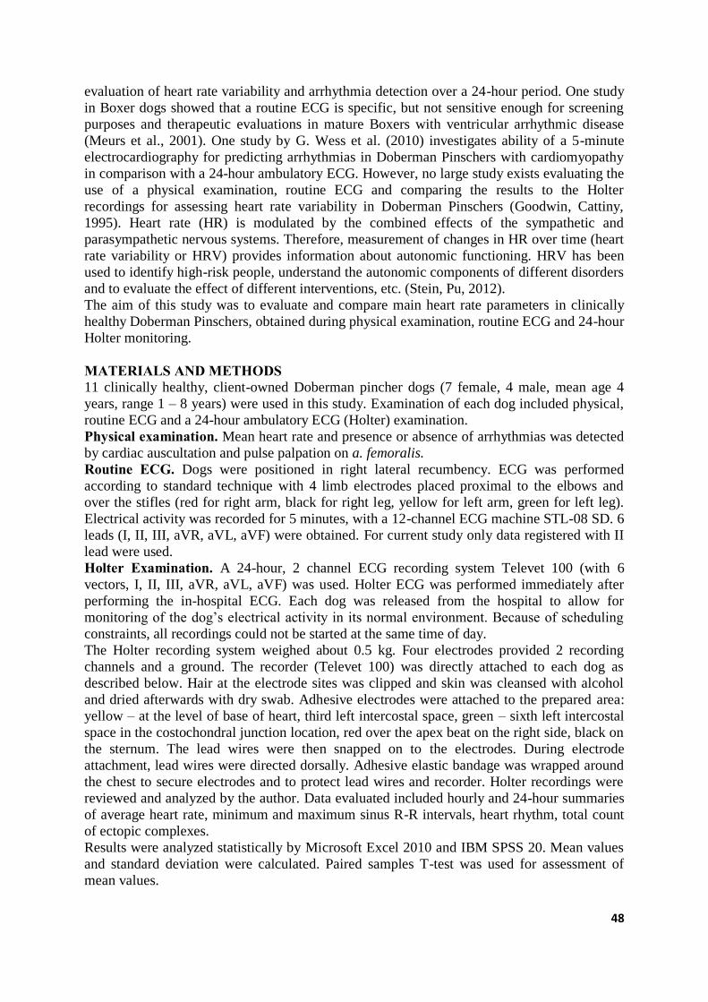
48
evaluation of heart rate variability and arrhythmia detection over a 24-hour period. One study
in Boxer dogs showed that a routine ECG is specific, but not sensitive enough for screening
purposes and therapeutic evaluations in mature Boxers with ventricular arrhythmic disease
(Meurs et al., 2001). One study by G. Wess et al. (2010) investigates ability of a 5-minute
electrocardiography for predicting arrhythmias in Doberman Pinschers with cardiomyopathy
in comparison with a 24-hour ambulatory ECG. However, no large study exists evaluating the
use of a physical examination, routine ECG and comparing the results to the Holter
recordings for assessing heart rate variability in Doberman Pinschers (Goodwin, Cattiny,
1995). Heart rate (HR) is modulated by the combined effects of the sympathetic and
parasympathetic nervous systems. Therefore, measurement of changes in HR over time (heart
rate variability or HRV) provides information about autonomic functioning. HRV has been
used to identify high-risk people, understand the autonomic components of different disorders
and to evaluate the effect of different interventions, etc. (Stein, Pu, 2012).
The aim of this study was to evaluate and compare main heart rate parameters in clinically
healthy Doberman Pinschers, obtained during physical examination, routine ECG and 24-hour
Holter monitoring.
MATERIALS AND METHODS 11 clinically healthy, client-owned Doberman pincher dogs (7 female, 4 male, mean age 4
years, range 1 – 8 years) were used in this study. Examination of each dog included physical,
routine ECG and a 24-hour ambulatory ECG (Holter) examination.
Physical examination. Mean heart rate and presence or absence of arrhythmias was detected
by cardiac auscultation and pulse palpation on a. femoralis.
Routine ECG. Dogs were positioned in right lateral recumbency. ECG was performed
according to standard technique with 4 limb electrodes placed proximal to the elbows and
over the stifles (red for right arm, black for right leg, yellow for left arm, green for left leg). Electrical activity was recorded for 5 minutes, with a 12-channel ECG machine STL-08 SD. 6
leads (I, II, III, aVR, aVL, aVF) were obtained. For current study only data registered with II
lead were used. Holter Examination. A 24-hour, 2 channel ECG recording system Televet 100 (with 6
vectors, I, II, III, aVR, aVL, aVF) was used. Holter ECG was performed immediately after
performing the in-hospital ECG. Each dog was released from the hospital to allow for
monitoring of the dog’s electrical activity in its normal environment. Because of scheduling
constraints, all recordings could not be started at the same time of day.
The Holter recording system weighed about 0.5 kg. Four electrodes provided 2 recording
channels and a ground. The recorder (Televet 100) was directly attached to each dog as
described below. Hair at the electrode sites was clipped and skin was cleansed with alcohol
and dried afterwards with dry swab. Adhesive electrodes were attached to the prepared area:
yellow – at the level of base of heart, third left intercostal space, green – sixth left intercostal
space in the costochondral junction location, red over the apex beat on the right side, black on
the sternum. The lead wires were then snapped on to the electrodes. During electrode
attachment, lead wires were directed dorsally. Adhesive elastic bandage was wrapped around
the chest to secure electrodes and to protect lead wires and recorder. Holter recordings were
reviewed and analyzed by the author. Data evaluated included hourly and 24-hour summaries
of average heart rate, minimum and maximum sinus R-R intervals, heart rhythm, total count
of ectopic complexes.
Results were analyzed statistically by Microsoft Excel 2010 and IBM SPSS 20. Mean values
and standard deviation were calculated. Paired samples T-test was used for assessment of
mean values.
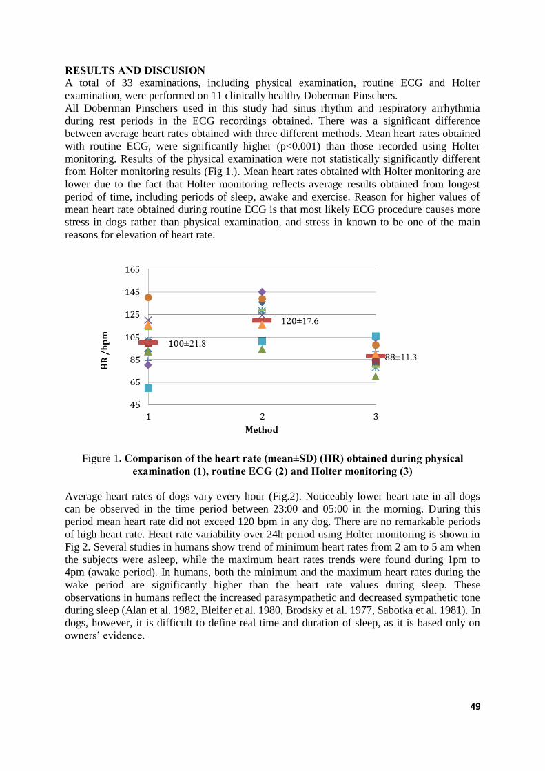
49
RESULTS AND DISCUSION A total of 33 examinations, including physical examination, routine ECG and Holter
examination, were performed on 11 clinically healthy Doberman Pinschers. All Doberman Pinschers used in this study had sinus rhythm and respiratory arrhythmia
during rest periods in the ECG recordings obtained. There was a significant difference
between average heart rates obtained with three different methods. Mean heart rates obtained
with routine ECG, were significantly higher (p<0.001) than those recorded using Holter
monitoring. Results of the physical examination were not statistically significantly different
from Holter monitoring results (Fig 1.). Mean heart rates obtained with Holter monitoring are
lower due to the fact that Holter monitoring reflects average results obtained from longest
period of time, including periods of sleep, awake and exercise. Reason for higher values of
mean heart rate obtained during routine ECG is that most likely ECG procedure causes more
stress in dogs rather than physical examination, and stress in known to be one of the main
reasons for elevation of heart rate.
Figure 1. Comparison of the heart rate (mean±SD) (HR) obtained during physical
examination (1), routine ECG (2) and Holter monitoring (3)
Average heart rates of dogs vary every hour (Fig.2). Noticeably lower heart rate in all dogs
can be observed in the time period between 23:00 and 05:00 in the morning. During this
period mean heart rate did not exceed 120 bpm in any dog. There are no remarkable periods
of high heart rate. Heart rate variability over 24h period using Holter monitoring is shown in
Fig 2. Several studies in humans show trend of minimum heart rates from 2 am to 5 am when
the subjects were asleep, while the maximum heart rates trends were found during 1pm to
4pm (awake period). In humans, both the minimum and the maximum heart rates during the
wake period are significantly higher than the heart rate values during sleep. These
observations in humans reflect the increased parasympathetic and decreased sympathetic tone
during sleep (Alan et al. 1982, Bleifer et al. 1980, Brodsky et al. 1977, Sabotka et al. 1981). In
dogs, however, it is difficult to define real time and duration of sleep, as it is based only on
owners’ evidence.
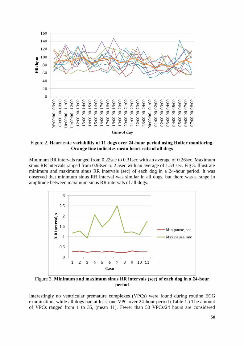
50
Figure 2. Heart rate variability of 11 dogs over 24-hour period using Holter monitoring.
Orange line indicates mean heart rate of all dogs
Minimum RR intervals ranged from 0.22sec to 0.31sec with an average of 0.26sec. Maximum
sinus RR intervals ranged from 0.93sec to 2.5sec with an average of 1.53 sec. Fig 3. Illustrate
minimum and maximum sinus RR intervals (sec) of each dog in a 24-hour period. It was
observed that minimum sinus RR interval was similar in all dogs, but there was a range in
amplitude between maximum sinus RR intervals of all dogs.
Figure 3. Minimum and maximum sinus RR intervals (sec) of each dog in a 24-hour
period
Interestingly no ventricular premature complexes (VPCs) were found during routine ECG
examination, while all dogs had at least one VPC over 24-hour period (Table 1.) The amount
of VPCs ranged from 1 to 35, (mean 11). Fewer than 50 VPCs/24 hours are considered
51
normal in Doberman Pinchers. Study by G. Wess et al. (2010) showed a low sensitivity but
high specificity of a 5-minute ECG to predict 4100 VPCs/24 hours, if at least 1 VPC is
detected within 5 minutes. The absence of VPCs in the 5-minute ECG should not lead to the
assumption that the dog is healthy, because false negative cases were found in 35.8% of the
examinations. A 5-minute ECG cannot replace a 24-hour ECG examination for the purpose of
detecting the occult phase of DCM in Doberman Pinschers and the absence of VPCs on a 5-
minute ECG does not preclude the occurrence of VPCs during the rest of the day (Wess et al.
2010). Current study shows that routine ECG corresponds to a small fraction of the dog’s
rhythm over a 24-hour period, and identification of abnormalities may be entirely incidental.
Table 1
Total number of ventricular premature complexes (VPCs) during routine ECG and 24-hour Holter examination
Case Nr. 1 2 3 4 5 6 7 8 9 10 11
Number of
VPCs during
routine ECG
0 0 0 0 0 0 0 0 0 0 0
Number of
VPCs during
24–hour Holter
examination
6 8 1 15 7 18 2 4 19 35 6
CONCLUSIONS 1. Physical examination method for evaluation of mean heart rate in dogs is more
objective as compared to routine ECG.
2. Mean heart rates obtained with routine ECG, were significantly higher (p<0.001) than
heart rate obtained during physical examination and recorded using Holter monitoring.
Presumably routine ECG procedure causes more stress in dogs since they are positioned
in a forced lateral recumbency.
3. Noticeably lower heart rate in all dogs can be observed in the time period between
23:00 and 05:00 in the morning. During this period mean heart rate did not exceed 120
bpm in any dog.
4. Respiratory sinus arrhythmia was observed in all dogs.
5. There is a wide range in amplitude between maximum sinus RR intervals of all dogs
and consequently heart rate is very variable.
6. Routine ECG is an inapplicable method for detecting small number of ventricular
premature complexes (VPCs).
7. Data obtained during this study illustrate normal cardio rhythmic functions in healthy
adult Doberman Pinchers, and may be used as a comparative measure in analyzing the
rhythm disturbances in patients with cardiac or non-cardiac problems.
REFERENCES 1. Alan DA, Bauernfeind RA, Ashley WW et al. (1982) Twenty-four hour continuous
ECG recordings in long - distance runners. Chest; 82: 19-24
2. Bleifer SB, Bleifer DJ, Mandel WJ. (1980) Holter monitor recording. In: Mandel WJ,
ed, Cardiac Arrhythmias. Philadelphia, PA: JB Lippincott; 470–513.
3. Brodsky M, Delon WU, Denes P et al. (1977) Arrhythmias documented by 24 hour
continuous electrocardiographic monitoring in 50 male medical students without
apparent heart disease. Am J Cardiol; 39: 390-6
52
4. Calvert CA. (1991) Long-term ambulatory electrocardiographic monitoring as an aid in
the diagnosis of occult cardiomyopathy in Doberman pinschers. Proc. 9th ACVIM
Forum, New Orleans, LA, 691– 692.
5. Calvert CA. (1995) Diagnosis and management of ventricular tachyarrhythmias in
Doberman Pinschers with cardiomyopathy. In: Bonagura JD, ed. Kirk’s Current
Veterinary Therapy XII. Philadelphia, PA: WB Saunders; 799–806.
6. Joanna Dukes-McEwan; Michele Borgarelli; Anna Tidholm; Andrea C Vollmar; Jens
Häggström. (2003) Proposed Guidelines for the Diagnosis of Canine Idiopathic Dilated
Cardiomyopathy. The ESVC Taskforce for Canine Dilated Cardiomyopathy. Journal of
Veterinary Cardiology, Vol.5, No. 2, November
7. Goodwin JK, Cattiny G. (1995) Further characterization of Boxer cardiomyopathy.
Proc. 13th ACVIM Forum, Lake Buena Vista, FL, 300 –302.
8. Goodwin JK, Lombard CW, Ginex DD. (1992) Results of continuous ambulatory
electrocardiography in a cat with hypertrophic cardiomyopathy. J Am Vet Med Assoc;
200:1352–1354.
9. Kennedy HL. (1992) Ambulatory (Holter) electrocardiography technology. Cardiol.
Clin.; 10:341–359.
10. Meurs KM, Spier AW, Wright NA, et al. (2001) Comparison of in-hospital versus 24-
hour ambulatory electrocardiography for detection of ventricular premature complexes
in mature Boxers. J Am Vet Med Assoc.; 218:222–224.
11. Morganroth J, Michelson EL, Horowitz LN, et al. (1978) Limitations of routine long-
term electrocardiographic monitoring to assess ventricular ectopic frequency.
Circulation; 58:408–414.
12. Marino DJ, Matthiesen DT, Fox PR, et al. (1994) Ventricular arrhythmias in dogs
undergoing splenectomy: A prospective study. Vet Surg; 23:101–106.
13. Moise NS, DeFrancesco (1995) T. Twenty-four hour ambulatory electrocardiography
(Holter monitoring). In: Bonagura JD, ed. Kirk’s Cur- rent Veterinary Therapy XII.
Philadelphia, PA: WB Saunders: 792–799.
14. Moise NS, Meyers-Wallen V, Flahive WJ, et al. (1994) Inherited ventricular
arrhythmias and sudden death in German Shepherd dogs. J Am Coll Cardiol; 24:233–
243.
15. Rush JE, Keene BW. (1989) ECG of the month. J Am Vet Med Assoc; 194:52.
16. Sabotka PA, Mayer JH, Bauernfiend RA et al. (1981) Arrythmias documented by 24-
hour continous ambulatory electrocardiographic monitoring in young women without
apparent heart disease. Am Heart J,; 101: 753-9.
17. Phyllis K. Stein, Yachuan Pu. (2012) Heart rate variability, sleep and sleep disorders.
Sleep Medicine Reviews Volume: 16, Issue: 1, Pages: 47-66
18. Stein PK, PU Y.(2012) Heart rate variability, sleep and sleep disorders. Sleep Med.
Rev. 2 Feb;16(1):47-66.
19. Talib, SH* PY Mulay**, AN Patil. (2005) Twenty-four Hour Ambulatory Holter
Monitoring and Heart Rate Variability in Healthy Individuals. JIACM; 6(2): 136-41.
20. Wendy A. Ware. (1999) Twenty-Four-Hour Ambulatory Electrocardiography in
Normal Cats. J Vet Intern Med; 13: 175–180
21. G. Wess, A. Schulze, N. Geraghty, and K. Hartmann. (2010) Ability of a 5-Minute
Electrocardiography (ECG) for Predicting Arrhythmias in Doberman Pinschers with
Cardiomyopathy in Comparison with a 24-Hour Ambulatory ECG. J Vet Intern Med;
24:367–371
22. G. Wess, A. Schulze, V. Butz, J. Simak, M. Killich, L.J.M. Keller, J. Maeurer, and, K.
Hartmann (2010) Prevalence of Dilated Cardiomyopathy in Doberman Pinschers in
Various Age Groups. J Vet Intern Med; 24:533–538

53
23. Zipes DP. (1997) Genesis of cardiac arrhythmias: Electrophysiological considerations.
In: Braunwald E, ed, Heart Disease: A Textbook of Cardiovascular Medicine, 5th ed.
Philadelphia, PA: WB Saunders: 548–592.
54
SYNOVIAL FLUID ANALYSIS, BIOMARKERS CRP (C-REACTIVE PROTEIN) AND COMP (CARTILAGE OLIGOMERIC MATRIX
PROTEIN) IMPORTANCE IN DIAGNOSTIC OF CANINE JOINT DISEASES
Ruta Noreikaite-Bulotiene, Vidmantas Bizokas
Department of Non-Infectious Diseases, Lithuanian University of Health Sciences, Veterinary
Academy, Tilžės str. 18, Kaunas, Lithuania
SUMMARY One of the main reasons dogs are going lame is a cranial cruciate ligament rupture (CCLR),
leading to the tibiofemoral joint instability (Johnston et al., 2001). There is a reason to believe
that the CCLR is a consequence of immune-mediated polyarthritis.
Recently, many scientists have focused on biomarker researches, which can provide
information about changes in the joints and efficiently monitor the progress of the disease,
when treatment is started. This is particularly important for investigating the diagnostics of
OA. The development of degenerative process can be prevented by the early taken necessary
measures. Although there are explored and described some of osteoarthritis biomarkers, to
date, none of them is in use in clinical practice (Tobias et al., 2012).
The aim was to evaluate changes in the canine synovial fluid physical properties and cells
quantity and composition, when the cranial cruciate ligament rupture has been diagnosed. It
was also investigated the concentration of biomarkers CRP and COMP in plasma and
synovial fluid by the cranial cruciate ligament rupture and by clinically healthy dogs. In total
there were 35 dogs tested, twenty-five with CCLR and ten healthy dogs from control group.
By researched group in canine synovial fluid there were found neutrophil and mononuclear
cells more than usual should be. In synovial fluid there were found a small amount of red
blood cells, although they are generally absent from synovial fluid. The study has been
informative and has confirmed the assumption that in most cases the CCLR is a secondary
disease caused by immune-mediated polyarthritis (IMP). Studies of biomarkers CRP and
COMP from plasma and synovial fluid showed that by the cranial cruciate ligament rupture,
the concentration of biomarkers was significantly increased compared with the results
obtained from the control group samples.
The aim of our study was to show the importance of the synovial fluid composition and cells
analysis by joint disease diagnostics and to promote the use of biomarkers in clinical practice.
KEY WORDS: dogs, synovial fluid indicators, cranial cruciate ligament rupture, biomarkers
CRP and COMP.
INTRODUCTION The cranial cruciate ligament rupture gives the tibiofemoral joint instability. This is one of the
main causes of dogs lameness (Johnson et al., 2001). Therefore, researchers focuses on the
ligament rupture causes and consequences. It is considered that the joint instability allows the
development of secondary joint pathology - osteoarthritis (Johnson et al., 2001). Osteoarthritis
(OA) is a condition where the articular cartilage is being damaged and this is resulting pain,
swelling, joint function limitations. It is known that 20 percent middle-aged dogs and 90
percent older dogs are suffering from OA. In the human population this percentage is even
higher (Venable et al., 2008).
Some authors maintain that osteoarthritis is a secondary disease after CCLR, other authors
propose that CCLR is a consequence of immune-mediated polyarthritis (IMP). The immune-
55
mediated arthritis can be indentified only by synovial fluid cells quantity changes, making
synovial fluid cells analysis (Viliers et al., 2005).
Recently, there is very often diagnosed the cranial cruciate ligament rupture by the dogs. By
most patients, arthrotomy reveals distinct articular cartilage defects, typical to OA. It was
observed also that very often, after a certain period of time, the problem will recur in another
limb. Because over the years, this problem takes on a huge scale, scientists are interested in
finding a way to provide early diagnosis of changes in the joints and prevent their
development. When OA of the joint is diagnosed, degenerative processes become
unstoppable. Therefore, there is only one way left - to treat symptomatic by inhibiting pain.
This makes concerns to the host, because it has to be regularly fed in drugs, the dog's quality
of life is getting worse, activity is limited. The problem is that there is often failed to
eliminate the sensation of pain completely, and the rest of the life a dog is suffering pain of
some degree, which is increasing with age.
The synovial fluid physical properties tests of color, turbidity, viscosity provide valuable
information. Turbid synovial fluid occurs by inflammatory joint diseases (Fossum et al.,
2007). The reduced viscosity of the synovial fluid indicates a decrease in the concentration of
hyaluronic acid (Slattery, 1995).
Under normal conditions, synovial fluid is not curdling, however, by patients with synovitis
the blood clots are formatting (Cohen et al., 1975). The results of the synovial fluid cells
research and evaluation of the cells quantity can be divided into several groups:
corresponding to the normal range, the typical for osteoarthritis, rheumatoid, infectious
arthritis, immune-mediated polyarthritis.
Synovial fluid test can provide more accurate information about the pathology. Research
includes microscopic evaluation, cell research, bacteriological and serological analysis.
Results by having septic arthritis may significantly differ from the norm (Brunberg, 2001).
The diagnosis of joint disease only from synovial fluid test is not possible. However, the
study results help to choose the appropriate additional analysis. For example, at cranial
cruciate ligament rupture, by a lack of joint fluid tests the primary reason - immune-mediated
polyarthritis may remain undetected.
Biomarkers’ goal is to convey information on the physiological and pathological state of
certain tissues and organs. Biomarkers can be used to achieve several goals:
1. as a diagnostic tool for the disease or pathological condition identification.
2. as a tool for determining the stage of disease and helping to classify the disease.
3. as an indicator of disease prognosis.
4. for monitoring of the clinical response to previous surgical intervention or medical
treatment (Tobias et al., 2012).
The most commonly biomarkers are studied from serum and plasma. However, it may be
measured from different body fluids - urine, saliva, synovial fluid, tears, cerebrospinal fluid,
samples from biopsies (Tobias et al., 2012).
For the last three decades many studies have been conducted in order to discover the ways of
early diagnosis of OA, monitoring the progress of the disease. The detection of cartilage
defects in the early stage of osteochondritis is the aimed result for diagnostic, prognostic and
therapeutic perspectives. Currently, diagnostic methods, such as arthroscopy, magnetic
resonance, are expensive, can only be applied to the animal in the general anesthetic state and
display the results only when a big area of the articular cartilage is injured or the cartilage
fibrillation occurred (Tobias et al. 2012).
In our study there were analyzed two key biomarkers - COMP and CRP. COMP - pentametric
non-collagenous glycoprotein - is an integral part of the articular cartilage. COMP is required
for the normal function, development, structure of the matrix of articular cartilage. A large
amount of COMP is found in articular cartilage, a small amount - in synovial fluid (Tobias et
56
al., 2012). In the early stage of OA metabolically active cartilage begins to decompose. A
number of processes that aim to correct the defects associated with an increased amount of
COMP in synovial fluid are proceeding in articular cartilage. In later stages of OA the
cartilage becomes less metabolically active; amount of COMP reduces (Tobias et al., 2012).
Up to date there have been few researches in order to assess the concentration of COMP in
dogs’ synovial fluid. Several studies, conducted with experimental canine OA models,
confirmed the importance of the COMP marker and the connection with cartilage impairment
(Tobias et al., 2012).
CRP is a protein produced by the liver. Its concentration is increased in the case of
inflammation or the healthy tissues injury. The main benefit obtained from the measurement
of CRP concentration is to assess the systemic inflammatory activity. Also, it is useful to
verify the effectiveness of the treatment - by CRP concentration measurement before
treatment, during it and after (Paul et al, 2011).
To date the monitoring of canine immune-mediated polyarthritis treatment with steroids has
been based on the monitoring of clinical symptoms. Danish researchers decided to use CRP
biomarkers for the assessment of inflammatory activity after initiation of treatment with
steroids. It was found that CRP is an objective biomarker helping accurately assess the
inflammatory processes occurring in the joints (Kjelgaard-Hansen et al., 2006).
The aim of the study is to perform joint fluid tests for dogs, which have been diagnosed with
the cranial cruciate ligament rupture and surgical treatment was employed.
The objectives of the study. To evaluate the physical properties of synovial fluid in two dog
groups - control and research’s - to compare the results. To analyze synovial fluid cells
changes in the canine control and research’s groups. To evaluate the CRP and COMP
biomarkers number in blood plasma and synovial fluid in the canine control and research’s
groups.
MATERIALS AND METHODS Work carried out in 2012 - 2014, in Lithuanian University of Health Sciences (LUHS)
Veterinary Academy, dr. Leonas Kriaučeliūnas small animal clinic and Evaldas Diržinauskas
private small animal veterinary clinic (Kaunas district).
Tested dogs were examined and treated for hind limb lameness, by the cranial cruciate
ligament rupture. All of these dogs suffered from joint instability caused by cranial cruciate
ligament rupture (CCLR). The patients were treated surgically (with DeAngelis method),
using the lateral parapatellar arthrotomy and extracapsular joint stabilization by implanting an
artificial ligament. During the operation the degree of manifestation of osteoarthritis was
estimated.
The control group consisted of dogs brought to veterinary clinics for target sterilization or
castration operations. Survey of patients’ owners and orthopedic study did not reveal any
orthopedic diseases.
Investigational arthrocentesis for tested dogs was performed in general anesthesia, following
asepsis-antisepsis requirements, before starting joint arthrotomy surgery.
Physical properties of the resulting synovial fluid were evaluated - the color, viscosity,
turbidity, mucinous clot formation. After the assessment of physical properties the sample
residue was sent to the medical clinic of the LUHS Medical Academy Laboratory for cells
research.
Research of synovial fluid’s physical properties. Synovial fluid color was determined by
visual assessment of synovial fluid taken without additives. Viscosity of the synovial fluid
was assessed by placing a drop on the thumb, putting indicating finger on it, slowly separating
the fingers and monitoring generated strings (Baker et al, 2000). Synovial fluid was divided
to: the low-viscous, when there were no strings or the strings were up to 1cm long, medium
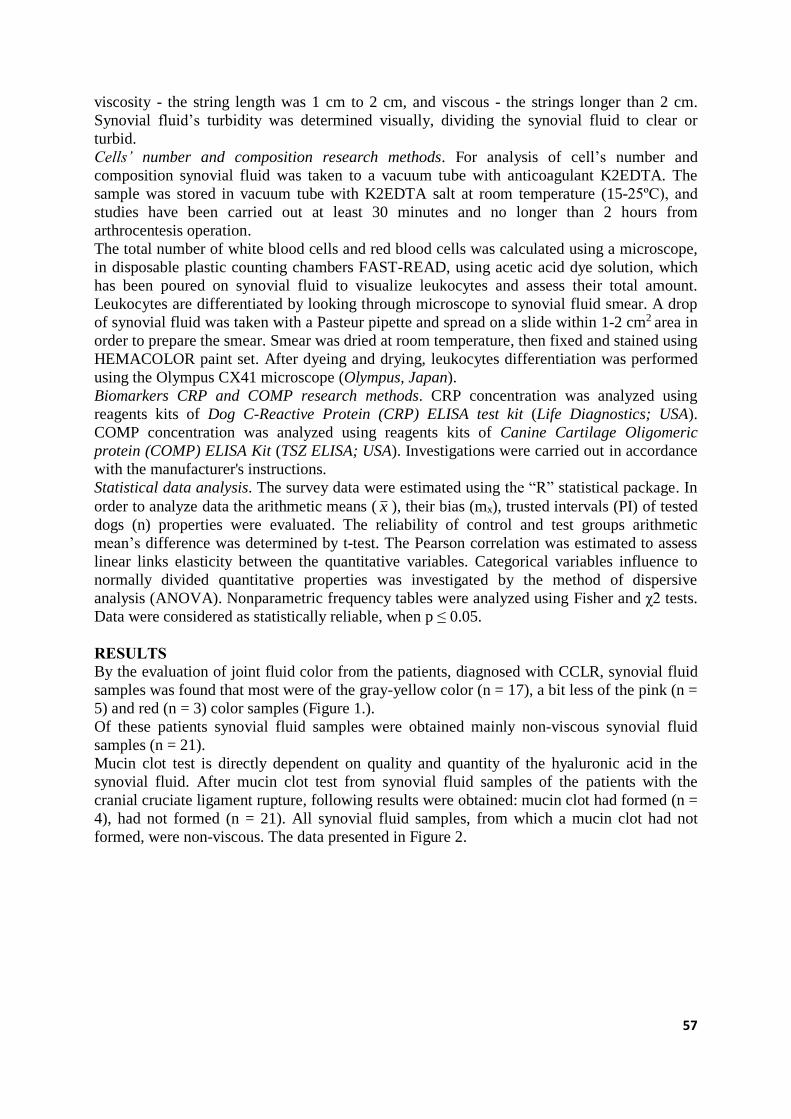
57
viscosity - the string length was 1 cm to 2 cm, and viscous - the strings longer than 2 cm.
Synovial fluid’s turbidity was determined visually, dividing the synovial fluid to clear or
turbid.
Cells’ number and composition research methods. For analysis of cell’s number and
composition synovial fluid was taken to a vacuum tube with anticoagulant K2EDTA. The
sample was stored in vacuum tube with K2EDTA salt at room temperature (15-25ºC), and
studies have been carried out at least 30 minutes and no longer than 2 hours from
arthrocentesis operation.
The total number of white blood cells and red blood cells was calculated using a microscope,
in disposable plastic counting chambers FAST-READ, using acetic acid dye solution, which
has been poured on synovial fluid to visualize leukocytes and assess their total amount.
Leukocytes are differentiated by looking through microscope to synovial fluid smear. A drop
of synovial fluid was taken with a Pasteur pipette and spread on a slide within 1-2 cm2 area in
order to prepare the smear. Smear was dried at room temperature, then fixed and stained using
HEMACOLOR paint set. After dyeing and drying, leukocytes differentiation was performed
using the Olympus CX41 microscope (Olympus, Japan).
Biomarkers CRP and COMP research methods. CRP concentration was analyzed using
reagents kits of Dog C-Reactive Protein (CRP) ELISA test kit (Life Diagnostics; USA).
COMP concentration was analyzed using reagents kits of Canine Cartilage Oligomeric
protein (COMP) ELISA Kit (TSZ ELISA; USA). Investigations were carried out in accordance
with the manufacturer's instructions.
Statistical data analysis. The survey data were estimated using the “R” statistical package. In
order to analyze data the arithmetic means ( x ), their bias (mx), trusted intervals (PI) of tested
dogs (n) properties were evaluated. The reliability of control and test groups arithmetic
mean’s difference was determined by t-test. The Pearson correlation was estimated to assess
linear links elasticity between the quantitative variables. Categorical variables influence to
normally divided quantitative properties was investigated by the method of dispersive
analysis (ANOVA). Nonparametric frequency tables were analyzed using Fisher and χ2 tests.
Data were considered as statistically reliable, when p ≤ 0.05.
RESULTS By the evaluation of joint fluid color from the patients, diagnosed with CCLR, synovial fluid
samples was found that most were of the gray-yellow color (n = 17), a bit less of the pink (n =
5) and red (n = 3) color samples (Figure 1.).
Of these patients synovial fluid samples were obtained mainly non-viscous synovial fluid
samples (n = 21).
Mucin clot test is directly dependent on quality and quantity of the hyaluronic acid in the
synovial fluid. After mucin clot test from synovial fluid samples of the patients with the
cranial cruciate ligament rupture, following results were obtained: mucin clot had formed (n =
4), had not formed (n = 21). All synovial fluid samples, from which a mucin clot had not
formed, were non-viscous. The data presented in Figure 2.
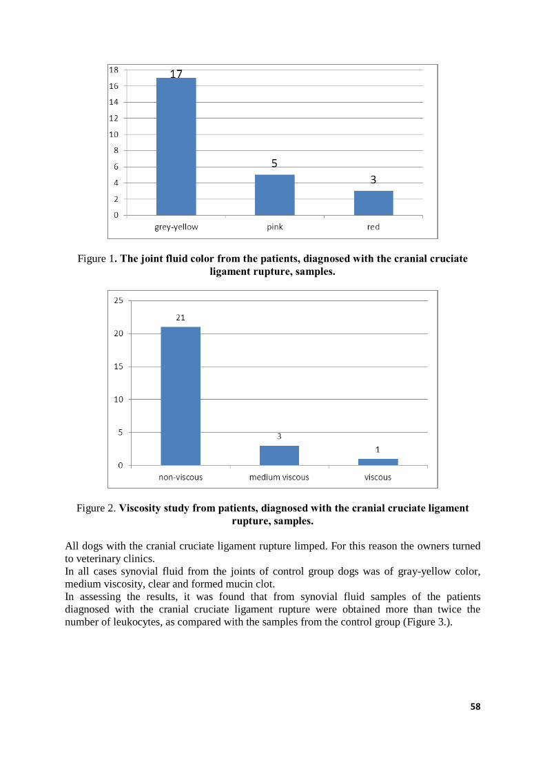
58
Figure 1. The joint fluid color from the patients, diagnosed with the cranial cruciate ligament rupture, samples.
Figure 2. Viscosity study from patients, diagnosed with the cranial cruciate ligament rupture, samples.
All dogs with the cranial cruciate ligament rupture limped. For this reason the owners turned
to veterinary clinics.
In all cases synovial fluid from the joints of control group dogs was of gray-yellow color,
medium viscosity, clear and formed mucin clot.
In assessing the results, it was found that from synovial fluid samples of the patients
diagnosed with the cranial cruciate ligament rupture were obtained more than twice the
number of leukocytes, as compared with the samples from the control group (Figure 3.).
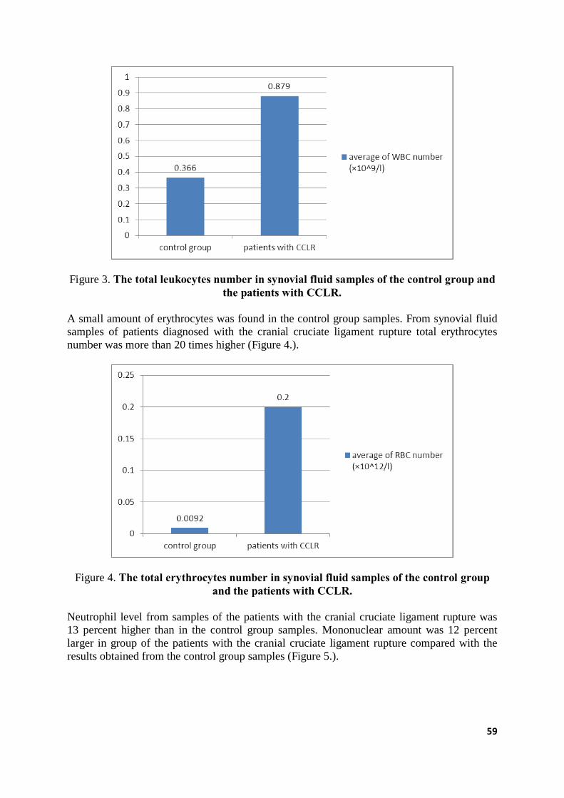
59
Figure 3. The total leukocytes number in synovial fluid samples of the control group and the patients with CCLR.
A small amount of erythrocytes was found in the control group samples. From synovial fluid
samples of patients diagnosed with the cranial cruciate ligament rupture total erythrocytes
number was more than 20 times higher (Figure 4.).
Figure 4. The total erythrocytes number in synovial fluid samples of the control group and the patients with CCLR.
Neutrophil level from samples of the patients with the cranial cruciate ligament rupture was
13 percent higher than in the control group samples. Mononuclear amount was 12 percent
larger in group of the patients with the cranial cruciate ligament rupture compared with the
results obtained from the control group samples (Figure 5.).
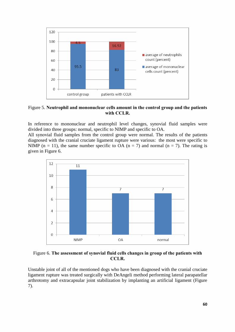
60
Figure 5. Neutrophil and mononuclear cells amount in the control group and the patients with CCLR.
In reference to mononuclear and neutrophil level changes, synovial fluid samples were
divided into three groups: normal, specific to NIMP and specific to OA.
All synovial fluid samples from the control group were normal. The results of the patients
diagnosed with the cranial cruciate ligament rupture were various: the most were specific to
NIMP (n = 11), the same number specific to OA (n = 7) and normal (n = 7). The rating is
given in Figure 6.
Figure 6. The assessment of synovial fluid cells changes in group of the patients with CCLR.
Unstable joint of all of the mentioned dogs who have been diagnosed with the cranial cruciate
ligament rupture was treated surgically with DeAngeli method performing lateral parapatellar
arthrotomy and extracapsular joint stabilization by implanting an artificial ligament (Figure
7).

61
Figure 7. Lateral parapatellar arthrotomy of the knee joint.
Synovial fluid and blood plasma biomarkers CRP tests were analyzed in the canine control
and study groups. It was found that the amount of CRP from synovial fluid and blood plasma
samples of the cranial cruciate ligament rupture group was higher than from the control group
(Figure 8.).
Figure 8. Biomarker CRP level from synovial fluid and blood plasma samples of the control and the patients with the cranial cruciate ligament rupture groups.
COMP biomarker from synovial fluid and blood plasma studies in the control and the cranial
cruciate ligament rupture groups showed that higher COMP amount was from synovial fluid
and blood plasma samples of the cranial cruciate ligament rupture group compared to the
62
results obtained from synovial fluid and blood plasma samples of the control group (Figure
9.).
Figure 9. Biomarker COMP level from synovial fluid and blood plasma samples of the control and the patients with the cranial cruciate ligament rupture groups.
Our research results showed that the tests of the physical properties, cells number and
composition, biomarkers of synovial fluid provide a lot of information, analyzing the causes
and consequences of lameness. In reference to the research results, we can say that a lot of
information about inflammation extent, articular cartilage damage can be obtained from
biomarker studies.
CONCLUSIONS 1. It was assessed, that 68 percent of joint fluid samples from dogs with the cranial
cruciate ligament rupture were of the gray-yellow color, 20 percent of the pink and 12
percent of the red.
2. 84 percent of synovial fluid samples from the joints of patients who have been
diagnosed with CCLR were non-viscous, 12 percent - medium viscous, 4 percent -
viscous. Mucin clot test in 84 percent of samples was negative, in 16 percent samples –
positive, all samples were clear.
3. Compared with the synovial fluid samples of the control group, in samples of the
patients with orthopedic diseases were evaluated twice as high leukocytes (WBC) and
almost 20 times higher erythrocytes (RBC) level.
4. Mononuclear cells count from the tested patient group’s synovial fluid samples was 12
percent higher, and the neutrophil count 13 percent higher compared to the control
group.
5. Neutrophil and mononuclear cells count in the canine control group’s samples was
within the mark, in 44 percent of cases of tested patients group these cells corresponded
to changes specific to nonerosive immune-mediated arthritis, 28 percent - changes
specific to osteoarthritis and 28 percent were within the mark.
6. Biomarker’s CRP level was higher in blood plasma, but not in synovial fluid.
Biomarker’s CRP level in blood plasma and synovial fluid samples of tested patients
group was almost twice as high as compared to the corresponding samples of the
control group.
63
7. Biomarker’s COMP level was twice higher in blood plasma. In synovial fluid samples
of the control group and tested patients this marker was similar, but it’s level in blood
plasma was higher of tested group patients samples.
REFERENCES 1. Bahr, T., Preisinger, R., Kalm, E. (1995) Untersuchungen zur Zellzahl und Melkbarkeit
beim Rind. Mitteilung: Genetische Parameter der Melkbarkeit. Ziichtungskunde 67.
P.105-116.
2. Baker R., Lumsden J. H. (2000) Color atlas of cytology of the dog and cat. USA,
Mosby, P. 209-215.
3. Bansal, B. K., J. Hamann, N. T. Grabowski, and K. B., Singh (2005) Variation in the
composition of selected milk fraction samples from healthy and mastitic quarters, and
its significance for mastitis diagnosis. J. Dairy Res. Vol. 72, P.144–152.
4. Brunberg L. (2001) Diagnosing lameness in dogs. Vienna-Berlin, Blackwell Sciences,
P.229.
5. Cohen A.S., Brandt K.D., Krey P.K. (1975) Synovial fluid in laboratory diagnostic
procedures. JAV, Brown and Co, P.1154 – 1195.
6. Cowell R. L., Tyler R. D. (1989) Diagnostic cytology of the dog and cat. USA,
American veterinary publications, P.121-136.
7. Davidson M., Else R.W., Lumsden J.H. (1998) Manual of small animal clinical
pathology. UK, BSAVA, P.135-136.
8. Fossum T.W., Duprey L.P., O’connor D. (2007) Small animal surgery. USA, Mosby
Elsevier, P.930-1333.
9. Houlton J.E.F., Cook J.L., Innes J.F., Bobbs S.J.L. (2006) Manual of canine and feline
musculosletal disorders. UK, BSAVA, P.21-26.
10. Johnson K. A., Hart R. C., Kochevar D., Hulse D. A. (2001) Concentrations of
chondroitin sulfate epitopes 3B3 and 7D4 in synovial fluid after itra-articular and
extracapsular reconstruction of the cranial cruciate ligament in dogs. AJVR; Vol. 62,
Nr.4, P.581-587.
11. Radin R.E., Meyer D.J. (2010) Canine and feline cytology a color atlas and
interpretation guide. USA, Saunders Elsevier, antras leidimas, P.309.
12. Slatter D. (1995) Textbook of small animal surgery. USA, W.B. Saunders company,
P.618-622.
13. Villiers E., Blackwood L. (2005) BSAVA Manual of canine and feline pathology. UK,
BSAVA, P.355-362.
14. Kjelgaard-Hansen M.(2004) Canine C-reactive protein – a study of applicability of
canine serum C-reactive protein. PhD thesis, Frederiksberg, Denmark, P.119.
15. Paul C., Hansson L.O., Seierstad S.L., Kriz K. (2011) Canine C-reactive protein – a
clinical guide. Life assays, Lund, Sweden, T. 4030, P.10.
16. Tobias K.M., Johnston S.A. (2012) Veterinary surgery small animal. UK. Elsevier
Saunders, P.1078-1111.
64
SLAUCAMO GOVJU ĶERMEŅA KONDĪCIJAS, DZĪVMASAS UN PIENA PRODUKTIVITĀTES SAKARĪBU ANALĪZE
DAIRY COWS BODY CONDITION SCORE, LIVE WEIGHT AND
MILK YIELD RELATIONSHIPS
Solvita Petrovska, Daina Jonkus LLU Lauksaimniecības fakultāte, Latvija; Faculty of Agriculture of LUA, Latvija
ABSTRACT Live weight and body condition score are important factors who affected milk quantity and
quality. Body condition score affected changes of live weight. It is important in early lactation
stage (until 120th lactation day). Research place was LUA Study and Research farm
‘Vecauce’. Data were collected from 16 primiparous and 16 multiparous from October 2013
to 2013 May 2014. Body condition score of primiparous was equal in 1st, 2nd and 3rd
recordings (2.72 points), but lower body condition score in multiparous was in 1st recording
(2.69 points). Live weight of primiparous was lower in 2nd recording (601 kg), but highest
live weight was in multiparous group in all recordings. Significantly highest milk yield and
energy corrected milk were in multiparous group in all recordings. Milk yield of multiparous
highest was in 2nd recording (49.2 ± 1.49 kg), but energy corrected milk yield of multiparous
highest was in 1st recording (46.4 ± 1.70 kg). Fat content was significantly highest in
multiparous group in 1st recording (4.62 ± 0.16%; p<0.05). Phenotypic correlation between
body condition score at calving and energy corrected milk yield per 100 kg live weight was
negative in both research groups, except 1st recording in multiparous and 2nd recording in
primiparous group, when correlation was positive.
KEY WORDS: live weight, body condition score, milk yield
IEVADS Slaucamo govju piena produktivitāti ietekmējošo faktoru izpēte var sekmēt saimniecību
ražošanas efektivitāti. Katrā saimniecībā var būt citāda faktoru kombinācija, tomēr nosacīti
faktorus var iedalīt ģenētiskajos un vides faktoros, kā arī attiecināmos uz govs fizioloģiju.
Govs laktāciju ir vēlams sadalīt periodos, lai efektīvāk varētu plānot govju barošanu un
analizēt notiekošās piena produktivitātes izmaiņas. Ikdienas izslaukums sasniedz maksimumu
apmēram 4 – 6 nedēļas pēc atnešanās. Tādēļ laktācijas sākuma periodā (apmēram līdz 120
laktācijas dienai) govis vēl nespēj uzņemt pietiekami daudz tilpumainās barības un līdz ar to
nepieciešamo enerģiju. Trīs nedēļas pēc atnešanās govīm sausnas uzņemšanas spēja
samazinās par 30 – 35%, kas atstāj ietekmi uz turpmāko laktācijas posmu (Grummer, 1995 ).
Trūkstošā enerģija tiek ņemta no dzīvnieka organisma rezervēm. Pirmkārt tiek izmantotas
tauku rezerves (Borner et al., 2013). Precīzāko informāciju par enerģijas deficītu var iegūt,
novērtējot pašu dzīvnieku, jo govīm laktācijas laikā izmainās ķermeņa kondīcija un
dzīvmasa. Ķermeņa kondīcija ir vizuāls faktors, ko izsaka piecu punktu skalā. Ja govs ir
pārāk vāja vai uzkrājusi pārāk lielas tauku rezerves, tas negatīvi ietekmē arī piena
produktivitāti un kvalitāti (Schroder, Staufenbiel, 2006).
Ķermeņa kondīcijas un dzīvmasas izmaiņu amplitūdu ietekmē arī laktācija (Wathes et al.,
2007). Augstražīgām govīm ir paaugstināts vielmaiņas stress, kas ietekmē lielākas ķermeņa
kondīcijas un dzīvmasas izmaiņas. Līdz ar to var novērot, ka vecāko laktāciju govīm ir lielāki
dzīvmasas zudumi. Tikai sabalansēta ēdināšana ļauj samazināt vielmaiņas stresu un tā tālākās
sekas (Wathes et al., 2007a). Apmēram līdz 15. laktācijas dienai vērojama enerģijas
65
iztrūkuma palielināšanās. Taču ķermeņa kondīcija turpina samazināties pat līdz 60. laktācijas
dienai (Block et al., 2001). Līdz ar to laktācijas sākuma periodā esošie procesi ietekmē
turpmāko laktācijas norisi.
Pētījuma mērķis bija vērtēt slaucamo govju ķermeņa kondīcijas, dzīvmasas, kā arī izslaukuma
un piena sastāva sakarību laktācijas sākuma periodā.
MATERIĀLS UN METODIKA Pētījuma vieta bija LLU MPS ‘Vecauce’. Dati, analizēti par dažādu šķirņu (Latvijas brūnās,
Holšteinas melnraibās un piena šķirņu krustojumu) 32 slaucamajām govīm, tās sadalot divās
grupās – pirmpienes (n=16) un vecāko laktāciju govis (n=16). Dati apkopoti laika periodā no
2013. gada oktobra līdz 2014. gada maijam. Govis turētas nepiesietajā turēšanas sistēmā,
slauktas trīs reizes dienā ar 8 stundu intervālu. Govis ēdinātas ar pilnīgi maisīto barības devu,
kura sastāvēja no kukurūzas skābbarības, zāles skābbarības, graudu miltiem, saulespuķu
spraukumiem, rapšu raušiem, sojas spraukumiem, melases, sāls, dzeramās sodas un
papildbarības Biotin Plus.
Dati par slaucamo govju ikmēneša piena produktivitāti iegūti no Lauksaimniecības datu
centra pārraudzības rezultātiem. Pirmā pārraudzības kontrole vidēji tikai veikta 18 ± 1.00
laktācijas dienā, otrā kontrole 49 ± 0.90 laktācijas dienā, trešā kontrole 81.86 laktācijas dienā,
bet ceturtā kontrole 111 ± 0.87 laktācijas dienā.
Somatiskās šūnas pārrēķinātas standartizētajās vienībās (SCS) pēc šādas formulas (Schutz,
Powell, 1993):
SCS=log2 (Somatic cell count/100000)+3 (1)
Enerģētiski koriģētais piens (EKP) aprēķināts izmantojot formulu (Garcia et al., 2006):
EKP = izslaukums, kg × ((0.393 × tauku saturs,%) + (0.242 ×
olbaltumvielu saturs,%) + 0.7832)/3.140 (2)
Dzīvmasa un ķermeņa kondīcija noteikta govīm atnesoties un dienā, kad veikta pārraudzības
kontrole. Dzīvmasa noteikta ar speciālu mērlentu, veicot krūšu apkārtmēra mērījumu.
Barojums vērtēts vizuāli 5 punktu sistēmā (1 punkts – vāja, 5 punkti – ļoti lielas zemādas
tauku rezerves).
Datu apstrādei izmantota SPSS 19.0 lietojumprogrammatūra. Analizēti dati par pētījuma
grupu vidējiem rādītājiem un to standartkļūdām. Izslaukuma un piena sastāva vidējo vērtību
būtiskās atšķirības pirmpienēm un vecāku laktāciju govīm atnesoties un turpmākajās
pārraudzības kontrolēs noteiktas, izmantojot T-testu (p<0.05). Sakarības ciešums starp EKP
un ķermeņa kondīcijas vērtējumu noteikts ar Pīrsona (Pearson) korelācijas koeficienta
palīdzību.
REZULTĀTI UN DISKUSIJA Ķermeņa kondīcijas izmaiņas atspoguļo zemādas tauku slāņa izmaiņas. Samazinoties zemādas
taukus slānim mainās ķermeņa kondīcija. Tas ieetkmē arī dzīvmasas izmaiņas. Gan ķermeņa
kondīcijas, gan dzīvmasas izmaiņas atstāj ietekmi uz piena produktivitāti, atražošanu un govs
veselību (Berry et al., 2006). Atnesoties vidējais ķermeņa kondīcijas vērtējums pirmpienēm
un vecāku laktāciju govīm būtiski neatšķirās, attiecīgi 2.78 un 2.77 punkti. Ķermeņa
kondīcijas zemākā vērtība tika novērota pirmajā kontrolē, kad pirmpienēm tā sasniedza 2.72
punktus, bet vecāko latāciju govīm 2.69 punktus. Vecāko latāciju govīm ķermeņa kondīcija
nākamajās kontrolēs pakāpeniski palielinājās. Pirmpienēm vidējā ķermeņa kondīcija pirmajā,
otrajā un trešajā kontrolē bija vienāda (2.72 punkti). Pirmpienēm ķermeņa kondīcija nedaudz

66
palielinājās ceturtajā kontrolē (2.73 punkti; 1. att). Saskaņā ar zinātnieku pētījumiem ķermeņa
kondīcijai jābūt no 2.75 līdz 3.00 punktiem. Zemākā ķermeņa kondīcija novērota no 50. līdz
75. laktācijas dienai, kad tā sasnidz 2.60 punktus (Bastin et al., 2010).
1. attēls. Ķermeņa kondīcijas izmaiņas laktācijas sākumā: □ pirmpienes; ■ vecāko laktāciju govis
Figure 1. Changes of body condition score in early lactation stage: □ primiparous; ■ multiparous
Vecāko laktāciju govīm atnesoties dzīvmasa bija lielāka (649 kg) kā pirmās laktācijas govīm
(609 kg). Arī pārējās četrās dzīvmasas kontroles reizēs vecāko laktāciju govīm novērota
lielāka dzīvmasa kā pirmpienēm. Vecāko laktāciju govīm zemākā dzīvmasa novērota otrajā
kontrolē (622 kg). Trešajā kontrolē govju vidējā dzīvmasa palielinājās par 18 kg, bet ceturtajā
jau sasniedza 652 kg.
Pirmās laktācijas govīm zemākā dzīvmasa bija pirmajā kontrolē (601 kg), bet augstāko
sasniedza trešajā kontrolē (615 kg; 2. att.). Dzīvmasas izmaiņas dažādu laktāciju govīm
analizētajās kontrolēs nebija būtiskas.
Saskaņā ar citu zinātnieku pētījumiem govju dzīvmasa otrajā laktācijā ir vidēji par 50
kilogramiem lielāka, salīdzinājumā ar pirmpienēm. Analizējot dzīvmasas izmaiņas laktācijas
laikā, zemākā dzīvmasa citu zinātnieku pētījumos novērota laika periodā no 50. – 100.
laktācijas dienai (Berry et al., 2011).
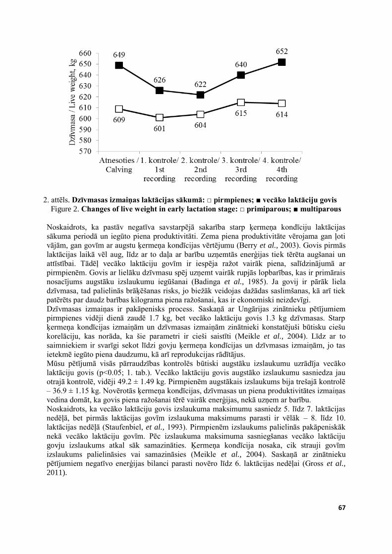
67
2. attēls. Dzīvmasas izmaiņas laktācijas sākumā: □ pirmpienes; ■ vecāko laktāciju govis
Figure 2. Changes of live weight in early lactation stage: □ primiparous; ■ multiparous
Noskaidrots, ka pastāv negatīva savstarpējā sakarība starp ķermeņa kondīciju laktācijas
sākuma periodā un iegūto piena produktivitāti. Zema piena produktivitāte vērojama gan ļoti
vājām, gan govīm ar augstu ķermeņa kondīcijas vērtējumu (Berry et al., 2003). Govis pirmās
laktācijas laikā vēl aug, līdz ar to daļa ar barību uzņemtās enerģijas tiek tērēta augšanai un
attīstībai. Tādēļ vecāko laktāciju govīm ir iespēja ražot vairāk piena, salīdzinājumā ar
pirmpienēm. Govis ar lielāku dzīvmasu spēj uzņemt vairāk rupjās lopbarības, kas ir primārais
nosacījums augstāku izslaukumu iegūšanai (Badinga et al., 1985). Ja govij ir pārāk liela
dzīvmasa, tad palielinās brāķēšanas risks, jo biežāk veidojas dažādas saslimšanas, kā arī tiek
patērēts par daudz barības kilograma piena ražošanai, kas ir ekonomiski neizdevīgi.
Dzīvmasas izmaiņas ir pakāpenisks process. Saskaņā ar Ungārijas zinātnieku pētījumiem
pirmpienes vidēji dienā zaudē 1.7 kg, bet vecāko laktāciju govis 1.3 kg dzīvmasas. Starp
ķermeņa kondīcijas izmaiņām un dzīvmasas izmaiņām zinātnieki konstatējuši būtisku ciešu
korelāciju, kas norāda, ka šie parametri ir cieši saistīti (Meikle et al., 2004). Līdz ar to
saimniekiem ir svarīgi sekot līdzi govju ķermeņa kondīcijas un dzīvmasas izmaiņām, jo tas
ietekmē iegūto piena daudzumu, kā arī reprodukcijas rādītājus.
Mūsu pētījumā visās pārraudzības kontrolēs būtiski augstāku izslaukumu uzrādīja vecāko
laktāciju govis (p<0.05; 1. tab.). Vecāko laktāciju govis augstāko izslaukumu sasniedza jau
otrajā kontrolē, vidēji 49.2 ± 1.49 kg. Pirmpienēm augstākais izslaukums bija trešajā kontrolē
– 36.9 ± 1.15 kg. Novērotās ķermeņa kondīcijas, dzīvmasas un piena produktivitātes izmaiņas
vedina domāt, ka govis piena ražošanai tērē vairāk enerģijas, nekā uzņem ar barību.
Noskaidrots, ka vecāko laktāciju govis izslaukuma maksimumu sasniedz 5. līdz 7. laktācijas
nedēļā, bet pirmās laktācijas govīm izslaukuma maksimums parasti ir vēlāk – 8. līdz 10.
laktācijas nedēļā (Staufenbiel, et al., 1993). Pirmpienēm izslaukums palielinās pakāpeniskāk
nekā vecāko laktāciju govīm. Pēc izslaukuma maksimuma sasniegšanas vecāko laktāciju
govju izslaukums atkal sāk samazināties. Ķermeņa kondīcija nosaka, cik strauji govīm
izslaukums palielināsies vai samazināsies (Meikle et al., 2004). Saskaņā ar zinātnieku
pētījumiem negatīvo enerģijas bilanci parasti novēro līdz 6. laktācijas nedēļai (Gross et al.,
2011).
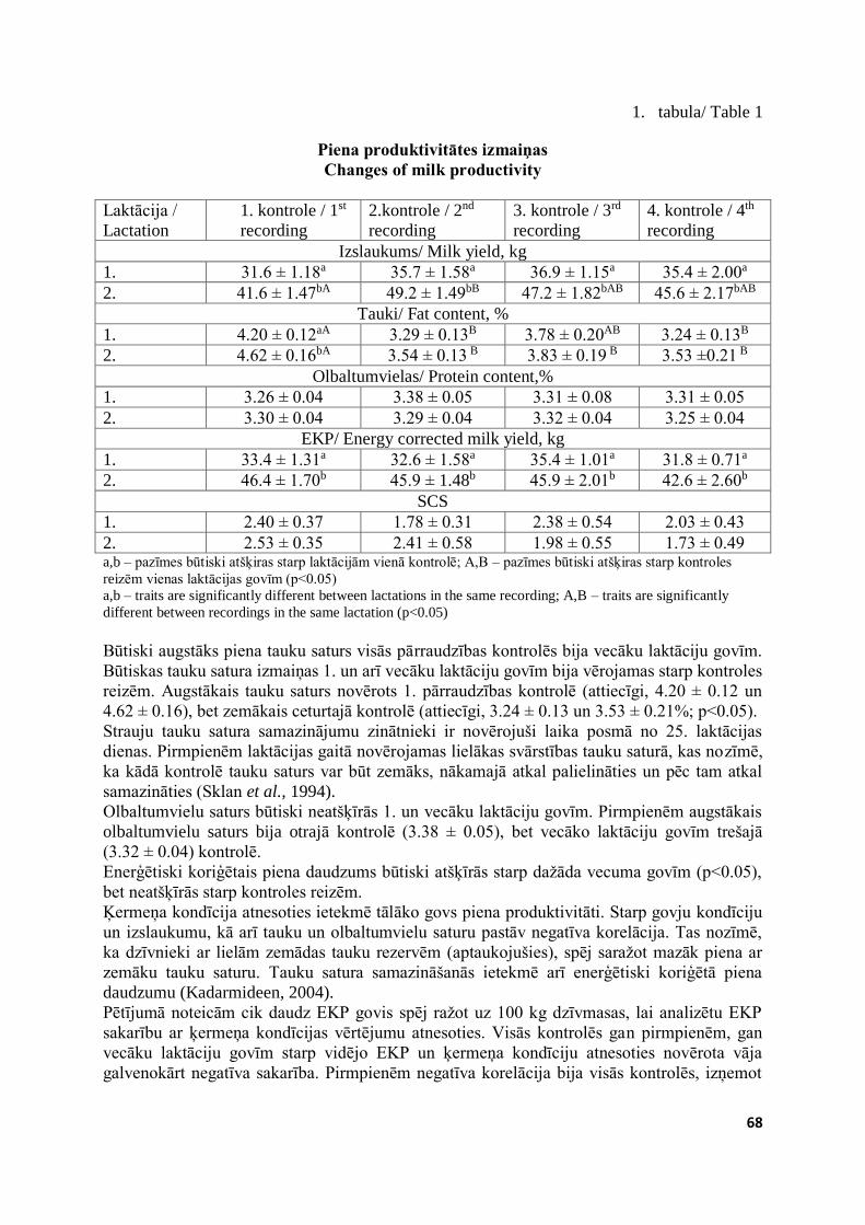
68
1. tabula/ Table 1
Piena produktivitātes izmaiņas Changes of milk productivity
Laktācija /
Lactation
1. kontrole / 1st
recording
2.kontrole / 2nd
recording
3. kontrole / 3rd
recording
4. kontrole / 4th
recording
Izslaukums/ Milk yield, kg
1. 31.6 ± 1.18a 35.7 ± 1.58a 36.9 ± 1.15a 35.4 ± 2.00a
2. 41.6 ± 1.47bA 49.2 ± 1.49bB 47.2 ± 1.82bAB 45.6 ± 2.17bAB
Tauki/ Fat content, %
1. 4.20 ± 0.12aA 3.29 ± 0.13B 3.78 ± 0.20AB 3.24 ± 0.13B
2. 4.62 ± 0.16bA 3.54 ± 0.13 B 3.83 ± 0.19 B 3.53 ±0.21 B Olbaltumvielas/ Protein content,%
1. 3.26 ± 0.04 3.38 ± 0.05 3.31 ± 0.08 3.31 ± 0.05
2. 3.30 ± 0.04 3.29 ± 0.04 3.32 ± 0.04 3.25 ± 0.04
EKP/ Energy corrected milk yield, kg
1. 33.4 ± 1.31a 32.6 ± 1.58a 35.4 ± 1.01a 31.8 ± 0.71a
2. 46.4 ± 1.70b 45.9 ± 1.48b 45.9 ± 2.01b 42.6 ± 2.60b
SCS
1. 2.40 ± 0.37 1.78 ± 0.31 2.38 ± 0.54 2.03 ± 0.43
2. 2.53 ± 0.35 2.41 ± 0.58 1.98 ± 0.55 1.73 ± 0.49 a,b – pazīmes būtiski atšķiras starp laktācijām vienā kontrolē; A,B – pazīmes būtiski atšķiras starp kontroles
reizēm vienas laktācijas govīm (p<0.05)
a,b – traits are significantly different between lactations in the same recording; A,B – traits are significantly
different between recordings in the same lactation (p<0.05)
Būtiski augstāks piena tauku saturs visās pārraudzības kontrolēs bija vecāku laktāciju govīm.
Būtiskas tauku satura izmaiņas 1. un arī vecāku laktāciju govīm bija vērojamas starp kontroles
reizēm. Augstākais tauku saturs novērots 1. pārraudzības kontrolē (attiecīgi, 4.20 ± 0.12 un
4.62 ± 0.16), bet zemākais ceturtajā kontrolē (attiecīgi, 3.24 ± 0.13 un 3.53 ± 0.21%; p<0.05).
Strauju tauku satura samazinājumu zinātnieki ir novērojuši laika posmā no 25. laktācijas
dienas. Pirmpienēm laktācijas gaitā novērojamas lielākas svārstības tauku saturā, kas nozīmē,
ka kādā kontrolē tauku saturs var būt zemāks, nākamajā atkal palielināties un pēc tam atkal
samazināties (Sklan et al., 1994).
Olbaltumvielu saturs būtiski neatšķīrās 1. un vecāku laktāciju govīm. Pirmpienēm augstākais
olbaltumvielu saturs bija otrajā kontrolē (3.38 ± 0.05), bet vecāko laktāciju govīm trešajā
(3.32 ± 0.04) kontrolē.
Enerģētiski koriģētais piena daudzums būtiski atšķīrās starp dažāda vecuma govīm (p<0.05),
bet neatšķīrās starp kontroles reizēm.
Ķermeņa kondīcija atnesoties ietekmē tālāko govs piena produktivitāti. Starp govju kondīciju
un izslaukumu, kā arī tauku un olbaltumvielu saturu pastāv negatīva korelācija. Tas nozīmē,
ka dzīvnieki ar lielām zemādas tauku rezervēm (aptaukojušies), spēj saražot mazāk piena ar
zemāku tauku saturu. Tauku satura samazināšanās ietekmē arī enerģētiski koriģētā piena
daudzumu (Kadarmideen, 2004).
Pētījumā noteicām cik daudz EKP govis spēj ražot uz 100 kg dzīvmasas, lai analizētu EKP
sakarību ar ķermeņa kondīcijas vērtējumu atnesoties. Visās kontrolēs gan pirmpienēm, gan
vecāku laktāciju govīm starp vidējo EKP un ķermeņa kondīciju atnesoties novērota vāja
galvenokārt negatīva sakarība. Pirmpienēm negatīva korelācija bija visās kontrolēs, izņemot
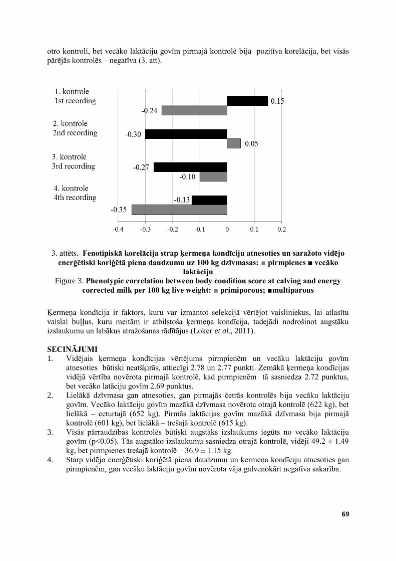
69
otro kontroli, bet vecāko laktāciju govīm pirmajā kontrolē bija pozitīva korelācija, bet visās
pārējās kontrolēs – negatīva (3. att).
3. attēts. Fenotipiskā korelācija strap ķermeņa kondīciju atnesoties un saražoto vidējo enerģētiski koriģētā piena daudzumu uz 100 kg dzīvmasas: ■ pirmpienes ■ vecāko
laktāciju Figure 3. Phenotypic correlation between body condition score at calving and energy
corrected milk per 100 kg live weight: ■ primiporous; ■multiparous
Ķermeņa kondīcija ir faktors, kuru var izmantot selekcijā vērtējot vaisliniekus, lai atlasītu
vaislai buļļus, kuru meitām ir atbilstoša ķermeņa kondīcija, tadejādi nodrošinot augstāku
izslaukumu un labākus atražošanas rādītājus (Loker et al., 2011).
SECINĀJUMI 1. Vidējais ķermeņa kondīcijas vērtējums pirmpienēm un vecāku laktāciju govīm
atnesoties būtiski neatšķirās, attiecīgi 2.78 un 2.77 punkti. Zemākā ķermeņa kondīcijas
vidējā vērtība novērota pirmajā kontrolē, kad pirmpienēm tā sasniedza 2.72 punktus,
bet vecāko latāciju govīm 2.69 punktus.
2. Lielākā dzīvmasa gan atnesoties, gan pirmajās četrās kontrolēs bija vecāku laktāciju
govīm. Vecāko laktāciju govīm mazākā dzīvmasa novērota otrajā kontrolē (622 kg), bet
lielākā – ceturtajā (652 kg). Pirmās laktācijas govīm mazākā dzīvmasa bija pirmajā
kontrolē (601 kg), bet lielākā – trešajā kontrolē (615 kg).
3. Visās pārraudzības kontrolēs būtiski augstāks izslaukums iegūts no vecāko laktāciju
govīm (p<0.05). Tās augstāko izslaukumu sasniedza otrajā kontrolē, vidēji 49.2 ± 1.49
kg, bet pirmpienes trešajā kontrolē – 36.9 ± 1.15 kg.
4. Starp vidējo enerģētiski koriģētā piena daudzumu un ķermeņa kondīciju atnesoties gan
pirmpienēm, gan vecāku laktāciju govīm novērota vāja galvenokārt negatīva sakarība.
70
LITERATŪRA 1. Badinga L., Collier R.J., Wilcox C.J., Thacher W.W. (1985). Interrelationships of milk
yield, body weight, and reproductive performance. Journal of dairy Science. Vol. 68,
pp. 1828–183.
2. Bastin C., Loker S., Gengler N., Sewalem A., Miglior F. (2010). Genetic relationships
between body condition score and reproduction traits in Canadian Holstein and Ayrshire
first-parity cows. Journal of Dairy Science. Vol 93, pp. 2215–2228.
3. Berry D.P., Buckley F., Dillon P., Evans R.D., Rath M., Veerkamp R.F. (2003). Genetic
relationships among body condition score, body weight, milk yield, and fertility in dairy
cows, Journal of Dairy Science. Vol. 86, pp. 2193–2204.
4. Berry D.P., Buckley F., Dillon P. (2011). Relationship between live weight and body
condition score in Irish Holstein-Friesian dairy cows. Irish Journal of Agricultural and
Food Research. Vol. 50, pp. 41–147.
5. Berry D.P., Macdonald K.A., Penno J.W., Roche J.R. (2006). Association between body
condition score and live weight in pasture-based Holstein-Friesian dairy cows. Journal
of Dairy Research. Vol. 73, pp. 487–491.
6. Block S.S., Butler W.R., Ehrhardt R.A., Bell A.W., Van Amburgh M.E., Boisclair Y.R.
(2001). Decreased concentration of plasma leptin in periparturient dairy cows is caused
by negative energy balance. Journal of Endocrinology, Vol. 171, pp. 339–348.
7. Borner S., Albrecht E., Schaff C., Hacke S., Kautzsch U., Derno M., Hammon H.M.,
Rontgen M., Sauerwein H., Kuhla B. (2013). Reduced AgRP activation in the
hypothalamus of cows with high extent of fat mobilization after parturition. General and
Comparative Endocrinology. Vol. 193, pp. 167 – 177.
8. Garcia O., Hemme T. Nho L.T., Tra H.T.H. (2006). The economics of milk production
in Hanoi, Vietnam, with particular emphasis on small-scale producers. PPLPI Working
Paper. No. 33, pp. 39 – 40.
9. Gross J., van Dorland H.A., Bruckmaier R.M., Schwarz F.J. (2011). Milk fatty acid
profile related to energy balance in dairy cows. Journal of Dairy Research. Vol 78, pp.
479 – 488.
10. Grummer R.R. (1995). Impact of changes in organic nutrient metabolism on feeding the
transition dairy cow. Journal of Animal Science. Vol. 73, pp. 2820-2833.
11. Kadarmideen H.N. (2004). Genetic correlations among body condition score, somatic
cell score, milk production, fertility and conformation traits in dairy cows. Animal
Science. Vol. 79, pp. 191-201.
12. Loker S., Bastin C., Miglior F., Sewalem A., Scheaffer L.R., Jamrozik J., Osborne V
(2011). Short communication: Estimates of genetic parameters of body condition score
in the first 3 lactations using a random regression animal model. Journal of Dairy
Science. Vol. 94, pp. 3693–3699.
13. Loker S., Bastin C., Miglior F., Sewalem A., Scheaffer L.R., Jamrozik J., Ali A.,
Osbornell V. (2012). Genetic and environmental relationships between body condition
score and milk production traits in Canadian Holsteins. Journal of Dairy Science. Vol.
95, pp. 410–419.
14. Meikle A., Kulcsar M., Chilliard Y., Febel H., Delavaud C., Cavestany D., Chilibroste
P. (2004). Effects of parity and body condition at parturition on endocrine and
reproductive parameters of the cow. Reproduction. Vol. 127, pp. 727–737.
15. Schroder U.J., Staufenbiel R. (2006). Invited Review: Methods to determine body fat
reserves in the dairy cow with special regard to ultrasonographic measurement of
backfat thickness. Journal of Dairy Science. Vol. 89, pp. 1–14.
16. Schutz M.M., Powell R.L. (1993). Genetic evaluations for somatic cell score. In:
Annual meeting of INTERBULL. August 20, 1993, Arhus, Denmark.
71
17. Sklan D., Kaim M., Moallem U., Folman Y. (1994). Effect of dietary calcium soaps on
milk yield, body weight, reproductive hormones, and fertility in first parity and older
cows. Journal of Dairy Science. Vol. 77 pp. 1652-1660.
18. Staufenbiel R., Staufenbiel B., Rossow N., Wiedemann F. (1993). Energie- und 15, 15.
Fettstoffwechsel des Rindes – Vergleich der Aussage der Rückenfettdicke mit anderen
Untersuchungsgrößen. Mh Vet-Med. Vol. 48, pp.167–174.
19. Wathes D.C., Cheng Z., Bourne N., Taylor V.J., Coffey M.P., Brotherstone S. (2007).
Differences between primiparous and multiparous dairy cows in the inter-relationships
between metabolic traits, milk yield and body condition score in the periparturient
period. Domestic Animal Endocrinology. Vol. 33, pp. 203-225.
20. Wathes D.C., Fenwick M., Cheng Z., Bourne N., Llewellyn S., Morris D.G., Kenny D.,
Murphy J., Fitzpatrick R. (2007a). Influence of negative energy balance on cyclicity and
fertility in the high producing dairy cow. Theriogenology. Vol. 68, pp. 232–241.
72
AR KALCIJU BAGĀTAS BARĪBAS PIEDEVAS PIELIETOŠANA SUŅIEM KAULAUDU VIELMAIŅAI
CALCIUM RICH ADDITIVES USE IN DOGS FOR BONE
METABOLISM
Ilga Šematoviča, Arturs Ivanovs LLU Veterinārmedicīnas fakultāte, Latvija; Faculty of Veterinary Medicine LUA, Latvia
ABSTRACT Morphological, biochemical analysis and bone turnover markers (BTM) in blood and urine
were performed for six dogs twice, in the start of the research, and in the end after calcium
(Ca) rich additives, without phosphorus (P), dosing for one month per os. All dogs were
clinically healthy at the start and in the end of work. No significant changes were observed
regarding to blood morphology. The level of Ca and P in blood before Ca additives dosing
was in physiologically reference values in five from six dogs. One dog had hypocalcaemia
and hyperphosphatemia in the start of the research. At the end of the research level of Ca was
increased for all dogs (p<0.05), while P were decreased in four from six dogs and was also in
reference values. Osteocalcin in blood decreased in all dogs (p<0.05) which show us decrease
of bone regenerative processes. The dog with expressed hypocalcemia at the start of the
research, were with considerably increased marker of bone resorption marker β-CTx at the
end. In conclusion whereas all dogs were clinically healthy in the end of the research, changes
regarding to BTM in blood are important point to evaluate influence of the treatment with Ca
rich additives to bone health. Although, the level of Ca increased in blood, the results of BTM
let suggest about negative effect on bone health. The need for P organism compensates with P
absorption from their own bones, thus reducing bones quality. Using feed additives for dogs
definitely must follow Ca and P proportion. KEY WORDS: dogs, calcium, bone turnover markers
IEVADS Pilnvērtīgas barības devas sabalansēšana suņu turētājiem joprojām ir izaicinājums. Viens no
aktuāliem jautājumiem ir minerālvielu, īpaši kalcija (Ca), nodrošinājums racionā. Kā
vispārzināms, Ca piemīt daudzveidīga loma organisma vielmaiņā, piemēram, tas ir
nepieciešams enzīmu un transmembrānu transporta reakcijās, asinsrecē, nervu impulsu
pārvadē, muskuļu kontrakcijās, asinsvadu un muskuļšķiedru tonusa nodrošināšanā, hormonu
sekrēcijā, glikogēna sintēzē aknās un dažādu organisma šūnu metabolismā kā arī kaulaudu
formēšanās un resorbcijas norisēs (Birchard, Sherding, 2006).
Līdz šim tīri empīriski, analizējot barības devu, ņemot vērā suņa šķirnes un vecuma īpatnības,
tika pieņemts lēmums par papildus Ca saturošu piedevu izēdināšanu. Fizioloģiski kopējā Ca
līmenis asinīs suņiem ir no 2.3 – 3.0 mmol/L (Jemeļjanovs et al., 2008; Birchard, Sherding,
2006). Galvenokārt Ca maiņu organismā nodrošina parathormons, D vitamīna metabolīti un
kalcitonīns.
Lai pieņemtu lēmumu par papildus Ca piedevu izēdināšanu, nevar ņemt par pamatu tikai
hipokalcēmiju asins bioķīmiskajās analīzēs, jo Ca līmeni asinīs ietekmē citas, ar Ca daudzumu
uzturā nesaistītas saslimšanas un patoloģiski procesi kā nervu, vairogdziedzera patoloģijas,
hipoalbuminēmija, eklampsija, u.c. (Willard et al., 1989).
Mazo dzīvnieku medicīnā trūkst konkrētu reprezentējošu kritēriju, pēc kuriem būtu indicēts
papildus izēdināt ar Ca bagātas barības piedevas profilaktiskos nolūkos, kā arī nav kritēriju,
73
pēc kuriem varētu spriest par šo piedevu iedarbību tieši uz kaulaudu veselību. Humānajā
medicīnā šis ir plašu pētījumu lauks, aktuāls dažādu saslimšanu un patfizioloģisku procesu
gadījumā, piemēram, lai profilaktētu kaulu lūzuma risku, sievietēm post-menopauzes vecumā,
pacientiem, kuriem ilgstoši veikta dialīze, u.c. (Civitelli et al., 2009; Tentori et al. 2008; Allen
et al., 2007).
Pētījuma mērķis bija pielietot humānajā medicīnā plaši pielietotu ar Ca bagātu barības
piedevu pieaugušiem suņiem un novērtēt tās ietekmi uz kaulaudu veselību.
MATERĀLS UN METODIKA Pētījums veikts 2014.gada jūlija mēnesī, tajā iekļaujot sešus klīniski veselus 2-7 gadus vecus
dažādu šķirņu un turēšanas veida suņus. No tiem četri kastrēti, visi vakcinēti un regulāri
attārpoti.
Asins paraugi, pētījumu uzsākot un noslēdzot, tika ņemti no v.jugularis vai v.saphena sterilos
vienreizējas lietošanas “Sarstedt Monovette” vakutaineros – asins morfoloģiskajai novērtēšnai
ar EDTA 1.2 ml, kā arī bioķīmiskai izmeklēšanai serumam Z/2.7 ml vakutaineros.
Izmeklējumi veikti sertificētās (akreditācijas apliecība LATAK-M-434-00-2011) SIA
„Centrālā laboratorija” (reģ. Nr. 215/L 430-C) laboratorijās, kur analīžu izpilde atbilst LVS
EN ISO 15189:2008 standarta prasībām, kā arī SIA „E. Gulbja laboratorija” kur tika noteikti
kaulaudu marķieri (β-CTx, osteokalcīns, P1NP, BALP), parathormons, D3 vitamīns atbilstoši
starptautiskam standartam LVS EN ISO/IEC 17025, kopš 2007.gada 8.oktobra - LVS EN ISO
15189:2007 un kopš 2008.gada 17. decembra - LVS EN ISO 15189:2008.
Pētījumā izvērtētie asins morfoloģiskie un bioķīmiskie parametri raksturo dzīvnieku vispārējo
veselību, kā arī reprezentē nieru veselības stāvokli. Kā kaulaudu veselību raksturojošie
parametri tika noteikti daži bioķīmiskie kaulu vielmaiņas rādītāji, sekojot humānajā medicīnā
ieteiktajiem (Civitelli et al., 2009; Allen et al, 2007):
kaulveides marķieri – kaulaudu fosfotāzes sārmainā frakcija (BALP), osteokalcīns, 1
tipa prokolagēna N-propeptīds (P1NP);
kaulaudu resorbcijas marķieri – 1 tipa kolagēna karboksi-termināla šķērssaites telopeptīds (CTX), deoksipiridinolīns (DPD) urīnā.
Pēc pirmreizējās analīžu noņemšanas un dzīvnieku nosvēršanas, suņiem vienu mēnesi tika
izēdinātas individuālas Ca saturošas barības piedevas saskaņā ar ieteicamajām Ca diennakts
profilaktiskajām devām (Birchard, Sherding, 2006; Ettinger, 1995). Pētījumā pielietotās
barības piedevas sastāvs ir identisks, kādu izmanto humānajā hipokalcēmijas profilaksē un šīs
konkrētās piedevas sastāvā ir Ca, Mg, K, F, D3.
Datu statistiskā apstrāde veikta izmantojot SPSS One sample t-test, descriptive statistics un
Wilcoxon test divu saistītu paraugkopu salīdzināšanai, kā arī veikta korelāciju analīze
kopsakarību atrašanai (Paura un Arhipova, 2002).
REZULTĀTI UN DISKUSIJA Pirms rezultātu interpretācijas jāpiebilst, ka 2014.gada jūlijs, kad tika veikta Ca piedevas
izēdināšana, bija īpaši saulains un vidējā gaisa temperatūra dienā bija virs 30 oC. Tā kā
dihidroksikalciferola daudzums, kā iepriekš minēts, ietekmē Ca vielmaiņu, atkarīgs no
uzņemtā D3 provitamīna un no saules staru daudzuma, tas varētu ietekmēt pētījuma rezultātu,
palielinot D vitamīna līmeni asinīs (Shikino et al., 2014).
Pētījumu uzsākot visi suņi bija klīniski veseli. Asins morfoloģiskie parametri visiem suņiem
kā uzsākot, tā noslēdzot pētījumu bija normas robežās, būtiskas izmaiņas nenovērojām, līdzīgi
citu autoru pētījumos humānajā medicīnā (Mitchell et.al., 2014).
Sākotnējās asins bioķīmiskajās analīzēs atklājās, ka diviem suņiem ir nedaudz paaugstināts
kopējā proteīna līmenis. To varētu izskaidrot ar nelielu dehidratāciju (Jemeļjanovs et al.,
2008; Birchard, Sherding, 2006; Ettinger, 1995), ko varēja radīt divu faktoru kombinācija -

74
karstais laiks un ceļojums uz klīniku. Vienam sunim (Alfai) pētījumu uzsākot tika konstatēts
samazināts albumīnu daudzums asinīs, hiperkalēmija, hipokalcēmija un viegla
hiperfosfatēmija. Turpmāk šo dzīvnieku nosacīti sauksim par „hipokalcēmisko” suni. Šajā
gadījumā būtu nepieciešama sīkāka dzīvnieka veselības stāvokļa izvērtēšana un papildus
analīzes. Tā kā Ca metabolisms ir cieši saistīts ar albumīnu daudzumu, tad būtu jāizvērtē
iespējamie hipoalbuminēmijas cēloņi (hroniskas aknu slimības, parazitāras slimības,
maldigestija, ilgstoša proteīnūrija nieru slimību dēļ, smagas eksudatīvas slimības u.c.)
(Jemeļjanovs et al., 2008). Hiperkalēmiju varēja radīt artefakts saistībā ar asins parauga
noņemšanas un uzglabāšanas procedūru eritrolīzes dēļ, kā arī to var novērot pie akūtas un
hroniskas nieru mazspējas, postrenālas urēmijas, audu bojājumiem, acidozes,
hiperadrenokorticisma un jatrogēni. Savukārt hroniska hipokalcēmija suņiem var būt pie
primāra hipoparatireoidisma, hiperkalcitonisma, malabsorbcijas, anoreksijas, nieru mazspējas,
D hipovitaminozes (Ettinger, 1995). Tā kā konkrētajam sunim pārējie asins bioķīmiskie
parametri bija normas robežās, parathormonu un D vitamīnu ieskaitot, tas jutās labi un
īpašniekam nebija sūdzību par suņa veselību, tika nolemts to tomēr iekļaut pētījumā.
Atkārtotajās bioķīmisko parametru analīzēs, pētījumu nobeidzot, tikai sunim, kuram pirmajā
reizē bija samazināts albumīnu un Ca līmenis, kā arī paaugstināts K un P līmenis asinīs,
joprojām palika samazināts albumīna daudzums asinīs, bet Ca daudzums pieauga līdz
minimālajai normas vērtībai, savukārt P un K daudzums samazinājās līdz normas vērtībai.
Kopumā Ca daudzums pētījumu uzsākot visu suņu asins serumā bija normas robežās, tas ir
2.4±0.08 mmol/L (izslēdzot „hipokalcēmisko”suni kā t.s. izlecošos datus), bet pielietojot One
sample t-test SPSS izrādījās, ka šis rādītājs bija statistiski nozīmīgi zemāks par vidējo
references vērtību 2.65 mmol/L (p<0.05). Mūsu pētījumā pēc ar Ca bagātu preparāta
pielietošanas Ca līmenis pieauga visiem dzīvniekiem (Wilcoxson test 2 saistītām
paraugkopām, p<0.05), un vidēji tas bija 2.5±0.14, kas vairs būtiski neatšķīrās no vidējās
pieļaujamās vērtības (p>0.05), (1.attēls).
Kaut gan Ca līmenis suņu asinīs iekļāvās normas robežās, pētījumā konstatējām, ka pat
neliela tendence uz Ca deficītu var tikt kompensēta, pielietojot ar Ca bagātas barības piedevas
uzturā.
P daudzums visiem suņiem, izņemot vienu („hipokalcēmiskais suns”), bija normas vērtību
ietvaros, kā uzsākot (1.3±0.20), tā arī noslēdzot pētījumu (1.2±0.12) un būtiski neatšķīrās no
vidējās pieļaujamās vērtības (p>0.05), (1.attēls).
1.attēls. Ca un P līmenis suņu asinīs pirms un pēc viena mēneša Ca piedevu kursa per os. Figure 1. Ca and P levels in the blood of dogs before and after one month dosing of the
Ca additive per os.
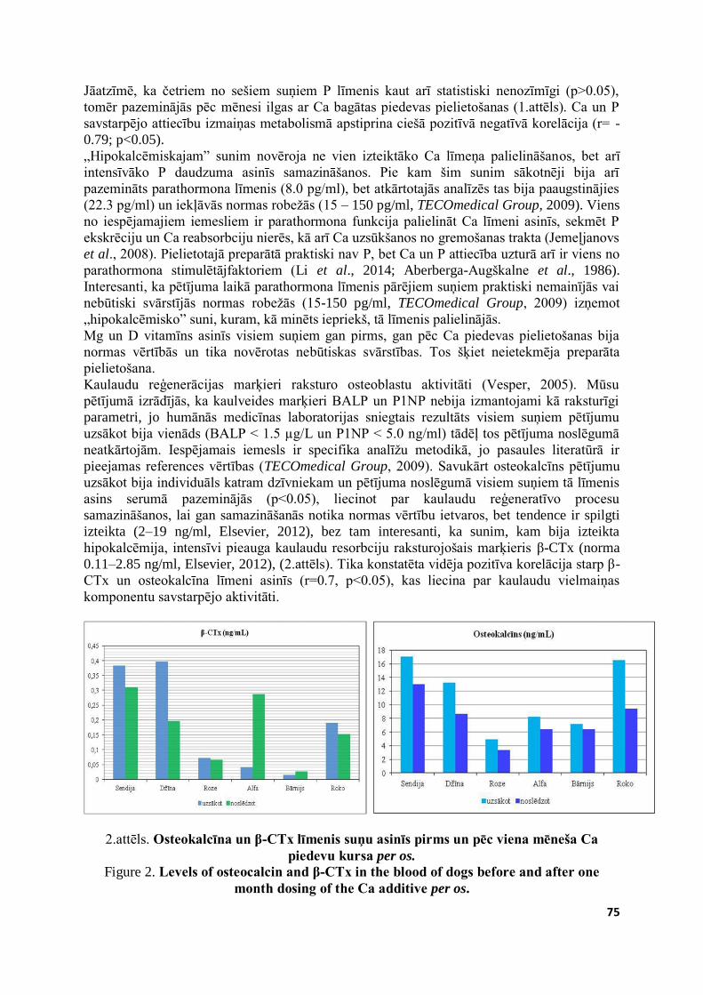
75
Jāatzīmē, ka četriem no sešiem suņiem P līmenis kaut arī statistiski nenozīmīgi (p>0.05),
tomēr pazeminājās pēc mēnesi ilgas ar Ca bagātas piedevas pielietošanas (1.attēls). Ca un P
savstarpējo attiecību izmaiņas metabolismā apstiprina ciešā pozitīvā negatīvā korelācija (r= -
0.79; p<0.05).
„Hipokalcēmiskajam” sunim novēroja ne vien izteiktāko Ca līmeņa palielināšanos, bet arī
intensīvāko P daudzuma asinīs samazināšanos. Pie kam šim sunim sākotnēji bija arī
pazemināts parathormona līmenis (8.0 pg/ml), bet atkārtotajās analīzēs tas bija paaugstinājies
(22.3 pg/ml) un iekļāvās normas robežās (15 – 150 pg/ml, TECOmedical Group, 2009). Viens
no iespējamajiem iemesliem ir parathormona funkcija palielināt Ca līmeni asinīs, sekmēt P
ekskrēciju un Ca reabsorbciju nierēs, kā arī Ca uzsūkšanos no gremošanas trakta (Jemeļjanovs
et al., 2008). Pielietotajā preparātā praktiski nav P, bet Ca un P attiecība uzturā arī ir viens no
parathormona stimulētājfaktoriem (Li et al., 2014; Aberberga-Augškalne et al., 1986).
Interesanti, ka pētījuma laikā parathormona līmenis pārējiem suņiem praktiski nemainījās vai
nebūtiski svārstījās normas robežās (15-150 pg/ml, TECOmedical Group, 2009) izņemot
„hipokalcēmisko” suni, kuram, kā minēts iepriekš, tā līmenis palielinājās.
Mg un D vitamīns asinīs visiem suņiem gan pirms, gan pēc Ca piedevas pielietošanas bija
normas vērtībās un tika novērotas nebūtiskas svārstības. Tos šķiet neietekmēja preparāta
pielietošana.
Kaulaudu reģenerācijas marķieri raksturo osteoblastu aktivitāti (Vesper, 2005). Mūsu
pētījumā izrādījās, ka kaulveides marķieri BALP un P1NP nebija izmantojami kā raksturīgi
parametri, jo humānās medicīnas laboratorijas sniegtais rezultāts visiem suņiem pētījumu
uzsākot bija vienāds (BALP < 1.5 µg/L un P1NP < 5.0 ng/ml) tādēļ tos pētījuma noslēgumā
neatkārtojām. Iespējamais iemesls ir specifika analīžu metodikā, jo pasaules literatūrā ir
pieejamas references vērtības (TECOmedical Group, 2009). Savukārt osteokalcīns pētījumu
uzsākot bija individuāls katram dzīvniekam un pētījuma noslēgumā visiem suņiem tā līmenis
asins serumā pazeminājās (p<0.05), liecinot par kaulaudu reģeneratīvo procesu
samazināšanos, lai gan samazināšanās notika normas vērtību ietvaros, bet tendence ir spilgti
izteikta (2–19 ng/ml, Elsevier, 2012), bez tam interesanti, ka sunim, kam bija izteikta
hipokalcēmija, intensīvi pieauga kaulaudu resorbciju raksturojošais marķieris β-CTx (norma
0.11–2.85 ng/ml, Elsevier, 2012), (2.attēls). Tika konstatēta vidēja pozitīva korelācija starp β-
CTx un osteokalcīna līmeni asinīs (r=0.7, p<0.05), kas liecina par kaulaudu vielmaiņas
komponentu savstarpējo aktivitāti.
2.attēls. Osteokalcīna un β-CTx līmenis suņu asinīs pirms un pēc viena mēneša Ca piedevu kursa per os.
Figure 2. Levels of osteocalcin and β-CTx in the blood of dogs before and after one month dosing of the Ca additive per os.

76
Kaulaudu deģenerācijas marķieri raksturo osteoklastu aktivitāti, - jo augstāka osteoklastu aktivitāte, jo lielāks kaulaudu deģenerācijas marķiera rādītājs (Vesper, 2005).
DPD urīnā rezultāti bija variabli un katram sunim individuāli. Tā daudzums pētījuma laikā
nebūtiski svārstījās. Diviem no sešiem nebūtiski pieauga, bez tam tika konstatēta vidēja
negatīva statistiski nozīmīga korelācija starp DPD urīnā un osteokalcīnu asinīs (r = - 0.59,
p<0.05), kas prezentē reģeneratīvo un kaulaudu atjaunošanās procesu pretējo dabu.
Paradokss, ka sekojot humānajā lietošanā plaši pielietotā preparāta anotācijai solījumam
panākt labāku kaulaudu veselību, stiprus kaulus, pielietojot šo ar Ca bagāto barības piedevu,
kurā nav P, asinīs gan panākam Ca līmeņa palielināšanos, bet kaulaudu marķieru analīžu
rezultāti liek domāt par negatīvu ietekmi uz kaulaudiem. Domājams, ka organisms
kompensējot P vajadzību to absorbē no saviem kaulaudiem, tādējādi samazinot kaulaudu
kvalitāti. Spilgts piemērs tam ir fakts, ka sunim ar izteiktu hronisku hipokalcēmiju novērojām
spilgtāko Ca pieaugumu, visizteiktāko kaulu resorbcijas rādītāja palielināšanos līdztekus
kaulaudu reģenerācijas marķiera samazinājumam. Nomierinošs ir fakts, ka visu barības
piedevu anotācijās ir norādīts, ka ar tām nedrīkst aizstāt pilnvērtīgu uzturu. Iespējams, tīri
instinktīvi cilvēks uzturā var sajust vajadzību racionā ietvert fosforu saturošus produktus,
piemēram, zivis. Mūsu draugiem suņiem nav šādas iespējas, tie ir pilnībā atkarīgi no īpašnieka
piedāvātā uztura, tādēļ sekas ir konstatējamas tik uzskatāmi.
Tā kā visi pētījumā iekļautie suņi pētījuma noslēgumā bija klīniski veseli, dzīvespriecīgi un ar
labu apetīti, jādomā, ka asinīs konstatētās izmaiņas ir agrīni kaulu vielmaiņas signālparametri.
Trūkst vispārpieņemtu, plaši pieejamu references vērtību kaulaudu veselības novērtēšanai. Arī
humānajā medicīnā tiek atzīmēts, ka kaulaudu marķieru vērtības atsevišķiem indivīdiem ir
visai variablas, tādēļ tie vairāk noder ne tik daudz kā Ca preparātu pielietošanas indikācijām,
bet gan kā ārstēšanas kursa novērtēšanas parametri (Civitelli et.al., 2009).
Lai precīzāk izpētītu kaulaudu veselību ietekmējošos parametrus suņiem, būtu nepieciešams
turpināt pētījumus, kuros būtu precīzāk izvērtējami ietekmējošie faktori kā suņu šķirnes,
vecums, dzimums, ēdināšanas un turēšanas veids. Lai veiktu korektu pētījumu, būtu
nepieciešama kontrolgrupa, taču dēļ references vērtību variabilitātes, pētāmajām grupām jābūt
pietiekami lielām, piemēram, Allen et. al.(2008) veikuši pētījumu uz 108 sieviešu kārtas
1.3±0.2 gadus vecām bīgla šķirnes kucēm. Lai veiktu kaulaudu densimetriju, ir nepieciešams
ilgāks laika periods. Ca:P attiecībai pieaugušu suņu uzturā jābūt 1.2:1 pretējā gadījumā,
pieaugušajiem suņiem varēs novērot osteoporozi, jaundzīvniekiem – rahītu.
SECINĀJUMI 1. Ar Ca bagātas barības piedevas pielietošana profilaktiskajās devās vienu mēnesi būtiski
neietekmēja nespecifiskos dzīvnieku vispārējās veselības rādītājus.
2. Pielietojot ar Ca bagātu barības piedevu, kas nesatur P, asinīs pieauga Ca, bet
samazinājās P līmenis, tomēr šo parametru ir par maz, lai spriestu par preparāta
noderīgumu vispārējai veselībai, jo ir jākontrolē arī šo preparātu ietekme uz kaulaudu
veselību.
3. Pielietojot ar Ca bagātu barības piedevu, kas nesatur P, visiem pētījumā iekļautajiem
suņiem samazinājās osteokalcīna līmenis (p<0.05), kas liecina par kaulaudu
reģeneratīvo jeb atjaunošanās procesu samazināšanos. Sunim ar izteiktu hipokalcēmiju,
ne tikai samazinājās osteokalcīna līmenis, bet arī palielinājās beta-CTx, kas liecina par
izteiktu kaulaudu deģeneratīvo procesu aktivizēšanos.
4. Suņiem nedrīkst pielietot Ca saturošus preparātus, kuros nav ievērota tiem fizioloģiski
nepieciešamā Ca:P attiecība, jo šajā gadījumā domājams, P vajadzības kompensēšanai
organisms to absorbē no saviem kaulaudiem, tādējādi mazinot kaulaudu kvalitāti.
Iespējams, šādā gadījumā Ca:P attiecības nodrošināšanas labad ir jāpārskata viss
77
izēdināmais racions kopumā, bet šāda uztura sabalansēšana prasa lielu pieredzi un uztura komponentu analīzi.
LITERATŪRA 1. Aberberga-Augškalne L., Āboltiņa-Āboliņa E., Aivars J., Gaile E., Valtneris A. (1986)
Cilvēka fizioloģija. „Zvaigzne”, 1986., 455 lpp.
2. Allen M.R., Gineyts E., Leeming D.J., Burr D.B., Delmas P.D. (2007) Bisphosphonates
alter trabecular bone collagen cross-linking and isomerization in beagle dog vertebra.
Osteoporosis international : a journal established as result of cooperation between the
European Foundation for Osteoporosis and the National Osteoporosis Foundation of the
USA. 2008 Mar;19(3):329-37.
3. Arhipova, I., Bāliņa, S. (2003). Statistika Ekonomikā. Risinājumi ar SPSS un Microsoft
Exel. Rīga: Datorzinību centrs, 352 lpp.
4. Birchard S.J., Sherding R.G. (2006). Saunders manual of small animal practise. Third
edition, USA, Elsevier, 2008 p.
5. Civitelli R., Armamento-Villareal R., Napoli N. (2009). Bone turnover markers:
understanding their vlue in clinical trials and clinical practise. Osteoporosis
international, 20:843-852
6. Ettinger S.J. (1995). Pocket companion to the fourth edition of textbook of veterinary
internal medicine. W.B.Saunders company, 877 p.
7. Jemeļjanovs Ļ., Dūrītis I., Beinerts J. (2008). Dzīvnieku iekšķīgo slimību laboratoriskā
diagnostika. Jelgava, 90 lpp.
8. Li F., Cao X., Zhao L., Wang J., Ding. Z. (2014). Effects of Mineral Additives on
Biochar Formation: Carbon Retention, Stability, and Properties. Environmental science
and technology. Washington DC : American Chemical Society,
http://www.ncbi.nlm.nih.gov/pubmed/25203840, resurss apskatīts 2014.09.01.
9. Mitchell N.J., Kumi J., Aleser M., Elmore S.E., Rychlik K.A., Zychowski K.E.,
Romoser A.A., Phillips T.D., Ankrah N.A. (2014). Short-Term Safety and Efficacy of
Calcium Montmorillonite Clay (UPSN) in Children. The American journal of tropical
medicine and hygiene. ISSN: 2014 Aug 18.,
http://www.ajtmh.org/content/early/2014/08/14/ajtmh.14-0093.full.pdf+html, resurss
apskatīts 2014.09.01.
10. Shikino K, Ikusaka M, Yamashita T. (2014). Vitamin D-deficient osteomalacia due to
excessive self-restrictions for atopic dermatitis. BMJ case reports. London : BMJ Pub.
Group, 2014 Jul 4;2014.
11. Tentori F., Blayney M.J., Albert J.M., Gillespie B.W., Kerr P.G., Bommer J., Young
E.W., Akizawa T., Akiba T., Pisoni R.L., Robinson B.M., Port F.K. (2008). Mortality
risk for dialysis patients with different levels of serum calcium, phosphorus, and PTH:
the Dialysis Outcomes and Practice Patterns Study (DOPPS). American journal of
kidney diseases : the official journal of the National Kidney Foundation. 52(3):519-30.
12. TECOmedical Group, Switzerland, © 04/2009, http://www.tecomedical.com
/download-file?item_file_id=2494&item_file_code=4ab6df96cf&file_key=0, resurss
apskatīts 3014.09.01.
13. Vesper H.W. (2005). Analytical and Preanalytical Issues in Measurement of
Biochemical Bone Markers. USA: Medscape, LabMedecine. 15.07.2014.,
http://www.medscape.com/viewarticle/509097_2 , resurss apskatīts 2014.09.02.
14. Willard M.D., Tvedten H., Turnwald G.H. (1989). Small animal clinical diagnosis by
laboratory methods. Saunders company, Philadelphia, 380 p.

78
MYCOPLASMA SYNOVIAE SEROPREVALENCE DĒJĒJVISTU GANĀMPULKĀ
SEROPREVALENCE OF MYCOPLASMA SYNOVIAE IN THE
COMMERCIAL LAYER FLOCK
Inita Zute1, Anda Valdovska2 1 A/S „Balticovo” 2 LLU Veterinārmedicīnas fakultāte, Latvija; Faculty of Veterinary Medicine LUA, Latvia
ABSTRACT Mycoplasma species are well-known pathogens of domestic poultry, causing significant
economic losses. Mycoplasma synoviae (M. synoviae) can cause respiratory disease, synovitis,
or result in a silent infection in chickens and turkeys. A total of 1543 serum samples from
non-vaccinated against M. synoviae 65 chicken flocks of different ages from 1 to 70 weeks
old were collected. Results show that the seroprevalence of M. synoviae was 56.1% in 2012
and 51.4% in 2013. Mycoplasma seropositivity in laying hens was the highest in summer
(79.1%) and lowest in winter (36.0%).
KEY WORDS: poultry, Mycoplasma synoviae, seroprevalence, ELISA
IEVADS Mikoplazmoze ir viena no nozīmīgākajām infekcijas slimībām putnkopības nozarē mūsdienu
intensīvās ražošanas apstākļos.
Mikoplazmozi izraisa Mollicutes klases Mycoplasma ģints baktērijas. Putniem mikoplazmozi
izraisa galvenokārt Mycoplasma gallisepticum (MG) un Mycoplasma synoviae (MS) un
inficēšanās notiek gan horizontālā, gan vertikālā ceļā, radot nozarei ekonomiskos zaudējumus
gan no elpošanas ceļu saslimšanām, kas izraisa imunitātes pazemināšanos un paver ceļu citām
bakteriālām (E.coli) (Kleven, Ferguson-Noel, 2008) un vīrusu (infekciozais bronhīts,
Ņūkāslas slimība, u.c.) saslimšanām, gan olu kvalitātes pasliktināšanās dēļ (pat par 10% -
20%) (Morrow et al., 1990; Bradbury, 2001).
Visu vecumu putni ir jutīgi pret mikoplazmozi, taču jaunputni ir daudz uzņēmīgāki pret
infekciju nekā pieaugušie (Nunoya et al., 1995). Slimības klīniskās pazīmes parādās dažu
dienu laikā no ierosinātāja iekļūšanas brīža putna organismā. Ierosinātājs no slimā putna ar
izdalījumiem no nāsīm tiešā kontakta (horizontālā) ceļā tiek pārnests uz veselajiem putniem
vai netiešā ceļā - no piesārņotās vides. MS ir viens no galvenajiem slimības ierosinātājiem,
kas izraisa respiratorās un/vai locītavu saslimšanas, kas putniem izpaužas ar klepu,
šķaudīšanu, gaisa maisu, deguna dobumu un blakusdobumu iekaisumu, augšanas
traucējumiem (Kleven, Ferguson-Noel, 2008).
Mikoplazmozes izplatību veicina arī mājputnu apkalpojošais personāls, kas infekciju pārnēsā
ar drēbēm, apaviem un inventāru. Palielinoties putnu un cilvēku blīvumam, palielinās arī
mikoplazmu izplatīšanās ātrums. Vertikālā transmisija (ar inficētām olām) ir otrs svarīgākais
slimības pārneses veids, īpaši, no vecāku ganāmpulka un broileru ganāmpulkos (Jordan,
1975).
Saslimstība ar mikoplazmozi ir ļoti plaši izplatīta un aktuāla visos mājputnu audzētāju
ganāmpulkos (Kleven, Ferguson-Noel, 2008). Arvien lielāka uzmanība tiek pievērsta
ražošanas izmaksu samazināšai un tāpēc aktuāla kļūst slimības ierobežošana. Pamatojoties uz
augstāk minēto, svarīga ir nepieciešamība izvērtēt slimības kontroles iespējas, ieviešot
uzraudzības programmas un vakcināciju, kas ir viens no efektīvākajiem līdzekļiem
79
mikoplazmozes izplatības kontrolē un profilaksē. Tādēļ mūsu darba mērķis ir noskaidrot Mycoplasma synoviae izplatību dējējvistu ganāmpulkā.
MATERIĀLS UN METODIKA Paraugu iegūšana. Pētījums veikts A/S „Balticovo” dējējvistu ganāmpulkā. 2012. un 2013.
gadā kopā tika noņemti 1543 asins paraugi no 65 putnu grupām vecumā no 1 dienas līdz 70
nedēļām, kuri nav bijuši vakcinēti pret Mycoplasma gallisepticum un Mycoplasma sinoviae.
Putniem asins paraugi iegūti no spārna vēnas, lietojot sterilas adatas un 1,5 ml stobriņus. Pēc
asins paraugu noņemšanas stobriņi tika glabāti 1 h istabas temperatūrā un pēc tam
centrifugējot 10 min/1500 apgr. paraugam atdalīts serums. Plazma ievietota sterilā 1.5 ml
stobriņā, nomarķēta un uzglabāta saldētavā -20 oC temperatūrā līdz tālākai testēšanai.
ELISA tests. Seruma izmeklēšana Mycoplasma synoviae (MS) antivielu titra noteikšanai tika
veikta ar ELISA metodi, izmantojot komerciālo kitu (BIOCHEK Smart Veterinary
Diagnostics, kataloga Nr CK115 MS), atbilstoši ražotāja instrukcijai. Pozitīvas reakcijas
gadījumā mikroplatē veidojās dzeltens krāsojums, kura intensitāte ir tieši atkarīga no anti-MS
antivielu klātbūtnes paraugā, t.i., jo vairāk anti-MS antivielu, jo tumšāka nokrāsa. Reakcijas
nolasīšanai tika izmantots spektrometrs ar viļņa garumu 405 nm. Saskaņā ar diagnostiskuma
ražotāja norādēm, seruma paraugi, kur S/P attiecība bija lielāka par 0,5 (t.i., titrs, kas
pārsniedz 1,076), tika uzskatīti par pozitīvu.
REZULTĀTI UN DISKUSIJA Pētījuma veikšanas mērķis ir noskaidrot Mycoplasma synoviae infekcijas seroprevalences
dinamiku komerciālā dējējvistu ganāmpulkā. Divu gadu monitoringa rezultāti (skat. 1.tabulu)
rāda, ka vidēji gandrīz puse putnu ir M. synoviae seroloģiski pozitīvi (43.9%).
Citu zinātnieku veiktajos pētījumos konstatēta dējējvistu ganāmpulkā lielāka inficētība ar M.
synoviae, salīdzinot ar mūsu rezultātiem, t.i., 66% (Heleili et al., 2012) un vairāk, piemēram,
Francijā ir 68% (Dufour-Gesber et al., 2006), Nīderlandē - 73% (Feberwee et al., 2008) un
Serbijā - 90% (Kapetanov et al., 2010) pozitīvi putni.
Salīdzinot inficēšanās biežumu dažādu šķirņu putniem, jāsecina, ka Hy-Line šķirnes putni ir
par 10.4% vairāk inficēti ar M. synoviae nekā Lohman šķirnes putni, kaut gan citu autoru
(Kapetanov et al., 2010) pētījumos ir atzīmēta augstāka saslimšana tieši Lohman šķirnes
putniem (76,6%). Pētījumā iegūtos rezultātus iespējams izskaidrot ar to, ka Hy-Line šķirnes
putni varētu būt inficēti vertikāli jau vecāku ganāmpulkā.
Visu vecumu putni ir jutīgi pret mikoplazmozi, taču jaunputni ir daudz uzņēmīgāki pret
infekciju nekā pieaugušie (Kleven, Ferguson-Noel, 2008). Pētījuma gaitā noskaidrojām
(skat.1.tabulu un 1.attēlu), ka, pieaugot putnu vecumam, Mycoplasma synoviae seropozitīvo
putnu skaits palielinās.
Pēc 1. attēla redzam, ka vislielāko putnu inficētību (34.7% 2012.gadā un 17.10% 2013.gadā)
ar M. synoviae novēro jau 20. nedēļā, kas atbilst produktivitātes cikla sākumam un ir
pagājušas jau 3-4 nedēļas pēc putnu pārvietošanas no jaunputnu zonas uz komerciālo
produktivitātes zonu. No tā var secināt, ka komerciālās produktivitātes zonā atrodas dažāda
vecuma putni, kuriem ir viena biodrošības telpa un vairāki riska faktori (stress, apkalpojošais
personāls, inventārs, kopējs olu transportieris u.c.), kas rezultātā būtiski veicina Mycoplasma
spp. izplatību. Lai arī kopējais seropozitīvo putnu skaits 2012.gadā bija par 4.7% lielāks nekā
2013.gadā, tomēr kopējais pozitīvo skaits, palielinoties vecumam, būtiski starp gadiem
neatšķiras.
80
1.tabula / Table 1
Mycoplasma synoviae izplatības dinamika dējējvistu ganāmpulkā Spread dynamics of Mycoplasma synoviae in laying hen flock
Riska faktori / Risk factors
Paraugu skaits
kopā / total
samples tested
Pozitīvo paraugu
skaits % / positive
samples %
jaunputni1 / poulets1 683 1.2
dēšanas cikla sākumā2 / beginning of laying cycle2 682 76.1
dēšanas cikla beigās3 / end of laying cycle3 178 84.8
Kopā / total 1543 43.9 Lohman Braun šķirne / Lohman Braun breed 1493 43.6
Hy-Line white šķirne / Hy-Line white breed 50 54.0
Kopā / total 1543 43.9 2012.gads / Year Vasara4 / Summer4 106 82.1
Rudens5 / Autumn5 162 51.2
Ziema6 / Winter6 194 36.6
Pavasaris7 / Spring7 272 62.9
2012.gads kopā / total 734 56.1 2013.gads / Year Vasara4 / Summer4 269 76.2
Rudens5 / Autumn5 99 57.6
Ziema6 / Winter6 229 35.4
Pavasaris7 / Spring7 212 34.4
2013.gads kopā/ total 809 51.4 1jaunputni - vecums līdz 16 nedēļām, 2dēšanas cikla sākums - no 17.-30. nedēļai, 3dēšanas cikla beigas - no 31.-70. nedēļai, 4vasara - no jūlija līdz septembrim, 5rudens - no oktobra līdz decembrim, 6ziema - no janvāra līdz martam, 7pavasaris - no aprīļa līdz jūnijam.
1.attēls. Mycoplasma synoviae izplatība (%) dažādu vecumu putniem 2012. un 2013. gadā
Figure 1. Prevalence of Mycoplasma synoviae (%) in birds of different ages in 2012 and 2013

81
Nākošo lielāko inficētību (29.9% 2012.gadā un 14.5% 2013.gadā) ar M. synoviae novērojām vidēji 28.nedēļā, t.i., periodā, kad putni sasniedz maksimālo produktivitātes līmeni. Arī stress
un dažādi ārējie faktori (gaisa temperatūra, alimentārie u.c) pazemina organisma imunitāti,
tādēļ arī M. synoviae izplatība turpina pieaugt. Citu autoru pētījumos, pieaugot putnu
vecumam, palielinās pozitīvo putnu skaits no 40.5% jaunputnu izaudzēšanas periodā uz 90% -
pieaugušo ganāmpulkā (Kapetanov et al., 2010). Jāsecina, ka pastāv arī zinātnieku
(Kapetanov et al., 2010) konstatējums, kad MS būtiski lielāku izplatību konstatē tieši
jaunputniem (85.1%) (Heleili et al., 2012) nevis vecākām dējējvistām. Tā kā augstāka
infekcijas izplatība tieši cāļu grupā var galvenokārt rasties mikroorganisma vertikālās
transmisijas rezultātā (Seifi, Shirzad, 2012), tad mūsu pētītā putnu ganāmpulka galvenais
slimības izplatības cēlonis tomēr ir ierosinātāja horizontālā pārnese.
Viens no mikoplazmozes izplatības pastiprinošiem faktoriem tiek minētas arī straujas
temperatūras maiņas (Kleven, Ferguson-Noel, 2008), kas kopumā arī var pazemināt putnu
rezistenci. Mūsu pētījumā iegūtie rezultāti par Mycoplasma synoviae seropozitivitāti saistībā
ar sezonalitāti redzami 2.attēlā.
2.attēls. Pozitīvo gadījumu skaita dinamika (%) 2012. un 2013. gadā
Figure 2. Number of positives cases (%) in 2012 and 2013
No 2. attēla redzams, ka vairāk MS pozitīvu putnu ir vasaras (vidēji 79.1%) nekā ziemas
(vidēji 36.0%) mēnešos. Viennozīmīga MS seropozitivitātes korelācija ar sezonalitāti nav
atrodama citu pētnieku konstatējumos, jo, piemēram, ir iegūtas mūsu pētījumam līdzīgas
sakarības – ka vasarā konstatē saslimšanu vairāk (91.25%) un ziemā – mazāk (46.7%) Alžīrijā
(Heleili et al., 2012), taču Dānijā (Feberwee et al., 2008) un Irānā (Seifi, Shirzad, 2012)
veiktajos pētījumos augstāka saslimšana tomēr ir novērota tieši ziemas mēnešos.
M. synoviae kontroles programmu pamatā ir infekcijas atklāšana un M. synoviae pozitīvo
putnu grupu likvidēšana. Tomēr šāda pieeja ir ilgtspējīga tikai tad, ja ganāmpulkā ir zema
slimības izplatība (Feberwee et al., 2008). Mūsu pētījumā iekļautajā saimniecībā, kur
augstāku mikoplazmozes izplatību novēro tieši dēšanas vecuma putniem, šādu grupu

82
likvidēšana būtu ekonomiski neizdevīgi, tāpēc vakcinācija varētu būt kā alternatīva iespēja slimības izplatības ierobežošanai.
SECINĀJUMI 1. Mikoplazmoze AS Balticovo dējējvistu ganāmpulkā ir aktuāla slimība, jo M. synoviae
seropozitīvo gadījumu skaits 2012. gadā vidēji ir 56.1%, bet 2013. gadā – 51.4%
putniem.
2. M. synoviae inficēšanos novēro visu vecumu putniem, taču būtiskāk 20.nedēļā (34.7%
2012.gadā un 17.1% 2013.gadā) un 28.nedēļā (29.9% 2012.gadā un 14.5% 2013.gadā),
norādot, ka infekcijas pārnese notiek galvenokārt horizontālā ceļā.
3. M. synoviae augstāku izplatību novēro putniem vasaras (vidēji 79.1%) nekā ziemas
(vidēji 36.0%) mēnešos.
LITERATŪRA 1. Bradbury J.M. (2001) Avian Mycoplasmosis. In: Frank Jordan et al. (eds.) Poultry
Diseases. 5th edn. W.B. Saunders Company, Iowa. pp: 178-193.
2. Dufour-Gesbert F., Dheilly A., Marois C., Kempf I. (2006) Epidemiological study on
Mycoplasma synoviae infection in layers. Veterinary Microbiology, 114, 148–154.
3. Feberwee A., de Vries T.S., Landman W.J.M. (2008) Seroprevalence of Mycoplasma
synoviae in Dutch commercial poultry farms. Avian Pathology, 37(6), 629-633.
4. Heleili N., Ayachi A., Mamache B., Chelihi A.J. (2012) Seroprevalence of Mycoplasma
synoviae and Mycoplasma gallisepticum at Batna Commercial poultry farms in Algeria.
Vet. World, 2012, Vol. 5(12).
5. Jordan F.T.W. (1975) Avian mycoplasma and pathogenecity – a review. Avian
Pathology, 4, 165–174.
6. Kapetanov M., Ordic D., Potkonjak D., Velhner M., Stojanov I., Milanov D., Stojanovic
D. (2010) Mycoplasma in poutry flocks in the Year 2009 compared ti the Year 2000 and
significance of the control measures. LUCRARI STIINłIFICE MEDICINA
VETERINARA VOL. XLIII (1)
7. Kleven S.H., Ferguson-Noel N. (2008) Mycoplasma synoviae infection. In: Saif Y.M
(ed.) Diseases of Poultry, 12th edn. Blackwell, pp. 843-851.
8. Morrow C.J., Whithear K.G., Kleven S.H. (1990) Restriction endonuclease analysis of
Mycoplasma synoviae strains. Avian Dis., 34, 611–616.
9. Nunoya T., Yagihashi T., Tajima M., Nagasawa Y. (1995) Occurrence of
keratoconjunctivitis apparently caused by Mycoplasma gallisepticum in layer chickens.
Vet. Pathol., 32: 11-18.
10. Seifi S., Shirzad M.R. (2012) Incidence and risk factors of Mycoplasma synoviae
infection in broiler breeder farms of Iran. Vet. World, 2012, Vol. 5(5)
83
PĀRSKATA RAKSTI
REVIEW PAPERS
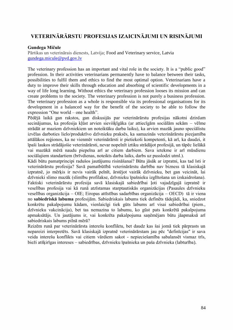
84
VETERINĀRĀRSTU PROFESIJAS IZAICINĀJUMI UN RISINĀJUMI
Gundega Mičule Pārtikas un veterinārais dienests, Latvija; Food and Veterinary service, Latvia
The veterinary profession has an important and vital role in the society. It is a “public good”
profession. In their activities veterinarians permanently have to balance between their tasks,
possibilities to fulfil them and ethics to find the most optimal option. Veterinarians have a
duty to improve their skills through education and absorbing of scientific developments in a
way of life long learning. Without ethics the veterinary profession looses its mission and can
create problems to the society. The veterinary profession is not purely a business profession.
The veterinary profession as a whole is responsible via its professional organisations for its
development in a balanced way for the benefit of the society to be able to follow the
expression “One world – one health”.
Pēdējā laikā gan rakstos, gan diskusijās par veterinārārstu profesijas nākotni dzirdam
secinājumus, ka profesija kļūst arvien sievišķīgāka (ar attiecīgām sociālām sekām – vēlme
strādāt ar maziem dzīvniekiem un noteiktāku darba laiku), ka arvien mazāk jauno speciālistu
izvēlas darboties lielo/produktīvo dzīvnieku praksēs, ka samazinās veterinārārstu pieejamība
attālākos reģionos, ka ne vienmēr veterinārārsti ir pietiekoši kompetenti, kā arī, ka daudzi, it
īpaši laukos strādājošie veterinārārsti, nevar nopelnīt iztiku strādājot profesijā, un tāpēc lielākā
vai mazākā mērā naudu piepelna arī ar citiem darbiem. Sava ietekme ir arī mūsdienu
sociālajiem standartiem (brīvdienas, noteikts darba laiks, darbs uz pusslodzi utml.).
Kādi būtu pamatprincipi radušos jautājumu risināšanai? Būtu jāsāk ar izpratni, kas tad īsti ir
veterinārārstu profesija? Savā pamatbūtībā veterinārārstu darbība nav bizness tā klasiskajā
izpratnē, jo mērķis ir nevis vairāk pelnīt, ārstējot vairāk dzīvnieku, bet gan veicināt, lai
dzīvnieki slimo mazāk (slimību profilakse, dzīvnieku īpašnieku izglītošana un izskaidrošana).
Faktiski veterinārārstu profesija savā klasiskajā sabiedrībai ļoti vajadzīgajā izpratnē ir
veselības profesija vai kā runā atzīstamas starptautiskās organizācijas (Pasaules dzīvnieku
veselības organizācija – OIE; Eiropas attīstības sadarbības organizācija – OECD) tā ir viena
no sabiedriskā labuma profesijām. Sabiedriskais labums tiek definēts tādejādi, ka, sniedzot
konkrētu pakalpojumu kādam, vienlaicīgi tiek gūts labums arī visai sabiedrībai (piem.,
dzīvnieku vakcinācija), bet tas nemazina to labumu, ko gūst pats konkrētā pakalpojuma
apmaksātājs. Un jautājums ir, vai konkrēta pakalpojuma saņēmējam būtu jāapmaksā arī
sabiedriskais labums pilnā mērā?
Reizēm runā par veterinārārsta interešu konfliktu, bet daudz kas šai jomā tiek pārprasts un
nepareizi interpretēts. Savā klasiskajā izpratnē veterinārārstam jau pēc "definīcijas" ir sava
veida interešu konflikts vai citiem vārdiem sakot - nepieciešamība sabalansēt vismaz trīs,
bieži atšķirīgas intereses – sabiedrības, dzīvnieku īpašnieka un paša dzīvnieka (labturība).

85
Sabiedrība
Dzīvnieks Dzīvnieku īpašnieks
Lai īstenotu nepieciešamību sabalansēt šo atšķirīgo iesaistīto pušu intereses, veterinārārstam
jābūt profesionāli neatkarīgam! Bet ne tikai – svarīgas attiecīgas zināšanas un prasmes
(izglītība un nepārtraukta kvalifikācijas celšana, specializācija utml visas dzīves garumā) un –
profesionālā ētika.
Veterinārmedicīnas izglītība
Zinātne LLL
Zināšanas un profesionālā ētika ir tas, kas ļauj veterinārārstam sabalansēt bieži vien krasi
atšķirīgās intereses – sabiedrības (ilgtspējīga attīstība, izvairīšanās no zoonozēm, no
piesārņotas pārtikas), dzīvnieku īpašnieku (ekonomiskais labums, peļņa, atsevišķos gadījumos
emocijas) dzīvnieku (labturība, veselība)! Vienīgi dzīvnieku īpašnieks kā konkrēta persona
var striktāk pastāvēt uz savu interešu pildīšanu. Sabiedrībai un dzīvniekam atliek paļauties uz
veterinārārsta goda prātu. No šī izriet, ka ne vienmēr veterinārārsts var vienkārši pelnīt naudu,
darot to, kas var būt ir iespējams tīri tehniski, bet būtu pārāk lielā pretrunā ar profesionālās
ētikas principiem.
Kas tad ir tas instruments, kas nodrošina šīs neatkarīgās un ētiskās profesionālās uzvedības
īstenošanos biznesa pasaulē? Tās ir prakses tiesības jeb kā mēs šobrīd tās dēvējam Latvijā –
prakses sertifikāts.
Veterinārārsts
86
Uzdevumi/darbi/tehnoloģijas
Prasmes Ētika
Ētikas kodekss
Veterinārārstu reģistrācija (licencēšana/sertifikācija) Veterinārārstu profesijas
disciplīna
Ja veterinārārsts negodprātīgi izmanto savu statusu, sertifikāts var tikt atņemts un attiecīgā
persona vairs nevar veikt veterinārārsta darbu. Latvijā un, godīgi sakot, arī citās valstīs nav
parasts šo instrumentu regulāri izmantot, bet fakts, ka biznesa intereses bieži ņem virsroku pār
veterinārārsta ētiku, liek pārdomāt šo jautājumu un atgādināt par šī instrumenta esamību, lai
stimulētu profesijas mērķu un sabiedriskā labuma sasniegšanu viss optimālākajā veidā.
Šajā ziņā nepārvērtējama ir dažādu valstu veterināro profesionālo organizāciju loma, kas
kolektīvi un profesionāli nosaka profesijas attīstības mērķus sabiedrības labumam, stimulē
attiecīgas veterinārārstu izglītības un specializācijas pieejamību, pieņem profesijas standartus,
kā ari uztur kompetences un profesionālās kvalitātes vērtēšanas līmeni. Tā kā tikai un vienīgi
pašai profesijai ir gan zināšanas gan to pielietošanas vērtēšanas iespējas, tad arī pašai
profesijai ir svēts pienākums uzturēt augstu profesionalitāti savu biedru vidu, saglabājot
neatkarīgu profesionālu lēmumu pieņemšanas iespējas, nepakļaujoties politiskām iegribām.
Te mēs nonākam pie nākamā jautājuma - kas ir tas veterinārārsta darbs, ko var veikt tikai
sertificēts veterinārārsts? Vienkāršotā veidā var teikt, ka tā ir dzīvnieku ārstniecība un zāļu
parakstīšana, bet juridiski nepieciešami precīzāki definējumi (par tiem diskutē un tos iekļauj
Veterinārārsts
Veterinārārsts
87
atsevišķu valstu likumdošanā; Eiropas Veterinārārstu federācija ir pieņēmusi t.s. "Veterinary act"; diskusijas uzsāktas arī Latvijas veterinārārstu vidū). Būtu pēdējais laiks veterinārā darba
precīzākas aprises iekļaut ari Latvijas likumdošanā.
Tagad varam šķetināt tālāk sākumā minētos profesijas izaicinājumus - mūsdienu Latvijā.
Tātad - ir darbi, kurus var veikt tikai veterinārārsts; tie ir saistīti ar veselības jomu (ne tikai ar
dzīvnieku, bet arī ar cilvēku veselību un pat vides nepiesārņošanu) un tie dod sabiedrisko
labumu. Visā Latvijas teritorijā iedzīvotājiem būtu jābūt iespējai saņemt šos veterinārārsta
pakalpojumus (būtu jātiek skaidrībā ar jautājumu - ko uzskatām par
apmierinošu/neapmierinošu iespēju dzīvnieku īpašniekam saņemt veterinārmedicīnisko
pakalpojumu - attālums līdz tuvākam veterinārārstam; laiks, kas paiet līdz veterinārārsts var
ierasties; papildus izmaksas par transportu utml). Ja adekvāta veterinārārstu pakalpojuma
nodrošināšana neizdodas brīvās veterinārārstu prakses un konkurences apstākļos (nav
piemērotu dzīves un darba apstākļu; nav pietiekoši daudz darba, lai nopelnītu iztiku utml.), tad
nepieciešama valsts iejaukšanās - papildus atbalsts, stimuli (piemaksas, dienesta dzīvokļi,
klīnikas īre, nodokļu atlaides vai tml.). Bet viennozīmīgi ir skaidrs, ka ar šādiem stimuliem
jābūt ļoti uzmanīgi izsvērtiem, lai nekropļotu veselīgu konkurenci. Atbalsts būtu jāsniedz par
konkrētām lietām (piem. par dzīvnieku īpašnieku izglītošanu, kvalifikācijas celšanu, pieredzes
apmaiņu, konsultācijām, ganāmpulku veselības plānu sagatavošanu, transporta izmaksām,
veterinārārstu kooperācijas attīstību, trakumsērgas vakcinācijas kampaņu, nodokļu atlaides par
lielo l/s dzīvnieku apkalpošanu, piemaksas par dežūrām brīvdienās un ārpus parastā darba
laika utml). Šāda veida pasākumiem būtu jānodrošina gan veterinārā pakalpojuma pieejamība
dzīvnieku īpašniekiem no vienas puses, gan, kas nav mazāk svarīgi, veterinārārsta iespējas
nopelnīt iztiku tieši ar veterināro darbu. Tas, ka veterinārārstam jāstrādā blakus darbi, lai
nopelnītu iztiku, noved pie tā, ka viņam nav iespējas iegūt pietiekošu profesionālo pieredzi,
samazinās veterinārārsta kompetence un sekojoši darba efektivitāte un rezultāti. Veterinārā
darba trūkums iespējams ne tikai teritorijās ar maz dzīvniekiem, bet ari tādās, kur dzīvnieku
daudz, bet arī veterinārārstu ir daudz. Tādejādi būtu nepieciešama regulāra situācijas
novērošana un ieteikumu izstrāde attiecībā uz optimālo veterinārārstu blīvumu dažādās
teritorijās.
SECINĀJUMI 1. Veterinārārstu profesijai ir sabiedrībai svarīgi uzdevumi, tā ir veselības un sabiedriskā
labuma profesija. 2. Tādejādi, lai vecinātu šo sabiedrisko labumu, veterinārārstiem savā darbībā jāievēro
stingra ētika un šo profesiju nevar uzskatīt par „tīru” biznesa profesiju
3. Veterinārārstiem savā darbībā jāsaglabā līdzsvars starp dažādiem aspektiem, t.sk. starp
veicamo darbu, prasmi veikt šo darbu un ētiku.
4. Sabiedrība ne vienmēr izprot veterinārārstu profesijas veikuma nozīmi, bet tā ir vitāli
atkarīga no veterinārārstu darba un tāpēc atsevišķos gadījumos nepieciešami papildus
stimuli profesijas pieejamībai un sabiedriskā labuma darbības nodrošināšanai.
P.S. shematiskie modeļi veidoti, ietekmējoties no EVF (Federation of Veterinarians of
Europe) notikušajām diskusijā par veterinārārstu profesiju. .
IZMANTOTIE MATERIĀLI: WVA; FVE; OIE; OECD mājas lapas; ES likumdošana par
profesionālās izglītības diploma atzīšanu un pakalpojumu pieejamību.
88
ELKOŅA LOCĪTAVAS DISPLĀZIJA SUŅIEM
CANINE ELBOW DYSPLASIA Armands Vekšins1, Oskars Kozinda1, Kerstin Amort Heike2 1 LLU Veterinārmedicīnas fakultāte, Latvija; Faculty of Veterinary Medicine LUA, Latvia 2 Justus Liebig universitāte, Vācija; Justus Liebig University, Germany
Elkoņa locītavas displāzija pēc IEWG (International elbow working group) definīcijas ietver
vairākas slimības: processus coronoideus medialis fragmentāciju, pleca kaula distālās daļas
osteohondrozi, nesavienojušos processus anconeus, locītavas skrimšļa bojājumus un elkoņa
locītavas nestabilitāti. Elkoņa locītavas displāzija bieži sastopama un aprakstīta labradora
šķirnes suņiem Anglijā ar 17% sastopamību no populācijas un bernes ganu šķirnes suņiem
Nīderlandē ar 70% sastopamību (Hazewinkel et al., 1995). Slimība galvenokārt skar liela
auguma (20 līdz 50 kg), ātri augošus suņus, vecumā no sešiem līdz 12 mēnešiem. Tomēr
slimība aprakstīta arī maza auguma, hondrodistrofiskām šķirnēm, tādām kā takšu šķirnes
suņiem un franču buldogiem (Narojek et al., 2008). Pētījumos minēts, ka suņi tiek skarti divas
reizes biežāk, nekā kuces. Klīniski saslimšana izpaužas suņu intensīvas augšanas periodā
(Meyer-Lindberg et al., 2006).
Pastāv uzskats, ka elkoņa locītavas displāzijas cēlonis ir iedzimtība. Ir vairāki pētījumi, kuros
meklēts ģenētiskais pamats elkoņa locītavas displāzijas attīstībai atkarībā no šķirnes. Ir
pierādījumi, ka dažādas elkoņa locītavas displāzijas izpausmes var pārmantot individuāli. Tā
kā elkoņa locītavas displāzija ir sindroms un tā attīstībā iekļauts gan ģenētiskais faktors, gan
vides un dažādu mehānisku faktoru iedarbība, tad ir maz ticams, ka tuvākajā nākotnē
ģenētiskā izmeklēšana sniegs precīzus rezultātus attiecībā uz slimības attīstības faktoru
noskaidrošanu (Jacob Michelsen., 2012).
Salīdzinoši bieži suņiem novēro elkoņa locītavas neatbilstību. Locītavas neatbilstība iedalāma
trīs tipos: 1) spieķa kaula un elkoņa kaula garuma neatbilstība; 2) pleca kaula un spieķa kaula
locītavas neatbilstība; 3) spieķa kaula un elkoņa kaula ieroba neatbilstība. Neatbilstības
noteikšana, izmantojot parasto rentgenogrāfisko izmeklēšanu, ir relatīvi neprecīza, tādēļ ir
nepieciešami datortomogrāfijas izmeklējumi, kas dod iespēju precīzi novērtēt locītavas
stāvokli un veikt morfometriskos mērījumus (Jacob Michelsen., 2012).
Osteohondroze jeb deģeneratīvs process skrimslī vai kaulā rodas endohondrālās osifikācijas
traucējumu rezultātā, un šī patoloģija var noritēt ar vai bez iekaisuma klātbūtnes.
Osteohondroze elkoņa locītavā ir raksturīga pleca kaula mediālajā epikondīlī, mediālajā
vainagizaugumā un elkoņa izaugumā. Osteohondrozes etiopatoģenēze līdz šim nav pilnībā
izprasta, bet kā izšķirošais faktors ir pieņemts asinsapgādes traucējumi hondrālajās un
subhondrālajās struktūrās.
Neskaitot locītavas inkongruenci un osteohondrozi, viena no biežākajām patoloģijām ir
elkoņa locītavas proc. coronoideus medialis slimības. Rentgenogrāfiski diagnosticēt šīs
mediālā vainagizauguma pārmaiņas ir grūti (Cook and Cook, 2009). Datortomogrāfijas jutība,
lai noteiktu proc. coronoideus medialis slimības ir no 71 līdz 88%, ar vidējo jutību 85%
(Jacob Michelsen., 2012).
Pētījums LLU Veterinārmedicīnas fakultātē LLU Veterinārmedicīnas fakultātei sadarbojoties ar Vācijas Justus Liebig Universitāti Gīsenē,
2014. gadā uzsākts pētījums, kurā plānots noteikt, vai pastāv saistība starp musculus biceps
brachii un musculus brachialis anatomiju un procesus coronoideus medialis slimībām.
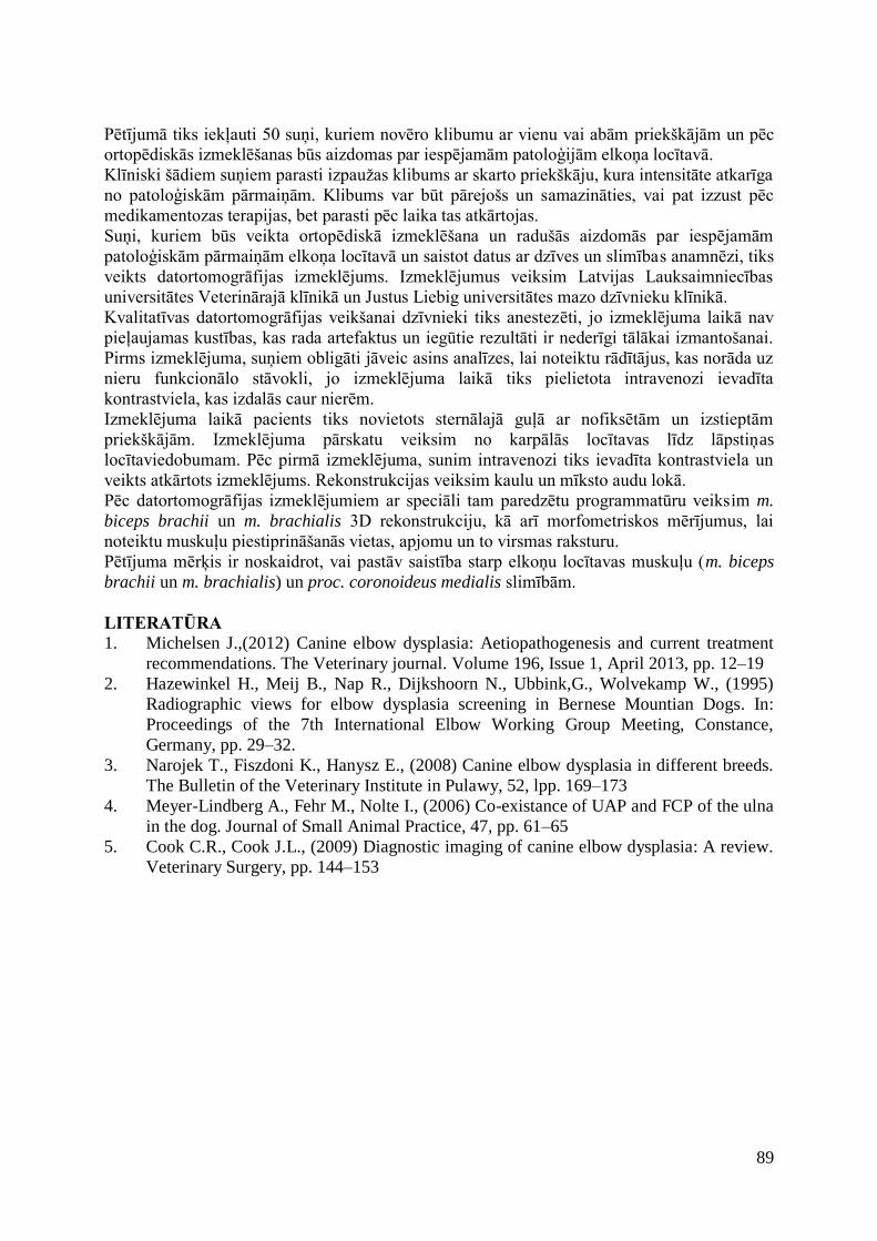
89
Pētījumā tiks iekļauti 50 suņi, kuriem novēro klibumu ar vienu vai abām priekškājām un pēc ortopēdiskās izmeklēšanas būs aizdomas par iespējamām patoloģijām elkoņa locītavā.
Klīniski šādiem suņiem parasti izpaužas klibums ar skarto priekškāju, kura intensitāte atkarīga
no patoloģiskām pārmaiņām. Klibums var būt pārejošs un samazināties, vai pat izzust pēc
medikamentozas terapijas, bet parasti pēc laika tas atkārtojas.
Suņi, kuriem būs veikta ortopēdiskā izmeklēšana un radušās aizdomās par iespējamām
patoloģiskām pārmaiņām elkoņa locītavā un saistot datus ar dzīves un slimības anamnēzi, tiks
veikts datortomogrāfijas izmeklējums. Izmeklējumus veiksim Latvijas Lauksaimniecības
universitātes Veterinārajā klīnikā un Justus Liebig universitātes mazo dzīvnieku klīnikā.
Kvalitatīvas datortomogrāfijas veikšanai dzīvnieki tiks anestezēti, jo izmeklējuma laikā nav
pieļaujamas kustības, kas rada artefaktus un iegūtie rezultāti ir nederīgi tālākai izmantošanai.
Pirms izmeklējuma, suņiem obligāti jāveic asins analīzes, lai noteiktu rādītājus, kas norāda uz
nieru funkcionālo stāvokli, jo izmeklējuma laikā tiks pielietota intravenozi ievadīta
kontrastviela, kas izdalās caur nierēm.
Izmeklējuma laikā pacients tiks novietots sternālajā guļā ar nofiksētām un izstieptām
priekškājām. Izmeklējuma pārskatu veiksim no karpālās locītavas līdz lāpstiņas
locītaviedobumam. Pēc pirmā izmeklējuma, sunim intravenozi tiks ievadīta kontrastviela un
veikts atkārtots izmeklējums. Rekonstrukcijas veiksim kaulu un mīksto audu lokā.
Pēc datortomogrāfijas izmeklējumiem ar speciāli tam paredzētu programmatūru veiksim m.
biceps brachii un m. brachialis 3D rekonstrukciju, kā arī morfometriskos mērījumus, lai
noteiktu muskuļu piestiprināšanās vietas, apjomu un to virsmas raksturu.
Pētījuma mērķis ir noskaidrot, vai pastāv saistība starp elkoņu locītavas muskuļu (m. biceps
brachii un m. brachialis) un proc. coronoideus medialis slimībām.
LITERATŪRA 1. Michelsen J.,(2012) Canine elbow dysplasia: Aetiopathogenesis and current treatment
recommendations. The Veterinary journal. Volume 196, Issue 1, April 2013, pp. 12–19
2. Hazewinkel H., Meij B., Nap R., Dijkshoorn N., Ubbink,G., Wolvekamp W., (1995)
Radiographic views for elbow dysplasia screening in Bernese Mountian Dogs. In:
Proceedings of the 7th International Elbow Working Group Meeting, Constance,
Germany, pp. 29–32.
3. Narojek T., Fiszdoni K., Hanysz E., (2008) Canine elbow dysplasia in different breeds.
The Bulletin of the Veterinary Institute in Pulawy, 52, lpp. 169–173
4. Meyer-Lindberg A., Fehr M., Nolte I., (2006) Co-existance of UAP and FCP of the ulna
in the dog. Journal of Small Animal Practice, 47, pp. 61–65
5. Cook C.R., Cook J.L., (2009) Diagnostic imaging of canine elbow dysplasia: A review.
Veterinary Surgery, pp. 144–153
90
KOPSAVILKUMI
ABSTRACTS
91
MEDĪJAMO DZĪVNIEKU TIESU VETERINĀRMEDICĪNAS EKSPERTĪZES LLU VETERINĀRMEDICĪNAS FAKULTĀTĒ
VETERINARY FORENSIC INVESTIGATIONS OF THE GAME
ANIMALS IN THE FACULTY OF VETERINARY MEDICINE, LUA Dace Bērziņa LLU Veterinārmedicīnas fakultāte, Preklīniskais institūts, Latvija
LUA, Faculty of Veterinary Medicine, Preclinical institute, Latvia
IEVADS. Tiesu veterinārmedicīnas ekspertīzes tiek veiktas, lai risinātu dažādus jautājumus
pārkāpumu izmeklēšanā, kas saistīti ar cietsirdīgu izturēšanos pret dzīvniekiem, nelikumīgām
medībām, aizsargājamo dzīvnieku iznīcināšanu un citos dzīvnieku aizsardzības noteikumu
neievērošanas gadījumos. Arvien aktuālākas kļūst medījamo dzīvnieku veterinārmedicīnas
ekspertīzes, kas noteiktas sakarā ar medību noteikumu pārkāpumiem un neatļautu ieroču
izmantošanu. Šī darba mērķis ir apkopot un izanalizēt pēdējo gadu laikā LLU
Veterinārmedicīnas fakultātē (VMF) veikto medījamo dzīvnieku līķu un to ķermeņa daļu tiesu
veterinārmedicīnas ekspertīžu rezultātus.
MATERIĀLS UN METODIKA. Darbā analizētas VMF veiktās tiesu veterinārmedicīnas
ekspertīzes medījamo dzīvnieku līķiem un to ķermeņu daļām, lai noskaidrotu atbildes uz
kriminālprocesos un administratīvo pārkāpumu lietās uzdotajiem jautājumiem. Dzīvnieka
nāves cēloņa un miesas bojājumu noskaidrošanai veikta līķu patologanatomiskā sekcija vai
ķermeņa daļu morfoloģiskā apskate, kā arī histoloģiskā izmeklēšana miesas bojājumu rašanās
laika noteikšanai. Metālu saturošu svešķermeņu – ložu, skrošu u.c. šāviņu daļu konstatēšanai
izmantota rentgenoloģiskā izmeklēšana.
REZULTĀTI. Kopumā LLU Veterinārmedicīnas fakultātē laika posmā no 2010.gada janvāra
līdz 2014.gada augustam veiktas medījamo dzīvnieku līķu un to ķermeņa daļu tiesu
veterinārmedicīnas ekspertīzes deviņām mežacūkām (Sus scrofa), septiņām stirnām
(Capreolus capreolus), pieciem aļņiem (Alces alces), diviem staltbriežiem (Cervus elaphus)
un vienam jenotsunim (Nyctereutes procyonoides), lai noskaidrotu dzīvnieku sugas piederību,
nāves cēloni, miesas bojājumu lokalizāciju, raksturu, rašanās mehānismu, smaguma pakāpi un
laiku, kā arī noteiktu svešķermeņu klātbūtni dzīvnieku ķermenī. Ekspertīzēm nosūtīti 12
dzīvnieku līķi un 12 dzīvnieku ķermeņa daļas. Patologanatomiskās sekcijas veiktas četru
mežacūku, piecu stirnu, divu aļņu un viena jenotsuņa līķim, kurās noskaidrots, ka astoņi
dzīvnieki miruši šautu ievainojumu dēļ, bet četros gadījumos vardarbīga nāve netika
konstatēta. Nošauto dzīvnieku nāves cēlonis bija ložu radīti miesas bojājumi, kas,
galvenokārt, konstatēti dzīvnieku krūšu un vēdera dobuma orgānos, izraisot nāvi ne vairāk kā
divu līdz trīs stundu laikā. Četros gadījumos dzīvnieka ķermenī konstatēti svešķermeņi –
lodes, kas nodotas kā lietiskais materiāls tālākai izmeklēšanai. Dzīvnieka ķermeņa daļu
veterinārmedicīnas ekspertīzes veiktas dzīvnieku galvām, ādām, iekšējiem orgāniem, kaulu un
muskuļu fragmentiem. Pēc sugas piederības noteikšanas konstatēts, ka uz ekspertīzi nosūtītas
piecu mežacūku, divu stirnu, triju aļņu un divu staltbriežu ķermeņa daļas. Sešās ekspertīzēs
dzīvnieka ķermeņa daļās konstatēti ložu radīti ievainojumi, galvenokārt, šautas ādas brūces un
muskuļu bojājumi, bet divās ekspertīzēs – skrošu radīti ievainojumi un skrotis ķermeņa daļās.
Trijās dzīvnieka ķermeņa daļu ekspertīzēs konstatēti galvaskausa lūzumi un galvas smadzeņu
ievainojumi bez šautiem ādas bojājumiem, kas varētu būt radušies trula priekšmeta iedarbības
92
rezultātā. Vienā ekspertīzē pēc morfoloģiskās apskates dzīvnieku ķermeņa daļās netika konstatēti dzīves laikā radušies miesas bojājumi.
SECINĀJUMI LLU Veterinārmedicīnas fakultātē laika posmā no 2010.gada janvāra līdz 2014.gada
augustam veiktas 24 medījamo dzīvnieku tiesu veterinārmedicīnas ekspertīzes, galvenokārt,
mežacūkām un stirnām, kā arī vairākiem aļņiem, staltbriežiem un vienam jenotsunim.
Ekspertīzēs izmeklēto dzīvnieku izplatītākais nāves cēlonis saistīts ar ložu radītiem
ievainojumiem. Dažos gadījumos dzīvnieku ķermeņa daļās konstatētas smagas galvas traumas
bez šautiem ādas bojājumiem.
93
TRICHINELLA SPECIES VARIETY IN PINE MARTEN (MARTES
MARTES) AND STONE MARTEN (MARTES FOINA) IN LATVIA AND LITHUANIA (KAUNAS REGION)
Zanda Bērziņa1,2, Inese Jahundoviča2, Muza Kirjušina2 1 Pārtikas drošības, dzīvnieku veselības un vides zinātniskais institūts „BIOR”, Latvija;
Institute of Food Safety, Animal Health and Environment „BIOR”, Latvia; 2 Sistemātikās Bioloģijas Institūts, Daugavpils Universitāte, Latvija; Institute of Systematic
Biology, Daugavpils University, Latvia
INTRODUCTION. Trichinella species are worldwide distributed zoonotic agents and
carnivores, as pine marten and stone marten, are common wildlife reservoirs for this
nematode. In wild and domestic animals from Latvia and Lithuania detected Trichinella
britovi, T. nativa, T. spiralis and T. pseudospiralis. Aim of this study was to obtain the
Trichinella species prevalence and diversity in stone and pine martens in Latvia and
Lithuania.
MATERIALS AND METHODS. Samples collected in hunting seasons from 2011 till 2014
in different regions of Latvia and in Kaunas region, Lithuania. Animals were killed by
licensed hunters accordingly to Latvian hunting law (MK rules No 760) and Lithuania hunting
law (LR MEDŽIOKLĖS ĮSTATYMAS Nr. IX-966). From each sampled animal (n=142)
collected muscle tissues from foreleg and stored frozen at -20° C until further analyzing.
Trichinella larvae detection was conducted separately for each animal. At least 25 g muscle
tissue was analyzed according to European Union Commission Regulation with reference
method (EC No 2075/2005). The number of Trichinella larvae per gram of muscle tissues (l.p.g.) was determined and larvae were collected and stored in 96% ethyl alcohol for further analyses. Trichinella species were identified by multiplex-polymerase chain reaction
(PCR) using species specific primers.
RESULTS AND CONCLUSIONS. Trichinella sp. prevalence in Martes sp. is 48.6%: in
Lithuania (Kaunas region) 20.0% but in Latvia 52.0%. In Latvia detected higher Trichinella
sp. prevalence in Martes sp. than in Lithuania (Kaunas region). There is no difference in
Trichinella sp. prevalence in pine marten (49.2%) and stone marten (45.8%) in Latvia and
Lithuania (Kaunas region). Trichinella sp. intensity is higher in pine marten (0.04-49.88
l.p.g.) than in stone marten (0.04-17.92 l.p.g.).
In Trichinella identification determined three species: T. britovi, T. nativa and T. spiralis.
There is higher Trichinella sp. diversity in Latvia than in Lithuania (Kaunas region).
Trichinella nativa is only common in mix invasion with T. britovi. The widespread species in
Latvia and Lithuania (Kaunas region) is T. britovi.
94
CYSTICERCOSIS: CURRENT SITUATION IN LATVIA
Gunita Deksne1, Zanda Esīte1, Bettija Ligere1, Evita Leitāne2 1 Institute of Food Safety, Animal Health and Environment “BIOR”, Latvia 2 Food and Veterinary service, Latvia
INTRODUCTION. The term cysticercosis refers to foodborne zoonotic infections with
larval tapeworms. The importance features of these zoonoses are that the larvae are meat-
borne and the adult stage develops only in the intestine of the human host. Taenia saginata
(beef tapeworm) and Taenia solium (pork tapeworm) are the most important causes of
taenisosis in humans. Cysticercosis is a tissue infection with the larval cysticercus or
metacestode stage, and occurs most commonly in pigs and cattle. Bovine cysticercosis, a
cosmopolitan disease caused by T. saginata, leads to economic losses due to carcass
devaluation at slaughter. Sanitary inspection at slaughterhouses, the routine diagnostic method
in Latvia, lacks the necessary sensitivity to detect the mildly infected cattle.
In time period from 2009 there are no positive findings for bovine cysticercosis. However,
during time period from 2003 there are seven human taenisosis/cysticercosis cases diagnosed
in Latvia.
The aim of present study was to assess the knowledge of meat inspectors through
questionnaires about cysticercosis and its causative agent, mainly T. saginata (Cysticercus
bovis). However, preliminary studies of T. saginata cysticercus seroprevalence in cattle were
set up.
MATERIALS AND METHODS. Questionnaire with 16 questions was developed for meat
inspectors to assess their knowledge about cysticercus biology, factors affecting transmission,
experience in diagnosing pathogen during routine sanitary expertise in cattle.
For seroprevalence study cattle `blood samples were collected within other National
surveillance programs. All animals were older than 24 month of age from different sex and
breeding type.
Viable metacestodes of Taenia spp. bovine serum samples were prepared with the use of a
monoclonal antibody-based sandwich ELISA developed in Institute of Tropical Medicine,
Department of Biomedical sciences Veterinary Helminthology, Antwerp, Belgium.
RESULTS. Overall 96 respondents, mainly meat inspectors, filled anonymous
questionnaires. During their practice 36.5 % of respondents at least once have detected viable
cysts in cattle (10.4%) or pigs (20.8%) or in both (6.3%). Meanwhile, 81.3% of respondents
consider that there is a risk for humans to acquire cysticercosis/taenisosis infection in Latvia
via consumption of pork, beef or both.
Seroprevalence study is still ongoing.
CONCLUSIONS. It would be useful to improve the detection sensitivity of infected
carcasses by investigating the use of risk-based meat inspection procedures and to improving
awareness of taenisosis/cysticercosis among veterinarians, meat animal producers and the
public through information and reiterative education.
95
STAPHYLOCOCCUS AUREUS IZPLATĪBA MĀJĀS RAŽOTOS PIENA PRODUKTOS
THE PREVALENCE OF STAPHYLOCOCCUS AUREUS IN HOME
MADE MILK PRODUCTS
Daiga Gāliņa, Anda Valdovska LLU Veterinārmedicīnas fakultāte, Latvija; Faculty of Veterinary Medicine LUA, Latvia
IEVADS. Latvijā tradicionāli ir attīstīta piena un piena produktu ražošana piemājas
saimniecībās, kā arī ir atļauta šo produktu tirdzniecība atklātajās tirdzniecības vietās un tirgos.
Vairums piemājas saimniecībās pielieto produktu pagatavošanas tehnoloģijas, kas paredz
izmantot nepasterizētu pienu. Galvenais avots piena kontaminācijai ar stafilokokiem, īpaši ar
Staphylococcus aureus, ir subklīnisks mastīts, kas ir daudzu ganāmpulku problēma.
Sekundāra loma S. aureus izplatībā ir piena produktu pagatavošanas tehnoloģijai,
uzglabāšanas un realizācijas apstākļiem, tāpēc darba mērķis bija noskaidrot S. aureus
izplatību pienā un mājās ražotos piena produktos dažādu tehnoloģisko procesu ietekmē.
MATERIĀLS UN METODIKA. Pētījumā izmantoti 45 piena un piena produktu paraugi
(krējums, biezpiens, biezpiena siers un svaiga piena siers) no 9 LB šķirnes govīm. SŠS
noteikšanai piena paraugi tika nosūtīti uz A/s “Latgales ciltslietu un mākslīgās apsēklošanas
staciju” piena analīžu laboratoriju. Paraugu sagatavošana mikrobioloģiskām pārbaudēm veikta
saskaņā ar LVS EN ISO 6887-5:2011E prasībām. Kopējā baktēriju skaita un S. aureus
klātbūtnes un skaita noteikšanai izmantotas vispārpieņemtās mikrobioloģijas metodes.
REZULTĀTI. Izvērtējot piena kvalitāti pēc SŠS, 56% paraugos tas pārsniedz 400 tūkst/ml.
Pētījumā konstatējām, ka vidējais kopējais baktēriju skaits pienā, krējumā, biezpienā,
biezpiena sierā un svaiga piena sierā ir attiecīgi 4.6 log10 kvv ml-1; 6.1 log10 kvv ml-1; 9.3
log10 kvv ml-1; 7.9 log10 kvv ml-1 un 3.5 log10 kvv ml-1.
S. aureus konstatējām 11% piena paraugos, turklāt visos no tiem pārsniedza normatīvajos
aktos noteikto pieļaujamo daudzumu. No mājās ražotajiem piena produktiem, S. aureus
visbiežāk tika konstatēts biezpienā (44% paraugos), krējumā un biezpiena sierā (22%
paraugos). S. aureus skaits, salīdzinot ar svaiga piena rezultātiem, būtiski pieauga biezpienā
(vidēji 10 reizes), kaut gan no tā gatavotā biezpiena sierā būtiski samazinās (vidēji 6 reizes).
Nevienā piena un piena produktu paraugā netika konstatēts S. aureus skaits virs 105 kvv ml-1.
Svaiga piena siera paraugos S. aureus netika konstatēts.
SECINĀJUMI. Vairums gadījumā piens (56% paraugu) neatbilst kvalitātes kritērijam,
vērtējot pēc SŠS, kas liecina par subklīniska mastīta esamību ganāmpulkos. Visos piena
paraugos ir atbilstošs kopējais baktēriju skaits, norādot par sanitāri - higiēnisko noteikumu
ievērošanu piena iegūšanas laikā. Baktēriju kopskaita izmaiņas piena produktos ir atkarīgas no
tehnoloģijas procesā pielietotās temperatūras, jo svaiga piena sierā tas ir būtiski mazāks kā
pienā, taču biezpiena sierā - būtiski augstāks. S. aureus skaits pienā un piena produktos
nepārsniedz 105 kvv ml-1, kas radītu aizdomas par enterotoksīna klātbūtni produktos. S. aureus
izplatība piena produktos ir atkarīga no izejvielas kontaminācijas un pārstrādes procesā
pielietotās temperatūras, jo, temperatūrā, kas ir tuva 100 0C, S. aureus tiek inaktivēts, bet 60 –
70 oC tas saglabājas.
96
MEANING OF CLINICAL SIGNS AND BLOOD HORMONE MEASUREMENTS, TO DETERMINE HYPERADRENOCORTICISM IN
FERRETS (Mustela putorius furo)
Silva Grīnblate, Aija Ilgaža Faculty of Veterinary Medicine LUA, Preclinical Institute, Latvia
INTRODUCTION. There are many research studies about hyperadrenocorticism (excessive
adrenal production of sex steroids related to adrenocortical adenomas and carcinomas) that is
a common disease in neutered male and female pet ferrets (Mustela putorius furo) and causes
significant morbidity. The chronic elevation in circulating luteinizing hormone (LH) that
follows ovariectomy or orchiectomy is a prerequisite for neoplastic transformation in the
adrenal glands. Without specific examination the detecting of adrenal disease can be difficult.
This is a pending matter in Europe and all over the globe, where ferrets are held as pets.
Incidence of ferret adrenal disease in Latvia is high because majority of them are
gonadectomized.
The aim of this research study was to evaluate clinical signs and blood hormone
measurements, to determine hyperadrenocorticism in ferrets.
MATERIALS AND METHODS. To find out the most relevant clinical signs of
hyperadrenocorticism 28 (n=15 male, n=13 female) pet ferrets, diagnosed with adrenal
disease, were included in this research (age 3 – 6 years). Full clinical examination was
performed, and from 10 ferrets out of 28 blood samples were taken, and levels of 17-
hydroxyprogesterone, estradiol and androstendione were detected. Hormonal values were
compared with adrenal panel (The University of Tennessee). To establish clinical signs,
noticed by ferret owners, a questionnaire was developed, and 10 owners were interviewed.
RESULTS. Blood sample test results indicated abnormal hormonal levels in all the animals.
The results of interviews showed that main clinical signs were – alopecia, observed to all
animals in various degrees, fragile skin, behavioral changes (ferrets became lethargic), weight
loss, recurrence of sexual behavior (vulvar swelling in females, mating aggression in males),
poliuria and polidipsia, pruritus and scaling (observed in most of ferrets). In individual
animals numerous clinical signs were observed. We would like to point out that in each ferret
approximately 4 – 6 clinical signs were noticed.
CONCLUSIONS. Most common clinical signs were alopecia, pruritus and behavioral
changes. Hormonal levels were abnormal in all animals, included in this research. Even if the
level is abnormal in one of the hormones, diagnosis hyperadrenocorticism is most likely to
occur. The most sensitive of sex steroids is androstendione. In this research androstendione
level was abnormal in all blood samples. Clinical signs and blood hormone measurements are
equally significant to diagnose hyperadrenocorticism.
97
COMPARATIVE STUDY OF GLUCOSE TRANSPORTERS GLUT-2 AND GLUT-5 IN OSTRICHES GASTROINTESTINAL TRACT
Piret Hussar1, Aleksandra Rotmistrova2, Ilmārs Dūrītis3, Martin Kärner2, Tõnu Järveots4, Arnis Mugurevičs3
1Institute of Anatomy, University of Tartu, Estonia 2Faculty of Science and Technology, University of Tartu, Estonia 3Faculty of Veterinary Medicine, Latvian University of Agriculture, Latvia 4Institute of Veterinary Medicine and Animal Sciences, Estonian University of Life Sciences,
Estonia
INTRODUCTION. The knowledges about transport of sugars in animals and birds
gastrointestinal tract are very important for science as carbohydrates are the main energy
source of food. As up to now there is few information about the localization of glucose
transporters in birds gastrointestinal tract the aim of the present study was to detect the
localization of glucose transporters GLUT-2 and GLUT-5 in different parts of ostriches
gastrointestinal tract comparatively in ostriches chicken after hatching and in 30 day old
ostriches.
MATERIAL AND METHODS. Material from three parts of gastrointestinal tract -
superficial gland zone of proventriculus, duodenum and the terminal zone of ileum - was
collected from two ostriches immediately after hatching and three 30 days old female
ostriches. Specimen were fixed with 10% formalin, embedded in paraffin, slices 7 μm thick
were cut followed by immunohistochemical staining. Rabbit anti-GLUT-2 and Rabbit anti-
GLUT-5 served as primary antibodies. Immunohistochemical staining was carried out
according to the manufacturers guidelines (IHC kit, Abcam, UK).
RESULTS. In ostriches chicken after hatching the staining for glucose transporters GLUT-2
and GLUT-5 occured to be very weak: positively was stained only the cytoplasm of some
epithelial and glandular cells of proventriculus and the epithelial cells of the apical parts of
intestinal villi. The brush border membranes of the villi were stained weakly and the Goblet
cells in the epithelium of small intestine were mostly unstained.
In 30 day old ostriches epithelial cells of proventriculus, the brush border of enterocytes as
well as the Goblet cells in the small intestine were stained strongly positively for GLUT-2 and
GLUT-5. Compared to GLUT-2 the terminal zone of ileum was more intensively stained by
GLUT-5.
CONCLUSIONS. The investigation provided comparative information about the localization
of GLUT-2 and GLUT-5 in gastrointestinal tract in ostriches of different age groups which is
a prerequisite for the knowledges about the transepithelial transport of sugars.
Staining for GLUT-2 and GLUT-5 occured to be weaker in different parts of gastrointestinal
tract of ostriches just after hatching compared to 30 day old ostriches which shows that the
gastrointestinal tract of ostriches immediately after hatching is not yet entirely able for
transportation of carbohydrates, hence the ostriches chickens mostly begin to eat about 3 days
after hatching.

98
STAPHYLOCOCCUS AUREUS SASTOPAMĪBA CŪKU KAUTUVĒS
OCCURRENCE OF STAPHYLOCOCCUS AUREUS IN PIG SLAUGHTERHOUSES
Meldra Ivbule1, Anda Valdovska2
1Pārtikas un veterinārais dienests, Latvija; Food and Veterinary Service, Latvia 2LLU Veterinārmedicīnas fakultāte, Latvija, Faculty of Veterinary Medicine LUA, Latvia [email protected]
IEVADS. Stafilokoki ir plaši izplatīti vides mikroorganismi un ir dzīvnieku un cilvēku ādas
mikrofloras dabiska sastāvdaļa. Tie ir izturīgi pret izžūšanu un apkārtējā vidē saglabājas
mēnešiem ilgi. S. aureus īpatnība veidot rezistenci pret dažādiem antibiotiku veidiem
vienlaicīgi ir viens no iemesliem infekciju plašiem uzliesmojumiem un izplatībai.
Lauksaimniecības sektors, it īpaši cūkkopība, kur pēdējos gados pēc ES valstu datiem S.
aureus sastopamība pieaug, ir viena no sfērām, kas veicina mikroorganismu antibiotiku
rezistences pieaugumu.
S. aureus ir viens no biežāk sastopamajiem pārtikas toksikoinfekciju ierosinātājiem, kas
izraisa gastroenterītu, kā arī mīksto audu infekcijas un septiskus procesus pacientiem
slimnīcās pēcoperāciju nodaļās. Kaušanas procesā var notikt liemeņu kontaminācija un
personāla inficēšanās no dzīvniekiem, kas ir S. aureus nēsātāji. Pētījuma mērķis bija noteikt S.
aureus izplatību cūku kautuvēs.
MATERIĀLS UN METODIKA. Paraugi tika iegūti no četrām kautuvēm ar dažādu
kaušanas jaudu. Divās kautuvēs kaušanas jauda bija zema (15-20 cūkas dienā), bet otrās divās
kautuvēs - augsta kaušanas jauda (100-300 cūkas dienā). Kautuvēs atsarošanu veica divējādi –
ar plaucēšanas un svilināšanas palīdzību. Tika iegūti 23 vides paraugi, 100 – nazālie un
rektālie paraugi pirms atsarošanas un 80 paraugi no liemeņiem pēc liemeņu pēdējās
skalošanas ar ūdeni. Mikrobioloģiskie gaisa paraugi iegūti, izmantojot Koha sedimentācijas
metodi. S. aureus izolēšana un identificēšana tika veikta ar mikrobioloģiskajām un
molekulārbioloģiskajām metodēm.
REZULTĀTI. S. aureus tika konstatēts 55% nazālajos un 48% rektālajos paraugos. S. aureus
sastopamība cūku paraugos pirms atsarošanas variē no 40-88%, bet liemeņos no 5%-28%.
Tika novērota tendence kautuvēs: jo lielāka kaušanas jauda, jo lielāka liemeņu kontaminācija
ar S. aureus. 15.5% gadījumu S. aureus tika konstatēts cūku liemeņu paraugos, kam veikta
atsarošana ar plaucēšanu, bet tikai 3% liemeņu, kam atsarošana veikta ar svilināšanu. Tikai
divos vides paraugos tika konstatēts S. aureus (n=23) – plaucēšanas vannas paraugā no
kautuves ar zemu kaušanas jaudu un eviscerācijas zonas grīdas paraugā no kautuves ar augstu
kaušanas jaudu.
SECINĀJUMI. 1. Ievērojot HACCAP principus, kautuvēs ir iespējams samazināt liemeņu un vides
kontamināciju ar S. aureus no cūkām, kas ir S. aureus nēsātājas.
2. S. aureus biežāk sastopams liemeņos, kam atsarošana veikta ar plaucēšanas metodi.
99
ANALYSIS OF DOGS’ HIP DISEASES
Dalia Juodzente, Ligita Zorgevica – Pockevica, Vita Riskeviciene Lithuanian University of Health Sciences, Veterinary Academy, Faculty of Veterinary
Medicine
INTRODUCTION. Hip malformations and diseases are very common problem that occurs
not only for pure breed dogs, but also for crossbreeds. That’s why, the aim of this work was to
determine the influence of hip diseases in manifestation of lameness.
MATERIALS AND METHODS. Research was carried out for dogs, which, because of
different reasons, were lame with hind limbs. Orthopedic and X-ray investigations were
carried out for 92 dogs. There were evaluated the influence of age (till 1 years, 1-5 years, 6-10
years, 11 years and older), breed (pure breed, crossbreed), sex (male, female), body condition
(1, 2, 3, 4, 5 degree), degree of lameness (0, 1, 2, 3, 4, 5) and the period of lameness (till 1
week, from 1 week to 1 month, from 1 month to 6 months, 6 months and more) in
manifestation of hip diseases. Statistical analysis was performed using statistical data
processing programme SPSS package version No 15 (2009).
RESULTS. Hip pathologies were found in 81,5 percent of dogs (n=92). Hip dysplasia was
found for 56 percent of dogs (n=42). Aseptic femoral head necrosis (14,7 percent, (n=11)),
acetabulum fractures (12 percent, (n=9)), hip luxation (9,3 percent (n=7)) were rarely
diagnosed. Both, femoral neck fractures and tumors in hip bones were diagnosed for 4 percent
of dogs (n=3). Dogs‘ age (1-5 years) had an impact on the occurence of hip dysplasia (54,8
percent, p<0,05) and aseptic femoral head necrosis (till 1 years of age) (72,7 percent, p<0,05).
Hip luxation, acetabulum fractures, femoral neck fractures and tumors of hip bones were
usually diagnosed in 1-5 years old dogs (respectively 71,4 percent and n=7, 77,8 percent and
n=9, 66,7 percent and n=3, 66,7 percent and n=3). Hip dysplasia was usually found in large
breed dogs, aseptic femoral head necrosis – in small breed dogs. Hip luxation, acetabulum
fractures, tumors of hip bones were usually diagnosed in pedigree dogs, while femoral neck
fractures – in crossbreed dogs. Excess body weight in dogs with hip dysplasia had an impact
on the development of hip dysplasia (71,4 percent, (n=42) p<0,05). Lameness time had an
impact in manifestation of lameness degree for dogs with aseptic femoral head necrosis, hip
luxation and acetabulum fractures. The longer the dog was lame, the higher degree of
lameness was determine (p<0,05). Dogs‘ with hip dysplasia usually had the 3rd lameness
degree (59,5 percent), 42,9 percent of dogs were lame for 6 months. Dogs‘ with femoral neck
fracture usually had the 4th lameness degree (66,7 percent), and were lame for 1 week.
CONCLUSIONS. Dogs‘ age (1-5 years), size (large breed), body weight (excess) had an
impact on the occurence of hip dysplasia, (p<0,05). Aseptic femoral head necrosis usually
occurs to young (till 1 years o fage), small breed dogs (p<0,05). Hip luxation, acetabulum
fractures, femoral neck fractures and tumors of hip bones were diagnosed mostly to 1-5 years
old dogs. Lameness time had an impact in manifestation of lameness degree.
Hip dysplasia had an impact in manifestation of cranial cruciate ligament rupture in 17 dogs
(40,5 percent) (n=45) (p<0,05).
100
DETERMINANTS OF CLINICAL OUTCOME OF CUTANEOUS MAST CELL TUMOR IN 15 DOGS (2012-2014)
Linda Kokoreviča1, Ilze Matīse-Van Houtana2 1 Faculty of Veterinary Medicine LUA, Latvia 2 Veterinary Pathology Service, Latvia
INTRODUCTION. Mast cell tumor (MCT) is second most common malignancy in dogs.
The biological behavior of MCT is variable therefore a lot of effort is taken to find
prognostically relevant factors. Some microscopic features correlate with clinical outcome of
MCT, therefore several histological grading systems have been developed. The aim of our
study was to assess prospectively the clinical outcome of MCT in Latvian dogs depending on
completeness of surgical excision and histopathological grade using newest proposed grading
system by M.Kiupel et al. (2011).
MATERIALS AND METHODS. Fifteen client-owned dogs diagnosed with MCT were
enrolled in the prospective study. Histological grading was done according to scheme
proposed by M.Kiupel et al. according to which MCT is assigned to one of two grades – low
or high depending on quantifiable microscopic criteria. The surgical margins were evaluated
as complete, close or incomplete if they were more than 3 mm, 1 to 3 mm or less than 1 mm
wide, respectively. The animals were followed for at least 12 months after surgery. Follow-up
included clinical examination of tumor area and draining lymph nodes and abdominal
ultrasound. The animal use was permitted by Food and Veterinary Service of Latvia (license
No. 45).
RESULTS. There were 9 (60%) low grade tumors and 6 (40%) high grade tumors. The tumor
control rate 12 months after initial surgery (no recurrence) was 67% for low grade and only
17% for high grade tumors. Median time to tumor recurrence was 304 days for low grade and
27 days for high grade MCT. Survival time for dogs with low grade MCT was 409 days
compared with only 60 days for dogs with high grade tumor. Dogs with high grade tumors
had significantly shorter survival time and worse prognosis than dogs with low grade tumors
(p=0.013).
Median time to MCT recurrence and median survival time was 326 and 376 days for complete
excision, 129 and 60 days for marginal and 32 and 55 days for incomplete excision,
respectively. Complete excision was associated with lower possibility and longer time to
tumor recurrence, as well as longer survival, however marginal surgical border status did not
have significant impact on time to tumor recurrence and survival. It was found that tumor
duration but not tumor size had significant impact on surgical margin status (p=0.047).
However, tumor size significantly affected survival time with larger tumors being correlated
with shorter survival (p=0.03).
It was also found that median tumor size (4.3 cm in diameter) was grater and median tumor
duration (121 days) in Latvian dogs was longer than reported previously in the literature.
These data suggest that surgeries for MCT in Latvia are delayed. CONCLUSIONS. The results of our study confirm that grade, tumor size and recurrence are
significant factors for MCT prognostication. Surgical excision of MCT must be done as soon
as tumor is detected. Determination of histological grade of the tumor is essential for clinician
to determine prognosis and to choose appropriate additional treatment.
101
CLINICAL OUTCOMES OF 17 LATVIAN CATS WITH INJECTION SITE SARCOMA TREATED WITH SURGERY ALONE
Linda Kokoreviča1, Ilze Matīse-Van Houtana2 1 Faculty of Veterinary Medicine LUA, Latvia 2 Veterinary Pathology Service, Latvia
INTRODUCTION. Injection site sarcomas (ISS) are malignant cutaneous tumors of cats that
develop as rare side effect after administration of vaccines and long-acting medications. ISS
are commonly diagnosed in Latvia which may be attributed to mandatory annual vaccination
against rabies until May 2013 and available vaccines. In the last three years 97% of sold
rabies vaccines for which cats are one of target species were inactivated vaccines with
aluminum salts or aluminum alkaline adjuvants. Aluminum compounds are thought to be
associated with ISS development. Estimated incidence of ISS in Latvia in 2013 was 7.5 per
10 000 feline vaccinations against rabies, and ISS were 14% of all feline biopsy submissions.
The aim of this study was to evaluate the clinical outcome of cats with ISS in Latvia where
only treatment modality available is surgery.
MATERIALS AND METHODS. Seventeen client-owned cats with ISS were prospectively
followed after surgical removal of the tumor between 2010 and 2014. Follow-up consisted of
periodic clinical examination of tumor area and 3-view thoracic radiography. All cats were
followed for at least 1 year unless they died earlier. Median follow-up period was 286 days
(range 135-1030). The animal use in this study was permitted by Food and Veterinary Service
of Latvia (Food and Veterinary Service license No. 45 for animal use in the experiment).
RESULTS. In 12/14 (86%) cats from which entire tumor was submitted surgical margins
were incomplete. In 11/17 (65%) cats tumor recurred after surgery; median time to recurrence
was 160 days (range 42-358). Two months after surgery 80% of cats were tumor-free, 5 and
12 months after – 53% and 33% respectively. Eight of 17 (47%) cats were euthanized due to
progression of ISS; median survival time was 400 days. In those cats that developed
recurrence and were treated with repeated surgery (n=6), survival time was longer (median
543 days; range 178-1030) than in cats that did not have repeated surgery (n=5; median 293
days; range 135-388).
CONCLUSIONS. ISS is common tumor in Latvian cat population that is characterized by
high recurrence rate that leads to decreased survival time in affected cats. Repeated surgery
may be an effective local treatment after ISS recurrence.
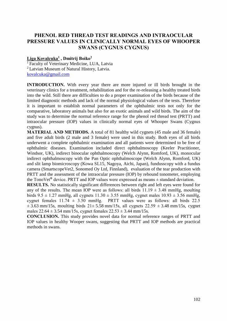
102
PHENOL RED THREAD TEST READINGS AND INTRAOCULAR PRESSURE VALUES IN CLINICALLY NORMAL EYES OF WHOOPER
SWANS (CYGNUS CYGNUS)
Liga Kovalcuka1 , Dmitrij Boiko2
1 Faculty of Veterinary Medicine, LUA, Latvia 2 Latvian Museum of Natural History, Latvia.
INTRODUCTION. With every year there are more injured or ill birds brought in the
veterinary clinics for a treatment, rehabilitation and for the re-releasing a healthy treated birds
into the wild. Still there are difficulties to do a proper examination of the birds because of the
limited diagnostic methods and lack of the normal physiological values of the tests. Therefore
it is important to establish normal parameters of the ophthalmic tests not only for the
comparative, laboratory animals but also for an exotic animals and wild birds. The aim of the
study was to determine the normal reference range for the phenol red thread test (PRTT) and
intraocular pressure (IOP) values in clinically normal eyes of Whooper Swans (Cygnus
cygnus).
MATERIAL AND METHODS. A total of 81 healthy wild cygnets (45 male and 36 female)
and five adult birds (2 male and 3 female) were used in this study. Both eyes of all birds
underwent a complete ophthalmic examination and all patients were determined to be free of
ophthalmic diseases. Examination included direct ophthalmoscopy (Keeler Practitioner,
Windsor, UK), indirect binocular ophthalmoscopy (Welch Alynn, Romford, UK), monocular
indirect ophthalmoscopy with the Pan Optic ophthalmoscope (Welch Alynn, Romford, UK)
and slit lamp biomicroscopy (Kowa SL15, Nagoya, Aichi, Japan), fundusscopy with a fundus
camera (SmartscopeVet2, Sonomed Oy Ltd, Finnland), evaluation of the tear production with
PRTT and the assessment of the intraocular pressure (IOP) by rebound tonometer, employing
the TonoVet® device. PRTT and IOP values were expressed as means ± standard deviation.
RESULTS. No statistically significant differences between right and left eyes were found for
any of the results. The mean IOP were as follows: all birds 11.19 ± 3.48 mmHg, moulting
birds 9.5 ± 1.27 mmHg, all cygnets 11.30 ± 3.55 mmHg, cygnet males 10.93 ± 3.56 mmHg,
cygnet females 11.74 ± 3.50 mmHg. PRTT values were as follows: all birds 22.5
± 3.63 mm/15s, moulting birds 21± 5.58 mm/15s, all cygnets 22.59 ± 3.48 mm/15s, cygnet
males 22.64 ± 3.54 mm/15s, cygnet females 22.53 ± 3.44 mm/15s. CONCLUSION. This study provides novel data for normal reference ranges of PRTT and
IOP values in healthy Wooper swans, suggesting that PRTT and IOP methods are practical
methods in swans.
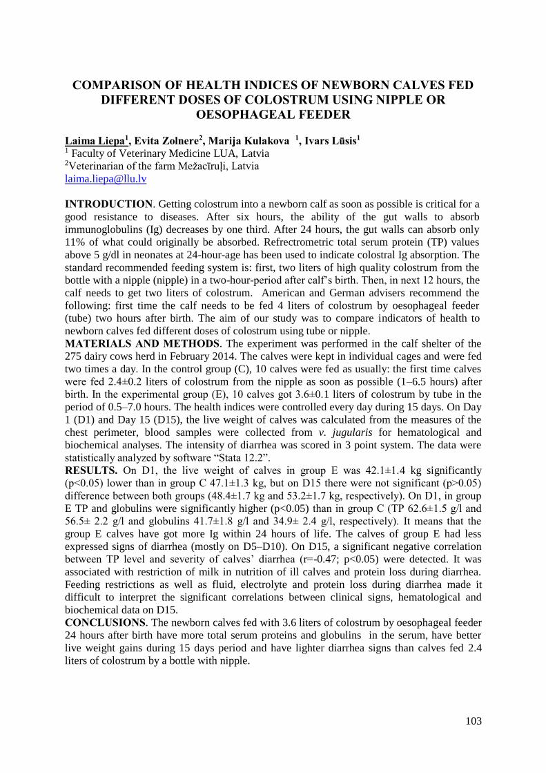
103
COMPARISON OF HEALTH INDICES OF NEWBORN CALVES FED DIFFERENT DOSES OF COLOSTRUM USING NIPPLE OR
OESOPHAGEAL FEEDER
Laima Liepa1, Evita Zolnere2, Marija Kulakova 1, Ivars Lūsis1 1 Faculty of Veterinary Medicine LUA, Latvia 2Veterinarian of the farm Mežacīruļi, Latvia
INTRODUCTION. Getting colostrum into a newborn calf as soon as possible is critical for a
good resistance to diseases. After six hours, the ability of the gut walls to absorb
immunoglobulins (Ig) decreases by one third. After 24 hours, the gut walls can absorb only
11% of what could originally be absorbed. Refrectrometric total serum protein (TP) values
above 5 g/dl in neonates at 24-hour-age has been used to indicate colostral Ig absorption. The
standard recommended feeding system is: first, two liters of high quality colostrum from the
bottle with a nipple (nipple) in a two-hour-period after calf’s birth. Then, in next 12 hours, the
calf needs to get two liters of colostrum. American and German advisers recommend the
following: first time the calf needs to be fed 4 liters of colostrum by oesophageal feeder
(tube) two hours after birth. The aim of our study was to compare indicators of health to
newborn calves fed different doses of colostrum using tube or nipple.
MATERIALS AND METHODS. The experiment was performed in the calf shelter of the
275 dairy cows herd in February 2014. The calves were kept in individual cages and were fed
two times a day. In the control group (C), 10 calves were fed as usually: the first time calves
were fed 2.4±0.2 liters of colostrum from the nipple as soon as possible (1–6.5 hours) after
birth. In the experimental group (E), 10 calves got 3.6±0.1 liters of colostrum by tube in the
period of 0.5–7.0 hours. The health indices were controlled every day during 15 days. On Day
1 (D1) and Day 15 (D15), the live weight of calves was calculated from the measures of the
chest perimeter, blood samples were collected from v. jugularis for hematological and
biochemical analyses. The intensity of diarrhea was scored in 3 point system. The data were
statistically analyzed by software “Stata 12.2”.
RESULTS. On D1, the live weight of calves in group E was 42.1±1.4 kg significantly
(p<0.05) lower than in group C 47.1±1.3 kg, but on D15 there were not significant (p˃0.05)
difference between both groups (48.4±1.7 kg and 53.2±1.7 kg, respectively). On D1, in group
E TP and globulins were significantly higher (p<0.05) than in group C (TP 62.6±1.5 g/l and
56.5± 2.2 g/l and globulins 41.7±1.8 g/l and 34.9± 2.4 g/l, respectively). It means that the
group E calves have got more Ig within 24 hours of life. The calves of group E had less
expressed signs of diarrhea (mostly on D5–D10). On D15, a significant negative correlation
between TP level and severity of calves’ diarrhea (r=-0.47; p<0.05) were detected. It was
associated with restriction of milk in nutrition of ill calves and protein loss during diarrhea.
Feeding restrictions as well as fluid, electrolyte and protein loss during diarrhea made it
difficult to interpret the significant correlations between clinical signs, hematological and
biochemical data on D15.
CONCLUSIONS. The newborn calves fed with 3.6 liters of colostrum by oesophageal feeder
24 hours after birth have more total serum proteins and globulins in the serum, have better
live weight gains during 15 days period and have lighter diarrhea signs than calves fed 2.4
liters of colostrum by a bottle with nipple.
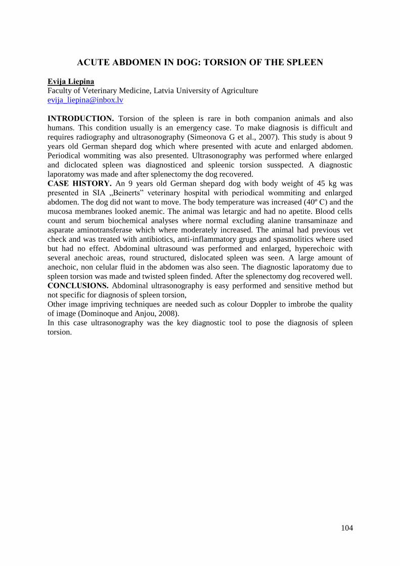
104
ACUTE ABDOMEN IN DOG: TORSION OF THE SPLEEN
Evija Liepina Faculty of Veterinary Medicine, Latvia University of Agriculture
INTRODUCTION. Torsion of the spleen is rare in both companion animals and also
humans. This condition usually is an emergency case. To make diagnosis is difficult and
requires radiography and ultrasonography (Simeonova G et al., 2007). This study is about 9
years old German shepard dog which where presented with acute and enlarged abdomen.
Periodical wommiting was also presented. Ultrasonography was performed where enlarged
and diclocated spleen was diagnosticed and spleenic torsion susspected. A diagnostic
laporatomy was made and after splenectomy the dog recovered.
CASE HISTORY. An 9 years old German shepard dog with body weight of 45 kg was
presented in SIA „Beinerts” veterinary hospital with periodical wommiting and enlarged
abdomen. The dog did not want to move. The body temperature was increased (40º C) and the
mucosa membranes looked anemic. The animal was letargic and had no apetite. Blood cells
count and serum biochemical analyses where normal excluding alanine transaminaze and
asparate aminotransferase which where moderately increased. The animal had previous vet
check and was treated with antibiotics, anti-inflammatory grugs and spasmolitics where used
but had no effect. Abdominal ultrasound was performed and enlarged, hyperechoic with
several anechoic areas, round structured, dislocated spleen was seen. A large amount of
anechoic, non celular fluid in the abdomen was also seen. The diagnostic laporatomy due to
spleen torsion was made and twisted spleen finded. After the splenectomy dog recovered well.
CONCLUSIONS. Abdominal ultrasonography is easy performed and sensitive method but
not specific for diagnosis of spleen torsion,
Other image impriving techniques are needed such as colour Doppler to imbrobe the quality
of image (Dominoque and Anjou, 2008).
In this case ultrasonography was the key diagnostic tool to pose the diagnosis of spleen
torsion.
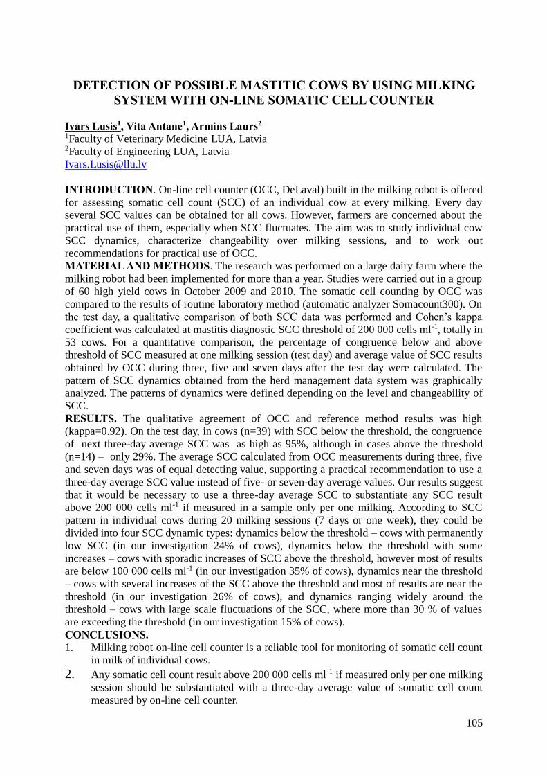
105
DETECTION OF POSSIBLE MASTITIC COWS BY USING MILKING SYSTEM WITH ON-LINE SOMATIC CELL COUNTER
Ivars Lusis1, Vita Antane1, Armins Laurs2
1Faculty of Veterinary Medicine LUA, Latvia 2Faculty of Engineering LUA, Latvia
INTRODUCTION. On-line cell counter (OCC, DeLaval) built in the milking robot is offered
for assessing somatic cell count (SCC) of an individual cow at every milking. Every day
several SCC values can be obtained for all cows. However, farmers are concerned about the
practical use of them, especially when SCC fluctuates. The aim was to study individual cow
SCC dynamics, characterize changeability over milking sessions, and to work out
recommendations for practical use of OCC.
MATERIAL AND METHODS. The research was performed on a large dairy farm where the
milking robot had been implemented for more than a year. Studies were carried out in a group
of 60 high yield cows in October 2009 and 2010. The somatic cell counting by OCC was
compared to the results of routine laboratory method (automatic analyzer Somacount300). On
the test day, a qualitative comparison of both SCC data was performed and Cohen’s kappa
coefficient was calculated at mastitis diagnostic SCC threshold of 200 000 cells ml-1, totally in
53 cows. For a quantitative comparison, the percentage of congruence below and above
threshold of SCC measured at one milking session (test day) and average value of SCC results
obtained by OCC during three, five and seven days after the test day were calculated. The
pattern of SCC dynamics obtained from the herd management data system was graphically
analyzed. The patterns of dynamics were defined depending on the level and changeability of
SCC.
RESULTS. The qualitative agreement of OCC and reference method results was high
(kappa=0.92). On the test day, in cows (n=39) with SCC below the threshold, the congruence
of next three-day average SCC was as high as 95%, although in cases above the threshold
(n=14) – only 29%. The average SCC calculated from OCC measurements during three, five
and seven days was of equal detecting value, supporting a practical recommendation to use a
three-day average SCC value instead of five- or seven-day average values. Our results suggest
that it would be necessary to use a three-day average SCC to substantiate any SCC result
above 200 000 cells ml-1 if measured in a sample only per one milking. According to SCC
pattern in individual cows during 20 milking sessions (7 days or one week), they could be
divided into four SCC dynamic types: dynamics below the threshold – cows with permanently
low SCC (in our investigation 24% of cows), dynamics below the threshold with some
increases – cows with sporadic increases of SCC above the threshold, however most of results
are below 100 000 cells ml-1 (in our investigation 35% of cows), dynamics near the threshold
– cows with several increases of the SCC above the threshold and most of results are near the
threshold (in our investigation 26% of cows), and dynamics ranging widely around the
threshold – cows with large scale fluctuations of the SCC, where more than 30 % of values
are exceeding the threshold (in our investigation 15% of cows).
CONCLUSIONS. 1. Milking robot on-line cell counter is a reliable tool for monitoring of somatic cell count
in milk of individual cows.
2. Any somatic cell count result above 200 000 cells ml-1 if measured only per one milking
session should be substantiated with a three-day average value of somatic cell count
measured by on-line cell counter.
106
NEURONAL CEROID LIPOFUSCINOSIS IN A BEAR FROM KALVENE ZOO
Ilze Matise-VanHoutana1, Anibal Armien2
1Veterinary Pathology Service, Latvia; 2Minnesota Veterinary Diagnostic Laboratory, USA.
In the spring of 2013 a five year old bear kept in Kalvene Zoo (Nature Park of Riga Zoo)
developed sudden motor disfunction, loss of coordination and difficulty to eat. During
summer neurological deficits progressed and bear was killed in September of 2013.
Postmortem necropsy perfomed at FVM showed no gross lesions in the brain, spinal cord or
muscles. Microscopic examination revealed accumulation of large amount of fine
eosinophilic-yellow vacuoles in nearly neurons of all levels of brain and spinal cord. Neurons
were often fragmented, necrotic or lost. Vacuoles were intensively positive by LFB stain and
moderately positive for PAS stain confirming lysosomal storage disease. The appearance of
vacuoles and staining properties suggested neurodegenerative storage disease -- neuronal
ceroid lipofuscinosis.
Neuronal ceroid lipofuscinosis (NCL) is a lysosomal storage disease characterized by
accumulation of lipopigment within central nervous system and peripheral tissues. This
occurs due to mutations in genes that code for a variety of proteins including lysosomal
enzymes and membranes of various compartments. Accumulation of lipopigment is
progressive and cumulative, resulting in disruption of neuronal integrity and imparing
neuronal functions.
The bear affected by NCL was offspring of close sibling / parent matings several generations
in a row which likely facilitated expression of this recessively inhereted disease. NCL is rare
disease best characterized in humans but also affecting domestic animals – most commonly
dogs, cats, sheep and cattle.
107
HEPATOCUTANEOUS SYNDROME IN ADULT DOG Ilze Matise-VanHoutana1, Olga Ponomarjova2
1Veterinary Pathology Service, Latvia; 2LUA Veterinary Hospital, Latvia.
Hepatocutaneous syndrome was suspected in 8-year female dalmation dog with erosive and
ulcerative skin lesions around mucocutaneous junction, ears and feet and elevated liver
enzymes. Biopsy samples from the skin, liver and pancreas were collected and submitted for
histopathologic evaluation which confirmed clinical diagnosis.
In the haired skin, microscopical changes typical of superficial necrolytic dermatitis were
seen: thick parakeratosis, irregular epidermal hyperplasia and pallor and multifocal apoptosis
of keratinocytes. There were also foci of full thickness epidermal necrosis and various
inflammatory cells scattered in the superficial dermis.
In the liver there was severe vacuolar hepatopathy with accumulation of lipid and water and
marked, multifocal nodular hyperplasia containing morphologically normal hepatocytes. In
the pancreas there was mild nodular hyperplasia.
Hepatocutaneous syndrome is a metabolic disease which most commonly occurs due to severe
vacuolar hepatopathy with or without concurrent diabetes mellitus. Less often similar skin
lesions are seen in association with glucagon-producing pancreatic endocrine tumor. The
proposed pathogenesis is metabolic dysfunction that causes cutaneous nutritional deprivation
or protein disbalance. Affected dogs have significantly decreased plasma amino acid levels.
The cause of hepatic lesions in this case was not apparent and the prognosis was guarded
because of limited therapy. Hepatocutaneous syndrome is uncommon disease of cats and
dogs which needs to be kept in mind in animals with crusty, exudative, symmetrical lesions in
the skin and elevated liver enzymes.
108
PREVALENCE AND ANTIMICROBIAL SUSCEPTIBILITY OF CAMPYLOBACTER SPP. IN ESTONIA
Mihkel Mäesaar1,2, Mati Roasto2
1 Veterinary and Food Laboratory, Kreutzwaldi 30, 51006, Tartu, Estonia, 2 Department of Food Hygiene, Institute of Veterinary Medicine and Animal Sciences,
Estonian University of Life Sciences, Kreutzwaldi 56/3, 51014 Tartu, Estonia
INTRODUCTION. Campylobacteriosis, caused by thermotolerant Campylobacter species, is
the most commonly reported zoonosis in the European Union. The main aim of the present
study was to estimate the prevalence and antimicrobial susceptibility of Campylobacter spp. at
Estonian retail level. Study was financially supported by the Estonian Scientific Council grant
No. 9315, and project T13057VLTH ordered by Ministry of Agriculture of Estonia.
MATERIALS AND METHODS. Sampling was performed during the twelve-month period
in 2012. In total, 606 poultry meat samples at Estonian retail level and 380 broiler chicken
caecal samples at slaughterhouse level were collected. Additionally, 28 Campylobacter
human isolates were obtained from Estonian hospitals from patients with severe enteric
infections during the period of 2011-2013. Campylobacter detection and enumeration were
determined according to the International Organization for Standardization (ISO) methods
described in ISO 10272-1:2006 and ISO 10272-2:2006. Conventional multiplex polymerase
PCR was used for the Campylobacter species identification and differentiation. Minimal
inhibitory concentration (MIC) test for a total of six antimicrobials (erythromycin,
ciprofloxacin, tetracycline, streptomycin, gentamicin and nalidixic acid) was performed to
determine antimicrobial susceptibility of 126 Campylobacter isolates obtained from Estonian,
Latvian and Lithuanian broiler chicken products and from Estonian patients with severe
enteric infections.
RESULTS. Total of 20.8% of the fresh broiler chicken meat and 39.2% of the caecal samples
were Campylobacter spp. positive. In chicken meat the positive samples mean number of
Campylobacter was 3.20 log10CFU/g. Distinct seasonal variation in the Campylobacter
contamination of broiler chicken meat was observed with the highest prevalence during the
warm summer months (p < 0.001). Estonian fresh poultry meat products had significantly
lower (p < 0.001) Campylobacter prevalence and counts compare to Latvian and Lithuanian
poultry products sold at Estonian retail level. Altogether from the 126 Campylobacter isolates
88.1% were harbouring C. jejuni specific hipO gene (hippuricase) and 11.9% had C. coli
specific glyA gene (serine hydroxymethyltransferase). Resistance to one or more
antimicrobials were detected in 82 (65.1%), and multidrug resistance was detected in 7 (5.6%)
of Campylobacter isolates. A high proportion of the broiler chicken meat isolates were
resistant to ciprofloxacin and nalidixic acid, 60.2% and 59.2% respectively. Twenty (71.4%)
human origin Campylobacter isolates were resistant to fluoroquinolones. Antimicrobial
resistance to one or more antimicrobials was less frequently detected (p < 0.05) in the
Estonian origin than Latvian or Lithuanian origin Campylobacter isolates.
CONCLUSIONS. Campylobacter related problems extend beyond the country in which a
food originates; therefore, both domestic and international interventions and agreements are
required.

109
GOAT KIDS GROWTH AND MORPHOLOGICAL DEVELOPMENT OF STOMACH IN FIRST 60 DAYS OF LIFE
Laura Otzule, Aija Ilgaža
Preclinical institute, Faculty of Veterinary Medicine LUA, Latvia
INTRODUCTION. In goats (Capra) the growth and functional development of certain parts
of the multi-chambered stomach continue during the first few months after birth. The aim of
this research was to clarify live weight gain and morphofunctional changes in the kids
stomach during the first 60 days of life.
MATERIALS AND METHODS. In research we used Saanen breed kids. In total 20 goat
kids were used in the research. In first group (DMG) were kids which were fed with dairy
(mother) milk ad libitum and lived with mothers (n=10), second group (MRG) kids were fed
with milk replacer and lived separate from mothers in cote (n=10). Each kid before
slaughtering was weighed (at day 45 and 60). The stomach complex were collected after
slaughter, weighed full and empty (abomasums; reticulorumen with omasum), and
immediately processed for morphological analyses.
RESULTS. Length of rumen on day 45 was 21±1.1 cm, but on day 60 the average length
was 25±0.8 cm. Rumen width on day 45 was 23±0.8cm, but on day 60 - 25±1.0 cm. In kids
which were fed with dairy milk the length of abomasum (curvature minor) on day 45 was
22±0.9 cm, but MRG at day 45 was 18±0.7 cm.
The relative stomach weight (p<0.05) on day 45 was 6.01±0.004% in MRG and 4.98±0.005%
DMG, while on day 60 in MRG it was 5.57 ±0.001 % and in DMG 5.88 ±0.11 %.
In MRG kids full abomasum weight (p<0.05) was 112 ±15.5g, but empty 30±2.54g and
rumen full weight was 1174.4±39.70g, empty 144±11.22g, while in DMG full abomasum
weight was 255.6±5.50g, but empty 69±4g, and rumen full weight was 1739±131.7g, empty
225.4±7.05g. Similar dynamics were found by other authors, so feeding of milk replacer,
which was intended for calves, not affect growth of kids stomach in the first two month of
life.
CONCLUSIONS. We confirm that the most important age of stomach development and kids
growth is approximately 45 day of age when the most significant differences can be
observed. Differences in kids growth was between groups – in day 45 higher live weight gain
was in DMG, but in day 60 better results was in MRG. In day 60 there are no significant
differences between goat kid group stomach developments.
110
HOMEOPĀTISKO LĪDZEKĻU IEGŪŠANA, SAGATAVOŠANA, IEDARBĪBA
Ilze Pētersone SIA "DVC diagnostika" klīnika "Mārupe", Latvija, Ltd. DVC diagnostics veterinary clinic
Mārupe, Latvia
IEVADS. Homeopātiskie zāļu līdzekļi tiek izgatavoti saskaņā ar homeopātisko zāļu
ražošanas procedūru, kura aprakstīta Eiropas homeopātiskajā farmakopejā vai, ja Eiropas
farmakopejā tā nav aprakstīta, farmakopejās, ko oficiāli lieto Eiropas Savienības dalībvalstis.
MATERIĀLS. Homeopātiskie zāļu līdzekļi tiek izgatavoti no dažādām izejvielām. To
iedalījums. Augu izejvielas: apmēram 70% no visiem homeopātiskajiem zāļu līdzekļiem tiek
iegūti izmantojot augu ekstraktus. Dzīvnieku valsts izejvielas: tiek izmantoti veselu
dzīvnieku audi, piem. bite kā arī veselu dzīvnieku izdalījumi, piem. čūsku, zirnekļu indes,
tāpat tiek izmantots arī patoloģiskais materiāls, piem. slima organisma izdalījumi.
Minerālvielas /ķīmiskās vielas: visbiežāk izmanto dabīgas izcelsmes vielas,npiem. kalcija
karbonāts tiek iegūts no austeres čaulas vidējās kārtas. Homeopātisko zāļu līdzekļu
sagatavošana. Pirmais solis no augiem iegūst ūdens/spirta ekstraktus, ko sauc par mātes
tinktūrām. Ūdenī nešķīstošas vielas piem. grafītu, zeltu saberž (titruācija) ar laktozes pulveri,
tik tālu, ka to var izmantot šķīduma pagatavošanai. No ūdenī šķīstošām vielām iegūst destilēta
ūdens vai spirta šķīdumus. Otrais solis ir ekstraktu un šķīdumu potencēšana, tos sērijveidā
atšķaida un pēc katras atšķaidīšanas reizes sakrata, ko sauc par potencēšanu vai arī par
dinamizāciju. Pastāv vairākas metodes šķīdumu atšķaidīšanai, visvairāk izmantotā ir
Hānemana metode – potencēšanas simtdaļu metode – vienam pilienam mātes tinktūras
pievieno 99 pilienus destilēta alkohola un ūdens, tiek veikta sakratīšana un pēc tam jauna
atšķaidīšana, katrai potencei lietojot tīru stikla trauku, līdz šķīdums sasniedz 3c, 6c, 12c, 30c,
200c utt. atšķaidījuma pakāpi. To pašu dara arī pēc desmitdaļu metodes, tad vienam pilienam
izejvielas šķīduma pievieno 9 pilienus atšķaidītāja, pārējais viss notiek kā pie 100 daļu
metodes. Iegūstot 12c potency, ir sasniegts sērijveida atšķaidīšanas kritiskais punkts, jeb
Avogardo skaitlis, jo pie 24x vai 12c (10 -24) šķīdums vairs nesatur nevienu oriģinālās
izejvielas molekulu vai atomu.
SECINĀJUMI. Sērijveida atšķaidīšanas un sakratīšanas procesā notiek pakāpeniska
atbrīvošanās no materiālās vielas, savukārt nesējvielā (ūdens/spirta šķīdumā) saglabājas
oriģinālās materiālās vielas nospiedumi augsti enerģētiskā veidā. Homeopātisko zāļu līdzekļu
iedarbība pieaug tieši proporcionāli to atšķaidījuma/sakratīšanas pakāpei, jeb tieši
proporcionāli to potencei.
111
RAW MILK SAFETY PILOT STUDY IN ESTONIA
Mati Roasto1, Piret Kalmus1, Toomas Kramarenko2, Kadrin Meremäe1, Arvo Viltrop1 1Institute of Veterinary Medicine and Animal Sciences, Estonian University of Life Sciences,
Kreutzwaldi 62, 51014, Tartu, Estonia, 2Estonian Veterinary and Food Laboratory, Kreutzwaldi 30, 51006, Tartu, Estonia [email protected]
INTRODUCTION. Raw milk has several microbiological and health benefits but due to its
nutritional properties, raw milk also creates good growth conditions for a variety of spoilage
and potentially pathogenic microorganisms, such as Shiga-toxin producing Escherichia coli
(STEC), Listeria monocytogenes, Salmonella enterica, Campylobacter spp., Yersinia spp. and
some others. Raw milk may also contain mastitis-causing agents, such as Staphylococcus
aureus, Streptococcus agalactiae and zoonotic pathogens, such as Coxiella burnetii,
Toxoplasma gondii, Leptospira hardjo, tick-borne encephalitis virus and some others which
can be excreted with milk and are potentially hazardous to humans. Present study was
supported by the Estonian Ministry of Agriculture project 8-2/T13091VLTO with the main
aim to estimate the safety and quality indicators of raw milk intended for direct selling to
customers.
MATERIALS AND METHODS. During the one month period from June to July 2013
without previous notification to the farmers’ in-line milk filters, bulk milk samples and milk
samples from selling points were collected from a total of 14 dairy farms in Estonia. These
farms were randomly selected out of the 35 farms that sold raw milk in large amounts directly
to consumers in Estonia in summer 2013. Of these 14 farms, six (42.8%) produced milk
organically. Among the selected herds, tie-stall and free-stall systems were equally used. All
samples were taken by official veterinarians. All analyses of the samples collected from the
farms began within 24 h after sampling. The samples obtained from the selling points were
analysed on the day of expiry date. For mastitis pathogens a commercial real-time PCR test
kit (Patho Proof Mastitis PCR Assay, Thermo Fisher Scientific, Espoo, Finland) was used for
bulk milk samples, and the analyses were performed in the Estonian Milk Recording
Laboratory. All other analyses were performed in the Estonian Veterinary and Food
laboratory in accordance with ISO standard methods including STEC genes determination
where PCR was used as described in ISO/TS 13136.
RESULTS. The somatic cell counts, total bacterial counts and the presence of L.
monocytogenes, Campylobacter spp., Salmonella spp. and STEC were studied in the milk.
The total bacterial counts exceeded 100,000 cfu/ml in three (21.4%) bulk milk samples and in
10 samples (71.4%) collected at the retail level. STEC genes were detected in 64.3% of the in-
line milk filter samples. More than one STEC serogroup-specific gene was detected in four
dairy farms. L. monocytogenes was found in 36% of the in-line milk filters. Neither
Salmonella spp. nor Campylobacter spp. were found in any samples.
CONCLUSIONS. The milk quality of one-third of the dairy herds from which raw milk was
directly sold to customers was not sufficient. Therefore, raw milk that is intended for direct
consumption cannot be considered to be microbiologically safe without heat treatment. The
current risk management methods that are applied in Estonia are not sufficient to ensure raw
milk safety for customers. We also suggest that the official criteria for raw milk that is
intended for direct consumption should be reviewed and improved in Estonia.
112
TEHNOLOĢISKO PROCESU IETEKME UZ KARTUPEĻU PRODUKTU MIKROBIOLOĢISKO DROŠĪBU UZGLABĀŠANAS
LAIKĀ
EFFECT OF TECHNOLOGICAL PROCESSES ON THE POTATO PRODUCT MICROBIOLOGICAL SAFETY DURING STORAGE
Aija Ruzaiķe, Sandra Muižniece-Brasava LLU Pārtikas tehnoloģijas fakultāte, Latvija; Faculty of Food Technology, LUA, Latvia [email protected]
IEVADS. Termiskā apstrāde ir plaši izmantota pārtikas produktu konservēšanas metode, bet
ne vienmēr tā nodrošina vēlamo kvalitāti uzglabāšanas laikā. Produktu apstrāde iepakojumā ir
viena no vadošajām apstrādes tehnoloģijām, kas nodrošina produktu ilgstošu uzglabāšanu
istabas temperatūrā. Izmantojot minēto metodi produktam iespējams saglabāt lielāko daļu
savas sākotnējās kvalitātes, papildus iegūstot ilgāku realizācijas laiku. Piemērota un
kvalitatīva iepakojuma un termiskā apstrādes režīma izmantošana nosaka lielu lomu gan
produkta kvalitātes saglabāšanā, gan arī patērētājam ērtā lietošanā.
Darba mērķis ir izvērtēt tehnoloģisko procesu ietekmi uz kartupeļu produktu kvalitāti
uzglabāšanas laikā.
MATERIĀLS UN METODIKA. Pētījumi veikti laika posmā no 2011. gada novembra līdz
2014. gada augustam. Produktu paraugi sagatavoti dārzeņu pārstrādes uzņēmumā SIA
„Paplāte Nr.1”, fizikālās analīzes (krāsa) noteiktas Latvijas Lauksaimniecības universitātes
Pārtikas tehnoloģijas katedras Iepakojuma materiālu īpašību izpētes laboratorijā.
Mikrobioloģisko analīžu noteikšana realizēta Pārtikas drošības, dzīvnieku veselības un vides
zinātniskā institūtā „BIOR”, Pārtikas un vides izmeklējumu laboratorijā. Pētījumam
izmantotas polietilēna/poliamīda (PE/PA) iepakojumā un polietilēnterefralāta/alumīnija/
poliamīda/polipropilēna (PET/ALU/PA/PP) iepakojumā fasētas, un, pēc to hermētiskas
aizkausēšanas, autoklāvā termiski apstrādātas kartupeļu šķēlītes ar sviesta un sēņu mērci
(viena parauga svars 500±5g). Fizikālās īpašība, kā arī mikrobioloģiskā kontrole veikta
produktu ražošanas dienā un pēc 1., 2., 3., 4., 6., 8., 10., 12., 14., 16., 18., 20. uzglabāšanas
mēneša.
REZULTĀTI. Nosakot kopējo mikroorganismu skaitu kartupeļu produktiem abu veidu
iepakojuma materiālos uzglabāšanas laikā, tas nepārsniedz <10 KVV*g-1, kā arī
sūlfītreducējošo klostrīdiju klātbūtne nevienā no pētījumā izmantotajiem paraugiem nav
konstatēta, kas savukārt liecina par izmantotās tehnoloģijas efektivitāti un spēju nodrošināt
produkta drošību un nekaitīgumu uzglabāšanas laikā.
Krāsa ir viena no nozīmīgākajiem raksturlielumiem, ko vērtē patērētājs kā produkta kvalitātes
rādītāju. Salīdzinot paraugus, kas iepakoti un uzglabāti abos iepakojuma materiālos divdesmit
mēnešus, var apgalvot, ka PET/ALU/PA/PP iepakojums nodrošina būtiski (p<0.05) mazākas
produkta krāsas izmaiņas nekā PE/PA iepakojums, tas skaidrojams ar iepakojumu materiāla
barjerīpašībām.
SECINĀJUMI 1. Izvēlētā termiskās apstrādes metode un pētījumā izmantoto iepakojuma materiālu veidi
nodrošina kartupeļu produktu nekaitīgumu un mikrobioloģisko drošību divdesmit
mēnešu uzglabāšanas laikā +18±2 oC temperatūrā.

113
2. Iegūtie rezultāti liecina par to, ka kartupeļu produkti, kas iepakoti PET/ALU/PA/PP iepakojumā, ņemot vērā pētītos kvalitāti noteicošos faktorus, ir droši lietošanai uzturā,
un to derīguma termiņš noteikts divdesmit mēneši.
114
SHIATSU FOR HORSES
Dmitry Sharafutdinov [email protected]
INTRODUCTION. What is equine shiatsu. History and development. Founder of equine
shiatsu methodology - Pamela Hannay. Current situation, practitioners, schools, associations.
MATERIALS AND METHODS. How does it work. Holistic approach vs. treatment of
symptoms. Concept of qi (chi) energy. Systems of energy meridians in horses. Diagnosis and
therapy. Necessary preparations, setting, session procedures, safety concerns. Trust. Knowing
your horse, her background and behavior. Taking responsibility for our own bodies. Patience.
After session. Role of a practitioner – facilitator for healing.
RESULTS. What can be expected. Shiatsu as a useful supplement to veterinary medicine.
Relationship between humans and horses.
CONCLUSIONS. Where to go from here. Self-study and practice. Available instructor-led
courses.
115
ANALYSIS OF PREVALENCE OF THE MOST COMMON CANINE SKIN AND MAMMARY TUMOURS
Donatas Šimkus1,2, Alius Pockevičius2, Petras Mačiulskis1,2,Virginija Šimkienė1, Ligita Zorgevica-Pockeviča2
1National Food and Veterinary Risk Assessment Institute, Lithuania 2Veterinary Faculty of the Lithuanian University of Health Sciences
INTRODUCTION. Skin tumours are the most common tumours in dogs, accounting for
approximately 30% of all diagnosed tumours. Mammary glands are the second most common
site for tumour development subsequent to dermal sites in dogs and account for approximately
82% of all tumours in reproductive organs.
MATERIALS AND METHODS. The samples of skin and mammary tumours were selected
from suspected dogs. Formalin buffer solution 10% was used as a fixative. A record was filled
in with the data about the age, sex and breed of dogs, number and location of tumours and
growth characteristics. The samples of tumours were embedded in paraffin blocks, cut into 4
µm thick sections and stained using the standard hematoxylin-eosin staining method.
Microscopic analysis was described in detail and photographed. The final histological
diagnosis was made according to the origin and differentiation of tumour cells.
RESULTS. Macroscopic and histological analyses of the skin and mammary biopsies were
performed on 116 dogs (56 males and 60 females). Benign tumours accounted for 50.8% and
malignant tumours for 49.2% of the total. The age of dogs at detection of mammary and skin
tumours ranged from 6 months to 19 years. The determined highest incidence (20.7%) of
mammary and skin tumours was from 6 to 8 years of age. The most common tumours were:
histiocytoma (11.1%) in young dogs (2.97 years of age), mast cell tumour (9.5%) in middle-
aged dogs (7.68 years) and simple mammary carcinoma (14.7%) in older dogs (9.72 years).
Females were more affected by simple mammary carcinoma (100%) and by lipoma (66.7%)
while males were more affected by hepatoid gland adenoma (83.3%) and by histiocytoma
(61.5%). The prevalence of tumours in purebred dogs accounted for 73.3% and in mongrels
26.7%. The skin tumours were most frequently located in the limb regions (28.5%). The
multiple tumours identified in 27.6% of the examined dogs were comprised of skin
melanomas (75.0%), hepatoid gland adenomas (66.7%) and sebaceous gland adenomas
(50.0%).
CONCLUSIONS. The age, sex and breed of dogs and anatomic location and number of
tumours are factors which markedly affect the incidence of skin and mammary tumours.
Young dogs are more predisposed to skin histiocytoma, dogs of middle age to mast cell
tumours and older dogs to simple mammary carcinoma. The incidence of simple mammary
carcinoma and of lipoma is higher among bitches whereas tumours of perianal glands and skin
histiocytoma are more frequent in males. The predisposition of purebred dogs to skin and
mammary tumours is higher than that of mixed breeds (mongrels). Skin tumours tend to
localise in the limb areas. The multiple tumours usually occur in the cases of skin melanoma,
adenoma of hepatoid glands and adenoma of sebaceous glands.
116
EVALUATION OF BACTERIAL MICROFLORA OF EUROPEAN EEL (ANGUILLA ANGUILLA) SKIN SAMPLES FROM LAKES IN LATVIA
Margarita Terentjeva1, Inga Eizenberga1, Olga Valciņa2, Aleksandr Novoslavskij3, Jevgēnija Ošmjana2, Aivars Bērziņš1,2 1 Faculty of Veterinary Medicine Latvia University of Agriculture, Latvia 2 Institute of Food Safety, Animal Health and Environment „BIOR”, Latvia
3 Lithuanian University of Health Sciences/Veterinary Academy, Lithuania
INTRODUCTION. Microflora of surfaces and skin of fish are continuously affected by
aquatic environment including bacteria present in water, sediment and contamination from
polluted wastewater. Above mentioned observations reveal that fish may serve as the
indicator of pollution of aquatic environment, because the changes in microflora of
surrounding environment of fish have an impact on number and composition of bacterial
microflora of fish. Limited studies on bacterial contamination and the presence of pathogens
in freshwater fish are conducted previously. Therefore the aim of the present study was to
detect the total bacterial count (TBC) and Enterobacteriaceae on eel skin as indicator of
hygienic status of environment and the presence of Salmonella spp. as possible zoonotic
pathogenic agent of intestinal origin.
MATERIALS AND METHODS. For the detection of total bacterial contamination of eel
the surface swabs were collected. Altogether 31 samples of eel skin were collected between
April-May in 2014 from eels from three different lakes in Latvia (Alūksnes, Usmas and
Sīvers). Skin samples were taken from live eel skin by processing a 10x10cm2 of skin with
abrasive sponge. Skin samples were plated onto PCA and VRBA agar with subsequent
incubation at 30°C and 37°C according to the ISO 6887 and ISO 21528 methods. For the
detection of Salmonella the ISO 6579 method was applied.
RESULTS. Depending on lake TBC of eel skin samples was varying from 5 up to 85000
cfu/cm2, there the lowest average number of 36 cfu/cm2 was identified on eel skin originated
from Alūksnes lake, and the highest average - 12750 cfu/cm2 in samples from Sīvers lake.
Number of Enterobacteriaceae was varying from 0 up to 198 cfu/cm2, there less than one
cfu/cm2 was identified in samples from Alūksnes and Usmas lakes, and the highest average
number - 36 cfu/cm2 in samples of Sīvers lake. All tested eel skin samples were Salmonella
negative.
CONCLUSIONS. Eel skin samples did not contain excessive amount of bacterial microflora
indicating the good hygienic status of the lake from there the eels were originated.
Differences detected in the contamination of eel skin samples with TBC and
Enterobacteriaceae showing a possible link between the area of origin of eel and bacterial
contamination of eel skin.
ACKNOWLEDGMENTS. This study was conducted within the ESF project No.
2013/0016/1DP/1.1.1.2.0/13/APIA/VIAA/055 „Iekšējo ūdeņu zivju resursu ķīmiskā un
bioloģiskā piesārņojuma pētniecības grupas izveide”.
117
KOORDINĀCIJAS TRAUCĒJUMI ZIRGIEM. KLĪNISKO GADĪJUMU ANALĪZE
Anna Vainute Privātpraktizējoša veterinārārste, Latvija; Self-employed veterinarian, Latvia
IEVADS. Neiroloģiskas pazīmes, kas rada koordinācijas traucējumus, var iedalīt 3
grupās: ataksija (ja bojājumi ir vienā no sekojošām nervu sistēmām: vestibulārie orgāni,
smadzenītes, mugurkauls), dismetria (raksturojas ar stīvumu, spazmām un pārmērīgu kāju
locītavu saliekšanu), vārgums. Šādas pazīmes rada kompresīvi kakla skriemeļu bojājumi,
alimentāra saindēšanās, baktēriju radītie toksīni, infekcijas slimības, ko izraisa vienšūņi un
vīrusi.
KLĪNISKAIS GADĪJUMS. Saimniecībā visiem sešiem zirgiem novēroja dažāda
smaguma koordinācijas traucējumus, stīvumu un novājēšanu ar dažu nedēļu starplaiku.
Dažiem zirgiem novēroja klepu un tahikardiju. Pirmajiem klīniskās pazīmes parādījās
diviem zirgiem, kuri pēc ārstēšanas atveseļojās. Pēc mēneša ar dažu dienu starplaiku
dažāda smaguma klīniskās pazīmes parādījās pārējiem zirgiem. Viens no zirgiem 9h pēc
klīnisko pazīmju parādīšanās nobeidzās. Tika veikta patologanatomiskā sekcija un
paņemti orgānu paraugi, paņemti asins paraugi visiem zirgiem seroloģiskai izmeklēšanai.
REZULTĀTI. Tā kā seroloģiskā izmeklēšana un patologanatomiskās sekcijas rezultāti
neuzrādīja konkrētu saslimšanas cēloni, tika veikta simptomātiska ārstēšana. Trīs zirgiem
tikai veikta konvencionāla simptomātiska ārstēšana, kas būtiski nemainīja stāvokli.
Visiem zirgiem tika pielietota homeopātiskā ārstēšana- katram zirgam tika piemeklēts savs
homeopātiskais līdzeklis, balstoties uz esošo klīnisko ainu un dzīvnieka uzvedības
pazīmēm.
Četriem zirgiem pēc ārstēšanas neiroloģiskas pazīmes nenovēroja. Vienam zirgam,
neskatoties uz ievērojamu veselības stāvokļa uzlabojumu, vēl novēro koordinācijas
traucējumus un novājēšanu.
SECINĀJUMI. Veicot simptomātisku ārstēšanu ar konvencionāliem ārstniecības
līdzekļiem, uzlabojumu nenovēroja, taču pēc homeopātisko līdzekļu lietošanas dažu dienu
laikā zirgiem tika novērots veselības stāvokļa uzlabojums, bet pilnīga atveseļošanās ilga
līdz pat mēnesim.
Homeopātisko līdzekļu izmantošana ļauj precīzāk piemeklēt nepieciešamo līdzekli
individuāli katram pacientam, neskatoties uz to, ka klīniskās pazīmes visiem ir līdzīgas. Ja
konvencionālie ārstniecības līdzekļi nedod vēlamo efektu, ir jāmeklē papildus iespējas
pacientu ārstēšanā. Homeopātija ir tā nozare, kas paplašina ārstēšanas iespējas.
118
DIROFILARIA REPENS SUŅIEM LATVIJAS PATVERSMĒS 2013. GADĀ
DIROFILARIA REPENS INFECTION AMONG DOGS IN LATVIAN
ANIMAL SHELTERS DURING 2013
Armands Vekšins, Anna Krūklīte, Dace Keidāne, Ilze Matīse-Van Houtana LLU Veterinārmedicīnas fakultāte, Latvija; Faculty of Veterinary Medicine LUA, Latvia
IEVADS. Pasaulē mainoties klimatiskajiem apstākļiem, kā arī pieaugot brīvās ceļošanas
iespējām, arvien biežāk tiek konstatētas dažādas slimības, kuras noteiktās klimatiskajās joslās
iepriekš netika novērotas. Latvijā pēdējo piecu gadu laikā suņiem bieži tiek konstatēta
Dirofilaria repens invāzija.
Dirofilaria repens ir nematode, kuru kāpuru - mikrofilāriju bioloģiskais vektors ir odi
(Anopheles, Aedes un Culex ģints). Pieaugušie parazīti lokalizējas dzīvnieka zemādā, bet
mikrofilārijas cirkulē asinīs (Genchi et al., 2009).
MATERIĀLS UN METODIKA. 2013.gada jūlija, augusta un septembra mēnešos veicām
suņu asins paraugu noņemšanu Saldus, Jelgavas, Slokas un Rīgas dzīvnieku patversmēs.
Mikrofilāriju noteikšanai asins paraugos izmantojām modificētā Knotta testu.
REZULTĀTI. Jūlijā, augustā un septembrī izmeklējām 139 asins paraugus, no kuriem 22
gadījumos diagnosticējām D.repens mikrofilārijas. Saldus dzīvnieku patversmē no
izmeklētajiem 26 asins paraugiem, mikrofilārijas netika konstatētas nevienā paraugā. No
Jelgavas mazo dzīvnieku izolatora iegūtajiem 21 asins paraugiem, mikrofilārijas konstatējām
trīs paraugos, kas sastāda IE (invāzijas ekstensitāte) 14,3%. No Slokas dzīvnieku patversmes
22 asins paraugiem, mikrofilārijas konstatētas sešos paraugos, kas sastāda IE 27,3%, bet no
Rīgas pilsētas dzīvnieku patversmes izmeklētajiem 70 asins paraugiem, 13 paraugos jeb
18,6% konstatētas mikrofilārijas.
Mikrofilāriju pozitīvie suņi 50% gadījumu bija vecāki par sešiem gadiem un 72,7% ar vidēja
garuma apmatojumu. Cirkulējošu mikrofilāriju pozitīvi suņi 59,1% bija vīrišķās, bet 40,9%
sievišķās kārtas.
Kopējā invāzijas ekstensitāte par pētījumā iekļautajiem mēnešiem bija 15,8% un invāzijas
intensitāte 3631 ± 1187.
SECINĀJUMI. Latvijā suņu saslimšanas gadījumi ar D. repens, ir samērā bieži sastopami,
kas, iespējams, izskaidrojams ar klimata maiņu. Patversmju suņu asins paraugos vislielākā
invāzijas ekstensitāte bija augusta mēnesī, bet vismazākā septembrī. 2014.gada septembrī
gaisa temperatūra bija zemāka par jūlija un augusta mēnešiem, kas liek domāt, ka D. repens
cirkulējošo mikrofilāriju skaits un aktivitāte ir atkarīga no apkārtējās vides temperatūras.
Augstākā IE tika konstatēta Slokas patversmē (27,3%), bet zemākā – Saldus patversmē (0%).
Izvirzītais apgalvojums, ka D. repens invāziju biežāk novēro suņiem ar īsu apmatojumu, mūsu
pētījumā neapstiprinājās, jo 72,7% gadījumu D. repens mikrofilārijas diagnosticējām suņiem,
ar vidēja garuma apmatojumu, 18,2% ar īsu un 9,1% ar garu apmatojumu. Iespējams, ka šāds
procentuālais sadalījums saistīts ar to, ka patversmēs liels īpatsvars atrodas tieši suņi ar vidēja
garuma apmatojumu, kas ir raksturīgs vairumam bezšķirnes suņu.
119
DIAGNOSIS OF ANTERIOR CRUCIATE LIGAMENT RUPTURE OF THE CANINE STIFLE
Ligita Zorgevica-Pockeviča, Alius Pockevičius, Dalia Juodžentė, Benas Noreikis Veterinary Faculty of the Lithuanian University of Health Sciences
INTRODUCTION. Anterior cruciate ligament disease of the canine stifle is a partial or total
rupture of the anterior cruciate ligament (ACL), or avulsion, where there is a loss of function
of the ligament due to a bone fracture at the ACL point of insertion. Consequences include
instability of the stifle joint, inflammation, the patient exhibits lameness, feels pain and
discomfort especially after physical load-bearing. This pathology is usually diagnosed in older
aged dogs.
MATERIALS AND METHODS. The lameness of dogs diagnosed with anterior cruciate
ligament rupture was evaluated via a point grading scale from 0 to 4. Instabilities of the stifle
joint were evaluated using the Cranial Drawer and Tibial Compression tests. Radiological
evaluations included mediolateral (ML) and craniocaudal projections. Osteoarthritic
symptoms were evaluated using ML projections according to the Brünnberg classification
index from 0 to 3. Effusion of the joint capsules were rated using a 0 to 3 point scale. A
macroscopic arthrotomy evaluated the condition of the joint capsule; signs of osteoarthritis –
osteophytes (0–none, 1–few, 2–many) as well as the condition of the meniscus and anterior
cruciate ligament. During the arthrotomy, samples were obtained from the joint capsule’s
sub-patellar bursa for histopathological evaluation which were fixed in buffered 10% formalin
solution, embedded in paraffin and stained with hematoxylin and eosin.
RESULTS. Dogs (n=21) diagnosed with anterior cruciate ligament rupture were from 5
months to 13 years of age. 59% of the examined dogs were of large breeds. Lameness in 28.5
% of the examined dogs scored 4 points, 47.6% - 3 points and 23.9 % - 2 points respectively.
Cranial Drawer and Tibial Compression test results were positive in 66.6 % of examined
dogs, 14.3 % were negative, and 19.1% were inconclusive. Joint capsule effusion grading
resulted as follows: 3 points - 33.3%, 2 points – 33.3%, 1 point – 14.3 % and 0 points in
19.1% of the examined dogs. Radiological examinations indicated that osteoarthritis grades
were valued at 3 points in 9.5%, 2 points in 9.5%, 1 point in 42.9% and 0 points in 38.1% of
cases. Joint capsule polyps were identified in 23.8% of examined dogs. During arthrotomy,
osteophyte grades were valued at 2 points in 19.1% and 1 point in 52.4% of examined dogs.
The remainder - 28.5%, were undetermined. A ruptured meniscus was identified in 61.9% of
examined dogs, 38.1% were undamaged. In 71.4% of examined dogs, the anterior cruciate
ligament was completely ruptured and partial ruptures comprised 28.6%. During arthrotomy,
redness and thickening of the joint capsules were observed in all patients. Histological
examinations of joint capsules confirmed inflammation, lubricant layer villous hyperplasia
and 23.8% of examined dogs were diagnosed with polyps.
CONCLUSIONS. Dogs with ruptures of the anterior cruciate ligament and observed
exhibiting continuous lameness should receive Cranial Drawer and Tibial Compression Tests
and radiological examinations. Ruptures of the anterior cruciate ligament indicate atypical
movement between joint surfaces which evokes inner meniscal pathologies and progressive
osteoarthritis. Increasingly common histological findings are characteristic to chronic
inflammation: thickening of the joint capsule, inflammatory cell infiltrations, lubricant layer
villous hyperplasia and polyps, observations of increased fibrocyte counts and collagen tissue.
120
AUTORU ALFABĒTISKAIS RĀDĪTĀJS
Antāne V. 25, 104 Armien A. 105 Ārne A. 8, Berģe V. 17 Bērziņa D. 90 Bērziņa Z. 92 Bērziņš A. 115 Birģele E. 36 Bizokas V. 53 Boiko D. 101 Deksne G. 93 Dūrītis I. 46, 96 Eizenberga I. 115 Esīte Z. 93 Ganola K. 21 Gāliņa D. 94 Grīnblate S. 95 Heike A.K. 87 Hussar P. 96 Ilgaža A. 8, 95, 108 Ivanovs A. 71 Ivbule M. 97 Jahundoviča I. 92 Järveots T. 96 Jonkus D. 63 Juodzente D. 98, 118 Kalmus P. 110 Kärner M. 96 Keidāne D. 17, 21, 117 Kirjušina M. 92 Kociņa I. 25 Kokoreviča L. 99, 100 Kondratjeva J. 36 Kovalcuka L. 101 Kozinda O. 87 Kramarenko T. 110 Krapauskis K. 41 Krūklīte A. 17, 21, 117
Kulakova M. 102 Laizāne D. 46 Laurs A. 104 Leitāne E. 93 Ligere B. 93 Liepa L. 102 Liepina E. 103 Lūsis I. 25, 102, 104 Mačiulskis P. 114 Matīse-Van Houtana I. 99, 100, 105, 106, 117 Mäesaar M. 107 Meremäe K. 110 Mičule G. 81 Mugurevičs A. 96 Muižniece-Brasava S. 111 Nātiņa L. 46 Noreikaite-Bulotiene R. 53 Noreikis B. 118 Novoslavskij A. 115 Osītis U. 41 Ošmjana J. 115 Otzule L. 108 Petrovska S. 63 Pētersone I. 109 Pockevičius A. 114, 118 Ponomarjova O. 106 Riskeviciene V. 98 Roasto M. 107, 110 Rotmistrova A. 96 Ruzaiķe A. 111
Sharafutdinov D. 113 Šematoviča I. 71 Šimkienė V. 114 Šimkus D. 114
Terentjeva M. 115 Trūpa A. 41

121
Vainute A. 116 Valciņa O. 115 Valdovska A. 77, 94, 97 Vekšins A. 87, 117 Viltrop A. 110 Zolnere E. 102 Zorgevica – Pockevica L. 98, 114, 118 Zute I. 77