Dr. B Ch 09_lecture_presentation
47
© 2012 Pearson Education, Inc. 9 The Muscular System: Skeletal Muscle Tissue and Organization PowerPoint ® Lecture Presentations prepared by Steven Bassett Southeast Community College Lincoln, Nebraska
Transcript of Dr. B Ch 09_lecture_presentation

© 2012 Pearson Education, Inc.
9The Muscular System: Skeletal Muscle Tissue and Organization
PowerPoint® Lecture Presentations prepared bySteven BassettSoutheast Community College Lincoln, Nebraska

© 2012 Pearson Education, Inc.
Introduction
There are three types of muscle tissue: Skeletal muscle—Skeletal muscle tissue moves the body by pulling on
bones of the skeleton. Skeletal muscle fibers arise from embryonic cells called myoblasts. Work voluntarily. Striated microscopic pattern.
Cardiac muscle—Cardiac muscle tissue pushes blood through the arteries and veins of the circulatory system.
Work involuntarily. Striated microscopic pattern. Intercalated discs. Branchd fibers.
Smooth muscle—Smooth muscle tissues push fluids and solids along the digestive tract and hollow organs and perform varied functions in other systems.
Work involuntarily. Smooth microscopic pattern.

© 2012 Pearson Education, Inc.
Introduction
Muscle tissues share four basic properties: Excitability: the ability to respond to stimulation
Skeletal muscles normally respond to stimulation by the nervous system.
Cardiac and smooth muscles respond to the nervous system and circulating hormones.
Contractility: the ability to shorten actively and exert a pull or tension that can be harnessed by connective tissues
Extensibility: the ability to continue to contract over a range of resting lengths
Elasticity: the ability of a muscle to rebound toward its original length after a contraction

© 2012 Pearson Education, Inc.
Functions of Skeletal Muscles
Skeletal muscles are contractile organs directly or indirectly attached to bones of the skeleton.
They work voluntarily. Skeletal muscles perform the following functions:
Produce skeletal movement Maintain posture and body position Support soft tissues and stabilize the joints. Regulate entering and exiting of material Maintain body temperature

© 2012 Pearson Education, Inc.
Skeletal muscle surrounding
• From inside out:• Sarcolema
• Plasma membrane of muscle fiber.• Endomysium
• Connective tissue surrounding the muscle fiber.• Perimysium
• Connective tissue surrounding the fascicle. Fascicle is a bundle of muscle fibers.
• Epimysium• Connective tissue surrounding the muscle.
• Deep fascia• Connective tissue surrounding the skeletal muscles together.

© 2012 Pearson Education, Inc.
Anatomy of Skeletal Muscles
• Connective Tissue of Muscle• Tendons and Aponeuroses
• Epimysium, perimysium, and endomysium converge to form tendons
• Tendons connect a muscle to a bone• Aponeuroses connect a muscle to a muscle

© 2012 Pearson Education, Inc.
Figure 9.1 Structural Organization of Skeletal Muscle
Epimysium
Muscle fascicle
Endomysium
Perimysium
Nerve
Muscle fibers
Blood vessels
SKELETAL MUSCLE(organ)
MUSCLE FASCICLE(bundle of cells)
Perimysium
Muscle fiber
Endomysium
Epimysium
Blood vesselsand nerves
Endomysium
Perimysium
Tendon
MUSCLE FIBER(cell)
Mitochondria
Sarcolemma
Myofibril
AxonSarcoplasm
Capillary
Endomysium
Myosatellitecell
Nucleus

© 2012 Pearson Education, Inc.
Anatomy of skeletal muscle fiber
• Sarcolemma• The plasma membrane of the muscle fiber.
• Sarcoplasmic reticulum• the ER that stores calcium ions.
• Terminal cisterna• an expanded part of ER next to T tube.
• T tube• an extension of sarcolema into the muscle fiber.
• Triad• The combination of one T tube and two flaked Terminal Cisterna.

© 2012 Pearson Education, Inc.
Figure 9.3ab The Formation and Structure of a Skeletal Muscle Fiber
Development of askeletal muscle fiber
External appearanceand histological view
Myoblasts
Muscle fibers developthrough the fusion ofmesodermal cellscalled myoblasts.
Myosatellite cell
Nuclei
Immaturemuscle fiber
© 2012 Pearson Education, Inc.
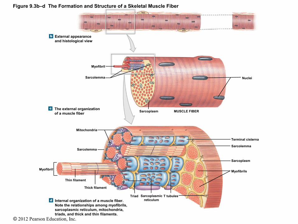
Figure 9.3b–d The Formation and Structure of a Skeletal Muscle Fiber
External appearanceand histological view
The external organizationof a muscle fiber
Internal organization of a muscle fiber.Note the relationships among myofibrils,sarcoplasmic reticulum, mitochondria,triads, and thick and thin filaments.
Myofibril
Sarcolemma
Sarcoplasm
Nuclei
MUSCLE FIBER
Mitochondria
Sarcolemma
Myofibril
Thin filament
Thick filament
Triad T tubulesSarcoplasmicreticulum
Terminal cisterna
Sarcolemma
Sarcoplasm
Myofibrils

© 2012 Pearson Education, Inc.
Figure 9.4b Sarcomere Structure
A corresponding view of a sarcomere in a myofibril inthe gastrocnemius muscle of the calf and a diagramshowing the various components of this sarcomere
Z line TitinH band
A bandI band
M lineZone of overlap Thinfilament
Thickfilament
Sarcomere
H band Z line
I band
Z line Zone of overlap M line
Sarcomere
TEM × 64,000
A band

© 2012 Pearson Education, Inc.
Anatomy of Skeletal Muscles
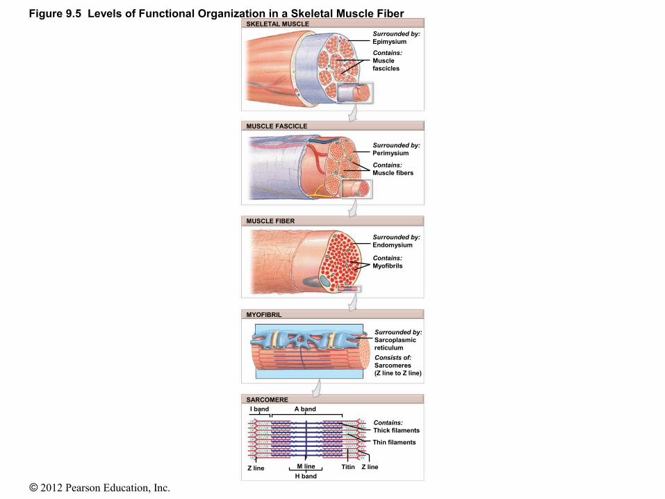
• Levels of Organization• Skeletal muscles consist of muscle fascicles• Muscle fascicles consist of muscle fibers• Muscle fibers consist of myofibrils• Myofibrils consist of sarcomeres• Sarcomeres consist of myofilaments• Myofilaments are made of actin and myosin

© 2012 Pearson Education, Inc.
Skeletal muscle
Sarcomere Organization Thick and thin filaments within a myofibril are organized in the
sarcomeres. All of the myofibrils are arranged parallel to the long axis of the
cell, with their sarcomeres lying side by side. A band: the dark area of sarcomere that contains thick filaments. I band: the light area between A bands. H band: the light area within the A band. M line: the dark line within the H band. Z line: the area of a myofibril, where actin filaments attach to one
another.
© 2012 Pearson Education, Inc.
Skeletal muscle
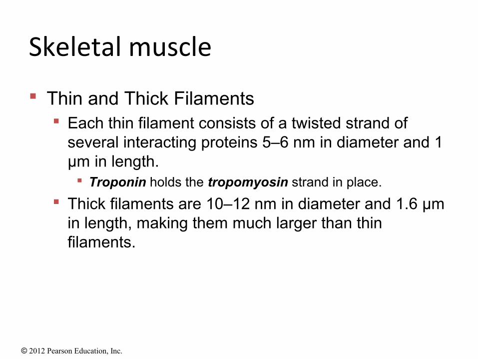
Thin and Thick Filaments Each thin filament consists of a twisted strand of
several interacting proteins 5–6 nm in diameter and 1 μm in length. Troponin holds the tropomyosin strand in place.
Thick filaments are 10–12 nm in diameter and 1.6 μm in length, making them much larger than thin filaments.
© 2012 Pearson Education, Inc.
Figure 9.5 Levels of Functional Organization in a Skeletal Muscle FiberSKELETAL MUSCLE
MUSCLE FASCICLE
MUSCLE FIBER
MYOFIBRIL
SARCOMERE
Surrounded by:Epimysium
Contains:Musclefascicles
Surrounded by:Perimysium
Contains:Muscle fibers
Surrounded by:Endomysium
Contains:Myofibrils
Surrounded by:Sarcoplasmicreticulum
Consists of:Sarcomeres(Z line to Z line)
Contains:Thick filaments
Thin filaments
I band A band
Z line M line
H band
Titin Z line

© 2012 Pearson Education, Inc.
Figure 9.6ab Thin and Thick Filaments
The attachmentof thin filamentsto the Z line
The detailed structure of a thin filament showingthe organization of G actin, troponin, andtropomyosin
Myofibril
Z line M line
H band
Sarcomere
Actinin Z line Titin
Troponin Nebulin TropomyosinActive
siteG actin
molecules
F actinstrand

© 2012 Pearson Education, Inc.
Figure 9.6cd Thin and Thick Filaments
Myofibril
Z line M line
H band
Sarcomere
A single myosin molecule detailing the structure andmovement of the myosin head after cross-bridgebinding occurs
The structure ofthick filaments
Titin
M line
Myosin tail
Myosin head
Hinge

© 2012 Pearson Education, Inc.
Muscle Contraction
Contracting muscle fibers exert a pull, or tension, and shorten in length.
Sarcoplasmic reticulum stores calcium ions.
Caused by interactions between thick and thin filaments in each sarcomere
Triggered by presence of calcium ions Contraction itself requires the presence
of ATP.

© 2012 Pearson Education, Inc.
Muscle Contraction
The Sliding Filament Theory Explains the following changes that occur
between thick and thin filaments during contraction: The H band and I band get smaller. The zone of overlap gets larger. The Z lines move closer together. The width of the A band remains constant
throughout the contraction.

© 2012 Pearson Education, Inc.
Figure 9.7 Changes in the Appearance of a Sarcomere during Contraction of a Skeletal Muscle Fiber
A relaxed sarcomere showing locationof the A band, Z lines, and I band
During a contraction, the A band stays the samewidth, but the Z lines move closer together andthe I band gets smaller. When the ends of amyofibril are free to move, the sarcomeresshorten simultaneously and the ends of themyofibril are pulled toward its center.
I band A band
Z line Z lineH band

© 2012 Pearson Education, Inc.
Muscle Contraction
The Start of a Contraction Triggered by calcium ions in the sarcoplasm Electrical events at the sarcolemmal surface
Trigger the release of calcium ions from the terminal cisternae
The calcium ions diffuse into the zone of overlap and bind to troponin.
Troponin changes shape, alters the position of the tropomyosin strand, and exposes the active sites on the actin molecules.

© 2012 Pearson Education, Inc.
Figure 9.10a The Neuromuscular Synapse
A diagrammatic view of aneuromuscular synapse
Motorneuron
Axon
Muscle fiber
Path of actionpotential
Neuromuscularsynapse
Motorend plate
Myofibril

© 2012 Pearson Education, Inc.
Figure 9.10ab The Neuromuscular Synapse
A diagrammatic view of aneuromuscular synapse
Motorneuron
Axon
Muscle fiber
Path of actionpotential
Neuromuscularsynapse
Motorend plate
Myofibril
One portion of aneuromuscular synapse
Myofibril
Mitochondrion
Sarcolemma
Glial cell
Synaptic terminal

© 2012 Pearson Education, Inc.
Figure 9.10bc The Neuromuscular Synapse
One portion of aneuromuscular synapse
Myofibril
Mitochondrion
Sarcolemma
Glial cell
Synaptic terminal
Detailed view of a terminal,synaptic cleft, and motor endplate. See also Figure 9.2.
Synaptic vesicles
ACh
AChreceptorsiteAChE
molecules
Junctionalfold
Sarcolemma ofmotor end plate
Arriving actionpotential
Synaptic cleft

© 2012 Pearson Education, Inc.
Skeletal muscle
The End of a Contraction When electrical stimulation ends:
The SR will recapture the Ca2+ ions. The troponin–tropomyosin complex will cover the
active sites. And, the contraction will end.

© 2012 Pearson Education, Inc.
Figure 9.11 The Events in Muscle Contraction
ACh released, bindingto receptors
Sarcoplasmicreticulumreleases Ca2+
Active-siteexposure,cross-bridgeformation
ActionpotentialreachesT tubule
Ca2+
Actin
Myosin
SarcolemmaT tubule
Motorend
plateSynapticterminal
Contractionbegins
STEPS IN INITIATING MUSCLE CONTRACTION STEPS IN MUSCLE RELAXATION
ACh removed by AChE
Sarcoplasmicreticulumrecaptures Ca2+
Active sitescovered, nocross-bridgeinteraction
Contractionends
Relaxation occurs,passive return toresting length

© 2012 Pearson Education, Inc.
Motor Units and Muscle Control
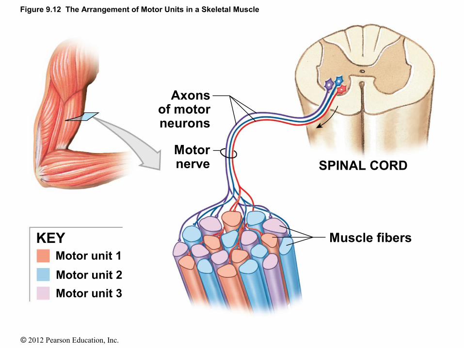
• A motor unit is all muscle fibers controlled by a single motor neurone.
• At the end of each motor neuron there are synaptic vesicles containing neurotransmitters. The neurotransmitter involved in the process of contraction is Acetylcholine.
• The enzyme in the synaptic cleft that destroys Ach and shuts down the contraction is Acetylcholineesterase (AchE)
© 2012 Pearson Education, Inc.
Figure 9.12 The Arrangement of Motor Units in a Skeletal Muscle
Motor unit 1
Motor unit 2
Motor unit 3
KEY
SPINAL CORD
Muscle fibers
Axonsof motorneurons
Motornerve

© 2012 Pearson Education, Inc.
Motor Units and Muscle Control
Muscle Tone Some of the motor units of muscles are always
contracting, producing a resting tension in a skeletal muscle that is called muscle tone.
Resting muscle tone stabilizes the position of bones and joints.

© 2012 Pearson Education, Inc.
Motor Units and Muscle Control
Muscle Hypertrophy and Atrophy Exercise causes increases in
Number of mitochondria Concentration of glycolytic enzymes Glycogen reserves Myofibrils
Each myofibril contains a larger number of thick and thin filaments.
The net effect is an enlargement, or hypertrophy, of the stimulated muscle.
Disuse of a muscle results in the opposite, called atrophy.

© 2012 Pearson Education, Inc.
Types of Skeletal Muscle Fibers
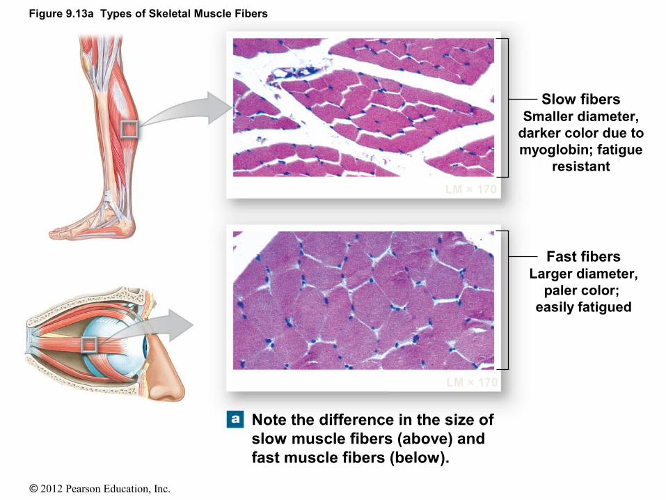
The features of fast fibers, or white fibers, are:
Large in diameter—due to many densely packed myofibrils
Large glycogen reserves Relatively few mitochondria
Their mitochondria are unable to meet the demand.
Fatigue easily Can contract in 0.01 seconds or less following
stimulation
© 2012 Pearson Education, Inc.
Figure 9.13a Types of Skeletal Muscle Fibers
Note the difference in the size ofslow muscle fibers (above) andfast muscle fibers (below).
LM × 170
LM × 170
Slow fibersSmaller diameter,
darker color due tomyoglobin; fatigue
resistant
Fast fibersLarger diameter,
paler color; easily fatigued

© 2012 Pearson Education, Inc.
Types of Skeletal Muscle Fibers
Slow fibers, or red fibers, features are Only about half the diameter of fast fibers Take three times as long to contract after
stimulation Contain abundant mitochondria Use aerobic metabolism Have a more extensive network of capillaries
than do muscles dominated by fast muscle fibers.
Red color because they contain the red pigment myoglobin

© 2012 Pearson Education, Inc.
Types of Skeletal Muscle Fibers
Intermediate fibers have properties intermediate between those of fast fibers and
slow fibers. Intermediate fibers contract faster than slow
fibers but slower than fast fibers. Intermediate fibers are similar to fast fibers
except They have more mitochondria. They have a slightly increased capillary supply. They have a greater resistance to fatigue.

© 2012 Pearson Education, Inc.
Figure 9.14ab Skeletal Muscle Fiber Organization
Parallel muscle(Biceps brachii muscle)
Parallel muscle with tendinous bands(Rectus abdominismuscle)
(h)
(d)
(g)
(a)
(b)
(e)
(c)
(f)
Fascicle
Cross section
Body (belly)

© 2012 Pearson Education, Inc.
Figure 9.14e Skeletal Muscle Fiber Organization
(h)
(d)
(g)
(a)
(b)
(e)
(c)
(f)
Unipennate muscle(Extensor digitorum muscle)
Extendedtendon

© 2012 Pearson Education, Inc.
Figure 9.14g Skeletal Muscle Fiber Organization
(h)
(d)
(g)
(a)
(b)
(e)
(c)
(f)
Multipennate muscle(Deltoid muscle)
Tendons
Cross section
© 2012 Pearson Education, Inc.
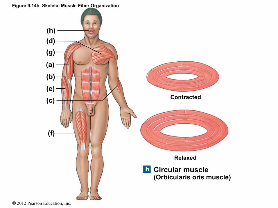
Figure 9.14h Skeletal Muscle Fiber Organization
(h)
(d)
(g)
(a)
(b)
(e)
(c)
(f)
Circular muscle(Orbicularis oris muscle)
Contracted
Relaxed

© 2012 Pearson Education, Inc.
Muscle Terminology
Origin: the end of the muscle that remains stationary Insertion: the end of the muscle that moves
Commonly the origin is proximal to the insertion.
Muscle Actions There are two methods of describing actions.
The first references the bone region affected. For example, the biceps brachii muscle is said to perform
“flexion of the forearm.”
The second method specifies the joint involved. For example, the action of the biceps brachii muscle is
described as “flexion of the elbow.”

© 2012 Pearson Education, Inc.
Muscle Terminology
Muscles can be grouped according to their primary actions into three types:
Prime movers (agonists): are muscles chiefly responsible for producing a particular movement
Synergists: assist the prime mover in performing that action
Antagonists: are muscles whose actions oppose that of the agonist If the agonist produces flexion, the antagonist will produce
extension.
• Fixators• Agonist and antagonist muscles contracting at the same time to
stabilize a joint

© 2012 Pearson Education, Inc.
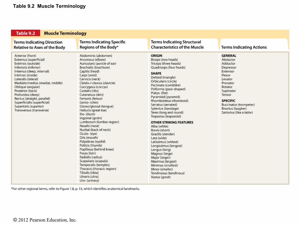
Organization of Skeletal Muscle Fibers
• Most muscle names provide clues to theiridentification or location
Size of the muscle Magnus: large Brevis: short
Specific body regions Brachialis
Shape of the muscle Trapezius
Orientation of muscle fibers Rectus, transverse, oblique
Specific or unusual features Biceps (two origins)
Identification of origin and insertion Sternocleidomastoid
Primary functions Flexor carpi radialis
References to actions Buccinators
© 2012 Pearson Education, Inc.
Table 9.2 Muscle Terminology
© 2012 Pearson Education, Inc.
Levers and Pulleys: A Systems Design for Movement
First-class levers Second-class levers
Characteristics of second-class levers are: The force is magnified. The resistance moves more slowly and covers a shorter distance.
Third-class levers The characteristics of the third-class lever are:
Speed and distance traveled are increased. The force produced must be great.

© 2012 Pearson Education, Inc.
Figure 9.15a The Three Classes of Levers
ResistanceFulcrum
Appliedforce
Movement completed
R
FAF
In a first-class lever, the applied force and the resistance are on oppositesides of the fulcrum. This lever can change the amount of force transmittedto the resistance and alter the direction and speed of movement.
R F
AF

© 2012 Pearson Education, Inc.
Figure 9.15b The Three Classes of Levers
Movement completed
AF
F
F
F
AF
In a second-class lever, the resistance lies between the applied force andthe fulcrum. This arrangement magnifies force at the expense of distanceand speed; the direction of movement remains unchanged.
R

© 2012 Pearson Education, Inc.
Figure 9.15c The Three Classes of Levers
In a third-class lever, the force is applied between the resistanceand the fulcrum. This arrangement increases speed and distancemoved but requires a larger applied force.
Movement completed
R
F
AF
AF
RF
© 2012 Pearson Education, Inc.
Aging and the Muscular System
Skeletal muscle fibers become smaller in diameter.
Skeletal muscles become smaller in diameter and less elastic.
Tolerance for exercise decreases.
The ability to recover from muscular injuries decreases.