Density and Duration of Pneumococcal Carriage Is Maintained by Transforming Growth Factor β1 and T...
10
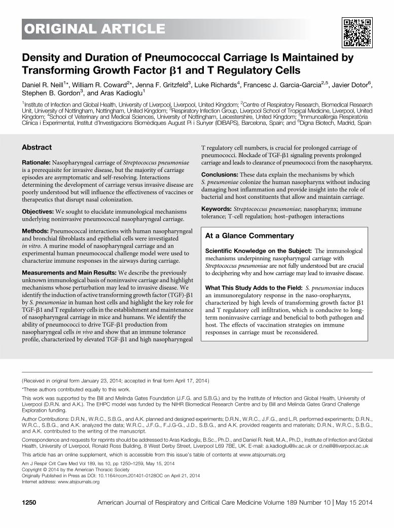
ORIGINAL ARTICLE Density and Duration of Pneumococcal Carriage Is Maintained by Transforming Growth Factor b1 and T Regulatory Cells Daniel R. Neill 1 *, William R. Coward 2 *, Jenna F. Gritzfeld 3 , Luke Richards 4 , Francesc J. Garcia-Garcia 2,5 , Javier Dotor 6 , Stephen B. Gordon 3 , and Aras Kadioglu 1 1 Institute of Infection and Global Health, University of Liverpool, Liverpool, United Kingdom; 2 Centre of Respiratory Research, Biomedical Research Unit, University of Nottingham, Nottingham, United Kingdom; 3 Respiratory Infection Group, Liverpool School of Tropical Medicine, Liverpool, United Kingdom; 4 School of Veterinary and Medical Sciences, University of Nottingham, Leicestershire, United Kingdom; 5 Immunoall ` ergia Respirat ` oria Cl´ ınica i Experimental, Institut d’Investigacions Biom ` ediques August Pi i Sunyer (IDIBAPS), Barcelona, Spain; and 6 Digna Biotech, Madrid, Spain Abstract Rationale: Nasopharyngeal carriage of Streptococcus pneumoniae is a prerequisite for invasive disease, but the majority of carriage episodes are asymptomatic and self-resolving. Interactions determining the development of carriage versus invasive disease are poorly understood but will influence the effectiveness of vaccines or therapeutics that disrupt nasal colonization. Objectives: We sought to elucidate immunological mechanisms underlying noninvasive pneumococcal nasopharyngeal carriage. Methods: Pneumococcal interactions with human nasopharyngeal and bronchial fibroblasts and epithelial cells were investigated in vitro. A murine model of nasopharyngeal carriage and an experimental human pneumococcal challenge model were used to characterize immune responses in the airways during carriage. Measurements and Main Results: We describe the previously unknown immunological basis of noninvasive carriage and highlight mechanisms whose perturbation may lead to invasive disease. We identify the induction of active transforming growth factor (TGF)-b1 by S. pneumoniae in human host cells and highlight the key role for TGF-b1 and T regulatory cells in the establishment and maintenance of nasopharyngeal carriage in mice and humans. We identify the ability of pneumococci to drive TGF-b1 production from nasopharyngeal cells in vivo and show that an immune tolerance profile, characterized by elevated TGF-b1 and high nasopharyngeal T regulatory cell numbers, is crucial for prolonged carriage of pneumococci. Blockade of TGF-b1 signaling prevents prolonged carriage and leads to clearance of pneumococci from the nasopharynx. Conclusions: These data explain the mechanisms by which S. pneumoniae colonize the human nasopharynx without inducing damaging host inflammation and provide insight into the role of bacterial and host constituents that allow and maintain carriage. Keywords: Streptococcus pneumoniae; nasopharynx; immune tolerance; T-cell regulation; host–pathogen interactions At a Glance Commentary Scientific Knowledge on the Subject: The immunological mechanisms underpinning nasopharyngeal carriage with Streptococcus pneumoniae are not fully understood but are crucial to deciphering why and how carriage may lead to invasive disease. What This Study Adds to the Field: S. pneumoniae induces an immunoregulatory response in the naso-oropharynx, characterized by high levels of transforming growth factor b1 and T regulatory cell infiltration, which is conducive to long- term noninvasive carriage and beneficial to both pathogen and host. The effects of vaccination strategies on immune responses in carriage must be reconsidered. ( Received in original form January 23, 2014; accepted in final form April 17, 2014 ) *These authors contributed equally to this work. This work was supported by the Bill and Melinda Gates Foundation (J.F.G. and S.B.G.) and by the Institute of Infection and Global Health, University of Liverpool (D.R.N. and A.K.). The EHPC model was funded by the NIHR Biomedical Research Centre and by Bill and Melinda Gates Grand Challenge Exploration funding. Author Contributions: D.R.N., W.R.C., S.B.G., and A.K. planned and designed experiments; D.R.N., W.R.C., J.F.G., and L.R. performed experiments; D.R.N., W.R.C., S.B.G., and A.K. analyzed the data; W.R.C., J.F.G., F.J.G-G., J.D., S.B.G., and A.K. provided reagents and materials; D.R.N., W.R.C., S.B.G., and A.K. contributed to the writing of the manuscript. Correspondence and requests for reprints should be addressed to Aras Kadioglu, B.Sc., Ph.D., and Daniel R. Neill, M.A., Ph.D., Institute of Infection and Global Health, University of Liverpool, Ronald Ross Building, 8 West Derby Street, Liverpool L69 7BE, UK. E-mail: [email protected] or [email protected] This article has an online supplement, which is accessible from this issue’s table of contents at www.atsjournals.org Am J Respir Crit Care Med Vol 189, Iss 10, pp 1250–1259, May 15, 2014 Copyright © 2014 by the American Thoracic Society Originally Published in Press as DOI: 10.1164/rccm.201401-0128OC on April 21, 2014 Internet address: www.atsjournals.org 1250 American Journal of Respiratory and Critical Care Medicine Volume 189 Number 10 | May 15 2014
Transcript of Density and Duration of Pneumococcal Carriage Is Maintained by Transforming Growth Factor β1 and T...
ORIGINAL ARTICLE
Density and Duration of Pneumococcal Carriage Is Maintained byTransforming Growth Factor b1 and T Regulatory CellsDaniel R. Neill1*, William R. Coward2*, Jenna F. Gritzfeld3, Luke Richards4, Francesc J. Garcia-Garcia2,5, Javier Dotor6,Stephen B. Gordon3, and Aras Kadioglu1
1Institute of Infection and Global Health, University of Liverpool, Liverpool, United Kingdom; 2Centre of Respiratory Research, Biomedical ResearchUnit, University of Nottingham, Nottingham, United Kingdom; 3Respiratory Infection Group, Liverpool School of Tropical Medicine, Liverpool, UnitedKingdom; 4School of Veterinary and Medical Sciences, University of Nottingham, Leicestershire, United Kingdom; 5Immunoallergia RespiratoriaClınica i Experimental, Institut d’Investigacions Biomediques August Pi i Sunyer (IDIBAPS), Barcelona, Spain; and 6Digna Biotech, Madrid, Spain
Abstract
Rationale: Nasopharyngeal carriage of Streptococcus pneumoniaeis a prerequisite for invasive disease, but the majority of carriageepisodes are asymptomatic and self-resolving. Interactionsdetermining the development of carriage versus invasive disease arepoorly understood but will influence the effectiveness of vaccines ortherapeutics that disrupt nasal colonization.
Objectives:We sought to elucidate immunological mechanismsunderlying noninvasive pneumococcal nasopharyngeal carriage.
Methods: Pneumococcal interactions with human nasopharyngealand bronchial fibroblasts and epithelial cells were investigatedin vitro. A murine model of nasopharyngeal carriage and anexperimental human pneumococcal challenge model were used tocharacterize immune responses in the airways during carriage.
Measurements and Main Results:We describe the previouslyunknown immunological basis of noninvasive carriage and highlightmechanisms whose perturbation may lead to invasive disease. Weidentify the induction of active transforming growth factor (TGF)-b1by S. pneumoniae in human host cells and highlight the key role forTGF-b1 andT regulatory cells in the establishment andmaintenanceof nasopharyngeal carriage in mice and humans. We identify theability of pneumococci to drive TGF-b1 production fromnasopharyngeal cells in vivo and show that an immune toleranceprofile, characterized by elevated TGF-b1 and high nasopharyngeal
T regulatory cell numbers, is crucial for prolonged carriage ofpneumococci. Blockade of TGF-b1 signaling prevents prolongedcarriage and leads to clearance of pneumococci from the nasopharynx.
Conclusions: These data explain the mechanisms by whichS. pneumoniae colonize the human nasopharynx without inducingdamaging host inflammation and provide insight into the role ofbacterial and host constituents that allow and maintain carriage.
Keywords: Streptococcus pneumoniae; nasopharynx; immunetolerance; T-cell regulation; host–pathogen interactions
At a Glance Commentary
Scientific Knowledge on the Subject: The immunologicalmechanisms underpinning nasopharyngeal carriage withStreptococcus pneumoniae are not fully understood but are crucialto deciphering why and how carriage may lead to invasive disease.
What This Study Adds to the Field: S. pneumoniae inducesan immunoregulatory response in the naso-oropharynx,characterized by high levels of transforming growth factor b1and T regulatory cell infiltration, which is conducive to long-term noninvasive carriage and beneficial to both pathogen andhost. The effects of vaccination strategies on immuneresponses in carriage must be reconsidered.
(Received in original form January 23, 2014; accepted in final form April 17, 2014 )
*These authors contributed equally to this work.
This work was supported by the Bill and Melinda Gates Foundation (J.F.G. and S.B.G.) and by the Institute of Infection and Global Health, University ofLiverpool (D.R.N. and A.K.). The EHPC model was funded by the NIHR Biomedical Research Centre and by Bill and Melinda Gates Grand ChallengeExploration funding.
Author Contributions: D.R.N., W.R.C., S.B.G., and A.K. planned and designed experiments; D.R.N., W.R.C., J.F.G., and L.R. performed experiments; D.R.N.,W.R.C., S.B.G., and A.K. analyzed the data; W.R.C., J.F.G., F.J.G-G., J.D., S.B.G., and A.K. provided reagents and materials; D.R.N., W.R.C., S.B.G.,and A.K. contributed to the writing of the manuscript.
Correspondence and requests for reprints should be addressed to Aras Kadioglu, B.Sc., Ph.D., and Daniel R. Neill, M.A., Ph.D., Institute of Infection and GlobalHealth, University of Liverpool, Ronald Ross Building, 8 West Derby Street, Liverpool L69 7BE, UK. E-mail: [email protected] or [email protected]
This article has an online supplement, which is accessible from this issue’s table of contents at www.atsjournals.org
Am J Respir Crit Care Med Vol 189, Iss 10, pp 1250–1259, May 15, 2014
Copyright © 2014 by the American Thoracic Society
Originally Published in Press as DOI: 10.1164/rccm.201401-0128OC on April 21, 2014
Internet address: www.atsjournals.org
1250 American Journal of Respiratory and Critical Care Medicine Volume 189 Number 10 | May 15 2014
Streptococcus pneumoniae (thepneumococcus) is an important humanpathogen that accounts for significantmorbidity and mortality worldwide,causing a range of invasive diseases,including pneumonia, meningitis, andsepsis. The pneumococcus is alsoa commensal of the human nasopharynx,and colonization of this importantecological niche is thought to bea prerequisite for invasive disease (1, 2).Where carriage rates are highest, risk ofinvasive pneumococcal disease is also high(3, 4). However, the actual incidence ofprogression from carriage to invasivedisease (the attack rate) is thought to be lessthan 1 in 10,000 (5), suggesting that theoccurrence of invasive disease is theexception rather than the norm. Thecontributory factors that lead to invasivedisease in the minority of such carriageevents are not known, but the host immunesystem and its interactions with colonizingpneumococci is certainly one aspect (3).
Transforming growth factor (TGF)-b1has a crucial immunosuppressive role ininnate and adaptive immunity (6). TGF-b1activity limits proinflammatory responsesand promotes tissue healing andremodeling after damage (6), and activationcan occur in response to damage to theepithelial layer (7). We have demonstratedthe importance of TGF-b1 in invasivepneumococcal infections, where inhibitionof TGF-b1 markedly increasedsusceptibility to invasive pneumonia innormally disease-resistant mice (8). Theimportance of TGF-b1 in the context ofpneumococcal infection of the lung relatesto its ability to drive T regulatory celldifferentiation and expansion, and thecorrelation of high TGF-b1 levels and Tregulatory cell numbers with reduced lungapoptosis suggests that immune regulationplays a key role in preventing bacterialdissemination by limiting inflammatorydamage to the lung epithelial barrier (8).
Correlative evidence suggests Tregulatory cells also play a role innasopharyngeal colonization: children withpneumococcus positive nasopharyngealswabs contained higher proportions of Tregulatory cells in adenoidal tissue thanchildren with pneumococcus negativecultures (9). We sought to elucidate thehost–pathogen interactions underpinningasymptomatic nasopharyngeal carriage.Identifying the immune phenotype in thecarrier state will aid our understanding of
how this equilibrium is disturbed ininvasive disease.
Some of the results of these studies havebeen previously reported in the form of anabstract (10).
Methods
Ethics StatementThe UK Home Office and University ofLiverpool ethics committee approved thestudy. Animal experiments were performedat the University of Liverpool. Ethicalapproval for human experimentalpneumococcal challenge was obtained fromthe National Health Service Research EthicsCommittee (08/H1001/52 and 11/NW/0592).
MiceFemale MF1 mice (9–14 wk of age)(Charles River, Margate, UK) wereacclimatized for 1 week before use.
BacteriaMouse-virulent serotype 2 Streptococcuspneumoniae strain D39 (NCTC 7466) or itsisogenic pneumolysin (Ply)-deficientmutant PLN-A were used throughout theexperiment. Infectious doses were preparedas described previously (11). Samplesfrom experimental human pneumococcalchallenge were taken from patientschallenged with serotype 6B pneumococci(12).
PneumolysinRecombinant Ply was expressed inEscherichia coli and purified as previouslydescribed (13). Additional details areprovided in the online supplement.
Infection of MiceAnimals were infected intranasally with 13105 colony-forming units (CFU) (low-dose)S. pneumoniae D39 or PLN-A or with1 3 107 CFU (high-dose) D39 in 10 ml PBSas previously described (14). Mice wereculled by cervical dislocation, andnasopharynx and lungs were prepared forassessment of bacterial CFU or leukocytes.A viable count of bacteria in murine tissuewas determined at prechosen intervals aspreviously described (14). Colonization for48 hours or more was considered a carriageevent. Additional details are provided inthe online supplement.
P17 Treatment of MiceAnimals were infected intranasally with 13105 CFU S. pneumoniae D39 and thenimmediately administered 500 mg of thepeptide TGF-b1 inhibitor P17 (15) or PBSby intraperitoneal injection. Further dosesof P17 or PBS were given at 1, 3, and 4 daysafter infection. This treatment regime waschosen to inhibit infection-induced TGF-b1 production without appreciably alteringhomeostatic TGF-b1 levels in thenasopharynx.
Experimental Human PneumococcalChallenge Model and Nasal WashHealthy adults with no respiratory diseaseand no natural carriage were recruited.Pneumococci were instilled in 100 ml salineinto each naris. Nasal wash was performedat 48 hours after inoculation (12, 16). Thepresence of carriage was assessed by serialdilution of nasal wash onto blood agar.Additional details are provided in theonline supplement.
Flow CytometryTissue cell suspensions were stained forsurface markers or transcription factors aspreviously described (8). Samples wereanalyzed using a FACScalibur flowcytometer (Becton Dickinson, Oxford, UK)running CellQuest acquisition and analyzedusing FlowJo (version 8.8.3; Tree Star,Ashland, OR). Additional details areprovided in the online supplement.
Cell CultureThe nasopharyngeal cell line Detroit 562(NE), immortalized human bronchialepithelial cells (J.W. Shay, University ofTexas, Dallas, TX) (17), primary humanbronchial fibroblasts (C.A. Feghali-Bostwick, University of Pittsburgh Schoolof Medicine, Pittsburgh, PA) (18), andhuman nasopharyngeal fibroblasts (Institutd’Investigacions Biomediques August Pi ISunyer and Centro de InvestigacionesBiomedicas en Red de EnfermedadesRespiratorias, Barcelona, Spain) (19) werecultured as described in the onlinesupplement. Cells were grown to confluencebefore addition of Ply or live or heat-killedD39. After 24 hours, culture supernatantswere collected and stored at 2808C.
MTT AssayCell toxicity was assessed by the 3-[4,5-dimethylthiazol-2-yl]-2,5-diphenyltetrazolium
ORIGINAL ARTICLE
Neill, Coward, Gritzfeld, et al.: Immune Regulation of Pneumococcal Carriage 1251

ORIGINAL ARTICLE
1252 American Journal of Respiratory and Critical Care Medicine Volume 189 Number 10 | May 15 2014
bromide (MTT) assay. Before the assay, culturesupernatants were centrifuged at 13,000 3 gfor 2 minutes, and the supernatant was passedthrough a 0.2-mm filter. Subsequently, 20 mlMTT (5 mg/ml) (Sigma Aldrich, Gillingham,UK) in culture media was added to cells andincubated for 1 hour. Medium was removed,and 100 ml of dimethyl sulfoxide (SigmaAldrich) was added for 30 minutes.Absorbance was read at 550 nm. Values areexpressed as % MTT absorbance of mediumcontrol.
Identification of CytokinesActive TGF-b1 within supernatants wasdetermined using luciferase-reportingtransformed mink lung epithelial cells (D.B.Rifkin, Langone Medical Center and Schoolof Medicine, New York University, NewYork) as previously described (20).Additional details are provided in theonline supplement.
StatisticsData were analyzed in GraphPad Prismusing a two-tailed unpaired Student’s t testor one- or two-way ANOVA with post-tests. Results with P values , 0.05 wereconsidered significant. Data representmean 6 SEM unless otherwise indicated.
Results
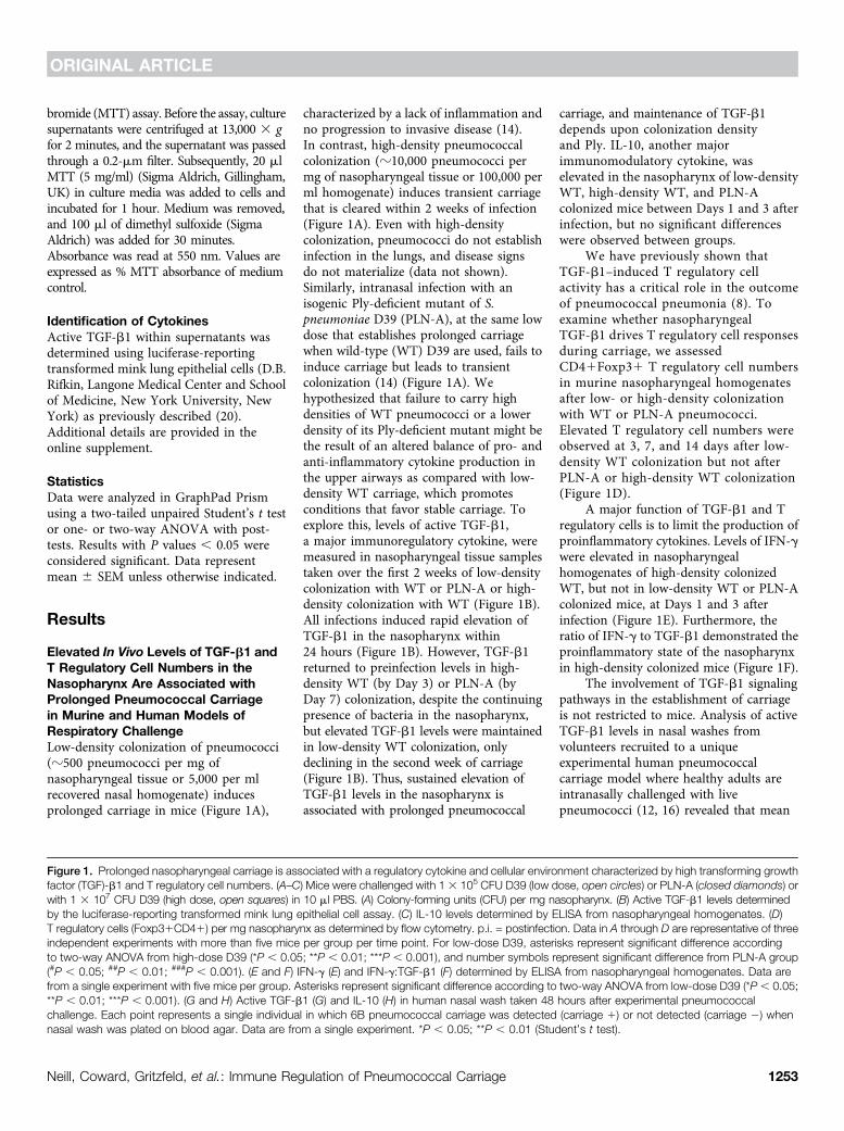
Elevated In Vivo Levels of TGF-b1 andT Regulatory Cell Numbers in theNasopharynx Are Associated withProlonged Pneumococcal Carriagein Murine and Human Models ofRespiratory ChallengeLow-density colonization of pneumococci(z500 pneumococci per mg ofnasopharyngeal tissue or 5,000 per mlrecovered nasal homogenate) inducesprolonged carriage in mice (Figure 1A),
characterized by a lack of inflammation andno progression to invasive disease (14).In contrast, high-density pneumococcalcolonization (z10,000 pneumococci permg of nasopharyngeal tissue or 100,000 perml homogenate) induces transient carriagethat is cleared within 2 weeks of infection(Figure 1A). Even with high-densitycolonization, pneumococci do not establishinfection in the lungs, and disease signsdo not materialize (data not shown).Similarly, intranasal infection with anisogenic Ply-deficient mutant of S.pneumoniae D39 (PLN-A), at the same lowdose that establishes prolonged carriagewhen wild-type (WT) D39 are used, fails toinduce carriage but leads to transientcolonization (14) (Figure 1A). Wehypothesized that failure to carry highdensities of WT pneumococci or a lowerdensity of its Ply-deficient mutant might bethe result of an altered balance of pro- andanti-inflammatory cytokine production inthe upper airways as compared with low-density WT carriage, which promotesconditions that favor stable carriage. Toexplore this, levels of active TGF-b1,a major immunoregulatory cytokine, weremeasured in nasopharyngeal tissue samplestaken over the first 2 weeks of low-densitycolonization with WT or PLN-A or high-density colonization with WT (Figure 1B).All infections induced rapid elevation ofTGF-b1 in the nasopharynx within24 hours (Figure 1B). However, TGF-b1returned to preinfection levels in high-density WT (by Day 3) or PLN-A (byDay 7) colonization, despite the continuingpresence of bacteria in the nasopharynx,but elevated TGF-b1 levels were maintainedin low-density WT colonization, onlydeclining in the second week of carriage(Figure 1B). Thus, sustained elevation ofTGF-b1 levels in the nasopharynx isassociated with prolonged pneumococcal
carriage, and maintenance of TGF-b1depends upon colonization densityand Ply. IL-10, another majorimmunomodulatory cytokine, waselevated in the nasopharynx of low-densityWT, high-density WT, and PLN-Acolonized mice between Days 1 and 3 afterinfection, but no significant differenceswere observed between groups.
We have previously shown thatTGF-b1–induced T regulatory cellactivity has a critical role in the outcomeof pneumococcal pneumonia (8). Toexamine whether nasopharyngealTGF-b1 drives T regulatory cell responsesduring carriage, we assessedCD41Foxp31 T regulatory cell numbersin murine nasopharyngeal homogenatesafter low- or high-density colonizationwith WT or PLN-A pneumococci.Elevated T regulatory cell numbers wereobserved at 3, 7, and 14 days after low-density WT colonization but not afterPLN-A or high-density WT colonization(Figure 1D).
A major function of TGF-b1 and Tregulatory cells is to limit the production ofproinflammatory cytokines. Levels of IFN-gwere elevated in nasopharyngealhomogenates of high-density colonizedWT, but not in low-density WT or PLN-Acolonized mice, at Days 1 and 3 afterinfection (Figure 1E). Furthermore, theratio of IFN-g to TGF-b1 demonstrated theproinflammatory state of the nasopharynxin high-density colonized mice (Figure 1F).
The involvement of TGF-b1 signalingpathways in the establishment of carriageis not restricted to mice. Analysis of activeTGF-b1 levels in nasal washes fromvolunteers recruited to a uniqueexperimental human pneumococcalcarriage model where healthy adults areintranasally challenged with livepneumococci (12, 16) revealed that mean
Figure 1. Prolonged nasopharyngeal carriage is associated with a regulatory cytokine and cellular environment characterized by high transforming growthfactor (TGF)-b1 and T regulatory cell numbers. (A–C) Mice were challenged with 13 105 CFU D39 (low dose, open circles) or PLN-A (closed diamonds) orwith 1 3 107 CFU D39 (high dose, open squares) in 10 ml PBS. (A) Colony-forming units (CFU) per mg nasopharynx. (B) Active TGF-b1 levels determinedby the luciferase-reporting transformed mink lung epithelial cell assay. (C) IL-10 levels determined by ELISA from nasopharyngeal homogenates. (D)T regulatory cells (Foxp31CD41) per mg nasopharynx as determined by flow cytometry. p.i. = postinfection. Data in A through D are representative of threeindependent experiments with more than five mice per group per time point. For low-dose D39, asterisks represent significant difference accordingto two-way ANOVA from high-dose D39 (*P , 0.05; **P , 0.01; ***P , 0.001), and number symbols represent significant difference from PLN-A group(#P , 0.05; ##P , 0.01; ###P , 0.001). (E and F) IFN-g (E) and IFN-g:TGF-b1 (F) determined by ELISA from nasopharyngeal homogenates. Data arefrom a single experiment with five mice per group. Asterisks represent significant difference according to two-way ANOVA from low-dose D39 (*P , 0.05;**P , 0.01; ***P , 0.001). (G and H) Active TGF-b1 (G) and IL-10 (H) in human nasal wash taken 48 hours after experimental pneumococcalchallenge. Each point represents a single individual in which 6B pneumococcal carriage was detected (carriage 1) or not detected (carriage 2) whennasal wash was plated on blood agar. Data are from a single experiment. *P , 0.05; **P , 0.01 (Student’s t test).
ORIGINAL ARTICLE
Neill, Coward, Gritzfeld, et al.: Immune Regulation of Pneumococcal Carriage 1253

TGF-b1 levels are higher in volunteers inwhom carriage successfully establishes afterpneumococcal challenge than in those whofail to develop carriage (Figure 1G).Furthermore, assessment of IL-10, anothermarker of immunomodulation andtolerance (21), revealed elevated levels incarriage-positive individuals but not inthose who fail to carry (Figure 1H).Carriage density in volunteers tested inthe study averaged 10,000 pneumococciper ml recovered nasal wash, which iscomparable to that seen when long-termcarriage is successfully established in mice(5,000 per ml nasopharyngeal homogenate)(Figure 1A).
These data demonstrate the increasedimmunoregulatory activity in thenasopharynx of mice and humans duringprolonged low-density episodes ofcarriage.
Carriage Is Associated with ReducedPolymorphonuclear LeukocyteInfiltration and an Accumulation ofAlternatively Activated Macrophagesin the NasopharynxTo assess the impact of increasedimmunoregulatory activity in thenasopharynx during carriage, neutrophiland macrophage numbers were assessed.High- or low-density colonization withWTor low-density colonization with PLN-Aled to infiltration of neutrophils into thenasopharynx by 24 hours (Figure 2A).However, neutrophil numbers werereduced by Day 3 after infection, and thisdecline was significantly more rapid inlow-density colonization with WT than inhigh-density WT or with PLN-A(Figure 2A).
Macrophage numbers increased in thenasopharynx during all infections and were
significantly elevated at Days 3 and 7 afterinfection as compared with prechallengelevels (Figure 2B). Although macrophagenumbers in nasopharynx at Day 7 afterinfection were comparable in all infectiongroups, a significantly higher proportion ofmacrophages from the low-density WTcolonization group (69%) expressedmannose receptor than from the high-density colonization with WT (29%) orPLN-A (26%) (Figure 2C). This resultsuggests that alternative activation ofmacrophages might occur in thenasopharynx of mice during long-termcarriage, and this is supported by theobservation of high levels of majorhistocompatibility complex class IIexpression on the mannosereceptor–expressing macrophages fromlow-density WT colonized mice, ascompared with mannose receptor–negative
Figure 2. Carriage is associated with decreased polymorphonuclear leukocyte (PMN) infiltration of the nasopharynx and alternative activation ofmacrophages. Mice were challenged with 1 3 105 colony-forming units (CFU) D39 (low dose, open circles) or PLN-A (closed diamonds) or with 1 3 107
CFU D39 (high dose, open squares) in 10 ml PBS. Neutrophil (PMN) (A) and macrophage (B) numbers per mg nasopharynx, as determined by flowcytometry. Neutrophils are defined as SSclowCD451Gr-1hiCD11bhiF4/80int/low; macrophages are defined as SSchiCD451Gr-12F4/80hi. p.i. = postinfection. CD206 (mannose receptor) (C) and major histocompatibility complex class (MHCII) expression on macrophages (D) at Day 7 after infection. Datain A through D are representative of three independent experiments with more than five mice per group per time point. For low-dose D39 in A and C,asterisks represent significant difference according to two-way ANOVA from high-dose D39 (*P , 0.05; **P , 0.01), and number symbols representsignificant difference from PLN-A (##P , 0.01).
ORIGINAL ARTICLE
1254 American Journal of Respiratory and Critical Care Medicine Volume 189 Number 10 | May 15 2014

macrophages or macrophages from high-density infections (Figure 2D).
Together, these data demonstrate thatlocal immune responses are altered in thenasopharynx during long-termpneumococcal carriage. High levels of TGF-b1 and T regulatory cells, reducedneutrophil infiltration, and alternativeactivation of macrophages characterize thisresponse, whereas lower TGF-b1 and Tregulatory cell levels and prolongedneutrophil infiltration aligned with classicalmacrophage activation favor clearance ofinfection.
Human Nasopharyngeal EpithelialCells and Fibroblasts Secrete ActiveTGF-b1 in Response to LiveS. pneumoniae and PlyTo elucidate the mechanism by whichpneumococcal colonization of thenasopharynx induced immunoregulatoryresponses, human nasopharyngealfibroblasts or epithelial cells were treatedwith pneumococci, resulting in significantinduction of active TGF-b1 (Figures 3A and3B). TGF-b1 production was completelyabrogated in assays performed with heat-killed D39 or PLN-A (Figures 3A–3D).TGF-b1 production drops off substantiallyat the highest D39 CFU numbers (2.5 3106) tested in fibroblasts and to a lesserextent in epithelial cells (Figures 3A–3D),suggesting differential host responses todiffering bacterial densities. This dropcannot be explained by increased cell deathalone in the case of fibroblasts because theyretain 100% viability even at 3 3 106 D39CFU (Figure 3E). By contrast, the slightdrop in TGF-b1 at high CFU numbers inepithelial cells (Figures 3B and 3D) may bedue to the significant cell death observed atthese bacterial concentrations (Figure 3F).Furthermore, consistent with theobservation that Ply is required forinduction of active TGF-b1 production,purified Ply was sufficient to induce TGF-b1 production from epithelial cells andfibroblasts (Figures 3G and 3H).
We have conducted preliminaryinvestigations into the mechanistic detail ofTGF-b1 induction in epithelial cells andfibroblasts of the upper and lower airways(see Figures E1 and E2 in the onlinesupplement). Our findings implicate theNLRP3 inflammasome (Figure E1) andsuggest the responses to pneumococcidiffer in the upper and lower airways(Figure E2).
TGF-b1 Blockade Prevents ProlongedCarriage of Pneumococci inthe NasopharynxTo determine whether immunomodulatoryresponses are essential for prolonged
pneumococcal carriage, we administered theP17 peptide inhibitor of TGF-b1 (8).Administration of P17 to mice 1 hourbefore intranasal administration ofa colonizing low dose of pneumococci, and
Figure 3. Pneumococci and pneumolysin induce active transforming growth factor (TGF)-b1production in nasopharyngeal fibroblasts and epithelial cells. Active TGF-b1 determined fromnasopharyngeal fibroblast (A, C, E, G) and nasopharyngeal epithelial cell (B, D, F, H) supernatants byluciferase-reporting transformed mink lung epithelial cell assay after stimulation with D39, heat-inactivated D39, pneumolysin (Ply)-deficient S. pneumoniae (PLN-A), or recombinant endotoxin free Ply.Survival of D39 and PLN-A stimulated nasopharyngeal fibroblasts (E) and nasopharyngeal epithelialcells (F ) determined by MTT assay. Medium alone was used as control. Open circles = D39; closedsquares = heat-inactivated D39 (A and B) or PLN-A (C–F). Asterisks in parentheses denote significantactive TGF-b1 induction induced by D39 over heat-inactivated D39 (A and B) or PLN-A (C and D)((*)P , 0.05; (**)P , 0.01). Asterisks denote significant active TGF-b1 induction induced by D39 overuntreated cells (*P , 0.05; **P , 0.01). Analysis is from two-way (A–D) or one-way (G) ANOVA.
ORIGINAL ARTICLE
Neill, Coward, Gritzfeld, et al.: Immune Regulation of Pneumococcal Carriage 1255
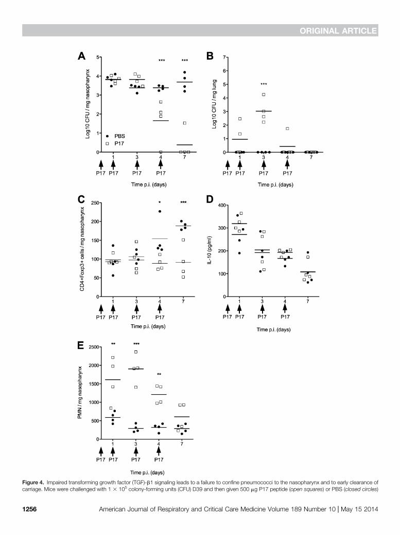
Figure 4. Impaired transforming growth factor (TGF)-b1 signaling leads to a failure to confine pneumococci to the nasopharynx and to early clearance ofcarriage. Mice were challenged with 1 3 105 colony-forming units (CFU) D39 and then given 500 mg P17 peptide (open squares) or PBS (closed circles)
ORIGINAL ARTICLE
1256 American Journal of Respiratory and Critical Care Medicine Volume 189 Number 10 | May 15 2014
subsequently at 1, 3, and 4 days afterinfection, profoundly reducedpneumococcal carriage in the nasopharynx(Figure 4A). Although initial colonizationdensity was unaffected in P17-treated mice,by 4 days after infection mice hadsignificantly lower bacterial numbers in thenasopharynx as compared with PBS-treatedcontrols (1.65 log10 mean CFU per mgtissue vs. 3.38 log10 CFU per mg), and by7 days after infection three out of four P17-treated mice had cleared carriage, whereasall control mice still had high densitiesof colonized bacteria in the nasopharynx.Before clearing of the infection, P17-treatedmice experienced a transient translocationof pneumococci from nasopharynx to lungsbetween Days 1 and 4 after infection(Figure 4B). This bacterial translocationwas completely absent in control animals.Blockade of TGF-b1 led to a failure toinduce T regulatory cell responses in thenasopharynx (Figure 4C), although IL-10production was unaffected (Figure 4D).TGF-b1 blockade was also associated withan exacerbated inflammatory responsecharacterized by increased neutrophil influxof the nasopharynx (Figure 4E).
Collectively, these data highlight thekey role of TGF-b1 and T regulatory cellsin limiting proinflammatory responses inthe nasopharynx and in allowing thepersistence of pneumococcal carriage. Itis likely that in different situationsdysregulation of TGF-b1 signaling wouldlead to either pathogen clearance orpathogen dissemination to deeper tissues.
Discussion
S. pneumoniae is a pathogen of significantclinical importance, and elucidating itsinteractions with host immunity iscrucial to our understanding of itspathogenesis. Here we identify the majorimmunoregulatory cytokine TGF-b1 asthe key determinant of the duration ofpneumococcal colonization of the upperairways. Low pneumococcal carriagedensity induces immunoregulatoryresponses characterized by sustained
elevation of nasopharyngeal TGF-b1 and Tregulatory cell numbers and alternativeactivation of macrophages. Consequently,pneumococci are maintained at low densityin the nasopharynx for prolonged periodswithout becoming invasive. By contrast,high-density colonization induces stronginflammatory responses that supersede theTGF-b1–driven pathway and thus clearbacteria from the nasopharynx. The use ofthe P17 peptide (8, 15, 22) to directlyinhibit TGF-b1 demonstrates the criticalrole of regulatory responses in mediatinglong-term pneumococcal carriage becausemice fail to raise T regulatory cell responsesin the nasopharynx and rapidly clearbacteria from the upper airways in theabsence of TGF-b1 signaling.
The suppression of inflammatoryresponses by S. pneumoniae–induced TGF-b1 and T regulatory cells may be importantto preserve the balance of flora and lessentissue damage, but it is clear that regulatoryresponses are also essential for theprolonged carriage of pneumococci in theupper airways; thus, targeting TGF-b1signaling may be a means to eliminatecarriage, a prerequisite for invasive disease.
TGF-b1 inhibition did not preventestablishment of pneumococcalcolonization in the nasopharynx, butbacteria were gradually cleared over thefirst week of infection. This correlated witha failure to induce T regulatory responses inthe nasopharynx in response to bacterialcolonization; hence, in line with our in vitrohuman airway epithelial cell culture data,we favor a model whereby pneumococciinduce airway epithelial cell TGF-b1production, which drives induced Tregulatory cell generation in thenasopharynx. These T regulatory cells limitproinflammatory responses, preventingtissue damage but also allowing persistenceof carriage. In support of this model,neutrophil influx into the nasopharynx wasgreatly enhanced in P17-treated mice,although we cannot rule out direct or Tregulatory cell–independent effects of TGF-b1 on neutrophil function, as has beenreported previously (23, 24). Theobservation that nasopharyngeal IL-10
levels were unaffected in P17-treated micesuggests that T regulatory cells are not theexclusive source of IL-10 and that IL-10alone is not sufficient to maintain carriage.However, the clear correlation betweenestablishment of carriage and elevated nasalwash IL-10 in experimental humanpneumococcal carriage suggests that IL-10may play some role in the colonizationprocess.
A consequence of the reduced Tregulatory cell activity in P17-treated micewas a transient colonization of the lung byS. pneumoniae. We postulate that thenormal role of T regulatory cells in thenasopharynx is to prevent this bacterialspread by limiting inflammatory tissuedamage. A prolonged but containedinfection is better for the host than anuncontrolled but shorter one.
In contrast to low-density colonization,high densities of pneumococcal colonization(a “high-risk” dose) induce only transientTGF-b1 production in the nasopharynx,with significantly reduced T regulatory cellinfiltration and, instead, an inflammatoryphenotype characterized by neutrophilinfiltration, classical activation ofmacrophages, and IFN-g productiondominates. Consequently, bacteria arecleared from the nasopharynx beforeinvasive disease can develop. We postulatethat deregulation of the balance betweenimmune tolerance and inflammatoryreactivity leads to invasive disease. We haveshown that Ply is a key driver of TGF-b1and regulatory responses, and it may followthat cases of human invasive disease withpneumococci that have Ply with reduced orno hemolytic activity (such as someserotype-1 strains) (25) result from thefailure to balance immune responses afterinfection
Our observations in the murinecarriage model are supported by dataobtained using a unique experimentalhuman pneumococcal carriage model(12, 16). We show that individuals whosuccessfully carry pneumococci in theirnasopharynx by 48 hours after challengehave significantly higher levels of TGF-b1and IL-10 in nasal wash samples than those
Figure 4. (Continued). at 0, 1, 3, and 4 days after infection. (A) CFU per mg nasopharynx. (B) CFU per mg lung. (C) T regulatory cells (defined asFoxp31CD41) per mg nasopharynx as determined by flow cytometry. (D) IL-10 levels in nasopharyngeal homogenates determined by ELISA. (E)Neutrophils (polymorphonuclear leukocytes [PMN] defined as SSclowCD451Gr-1hiCD11bhiF4/80int/low) per mg nasopharynx as determined byflow cytometry. Data are from a single experiment with 16 mice per treatment. Asterisks represent significant difference according to two-way ANOVA(*P , 0.05; **P , 0.01; ***P , 0.001).
ORIGINAL ARTICLE
Neill, Coward, Gritzfeld, et al.: Immune Regulation of Pneumococcal Carriage 1257
who clear the challenge dose. To thebest of our knowledge, this is the firstdemonstration in mouse and humanmodels of nasopharyngeal colonization thatregulatory cytokines play a key role insustaining pneumococcal colonization.Carriage density in colonized humanvolunteers was comparable to what weobserved when long-term carriage issuccessfully established in mice.
We have demonstrated thatS. pneumoniae and Ply directly stimulateproduction of TGF-b1 from human airwayepithelial cells and fibroblasts. We showthat host pattern recognition receptorsplay an essential role in this interactionand that the dependency upon ion efflux,cathepsin B, and lysosomal degradationimplicates the NLRP3 inflammasome inthe signaling process. Furthermore, wehave demonstrated that this occursthrough a mechanism dependent uponpotassium efflux, as has beendemonstrated for cytokine productioninduced by the pore-forming toxins ofStaphylococcus aureus (26) and Aeromonashydrophilia (27).
Alternative activation of macrophagesinduces up-regulation of a characteristic setof surface markers, including the mannosereceptor and major histocompatibilitycomplex class II (28), and appears to bea characteristic of prolonged pneumococcalcarriage in our model. Production of TGF-b1 by alternatively activated macrophageshas been documented (29), and the cellshave been implicated in tissue repair andremodeling (30) and in protection ofthe host from damaging inflammationduring pathology (31, 32). In the contextof pneumococcal nasopharyngealcolonization, they may limit inflammationto prevent tissue damage and thus promoteprolonged carriage.
Our results highlight the delicatebalance between the requirements of thehost to maintain epithelial barrier integrityand prevent inflammatory tissue damagewith the need to combat potentiallydangerous pathogen colonization. Thisequilibrium needs to be considered in thedesign of anti-pneumococcal therapeuticsand vaccines that alter the density ofpneumococcal carriage and hence affect
upper airways immune responses. Wehypothesize that the prolonged butcontained and controlled carriage mediatedby T regulatory cells allows the host time tomount protective memory immuneresponses against the pneumococcus thatmay protect against invasive disease. Wehave previously demonstrated the potentialfor natural carriage to boost vaccineresponses (33). Furthermore, elucidationof factors conducive to carriage orclearance may open up avenues fortreatments tailored to patients, wherelevels of immunomodulatory cytokine innasal wash might be used as a predictorof future responses to infection orvaccination. n
Author disclosures are available with the textof this article at www.atsjournals.org.
Acknowledgment: The authors thank thevolunteers in the Experimental HumanPneumococcal Carriage study, the clinical andlaboratory team, particularly Angie Wright,Andrea Collins and Daniela Ferreira, as well as theteam at the Clinical Research Unit of the RoyalLiverpool University Hospital.
References
1. Kadioglu A, Weiser JN, Paton JC, Andrew PW. The roleof Streptococcus pneumoniae virulence factors in hostrespiratory colonization and disease. Nat Rev Microbiol 2008;6:288–301.
2. Weiser JN. The pneumococcus: why a commensal misbehaves. J MolMed (Berl) 2010;88:97–102.
3. Bogaert D, De Groot R, Hermans PW. Streptococcus pneumoniaecolonisation: the key to pneumococcal disease. Lancet Infect Dis2004;4:144–154.
4. Hausdorff WP, Feikin DR, Klugman KP. Epidemiological differencesamong pneumococcal serotypes. Lancet Infect Dis 2005;5:83–93.
5. Sleeman KL, Griffiths D, Shackley F, Diggle L, Gupta S, Maiden MC,Moxon ER, Crook DW, Peto TE. Capsular serotype-specific attackrates and duration of carriage of Streptococcus pneumoniae ina population of children. J Infect Dis 2006;194:682–688.
6. Li MO, Wan YY, Sanjabi S, Robertson AK, Flavell RA. Transforminggrowth factor-beta regulation of immune responses. Annu RevImmunol 2006;24:99–146.
7. Howat WJ, Holgate ST, Lackie PM. TGF-beta isoform release andactivation during in vitro bronchial epithelial wound repair. Am JPhysiol Lung Cell Mol Physiol 2002;282:L115–L123.
8. Neill DR, Fernandes VE, Wisby L, Haynes AR, Ferreira DM, Laher A,Strickland N, Gordon SB, Denny P, Kadioglu A, et al. T regulatory cellscontrol susceptibility to invasive pneumococcal pneumonia in mice.PLoS Pathog 2012;8:e1002660.
9. Zhang Q, Leong SC, McNamara PS, Mubarak A, Malley R, Finn A.Characterisation of regulatory T cells in nasal associated lymphoidtissue in children: relationships with pneumococcal colonization.PLoS Pathog 2011;7:e1002175.
10. Neill DR, Coward WR, Gritzfeld JF, Richards L, Garcia-Garcia FJ, DotorJ, Gordon SB, Kadioglu A. Density and duration of pneumococcalcarriage is maintained by TGFB1 and T regulatory cells. Presented atthe British Society of Immunology Congress 2014, Liverpool, UK.
11. Kadioglu A, Gingles NA, Grattan K, Kerr A, Mitchell TJ, Andrew PW.Host cellular immune response to pneumococcal lung infection inmice. Infect Immun 2000;68:492–501.
12. Gritzfeld JF, Wright AD, Collins AM, Pennington SH, Wright AK,Kadioglu A, Ferreira DM, Gordon SB. Experimental humanpneumococcal carriage. J Vis Exp 2013; (72).
13. Gilbert RJ, Rossjohn J, Parker MW, Tweten RK, Morgan PJ, MitchellTJ, Errington N, Rowe AJ, Andrew PW, Byron O. Self-interaction ofpneumolysin, the pore-forming protein toxin of Streptococcuspneumoniae. J Mol Biol 1998;284:1223–1237.
14. Richards L, Ferreira DM, Miyaji EN, Andrew PW, Kadioglu A. Theimmunising effect of pneumococcal nasopharyngeal colonisation;protection against future colonisation and fatal invasive disease.Immunobiology 2010;215:251–263.
15. Dotor J, Lopez-Vazquez AB, Lasarte JJ, Sarobe P, Garcıa-Granero M,Riezu-Boj JI, Martınez A, Feijoo E, Lopez-Sagaseta J, Hermida J, et al.Identification of peptide inhibitors of transforming growth factor beta 1using a phage-displayed peptide library. Cytokine 2007;39:106–115.
16. Ferreira DM, Neill DR, Bangert M, Gritzfeld JF, Green N, Wright AK,Pennington SH, Bricio-Moreno L, Moreno AT, Miyaji EN, et al.Controlled human infection and rechallenge with Streptococcuspneumoniae reveals the protective efficacy of carriage in healthyadults. Am J Respir Crit Care Med 2013;187:855–864.
17. Ramirez RD, Sheridan S, Girard L, Sato M, Kim Y, Pollack J, Peyton M,Zou Y, Kurie JM, Dimaio JM, et al. Immortalization of humanbronchial epithelial cells in the absence of viral oncoproteins. CancerRes 2004;64:9027–9034.
18. Coward WR, Watts K, Feghali-Bostwick CA, Jenkins G, Pang L.Repression of IP-10 by interactions between histone deacetylationand hypermethylation in idiopathic pulmonary fibrosis. Mol Cell Biol2010;30:2874–2886.
19. Roca-Ferrer J, Garcia-Garcia FJ, Pereda J, Perez-Gonzalez M, Pujols L,Alobid I, Mullol J, Picado C. Reduced expression of coxs andproduction of prostaglandin e(2) in patients with nasal polyps with orwithout aspirin-intolerant asthma. J Allergy Clin Immunol 2011;128:66–72, e61.
ORIGINAL ARTICLE
1258 American Journal of Respiratory and Critical Care Medicine Volume 189 Number 10 | May 15 2014
20. Abe M, Harpel JG, Metz CN, Nunes I, Loskutoff DJ, Rifkin DB. An assayfor transforming growth factor-beta using cells transfected witha plasminogen activator inhibitor-1 promoter-luciferase construct.Anal Biochem 1994;216:276–284.
21. Rubtsov YP, Rasmussen JP, Chi EY, Fontenot J, Castelli L, Ye X,Treuting P, Siewe L, Roers A, Henderson WR Jr, et al. RegulatoryT cell-derived interleukin-10 limits inflammation at environmentalinterfaces. Immunity 2008;28:546–558.
22. Gil-Guerrero L, Dotor J, Huibregtse IL, Casares N, Lopez-VazquezAB, Rudilla F, Riezu-Boj JI, Lopez-Sagaseta J, Hermida J, VanDeventer S, et al. In vitro and in vivo down-regulation of regulatoryT cell activity with a peptide inhibitor of TGF-beta1. J Immunol 2008;181:126–135.
23. Ganeshan K, Johnston LK, Bryce PJ. TGF-b1 limits the onset of innatelung inflammation by promoting mast cell-derived IL-6. J Immunol2013;190:5731–5738.
24. Steiger S, Harper JL. Neutrophil cannibalism triggers transforminggrowth factor b1 production and self regulation of neutrophilinflammatory function in monosodium urate monohydratecrystal-induced inflammation in mice. Arthritis Rheum 2013;65:815–823.
25. Jefferies JM, Johnston CH, Kirkham LA, Cowan GJ, Ross KS, Smith A,Clarke SC, Brueggemann AB, George RC, Pichon B, et al. Presenceof nonhemolytic pneumolysin in serotypes of Streptococcuspneumoniae associated with disease outbreaks. J Infect Dis 2007;196:936–944.
26. Walev I, Reske K, Palmer M, Valeva A, Bhakdi S. Potassium-inhibitedprocessing of IL-1 beta in human monocytes. EMBO J 1995;14:1607–1614.
27. Gurcel L, Abrami L, Girardin S, Tschopp J, van der Goot FG. Caspase-1activation of lipid metabolic pathways in response to bacterial pore-forming toxins promotes cell survival. Cell 2006;126:1135–1145.
28. Gordon S. Alternative activation of macrophages. Nat Rev Immunol2003;3:23–35.
29. Lee CG, Homer RJ, Zhu Z, Lanone S, Wang X, Koteliansky V, ShipleyJM, Gotwals P, Noble P, Chen Q, et al. Interleukin-13 induces tissuefibrosis by selectively stimulating and activating transforming growthfactor beta(1). J Exp Med 2001;194:809–821.
30. Gratchev A, Guillot P, Hakiy N, Politz O, Orfanos CE, Schledzewski K,Goerdt S. Alternatively activated macrophages differentially expressfibronectin and its splice variants and the extracellular matrix proteinbetaIG-H3. Scand J Immunol 2001;53:386–392.
31. Ponomarev ED, Maresz K, Tan Y, Dittel BN. CNS-derived interleukin-4is essential for the regulation of autoimmune inflammation andinduces a state of alternative activation in microglial cells. J Neurosci2007;27:10714–10721.
32. Cao Y, Brombacher F, Tunyogi-CsapoM, Glant TT, Finnegan A. Interleukin-4 regulates proteoglycan-induced arthritis by specifically suppressing theinnate immune response. Arthritis Rheum 2007;56:861–870.
33. Neill DR, Smeaton S, Bangert M, Kadioglu A. Nasopharyngeal carriagewith Streptococcus pneumoniae augments the immunizing effect ofpneumolysin toxoid B. J Allergy Clin Immunol 2013;131:1433–1435, e1.
ORIGINAL ARTICLE
Neill, Coward, Gritzfeld, et al.: Immune Regulation of Pneumococcal Carriage 1259