Cytochemical and autoradiographic study of the early nuclear lesions induced by an ellipticine...
15
Eurj Comer Cfin Chcol, Vol. 18. No. 3. pp. 29-306. 1982 Printed in Great Britain. 0277-5379/82/030291-15~3.w/0 @I 1982 Pergamon Press Ltd. Cytochemical and Autoradiographic Study of the Early Nuclear Lesions Induced by an Ellipticine Derivative in Isolated Rat Hepatocytes NICOLE SALkS* and EDMOND PUVIONt Laboratoire de Microscopic Electronique, Znstitut de Recherches Scientifiques sur le Cancer, B.Z? no&$F-94802, Villejuif, Cedex, France Abstract-Zsolated rat liver cells maintained in primary culture were treated with 9-OH ellipticinium (g-OH E + ). The effects of this drug on the nuclear ultrastructure and on the chromatin transcriptional activity were studied by the combination of cytochemistry with high resolution autoradiography. At a low concentration (1 pg/ml) for periods ranging from 10 min to 3 hr, S-OH E + induced chromatin clumping, nucleolar microsegregation and a diminution in the number of perichromatin and interchromatin fibrils. Autoradiography revealed that this compound inhibited rapidly the incorporation of [5-‘HI-uridine in the nucleus, preferentially but not exclusively in the nucleolar area. In addition, the distribution of the radioactivity in the nucleoli proved that the processing of the pre-ribosomal ribonucleic acids (fire-rRNA) syn- thesized in the presence of the drug was blocked while the processing during 9-OH E + treatment of normally synthesizedfire-rRNA was not altered. These findings suggest that the inhibition of pre-rRNA processing might result from an impairment of factors controlling this processing rather than from a direct action of 9-OH E + on fire-rRNA molecules. INTRODUCTION ELLIPTICINE (E) is a natural substance which has been extracted from plants belonging to the botanical family of Apocynaceae, such as Och- rosia ellifitica. The dimethyl pyridocarbazole structure of this alkaloid endows it with inter- calative properties towards DNA [l, 23. The antitumoral activity of E [3], initially men- tioned by Dalton et al. [4] and by Hartwell et al. [5], has been subsequently studied by LeMen et al. [6], MathC et al. [7] and Le Pecq et al. [8]. The chemotherapeutic index appeared sufficiently interesting to initiate clinical trials, but these were soon interrupted because of undesirable secondary effects such as cardio- vascular lesions or cerebellar ataxia. Ellipticiniums (E + ) are synthetic substituted derivatives bearing a quaternarized ammonium Accepted 20 October 1981. *Present address: U.E.R. des sciences pharmaceutiques et biologiques, UniversitC Paris V, 4 avenue de I’Observatoire 75006, Paris, France. tTo whom correspondence and requests for reprints should be addressed. [9]. The affinity of these molecules to DNA is much (up to loo-fold) higher than that of the natural alkaloid. At the same time, the anti- tumoral activity is increased [ 10,111, which has stimulated renewed interest for the continua- tion of clinical trials. However, only a few works deal with the morphological effects of these drugs on cell structure. Dufer et al. [12] studied by means of optic microscopy the modifications of leukocytes in rats after 10 days of treatment with methoxy9-ellipticine. They histochemically detected quantitative variations in acid phosphatases and esterases. Structural abnormalities of mitochondria were reported at the electron microscope level in HeLa cells treated with E derivatives [IS], as well as in L 1210 cells [ll]. Concerning the nucleus, multi- lobation [l l] and appearance of punctiform chromatin [IS] were observed by the same authors. But, to our knowledge, no precise ultrastructural description has been made of nuclear lesions. Isolated rat liver cells maintained in vitro offer a suitable experimental model for study- ing modifications of nuclear structural com- ponents. Their nuclear morphology remains 291
Transcript of Cytochemical and autoradiographic study of the early nuclear lesions induced by an ellipticine...

Eurj Comer Cfin Chcol, Vol. 18. No. 3. pp. 29-306. 1982 Printed in Great Britain.
0277-5379/82/030291-15~3.w/0 @I 1982 Pergamon Press Ltd.
Cytochemical and Autoradiographic Study of the Early Nuclear Lesions Induced by an Ellipticine Derivative in Isolated Rat Hepatocytes
NICOLE SALkS* and EDMOND PUVIONt Laboratoire de Microscopic Electronique, Znstitut de Recherches Scientifiques sur le Cancer, B.Z? no&$ F-94802, Villejuif,
Cedex, France
Abstract-Zsolated rat liver cells maintained in primary culture were treated with 9-OH ellipticinium (g-OH E + ). The effects of this drug on the nuclear ultrastructure and on the chromatin transcriptional activity were studied by the combination of cytochemistry with high resolution autoradiography. At a low concentration (1 pg/ml) for periods ranging from 10 min to 3 hr, S-OH E + induced chromatin clumping, nucleolar microsegregation and a diminution in the number of perichromatin and interchromatin fibrils. Autoradiography revealed that this compound inhibited rapidly the incorporation of [5-‘HI-uridine in the nucleus, preferentially but not exclusively in the nucleolar area. In addition, the distribution of the radioactivity in the nucleoli proved that the processing of the pre-ribosomal ribonucleic acids (fire-rRNA) syn- thesized in the presence of the drug was blocked while the processing during 9-OH E + treatment of normally synthesized fire-rRNA was not altered. These findings suggest that the inhibition of pre-rRNA processing might result from an impairment of factors controlling this processing rather than from a direct action of 9-OH E + on fire-rRNA molecules.
INTRODUCTION ELLIPTICINE (E) is a natural substance which has been extracted from plants belonging to the botanical family of Apocynaceae, such as Och- rosia ellifitica. The dimethyl pyridocarbazole structure of this alkaloid endows it with inter- calative properties towards DNA [l, 23. The antitumoral activity of E [3], initially men- tioned by Dalton et al. [4] and by Hartwell et al. [5], has been subsequently studied by LeMen et al. [6], MathC et al. [7] and Le Pecq et al. [8]. The chemotherapeutic index appeared sufficiently interesting to initiate clinical trials, but these were soon interrupted because of undesirable secondary effects such as cardio- vascular lesions or cerebellar ataxia.
Ellipticiniums (E + ) are synthetic substituted derivatives bearing a quaternarized ammonium
Accepted 20 October 1981. *Present address: U.E.R. des sciences pharmaceutiques et
biologiques, UniversitC Paris V, 4 avenue de I’Observatoire 75006, Paris, France.
tTo whom correspondence and requests for reprints should be addressed.
[9]. The affinity of these molecules to DNA is much (up to loo-fold) higher than that of the natural alkaloid. At the same time, the anti- tumoral activity is increased [ 10,111, which has stimulated renewed interest for the continua- tion of clinical trials. However, only a few works deal with the morphological effects of these drugs on cell structure. Dufer et al. [12] studied by means of optic microscopy the modifications of leukocytes in rats after 10 days of treatment with methoxy9-ellipticine. They histochemically detected quantitative variations in acid phosphatases and esterases. Structural abnormalities of mitochondria were reported at the electron microscope level in HeLa cells treated with E derivatives [IS], as well as in L 1210 cells [ll]. Concerning the nucleus, multi- lobation [l l] and appearance of punctiform chromatin [IS] were observed by the same authors. But, to our knowledge, no precise ultrastructural description has been made of nuclear lesions.
Isolated rat liver cells maintained in vitro offer a suitable experimental model for study- ing modifications of nuclear structural com- ponents. Their nuclear morphology remains
291

292 Nicole Salk and Edmond Puvion
for at least 36 hr similar to that of in situ hepa- tocytes, with clearly defined nucleolar, con- densed chromatin and interchromatin areas. Such structural features facilitate the obser- vation of ribonucleoproteic (RNP) structures after appropriate staining. In addition, these cells lend themselves easily to labeling. Con- sequently, several studies carried out recently on isolated rat liver cells resulted in a better knowledge of the functional significance of the different nuclear RNP structures [14].
The purpose of this experiment was to in- vestigate, by means of the combination of cytochemistry with high resolution autoradio- graphy, the early effects of E on the nucleus of isolated rat liver cells. Our findings are dis- cussed in the light of recent results on metabolic and structural lesions induced by various inhibitors of RNA metabolism.
MATERIALS AND METHODS
Isolation of adult rat hepatocytes Three-month-old male rats ,(strain WAG)
were used after a fasting period of 18 hr. The hepatocytes were isolated as previously des- cribed [15]. The pellets of liver cells were then resuspended in Eagle’s Minimum Essential Medium (MEM), supplemented with 10% calf serum (700,000 cells per ml). Three ml of this suspension were put into 25cmz Falcon flasks. The incubation was carried out at 37°C for 3 hr in an atmosphere of 95% air and 5% COr. Then the medium was changed and the incubation carried out for about 20 hr before the beginning of the experiment.
Ellifiticine treatment The following compounds were used: ellip-
ticine (E), 9-OH ellipticine (g-OH E) and the same substances with a quaternarized am- monium, i.e., ellipticinium iodo-methylate (E +) and 9-OH ellipticinium aceto-methylate (9- OH E+).
Twenty-five ~1 of a 2 mg/ml solution (either in water or in 10-z M HCl or in 10% dimethyl sulfoxide) were added to 5 ml of medium in order to obtain a final concentration of 10 pg/ml. Concentrations of 5 pg/ml, 1 pg/ml and 0.1 pg/ml were prepared by l/2 and suc- cessive l/10 dilutions of the 2 mg/ml solution with water and addition of 25 ~1 of these solu- tions per flask. Controls were made by addition of 25 ~1 of each solvent used per flask.
Cells were treated at various concentrations from 5 min to 24 hr.
Electron microscopy At the end of each experiment, the cells were
fixed in the flask by replacing the medium with 1.6% glutaraldehyde in Siirensen’s phosphate buffer (0.1 M, pH 7.4) at +4”C for 45 min.
The cells were then rinsed in the same buffer, scraped and sedimented by centrifuga- tion (10 min at 3500g). Dehydration of the pellets was carried out in ascending concen- trations of ethanol and Epon inclusion, achieved by the usual technique.
Thin sections were stained either with the EDTA regressive stain for ribonucleoproteins [S] or with uranyl acetate and lead citrate.
Autoradiography Two types of experiments were carried out.
In the first type, cells were incubated for periods of 15 min or 1 hr in ellipticine-contain- ing medium and then labeled by addition of 80 PCi of triated uridine ([5-‘HI-uridine, C.E.A., Saclay, France; specific activity: 20- 30 Ci/mmol). After SO- or 60-min exposures to [5-SH]-uridine, the hepatocytes were briefly rinsed with serum-free MEM containing 1 mg/ml of non-radioactive uridine at 0°C. They were then fixed in 1.6% glutaraldehyde and embedded in Epon. Controls were treated in the same way in a medium deprived of ellipticine.
In the second type of experiment, the cells were labeled for 15 min in the presence of 100 &i/ml of [5-‘HI-uridine, then rinsed briefly as above and post-incubated for 1 or 3 hr in the presence of ellipticine and 100 pg/ml of non-radioactive uridine. Controls were fixed immediately after labeling and following 1 or 3 hr of post-incubation in the presence of un- labeled uridine. Ilford L emulsion was applied onto ultra-thin sections mounted on formvar- coated grids by the loop technique [16]. After 3-4 months of exposure, the autoradiographs were developed with the gold latensification technique [17]. The grids were then stained preferentially for RNP by the regressive stain of Bernhard [3] modified for autoradiography [18], or conventionally by uranyl acetate and lead citrate. A quantitative analysis of the in- corporation of [5-3H]-uridine in the nucleolar and extra-nucleolar areas and in the cytoplasm was carried out in the first type of experiment, following 30 min of labeling. Grain counting was effected on nuclei photographed under controlled magnification. Measurements of areas, determination of grain densities and statis- tical calculations and comparisons were per- formed by using a Hewlett-Packard 9820 cal- culator fitted to a graphic digitizer.

Ellipticine Effects on Hepatocyte Nuclei 293
Fig. l(o).
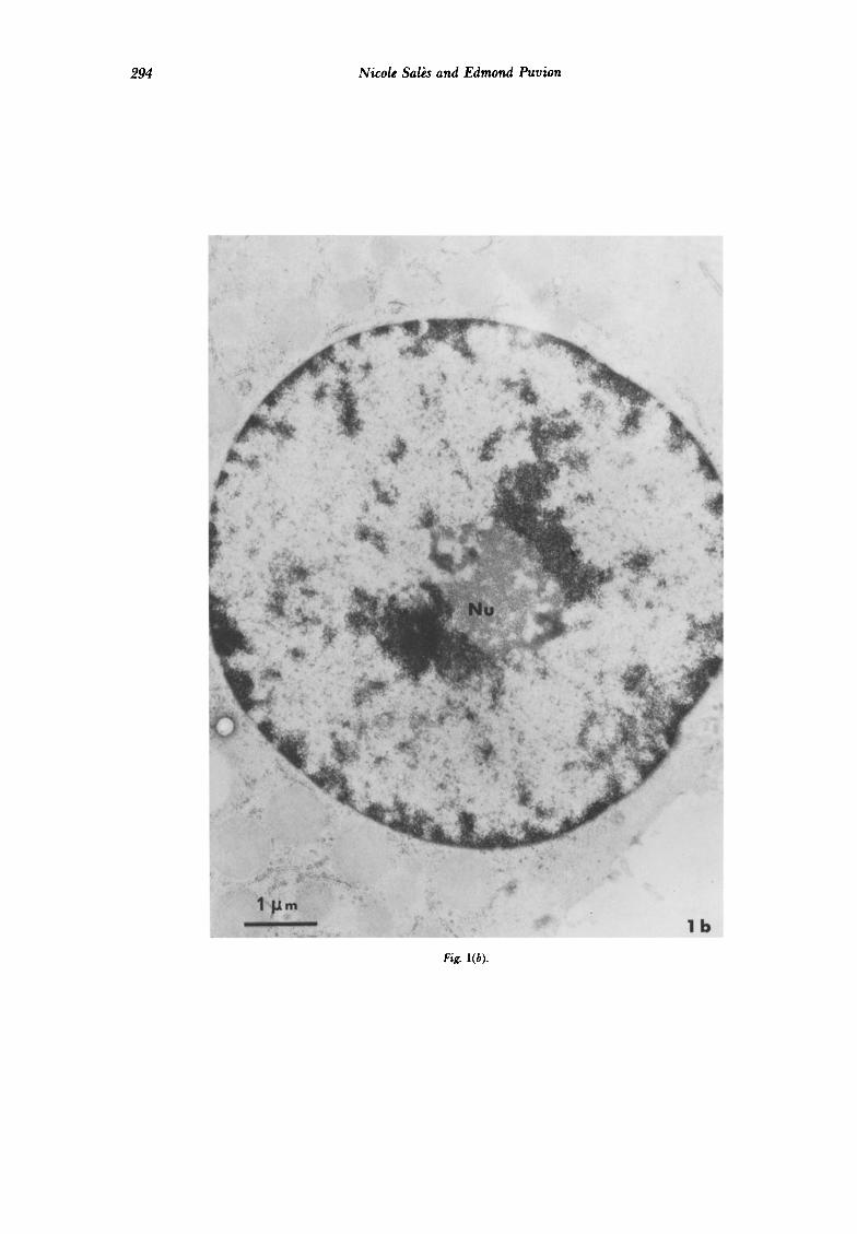
294 Nicole Salb and Edmond Puvion
Fig. l(b).

Ellipticine Efects on Hepatocyte Nuclei 295
Fig. l(c).

296 Nicole Salk and Edmond Puvion
Fig. l(d).
Fig. 1. Nuclei of isoluted rat liver cells. (a) Non-treakd control cell. 7’he nuckolus (Nu) exhibits classically intermingled strands of fibrillar and granular material. Uranyl acetate and lead citrak conventional stain. (b) Ten min treatment with 9-OH E + (1 &ml). The nucleolus is now compact and round-shaped. Uranyl and lead stain. (c) Non-treakd control cell. RNPstain. The condensed chromatin has been bleached. Perichromatin (-) and interchromatin lfbrils (+) are revealed. (d) Ten min treatment with 9-OH Ei(1 pglml). RNPstain. The bleached chromatin is more abundant at the nuclearperifihcrp and around the nucleolus. In the interchronuatin space most interchromutin lfbrils have disatieared, whereas perichromutin ,fibrils are less dense.

Eliifiticine Efects on Hepatocyte Nuclei 297
Fig. 2(a).

Nicole Saks and Edmond Puvion
Fig. 2(b).
Fig. 2. Nucleoli of isolated rat liver celLs. (a) Two hr treatment with g-OH E + (1 pglml). Conventional shining reveals the jnzsence of a thin rim of less condensed mute&l ( WYOW) separating the nuckolar body from the surrounding condensed chromatin. 7% modification can also be observed after a IO-min treatment. A longer @riod of contact with the drug (1 pg/ml) has not resulted in further nuckar changes (compare with Fig. 1 b). (b) Two hr treatment with 9-OH E + (5 pg/ml). A t+@ical nuckolar segwgation occurs; it is characterized by the sgaration of nuckokar com@nents into fi6ril.s (F), gsanuks ( C) and a clear jibrillar area ( C). Uranyl and kad
stain.

Ellifiticine Efects on Hepatocyte Nuclei 299
Fig. 3. Autoradiografihs of isolated rat liver cells. (a) The cells have been labeled for ‘30 min with [5-‘HI-uridine in a drug-free medium. The nucleolus is labeled homogeneously over both its granular and fibrillar components. (b) The cells have been pretreated for 15 min with 9-OH E + (1 Fg/ml), then labeled for 30 min in the presence of the drug at the same concentration. The overall number of silver grains present in the
nuclolar area is strongly reduced and there is a complete luck of labeling over the granular zone (G).

300 Nicole Sal& and Edmond Puvion
Fig. 4(a, b).

Ellipticine Effects on Hefiatocyte Nuclei 301
Fig. 4(c).
Fig. 4. Autoradiografihs of isolakd rat liver cells. (a) Non-treated control cell. The Matocytes have been labeled for 15 min then jixcd immediakly. Silver grains are present over the jibrilkzr zone of the nucleolus but not over the granular zone (G). (b) Non-treakd cell. Fifken min labeling followed by a 1-hr chase. 77~ nucleolus is homogeneously labeled on its fibrillar and granular com@nents. (c) Treakd cell. The watocyks have been label- ed as in Figs. 4a and 4b, followed by I-hr chase in the presence of 9-OH E + (1 pglml). 7’he overall labeling of the nucleolus is reduced, but there has been a shift of the radioactivity from the fibrillar (F) to the granular (C)
com@k?nt.

Ellifiticine Efects on Hefiatocytes Nuclei 303
RESULTS Table 1. Quantitative autoradiography: incorporation of Morphological alterations
Very similar morphological obtained with E, E + , g-OH E
[5-‘HI-uridine in hepatocyte nuclei
alterations are and 9-OH E + , Nucleolar
area Extranucleolar
area but 5- to IO-fold higher concentrations were necessary with compounds lacking the quater- nary ammonium. So, only modifications due to g-OH E + will be described here.
Comparison of control cells (Fig. la) and cells treated with the ellipticine derivative at a dose of 1 pg/ml for only 10 min reveals that the drug induces a condensation of chromatin which appears more contrasted both along the nuclear envelope and around the nucleolar body (Fig. lb). Clumping of chromatin is also visible within the faintly stained nucleoplasm. Following staining of only RNP with the method of Bernhard, nucleolar and extra- nucleolar RNP are revealed. The nucleoplasm of control cells contains many perichromatin fibrils at the border of the condensed chroma- tin and interchromatin fibrils within the inter- chromatin space (Fig. lc). In ellipticine-treated cells, perichromatin fibrils adjacent to the condensed chromatin clumps are less abun- dant, whereas interchromatin fibrils have al- most completely disappeared in the inter- chromatin space (Fig. Id). The nucleolus of treated cells is more compact and has a roun- ded shape. The separation of fibrillar and granular nucleolar RNP is already visible and, generally, fibrils form dense strands clearly distinct from the granular zones. In most cases, the nucleolar body is separated from the sur- rounding condensed chromatin by a thin rim of less condensed material (Fig. 2a). Longer periods of contact with ellipticine at the same dose do not result in further nuclear structural changes. However, at a dose of 5 pg/ml, the lesions described above evolve within 2 hr towards a complete and typical nucleolar segregation characterized by the separation of nucleolar components into three distinct zones: fibrils, granules and clear fibrillar area (Fig. 2b). This is followed very rapidly by cell death.
Autoradiographic observations The quantitative determinations of [5-‘HI-
uridine incorporation in the two nucleolar and extra-nucleolar areas of the nuclei followed by statistical analysis demonstrates no statistically significant difference between 15-min and 1-hr pretreated cells. On the other hand, a highly significant difference in silver grain densities (risk less than 0.01, as estimated by t test) exists between either of these treated groups and the untreated controls. The labeling of the nucleolar area is strongly reduced in treated
Controls 36.892 5.1* 7.87 + 0.9 15 min 9-OH E + 10.86 + 1.5 3.39 f 0.4 lhr9-OHE+ 7.08 + 1.9 4.16 k 0.4
‘Silver grain densities are expressed in grains per square pm-mean + S.D.
cells (73 and 80% after 15 and 60min respec- tively). In the extra-nucleolar area, the decrease of the incorporation of [5-‘HI-uridine is of 47 and 57% respectively (Table 1).
Figure 3a represents a normal control nucleus labeled for 30 min with [5-‘HI-uridine. Radioactivity is dispersed all over the nucleo- plasm. The nucleolus is homogeneously labeled over fibrillar and granular components. On the other hand, in the cells pre-treated either for 15 min or 1 hr with 9-OH E+ (1 pg/ml) fol- lowed by 30 min of labeling the silver grains are present exclusively over the fibrillar nucleolar strands; the granular component is not labeled (Fig. 3b).
Contrary to the above results, the com- parison of autoradiograms of hepatocytes after a 15-min pulse with [5-‘HI-uridine followed by a I-hr or a 3-hr chase with or without 9-OH E+ does not reveal any difference in the local- ization of silver grains in the nucleoli of treated and untreated cells. At the end of the 15-min period of labeling, the association of the radioactivity with the fibrillar elements of the nucleoli is evident (Fig. 4a). Following 1 or 3 hr of subsequent post-incubation, the nucleoli of control cells exhibit a progressive migration of the radioactivity from the fibrillar to the granular compartment (Fig. 4b).
When the chase is carried out in the presence of 9-OH E+ , a moderate reduction of the density of silver grains is observed over the extra-nucleolar and nucleolar areas. In the nucleolus, a shift of the radioactivity from the fibrillar towards the granular compartment takes place (Fig. 4~).
DISCUSSION
Our observations of structural and functional nuclear lesions induced by an ellipticine derivative in isolated rat liver cells must be discussed in relation to our recent knowledge of the significance of nuclear RNP components and on the basis of biochemical data dealing with ellipticine action on cellular metabolism.

304 Nicole Salb and Edmond Puvion
Quantitative high resolution autoradio- graphy has shown a rapid decrease of [5-3H]- uridine incorporation with moderate selectivity for nucleolar incorporation. This agrees well with biochemical determination of Kann and Kohn [19] carried out on leukemia L 1210 cells. Our results can therefore be interpreted as reflecting an inhibition of nucleolar and extra- nucleolar RNA synthesis, though the hypothesis of a drug-related effect on uridine uptake or phosphorylation cannot be completely excluded.
From a structural point of view, the func- tional disturbances were associated with the segregation of nucleolar components and in- duced the disappearance of perichromatin fibrils and interchromatin fibrils.
Nucleolar segregation characterized by the coalescence of nucleolar RNP components into distinct zones has been extensively studied [20]. From the studies of Simard [21] and of Simard and Bernhard [22], it is now well-established that this nucleolar lesion can be considered as specific for the action of drugs binding to DNA and affecting the template activity of ribosomal DNA. The agents known to induce nucleolar segregation include intercalating drugs [22]. However, in the case of ellipticine, relatively high doses (5 wg/ml) which lead rapidly to cell death were required to induce a complete segregation of nucleolar components similar to that caused by actinomycin D. This might be imputable to the relatively low selectivity of ellipticine binding to rDNA, and confirms that nucleolar segregation is a dose-dependent lesion [20].
Several recent studies have demonstrated that perichromatin fibrils formed initially at the border of the condensed chromatin contain the newly synthesized heterogeneous nuclear RNA [14] and that they migrate subsequently towards the interchromatin space [23]. Thus, the almost complete disappearance of perich- romatin and interchromatin fibrils, which occurred simultaneously with the condensation of chromatin as rapidly as 15 min after the initiation of the ellipticine treatment, can be considered as reflecting the inhibition of 43% of uridine uptake in the nucleoplasm.
Since the earlier works of Granboulan and Granboulan [24], later on confirmed by many investigators [14,25], it is well-established that the fibrillar RNP component of the nucleolus is labeled first following a short incubation time with [5-‘HI-uridine and that the subsequent shift of radioactivity from the fibrils to the nucleolar granules represents the structural counterpart of pre-rRNA processing. Therefore,
the selective and persistent accumulation of the radioactivity over the nucleolar fibrils following different labeling periods with [5-‘HI-uridine of ellipticine-treated cells represents a block of pre- rR NA processing.
Similar effects have been reported under the action of unrelated drugs such as cordycepin [26] and camptothecin [27]. In both cases, the inhibition of pre-rRNA processing was related to the formation of shortened pre-rRNA molecules which could not be normally proces- sed. According to Kann and Kohn [19], such pre-rRNA chain shortening does not occur in the presence of ellipticine, due probably to the rapid reversibility of the complex formed by this drug with DNA. Snyder et al. [28] proposed that the inhibition of the delay in the onset of the pre-rRNA processing induced by ellipticine might be related to a direct binding of the drug at hypothetical sites of these RNA molecules.
However, our observations differ from those of these authors since we observed in isolated rat liver cells that the processing of the nor- mally synthesized pre-rRNA was not blocked during post-incubation in the presence of ell- ipticine. Thus, the possibility that the inhibition of the processing of pre-r-RNA synthesized in the presence of the drug might be secondary to the inhibition of heterogeneous nuclear RNA synthesis cannot be excluded. Such an effect was proposed by Granick [29] to account for the partial inhibition of pre-rRNA processing and the further degradation of pre-ribosomes in nucleoli of chick fibroblasts treated with the adenosine analogue, 5-6 dichloro I-/3n-ribo- furanosyl benzimidazole (DRB).
Furthermore, as shown by Geuskens and Bernhard [30] with actinomcyin D, nucleolar segregation still permits the normal processing of pre-rRNA synthesized prior to the action of the antibiotic. Hadjiolov and Nikolaev [31] generalized these observations and postulated that agents inducing nucleolar segregation have the particularity of blocking pre-rRNA transcription without affecting pre-rRNA processing.
Another point to be considered is the ab- sence in ellipticine-treated cells of newly for- med perichromatin granules. It was indeed reported in several studies [26,27,32-351 that drugs blocking totally or partially pre-rRNA processing induced the appearance of perich- romatin granules at the level of intra and peri- nucleolar chromatin. Surprisingly, such granules were never observed following ellip- ticine treatment. Since, as discussed above, E does not induce pre-rRNA shortening, this finding indirectly supports our initial hypo-

Elliflticine Efects on H@atocyte Nuclei 505
thesis (see Puvion and Moyne [36] for a review) Dr. W. Bernhard (I.R.S.C., Villejuif) and to Prof. C. that the nucleolar perichromatin granules do Paoletti (Laboratoire de ChimiothCrapie ExpCri-
represent degradation forms of abnormally mentale, I.G.R., Villejuif), who initiated this work.
shortened pre-rRNA. They thank Prof. C. Paoletti for providing ellip- ticine derivatives, Prof. E. Leduc (Brown University, Providence) for critical reading of the manuscript
Acknowledgements-The authors are indebted to and Mrs J. Bnrglen for technical assistance.
5.
6.
7.
8.
9.
10.
11.
12.
13.
14.
15.
16.
17.
18.
19.
20.
REFERENCES
FESTY B, POISSON J, PAOLETTI C. A new DNA intercalating drug: methoxy-g- ellipticine. FEBS Left 1971, 17, 321-325. KOHN KW, WARING MJ, GLAUBIGER D, FRIEDMAN CA. Intercalative binding of ellipticine NSC-71795 to DNA. Cancer Res 1975, 35, 71-76. BERNHARD W. A new staining procedure for electron microscopical cytology. J Ultrastruct Res 1969, 27, 25&267. DALTON LK, DEMERAC S, ELMES BC, LODER JW, SWAN JM, TEITEI T. Synthesis of the tumor-inhibitory alkaloids, ellipticine, 9-methoxy ellipticine and related pyrido (4-3,b) carbazoles Aust J Chem 1967, 20, 2715-2722. HARTWELL JL, ABBOTT BJ. Antineoplastic principles in plants. Recent develop- ments in the field. Adv Z’harmacol Chemother 1969,7, 117-209. LEMEN J, HAYAT M, MATHS G et al. Methoxy-g-ellipticine lactate. I-Experimental study (oncostatic and immuno-suppressive actions; preclinical pharmacology). Rev Eur Et Clin Biol 1970, 15, 534-538. MATH& G, HAYAT M, DE VASSAL F et al. Methoxy-9-ellipticine lactate. III-Clinical screening: its action in acute myeloblastic leukemia. Rev Eur Et Clin Biol 1970, 15, 541-545. LE PECQ JB, GOSSE C, DAT-XUONG N, PAOLETTI C. Un nouveau composC anti- tumoral:l’hydroxy-9-ellipticine. Action sur la leuctmie L 1210 de la souris. C R Acad Sci [D] (Paris) 1973,277, 2289-2291. LE PECQ JB, GOSSE C, DAT-XUONG N, PAOLETTI C. Deux nouveaux derives anti-tumoraux:l’hydroxy-9-methyl-2-ellipticinium et l’hydroxy-9-dimethyl-2-6-ellip- ticinium. Action sur la leucemie L 1210 de la souris. CR Acad Sci [D] (Paris) 1975,481, 1365-1367. LE PECQ JB, GOSSE C, DAT-XUONG N, CROS S, PAOLE’ITI C. Anti-tumor activity of 9-hydroxy ellipticine NSC210717 on L 1210 mouse leukemia and the effect of route of injection. Cancer Res 1976, 36, 3067-3076. PAOLETTI C, CROS S, DAT-XUONG N, LECOINTE P, MOISAND A. Comparative cytotoxic and antitumoral effects of ellipticine derivatives on mouse L 1210 leuke- mia. Chem Biol Interact 1979, 25, 45-58. DUFER J, DESPLACES A, AUROUSSEAU M. Cytochemical study of rat leukocytes treated by methoxy-9-ellipticine. Ann Z’harm Fr 1973, 31, 441-450. OUSTRIN ML, PIERAGGI MT. Action des hydroxy-9 et methoxy-9 ellipticines sur la cellule HeLa Ss on culture “in vitro”. CR Acad Sci [D] (Paris) 1974,278,1967-1970. FAKAN S, PWION E. The ultrastructural visualization of nucleolar and extra- nucleolar RNA synthesis and distribution. Znt Rev Cytol 1980,65, 255-299. PWION E, GARRIDO G, VIRON A. Technique rapide d’isolement et de survie en monocouches d’htpatocytes de rats adultes. C R Acad Sci [D] (Paris) 1974, 279, 509-512. HAASE G, JUNG G. Herstellung von Einkorn-schichten aus photographischen emulsionen. Natunvissenschaftm 1964, 51, 4WO5. BOUTEILLE M, DUPUY-COIN AM, MOYNE G. Techniques of localization of proteins and nucleoproteins in the cell nucleus by high resolution autoradiography and cytochemistry. In: O’MALLEY BW, HARDMAN JG, eds. Methods in Enzymology. New York, Academic Press, 1975,40, s-41. FAKAN S, BERNHARD W. Localization of rapidly and slowly labeled nuclear RNA as visualized by high resolution autoradiography. Exp Cell Res 1971, 67, 129-141. KANN HE, KOHN KW. Effects of deoxyribonucleic acid-reactive drugs on ribo- nucleic acid synthesis in leukemia L 1210 cells. Mol Pharmacol 1972, 8, 551660: SIMARD R, LANGELIER Y, MANDEVILLE R, MAESTRACCI N, ROYAL A. Inhibitors as tools in elucidating the structure and function of the nucleus. In: BUSCH H, ed. The Cell Nucleus. New York, Academic Press, 1974 Vol. III, 447487.

306 Nicole Salk and Edmund Puvion
21.
22.
23.
24.
25.
26.
27.
28.
29.
30.
31.
32.
33.
34.
35.
36.
SIMARD R. Specific nuclear and nucleolar ultrastructural lesions induced by proflavin and similarly acting anti-metabolites in tissue culture. Cancer Res 1966,26, 23162328. SIMARD R, BERNHARD W. Le phenomene de la segregation nucleolaire: specificite d’action de certains anti-mitabolites. Znt J Cancer 1966, 1, 463-479. PUVION E, MOYNE G. Intranuclear migration of newly synthesized extra-nucleolar ribonucleoproteins. A high resolution quantitative autoradiographical and cyto- chemical study. Exp Cell Res 1978, 115, 79-88. GRANBOULAN N, GRANBOULAN P. Cytochimie ultrastructurale du nucleole. II. Etude des sites de synthese du RNA dans le nucliole et le noyau. Exp Cell Res 1965, 38, 604-619. FAKAN S. High resolution autoradiography studies on chromatin functions. In: BUSCH H, ed. The Cell Nucleus, Chromatin-Part B. New York, Academic Press, 1978, Vol. V, 3-53. PUVION E, MOYNE G, BERNHARD W. Action of 3’ deoxyadenosine (cordycepin) on the nuclear ribonucleoproteins of isolated liver cells. J Microsc Biol Cell 1976, 25, 17-32. GAJKOWSKA B, PUVION E, BERNHARD W. Unusual perinucleolar accumulation of ribonucleoprotein granules induced by camptothecin in isolated liver cells. J Ultrastruct Res 1977,60, 335-347. SNYDER AL, KANN HE, KOHN KW. Inhibition of the processing of ribosomal precursor RNA by intercalating agents. J Mol Biol 1971, 58, 555-565. GRANICK D. Nucleolar necklaces in chick embryo fibroblast cells. J Cell Biol 1975,65, 389-417. GEUSKENS M, BERNHARD W. Cytochimie ultrastructurale du nucleole. III. Action de I’actinomycine D sur le metabolisme du RNA nucleolaire. Exp Cell Res 1966, 44, 579-598. HADJIOLOV AA, NIKOLAEV N. Maturation of ribosomal ribonucleic acids and the biogenesis of ribosomes. Z’rog Biophys Mol Bill 1976, 31, 95-144. RECHER L, CHAN H, BRICCS L, PARRY N. Ultrastructural changes inducible with the plant alkaloid camptothecin. Cancer Res 1972, 32, 2495-2501. DERENZINI M, MOYNE G. The nucleolar origin of certain perichromatin-like granules. A study with a amanitin. J Ultrastruct Res 1978, 62, 213-219. PUVION E, BACHELLERIE JP, BURGLEN MJ. Nucleolar perichromatin granules induced by dichlorobenzimidazole-riboside. J Ultras&u& Res 1979, 69, 1-12. VAZQUEZ-NIN G, ECHEVERRIA OM, PEDRON J. Ultrastructural and autoradio- graphic study of the effects of bleomycin on the interphase nucleus of cultured normal cells. Cancer Res 1979, 39, 4218-4223. PUVION E, MOYNE G. In situ localization of RNA structures In: BUSCH H, ed. The Cell Nucleus, Nuclear Particles-Part A. New York, Academic Press, 1981, Vol. VIII, 59-116.