
Concept Revisie Richtlijn cervixcarcinoom · 5 Concept Revisie Richtlijn Cervixcarcinoom -...
192
Mogelijk gemaakt in samenwerking met: Concept Revisie Richtlijn cervixcarcinoom 1 2 Inhoudsopgave 3 1. ALGEMEEN ............................................................................................................................................... 3 4 1.1 Epidemiologie/Etiologie ................................................................................................. 5 5 2. SCREENING .............................................................................................................................................. 6 6 3. PATHOLOGIE ............................................................................................................................................ 7 7 4. DIAGNOSTIEK ........................................................................................................................................... 9 8 4.1 Medisch Technisch ............................................................................................................. 9 9 4.1.1 Symptomen ................................................................................................................. 9 10 4.1.2 Lichamelijk onderzoek ................................................................................................. 9 11 4.1.3 Laboratorium onderzoek .............................................................................................. 9 12 4.1.4 Voorlichting en communicatie ...................................................................................... 9 13 4.1.5 Continuïteit van zorg / organisatie ................................................................................ 9 14 4.1.6 Spreiding en concentratie, infrastructuur .................................................................... 10 15 4.2 Primaire tumor .................................................................................................................. 11 16 4.3 Lymfekliermetastasen ....................................................................................................... 15 17 4.4 Schildwachtklierbiopt ........................................................................................................ 19 18 4.5 Metastasen op afstand ..................................................................................................... 20 19 4.6 Tumormarkers .................................................................................................................. 21 20 5. BEHANDELING ....................................................................................................................................... 22 21 5.1 Medisch technisch ......................................................................................................... 22 22 5.1.1 Stadium IA1 ............................................................................................................... 22 23 5.1.2 Stadium IA2 ............................................................................................................... 22 24 5.1.3 Stadium IB1 en IIA ..................................................................................................... 22 25 5.1.4 Stadium IB2, IIA-IVA .................................................................................................. 23 26 5.1.5 Stadium IVb ............................................................................................................... 23 27 5.2 Voorlichting en communicatie ........................................................................................... 24 28 5.3 continuïteit van zorg ......................................................................................................... 24 29 5.4 Spreiding en concentratie, infrastructuur .......................................................................... 24 30 5.5 Fertiliteitsparende behandeling ......................................................................................... 25 31 5.5.1 Algemeen .................................................................................................................. 25 32 5.5.2 Conisatie.................................................................................................................... 25 33 5.5.3 Radicale trachelectomie............................................................................................. 28 34 5.5.4 Vaginale radicale trachelectomie ............................................................................... 29 35 5.5.5 Abdominale radicale trachelectomie .......................................................................... 30 36 5.5.6 Experimentele protocollen ......................................................................................... 32 37 6. ADJUVANTE SYSTEMISCHE BEHANDELING NA INITIËLE BEHANDELING ................................... 33 38 6.1. Medisch technisch ........................................................................................................... 33 39 6.1.1. Postoperatieve radiotherapie .................................................................................... 33 40 6.1.2. Salvage chirurgie ...................................................................................................... 33 41 6.1.3. Postoperatieve chemoradiatie................................................................................... 34 42 7. NACONTROLE EN NAZORG.................................................................................................................. 36 43 7.1 Algemeen ......................................................................................................................... 36 44 7.2 Gevolgen en aanpak 1e jaar ............................................................................................. 36 45 7.3 Detectie nieuwe kankermanifestaties................................................................................ 39 46 7.3.1 Op welke termijn kunnen nieuwe kankermanifestaties optreden? .............................. 39 47 7.3.2 Bestaat er voor deze kankermanifestaties een effectieve behandeling? .................... 39 48 7.3.3 Is de behandeleffectiviteit hoger naarmate het recidief eerder wordt gediagnosticeerd? 49 ........................................................................................................................................... 39 50 7.3.4 Welke diagnostiek is het meest geschikt om behandelbare nieuwe 51 kankermanifestaties vroeg en accuraat te diagnosticeren? ................................................. 42 52
Transcript of Concept Revisie Richtlijn cervixcarcinoom · 5 Concept Revisie Richtlijn Cervixcarcinoom -...
Mogelijk gemaakt in samenwerking met:
Concept Revisie Richtlijn cervixcarcinoom 1 2 Inhoudsopgave 3 1. ALGEMEEN ............................................................................................................................................... 3 4
1.1 Epidemiologie/Etiologie ................................................................................................. 5 5 2. SCREENING .............................................................................................................................................. 6 6 3. PATHOLOGIE ............................................................................................................................................ 7 7 4. DIAGNOSTIEK ........................................................................................................................................... 9 8
4.1 Medisch Technisch ............................................................................................................. 9 9 4.1.1 Symptomen ................................................................................................................. 9 10 4.1.2 Lichamelijk onderzoek ................................................................................................. 9 11 4.1.3 Laboratorium onderzoek .............................................................................................. 9 12 4.1.4 Voorlichting en communicatie ...................................................................................... 9 13 4.1.5 Continuïteit van zorg / organisatie ................................................................................ 9 14 4.1.6 Spreiding en concentratie, infrastructuur .................................................................... 10 15
4.2 Primaire tumor .................................................................................................................. 11 16 4.3 Lymfekliermetastasen ....................................................................................................... 15 17 4.4 Schildwachtklierbiopt ........................................................................................................ 19 18 4.5 Metastasen op afstand ..................................................................................................... 20 19 4.6 Tumormarkers .................................................................................................................. 21 20
5. BEHANDELING ....................................................................................................................................... 22 21 5.1 Medisch technisch ......................................................................................................... 22 22
5.1.1 Stadium IA1 ............................................................................................................... 22 23 5.1.2 Stadium IA2 ............................................................................................................... 22 24 5.1.3 Stadium IB1 en IIA ..................................................................................................... 22 25 5.1.4 Stadium IB2, IIA-IVA .................................................................................................. 23 26 5.1.5 Stadium IVb ............................................................................................................... 23 27
5.2 Voorlichting en communicatie ........................................................................................... 24 28 5.3 continuïteit van zorg ......................................................................................................... 24 29 5.4 Spreiding en concentratie, infrastructuur .......................................................................... 24 30 5.5 Fertiliteitsparende behandeling ......................................................................................... 25 31
5.5.1 Algemeen .................................................................................................................. 25 32 5.5.2 Conisatie.................................................................................................................... 25 33 5.5.3 Radicale trachelectomie ............................................................................................. 28 34 5.5.4 Vaginale radicale trachelectomie ............................................................................... 29 35 5.5.5 Abdominale radicale trachelectomie .......................................................................... 30 36 5.5.6 Experimentele protocollen ......................................................................................... 32 37
6. ADJUVANTE SYSTEMISCHE BEHANDELING NA INITIËLE BEHANDELING ................................... 33 38 6.1. Medisch technisch ........................................................................................................... 33 39
6.1.1. Postoperatieve radiotherapie .................................................................................... 33 40 6.1.2. Salvage chirurgie ...................................................................................................... 33 41 6.1.3. Postoperatieve chemoradiatie ................................................................................... 34 42
7. NACONTROLE EN NAZORG.................................................................................................................. 36 43 7.1 Algemeen ......................................................................................................................... 36 44 7.2 Gevolgen en aanpak 1e jaar ............................................................................................. 36 45 7.3 Detectie nieuwe kankermanifestaties ................................................................................ 39 46
7.3.1 Op welke termijn kunnen nieuwe kankermanifestaties optreden? .............................. 39 47 7.3.2 Bestaat er voor deze kankermanifestaties een effectieve behandeling? .................... 39 48 7.3.3 Is de behandeleffectiviteit hoger naarmate het recidief eerder wordt gediagnosticeerd?49 ........................................................................................................................................... 39 50 7.3.4 Welke diagnostiek is het meest geschikt om behandelbare nieuwe 51 kankermanifestaties vroeg en accuraat te diagnosticeren? ................................................. 42 52

2 Concept Revisie Richtlijn Cervixcarcinoom - September 2011
7.3.5 Scenario voor vroegdetectie ...................................................................................... 44 53 7.4 Evaluatie medisch handelen ............................................................................................. 46 54 7.5 Organisatie van nazorg ..................................................................................................... 46 55 7.6 Seksuele dysfunctie .......................................................................................................... 50 56
7.6.1 Psycho-educatie ........................................................................................................ 50 57 7.6.2 Medicatie ................................................................................................................... 51 58 7.6.3 Vaginale dilatatie ....................................................................................................... 53 59
8. DIAGNOSTIEK EN BEHANDELING LOKAAL / LOKOREGIONAAL RECIDIEF .................................. 55 60 8.1. Medisch technisch ........................................................................................................... 55 61
8.1.1 Lokaal recidief zonder eerdere radiotherapie ............................................................. 55 62 8.1.2 Lokaal recidief na eerdere radiotherapie .................................................................... 56 63
8.2 Voorlichting en communicatie ........................................................................................... 57 64 8.3 Continuïteit van zorg ......................................................................................................... 57 65 8.4 Spreiding en concentratie, infrastructuur .......................................................................... 57 66
9. DIAGNOSTIEK EN BEHANDELING METASTASEN OP AFSTAND ..................................................... 58 67 9.1 Medisch technisch ............................................................................................................ 58 68
9.1.1 Recidief op afstand, al dan niet met lokaal recidief .................................................... 58 69 9.2 Voorlichting en communicatie ........................................................................................... 59 70 9.3 Continuïteit van zorg ......................................................................................................... 59 71
10. PALLIATIEVE ZORG ............................................................................................................................. 60 72 11. TNM CLASSIFICATIE ............................................................................................................................ 61 73 12. SAMENVATTING ................................................................................................................................... 62 74
Bijlage 1 Samenstelling werkgroep ......................................................................................... 63 75 Bijlage 2 Werkgroepleden ...................................................................................................... 63 76 Bijlage 3 Onafhankelijkheid werkgroepleden .......................................................................... 64 77 Bijlage 4 Betrokken en autoriserende verenigingen ................................................................ 64 78 Bijlage 5 Wetenschappelijke onderbouwing ............................................................................ 64 79 Bijlage 6 Indeling van onderzoeksresultaten naar mate van bewijskracht ............................... 65 80 Bijlage 7 Format „Overige overwegingen‟ en „Formuleren van aanbevelingen‟ ....................... 67 81 Bijlage 8 Uitgangsvragen ........................................................................................................ 69 82 Bijlage 9 Literatuur searches .................................................................................................. 70 83 Bijlage 10 Evidence tabellen ................................................................................................... 82 84 Bijlage 11 Tabellen overige overwegingen ........................................................................... 148 85 Bijlage 12 Voorbeeld nazorgplan cervixcarcinoom ............................................................... 155 86 Bijlage 13 Actualisatie .......................................................................................................... 157 87 Bijlage 14 Houderschap richtlijn ........................................................................................... 158 88 Bijlage 15 Juridische betekenis van richtlijnen ...................................................................... 158 89 Bijlage 16 Verantwoording .................................................................................................... 158 90 Bijlage 17 Implementatie en evaluatie .................................................................................. 158 91 Bijlage 18 Kennislacunes ..................................................................................................... 159 92 Bijlage 19 Referenties .......................................................................................................... 161 93
94
3 Concept Revisie Richtlijn Cervixcarcinoom - September 2011
1. ALGEMEEN 95
96 Aanleiding 97 In 2010 is de richtlijn cervixcarcinoom consensus based gereviseerd. De richtlijn is nu evidence based 98 gereviseerd voor 6 onderwerpen. Daarbij is samengewerkt met het Federaal Kenniscentrum voor de 99 Gezondheidszorg (KCE) uit België. 100 101 Doelstelling 102 Een richtlijn is een aanbeveling ter ondersteuning van de belangrijkste knelpunten uit de dagelijkse praktijk. 103 Deze richtlijn is zoveel mogelijk gebaseerd op wetenschappelijk onderzoek of consensus. 104 105 Doelgroep 106 Deze richtlijn is bestemd voor alle professionals die betrokken zijn bij de diagnostiek, behandeling en 107 begeleiding van patiënten met cervixcarcinoom: gynaecologen, radiotherapeuten, medisch oncologen, 108 radiologen, pathologen, oncologieverpleegkundigen, huisartsen, IKNL-consulenten, maatschappelijk 109 werkers en psychologen. Tevens wordt deze richtlijn gebruikt voor het maken van patiënten 110 informatiemateriaal in samenwerking met het KWF. 111 112 Werkwijze werkgroep consensus based richtlijn 2010 113 De consensus based richtlijn cervixcarcinoom uit 2010 is gemaakt binnen de Commissie Richtlijnen 114 Gynaecologische Oncologie (CRGO) Dit is een landelijke multidisciplinair samengestelde commissie. Er is 115 gewerkt op basis van consensus. De meest recente literatuur is gehanteerd. De richtlijnen zijn in alle 116 regio's besproken. Tevens zijn zij aan alle leden van de NVOG, LPRGT en NVMO ter goedkeuring 117 voorgelegd. De richtlijnen hebben een adviserend karakter. De revisie van de richtlijn in 2010 heeft 118 dusdanig beperkte aanpassingen opgeleverd ten aanzien van de richtlijn uit 2006, dat deze niet opnieuw 119 uitgebreid aan het veld is voorgelegd 120 121 Werkwijze werkgroep evidence based richtlijn 2011 122 In november 2010 is er een knelpunteninventarisatie gehouden in het veld bij professionals en 123 patiënten(vertegenwoordigers). De meest relevante knelpunten zijn uitgewerkt tot zes uitgangsvragen (zie 124 bijlage 8). 125 Vier uitgangsvragen zijn tussen KCE en het IKNL verdeeld. Deze samenwerking bestond uit de verdeling 126 van het literatuuronderzoek, de critical appraisal, evidence tabellen en de literatuurbespreking. Hierbij heeft 127 KCE uitgangsvraag 2 en 3 uitgewerkt, het IKNL uitgangsvraag 1 en 4. Beide partijen hebben elkaars 128 resultaten gevalideerd. In het geval van discrepanties werd consensus bereikt door middel van discussie. 129 Daarnaast heeft het IKNL de resultaten van de uitgangsvragen 5 en 6 van KCE ontvangen. 130 131 Voor iedere uitgangsvraag werd uit de richtlijnwerkgroep een subgroep geformeerd. De werkgroepleden 132 schreven afzonderlijk of in subgroepen de teksten die uiteindelijk door de hele richtlijnwerkgroep zijn 133 geaccordeerd. De teksten van de richtlijn cervixcarcinoom uit 2010 zijn waar nodig geactualiseerd. 134 De concept richtlijn is in september 2011 naar alle betrokken wetenschappelijke-, beroeps- en 135 patiëntenverenigingen en de landelijke en regionale tumorwerkgroepen gestuurd voor commentaar. Na 136 verwerking van het commentaar wordt de richtlijn in december 2011 naar de mandaterende 137 wetenschappelijke- en beroepsverenigingen gestuurd ter autorisatie. 138 139 Meer informatie over 140 Samenstelling werkgroep (zie bijlage 1) 141 Werkgroepleden (zie bijlage 2) 142 Onafhankelijkheid werkgroepleden (zie bijlage 3) 143 Betrokken en autoriserende verenigingen (zie bijlage 4) 144 Wetenschappelijke onderbouwing (zie bijlage 5) 145 Indeling van onderzoeksresultaten naar mate van bewijskracht (zie bijlage 6) 146 Format „Overige overwegingen‟ en „formuleren van aanbevelingen‟ (zie bijlage 7) 147 Uitgangsvragen (zie bijlage 8) 148 Literatuursearches (zie bijlage 9) 149 Evidence tabellen (zie bijlage 10) 150 Tabellen overige overwegingen (zie bijlage 11) 151 Actualisatie (zie bijlage 13) 152

4 Concept Revisie Richtlijn Cervixcarcinoom - September 2011
Houderschap richtlijn (zie bijlage 14) 153 Juridische betekenis (zie bijlage 15) 154 Verantwoording (zie bijlage 16) 155 Implementatie en evaluatie (zie bijlage 17) 156 Kennislacunes (zie bijlage 18) 157 158 159
160
5 Concept Revisie Richtlijn Cervixcarcinoom - September 2011
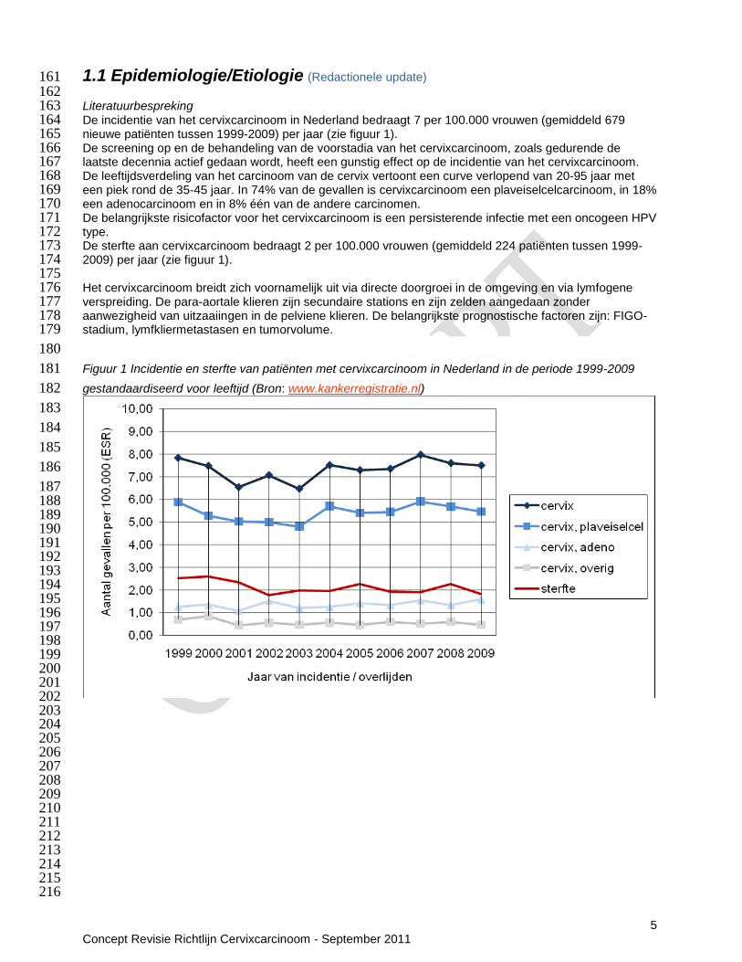
1.1 Epidemiologie/Etiologie (Redactionele update) 161 162 Literatuurbespreking 163 De incidentie van het cervixcarcinoom in Nederland bedraagt 7 per 100.000 vrouwen (gemiddeld 679 164 nieuwe patiënten tussen 1999-2009) per jaar (zie figuur 1). 165 De screening op en de behandeling van de voorstadia van het cervixcarcinoom, zoals gedurende de 166 laatste decennia actief gedaan wordt, heeft een gunstig effect op de incidentie van het cervixcarcinoom. 167 De leeftijdsverdeling van het carcinoom van de cervix vertoont een curve verlopend van 20-95 jaar met 168 een piek rond de 35-45 jaar. In 74% van de gevallen is cervixcarcinoom een plaveiselcelcarcinoom, in 18% 169 een adenocarcinoom en in 8% één van de andere carcinomen. 170 De belangrijkste risicofactor voor het cervixcarcinoom is een persisterende infectie met een oncogeen HPV 171 type. 172 De sterfte aan cervixcarcinoom bedraagt 2 per 100.000 vrouwen (gemiddeld 224 patiënten tussen 1999-173 2009) per jaar (zie figuur 1). 174 175 Het cervixcarcinoom breidt zich voornamelijk uit via directe doorgroei in de omgeving en via lymfogene 176 verspreiding. De para-aortale klieren zijn secundaire stations en zijn zelden aangedaan zonder 177 aanwezigheid van uitzaaiingen in de pelviene klieren. De belangrijkste prognostische factoren zijn: FIGO-178 stadium, lymfkliermetastasen en tumorvolume. 179
180
Figuur 1 Incidentie en sterfte van patiënten met cervixcarcinoom in Nederland in de periode 1999-2009 181
gestandaardiseerd voor leeftijd (Bron: www.kankerregistratie.nl) 182
183
184
185
186
187 188 189 190 191 192 193 194 195 196 197 198 199 200 201 202 203 204 205 206 207 208 209 210 211 212 213 214 215 216

6 Concept Revisie Richtlijn Cervixcarcinoom - September 2011
Tabel 1 Vijfjaarsoverleving van patiënten met cervixcarcinoom per stadium 217 (TNM 6e editie; Bron: www.kankerregistratie.nl) 218 219
Aantal jaren na diagnose
Stadium (TNM 6e editie) 0 1 2 3 4 5
Ia 100% 100% 99% 99% 99% 99%
Ib 100% 99% 95% 93% 92% 90%
IIa 100% 96% 86% 82% 79% 80%
IIb 100% 93% 82% 74% 70% 68%
IIIa 100% 77% 58% 54% 48% 37%
IIIb 100% 84% 69% 62% 58% 56%
IVa 100% 48% 31% 24% 23% 22%
IVb 100% 36% 18% 13% 11% 10%
220 221 222 Figuur 2 Vijfjaarsoverleving van patiënten met cervixcarcinoom per stadium 223 (TNM 6e editie; Bron: www.kankerregistratie.nl) 224 225
226
227
228
2. SCREENING (Redactionele update) 229 Literatuurbespreking 230 In Nederland bestaat het bevolkingsonderzoek baarmoederhalskanker. Tussen 30 en 60 jaar wordt eens 231 per vijf jaar een uitstrijkje gemaakt. Zie verder richtlijn Cervicale Intra-epitheliale Neoplasie (CIN). 232 233
234
7 Concept Revisie Richtlijn Cervixcarcinoom - September 2011
3. PATHOLOGIE (Redactionele update) 235 236 Literatuurbespreking 237 Classificatie van maligne cervix tumoren 238 Classificatie van maligne cervix tumoren geschiedt volgens de WHO. Hier worden alleen de hoofdgroepen 239 genoemd: 240 Plaveiselcelcarcinoom 241 Adenocarcinoom, waaronder het adenosquameus carcinoom. 242 Andere epitheliale tumoren, waaronder neuroendocriene tumoren (zeldzaam) 243 Mesenchymale tumoren (bijv. leiomyosarcoom, rhabdomyosarcoom) 244 Mixed epitheliale en mesenchymale tumoren 245 Maligne lymfomen 246 Doorgroei endometriumcarcinoom 247 Metastasen 248 249 Plaveiselcelcarcinoom 250 Bij 70% van de cervixcarcinomen is er sprake van de plaveiselcel vorm. Graderen bij 251 plaveiselcelcarcinoom is slecht reproduceerbaar en heeft geen goede relatie met het klinisch beloop. Om 252 die reden wordt graderen van plaveiselcelcarcinoom niet aanbevolen. Wel maakt de WHO een 253 onderscheid tussen grootcellig verhoornend en grootcellig niet-verhoornend plaveiselcelcarcinoom. Indien 254 een of meerdere hoornparels worden aangetroffen is er sprake van een grootcellig verhoornend 255 plaveiselcarcinoom. 256 257 Adenocarcinoom 258 Indien endocervicale klierbuisjes met een atypische epitheelbekleding zoals bij adenocarcinoom in situ 259 microscopisch niet meer in een normale architecturele rangschikking gelegen zijn dan wel dieper gelegen 260 zijn dan het niveau van de pre-existente endocervicale klierbuizen, is er sprake van een invasief 261 adenocarcinoom. “Early invasion” is bij adenocarcinomen soms lastig vast te stellen. Suggestief hiervoor 262 zijn cribriforme groei, focale squameuze cytoplasmatische veranderingen, het langgerekt worden van de 263 atypische klierbuizen en een stroma-reactie. Ook bij adenocarcinoom dient de invasiediepte te worden 264 gerapporteerd. 265 De adenocarcinomen van de cervix zijn histologisch meestal van het mucineuze endocervicale type, soms 266 van het intestinale type of zegelringtype. Daarnaast kunnen, zoals ook in de overige organen van de 267 tractus genitalis, verschillende differentiatievormen zoals mucineus, papillair, endometrioïd en clear cell 268 worden aangetroffen. 269 Aparte vermelding verdient het adenosquameuze carcinoom, dat opgebouwd is uit een vermenging van 270 glandulaire en squameuze elementen. Deze tumoren zijn meestal weinig gedifferentieerd, hetgeen past bij 271 het slechtere beloop, zoals dat door sommige auteurs wordt gevonden. 272 273 Histopathologische kenmerken en verslaglegging 274 De diepte van de invasie wordt gemeten vanaf de basis van het epitheel vanaf het punt van ontstaan tot 275 het diepste punt van invasie. Indien het invasieve carcinoom uitgaat van een door CIN ingenomen 276 endocervicale klierbuis, dient de invasiediepte gemeten te worden vanaf de basis van het epitheel van de 277 klierbuis. In het geval van een adenocarcinoom wordt de diepte van de invasie gemeten vanaf de dichtstbij 278 gelegen klierbuis met adenocarcinoma in situ. Bij aanwezigheid van multifocaal carcinoom worden 279 dieptegroei en diameter van de afzonderlijke laesies vermeld. Indien de absolute invasiediepte niet 280 gemeten kan worden, bijvoorbeeld omdat de tumor tot in de bodem van het biopt reikt, wordt de minimale 281 invasiediepte aangegeven. 282 283 In het pathologieverslag dienen bij een biopt, lis of conus derhalve de volgende kenmerken te worden 284 genoemd: histologisch type, invasiediepte, vaatinvasie en oppervlakte uitbreiding. Indien een conisatie 285 wordt gedaan, dient al het materiaal te worden ingesloten voor histologisch onderzoek. De status van de 286 snijranden wordt hierbij aangeven. Het al dan niet aangeven van de status van de snijranden bij een 287 lisexcisie wordt lokaal in overleg met de gynaecoloog en patholoog bepaald. 288 289 Na een radicale hysterectomie en pelviene lymfklierdissectie dienen de volgende aspecten te worden 290 aangegeven, omdat deze mede bepalend zijn voor eventuele aanvullende behandeling: de grootste 291 diameter van de tumor, de dieptegroei, de horizontale uitbreiding van de tumor, al dan niet vrij zijn van de 292 resectieranden, ook aan de voor- en achterzijde van de cervix, de afstand tot de resectieranden, de aan- of 293

8 Concept Revisie Richtlijn Cervixcarcinoom - September 2011
afwezigheid van (lymfe)vaatinvasie, en het aantal positieve lymfklieren van het totaal aantal lymfklieren. 294 Indien positieve lymfklieren worden gevonden, dient de lokalisatie van deze lymfklieren en eventuele 295 kapseldoorbraak aangegeven te worden i.v.m. postoperatieve radiotherapie en eventueel chemotherapie. 296 297 298 299
300

9 Concept Revisie Richtlijn Cervixcarcinoom - September 2011
4. DIAGNOSTIEK (Redactionele update) 301
302
4.1 Medisch Technisch (Redactionele update) 303 Literatuurbespreking 304 Na het stellen van de diagnose cervixcarcinoom d.m.v. histologisch onderzoek vindt klinische stadiering 305 plaats conform de FIGO richtlijnen (door een oncologisch gynaecoloog en radiotherapeut tezamen). De 306 klinische stadiëring wordt na vaststellen niet meer veranderd. Wanneer er twijfel- bestaat over het klinisch 307 stadium dient het laagste stadium te worden gekozen. Het is bekend dat de klinische stadiëring slechts in 308 circa 60% correleert met de histopathologische bevindingen (chirurgische stadiëring), indien deze bekend 309 zijn. Zie hoofdstuk 12 (TNM classificatie) 310 311 4.1.1 Symptomen (Redactionele update) 312 Literatuurbespreking 313 In de beginfase van het cervixcarcinoom heeft de patiënt vaak geen klachten en wordt de diagnose 314 meestal na afname cervixcytologie (screening) gesteld. Bij toename van de tumormassa kunnen door de 315 necrotische en proliferatieve veranderingen van het cervixweefsel de volgende klachten optreden: 316 abnormaal (intermenstrueel) vaginaal bloedverlies, contactbloedingen en abnormale fluor vaginalis. In 317 hogere stadia kunnen mictiestoornissen (dysurie), defaecatiestoornissen, pijn in de onderbuik of rug en 318 lymfoedeem optreden. 319 320 4.1.2 Lichamelijk onderzoek (Redactionele update) 321 Literatuurbespreking 322 Het gynaecologisch onderzoek bestaat uit inspectie en palpatie van de genitalia interna en de parametria. 323 Tenzij de patiënt goed poliklinisch te onderzoeken is, wordt dit onderzoek onder narcose verricht door een 324 oncologisch gynaecoloog en radiotherapeut tezamen. Hierbij wordt een schatting gemaakt van de grootte 325 van de tumor en beoordeeld of er ingroei is in de parametria en de vagina. Indien er verdenking bestaat op 326 doorgroei van de tumor in de blaas of in het rectum wordt tevens een cystoscopie resp. proctoscopie 327 uitgevoerd. Bij het algemeen lichamelijk onderzoek wordt gelet op vergrote klieren in de liezen en in de 328 fossa supraclavicularis met name links. 329 330 Conclusie 331 Met lichamelijk onderzoek door gynaecoloog en radiotherapeut samen (al dan niet onder narcose, 332 afhankelijk van de beoordeelbaarheid) bestaat een redelijke inschatting van de lokale uitbreiding van de 333 tumor. 334 335 4.1.3 Laboratorium onderzoek (Redactionele update) 336 Literatuurbespreking 337 Routine (pre-operatief) bloedonderzoek. In geval van chemotherapie: nierfunctie. 338 339 4.1.4 Voorlichting en communicatie (Redactionele update) 340 Literatuurbespreking 341 Patiënte wordt ingelicht over de aard en het doel van de voorgestelde onderzoeken. Gaat patiënte akkoord 342 met de voorgestelde onderzoeken? Indien voorhanden wordt voorlichtingsmateriaal over de onderzoeken 343 meegegeven. 344 De KWF Kankerbestrijding folder Baarmoederhalskanker is hier te downloaden. 345 De NVOG voorlichtingsfolder baarmoederhalskanker is hier te downloaden. 346 De stichting OLIJF voor vrouwen met gynaecologische kanker geeft informatie en verzorgt lotgenoten 347 contact. 348 349 4.1.5 Continuïteit van zorg / organisatie (Redactionele update) 350 Literatuurbespreking 351 Intramuraal 352 Overleg met de consulent/ in de oncologie bespreking voorafgaand aan de behandeling. 353 354 Transmuraal 355 Bericht huisarts over aard van diagnose en welke informatie aan patiënte en familie is gegeven. 356 357 358

10 Concept Revisie Richtlijn Cervixcarcinoom - September 2011
359 4.1.6 Spreiding en concentratie, infrastructuur (Redactionele update) 360 Literatuurbespreking 361 De stadiering en behandeling van het cervixcarcinoom vindt plaats in een erkend gynaecologisch-362 oncologisch centrum. Behandeling van het stadium IA1 kan na overleg met het centrumziekenhuis 363 plaatsvinden in een niet-centrum ziekenhuis. 364 365
366

11 Concept Revisie Richtlijn Cervixcarcinoom - September 2011
4.2 Primaire tumor (Evidence based tekst n.a.v. uitgangsvraag 1: Diagnostische technieken) 367 Literatuurbespreking 368 Eén systematische review met meta-analyse beoordeelde de waarde van CT en MRI voor detectie van 369 parametrium-, blaas- en rectum invasie met histopathologie als referentie standaard (Bipat, Glas et al. 370 2003). In dit review werden 57 studies geïncludeerd waarbij de meeste studies methodologische 371 beperkingen hadden. De sensitiviteit voor het detecteren van parametrium invasie was voor MRI significant 372 hoger dan voor CT (sensitiviteit MRI: 74% (95%CI: 68-79); N=52 studies vs. CT 55% (95%CI 44-66); N=9 373 studies; p=0.0027). Voor de uitkomsten blaasinvasie en rectuminvasie waren de verschillen tussen CT en 374 MRI niet significant. De sensitiviteit voor het detecteren van blaasinvasie van MRI was 75% ((95%CI: 66-375 83); specificiteit: 91% (95%CI: 83-95); N=16 studies) vs. sensitiviteit CT 64% ((95%CI: 39-82%); 376 specificiteit 73% (95%CI: 52-87); N=3 studies). De sensitiviteit van MRI voor het detecteren van 377 rectuminvasie was 71% ((95%CI: 53-83); specificiteit: 83%; N=9 studies) vs. een sensitiviteit voor CT van 378 45% ((95%CI: 20-73); specificiteit: 83%; N=2 studies). 379 380 Primaire studies 381 Na de laatste zoekdatum van deze systematische review werden er nog 19 primaire studies gepubliceerd, 382 die CT en/of MRI evalueerden voor de detectie van verschillende tumorkenmerken (parametrium-, blaas-, 383 rectuminvasie). 384 385 Primaire tumor CT 386 Vier primaire studies beschreven verschillende uitkomsten van CT (Zie tabel 1) (Hertel, Kohler et al. 2002; 387 Kokka, Vorgias et al. 2003; Mitchell, Snyder et al. 2006; Sharma, Thulkar et al. 2010). Twee studies 388 rapporteerden zeer uiteenlopende waarden voor de accuratesse van de CT voor de detectie van 389 blaasinvasie: sensitiviteit 100% ,specificiteit: 92%; (NPV: 100%; PPV: 40%; N=305) vs sensitiviteit 9% 390 (specificiteit: 73%; PPV: 4%; NPV: 85%; N=109) (Hertel, Kohler et al. 2002; Sharma, Thulkar et al. 2010). 391 Voor detectie van invasie van de urinewegen rapporteerde een derde studie een sensitiviteit van 100% 392 (specificiteit: 99,7%; NPV: 100%; PPV; 75%) (Kokka, Vorgias et al. 2003). In dezelfde studie werd een 393 sensitiviteit van 50% (specificiteit: 99,7%; NPV: 99,7%; PPV: 50%) gerapporteerd voor een rectum invasie; 394 tegenover een gerapporteerde sensitiviteit van 0% (specificiteit: 85%; PPV:0%; NPV: 92%) voor de 395 detectie van rectum invasie in de studie van Hertel et al (Hertel, Kohler et al. 2002). Waarschijnlijk liggen 396 verschillen in patiëntenkarakteristieken, in de definities van uitkomstmaten en in de methodes van 397 onderzoek mede ten grondslag aan de uiteenlopende uitkomsten. 398 399 Tabel 1 Sensitiviteit en specificiteit van CT voor detectie blaasinvasie en rectuminvasie 400 Referentie Aantal
patiënten in analyse
Detectie invasie urinewegen/blaas
Detectie rectum invasie
(Hertel, Kohler et al. 2002; Sharma, Thulkar et al. 2010)
305 Se 100%;Sp 92%; NPV 100%;PPV 40%
Hertel, Kohler et al. 2002
91 Se 9%;Sp 73%; NPV 85%;PPV 4%;
Se 0%;Sp 85%; NPV 92%;PPV 0%;
(Kokka, Vorgias et al. 2003)
309 Se 100%;Sp 99.7%; NPV 100%;PPV 75%
Se 50%;Sp 99.7%; NPV 99.7%;PPV 50%
Mediane sensitiviteit (range) 100% (9-100%) 25% (0-50%)
Mediane specificiteit (range) 92% (73-99.7%) 92% (85-99.7%)
401 402 Primaire tumor MRI 403 Zeventien primaire studies evalueerden de waarde van MRI voor het beschrijven van verschillende 404 tumorkarakteristieken (ingroei parametrium, blaas en rectum). De uitkomsten van de studies worden 405 hieronder weergegeven per karakteristiek. 406 407 Parametrium invasie 408 Parametrium invasie werd beoordeeld in één goed uitgevoerde studie en acht matig uitgevoerde studies 409 (Tabel 2). Over het algemeen rapporteerden de studies een lage sensitiviteit en een hoge specificiteit. In 410 de enige dubbelblind beoordeelde, prospectieve cohort studie (N=73) had fast-spin echo T2 weighted MRI 411 een sensitiviteit van 79% ((95%CI: 61-84); specificiteit: 81% (95%CI: 68-89); NPV: 95% (95%CI: 75-98); 412 PPV: 73% (95%CI: 61-86)) (Sironi, Bellomi et al. 2002). Waarom de specificiteit van deze studie lager ligt 413
12 Concept Revisie Richtlijn Cervixcarcinoom - September 2011
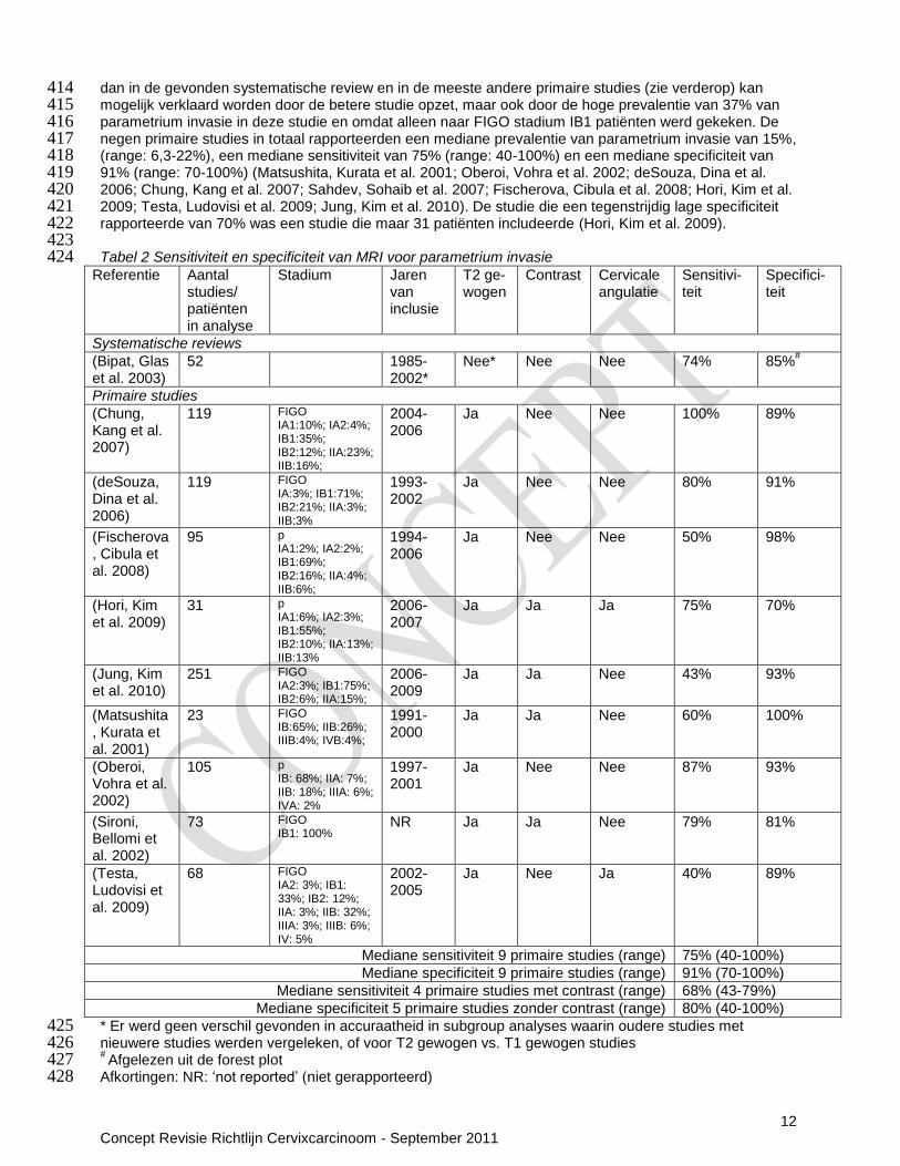
dan in de gevonden systematische review en in de meeste andere primaire studies (zie verderop) kan 414 mogelijk verklaard worden door de betere studie opzet, maar ook door de hoge prevalentie van 37% van 415 parametrium invasie in deze studie en omdat alleen naar FIGO stadium IB1 patiënten werd gekeken. De 416 negen primaire studies in totaal rapporteerden een mediane prevalentie van parametrium invasie van 15%, 417 (range: 6,3-22%), een mediane sensitiviteit van 75% (range: 40-100%) en een mediane specificiteit van 418 91% (range: 70-100%) (Matsushita, Kurata et al. 2001; Oberoi, Vohra et al. 2002; deSouza, Dina et al. 419 2006; Chung, Kang et al. 2007; Sahdev, Sohaib et al. 2007; Fischerova, Cibula et al. 2008; Hori, Kim et al. 420 2009; Testa, Ludovisi et al. 2009; Jung, Kim et al. 2010). De studie die een tegenstrijdig lage specificiteit 421 rapporteerde van 70% was een studie die maar 31 patiënten includeerde (Hori, Kim et al. 2009). 422 423 Tabel 2 Sensitiviteit en specificiteit van MRI voor parametrium invasie 424
Referentie Aantal studies/ patiënten in analyse
Stadium Jaren van inclusie
T2 ge-wogen
Contrast Cervicale angulatie
Sensitivi- teit
Specifici- teit
Systematische reviews
(Bipat, Glas et al. 2003)
52 1985-2002*
Nee* Nee Nee 74% 85%#
Primaire studies
(Chung, Kang et al. 2007)
119 FIGO IA1:10%; IA2:4%; IB1:35%; IB2:12%; IIA:23%; IIB:16%;
2004-2006
Ja Nee Nee 100% 89%
(deSouza, Dina et al. 2006)
119 FIGO IA:3%; IB1:71%; IB2:21%; IIA:3%; IIB:3%
1993-2002
Ja Nee Nee 80% 91%
(Fischerova, Cibula et al. 2008)
95 p IA1:2%; IA2:2%; IB1:69%; IB2:16%; IIA:4%; IIB:6%;
1994-2006
Ja Nee Nee 50% 98%
(Hori, Kim et al. 2009)
31 p IA1:6%; IA2:3%; IB1:55%; IB2:10%; IIA:13%; IIB:13%
2006-2007
Ja Ja Ja 75% 70%
(Jung, Kim et al. 2010)
251 FIGO IA2:3%; IB1:75%; IB2:6%; IIA:15%;
2006-2009
Ja Ja Nee 43% 93%
(Matsushita, Kurata et al. 2001)
23 FIGO IB:65%; IIB:26%; IIIB:4%; IVB:4%;
1991-2000
Ja Ja Nee 60% 100%
(Oberoi, Vohra et al. 2002)
105 p IB: 68%; IIA: 7%; IIB: 18%; IIIA: 6%; IVA: 2%
1997-2001
Ja Nee Nee 87% 93%
(Sironi, Bellomi et al. 2002)
73 FIGO IB1: 100%
NR Ja Ja Nee 79% 81%
(Testa, Ludovisi et al. 2009)
68 FIGO IA2: 3%; IB1: 33%; IB2: 12%; IIA: 3%; IIB: 32%; IIIA: 3%; IIIB: 6%; IV: 5%
2002-2005
Ja Nee Ja 40% 89%
Mediane sensitiviteit 9 primaire studies (range) 75% (40-100%)
Mediane specificiteit 9 primaire studies (range) 91% (70-100%)
Mediane sensitiviteit 4 primaire studies met contrast (range) 68% (43-79%)
Mediane specificiteit 5 primaire studies zonder contrast (range) 80% (40-100%)
* Er werd geen verschil gevonden in accuraatheid in subgroup analyses waarin oudere studies met 425 nieuwere studies werden vergeleken, of voor T2 gewogen vs. T1 gewogen studies 426 # Afgelezen uit de forest plot 427
Afkortingen: NR: „not reported‟ (niet gerapporteerd) 428
13 Concept Revisie Richtlijn Cervixcarcinoom - September 2011
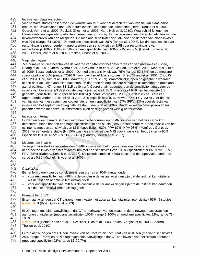
Invasie van blaas en rectum 429 Vier primaire studies beschreven de waarde van MRI voor het detecteren van invasie van blaas en/of 430 rectum, met zowel voor blaas- als rectuminvasie uiteenlopende uitkomsten (Hertel, Kohler et al. 2002; 431 Oberoi, Vohra et al. 2002; Rockall, Ghosh et al. 2006; Nam, Huh et al. 2010). Waarschijnlijk liggen de 432 kleine aantallen ingesloten patiënten hieraan ten grondslag. Echter, ook een verschil in de definities van de 433 uitkomstwaarden kan een rol spelen. De mediane sensitiviteit van MRI voor de detectie van blaas invasie 434 was 97% (range: 64-100%). De mediane specificiteit was 88% (range: 63-100%). De drie studies die over 435 rectuminvasie rapporteerden, rapporteerden een sensitiviteit van MRI voor rectuminvasie van 436 respectievelijk 100%, 100% en 50%; en een specificiteit van 100%, 91% en 86% (Hertel, Kohler et al. 437 2002; Oberoi, Vohra et al. 2002; Rockall, Ghosh et al. 2006) 438 439 Vaginale invasie 440 Zes primaire studies beschreven de waarde van MRI voor het detecteren van vaginale invasie (Sheu, 441 Chang et al. 2001; Oberoi, Vohra et al. 2002; Choi, Kim et al. 2004; Hori, Kim et al. 2009; Manfredi, Gui et 442 al. 2009; Testa, Ludovisi et al. 2009). De mediane sensitiviteit was 75% (range: 67-87%) en de mediane 443 specificiteit was 80% (range: 72-92%) voor vier vergelijkbare studies (Sheu, Chang et al. 2001; Choi, Kim 444 et al. 2004; Hori, Kim et al. 2009; Manfredi, Gui et al. 2009). Waarschijnlijk lopen de geschatte waardes 445 uiteen door de kleine aantallen patiënten, en daarmee de nog kleinere aantallen uitkomstmaten (mediaan 446 aantal patiënten: 47; range: 31-115 patiënten). Oberoi et al. rapporteerden de sensitiviteit apart voor een 447 invasie van bovenste 2/3 deel van de vagina (sensitiviteit: 83%; specificiteit 94%) en het laagste 1/3 448 gedeelte (sensitiviteit 78%; specificiteit 100%) (Oberoi, Vohra et al. 2002). De studie van Testa et al. 449 rapporteerde verder een sensitiviteit van 100% (specificiteit: 97%; NPV: 100%; PPV: 33%) voor detectie 450 van invasie van het septum vesicovaginale; en een specificiteit van 97% (PPV: 33%) voor detectie van 451 invasie van het septum rectovaginale (Testa, Ludovisi et al. 2009). Omdat er respectievelijk één en nul 452 patiënten waren met deze uitkomstmaten lijken deze gegevens weinig betrouwbaar. 453 454 Invasie os interna 455 Er werden twee primaire studies gevonden die beoordeelden of MRI invasie van het os interna kon 456 detecteren; beide hadden een hoge specificiteit. In één studie (N=53) detecteerde MRI een invasie van het 457 os interna met een sensitiviteit van 86% (specificiteit: 93%; PPV 67%; NPV 98%) (Manfredi, Gui et al. 458 2009). In een grotere studie (N=150) was de sensitiviteit van MRI voor invasie van het os interna 90% 459 (specificiteit: 98%; NPV: 98%; PPV: 86%) (Sahdev, Sohaib et al. 2007). 460 461 Myometrium invasie 462 Twee primaire studies beoordeelden of MRI invasie van het myometrium kan detecteren. Eén studie 463 beoordeelde invasie van het myometrium met een sensitiviteit van 100% (specificiteit: 99%; NPV: 100%; 464 PPV: 88%) (Sahdev, Sohaib et al. 2007). De tweede studie (N=208) beschreef de oppervlakte onder de 465 curve als 0,80 (Mitchell, Snyder et al. 2006). 466 467 468 Conclusies 469 Bij het formuleren van de conclusies is een grens van 90% aangehouden: 470 voor een sensitiviteit van ≥90% is de conclusie dat er aanwijzingen zijn dat de test het kan uitsluiten 471
als de test een negatieve test uitslag geeft 472 voor een specificiteit van ≥90% is de conclusie dat er aanwijzingen zijn dat de test het kan aantonen 473
als de test een afwijkende uitslag geeft 474 475 Primaire tumor CT 476 Er zijn aanwijzingen dat CT parametrium invasie niet accuraat kan uitsluiten (sensitiviteit 55%, 9 studies) 477 Niveau 3: B (Bipat, Glas et al. 2003) 478 479 Er zijn tegengestelde aanwijzingen dat CT tumorinvasie van de blaas en de urinewegen accuraat kan 480 aantonen of uitsluiten (mediane sensitiviteit 100%; range 9-100% en mediane specificiteit 92%; range 73- 481 100%). 482 Niveau 3: B (Hertel, Kohler et al. 2002; Bipat, Glas et al. 2003; Kokka, Vorgias et al. 2003; Sharma, 483 Thulkar et al. 2010) 484 485 Er zijn aanwijzingen dat CT een invasie van het rectum niet accuraat kan uitsluiten (mediane sensitiviteit 486 25%; range 0-50%) en er zijn tegengestelde aanwijzingen dat CT een invasie van het rectum aantonen 487 (mediane specificiteit 92%; range 85-99.7%) 488
14 Concept Revisie Richtlijn Cervixcarcinoom - September 2011
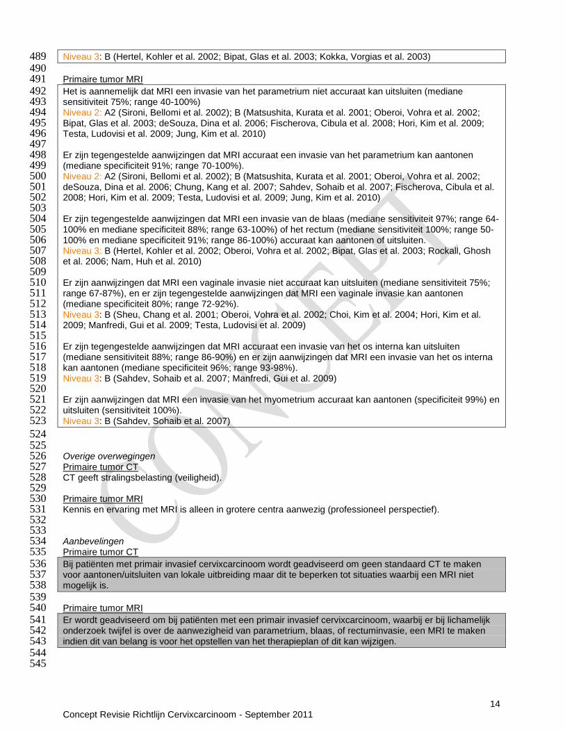
Niveau 3: B (Hertel, Kohler et al. 2002; Bipat, Glas et al. 2003; Kokka, Vorgias et al. 2003) 489 490 Primaire tumor MRI 491 Het is aannemelijk dat MRI een invasie van het parametrium niet accuraat kan uitsluiten (mediane 492 sensitiviteit 75%; range 40-100%) 493 Niveau 2: A2 (Sironi, Bellomi et al. 2002); B (Matsushita, Kurata et al. 2001; Oberoi, Vohra et al. 2002; 494 Bipat, Glas et al. 2003; deSouza, Dina et al. 2006; Fischerova, Cibula et al. 2008; Hori, Kim et al. 2009; 495 Testa, Ludovisi et al. 2009; Jung, Kim et al. 2010) 496 497 Er zijn tegengestelde aanwijzingen dat MRI accuraat een invasie van het parametrium kan aantonen 498 (mediane specificiteit 91%; range 70-100%). 499 Niveau 2: A2 (Sironi, Bellomi et al. 2002); B (Matsushita, Kurata et al. 2001; Oberoi, Vohra et al. 2002; 500 deSouza, Dina et al. 2006; Chung, Kang et al. 2007; Sahdev, Sohaib et al. 2007; Fischerova, Cibula et al. 501 2008; Hori, Kim et al. 2009; Testa, Ludovisi et al. 2009; Jung, Kim et al. 2010) 502 503 Er zijn tegengestelde aanwijzingen dat MRI een invasie van de blaas (mediane sensitiviteit 97%; range 64-504 100% en mediane specificiteit 88%; range 63-100%) of het rectum (mediane sensitiviteit 100%; range 50-505 100% en mediane specificiteit 91%; range 86-100%) accuraat kan aantonen of uitsluiten. 506 Niveau 3: B (Hertel, Kohler et al. 2002; Oberoi, Vohra et al. 2002; Bipat, Glas et al. 2003; Rockall, Ghosh 507 et al. 2006; Nam, Huh et al. 2010) 508 509 Er zijn aanwijzingen dat MRI een vaginale invasie niet accuraat kan uitsluiten (mediane sensitiviteit 75%; 510 range 67-87%), en er zijn tegengestelde aanwijzingen dat MRI een vaginale invasie kan aantonen 511 (mediane specificiteit 80%; range 72-92%). 512 Niveau 3: B (Sheu, Chang et al. 2001; Oberoi, Vohra et al. 2002; Choi, Kim et al. 2004; Hori, Kim et al. 513 2009; Manfredi, Gui et al. 2009; Testa, Ludovisi et al. 2009) 514 515 Er zijn tegengestelde aanwijzingen dat MRI accuraat een invasie van het os interna kan uitsluiten 516 (mediane sensitiviteit 88%; range 86-90%) en er zijn aanwijzingen dat MRI een invasie van het os interna 517 kan aantonen (mediane specificiteit 96%; range 93-98%). 518 Niveau 3: B (Sahdev, Sohaib et al. 2007; Manfredi, Gui et al. 2009) 519 520 Er zijn aanwijzingen dat MRI een invasie van het myometrium accuraat kan aantonen (specificiteit 99%) en 521 uitsluiten (sensitiviteit 100%). 522 Niveau 3: B (Sahdev, Sohaib et al. 2007) 523
524 525 Overige overwegingen 526 Primaire tumor CT 527 CT geeft stralingsbelasting (veiligheid). 528 529 Primaire tumor MRI 530 Kennis en ervaring met MRI is alleen in grotere centra aanwezig (professioneel perspectief). 531 532 533 Aanbevelingen 534 Primaire tumor CT 535 Bij patiënten met primair invasief cervixcarcinoom wordt geadviseerd om geen standaard CT te maken 536 voor aantonen/uitsluiten van lokale uitbreiding maar dit te beperken tot situaties waarbij een MRI niet 537 mogelijk is. 538 539 Primaire tumor MRI 540 Er wordt geadviseerd om bij patiënten met een primair invasief cervixcarcinoom, waarbij er bij lichamelijk 541 onderzoek twijfel is over de aanwezigheid van parametrium, blaas, of rectuminvasie, een MRI te maken 542 indien dit van belang is voor het opstellen van het therapieplan of dit kan wijzigen. 543 544
545

15 Concept Revisie Richtlijn Cervixcarcinoom - September 2011
4.3 Lymfekliermetastasen (Evidence based tekst n.a.v. uitgangsvraag 1: Diagnostische 546 technieken) 547 548 Literatuurbespreking 549 Systematische reviews 550 Vier systematische reviews beoordeelden beeldvormende technieken voor de detectie van 551 lymfekliermetastasen (Bipat, Glas et al. 2003; Havrilesky, Kulasingam et al. 2005; Selman, Mann et al. 552 2008; Kang, Kim et al. 2010) (zie ook tabel 3 en 4). 553 554 Eén systematische review werd eerder besproken onder tumorkarakteristieken parametrium, blaas of 555 rectum invasie (Bipat, Glas et al. 2003). Hier was de sensitiviteit voor het detecteren van 556 lymfekliermetastasen (lokatie niet gespecificeerd) voor MRI 60% (95% CI 52-68; N=25); significant hoger 557 dan voor CT (43%; 95% CI 37-57; N=17) (P=0.047). 558 De tweede review van Havrilseky et al. includeerde 15 kleine studies, waarvan geen enkele een blindering 559 voor de beoordeling van de beeldvorming rapporteerde (Havrilesky, Kulasingam et al. 2005). In een meta-560 analyse van twee van de geïncludeerde studies was de sensitiviteit van CT om pelviene 561 lymfekliermetastasen te detecteren 47% (95%CI: 21–73)(referentie: histologie of follow-up); Er werden 562 onvoldoende data gepubliceerd voor het berekenen van de specificiteit. Voor MRI was de sensitiviteit 72% 563 (95%CI: 53–87) en de specificiteit 96% (95%CI: 92–98) (meta-analyse van twee studies met histologie of 564 follow-up als referentie); en voor PET was de sensitiviteit 79% (95%CI: 65–90) met een specificiteit van 565 99% (95%CI: 96–99) (meta-analyse van vier studies met histologie of follow-up als standaard). Eén van de 566 geïncludeerde studies rapporteerde een sensitiviteit van 67% (95%CI: 9-99) en een specificiteit van 100% 567 (95%CI: 66-100) voor de detectie van para-aortale lymfekliermetastasen door middel van MRI (referentie: 568 histologie). In een meta-analyse van vier van die geïncludeerde studies was de sensitiviteit van PET voor 569 de detectie van para-aortale lymfekliermetastasen 84% (95%CI: 68-94%) en de specificiteit was 95% 570 (95%CI: 89-98) (referentie: histologie). 571 572 De (derde) review van Kang et al. includeerde tien studies die de detectie van para-aortale 573 lymfekliermetastasen met behulp van PET of PET/CT, waarvan er acht prospectief van opzet waren en 574 waarvan zes de beeldvorming geblindeerd beoordeelden (Kang, Kim et al. 2010). Een meta-analyse van 575 de tien geïncludeerde studies (vijf studies naar PET en vijf studies naar PET/CT, steeds met histologie als 576 referentie standaard) liet een sensitiviteit zien van 34% (95%CI: 10-72); een specificiteit van 97% (95%CI: 577 93-99%); een negatieve likelihood ratio van 0.68 (95%CI: 0.40-1.15); en een positieve likelihood ratio van 578 12.49 (95%CI: 4.64-33.62). De sensitiviteit en specificiteit voor PET waren 66% (95%CI: 33-89) en 97% 579 (95%CI: 90-99); en voor PET/CT 13% (95%CI: 2-56) en 98% (95%CI: 78-100). De sensitiviteit van de 580 PET/CT voor lymfkliermetastasen was extreem heterogeen; de auteurs concludeerden dat bias in studies 581 met een lagere prevalentie van lymfeklier metastasen een grote rol moet hebben gespeeld. In de vijf 582 studies met een hoge prevalentie (>15%) van lymfekliermetastasen was de sensitiviteit 73% (95%CI: 53-583 87%); en de specificiteit: 93% (95%CI: 86-97%). 584 585 In de vierde en laatste review rapporteerden Selman et al. 95 test resultaten van 72 geïncludeerde studies 586 met histologie als referentie standaard, waarbij zowel studies die naar pelviene lymfeklieren keken als 587 studies die naar para-aortale lymfeklieren keken werden meegenomen (Selman, Mann et al. 2008). Alle 588 primaire studies hadden methodologische beperkingen. Een meta-analyse van 32 studies die CT 589 evalueerden liet een sensitiviteit zien van 58% ((95% CI 54-61); specificiteit: 92% (95% CI 92-94); 590 positieve likelihood ratio: 4,3 (95% CI 3,0-6,2); negatieve likelihood ratio: 0,58 (95% CI 0,48-0,70)). Een 591 meta-analyse van 24 studies naar MRI liet een sensitiviteit zien van 56% ((95% CI 49-62); specificiteit: 592 93% (95%CI 91-94); positieve likelihood ratio: 6,4 (95% CI 4,9-8,3); negatieve likelihood ratio: 0,50 (95% 593 CI 0,39-0,64)). 594 Een meta-analyse van 8 studies naar PET liet een sensitiviteit zien van 75% ((95% CI 63-84); specificiteit: 595 98% (95%CI 95-99); positieve likelihood ratio: 15,3 (95% CI 7,9-29,6); negatieve likelihood ratio: 0.27 (95% 596 CI 0,11-0,66). PET was meer accuraat dan MRI voor het aantonen van pelviene en para-aortale 597 kliermetastasen (odds ratio 3,84; 95%CI 1,22-12,12). CT en MRI waren even accuraat (odds ratio 0,63; 598 95%CI 0,36-1,12). 599 600 Primaire studies 601 Na de laatste zoekdatum van de systematische reviews werd er één primaire studie gevonden met 602 informatie over CT; en negen primaire studies die MRI evalueerden; drie studies die naar PET keken; en 603 13 studies over PET/CT. 604
16 Concept Revisie Richtlijn Cervixcarcinoom - September 2011
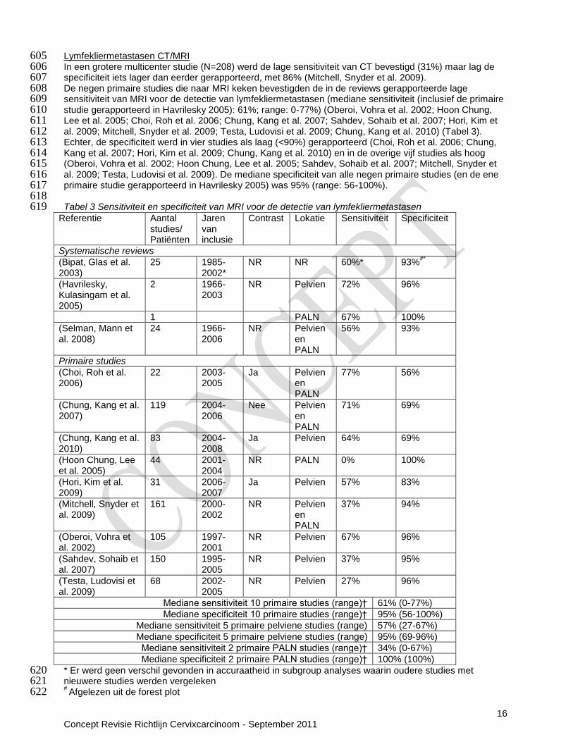
Lymfekliermetastasen CT/MRI 605 In een grotere multicenter studie (N=208) werd de lage sensitiviteit van CT bevestigd (31%) maar lag de 606 specificiteit iets lager dan eerder gerapporteerd, met 86% (Mitchell, Snyder et al. 2009). 607 De negen primaire studies die naar MRI keken bevestigden de in de reviews gerapporteerde lage 608 sensitiviteit van MRI voor de detectie van lymfekliermetastasen (mediane sensitiviteit (inclusief de primaire 609 studie gerapporteerd in Havrilesky 2005): 61%; range: 0-77%) (Oberoi, Vohra et al. 2002; Hoon Chung, 610 Lee et al. 2005; Choi, Roh et al. 2006; Chung, Kang et al. 2007; Sahdev, Sohaib et al. 2007; Hori, Kim et 611 al. 2009; Mitchell, Snyder et al. 2009; Testa, Ludovisi et al. 2009; Chung, Kang et al. 2010) (Tabel 3). 612 Echter, de specificiteit werd in vier studies als laag (<90%) gerapporteerd (Choi, Roh et al. 2006; Chung, 613 Kang et al. 2007; Hori, Kim et al. 2009; Chung, Kang et al. 2010) en in de overige vijf studies als hoog 614 (Oberoi, Vohra et al. 2002; Hoon Chung, Lee et al. 2005; Sahdev, Sohaib et al. 2007; Mitchell, Snyder et 615 al. 2009; Testa, Ludovisi et al. 2009). De mediane specificiteit van alle negen primaire studies (en de ene 616 primaire studie gerapporteerd in Havrilesky 2005) was 95% (range: 56-100%). 617 618 Tabel 3 Sensitiviteit en specificiteit van MRI voor de detectie van lymfekliermetastasen 619
Referentie Aantal studies/ Patiënten
Jaren van inclusie
Contrast Lokatie Sensitiviteit Specificiteit
Systematische reviews
(Bipat, Glas et al. 2003)
25 1985-2002*
NR NR 60%* 93%#*
(Havrilesky, Kulasingam et al. 2005)
2 1966-2003
NR Pelvien 72% 96%
1 PALN 67% 100%
(Selman, Mann et al. 2008)
24 1966-2006
NR Pelvien en PALN
56% 93%
Primaire studies
(Choi, Roh et al. 2006)
22 2003-2005
Ja Pelvien en PALN
77% 56%
(Chung, Kang et al. 2007)
119 2004-2006
Nee Pelvien en PALN
71% 69%
(Chung, Kang et al. 2010)
83 2004-2008
Ja Pelvien 64% 69%
(Hoon Chung, Lee et al. 2005)
44 2001-2004
NR PALN 0% 100%
(Hori, Kim et al. 2009)
31 2006-2007
Ja Pelvien 57% 83%
(Mitchell, Snyder et al. 2009)
161 2000-2002
NR Pelvien en PALN
37% 94%
(Oberoi, Vohra et al. 2002)
105 1997-2001
NR Pelvien 67% 96%
(Sahdev, Sohaib et al. 2007)
150 1995-2005
NR Pelvien 37% 95%
(Testa, Ludovisi et al. 2009)
68 2002-2005
NR Pelvien 27% 96%
Mediane sensitiviteit 10 primaire studies (range)† 61% (0-77%)
Mediane specificiteit 10 primaire studies (range)† 95% (56-100%)
Mediane sensitiviteit 5 primaire pelviene studies (range) 57% (27-67%)
Mediane specificiteit 5 primaire pelviene studies (range) 95% (69-96%)
Mediane sensitiviteit 2 primaire PALN studies (range)† 34% (0-67%)
Mediane specificiteit 2 primaire PALN studies (range)† 100% (100%)
* Er werd geen verschil gevonden in accuraatheid in subgroup analyses waarin oudere studies met 620 nieuwere studies werden vergeleken 621 # Afgelezen uit de forest plot 622

17 Concept Revisie Richtlijn Cervixcarcinoom - September 2011
† Inclusief de primaire studie gerapporteerd in Havrilesky 2005 623 Afkortingen: NR: „not reported‟ (niet gerapporteerd); PALN: para-aortale lymfeklieren 624 625 Lymfekliermetastasen PET 626 Twee primaire studies evalueerden PET. De eerste studie betrof patiënten (N=60) die geen 627 lymfekliermetastasen hadden op de MRI (Chou, Chang et al. 2006). In deze patiëntengroep was de 628 sensitiviteit van PET voor het detecteren van pelviene lymfekliermetastasen 10% (specificiteit: 94%, NPV 629 84%, PPV: 25%). In de tweede studie (N=47) hadden alle patiënten verdenking op een para-aortale, 630 inguinale en/of supraclaviculaire lymfekliermetastase (Chao, Ho et al. 2008). 9/47 patiënten werd met 631 PET/CT geëvalueerd, maar deze resultaten werden niet apart van PET gerapporteerd. In alle patiënten 632 groep was de sensitiviteit van PET of PET/CT voor de detectie van para-aortale lymfeklieren 97% 633 (specificiteit 90%; NPV: 90%; PPV: 97%); voor de detectie van inguinale lymfeklieren 80% (specificiteit 634 86%; NPV: 97%; PPV: 40%); en voor de detectie van supraclaviculaire lymfeklieren 85% (specificiteit: 635 100%; NPV: 94%; PPV: 100%). 636 637 Lymfekliermetastasen PET/CT 638 Dertien primaire studies evalueerden PET/CT. Alle studies hadden methodologische tekortkomingen. De 639 mediane sensitiviteit voor twaalf min of meer vergelijkbare studies was 39% (range: 0-100%) en de 640 mediane specificiteit was 94% (range: 56-100%) (Amit, Beck et al. 2006; Choi, Roh et al. 2006; Sironi, 641 Buda et al. 2006; Loft, Berthelsen et al. 2007; Chung, Park et al. 2009; Kim, Choi et al. 2009; Chung, Kang 642 et al. 2010; Goyal, Singh et al. 2010; Ramirez, Jhingran et al. 2010; Leblanc, Gauthier et al. 2011; Sandvik, 643 Jensen et al. 2011; Yu, Jia et al. 2011) (Tabel 4). Er waren geen duidelijke aanwijzingen waarom sommige 644 studies tegenstrijdige resultaten rapporteerden, zoals de studie van Loft et al. die als enige studie een 645 sensitiviteit boven de 90% rapporteerde, en de studies van Kim et al. en Choi et al, die als enigen een 646 specificiteit onder de 90% rapporteerden (Choi, Roh et al. 2006; Loft, Berthelsen et al. 2007; Kim, Choi et 647 al. 2009). Eén kleine studie (N=16) rapporteerde een sensitiviteit van 0% voor het detecteren van bilaterale 648 lymfekliermetastasen bij een selectieve subgroep die zowel MRI en PET/CT hadden ondergaan voor 649 chirurgie (NPV 88%) (Bentivegna, Uzan et al. 2010). 650 651 Tabel 4 Sensitiviteit en specificiteit van PET/CT voor de detectie van lymfekliermetastasen 652
Referentie Aantal studies/ patiënten
Jaren van inclusie
Lokatie Sensitiviteit Specificiteit
Systematische reviews
(Kang, Kim et al. 2010) 5 1980-2009 PALN 13% 98%
Primaire studies
(Amit, Beck et al. 2006) 16 NR NR 0% NR
(Choi, Roh et al. 2006) 22 2003-2005 Pelvien en PALN 77% 56%
(Chung, Park et al. 2009) 34 2003-2007 Pelvien 41% 94%
(Chung, Kang et al. 2010) 83 2004-2008 Pelvien 29% 84%
(Goyal, Singh et al. 2010) 80 2007-2009 Pelvien 58% 93%
(Kim, Choi et al. 2009) 79 2001-2007 Pelvien en PALN 47% 71%
(Leblanc, Gauthier et al. 2011)
125 2004-2008 PALN 33% 94%
(Loft, Berthelsen et al. 2007)
119 2002-2005 PALN 100% 99%
(Ramirez, Jhingran et al. 2010)
60 2004-2009 PALN 36% 96%
(Sandvik, Jensen et al. 2011)
36 2006-2007 Pelvien 20% 90%
(Sironi, Buda et al. 2006) 47 2003-2004 Pelvien 73% 97%
(Yu, Jia et al. 2011) 16 NR NR 0% 100%
Mediane sensitiviteit 12 primaire studies (range) 39% (0-100%)
Mediane specificiteit 11 primaire studies (range) 94% (56-100%)
Mediane sensitiviteit 5 primaire pelviene studies (range) 41% (20-73%)
Mediane specificiteit 5 primaire pelviene studies (range) 93% (84-97%)
Mediane sensitiviteit 3 primaire PALN studies (range) 36% (33-100%)
Mediane specificiteit 3 primaire PALN studies (range) 96% (94-99%)
Afkortingen: NR: „not reported‟ (niet gerapporteerd); PALN: para-aortale lymfeklieren 653

18 Concept Revisie Richtlijn Cervixcarcinoom - September 2011
Conclusies Lymfekliermetastasen CT/MRI 654 Er zijn aanwijzingen dat CT (sensitiviteit 31%) of MRI (mediane sensitiviteit 61%; range 0-77%) de 655 aanwezigheid van pelviene of para-aortale lymfekliermetastasen niet accuraat kunnen uitsluiten 656 Niveau 3: B (Oberoi, Vohra et al. 2002; Bipat, Glas et al. 2003; Havrilesky, Kulasingam et al. 2005; Hoon 657 Chung, Lee et al. 2005; Choi, Roh et al. 2006; Chung, Kang et al. 2007; Sahdev, Sohaib et al. 2007; 658 Selman, Mann et al. 2008; Hori, Kim et al. 2009; Mitchell, Snyder et al. 2009; Testa, Ludovisi et al. 2009; 659 Chung, Kang et al. 2010) 660 661 Er zijn aanwijzingen dat CT (specificiteit 86%) of MRI (mediane specificiteit 95%; range 56-100%) de 662 aanwezigheid van pelviene of para-aortale lymfekliermetastasen accuraat kunnen aantonen. 663 Niveau 3: B (Oberoi, Vohra et al. 2002; Bipat, Glas et al. 2003; Havrilesky, Kulasingam et al. 2005; Hoon 664 Chung, Lee et al. 2005; Sahdev, Sohaib et al. 2007; Selman, Mann et al. 2008; Mitchell, Snyder et al. 665 2009; Testa, Ludovisi et al. 2009) 666 667 Conclusies lymfekliermetastasen PET(/CT) 668 Er zijn aanwijzingen dat PET de aanwezigheid van lymfekliermetastasen niet accuraat kan uitsluiten 669 (mediane sensitiviteit 75%; range 66-79%), maar wel accuraat kan aantonen (mediane specificiteit 98%; 670 range 97-99%). 671 Niveau 3: B (Havrilesky, Kulasingam et al. 2005; Selman, Mann et al. 2008; Kang, Kim et al. 2010) 672 673 Er zijn aanwijzingen dat PET pelviene lymfekliermetastasen accuraat kan aantonen (specificiteit 94%), 674 maar niet kan uitsluiten (sensitiviteit 10%), in patiënten die geen aanwijzingen hadden voor 675 lymfekliermetastasen op MRI. 676 Niveau 3: B (Chou, Chang et al. 2006) 677 678 Er zijn aanwijzingen dat PET/CT pelviene en para-aortale lymfeklier metastasen accuraat kan aantonen 679 (mediane specificiteit 94%; range 56-100%), maar niet kan uitsluiten (mediane sensitiviteit 39%; range 0-680 100%), 681 Niveau 3: B (Amit, Beck et al. 2006; Choi, Roh et al. 2006; Sironi, Buda et al. 2006; Loft, Berthelsen et al. 682 2007; Chung, Park et al. 2009; Kim, Choi et al. 2009; Chung, Kang et al. 2010; Goyal, Singh et al. 2010; 683 Kang, Kim et al. 2010; Ramirez, Jhingran et al. 2010; Leblanc, Gauthier et al. 2011; Yu, Jia et al. 2011) 684 685 686 Overige overwegingen 687 Lymfekliermetastasen CT/MRI 688 Indien er een MRI wordt gemaakt voor het bepalen van de tumorgrootte, parametrium, blaas en rectum 689 invasie, heeft dit ook de voorkeur bij het bepalen van de lymfeklierstatus zodat kan worden volstaan met 690 één diagnostiekvorm (kosteneffectiviteit). Verder geeft een CT stralingsbelasting (veiligheid). Kennis en 691 ervaring met MRI is alleen in de grotere centra aanwezig (professioneel perspectief). Beschikbaarheid is 692 echter minder goed geregeld (langere wachttijden) 693 694 Lymfekliermetastasen PET(/CT) 695 PET(CT) geeft stralingsbelasting (veiligheid); De expertise met PET(CT) is alleen in grotere centra 696 aanwezig (professioneel perspectief); PET(CT) is duur en kosteneffectiviteitsanalyses zijn er nog niet in 697 voldoende mate (kosteneffectiviteit); PET(CT) is alleen in de grotere ziekenhuizen aanwezig (organisatie) 698 699 700 Aanbevelingen 701 Lymfekliermetastasen CT/MRI 702 Bij patiënten met primair invasief cervixcarcinoom wordt geadviseerd om geen standaard CT te maken 703 voor het aantonen van lymfekliermetastasen. 704 705 Lymfekliermetastasen MRI 706 Bij patiënten met primair invasief cervixcarcinoom waarbij er onduidelijkheid bestaat over de lokale 707 uitbreiding wordt geadviseerd om naast de MRI voor lokale uitbreiding ook direct de MRI te gebruiken om 708 eventuele lymfekliermetastasen aan te tonen. 709 710 711 712

19 Concept Revisie Richtlijn Cervixcarcinoom - September 2011
Lymfekliermetastasen PET(/CT) 713 Er wordt geadviseerd om bij patiënten met primair invasief cervixcarcinoom alleen op indicatie een PET/CT 714 te maken om lymfekliermetastasen in hogere stations uit te sluiten/aan te tonen. Indicaties kunnen zijn 715 positieve klieren bij operatie of aanwijzingen voor lymfekliermetastasen op de MRI. 716 717 718 719
4.4 Schildwachtklierbiopt (Evidence based tekst n.a.v. uitgangsvraag 1: Diagnostische 720 technieken) 721 722 Literatuurbespreking 723 Systematische reviews 724 Twee systematische reviews en vijf primaire studies beoordeelden de waarde van een schildwachtklier 725 biopsie in de stadiering van cervix carcinoom. Eén systematische review bevatte ook informatie over CT, 726 MRI en PET en werd eerder besproken (Selman, Mann et al. 2008). Deze review liet in een meta-analyse 727 van 31 studies zien dat bij 96% van de patiënten een schildwachtklier werd gevonden, als zowel 728 techneticum als blue dye werden gebruikt. De sensitiviteit van een schildwachtklier biopsie was 91% (95% 729 CI 87-95) (specificiteit: 100% (95%CI 99,6-100); positieve likelihood ratio: 40.8 (95%CI 24,6-67,6); 730 negatieve likelihood ratio: 0,18 (95%CI 0,14-0,24)). Een schildwachtklier biopsie was accurater dan MRI 731 voor het aantonen van lymfkliermetastasen (odds ratio: 18,49; 95%CI 3,59-95,17; p<0,01). 732 733 De tweede systematische review verrichtte een meta-analyse van 22 studies met histologie als referentie 734 standaard (van de Lande, Torrenga et al. 2007). In geen van de studies werd de referentie standaard 735 geblindeerd geëvalueerd. De sensitiviteit van een schildwachtklier biopsie was 89% (95%CI: 83-94). Bij 736 95% van de patiënten werd een schildwachtklier gevonden, als zowel technetium als blue dye werden 737 gebruikt. 738 739 Primaire studies 740 Na de laatste zoekdatum van de systematische reviews werden drie studies met meer dan 100 patiënten 741 gevonden, en twee studies die naar een intra-operatieve beoordeling van de schildwachtklier keken. In 84-742 90% van de patiënten werd tenminste één schildwachtklier gevonden, als zowel technetium als blue dye 743 werden gebruikt (Wydra, Sawicki et al. 2006; Altgassen, Hertel et al. 2008; Darlin, Persson et al. 2010). In 744 59-66% van de patiënten werd aan twee kanten een schildwachtklier gevonden (Wydra, Sawicki et al. 745 2006; Darlin, Persson et al. 2010). In de drie grotere patiëntenseries lag de sensitiviteit van een 746 schildwachtklier biopt tussen de 77% en 94% voor het uitsluiten van lymfkliermetastasen (range NPV: 94-747 96%). De sensitiviteit was het hoogst in de subgroep patiënten met een tumor van 2 cm of kleiner (91%), 748 en wanneer er bij patiënten aan twee kanten een schildwachtklier werd gevonden (87%) (Altgassen, Hertel 749 et al. 2008) 750 751 Als de schildwachtklier nog tijdens de operatie werd beoordeeld wisselde de sensitiviteit sterk, in de twee 752 studies die hiernaar keken. In de eerste studie (N=38) lag de sensitiviteit beduidend lager (33% intra-753 operatief vs. 83% postoperatief; NPV: 89% vs. 97%) (Fader, Edwards et al. 2008), terwijl die in de tweede 754 studie (N=58%) hoog lag (100% intra-operatief; NPV: 100%) (Yamashita, Katayama et al. 2009). 755 756 Conclusies 757 Er zijn tegengestelde aanwijzingen dat een schildwachtklier biopt lymfekliermetastasen accuraat kan 758 uitsluiten (sensiviteit range 33-100%) 759 Niveau 3: B (Wydra, Sawicki et al. 2006; van de Lande, Torrenga et al. 2007; Altgassen, Hertel et al. 2008; 760 Fader, Edwards et al. 2008; Selman, Mann et al. 2008; Yamashita, Katayama et al. 2009; Darlin, Persson 761 et al. 2010) 762 763 764 Overige overwegingen 765 Aangezien bewijs ontbreekt dat een schildwachtklier biopt lymfekliermetastasen accuraat kan uitsluiten, 766 hebben verdere overige overwegingen met betrekking tot deze diagnostiek geen betekenis. 767 768 769 770 771

20 Concept Revisie Richtlijn Cervixcarcinoom - September 2011
Aanbevelingen 772 Bij patiënten met primair invasief cervixcarcinoom wordt geadviseerd om de schildklierwachtprocedure 773 alleen in onderzoeksverband uit te voeren. 774 775 776
4.5 Metastasen op afstand (Evidence based tekst n.a.v. uitgangsvraag 1: Diagnostische 777 technieken) 778 779 Literatuurbespreking 780 Vier primaire studies evalueerden de waarde van verschillende diagnostische technieken voor het 781 opsporen van metastasen op afstand. In de eerste studie (N=165) evalueerden Liu et al. de waarde van 782 CT, MRI en PET voor de detectie van hematogene botmetastasen in patiënten met FIGO stadium III of IV 783 (Liu, Yen et al. 2009). PET was het meest accuraat in het diagnosticeren van hematogene botmetastasen, 784 met een sensitiviteit van 100% (specificiteit: 99%; NPV: 100%; PPV: 91%). MRI en CT presteerden minder 785 goed (sensitiviteit: 80%; specificiteit: 99%; NPV: 99%; PPV: 80% vs. sensitiviteit: 25%; specificiteit: 100%; 786 NPV: 92%; PPV: 100%). De accuratesse van PET of PET/CT werd bevestigd in een tweede serie (N=47) 787 patiënten, allen met verdenking van distale lymfekliermetastasen (sensitiviteit detectie botmetastasen: 788 100%; specificiteit: 98%; NPV 100%; PPV 50%) (Chao, Ho et al. 2008). In dezelfde studie was de 789 sensitiviteit van PET of PET/CT voor het detecteren van andere, niet skeleteuze metastasen op afstand 790 100% (specificiteit: 91%; NPV: 100%; PPV: 33%). De derde, kleine (N=17) serie patiënten die routine 791 preoperatieve onderzoeken, inclusief CT en MRI, hadden ondergaan, werd er met PET bij 5 patiënten 792 (29%) nieuwe metastasen ontdekt (sensitiviteit: 83%; specificiteit 100%; NPV: 92%; PPV: 100%)) 793 (Bjurberg, Kjellen et al. 2007). De vierde studie (N=119) evalueerde PET/CT (Loft, Berthelsen et al. 2007). 794 De sensitiviteit voor detecteren van metastasen op afstand was 100% (specificiteit: 94%; PPV 53%; NPV: 795 100%). 796 797 Conclusies 798 Er zijn aanwijzingen dat CT en MRI hematogene botmetastasen niet kunnen uitsluiten (sensitiviteit CT 799 25% en MRI 80%), maar wel accuraat kunnen aantonen (specificiteit CT 100% en 99%). 800 Niveau 3: B (Liu, Yen et al. 2009) 801 802 Er zijn aanwijzingen dat PET en PET/CT botmetastasen kunnen uitsluiten (mediane sensitiviteit 100%) en 803 aantonen (mediane specificiteit 99%). 804 Niveau 3: B (Chao, Ho et al. 2008; Liu, Yen et al. 2009) 805 806 Er zijn aanwijzingen dat PET nieuwe metastasen niet accuraat kan uitsluiten (sensitiviteit 83%), maar wel 807 accuraat kan aantonen (specificiteit 100%), in patiënten die routine preoperatieve onderzoeken, inclusief 808 CT en MRI, hebben ondergaan. 809 Niveau 3: B (Bjurberg, Kjellen et al. 2007) 810 811 Er zijn aanwijzingen dat PET en PET/CT de aanwezigheid van metastasen op afstand kan uitsluiten 812 (mediane sensitiviteit 100%) en aantonen (mediane specificiteit 97%; range 94-100%). 813 Niveau 3: B (Loft, Berthelsen et al. 2007; Chao, Ho et al. 2008) 814 815 Overige overwegingen 816 CT geeft stralingsbelasting (veiligheid). 817 Kennis en ervaring met MRI is alleen in grotere centra aanwezig (professioneel perspectief) 818 PET(CT) geeft stralingsbelasting (veiligheid); De expertise met PET(CT) is alleen in grotere centra 819 aanwezig (professioneel perspectief); PET(CT) is duur (kosteneffectiviteit); PET(CT) is alleen in de grotere 820 ziekenhuizen aanwezig (organisatie) 821 822 Aanbevelingen 823 Bij patiënten met primair invasief cervixcarcinoom wordt geadviseerd om onderzoek naar metastasen op 824 afstand uitsluitend op indicatie uit te voeren door middel van PET/CT. 825 Indicaties kunnen zijn positieve klieren bij operatie of indicatie voor primaire chemoradiatie. 826 827 828 829

21 Concept Revisie Richtlijn Cervixcarcinoom - September 2011
4.6 Tumormarkers (Evidence based tekst n.a.v. uitgangsvraag 1: Diagnostische technieken) 830 831 Literatuurbespreking 832 CA125 Twee studies evalueerden CA125 voor de detectie van lymfekliermetastasen. Bij een afkappunt 833 van ≥30 U/mL was de sensitiviteit van CA125 voor de detectie van lymfekliermetastasen 67% (specificiteit 834 84%; NPV: 92%; PPV: 46%) in vergelijking met het histologisch preparaat bij patiënten met 835 adenocarcinoom of adenosquameus carcinoom (Bender, Sorosky et al. 2003). 836 Bij een afkappunt van ≥5,0 ng/ml was de sensitiviteit van CA125 voor de detectie van 837 lymfekliermetastasen 18% (specificiteit 60%; NPV: 66%; PPV: 15%), waarbij werd vergeleken met het 838 histologisch preparaat of CT bij patiënten met cervixcarcinoom. (Kotowicz, Fuksiewicz et al. 2008). De 839 geïncludeerde patiënten hadden adenocarcinoom of plaveiselcarcinoom. De uitkomsten werden echter 840 niet uitgesplitst naar histologisch subtype. 841 842 SCC (Squamous cell carcinoma antigen) 843 Vier studies gepuliceerd vanaf 2001 evalueerden verschillende afkappunten voor SCC bij patiënten met 844 plaveiselcelcarcinoom. 845 Eén studie evalueerde of SCC een tumorgrootte van 4 cm of meer kon detecteren. In een studie waarin 846 SCC werd vergeleken met CT (of klinisch onderzoek, was de sensitiviteit bij een afkappunt van ≥2,0 ng/ml 847 64% (specificiteit 31%; NPV: 8%; PPV: 91%) (Chen, Liang et al. 2008). 848 Drie studies evalueerden of SCC lymfekliermetastasen kon detecteren. In de eerste studie, die SCC 849 vergeleek met CT, was de sensitiviteit van SCC ≥2.0 ng/ml 77% (specificiteit: 38%; NPV: 91%; PPV: 18%) 850 (Chen, Liang et al. 2008). De tweede studie vergeleek SCC ≥1,5 ng/ml met histologie, en mat een 851 sensitiviteit van 79% (specificiteit 56%; NPV: 88%; PPV: 40%) (Takeda, Sakuragi et al. 2002). De derde 852 studie vergeleek SCC ook met histologie, maar dan in twee verschillende subgroepen (van de Lande, 853 Davelaar et al. 2009). Bij patiënten met stadium IB1 was de sensitiviteit van SCC ≥1,65 ng/ml 53% 854 (specificiteit: 84%; NPV: 85%; PPV: 50%); en bij patiënten met stadium IIB2 of IIA was dat 63% 855 (specificiteit: 46%; NPV: 63%; PPV: 40%). 856 Eén studie die CA125 evalueerde, keek ook naar SCC voor het detecteren van lymfkliermetastasen bij 857 patiënten met adenocarcinoom of plaveiselcelcarcinoom. In deze studie, waarbij een vergelijking werd 858 gemaakt met histologie of CT, was de sensitiviteit van SCC ≥1,5 ng/ml 67% (specificiteit: 84%; NPV: 92%; 859 PPV: 46%) (Kotowicz, Fuksiewicz et al. 2008). 860 861 Conclusies 862 Er zijn geen aanwijzingen dat er een goed afkapppunt is van CA125 voor het definiëren van een hoog 863 risicogroep bij patiënten met adenocarcinoom/adenosquameus cervixcarcinoom met betrekking tot 864 lymfekliermetastasen (mediane sensitiviteit 43% (range 18-67%); mediane specificiteit 72% (range 60-865 84%) 866 Niveau 3: B (Bender, Sorosky et al. 2003; Kotowicz, Fuksiewicz et al. 2008) 867 868 Er zijn geen aanwijzingen dat er een goed afkappunt is van SCC voor het definiëren van een hoog 869 risicogroep bij patiënten met plaveiselcelcarcinoom met betrekking tot een tumor grootte van meer dan 4 870 cm (sensitiviteit 64%; specificiteit 31%) 871 Niveau 3: B ((Yuan, Wang et al. 2002; Chen, Liang et al. 2008) 872 873 Er zijn geen aanwijzingen dat er een goed afkappunt is van SCC voor het definiëren van een hoog 874 risicogroep bij patiënten met plaveiselcelcarcinoom met betrekking tot lymfekliermetastasen (mediane 875 sensitiviteit 67%; range 53-79%; mediane specificiteit 56%; range 38-84%) , 876 Niveau 3: B (Takeda, Sakuragi et al. 2002; Chen, Liang et al. 2008; Kotowicz, Fuksiewicz et al. 2008; van 877 de Lande, Davelaar et al. 2009) 878 879 Overige overwegingen 880 Aangezien bewijs ontbreekt dat met tumormarkers een hoog risicogroep voor locale tumoruitbreiding of 881 (lymfeklier) metastasen adequaat kan worden gedefinieerd, hebben verdere overige overwegingen met 882 betrekking tot deze diagnostiek geen betekenis. 883 884 Aanbevelingen 885 De werkgroep is van mening dat er nog onvoldoende literatuur is over de klinische toepassing van de 886 tumormarkers SCC en CA125 voor het definiëren van een hoog risicogroep met betrekking tot prognose 887 en lymfekliermetastasen. 888
889
22 Concept Revisie Richtlijn Cervixcarcinoom - September 2011
5. BEHANDELING 890
5.1 Medisch technisch (Redactionele update) 891 Literatuurbespreking 892 Dit deel heeft betrekking op het plaveiselcel- en het adeno(squameus) carcinoom. 893 De behandeling van het cervixcarcinoom bestaat primair uit chirurgie of radiotherapie. Bij tumoren beperkt 894 tot de cervix of met minimale uitbreiding naar de proximale vagina wordt primair gekozen voor radicale 895 chirurgie. Bij uitbreiding buiten de cervix wordt primair gekozen voor radiotherapie, op indicatie in 896 combinatie met chemotherapie of hyperthermie. 897 898 5.1.1 Stadium IA1 (diagnose na goed te beoordelen conus) (Redactionele update) 899 Literatuurbespreking 900 Conisatie (bij kinderwens of andere individuele reden). 901 Uterusextirpatie (indien geen kinderwens). 902 In geval van stadium IA1 zal er zelden sprake zijn van lymfangio-invasieve groei. In die gevallen waarin dat 903 dit wel gevonden wordt is het onduidelijk of hieraan consequenties moeten worden verbonden. In de 904 minderheid van studies is lymfangio-invasieve groei een onafhankelijk prognostische factor voor overleving. 905 906 5.1.2 Stadium IA2 (diagnose na optimale c.q. goed te beoordelen conus) (Redactionele update) 907 Literatuurbespreking 908 Conisatie (bij kinderwens of andere individuele reden) 909 Uterusextirpatie (indien geen kinderwens) 910 In geval van lymfangio invasieve groei wordt een pelviene lymfadenectomie geadviseerd, omdat er hier 911 een duidelijke relatie bestaat met aanwezigheid van lymfkliermetastasen. 912 Deze aanbeveling wordt gebaseerd op een recent review uit 2009 waarbij 20 studies met 1063 patienten 913 met stadium IA2 cervixcarcinoom geincludeerd werden (Sedlis, Bundy et al. 1999). De meeste patiënten 914 werden primair behandeld met een radicale hysterectomie en pelviene lymfadenectomie, Bij de 805 915 patienten die een lymfadenectomie ondergingen, werden in totaal 4,8% lymfkliermetastasen gevonden. Bij 916 535 van de 805 patienten werd gerapporteerd of er sprake was van lymfangio invasie. In geval er sprake 917 was van lymfangio-invasieve groei was het percentage lymfkliermetastasen 12% in tegenstelling tot 1,3% 918 lymfkliermetastasen in geval er geen lymfangio-invasie was. In slechts 6 van de 20 studies uit dit review 919 werd gerapporteerd of er wel of niet sprake was van parametriuminfiltratie: in geen van deze studies werd 920 parametriuminfiltratie gevonden bij stadium IA2. 921 922 Er bestaat in Nederland geen consensus over de radicaliteit van de uit te voeren hysterectomie in geval 923 van lymfangio invasieve groei bij het stadium IA2. 924 In geval van kinderwens zie Hoofdstuk fertiliteitsparende behandeling. 925 926 5.1.3 Stadium IB1 en IIA (Redactionele update) 927 Literatuurbespreking 928 Radicale uterusextirpatie met pelviene lymfadenectomie of primaire radiotherapie. De keuze wordt 929 gemaakt op basis van co-morbiditeit en leeftijd. Bij patiënten zonder contra-indicatie voor een operatie 930 wordt meestal gekozen voor primaire chirurgie met het oog op het effect van radiotherapie op de ovaria 931 en/of de sexuele functie. In geval van kinderwens zie hoofdstuk fertiliteitsparende behandeling 932 Het verdient aanbeveling bulky lymfklieren (>2 cm) retroperitoneaal te reserceren voorafgaand aan 933 radiotherapie. 934 In geval van adeno(squameus) carcinoom is de behandeling gelijk aan die van het plaveiselcelcarcinoom. 935 936 Radicale hysterectomie en pelviene lymfadenectomie 937 Bilaterale pelviene lymfadenectomie: de volgende klierstations worden verwijderd: lateraal van de iliaca 938 externa tot juist boven de bifurcatie met de iliaca interna; tussen de iliaca externa en interna; uit de fossa 939 obturatoria; para-aorale klieren (alleen bij palpatoire afwijkingen). Extirpatie uterus, parametria en 940 paracervicaal weefsel. Verwijderen van voldoende ruime vaginamanchet. Bij positieve pelviene klieren 941 (vriescoupe) verdient het in ieder geval aanbeveling om de bulky klieren (>2 cm) te verwijderen. Voltooien 942 van de lymphadenectomie verdient de voorkeur. Achtergebleven macroscopische tumor clippen. Het nut 943 van voltooien van de (radicale) uterusextirpatie bij positieve klieren is omstreden met het oog op de te 944 geven radiotherapie. Het inhechten van omentum of een dexon- of vicrylnetje of het inbrengen van een 945
23 Concept Revisie Richtlijn Cervixcarcinoom - September 2011
tissue-expander evenals het verplaatsen van de ovaria uit het bestralingsgebied (bij premenopauzale 946 vrouwen) dient overwogen te worden. 947 In geval van kinderwens zie Hoofdstuk fertiliteitsparende behandeling. 948 949 5.1.4 Stadium IB2, IIA-IVA (Redactionele update) 950 Literatuurbespreking 951 (Chemo)-radiatie; bestaande uit simultane combinatie radiotherapie en chemotherapie. 952 953 Radiotherapie: uit- en inwendige radiotherapie. 954 Techniek: het klinische doelvolume bevat de cervicale tumor, de uterus, de parametria, het proximale 955 gedeelte van de vagina, de iliacale en obturator klieren. Uitwendige radiotherapie bij voorkeur op basis van 956 3D bestralingsplan. Bij stadium IIIA gehele vagina en tevens liesklieren. Bij pathologische klieren kan 957 worden overwogen om 1 station hoger mee te bestralen. 958 Voor het bepalen van het doelvolume rekening houden met interne mobiliteit van het klinische doelvolume 959 en de omgevende organen: dosis: 45-50 Gy, 5 fracties per week in 5 weken. Planning middels CT 960 planscan. Tevens brachytherapie: HDR, PDR of LDR tot een dosisequivalent van minimaal 80 Gy in punt 961 A, danwel volgens GEC-ESTRO richtlijn voor 3D brachy. Er dient rekening te worden gehouden met de 962 bestralingstolerantie van de omgevende organen. (ICRU richtlijn en naar GEC-ESTRO richtlijn. De totale 963 behandelingsduur bedraagt maximaal 7 weken. 964 965 Chemotherapie 966 Het meest gebruikte schema bestaat uit 5-6 kuren single agent cisplatine 40 mg/m2 (in 250 ml 3% NaCl in 967 90-180 minuten i.v.) eens per week tijdens de radiotherapie. 968 Alternatieve platinum- en non-platinum bevattende chemotherapieschema's lijken vergelijkbaar qua 969 effectiviteit. Ook kan voor een 3-wekelijks schema cisplatina 50-70 mg/m2 (in 1 L NaCl 0.9%) 2-4 kuren 970 worden gekozen met een zelfde effectiviteit. 971 Of er een plaats is voor neo-adjuvante chemotherapie (inductie chemotherapie voorafgaand aan chirurgie) 972 is nog onderwerp van discussie en studie. Er zijn duidelijke aanwijzingen dat met name bij bulky tumoren 973 in de vroege stadia door pre-operatieve chemotherapie een overlevingsvoordeel behaald kan worden. 974 975 Hyperthermie: 976 Hyperthermie is een alternatief voor chemotherapie bij stadium IIB-IIIB-IVA tumoren en kan in overige 977 gevallen overwogen worden indien er een contra-indicatie bestaat voor chemotherapie. Tijdens uitwendige 978 bestraling 1x/week diepe hyperthermie. Totaal 5 sessies. 979 Uit een cochrane review (Lutgens, van der Zee et al. 2010) aangaande de effectiviteit van hyperthermie in 980 combinatie met radiotherapie bij patiënten met een locally advanced cervixcarcinoom blijkt dat er geen 981 harde conclusies te trekken zijn en dat het merendeel, i.e. driekwart, van de data betrekking heeft op 982 patiënten met een FIGO stadium IIIB. Bij deze patiënten wordt een winst gevonden m.b.t. lokale controle 983 en overall survival in vergelijking met radiotherapie alleen. 984 985 Chirurgie 986 Er blijven voorstanders voor radicale chirurgie van het stadium Ib-2 cervixcarcinoom waarbij een aantal 987 voordelen benadrukt worden: 988 · accurate stagering 989 · resectie bulky klieren 990 · verwijdering primaire tumor waarmee resttumor na radiotherapie voorkomen wordt 991 · preservatie ovariumfunctie premenopauzaal 992 993 994 5.1.5 Stadium IVb (Redactionele update) 995 Literatuurbespreking 996 Individualisatie van palliatieve behandeling (zie gemetastaseerde ziekte) 997 998 999 1000

24 Concept Revisie Richtlijn Cervixcarcinoom - September 2011
5.2 Voorlichting en communicatie (Redactionele update) 1001 Literatuurbespreking 1002 Voorafgaand aan de behandeling wordt patiënte geïnformeerd over de aard ervan, de eventuele 1003 alternatieven, de gevolgen van deze behandeling en de mogelijke bijwerkingen. Hierbij wordt onderscheid 1004 gemaakt tussen bijwerkingen van: 1005 1006 Operatie 1007 Er wordt aandacht besteed (indien van toepassing) aan het verlies van fertiliteit, de kans op 1008 mictiestoornissen, veranderd defaecatiepatroon, lymfoedeem, de kans op het wegvallen van de ovariële 1009 functie en de mogelijke gevolgen voor de seksualiteit. Zie Hoofdstuk Seksuele dysfunctie. 1010 1011 Radiotherapie 1012 Hierbij wordt aandacht besteed aan het optreden van darm- en mictieklachten, eventuele infertiliteit, 1013 lymfoedeem en het en de gevolgen voor de seksualiteit, met name door vaginastenose en het preventieve 1014 gebruik van vaginale vaseline tampons en/of pelotes. Zie Hoofdstuk Seksuele dysfunctie. 1015 1016 Chemotherapie 1017 Afhankelijk van de keuze van chemotherapeutica wordt aandacht besteed aan het optreden van 1018 misselijkheid, haarverlies, beenmergdepressie, verhoogde infectiekans, neuropathie, verminderde 1019 nierfunctie en schade aan het gehoor. 1020 1021 Hyperthermie 1022 Er wordt aandacht besteed aan passagiere subcutane vetnecrose (CTC graad 2). Dit wordt in 3-12% van 1023 de gevallen beschreven waarvoor zo nodig tijdelijk pijnstilling. 1024 1025 Los van de gevolgen van de behandeling kunnen ook meer algemene klachten en klachten van „distress‟ 1026 (angst, depressie en vermoeidheid) optreden. 1027 1028 Met patiënte 1029 Nagegaan wordt of patiënte het behandelingsvoorstel en de evt. aangeboden alternatieven heeft begrepen. 1030 Nagegaan wordt of de patiënte akkoord gaat met het behandelingsvoorstel. 1031 De KWF kankerbestrijdingfolder Baarmoederhalskanker is hier te downloaden. 1032 De NVOG folder Baarmoederhalskanker is hier te downloaden. 1033 De stichting OLIJF voor vrouwen met gynaecologische kanker geeft informatie en verzorgt lotgenoten 1034 contact. 1035 1036 1037
5.3 continuïteit van zorg (Redactionele update) 1038 Literatuurbespreking 1039 Intramuraal 1040 Overleg in multidisciplinaire bespreking. 1041 1042 Transmuraal 1043 Bericht huisarts over aard van de ziekte en behandeling en de informatie welke aan patiënte is gegeven. 1044 1045 1046
5.4 Spreiding en concentratie, infrastructuur (Redactionele update) 1047 Literatuurbespreking 1048 De behandeling van het cervixcarcinoom (van alle stadia m.u.v. stadium IA1) vindt plaats in een centrum. 1049 Hyperthermie kan slechts in twee centra in Nederland plaatsvinden (AMC en Erasmus MC). 1050
1051
25 Concept Revisie Richtlijn Cervixcarcinoom - September 2011
5.5 Fertiliteitsparende behandeling (Evidence based tekst n.a.v. uitgangsvraag 2: 1052 Fertiliteitsparende behandeling) 1053
5.5.1 Algemeen 1054
Literatuurbespreking 1055 Bij de beantwoording van deze vraag is uitgegaan van de SIGN richtlijn wat betreft vertaling van de 1056 literatuur tot aan 2005 (Scottish Intercollegiate Guidelines Network 2008). Daarnaast is een 1057 literatuuronderzoek gedaan vanaf 2005. Daarbij werden drie beschrijvende reviews waarin case studies en 1058 retrospectief vergelijkende studies aan de orde komen gebruikt voor het controleren van referenties (Rob, 1059 Pluta et al. 2010; Rob, Skapa et al. 2011). 1060 In de SIGN richtlijn en met het literatuuronderzoek vanaf 2005 werden geen RCTs gevonden die 1061 fertiliteitsparende chirurgie met radicale hysterectomie vergeleken. Een dergelijke RCT lijkt niet uitvoerbaar 1062 vanwege de ethische kwesties rondom de werving van vrouwelijke proefpersonen die vruchtbaar wensen 1063 te blijven, en de grote steekproefomvang die nodig zou zijn (Rob, Skapa et al. 2011). Het bleek al even 1064 moeilijk om RCT's te vinden, gericht op de vergelijking van verschillende vormen van fertiliteitsparende 1065 chirurgie. Eén review combineerde casestudies door het aantal recidieven en overlijdensgevallen te delen 1066 door het aantal proefpersonen (Gien and Covens 2010). De validiteit van deze procedure is echter 1067 twijfelachtig vanwege grote verschillen in follow-up tijd tussen proefpersonen, en in de gemiddelde follow-1068 up tijd tussen studies. Daarom werd ook gezocht naar vergelijkende observationele studies en naar case 1069 studies met meer dan 50 proefpersonen, gepubliceerd sinds 2005. 1070 De conclusies van de SIGN richtlijn in combinatie met de meest recente literatuur leidt tot de 1071 aanbevelingen in dit hoofdstuk. 1072 De aanbeveling betreft 3 fertiliteitsparende behandel opties bij het vroeg stadium cervixcarcinoom (stadium 1073 IA1, IA2, IB1): 1074 1. Conisatie: (koud mes conisatie, LLETZ, of laser conisatie) 1075 2. Conisatie met pelviene lymfeklier dissectie (per laparotomie of laparoscopisch) 1076 3. Radicale trachelectomie (vaginaal of abdominaal) met pelviene lymfeklier dissectie (laparoscopisch of 1077
per laparotomie). 1078 1079
5.5.2 Conisatie 1080
(Evidence based tekst n.a.v. uitgangsvraag 2: Fertiliteitsparende behandeling) 1081 Literatuurbespreking 1082 1083 De volgende tekst is een vertaling van de SIGN richtlijn over studies gepubliceerd voor 2005: 1084 Conisatie met het chirurgische „koude mes‟ of LLETZ (Large Loop Excision of the Transformation 1085 Zone=grote lus excisie van de transformatie zone) is een adequate behandeling voor vrouwen met 1086 stadium IA1 cervixcarcinoom die fertiliteitsparend behandeld willen worden. Als lymfangio invasie 1087 aanwezig is, moet een pelviene lymfklierdissectie overwogen worden (Mota 2003). 1088 De Nederlandse richtlijnwerkgroep merkt hierbij op dat deze aanbeveling in de SIGN richtlijn is gebaseerd 1089 op literatuur gepubliceerd voor 2005 waarbij er niet altijd een juiste definitie van stadium IA1 werd 1090 gehanteerd. 1091
Eén studie rapporteerde dat vrouwen met cervixcarcinoom met een tumordiameter ≤ 2cm en minder dan 1092 10mm infiltratiediepte een laag risico op parametrium invasie hebben. Van de 103 patiënten (FIGO 1093 stadium IA-IB1 en een tumorgrotte kleiner dan 2cm) die in deze studie waren behandeld met radicale 1094 hysterectomie en pelviene lymfklierdissectie, hadden slechts 2 patiënten (1,94%) parametrium invasie. 1095 Deze twee patiënten hadden cervixcarcinoom FIGO stadium IB1, negatieve pelviene lymfeklieren en wel 1096 lymfangio invasie (Stegeman, Louwen et al. 2007).. 1097
Deze studie gaf ook een overzicht van de literatuur van 696 patiënten met FIGO stadium IA of IB, 1098 behandeld met radicale hysterectomie en pelviene lymfklierdissectie. Selectie criteria waren tumor <2cm, 1099 infiltratiediepte <10mm en negatieve pelviene lymfklieren. Slechts drie patiënten (0.43%) hadden 1100 parametrium invasie. De aanwezigheid van lymfangio invasie was onduidelijk. Deze studie concludeerde 1101 dat voor tumoren met een tumordiameter kleiner dan 2cm, minder dan 10mm infiltratiediepte, negatieve 1102 pelviene lymfeklieren en geen lymfangio invasie, het overall risico van parametrium invasie 0,63% is 1103 (Stegeman, Louwen et al. 2007). 1104
SIGN suggereert op basis van extrapolatie van bovenstaande onderzoeksresultaten dat koude mes 1105 conisatie met pelviene lymfklierdissectie of LLETZ met pelviene lymfklierdissectie ook een adequate 1106
26 Concept Revisie Richtlijn Cervixcarcinoom - September 2011
behandeling is voor vrouwen met FIGO stadium IA2 en microscopisch IB1 cervixcarcinoom zonder 1107 lymfangio invasie, in plaats van radicale trachelectomie met pelviene lymfklierdissectie (Koliopoulos, 1108 Sotiriadis et al. 2004; Naik, Cross et al. 2007; Stegeman, Louwen et al. 2007). Microscopisch IB1 1109 cervixcarcinoom is een term die niet in Nederland maar soms wel in de literatuur wordt gebruikt. 1110
Op basis van bovenstaande literatuur geeft de SIGN richtlijn de aanbeveling dat bij vrouwen met een vroeg 1111 stadium cervixcarcinoom zonder lymfangio invasie (FIGO IA2 en microscopisch IB1) die fertiliteitsparend 1112 behandeld willen worden, conisatie met het chirurgische koude mes of LLETZ in combinatie met pelviene 1113 lymfadenectomie aangeboden kan worden. 1114
Er is onvoldoende bewijs om koude mes conisatie of LLETZ gecombineerd met pelviene 1115 lymfeklierdissectie aan te bevelen wanneer lymfangio invasie aanwezig is. De SIGN richtlijnwerkgroep is 1116 van mening dat vrouwen met vroeg stadium cervixcarcinoom (FIGO stadium IA2 en microscopisch IB1) 1117 en lymfangio invasie die fertiliteitsparend behandeld willen worden een verhoogd risico hebben op een 1118 lokaal recidief en dat deze behandeling moet worden geindividualiseerd. 1119
Aanvullend wordt opgemerkt dat vrouwen die een LLETZ hebben ondergaan hebben in vergelijking met 1120 vrouwen die dit niet hebben ondergaan een significant grotere kans hebben om pre-term te bevallen (11% 1121 versus 7%), baby‟s met een laag geboortegewicht (8% versus 4%), en prematuur breken van de vliezen 1122 (5% versus 2%) in een navolgende zwangerschap (Kyrgiou, Koliopoulos et al. 2006). 1123 1124 Primaire studies gepubliceerd vanaf 2005 1125 Met het literatuuronderzoek vanaf 2005 werden 5 retrospectieve cohort studies gevonden waarin conisatie 1126 met hysterectomie werd vergeleken. De conclusies van deze studies sluiten aan bij de aanbevelingen van 1127 SIGN richtlijn. 1128
De proefpersonen die aan deze studies deelnamen, waren voornamelijk patiënten met FIGO-stadium IA1 1129 (N=393 in totaal, waarvan 123 patiënten een conisatie ondergingen, 209 patiënten simpele hysterectomie 1130 en 61 patiënten radicale hysterectomie) (Bisseling, Bekkers et al. 2007; Lee, Kim et al. 2009; Hefler, 1131 Polterauer et al. 2010; Kim, Chang et al. 2010; Reynolds, Tierney et al. 2010; Yahata, Nishino et al. 2010). 1132 Bisseling et al. lieten ook 9 patiënten met stadium IA2 deelnemen (2 conisatie, geen recidief) (Bisseling, 1133 Bekkers et al. 2007), terwijl Reynolds et al. 14 patiënten met stadium IA2 lieten deelnemen (1 conisatie, 1134 geen recidief) (Reynolds, Tierney et al. 2010). Alle studies hadden methodologische tekortkomingen, 1135 vooral wat betreft de vergelijkbaarheid van de controlegroep en het statistische onderscheidingsvermogen. 1136 De gemiddelde follow-up varieerde van 34 tot 80 maanden. De validiteit van deze cohort studies is daarom 1137 vergelijkbaar met die van case studies. 1138
Bij geen van deze studies werden recidieven waargenomen in de controlegroep. 4 van deze studies 1139 rapporteerden het ontbreken van recidieven tijdens de follow-up periode bij zowel primaire conisatie als 1140 hysterectomie (Bisseling, Bekkers et al. 2007; Lee, Kim et al. 2009; Reynolds, Tierney et al. 2010; Yahata, 1141 Nishino et al. 2010). Alleen Kim et al. maakten melding van 10% (n=7) recidieven in de conisatie-groep 1142 (n=68, stadium IA1). Bij al deze patiënten met recidief was sprake van een positief snijvlak bij primaire 1143 conisatie (Kim, Chang et al. 2010). 1144
De richtlijnwerkgroep merkt echter op dat volgens de FIGO definitie het stadium pas bepaald kan worden 1145 als de snijranden van de conus vrij zijn. Dit betekent dat het in deze studie onzeker is of er inderdaad 1146 sprake was van stadium IA1. 1147
Zes van deze patiënten werden vervolgens met succes behandeld middels herhaalde conisatie of middels 1148 hysterectomie zonder ovariëctomie. Eén patiënt (stadium IA1) die gedurende 6 jaar niet beschikbaar was 1149 voor follow-up, werd met chemotherapie behandeld wegens progressie van de ziekte. 1150 1151 Conclusies 1152 Het is aannemelijk dat het recidief percentage bij patiënten met cervixcarcinoom FIGO-stadium IA1 zonder 1153 lymfangio invasie die worden behandeld met alleen een primaire conisatie (met vrije snijranden) laag is. 1154 Niveau 2: (Bisseling, Bekkers et al. 2007)(B); (Kim, Chang et al. 2010) (B); (Lee, Kim et al. 2009)(B), 1155 (Yahata, Nishino et al. 2010)(B), (Reynolds, Tierney et al. 2010)(B) 1156 1157 De werkgroep is van mening dat in geval van stadium IA1 er zelden sprake zal zijn van lymfangio-1158 invasieve groei. 1159 Niveau 4: Mening van de werkgroep 1160 Het is aannemelijk dat bij cervixcarcinoom stadium IA2 en IB1 met een tumordiameter kleiner dan 2cm, 1161 minder dan 10mm infiltratiediepte en negatieve pelviene lymfeklieren het overall risico van parametrium 1162

27 Concept Revisie Richtlijn Cervixcarcinoom - September 2011
invasie zeer laag is. Zeker in geval er geen lymfangio invasie is, is het risico op parametriuminfiltratie in 1163 deze geselecteerde groep zeer laag (<1%). 1164 Niveau 2: (Stegeman, Louwen et al. 2007 in Scottish Intercollegiate Guidelines Network 2008) (B). 1165 1166 Er is nog onvoldoende bewijs of bij patiënten met een vroeg stadium cervixcarcinoom (stadium IA2 met 1167 lymfangio invasie of IB1 kleiner dan 2 cm) die fertiliteitsparende behandeld willen worden het uitvoeren van 1168 alleen een conisatie met pelviene lymfadenectomie een vergelijkbare prognose geeft als een radicale 1169 trachelectomie met pelviene lymfadenectomie. 1170 Niveau 4: Mening van de werkgroep 1171 1172 1173 Overige overwegingen conisatie 1174 Conisatie heeft een laag risico op complicaties en is een eenvoudige behandeling (veiligheid). Conisatie is 1175 een snelle, weinig belastende behandeling voor de patiënt waarbij de fertiliteitskans behouden blijft 1176 (patiëntenperspectief). De behandeling kan door iedere gynaecoloog worden uitgevoerd (professioneel 1177 perspectief). De behandeling kan in ieder ziekenhuis worden uitgevoerd (organisatie). De behandeling is 1178 goedkoop (kosten effectiviteit) waardoor deze kostenbesparend werkt (maatschappij). De SIGN richtlijn 1179 beschrijft verder dat het diagnosticeren en meten van vroeg adenocarcinoom en squamous cell carcinoom 1180 zou moeten worden gedaan door een gespecialiseerd gynaecologisch patholoog (professioneel 1181 perspectief). Deze mening wordt ondersteund door de richtlijnwerkgroep. 1182 1183 1184 Overige overwegingen conisatie met pelviene lymfklierdissectie 1185 Deze behandeling heeft een laag recidief percentage in een goed geselecteerde patiënten groep en is 1186 hiermee veilig en een minder radicale ingreep dan een radicale trachelectomie voor de patiënte 1187 (veiligheid). De patiënte behoudt fertiliteitskans maar deze behandeling heeft wel meer bijwerkingen dan 1188 conisatie alleen, maar mogelijk minder complicaties in vergelijking met een radicale trachelectomie 1189 (patiëntenperspectief). Niet iedere gynaecoloog kan deze behandeling uitvoeren (professioneel 1190 perspectief) waardoor centralisatie van de behandeling dient plaats te vinden (organisatie). De 1191 behandeling is duurder dan conisatie alleen (kosten effectiviteit) 1192 1193 1194 Aanbevelingen Conisatie 1195 Bij vrouwen met stadium IA1 cervixcarcinoom zonder lymfangio invasie en met wens tot behoud van 1196 fertiliteit dient een conisatie uitgevoerd te worden. Voorwaarde is dat de snijranden goed beoordeelbaar en 1197 tumorvrij zijn om er zeker van te zijn dat het een stadium IA1 betreft. 1198 1199 De werkgroep is van mening dat bij vrouwen met stadium IA1 cervixcarcinoom zonder lymfangio invasie 1200 en met wens tot behoud van fertiliteit het uitvoeren van een “conusvormige” lisexcisie (LLETZ) kan worden 1201 overwogen, mits de snijvlakken tumorvrij en goed beoordeelbaar zijn en de laesie bij voorkeur in 1 1202 preparaat/lis wordt gevat (in verband met adequate beoordeling van de grootte van de laesie). Revisie 1203 door een gespecialiseerd gynaecologisch patholoog wordt aanbevolen. 1204 1205 De werkgroep is van mening dat bij patiënten met een cervixcarcinoom FIGO-stadium IA1 met lymfangio 1206 invasie (zelden voorkomend) die fertiliteitsparend behandeld willen worden, het onduidelijk is welke 1207 consequenties hieraan verbonden moeten worden. Zeer waarschijnlijk kan ook hier volstaan worden met 1208 een conisatie. 1209 1210 De werkgroep is van mening dat bij vrouwen met stadium IA2 zonder lymfangio invasie die 1211 fertiliteitsparend behandeld willen worden, een conisatie aangeboden kan worden zonder pelviene 1212 lymfadenectomie. Het risico op parametrium infiltratie en lymfkliermetastasen is in dergelijke gevallen zeer 1213 laag. 1214
1215 Aanbevelingen conisatie met pelviene lymfklierdissectie 1216 De werkgroep is van mening dat bij een patiënt met stadium IA2 cervixcarcinoom met lymfangio invasie en 1217 wens tot behoud van de fertiliteit er geen bewijs is of een conisatie, eenvoudige trachelectomie of radicale 1218 trachelectomie de voorkeur heeft. Er dient daarbij in ieder geval een pelviene lymfadenectomie te worden 1219 verricht in verband met de reële kans op lymfkliermetastasen. 1220 1221

28 Concept Revisie Richtlijn Cervixcarcinoom - September 2011
De werkgroep is van mening dat bij een patiënt met stadium IB1 cervixcarcinoom kleiner dan 2 cm 1222 tumorgrootte en wens tot behoud van de fertiliteit een radicale trachelectomie met pelviene 1223 lymfklierdissectie de voorkeur heeft boven een conisatie met pelviene lymfklierdissectie. (Zie hoofdstuk 1224 trachelectomie). Echter in die gevallen waarbij de lymfklieren tumor negatief zijn, is het risico op 1225 parametriuminfiltratie zeer gering. 1226
1227
5.5.3 Radicale trachelectomie 1228
(Evidence based tekst n.a.v. uitgangsvraag 2: Fertiliteitsparende behandeling) 1229 1230 Literatuurbespreking 1231 Informatie uit SIGN richtlijn over studies gepubliceerd voor 2005: 1232 De SIGN richtlijn beschrijft het volgende over de radicale trachelectomie refererend naar studies 1233 gepubliceerd voor 2005 (Scottish Intercollegiate Guidelines Network 2008). 1234 Bij vrouwen die fertiliteitsparend behandeld willen worden, is een radicale trachelectomie een alternatief 1235 voor (radicale) hysterectomie. Dit bestaat uit een vaginale resectie van de cervix (opmerking werkgroep: 1236 ook abdominale benadering is mogelijk), het bovenste 1-2 cm van de vaginale manchet en het mediale 1237 deel van de cardinale en uterosacrale ligamenten. De cervix wordt doorgesneden bij het onderste uteriene 1238 segement en er wordt een profylactische cerclage geplaatst tijdens de operatie. 1239 Een radicale trachelectomie blijkt de kans op een recidief niet te vergroten, indien de tumor diameter niet 1240 groter is dan 2 cm en er geen sprake is van lymfangio invasie (LVSI) (Suprasert, Srisomboon et al. ; 1241 Covens, Shaw et al. 1999; Dargent, Martin et al. 2000; McHale, Le et al. 2001; Kwon and Case 2002; 1242 Bernardini, Barrett et al. 2003; Koliopoulos, Sotiriadis et al. 2004; Plante, Renaud et al. 2005). Een radicale 1243 trachelectomie moet gecombineerd worden met een pelviene lymfklierdissectie bij cervixcarcinoom 1244 stadium IA2 en IB1 (Covens, Shaw et al. 1999; Dargent, Martin et al. 2000; McHale, Le et al. 2001; Kwon 1245 and Case 2002; Bernardini, Barrett et al. 2003; Koliopoulos, Sotiriadis et al. 2004; Plante, Renaud et al. 1246 2005). 1247 De veiligheid van een radicale trachelectomie bij vrouwen met een tumordiameter groter dan 2 cm is 1248 onduidelijk omdat de meerderheid van de gerapporteerde radicale trachelectomieën zijn verricht bij 1249 vrouwen met een tumordiameter kleiner dan 2 cm. 1250 Een prospectieve multicenter studie waarin een radicale vaginale trachelectomie werd gecombineerd met 1251 een laparoscopische pelviene lymfklierdissectie rapporteerde drie recidieven in 100 behandelde patiënten 1252 (FIGO stadium IA1, IA2 en IB1). De mediane follow-up tijd was 29 maanden (Hertel, Kohler et al. 2006). 1253 Een meta-analyse van 346 patiënten met een vroeg stadium cervixcarcinoom behandeld met een radicale 1254 trachelectomie rapporteerde een recidief percentage van 4,1% (13 patiënten) bij een mediane follow-up tijd 1255 van 44 maanden (Plante, Renaud et al. 2004). In deze meta-analyse rapporteerde één studie 2 patiënten 1256 met een recidief, beide patiënten hadden een tumorgrootte > 2cm. Het risico op recidief kwam significant 1257 vaker voor wanneer bij tumorgrootte > 2cm in vergelijking met lesies ≤ 2cm (P=0.03). In de studie met 4 1258 recidieven was het risico op recidief ook significant vaker bij een tumorgrootte > 2 cm (P=0.02). Van de 1259 studie met 7 recidieven konden deze data niet worden achterhaald. 1260 1261 Na een radicale trachelectomie wordt een meerderheid van de vrouwen spontaan zwanger en bevalt à 1262 term (Kwon and Case 2002; Koliopoulos, Sotiriadis et al. 2004; Plante, Renaud et al. 2005). Het aantal 1263 miskramen in het eerste en tweede trimester is vergelijkbaar als in de algemene populatie (Plante, Renaud 1264 et al. 2005). 1265 In een studie van obstetrische resultaten na een radicale trachelectomie, voldroeg 72% van de patiënten 1266 de zwangerschap tot in het derde trimester (Plante, Renaud et al. 2005). Van deze patiënten beviel de 1267 meerderheid van de patiënten (78%) à term (>37 weken zwangerschap). Het aantal vroeggeboortes was 1268 iets hoger dan in de algemene populatie (16% in vergelijking met 12%) (Plante, Renaud et al. 2005). In 1269 een andere studie (n=123) streefden 63 vrouwen na een radicale trachelectomie een zwangerschap na. Er 1270 waren 28 levengeborenenen bij 19 vrouwen (Shepherd, Spencer et al. 2006). In alle studies vond de 1271 bevalling meestal plaats door middel van een sectio caesarea (Covens, Shaw et al. 1999; Dargent, Martin 1272 et al. 2000; McHale, Le et al. 2001; Kwon and Case 2002; Bernardini, Barrett et al. 2003; Koliopoulos, 1273 Sotiriadis et al. 2004; Plante, Renaud et al. 2005; Shepherd, Spencer et al. 2006). 1274 Op basis van bovenstaande literatuur geeft de SIGN richtlijn de aanbeveling dat vrouwen die voorkeur 1275 hebben voor een fertiliteitsparende behandeling een radicale trachelectomie en een pelviene 1276 lymfklierdissectie zou moeten worden aangeboden, indien de tumordiameter kleiner is dan 2 cm en er 1277 geen sprake is van lymfangio invasie (Scottish Intercollegiate Guidelines Network 2008). 1278

29 Concept Revisie Richtlijn Cervixcarcinoom - September 2011
Primaire studies gepubliceerd vanaf 2005 1279 Hieronder wordt de literatuur besproken vanaf 2005 onderverdeeld in vaginale en abdominale radicale 1280 trachelectomie. 1281 1282 5.5.4 Vaginale radicale trachelectomie 1283 (Evidence based tekst n.a.v. uitgangsvraag 2: Fertiliteitsparende behandeling) 1284 1285 Literatuurbespreking 1286 2 gecontroleerde studies vergeleken LAVRT (Laparoscopic-Assisted Vaginal Radical Trachelectomy: 1287 vaginale radicale trachelectomie met laparoscopie) met LAVRH (Laparoscopic-Assisted Vaginal Radical 1288 Hysterectomy: vaginale radicale hysterectomie met laparoscopie). Marchiole et al. publiceerden een studie 1289 die LAVRT (n=118) met LAVRH (n=139) vergeleek bij patiënten met cervixcarcinoom FIGO-stadium I–IIA 1290 (Marchiole, Benchaib et al. 2007). De mediane follow-up duur was 95 maanden (range 31–234). Bij 1291 LAVRT werd 7 maal (5,2%) een recidief geconstateerd en bij LAVRH 9 maal (6,5%). Wat betreft de 1292 peroperatieve (2,5% bij LAVRT en 5,8% bij LAVRH, p=ns) en postoperatieve complicaties (21,2% bij 1293 LAVRT en 19,4% bij LAVRH, p=ns) ontliepen de beide behandelingsmethoden elkaar niet veel. Op de 1294 recidiefvrije overleving werd Cox-regressie toegepast voor de volgende risicofactoren: tumorgrootte, 1295 lymfeklierstatus, LVSI, histologie, leeftijd en operatietype. Deze statistische bewerking leidde niet tot 1296 andere conclusies, maar meer informatie werd hierover niet verstrekt. 1297 In een retrospectieve cohort studie selecteerde Beiner et al. 90 LAVRT-patiënten en 90 LAVRH-patiënten 1298 die overeenkwamen in leeftijd (±5 jaar) tumorgrootte (±1 mm), histologie, stadium, dieptegroei (±1 mm), 1299 aanwezigheid van lymfangio-invasie, en aanvullende radiotherapie (Beiner, Hauspy et al. 2008). De 1300 selectiecriteria waren: patiënten met cervixcarcinoom met een duidelijke voorkeur voor een 1301 fertiliteitsparende behandeling, een tumorgrootte van ≤2 cm, geen micro-invasief carcinoom volgens de 1302 definitie van de Society of Gynecologic Oncologists (plaveiselcelcarcinoom, minder dan 3 mm invasief en 1303 geen lymfangio-invasie). In de LAVRT- en de LAVRH-groep werden respectievelijk 5 en 1 recidieven 1304 geconstateerd. De recidiefvrije overleving na 5 jaar was respectievelijk 95% en 100% (p=0,17). Daarnaast 1305 kwamen 3 en 1 patiënten respectievelijk te overlijden, wat resulteerde in een 5-jaars overleving van 1306 respectievelijk 99% en 100% (p=0,55). 1307 Het ontbreken van verschillen in beide studies moet voorzichtig worden geïnterpreteerd vanwege een 1308 gering statistisch onderscheidingsvermogen en vanwege niet-vermelde betrouwbaarheidsintervallen. 1309 Naast deze gecontroleerde studies, werden diverse case studies gevonden. De grootste case studie, van 1310 Plante et al. omvatte 125 patiënten met cervixcarcinoom met een vroeg stadium (stadium IA1 met 1311 lymfangio invasie, IA2, IB en IIA) en met een duidelijke voorkeur voor een fertiliteitsparende behandeling 1312 (Plante, Gregoire et al. 2011). Er werd bij deze patiënten een vaginale radicale trachelectomie uitgevoerd 1313 111 patiënten hadden een tumordiameter ≤ 2 cm en 14 patiënten een tumor diameter ≥ 2 cm. De 1314 gemiddelde follow-up duur was 93 maanden (range 4-225 maanden). 6 patiënten kregen een recidief 1315 (4,8%) en 2 patiënten kwamen te overlijden (1,6%). 3 van de 6 recidieven waren in patiënten met lesies ≥ 1316 2 cm, stadium niet bij allen vermeld. De actuariële recidiefvrije overleving na 5 jaar was 95,8% (95% CI: 1317 0,90-0,98) voor de hele groep, en 79% (95% CI: 0,49-0,93) in de groep waarin geen RVT werd uitgevoerd 1318 (maar een vaginale radicale hysterectomy of „Schauta operatie‟) (p=0,001). 1319 Een tumorgrootte van >2 cm werd in verband gebracht met een grotere recidiefkans (p=0,002) (3 1320 recidieven bij 14 patiënten met tumor groter dan 2 cm in vergelijking met 3 recidieven bij 111 patiënten met 1321 tumor diameter niet groter dan 2 cm). 58 vrouwen werden in totaal 106 keer zwanger: 77 1322 zwangerschappen (73%) bereikten het derde trimester, en 58 zwangerschappen (75%) werden volledig 1323 uitgedragen. Het aantal nagestreefde zwangerschappen werd niet vermeld. 1324 Hertel et al. publiceerden een case studie met 108 patiënten (TNM-stadium 1A1, L1 (lymf invasie) n=18; 1325 1A2 n=21; 1B1 n=69) die RVT met pelviene lymfadenectomie ondergingen (Hertel, Kohler et al. 2006). 8 1326 patiënten werden uitgesloten omdat na RVT niet aan de studiecriteria bleek te zijn voldaan (tumorgrootte 1327 >2 cm, neuro-endocrien tumortype, positief snijvlak of positieve pelviene lymfeklieren). Drie recidieven 1328 werden geconstateerd bij 100 patiënten (3%) die volgens het protocol waren behandeld. De recidiefvrije 1329 overleving na 5 jaar en de totale overleving na 5 jaar waren respectievelijk 97% en 98%. De mediane 1330 follow-up duur was 29 (1-128) maanden. 1331 Shepherd et al. publiceerden een case studie (n=123) met patiënten die waren behandeld middels RVT 1332 met pelviene lymfadenectomie voor cervixcarcinoom (2 patiënten stadium IA2; 121 patiënten stadium 1333 IB1).(Shepherd, Spencer et al. 2006) 2 patiënten ondergingen vervolgens een radicale hysterectomy en 9 1334 patiënten kregen vervolgens chemoradiatie in verband met lymfkliermetastasen of niet tumorvrije 1335 snijranden (stadium niet beschreven). 1336 De gemiddelde follow-up duur was 45 maanden (SD 32 maanden, range 1-120 maanden). Er ontstonden 1337 3 recidieven (2,7%) bij patiënten zonder aanvullende chirurgie of chemoradiatie, en 2 recidieven (18.2%) 1338

30 Concept Revisie Richtlijn Cervixcarcinoom - September 2011
bij patiënten met aanvullende chemoradiatie. 63 patiënten probeerden zwanger te worden. 26 vrouwen 1339 werden 55 maal zwanger, wat resulteerde in 28 levendgeborenen bij 19 vrouwen. Er werden 6 1340 perioperatieve en 26 postoperatieve complicaties waargenomen. 1341 Nam et al. publiceerden een kleinere casestudie (Park, Kim et al.) (n=59) met 2 recidieven en 1 sterfgeval 1342 na een mediane follow-up duur van 31 maanden (bereik 7-70). Stadia werden niet beschreven. 16 1343 patiënten probeerden zwanger te worden. 8 vrouwen werden zwanger wat resulteerde in 3 gezonde 1344 pasgeborenen. 1345 1346 5.5.5 Abdominale radicale trachelectomie 1347 (Evidence based tekst n.a.v. uitgangsvraag 2: Fertiliteitsparende behandeling) 1348 1349 Over de vaginale radicale trachelectomie zijn meer studies gepubliceerd dan over de abdominale 1350 benadering. 1351 Nishio et al. publiceerden een case studie (n=61) over Japanse patiënten met FIGO-stadium IA1 plus 1352 lymfangio -invasie, FIGO-stadium IA2 of FIGO-stadium IB1, en een duidelijke voorkeur voor een 1353 fertiliteitsparende behandeling (Nishio, Fujii et al. 2009). De mediane follow-up duur was 27 maanden 1354 (range 1-79 maanden). 6 maal werd een recidief vastgesteld (9,8%): 1 patiënt met een adenocarcinoom < 1355 2 cm, de andere 5 patiënten hadden een tumorgrootte ≥ 2 cm. 1356 29 patiënten probeerden zwanger te worden. 4 vrouwen werden daadwerkelijk zwanger, Alle 4 1357 zwangerschappen leidden tot levendgeborenen: 2 vroeggeboortes en 2 zwangerschappen werden volledig 1358 uitgedragen. 1359 Li et al. meldden de afwezigheid van recidieven na abdominale radicale trachelectomie wegens 1360 cervixcarcinoom bij 64 patiënten (16 patiënten stadium IA1; 7 patiënten stadium IA2; 36 patiënten stadium 1361 IB1) De mediane follow-up duur was 22,8 maanden (range 1-78 maanden) (Li, Li et al. 2011). Veertien 1362 patiënten hadden een tumorgrootte > 2cm. 1363 Kim et al. publiceerden een case studie met 105 patiënten met cervixcarcinoom die radicale abdominale 1364 trachelectomie ondergingen (RAT, n=49), via de vagina (RVT, n=52) of middels robotchirurgie (RRT, n=4) 1365 (Kim, Abu-Rustum et al. 2011; Kim, Chang et al. 2011). De FIGO-stadia waren als volgt: 12 patiënten met 1366 stadium IA1 (12%), 12 patiënten met stadium IA2 (12%) en 81 patiënten met stadium IB2 (77%). Eén 1367 patiënt (stadium niet gerapporteerd) kreeg een recidief; 24 maanden na de operatie stierf zij aan 1368 progressieve ziekte. 2 patiënten stierven een niet-oncologische dood tijdens een mediane follow-up van 29 1369 maanden (bereik 0,1-99,8). 1370 1371 Fertiliteitsparende behandeling en tumorgrootte 1372 Sommige van de hierboven genoemde studies excludeerden patiënten met een tumorgrootte >2cm(Beiner, 1373 Hauspy et al. 2008) (Hertel, Kohler et al. 2006) terwijl andere studies geen onderscheid maakten tussen 1374 tumorgrootte(Shepherd, Spencer et al. 2006) (Park, Kim et al.) (Kim, Abu-Rustum et al. 2011; Kim, Chang 1375 et al. 2011). Marchiole et al. rapporteerde 6 recidieven bij 21 patiënten met een tumorgrootte > 1376 2cm(Marchiole, Benchaib et al. 2007), Plante et al. 3/13(Plante, Gregoire et al. 2011), Nishio et al. 1377 5/13(Nishio, Fujii et al. 2009) and Li et al. rapporteerden geen recidief bij 14 patiënten met een tumor 1378 grootte > 2cm. Het poolen van deze data is niet valide door verschil in follow-up duur. Echter, deze 1379 resultaten geven extra ondersteuning aan de aanbevelingen uit de SIGN richtlijn dat patiënten met een 1380 tumorgrootte > 2cm een hoger recidief percentage hebben en dat de behandeling in dergelijke gevallen 1381 geindividualiseerd dient te worden en gecentraliseerd in een gespecialiseerd centrum. 1382 1383 Conclusies radicale trachelectomie versus conisatie 1384 Er is nog onvoldoende bewijs of bij patiënten met een vroeg stadium cervixcarcinoom (stadium IA2 met 1385 lymfangio invasie of IB1 kleiner dan 2 cm) die fertiliteitsparend behandeld willen worden het uitvoeren van 1386 alleen een conisatie met pelviene lymfadenectomie een vergelijkbare prognose geeft als een radicale 1387 trachelectomie met pelviene lymfadenectomie. 1388 Niveau 4: Mening van de werkgroep 1389 1390 1391 Conclusies radicale trachelectomie versus radicale hysterectomie 1392 Het is aannemelijk dat patiënten met cervixcarcinoom stadium IA1 (met lymfangio-invasie), IA2 en IB1 met 1393 een tumorgrootte ≤ 2 cm die een radicale trachelectomie met pelviene lymfklierdissectie ondergaan geen 1394 hoger recidiefpercentage hebben in vergelijking met een radicale hysterectomie met pelviene 1395 lymfklierdissectie. 1396
31 Concept Revisie Richtlijn Cervixcarcinoom - September 2011
Niveau 2: (Scottish Intercollegiate Guidelines Network 2008) (C); (Hertel, Kohler et al. 2006) (C) ; (Beiner, 1397 Hauspy et al. 2008) (B); (Shepherd, Spencer et al. 2006) (C); (Kim, Abu-Rustum et al. 2011; Kim, Chang et 1398 al. 2011) (C); (Park, Kim et al.) (C); (Marchiole, Benchaib et al. 2007) (B); (Plante, Gregoire et al. 2011) 1399 (C); (Nishio, Fujii et al. 2009) (C); (Li, Li et al. 2011) 2011(C) 1400 1401 Er zijn aanwijzingen dat er bij een tumorgrootte van >2cm een grotere kans op recidief is na radicale 1402 trachelectomie in vergelijking met patiënten waarbij de tumor diameter niet groter is dan 2cm. Het is echter 1403 onbekend of dit recidiefpercentage hoger is in vergelijking met de uitvoer van een radicale hysterectomie 1404 voor een vergelijkbare patiëntengroep met vergelijkbare risicofactoren (tumor grootte, lymfkliermetastasen, 1405 lymfangio-invasie, infiltratie diepte) 1406 Niveau 3: (Scottish Intercollegiate Guidelines Network 2008) (C); (Marchiole, Benchaib et al. 2007) (B); 1407 (Plante, Gregoire et al. 2011) (C); (Nishio, Fujii et al. 2009) (C); (Li, Li et al. 2011) (C) 1408 1409 Conclusie radicale vaginale trachelectomie versus radicale abdominale trachelectomie 1410 Er is geen literatuur beschikbaar over het verschil in uitkomst tussen een radicale vaginale trachelectomie 1411 en radicale abdominale trachelectomie 1412 1413 De werkgroep is van mening dat de prognose en uitkomst wat betreft complicaties en zwangerschap-1414 percentage vergelijkbaar is tussen een radicale vaginale of radicale abdominale trachelectomie 1415 Niveau 4: mening van de werkgroepleden 1416 1417 1418 Overige overwegingen trachelectomie versus radicale hysterectomie 1419 De radicale trachelectomie en radicale hysterectomie zijn beide veilige technieken (veiligheid) met 1420 waarschijnlijk vergelijkbare kosten (geen vergelijkbare studies verricht) (kosten effectiviteit). De radicale 1421 trachelectomie is een fertiliteitsparende behandeling die invasiever is dan een conisatie 1422 (patiëntenperspectief). Een radicale hysterectomie is gecentraliseerd in alle oncologische centra, terwijl 1423 een radicale trachelectomie alleen in bepaalde oncologische centra plaatsvindt (organisatie). 1424 1425 Overige overwegingen radicale vaginale trachelectomie versus radicale abdominale trachelectomie 1426 De vaginale en abdominale radicale trachelectomie hebben vergelijkbare uitkomsten qua prognose en 1427 morbiditeit (veiligheid). De patiënt heeft de mogelijkheid om een individuele keuze te maken voor één van 1428 beide technieken (patiëntenperspectief). De gynaecoloog heeft een eigen voorkeur (professioneel 1429 perspectief). Er is geen zicht op het verschil in kosten (kosteneffectiviteit). Voor beide technieken is 1430 centralisatie nodig (organisatie). 1431 1432 Aanbevelingen trachelectomie versus radicale hysterectomie 1433 Bij vrouwen met een cervixcarcinoom stadium IB1 met een tumor diameter ≤ 2 cm met sterke wens tot 1434 behoud van de fertiliteit wordt een radicale trachelectomie met pelviene lymfklierdissectie geadviseerd in 1435 een gespecialiseerd centrum. 1436 1437 De werkgroep is van mening dat bij een patiënt met stadium IA2 cervixcarcinoom met lymfangio invasie en 1438 wens tot behoud van de fertiliteit er geen bewijs is of een conisatie, eenvoudige trachelectomie of radicale 1439 trachelectomie de voorkeur heeft. Er dient daarbij in ieder geval een pelviene lymfadenectomie te worden 1440 verricht. 1441 1442 Aanbevelingen radicale vaginale trachelectomie versus radicale abdominale trachelectomie 1443 De werkgroep is van mening dat de prognose en uitkomst wat betreft complicaties en 1444 zwangerschappercentage vergelijkbaar is tussen een radicale vaginale of radicale abdominale 1445 trachelectomie. De ingreep dient in een gespecialiseerd centrum plaats te vinden. 1446
1447
1448

32 Concept Revisie Richtlijn Cervixcarcinoom - September 2011
5.5.6 Experimentele protocollen 1449
(Evidence based tekst n.a.v. uitgangsvraag 2: Fertiliteitsparende behandeling) 1450 1451 Literatuurbespreking 1452 Er werden 2 kleine observationele studies gepubliceerd die gericht waren op experimentele 1453 behandelingsstrategieën. 1454 1455 Eenvoudige trachelectomie (“simple trachelectomy”) 1456 Rob et al. publiceerden een kleine pilotstudie met de toepassing van een minder radicale trachelectomie 1457 (Rob, Pluta et al. 2008). De inclusiecriteria waren tumorgrootte kleiner dan 20 mm en infiltratiediepte 1458 minder dan de helft van het stroma van de cervix op de MRI. 3 patiënten hadden stadium IA1 met 1459 aanwezigheid LSVI, 10 patiënten hadden stadium IA2 en 27 patiënten hadden stadium IB1 1460 cervixcarcinoom 1461 40 vrouwen ondergingen laparoscopische schildwachtklier procedure gevolgd door een complete 1462 laparoscopische pelviene lymfklierdissectie. Patiënten met negatieve pelviene lymfeklieren en stadium IA2 1463 werden behandeld met conisatie (grote conus met LLETZ apparatuur) (n=10; 40% LVSI). Patiënten met 1464 negatieve pelviene lymfklieren en stadium IB1 (n=27; 38.5% LVSI) werden vervolgens behandeld met een 1465 eenvoudige trachelectomie. Bij de eenvoudige trachelectomie werd een vaginale portioamputatie verricht 1466 met kleine vaginamanchet, zonder parametriumresectie. Er werd geen cerclage aangelegd. In geval de 1467 klieren tumorpositief waren (3 patiënten met stadium IB) werd de pelviene lymfadenectomie gevolgd door 1468 een radicale hysterectomie type III (Wertheim procedure). 1469 De mediane follow-up duur was 47 maanden (range 12-102 maanden). Eén centraal recidief trad op (in de 1470 isthmus uteri) 14 maanden na de operatie bij een patiënt (stadium en primaire behandeling niet 1471 beschreven). Deze patiënt werd behandeld met chemoradiatie en was tumorvrij na 60 maanden follow-up. 1472 24 van de 32 vrouwen waarvan de fertiliteit behouden was gebleven streefden een zwangerschap na. Er 1473 waren 23 zwangerschappen bij 17 vrouwen; 2 vrouwen na IUI en 2 vrouwen na IVF. Elf vrouwen kregen 1474 12 gezonde kinderen. In drie gevallen was er wel sprake van vroeggeboorte (24 weken, 34 weken en 35 1475 weken). Twee vrouwen hadden een electieve abortus ondergaan, waarvan 1 bij een extra-uteriene 1476 zwangerschap. Eén vrouw had tweemaal een miskraam in het 1
e trimester. Er waren 3 spontane 1477
miskramen in het 2e trimester; 3 zwangerschappen waren nog gaande. 1478
1479 Neo-adjuvante therapie 1480 Maneo et al. beschreven een het gebruik van neoadjuvante chemotherapie (3 cycli van cisplatin, 1481 paclitaxel en ifosfamide) gevolgd door een conisatie met het chirurgische „koude mes‟ en pelviene 1482 lymfadenectomie voor cervixcarcinoom stadium IB1 (tumorgrootte < 3cm) bij 21 patiënten met een 1483 duidelijke voorkeur voor een fertiliteitsparende behandeling (Maneo, Chiari et al. 2008). Bij een mediane 1484 follow-up duur van 69 maanden traden geen recidieven op. Het doel van de neo-adjuvante chemotherapie 1485 is pogen de tumor te verkleinen en de operabiliteit te verbeteren en mogelijk de prognose te verbeteren. 1486 1487 1488 Conclusies 1489 De werkgroep is van mening dat er nog onvoldoende bekend is over het gebruik van neo-adjuvante 1490 chemotherapie bij het vroeg stadium cervixcarcinoom om hierover conclusies te formuleren. De toepassing 1491 hiervan heeft een experimenteel karakter en dient geindividualiseerd te worden. 1492 1493 1494 Overige overwegingen neo-adjuvante therapie 1495 Neo-adjuvante chemotherapie heeft een experimenteel karakter waarvan de invloed op de fertiliteit nog 1496 onbekend is. Zie richtlijn Behoud ovariële functie na kankerbehandeling 1497 1498 1499 Aanbevelingen 1500 1501
1502 De werkgroep is van mening dat er nog onvoldoende bekend is over het gebruik van neo-adjuvante chemotherapie bij het vroeg stadium cervixcarcinoom om hierover aanbevelingen te formuleren. De toepassing hiervan heeft een experimenteel karakter en dient geïndividualiseerd te worden.
33 Concept Revisie Richtlijn Cervixcarcinoom - September 2011
6. ADJUVANTE SYSTEMISCHE BEHANDELING NA INITIËLE 1503
BEHANDELING 1504
6.1. Medisch technisch 1505
6.1.1. Postoperatieve radiotherapie (Redactionele update) 1506 Literatuurbespreking 1507 Op grond van een GOG-studie kan in aanwezigheid van prognostisch ongunstige factoren (ten minste 1508 twee van de drie factoren vaso-invasie, tumordiameter > 4 cm, invasiediepte > 2/3 of > 15 mm) 1509 postoperatieve radiotherapie op het oorspronkelijk tumorgebied en de parametria worden overwogen 1510 (Sedlis, Bundy et al. 1999). Hiermee wordt een betere bekkencontrole bereikt. Er is echter geen voordeel 1511 ten aanzien van overleving. 1512 1513 6.1.2. Salvage chirurgie (Consensus based tekst) 1514 Literatuurbespreking 1515 Salvage operatie na primaire radiotherapie valt alleen dan te overwegen indien er na minimaal 10 weken 1516 na de radiotherapie nog histologisch bewezen vitaal tumorweefsel wordt aangetroffen. Het voordeel 1517 hiervan voor de overleving is nooit aangetoond indien salvage chirurgie aan alle patiënten na RCT wordt 1518 aangeboden. In geval van selectie van patiënten met residu tumor in het centrale biopt 10-12 weken na 1519 RCT worden langdurige overleving beschreven (Nijhuis, van der Zee et al. 2006). 1520 1521
1522

34 Concept Revisie Richtlijn Cervixcarcinoom - September 2011
6.1.3. Postoperatieve chemoradiatie (Evidence based tekst n.a.v. uitgangsvraag 3: Postoperatieve 1523 chemoradiatie/radiotherapie) 1524 1525 Literatuurbespreking 1526 Patiënten met een vroeg stadium cervixcarcinoom en risicofactoren zoals een grote tumor, diepe stromale 1527 invasie, lymfangio-invasie, positieve lymfeklieren, parametrium invasie of een positief resectievlak hebben 1528 een groter risico op recidief in vergelijking tot patiënten met een vroeg stadium cervixcarcinoom zonder 1529 deze risicofactoren(Petignat and Roy 2007). 1530 Volgens de SIGN richtlijn uit 2008 moet bij patiënten met positieve lymfeklieren die chirurgie hebben 1531 ondergaan voor cervixcarcinoom, adjuvante behandeling worden overwogen met gelijktijdige cisplatine-1532 gebaseerde chemoradiotherapie. SIGN baseert deze aanbeveling op de studie van Peters (2000) en Monk 1533 (2005) die hieronder worden beschreven. 1534 Bij patiënten met negatieve lymfeklieren en twee van de volgende risicofactoren moet adjuvante 1535 radiotherapie overwogen worden, als de patiënt hiervoor fit genoeg is: 1) meer dan 1/3 stromale invasie; 2) 1536 lymfangio-invasie; 3) tumordiameter >4 cm.(Scottish Intercollegiate Guidelines Network 2008) 1537 Na de datum van het literatuuronderzoek van de SIGN richtlijn (2005) is er één Cochrane systematische 1538 review verschenen(Rosa, Medeiros et al. 2009) over patiënten met een vroeg stadium cervixcarcinoom 1539 (FIGO-stadium IA2-IIA) met risicofactoren voor recidief. Deze systematische review van Rosa et al. 1540 includeerde drie trials (n=368) ((Tattersall, Ramirez et al. 1992; Peters, Liu et al. 2000)Protocol CE30054) 1541 waarin adjuvante radiotherapie alleen vergeleken werd met adjuvante radiotherapie en cisplatine-1542 gebaseerde chemotherapie na radicale chirurgie voor vroeg stadium cervixcarcinoom (stadium IA2, IB1 of 1543 IIA). 1544 De grootste trial van Peters et al.(Peters, Liu et al. 2000) (N=243) vergeleek gelijktijdige 1545 chemoradiotherapie en chemotherapie na afloop van de radiotherapie met radiotherapie alleen. In deze 1546 trial hadden de patiënten één of meer van de volgende risicofactoren: positieve pelviene lymfeklieren en/of 1547 positief resectievlak en/of microscopische invasie van het parametrium. Het chemotherapieschema in deze 1548 trial bestond uit cisplatine op dag 1 en 5Fu op dag 1-4. Het schema werd herhaald op dag 22 tijdens de 1549 radiotherapie en nog tweemaal na afloop van de radiotherapie. Protocol CE30054 (N=54) vergeleek 1550 gelijktijdige chemoradiotherapie met radiotherapie alleen. 1551 Meta-analyse van deze 2 RCT‟s toonde bij chemoradiotherapie-arm een significant kleinere sterftekans 1552 (HR=0,56, 95% CI 0,36-0,87) en een significant kleinere kans op ziekteprogressie (HR=0,47, 95% CI 0,30-1553 0,74) dan in de radiotherapie-arm, zonder heterogeniteit tussen de studies (I2=0% voor beide meta-1554 analyses). 1555 De gepoolde RR voor graad 4-toxiciteit in beide studiearmen was 5,7 (95% CI 2,1-14,5) zonder 1556 studieheterogeniteit (I2=0%), en toonde daarmee een significant grotere kans op ernstige bijwerkingen in 1557 de chemoradiotherapie-arm dan in de radiotherapie-arm. Het gaat hierbij vooral om haematologische 1558 toxiciteit gr IV (Peters: 16% vs 1%) en nauwelijks GE toxiciteit (5% vs 1%). Opgemerkt moet worden dat er 1559 geen data zijn over de late toxiciteit. De mediane follow-up duur varieerde van 29 tot 42 maanden. 1560 Voor één van deze trials (Protocol CE3005)(Cancer Research UK 2001) waren alleen de niet 1561 gepubliceerde individuele patiënten data beschikbaar. Echter, aangezien er geen studieheterogeniteit was, 1562 de resultaten in beide studies dezelfde kant op wezen en het risico op bias volgens Rosa et al., beperkt 1563 was, is de studie van Peters et al. (Peters, Liu et al. 2000) hier niet apart geëvalueerd. 1564 In de derde studie (n=71)(Tattersall, Ramirez et al. 1992) werd chemotherapie gevolgd door radiotherapie, 1565 vergeleken met alleen radiotherapie in patiënten met klinisch stadium IB en IIA cervixcarcinoom, initieel 1566 behandeld met radicale hysterectomie en pelviene lymfadenectomie. Deze patiënten hadden positieve 1567 pelviene lymfeklieren (1 tot >5). De resultaten rapporteerde geen significant verschil in ziekteprogressie 1568 (HR=1,34; 95% CI 0,24-7,66). Deze studie rapporteerde geen data over toxiciteit door de behandeling. De 1569 mediane follow-up was 30 maanden. 1570 De grootste potentiële bias in de Cochrane review is dat de studie van Peters et al. was gebaseerd op een 1571 interim analyse welke de nul hypothese (geen voordeel van chemotherapie) verwerpt. Het voordeel van de 1572 combinatie van radiotherapie en chemotherapie kan hierdoor overschat zijn(Rosa, Medeiros et al. 2009). 1573 In 2005, analyseerde Monk et al.(Monk, Wang et al. 2005) retrospectief de data van de Peters trial 1574 waarbij de histopathologische en klinische factoren werden onderzocht die de kans op recidief zouden 1575 kunnen voorspellen. Hun verkennende, hypothesegenererende analyse rapporteerde een hogere 5-jaars 1576 overleving in de chemoradiotherapie groep (80%) dan in de radiotherapie groep (66%), een bevestiging 1577 van de eerdere resultaten. 1578 De leeftijd van de patiënt, histologisch type of gradering van de tumor waren niet sterk geassocieerd met 1579 een gunstig effect van adjuvante chemoradiotherapie. De absolute verbetering in 5-jaars overleving voor 1580 adjuvante chemoradiotherapie in patiënten met een tumor > 2cm was 19% (58% vs. 77%), terwijl dit voor 1581 tumoren ≤2cm slechts 5% was (77% vs. 82%). Vergelijkbaar, de verbetering in 5-jaars overleving was 1582

35 Concept Revisie Richtlijn Cervixcarcinoom - September 2011
duidelijker wanneer tenminste 2 lymfeklieren positief waren (55% versus 75%) dan bij patiënten met één 1583 lymfeklier metastase (79% versus 83%). 1584 Omdat deze studie retrospectief is kan geen definitieve conclusie getrokken worden, en een 1585 gerandomiseerde studie die de toegevoegde waarde van chemoradiotherapie na radicale hysterectomie 1586 onderzoekt in patiënten met ≥ 2 positieve lymfeklieren is nodig. 1587 Er zijn verder geen andere RCT‟s gevonden. 1588 1589 Conclusies 1590 Er zijn aanwijzingen dat in vergelijking met adjuvante radiotherapie alleen, adjuvante cisplatine-1591 gebaseerde chemoradiotherapie de progressie-vrije en algehele overleving significant verbetert bij 1592 patiënten met een vroeg stadium cervixcarcinoom die een radicale hysterectomie en een pelviene 1593 lymfadenectomie hebben ondergaan en risicofactoren hebben voor een recidief (positieve pelviene 1594 lymfeklieren en/of positief resectievlak en/of microscopische invasie van het parametrium). 1595 Niveau 3: B: (Rosa et al.3) 1596 1597 Er zijn aanwijzingen dat aanvullende cisplatine-gebaseerde chemoradiotherapie is geassocieerd met een 1598 grotere kans op met name hematologische toxiciteit in vergelijking met alleen radiotherapie na chirurgie 1599 (veiligheid). 1600 Niveau 3: B: (Rosa et al.3) 1601 1602 Overige overwegingen 1603 Professionals hebben bij het cervixcarcinoom weinig ervaring met het chemoradiotherapie schema zoals 1604 aanbevolen in de Peters studie: gecombineerd concurrent en sequentiële chemotherapie in de vorm van 1605 cisplatine (70 mg/m2 q 1 x 4) en 5FU (100 mg/m2 q 4 x 4) (professioneel perspectief). Er zijn geen 1606 bewijzen dat concurrent chemotherapie alleen in de vorm van cisplatinum (40 mg/m2 x 5) even effectief is. 1607 1608 Aanbevelingen 1609 Er wordt geadviseerd bij patiënten die een radicale hysterectomie hebben ondergaan en waarbij positieve 1610 klieren, en/of parametriumingroei en/of positieve resectievlakken worden gevonden, aanvullende 1611 radiotherapie op het bekken te combineren met (cisplatine houdende) chemotherapie. In de studie waarop 1612 deze aanbeveling is gebaseerd werd gebruik gemaakt van zowel concurrent als sequentiële 1613 chemotherapie tijdens en na de adjuvante radiotherapie. 1614 1615
1616
36 Concept Revisie Richtlijn Cervixcarcinoom - September 2011
7. NACONTROLE EN NAZORG 1617
7.1 Algemeen (Consensus based tekst) 1618 In de praktijk worden de begrippen nacontrole, nazorg en follow-up niet altijd duidelijk van elkaar 1619 onderscheiden. In het rapport van de Gezondheidsraad „Nacontrole in de oncologie‟ worden de begrippen 1620 nazorg en nacontrole gedefinieerd [Gezondheidsraad, 2007]. 1621 Nazorg is een essentieel onderdeel van de individuele patiëntenzorg na behandeling voor kanker. Het 1622 behelst drie elementen: 1623 1. Voorlichting, begeleiding, ingaan op klachten en symptomen, signaleren van directe of late effecten 1624
van ziekte en behandeling en aandacht voor sociale gevolgen. 1625 2. Detectie van nieuwe manifestaties van de primair behandelde kanker of nieuwe daarmee 1626
geassocieerde maligniteiten. 1627 3. Evaluatie van het medisch handelen en de gevolgen daarvan. 1628 Nazorg is ook voorzorg. Fysieke en psychosociale gevolgen van kanker en de behandeling daarvan 1629 kunnen reeds direct na diagnose en tijdens behandeling optreden. Tijdige behandeling van klachten door 1630 vroegsignalering startend direct na diagnose kan ziektelast verminderen en erger voorkomen. 1631 1632 Het initiatief voor een contact kan zowel uitgaan van de arts als van de patiënt. Nazorg heeft als eerste 1633 doel om ziektelast te beperken door verbetering van de kwaliteit van leven en verlenging van de 1634 levensduur. In dit hoofdstuk worden de drie elementen van nazorg gehanteerd, zoals gedefinieerd door de 1635 Gezondheidsraad. Nacontrole is gedefinieerd als de programmatische aanpak van nazorg, die bestaat uit 1636 terugkerende contacten tussen de patiënt en zijn behandelaren en die verband houdt met de behandelde 1637 vorm van kanker. 1638 Dit hoofdstuk is gebaseerd op de richtlijnen Herstel na Kanker (IKNL, 2011), Detecteren behoefte 1639 psychosociale zorg (IKNL, 2010) en Oncologische revalidatie (IKNL, 2011). 1640 1641
7.2 Gevolgen en aanpak 1e jaar (Consensus based tekst) 1642 Literatuurbeschrijving 1643 De Gezondheidsraad [2007] concludeert dat veel patiënten na een, in opzet, curatieve behandeling voor 1644 kanker met klachten kampen. Deze klachten kunnen beperkt dan wel uitgebreid zijn, zowel van 1645 lichamelijke als psychische aard zijn, en vroeg dan wel later optreden. Het gaat hierbij veelal om 1646 lichamelijke gevolgen die duidelijk verbonden zijn aan de specifieke aard van de kanker en van de 1647 behandeling, psychosociale problemen en algemene klachten. Bij deze richtlijn is er specifiek gekeken 1648 naar seksuele dysfunctie, zie paragraaf 7.3. 1649 Algemene problemen kunnen zijn op het gebied van relaties met partner en gezin, sociale contacten, 1650 problemen met maatschappelijke participatie, arbeidsparticipatie en financiële problemen. 1651 1652 Vroege gevolgen 1653 Vroege gevolgen zijn die gevolgen die de patiënt direct na diagnose en tijdens behandeling of in de eerste 1654 periode (tot één jaar) na de behandeling ervaart. De Gezondheidsraad stelt dat tijdige behandeling door 1655 vroege signalering, de ziektelast van vroege gevolgen kan verminderen. De zorg met betrekking tot vroege 1656 gevolgen valt primair onder de verantwoordelijkheid van de behandelend specialist. Uiteraard kunnen 1657 hierbij andere hulpverleners ingeschakeld worden. 1658 Voorbeelden van vroege gevolgen bij de behandeling van cervixcarcinoom zijn seksuele dysfunctie, 1659 infertiliteit, lymfoedeem, vervroegde overgang (Maher and Denton 2008), Kwaliteitscriteria Stichting Olijf). 1660 Bij chemotherapie kunnen er klachten zijn van neuropathie, misselijkheid, braken en infecties. 1661 Psychische klachten die voorkomen zijn problemen met concentratie en geheugen, angst, depressie, 1662 woede, verdriet, verminderd zelfbeeld, problemen met seksualiteit en infertiliteit, vermoeidheid, 1663 eenzaamheid, twijfels over de prognose en psychische problemen met betrekking tot controles (Richtlijn 1664 Herstel na Kanker; Kwaliteitscriteria Stichting Olijf). Wanneer gekeken wordt naar kwaliteit van leven 1665 hebben patiënten behandeld met radiotherapie meer klachten dan patiënten behandeld met chirurgie met 1666 of zonder chemotherapie (Goncalves 2010). 1667 1668 Late gevolgen 1669 Late gevolgen zijn dìe gevolgen die nog niet bestaan, of althans nog geen klachten geven, bij het einde 1670 van de behandeling. Zolang onduidelijk is of de detectie van deze late gevolgen in een asymptomatische 1671 fase voor de patiënt gezondheidswinst oplevert, bestaat er onvoldoende rechtvaardiging om alle patiënten 1672 langdurig onder controle te houden. Het is van belang de patiënt en haar huisarts goede voorlichting en 1673
37 Concept Revisie Richtlijn Cervixcarcinoom - September 2011
instructie over mogelijke late gevolgen te geven en hoe daarmee om te gaan. De patiënt moet 1674 geïnformeerd worden bij welke zorgverlener zij bij het optreden van deze klachten terecht kan. 1675 Voorbeelden van late gevolgen zijn lymfoedeem, pijn/neuropathie, klachten van het bewegingsapparaat, 1676 en chronische moeheid. Blaasproblemen na bestraling kunnen bestaan uit incontinentie, dysurie, 1677 haematurie, ulceratie en kans op perforatie en fistel vorming. Gastro-intestinale gevolgen van pelviene 1678 radiotherapie zijn vooral acute en late proctitis en adhesies in de dunne darm die acute of chronische 1679 obstructie kunnen veroorzaken. Klachten zijn diarree, faecale incontinentie, pijn en overgeven. 1680 Al deze klachten kunnen echter ook al in een vroege fase optreden. De onderverdeling vroege en late 1681 gevolgen is niet altijd goed te maken, vroege gevolgen kunnen blijvende gevolgen worden. 1682 1683 Signalering 1684 Nazorg begint met het systematisch signaleren van klachten. Een basisset van klachtensignalering dient 1685 bij elke patiënt standaard toegepast te worden. In de richtlijn Detecteren van behoefte aan psychosociale 1686 zorg (NVPO, 2009) wordt een signaleringsinstrument (de Lastmeter) aanbevolen. Aanvullend kunnen 1687 signalerings- en diagnostische instrumenten worden ingezet voor specifieke klachten. Voor deze 1688 instrumenten wordt verwezen naar de richtlijn Oncologische revalidatie (IKNL, 2011). 1689 1690 Zelfmanagement 1691 Veel nazorg is zelfzorg door de patiënt. Zorgprofessionals hebben de belangrijke taak de patiënt te 1692 ondersteunen in deze zelfzorg, het zogenaamde zelfmanagement. Informatie over diagnose en 1693 behandeling blijkt meestal goed voorhanden, maar informatie betreffende psychosociale gevolgen, 1694 gevolgen op langere termijn, leefstijl en financiële consequenties is vaak gebrekkig. Zorgverleners kunnen 1695 de besluitvorming van patiënten ondersteunen door het gebruik van nazorgplannen (zie bijlage 12 1696 voorbeeld nazorgplan cervixcarcinoom) en verwijzingen naar betrouwbare bronnen voor informatie 1697 (bijvoorbeeld patiëntenfolders van NVOG en KWF) [Hoffman 2006]. Bij de patiëntenvereniging kan men 1698 terecht voor informatie, lotgenotencontact en belangenbehartiging. De patiëntenvereniging voor vrouwen 1699 met gynaecologische kanker is Stichting Olijf (www.olijf.nl). Leefstijladviezen en -interventies kunnen de 1700 kwaliteit van leven van patiënten bevorderen en kunnen mogelijk het risico op late gevolgen van kanker en 1701 op andere ziekten verlagen [Demark-Wahnefried 2006]. Denk hierbij onder andere aan stop-met-roken 1702 interventies. 1703 1704 Behandeling 1705 Naast de standaardbegeleiding, zoals voorlichting, steun en advies bij zelfzorg, zijn verschillende 1706 specifieke behandelingen voor lichamelijke, psychische en sociale gevolgen van kanker effectief gebleken. 1707 Bij lymfoedeem wordt het gebruik van ontstuwingstherapie met manuele lymfdrainage aanbevolen. 1708 Behandelingsmogelijkheden van blaasproblemen zijn katheterisatie en indien nodig antibiotica, of 1709 operatie (Maher and Denton 2008). Mogelijkheden voor behandeling van gastro-intestinale klachten zijn 1710 toilettraining, biofeedback en medicatie. Ten aanzien van hormonale substitutie dient HRT (Hormone 1711 Replacement Therapy) gegeven te worden aan alle vrouwen die cervixcarcinoom hebben gehad en door 1712 de behandeling vroegtijdig in de overgang gekomen zijn. Contra-indicaties zijn mamma- en 1713 endometriumcarcinoom. Ploch vond in zijn onderzoek onder 120 vrouwen zonder ovariumfunctie, die 1714 behandeld waren wegens cervixcarcinoom (stadium I en II), dat noch de recidiefkans, noch de 1715 vijfjaarsoverleving negatief werd beïnvloed door HRT (Ploch 1987). 1716 Naast medicamenteuze behandeling van klachten zijn psychologische behandeling en oncologische 1717 revalidatie in te zetten bij klachten en ter verbetering van de kwaliteit van leven. In de richtlijn Oncologische 1718 revalidatie (IKNL, 2011) zijn beslisbomen opgenomen voor de verwijzing en revalidatie bij specifieke 1719 klachten. De blauwdruk Kanker en Werk (NVAB, 2009) geeft aanbevelingen voor arbeidsre-integratie. De 1720 richtlijn cervix- en endometriumcarcinoom van de Landelijke Werkgroep Diëtisten Oncologie 1721 omvat tumorspecifieke voedingsaandachtspunten en voedings- en dieetadviezen. 1722 1723 Heroverweging na één jaar 1724 Gemiddeld genomen nemen algemene klachten en klachten van distress af in de loop van één tot twee 1725 jaar [Stanton 2006, Parker 2007]. Het advies is dan ook om één jaar na afronding van de primaire 1726 kankerbehandeling de systematiek van signalering en nazorg ten aanzien van restklachten te 1727 heroverwegen en zo mogelijk af te sluiten. Zo nodig kan dit leiden tot een deelscenario voor nazorg of tot 1728 verwijzing. 1729 1730 Individueel nazorgplan 1731 Het advies van de Gezondheidsraad is om na afloop van de kankerbehandeling voor elke patiënt 1732
38 Concept Revisie Richtlijn Cervixcarcinoom - September 2011
een nazorgplan te maken, dat ter beschikking komt van de patiënt, de huisarts en andere betrokken 1733 partijen. Het nazorgplan bevat op zijn minst informatie over lichamelijke en psychosociale gevolgen van 1734 ziekte en behandeling, over de wenselijkheid en inrichting van de nazorg, het moment van heroverweging 1735 en over blijvende aandachtspunten. Het individuele nazorgplan is tumorspecifiek gemaakt voor 1736 cervixcarcinoom en is opgenomen in bijlage 18. 1737 Een herziening van het nazorgplan is nodig indien nieuwe gegevens bekend zijn over de (late) gevolgen 1738 van kanker en als zich nieuwe zorgvragen voordoen. Bijvoorbeeld het moment van heroverweging van de 1739 nazorg, een jaar na afronding van de primaire behandeling. Als vervolgens de nazorg wordt afgesloten, 1740 wordt aanvullende informatie in het nazorgplan opgenomen: mogelijk specifieke late gevolgen van de 1741 behandeling, signalen die aanleiding moeten zijn om een arts te raadplegen en de afspraken over de 1742 coördinatie en taakverdeling tussen hulpverleners. 1743 Het standaard opnemen van nazorgplannen van patiënten in elektronische databases en dossiers biedt 1744 een goede mogelijkheid voor monitoring van zorgpatronen, evaluatie van kwaliteit en beleidsontwikkeling 1745 [Craig 2007]. 1746 1747 Aanbevelingen 1748 Vroege gevolgen vragen aanpak 1749 De vroege gevolgen van kanker (zowel fysiek als psychosociaal) vergen een systematische aanpak. Deze 1750 bestaat uit: 1751 regelmatig signaleren van de vroege gevolgen van kanker met behulp van (gevalideerde) 1752
signaleringsinstrumenten, startend vanaf diagnose. 1753 informeren van de patiënt over de mogelijke klachten en behandelingen via zelfmanagement en 1754
professionele zorg. 1755 behandelen van de vroege gevolgen en verwijzen op indicatie. 1756 1757 Heroverweging na één jaar en informatie over late gevolgen 1758 Verken één jaar na afronding van de primaire kankerbehandeling de resterende gevolgen van (de 1759
behandeling van) kanker en de behoefte aan nazorg hierbij, en sluit het nazorgtraject zo mogelijk af. 1760 Zo nodig kan dit leiden tot een deelscenario voor nazorg of tot verwijzing. 1761
Geef bij het afsluiten van de nazorg de patiënt en haar huisarts goede voorlichting en instructie over 1762 mogelijke late gevolgen en hoe daarmee om te gaan. Informeer de patiënt bij welke zorgverlener zij bij 1763 het optreden van klachten terecht kan en neem dit op in het nazorgplan. 1764
Zorg voor overdracht naar de huisarts. 1765 1766 Individueel nazorgplan (zie voorbeeld in Bijlage 12) 1767 Geadviseerd wordt om voor elke patiënt een individueel nazorgplan te maken dat is afgestemd op 1768
haar restklachten en behoeften. 1769 Het nazorgplan wordt ingezet, tenminste op de volgende momenten: bij ontslag uit het ziekenhuis, bij 1770
de afronding van de primaire kankerbehandeling, bij heroverweging van de nazorg één jaar na 1771 afronding van de behandeling, en indien wijzigingen optreden in de medisch en/of psychosociale 1772 situatie van de patiënt of andere momenten van heroverweging van de nazorg. 1773
Geadviseerd wordt om het nazorgplan van de patiënt op te nemen in elektronische databases en 1774 dossiers en te gebruiken voor interdisciplinare overdracht (o.a. huisarts).. 1775
1776 1777
1778

39 Concept Revisie Richtlijn Cervixcarcinoom - September 2011
7.3 Detectie nieuwe kankermanifestaties (Evidence based tekst n.a.v. uitgangsvraag 4: 1779 Follow-up) 1780
7.3.1 Op welke termijn kunnen nieuwe kankermanifestaties optreden? 1781
Literatuurbespreking 1782 In de systematische review van Elit et al varieerde het recidief percentage na behandeling voor 1783 cervixcarcinoom van 8-26% (betreft loco-regionaal recidief en metastsaen op afstand) (Elit, Fyles et al. 1784 2009). In 6 studies waarbij 90% of meer van de patiënten stadium IB heeft, varieerde het recidief 1785 percentage van 10-18%. 14-57% van de recidieven werden in het bekken gevonden; en 15-61% van de 1786 recidieven waren metastasen op afstand of op meerdere plaatsen (onder andere lymfeklieren, longen, 1787 lever en bot). 1788 1789 De mediane tijd tot aan een recidief na primaire behandeling/diagnose van cervixcarcinoom varieerde van 1790 7 tot 36 maanden (Elit, Fyles et al. 2009). Voor symptomatische patiënten was dit 14-36 maanden en voor 1791 asymptomatische patiënten 11-24 maanden. 1792 In de eerste twee jaar werd 62-89% van alle recidieven gevonden; in de eerste drie jaar 75-85%; en in de 1793 eerste vijf jaar 89-99%. 1794 1795 De volgende klachten of symptomen kunnen optreden bij locoregionaal recidief cervixcarcinoom: vaginaal 1796 bloedverlies, veranderde of toegenomen afscheiding, pijn lokaal in bekken of uitstralend naar rug of benen, 1797 toename of ontstaan van lymfoedeem benen (met name eenzijdig, optredend), thrombosebeen. 1798 1799 Tabel 1: percentage opgespoorde recidieven na behandeling van cervixcarcinoom 1800 Eerste twee jaar 62-89%
Eerste drie jaar 75-85%
Eerste vijf jaar 89-99%
Opmerking: als gevolg van verschillende primaire studies in het review sluiten deze getallen niet op 1801 elkaar aan. 1802 1803 Conclusies 1804 Er zijn aanwijzingen dat 8-26% van de patiënten behandeld voor cervixcarcinoom een recidief krijgt. 1805 Niveau 2: B (Elit, Fyles et al. 2009) 1806 1807 Er zijn aanwijzingen dat 89% van een recidief cervixcarcinoom binnen twee jaar na primaire behandeling 1808 wordt opgespoord 1809 Niveau 2: B (Elit, Fyles et al. 2009) 1810 1811
7.3.2 Bestaat er voor deze kankermanifestaties een effectieve behandeling? 1812
Literatuurbespreking 1813 Zie literatuurbespreking hoofdstuk Diagnostiek en behandeling lokaal/locoregionaal recidief. 1814 1815 Conclusies 1816 Zie conclusies hoofdstuk Diagnostiek en behandeling lokaal/locoregionaal recidief. 1817 1818 Aanbevelingen 1819 Zie aanbevelingen hoofdstuk Diagnostiek en behandeling lokaal/locoregionaal recidief 1820 1821
7.3.3 Is de behandeleffectiviteit hoger naarmate het recidief eerder wordt gediagnosticeerd? 1822
Literatuurbespreking 1823 Er werden geen gerandomiseerde studies gevonden die over overleving of kwaliteit van leven 1824 rapporteerden. Bij observationele studies bestaat er risico op lead time bias en length time bias. 1825 De lead time is de tijd tussen detectie van een asymptomatisch recidief en het ontstaan van symptomen. 1826 Bij lead time bias lijkt er een langere overleving te bestaan bij het gebruik van (in dit geval) screening op 1827 recidief, terwijl er in werkelijkheid alleen een langere tijd is waarin er weet is van het recidief. Het risico op 1828 lead time bias kan (in observationele studies) worden omzeild door niet naar de overleving vanaf de 1829 detectie van een recidief te kijken, maar naar overleving vanaf de primaire diagnose/therapie. Length time 1830
40 Concept Revisie Richtlijn Cervixcarcinoom - September 2011
bias bestaat uit het feit dat patiënten met snel groeiende tumoren (die vaak een slechtere prognose 1831 hebben) een kortere asymptomatische fase hebben dan langzaam groeiende tumoren. Langzaam 1832 groeiende tumoren worden hierdoor vaker gediagnosticeerd door middel van screening en zullen de 1833 overleving van screen-gedetecteerde recidieven vertekenen. Men kan proberen voor length time bias te 1834 corrigeren door een multivariate analyse uit te voeren waarin de tumorkarakteristieken en prognose 1835 worden betrokken. 1836 Echter, om echter een evaluatie te doen zonder deze vormen van bias zijn gerandomiseerde studies nodig. 1837 1838 Eén systematische review evalueerde de aard en de frequentie van follow-up na primaire behandeling (Elit, 1839 Fyles et al. 2009). Deze review vond ook geen gerandomiseerde of gecontroleerde studies, en includeerde 1840 17 ongecontroleerde, retrospectief observationele studies met zeer verschillende patiëntenpopulaties. De 1841 frequentie van follow-up was meestal iedere 3-4 maanden in de eerste twee jaar, elk half jaar tot vijf jaar 1842 en jaarlijks tot 10 jaar na de primaire behandeling. 1843 1844 Deze review beschrijft een mediane overleving van 8-38 maanden na een recidief voor symptomatische 1845 patiënten; voor asymptomatische patiënten varieerde dat tussen de acht maanden tot een mediane 1846 overleving die nog niet was bereikt na 53 maanden follow-up (Elit, Fyles et al. 2009). Acht studies, 1847 beschreven in deze review, die na 1995 werden gepubliceerd, worden in meer detail beschreven in Tabel 1848 1. Van deze acht studies rapporteerden zeven studies data over overleving. Hierbij werden echter de 1849 gegevens over de behandeling niet meegenomen. Verder werd hierbij geen onderscheid gemaakt tussen 1850 locoregionaal recidief en metastasen op afstand. In slechts enkele studies werd rekening gehouden met de 1851 „lead time‟ en „length time‟. 1852 1853 In vier van de zeven observationele studies werd geen verschil in overleving gevonden tussen 1854 asymptomatische en symptomatische patiënten (Esajas, Duk et al. 2001; Duyn, Van Eijkeren et al. 2002; 1855 Morice, Deyrolle et al. 2004) of tussen patiënten die zelf een afspraak maakten of tijdens een routine 1856 follow-up werden gediagnosticeerd (Lim, Howells et al. 2004). Echter deze resultaten lijken niet valide: 1857 Al deze studies waren klein (minder dan 100 patiënten met een recidief). 1858 Al deze studies hielden bij het berekenen van de overleving niet duidelijk rekening met de „lead time‟ 1859 Slechts één van de vier studies (Lim, Howells et al. 2004). corrigeerde voor vertekenende factoren in 1860
een multivariate analyse (zoals leeftijd, stadium en post-operatieve adjuvante therapie). Echter de 1861 subgroepen waren hierbij zo klein dat deze resultaten weinig betrouwbaar zijn. 1862
1863 Drie van de zeven observationele studies vonden wel een verschil in overleving tussen asymptomatische 1864 en symptomatische patiënten. 1865 Eén kleine studie (N=27 recidieven) zonder multivariate analyse vond een lagere sterfte in 1866 asymptomatische patiënten vergeleken met symptomatische patiënten (Samlal, Van Der Velden et al. 1867 1998). 1868 Twee grote series (N=133, N=327 recidieven) vonden een significant langere overleving (vanaf de primaire 1869 behandeling) voor asymptomatische patiënten met een recidief, in vergelijking met symptomatische 1870 patiënten met een recidief (Bodurka-Bevers, Morris et al. 2000; Zola, Fuso et al. 2007). Beide studies 1871 corrigeerden voor vertekenende factoren met een multivariate analyse (onder andere stadium, 1872 tumorgrootte en lymfeklierstatus). Ook hielden ze rekening met de ´lead time´, waardoor de resultaten 1873 valide lijken. 1874 Bodurka bekeek retrospectief 133 patiënten met recidief cervixcarcinoom (Bodurka-Bevers, Morris et 1875
al. 2000) . Primair hadden deze patiënten stadium IB. 86% van de patiënten was symptomatisch en 1876 14% asymptomatisch. 30% van de patiënten was aanvankelijk behandeld met chirurgie als primaire 1877 behandeling; 70% van de patiënten kreeg aanvankelijk radiotherapie als primaire behandeling. De 1878 mediane overleving vanaf primaire diagnose was 31 maanden voor symptomatische en 83 maanden 1879 voor asymptomatische patiënten (p<0.001). 1880
Zola beschreef retrospectief 327 patiënten met recidief cervixcarcinoom (Zola, Fuso et al. 2007). 1881 Primair hadden 36% van de patiënten stadium IB1, 45% stadium IB2-IIB, 14% > IIB en 6% onbekend. 1882 50% van de patiënten was symptomatisch en 50% was asymptomatisch. 36% van de patiënten had 1883 primaire neo-adjuvante chemotherapie ondergaan gevolgd door chirurgie met of zonder adjuvante 1884 behandeling (radiotherapie of chemotherapie); 28% had chirurgie ondergaan met adjuvante 1885 radiotherapie; 26% had alleen chirurgie ondergaan; 7% chemoradiotherapie en 3% alleen 1886 radiotherapie. De mediane overall survival vanaf primaire behandeling was 37 maanden voor 1887 symptomatische versus 109 maanden voor asymptomatische patiënten (p=0.00001). 1888
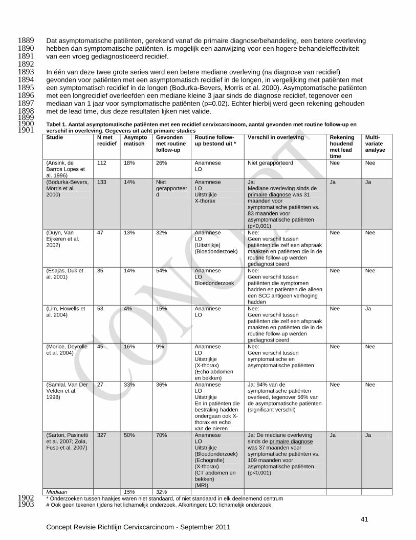
41 Concept Revisie Richtlijn Cervixcarcinoom - September 2011
Dat asymptomatische patiënten, gerekend vanaf de primaire diagnose/behandeling, een betere overleving 1889 hebben dan symptomatische patiënten, is mogelijk een aanwijzing voor een hogere behandeleffectiviteit 1890 van een vroeg gediagnosticeerd recidief. 1891 1892 In één van deze twee grote series werd een betere mediane overleving (na diagnose van recidief) 1893 gevonden voor patiënten met een asymptomatisch recidief in de longen, in vergelijking met patiënten met 1894 een symptomatisch recidief in de longen (Bodurka-Bevers, Morris et al. 2000). Asymptomatische patiënten 1895 met een longrecidief overleefden een mediane kleine 3 jaar sinds de diagnose recidief, tegenover een 1896 mediaan van 1 jaar voor symptomatische patiënten (p=0.02). Echter hierbij werd geen rekening gehouden 1897 met de lead time, dus deze resultaten lijken niet valide. 1898 1899 Tabel 1. Aantal asymptomatische patiënten met een recidief cervixcarcinoom, aantal gevonden met routine follow-up en 1900 verschil in overleving. Gegevens uit acht primaire studies 1901 Studie N met
recidief Asymptomatisch
Gevonden met routine follow-up
Routine follow-up bestond uit *
Verschil in overleving Rekening houdend met lead time
Multi- variate analyse
(Ansink, de Barros Lopes et al. 1996)
112 18% 26% Anamnese LO
Niet gerapporteerd Nee Nee
(Bodurka-Bevers, Morris et al. 2000)
133 14% Niet gerapporteerd
Anamnese LO Uitstrijkje X-thorax
Ja: Mediane overleving sinds de primaire diagnose was 31 maanden voor symptomatische patiënten vs. 83 maanden voor asymptomatische patiënten (p<0,001)
Ja Ja
(Duyn, Van Eijkeren et al. 2002)
47 13% 32% Anamnese LO (Uitstrijkje) (Bloedonderzoek)
Nee: Geen verschil tussen patiënten die zelf een afspraak maakten en patiënten die in de routine follow-up werden gediagnosticeerd
Nee Nee
(Esajas, Duk et al. 2001)
35 14% 54% Anamnese LO Bloedonderzoek
Nee: Geen verschil tussen patiënten die symptomen hadden en patiënten die alleen een SCC antigeen verhoging hadden
Nee Nee
(Lim, Howells et al. 2004)
53 4% 15% Anamnese LO
Nee: Geen verschil tussen patiënten die zelf een afspraak maakten en patiënten die in de routine follow-up werden gediagnosticeerd
Nee Ja
(Morice, Deyrolle et al. 2004)
45 16% 9% Anamnese LO Uitstrijkje (X-thorax) (Echo abdomen en bekken)
Nee: Geen verschil tussen symptomatische en asymptomatische patiënten
Nee Nee
(Samlal, Van Der Velden et al. 1998)
27 33% 36% Anamnese LO Uitstrijkje En in patiënten die bestraling hadden ondergaan ook X-thorax en echo van de nieren
Ja: 94% van de symptomatische patiënten overleed, tegenover 56% van de asymptomatische patiënten (significant verschil)
Nee Nee
(Sartori, Pasinetti et al. 2007; Zola, Fuso et al. 2007)
327 50% 70% Anamnese LO Uitstrijkje (Bloedonderzoek) (Echografie) (X-thorax) (CT abdomen en bekken) (MRI)
Ja: De mediane overleving sinds de primaire diagnose was 37 maanden voor symptomatische patiënten vs. 109 maanden voor asymptomatische patiënten (p<0,001)
Ja Ja
Mediaan 15% 32%
* Onderzoeken tussen haakjes waren niet standaard, of niet standaard in elk deelnemend centrum 1902 # Ook geen tekenen tijdens het lichamelijk onderzoek. Afkortingen: LO: lichamelijk onderzoek 1903
42 Concept Revisie Richtlijn Cervixcarcinoom - September 2011
Conclusies 1904 Uit de literatuur komt naar voren dat het aannemelijk is dat de overleving vanaf primiare 1905 behandeling/diagnose van patiënten die asymptomatisch waren ten tijde van de diagnose recidief beter is 1906 dan de overleving vanaf primaire behandeling/diagnose van patiënten die symptomatisch waren ten tijde 1907 van de diagnose recidief. 1908 Gerandomiseerde studies zijn echter nodig om directe uitspraken te kunnen doen of het zinvol zou kunnen 1909 zijn om recidief eerder op te sporen. 1910 Niveau 2: B (Bodurka-Bevers, Morris et al. 2000; Zola, Fuso et al. 2007) 1911 1912 1913 7.3.4 Welke diagnostiek is het meest geschikt om behandelbare nieuwe kankermanifestaties vroeg 1914 en accuraat te diagnosticeren? 1915 Literatuurbespreking 1916 De systematische review van Elit et al. beschreef 17 retrospectieve studies, waarin voor alle studies te 1917 samen 29–71% van de recidieven met een lichamelijk onderzoek werd ontdekt; met een X-thorax 20-47%; 1918 met CT 0-34%; met een vaginale uitstrijk 0-17%; met echo 0-2%; met een intraveneus pyelogram 0%; met 1919 behulp van tumor markers 0-26%; en met andere onderzoeken 11-33% (Elit, Fyles et al. 2009). Hierbij 1920 wordt geen onderscheid gemaakt tussen locoregionaal recidief en metastasen op afstand. 1921 Als de acht van de 17 door Elit geïncludeerde studies in meer detail worden bekeken (zie tabel 1), waren 1922 veruit de meeste patiënten symptomatisch ten tijde van de diagnose recidief. Een mediaan van 15% 1923 (range: 4-50%) van de patiënten was asymptomatisch (Tabel 1). Daarnaast had een klein deel (9%) (Duyn, 1924 Van Eijkeren et al. 2002) van de asymptomatische patiënten tekenen van een recidief bij lichamelijk 1925 onderzoek in één studie waarin dat werd beschreven. 1926 Wat betreft lokale recidieven werden in één studie alle acht asymptomatische recidieven in het kleine 1927
bekken met lichamelijk onderzoek gevonden (Bodurka-Bevers, Morris et al. 2000). 1928 In een andere studie werden twee van de zeven asymptomatische lokale recidieven met lichamelijk 1929
onderzoek gevonden; één lokaal recidief werd met een uitstrijkje gevonden, twee lokale recidieven met 1930 X-thorax en twee met CT. (Morice, Deyrolle et al. 2004). 1931
Samlal et al. beschreven dat 41% van alle recidieven met behulp van een lichamelijk onderzoek werd 1932 gevonden en 52% met een routine radiologisch onderzoek (Samlal, Van Der Velden et al. 1998). In 1933 deze studie werd in de routine follow-up een X-thorax en een echo van de urinewegen verricht bij 1934 patiënten die radiotherapie hadden ondergaan. 1935
In een serie recidieven beschreven door Sartori et al. was de diagnose gebaseerd op een CT in 41% 1936 van de recidieven; op een lichamelijk onderzoek in 27%; op een echo in 10%; op histologie in 8%; op 1937 een X-thorax in 6%; op een MRI in 5% en op cytologie in 3% van de recidieven (Sartori, Pasinetti et al. 1938 2007). Een mediaan van 32% (range: 9-70%) van de diagnosen recidief werd ten tijde van een routine 1939 follow-up gesteld (Tabel 1). 1940
Asymptomatische longmetastasen werden in alle beschreven gevallen (respectievelijk N=8 en N=2) 1941 met een X-thorax gevonden (Bodurka-Bevers, Morris et al. 2000; Morice, Deyrolle et al. 2004). 1942
Geen van deze zeven studies evalueerde de accuraatheid van verschillende diagnostische technieken 1943 voor het stellen van de diagnose recidief. 1944 1945 PET voor de detectie van recidief (lokaal of op afstand) cervix carcinoom 1946 Er zijn geen studies gevonden die de waarde van PET onderzoeken op de overleving bij de detectie van 1947 recidief cervixcarcinoom. 1948 Èén systematische review evalueerde PET voor het opsporen van een recidief cervixcarcinoom 1949 (Havrilesky, Kulasingam et al. 2005). Er werden zes retrospectieve studies geïncludeerd. De sensitiviteit 1950 van PET was 96% ((95% CI: 87–99%) en de specificiteit 81% (95% CI: 58–94%)) in een meta-analyse van 1951 drie studies die de detectie van een recidief in patiënten met een klinische verdenking evalueerden. 1952 Wanneer er geen klinische verdenking was, was de sensitiviteit van PET 92% ((95% CI: 77–98%) en de 1953 specificiteit 75% (95% CI: 69–80%), meta-analyse van 2 studies). Voor één van de zes geïncludeerde 1954 studies werd geen onderscheid gemaakt tussen patiënten met of zonder een klinische verdenking. In deze 1955 studie was de sensitiviteit van PET 100% (specificiteit 77% ). 1956 1957 SCC antigeen voor de detectie van recidief cervix carcinoom 1958 Er zijn geen goede studies gevonden die de waarde van SCC antigeen bepaling onderzoeken op de 1959 overleving bij de detectie van recidief cervixcarcinoom (geen multivariate analyse). 1960 Vier studies evalueerden de accuratesse van SCC antigeen bepalingen in de follow-up van patiënten met 1961 behandeld cervixcarcinoom (Forni, Ferrandina et al. 2007; Yoon, Shin et al. 2010);(Chan, Ng et al. 1962

43 Concept Revisie Richtlijn Cervixcarcinoom - September 2011
2002).(Esajas, Duk et al. 2001). De mediane sensitiviteit was 74% (range 61-95%) en de mediane 1963 specificiteit was 96% (range 96-98%). De studies gebruiken verschillende afkappunten (range 1,4-2,0 1964 ng/ml). 1965 Eén studie (N=135 patiënten) evalueerde tevens de accuratesse van SCC antigeen bepaling in combinatie 1966 met gynaecologisch onderzoek (sensitiviteit 95% en specificiteit 96%) (Forni, Ferrandina et al. 2007. 1967 1968 Het uitstrijkje voor de detectie van recidief cervix carcinoom 1969 Er zijn geen studies gevonden die de waarde van het uitstrijkje onderzoeken op de overleving bij de 1970 detectie van recidief cervixcarcinoom. 1971 Er werd één retrospectieve studie gevonden die de waarde van uitstrijkjes in de follow-up van cervix 1972 carcinoom patiënten die radiotherapie hadden ondergaan evalueerden. (Chien, Ting et al. 2005). De 1973 sensitiviteit van een maligne uitslag van een uitstrijkje voor het aantonen van een centraal recidief was 1974 66% (specificiteit 100%); en van een maligne of een „high-grade squamous intraepithelial lesion‟ uitslag 1975 voor het aantonen van een centraal recidief was 83% (specificiteit 95%). 1976 1977 Conclusies 1978 Het is aannemelijk dat slechts een klein deel van de patiënten met recidief cervixcarcinoom (mediaan 1979 15% ) asymptomatisch is ten tijde van detectie van het recidief. 1980 Niveau 2: B (Ansink, de Barros Lopes et al. 1996) (Bodurka-Bevers, Morris et al. 2000) (Duyn, Van 1981 Eijkeren et al. 2002) (Esajas, Duk et al. 2001) (Lim, Howells et al. 2004) (Morice, Deyrolle et al. 2004) 1982 (Samlal, Van Der Velden et al. 1998) (Sartori, Pasinetti et al. 2007; Zola, Fuso et al. 2007) 1983 1984 Het is aannemelijk dat het merendeel van de recidieven kan worden aangetoond door middel van 1985 lichamelijk onderzoek (29-71%). 1986 Niveau 3: B (Bodurka-Bevers, Morris et al. 2000; Morice, Deyrolle et al. 2004) 1987 1988 Conclusies PET 1989 Er zijn tegengestelde aanwijzingen dat PET een recidief cervixcarcinoom accuraat kan uitsluiten bij 1990 patiënten met een klinische verdenking op recidief cervixcarcinom (sensitiviteit 96%; 95%CI 87-99%). 1991 Niveau 3: B (Havrilesky, Kulasingam et al. 2005; Chung, Kim et al. 2006; Kitajima, Murakami et al. 2008; 1992
Brooks, Rader et al. 2009) 1993 1994 Er zijn aanwijzingen dat PET een recidief cervixcarcinoom niet accuraat kan aantonen bij patiënten met 1995 een klinische verdenking op recidief cervixcarcinoom (specificiteit 81% (95% CI 58-94%) 1996 Niveau 3: B (Havrilesky, Kulasingam et al. 2005; Chung, Kim et al. 2006; Kitajima, Murakami et al. 2008) 1997 1998 Conclusies SCC antigeen 1999 Er zijn aanwijzingen dat SCC antigeen een recidief accuraat kan aantonen (mediane specificiteit 96%; 2000 range 96-98%) maar niet accuraat kan uitsluiten (mediane sensitiviteit 74%; range 61-95%). 2001 Niveau 3: B (Esajas, Duk et al. 2001; Chan, Ng et al. 2002; Forni, Ferrandina et al. 2007; Yoon, Shin et al. 2002 2010) 2003 2004 Er zijn aanwijzingen dat SCC antigeen in combinatie met een gynaecologisch onderzoek een recidief 2005 cervixcarcinoom accuraat kan uitsluiten (sensitiviteit 95%) en aantonen (specificiteit 96%). 2006 Niveau 3: B (Forni, Ferrandina et al. 2007) 2007 2008 Conclusies uitstrijkje 2009 Er zijn aanwijzingen dat post-trachelectomie uitstrijkjes een centraal recidiverend cervixcarcinoom accuraat 2010 kunnen aantonen (specificiteit 100%) maar niet kunnen uitsluiten (sensitiviteit 66%). 2011 Niveau 3: B (Chien, Ting et al. 2005) 2012
2013 Overige overwegingen 2014 De follow-up na behandeling van cervixcarcinoom is veilig, indien het routine gynaecologisch onderzoek 2015 betreft en eventueel bloedafnames. Ook het verrichten van beeldvormig is veilig, mits aandacht voor 2016 eventueel allergische reacties en benodigde voorbereidingen bij verminderde nierfunctie (in geval van 2017 contrastoediening). 2018 Echter de routine nacontroles kunnen angst of pre-occupatie voor recidief geven bij de patiënt en daardoor 2019 een negatief effect hebben op de kwaliteit van leven. Anderszijds kan een patiënt het ook als zeer “ veilig” 2020
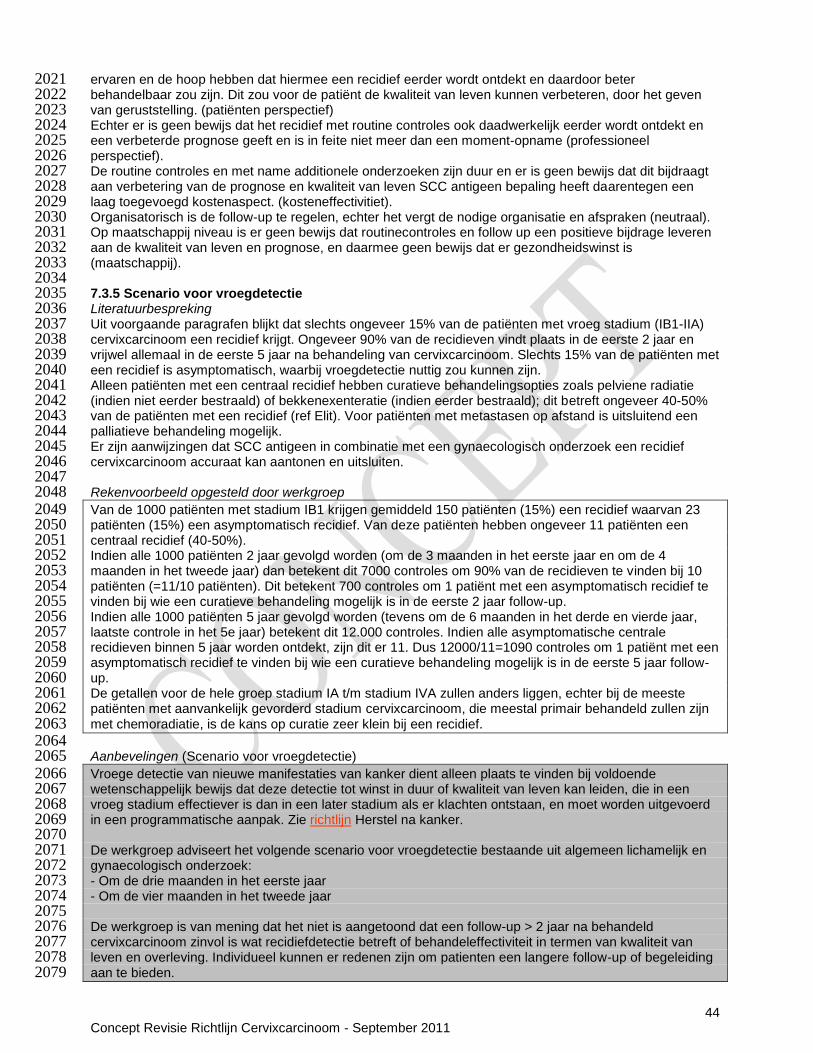
44 Concept Revisie Richtlijn Cervixcarcinoom - September 2011
ervaren en de hoop hebben dat hiermee een recidief eerder wordt ontdekt en daardoor beter 2021 behandelbaar zou zijn. Dit zou voor de patiënt de kwaliteit van leven kunnen verbeteren, door het geven 2022 van geruststelling. (patiënten perspectief) 2023 Echter er is geen bewijs dat het recidief met routine controles ook daadwerkelijk eerder wordt ontdekt en 2024 een verbeterde prognose geeft en is in feite niet meer dan een moment-opname (professioneel 2025 perspectief). 2026 De routine controles en met name additionele onderzoeken zijn duur en er is geen bewijs dat dit bijdraagt 2027 aan verbetering van de prognose en kwaliteit van leven SCC antigeen bepaling heeft daarentegen een 2028 laag toegevoegd kostenaspect. (kosteneffectivitiet). 2029 Organisatorisch is de follow-up te regelen, echter het vergt de nodige organisatie en afspraken (neutraal). 2030 Op maatschappij niveau is er geen bewijs dat routinecontroles en follow up een positieve bijdrage leveren 2031 aan de kwaliteit van leven en prognose, en daarmee geen bewijs dat er gezondheidswinst is 2032 (maatschappij). 2033 2034 7.3.5 Scenario voor vroegdetectie 2035 Literatuurbespreking 2036 Uit voorgaande paragrafen blijkt dat slechts ongeveer 15% van de patiënten met vroeg stadium (IB1-IIA) 2037 cervixcarcinoom een recidief krijgt. Ongeveer 90% van de recidieven vindt plaats in de eerste 2 jaar en 2038 vrijwel allemaal in de eerste 5 jaar na behandeling van cervixcarcinoom. Slechts 15% van de patiënten met 2039 een recidief is asymptomatisch, waarbij vroegdetectie nuttig zou kunnen zijn. 2040 Alleen patiënten met een centraal recidief hebben curatieve behandelingsopties zoals pelviene radiatie 2041 (indien niet eerder bestraald) of bekkenexenteratie (indien eerder bestraald); dit betreft ongeveer 40-50% 2042 van de patiënten met een recidief (ref Elit). Voor patiënten met metastasen op afstand is uitsluitend een 2043 palliatieve behandeling mogelijk. 2044 Er zijn aanwijzingen dat SCC antigeen in combinatie met een gynaecologisch onderzoek een recidief 2045 cervixcarcinoom accuraat kan aantonen en uitsluiten. 2046 2047 Rekenvoorbeeld opgesteld door werkgroep 2048 Van de 1000 patiënten met stadium IB1 krijgen gemiddeld 150 patiënten (15%) een recidief waarvan 23 2049 patiënten (15%) een asymptomatisch recidief. Van deze patiënten hebben ongeveer 11 patiënten een 2050 centraal recidief (40-50%). 2051 Indien alle 1000 patiënten 2 jaar gevolgd worden (om de 3 maanden in het eerste jaar en om de 4 2052 maanden in het tweede jaar) dan betekent dit 7000 controles om 90% van de recidieven te vinden bij 10 2053 patiënten (=11/10 patiënten). Dit betekent 700 controles om 1 patiënt met een asymptomatisch recidief te 2054 vinden bij wie een curatieve behandeling mogelijk is in de eerste 2 jaar follow-up. 2055 Indien alle 1000 patiënten 5 jaar gevolgd worden (tevens om de 6 maanden in het derde en vierde jaar, 2056 laatste controle in het 5e jaar) betekent dit 12.000 controles. Indien alle asymptomatische centrale 2057 recidieven binnen 5 jaar worden ontdekt, zijn dit er 11. Dus 12000/11=1090 controles om 1 patiënt met een 2058 asymptomatisch recidief te vinden bij wie een curatieve behandeling mogelijk is in de eerste 5 jaar follow-2059 up. 2060 De getallen voor de hele groep stadium IA t/m stadium IVA zullen anders liggen, echter bij de meeste 2061 patiënten met aanvankelijk gevorderd stadium cervixcarcinoom, die meestal primair behandeld zullen zijn 2062 met chemoradiatie, is de kans op curatie zeer klein bij een recidief. 2063 2064 Aanbevelingen (Scenario voor vroegdetectie) 2065 Vroege detectie van nieuwe manifestaties van kanker dient alleen plaats te vinden bij voldoende 2066 wetenschappelijk bewijs dat deze detectie tot winst in duur of kwaliteit van leven kan leiden, die in een 2067 vroeg stadium effectiever is dan in een later stadium als er klachten ontstaan, en moet worden uitgevoerd 2068 in een programmatische aanpak. Zie richtlijn Herstel na kanker. 2069 2070 De werkgroep adviseert het volgende scenario voor vroegdetectie bestaande uit algemeen lichamelijk en 2071 gynaecologisch onderzoek: 2072 - Om de drie maanden in het eerste jaar 2073 - Om de vier maanden in het tweede jaar 2074 2075 De werkgroep is van mening dat het niet is aangetoond dat een follow-up > 2 jaar na behandeld 2076 cervixcarcinoom zinvol is wat recidiefdetectie betreft of behandeleffectiviteit in termen van kwaliteit van 2077 leven en overleving. Individueel kunnen er redenen zijn om patienten een langere follow-up of begeleiding 2078 aan te bieden. 2079
45 Concept Revisie Richtlijn Cervixcarcinoom - September 2011
2080 De werkgroep is van mening dat SCC bepaald kan worden in de follow-up; de klinische waarde hiervan is 2081 echter nog niet overtuigend bewezen. 2082 2083 De controles kunnen (eventueel afwisselend) uitgevoerd worden door de behandelend of verwijzend 2084 gynaecoloog of door gynaecoloog en radiotherapeut. 2085 2086 2087 Informeer de patiënt over de mogelijkheden en beperkingen van vroege detectie van nieuwe manifestaties 2088 van kanker. Eerlijkheid en beperkingen verdient de voorkeur boven het zinloos opsporen van 2089 onbehandelbare ziekte. Het voorkomt valse hoop en gaat onnodige medicalisering tegen. Een instructie 2090 voor deze patiëntenvoorlichting vindt u in de richtlijn Herstel na kanker (IKNL, 2011). 2091 2092 Bij klachten 2093 Gericht laboratorium- en beeldvormend onderzoek (CT/MRI/PET/X-thorax), afhankelijk van de klachten. 2094 2095 2096
2097
46 Concept Revisie Richtlijn Cervixcarcinoom - September 2011
7.4 Evaluatie medisch handelen (Consensus based tekst) 2098 Literatuurbespreking 2099 Het rapport van de Gezondheidsraad [2007] beschrijft dat de evaluatie van de medische zorg vaak wordt 2100 genoemd als doelstelling van nazorg. Deze doelstelling komt de individuele patiënt slechts indirect ten 2101 goede. De kwaliteit van de medische zorg kan geëvalueerd worden onder voorwaarden. De richtlijn Herstel 2102 na kanker (IKNL, 2011) beveelt aan dat indien de nazorg benut wordt voor de evaluatie van het medisch 2103 handelen, dit enkel met expliciete voorlichting en informed consent van de patiënt kan gebeuren. 2104 Daarbij wordt voor de evaluatie van het medisch handelen een systematische aanpak aanbevolen. 2105 Effectiviteit en kwaliteit van de oncologische zorg kan systematisch geëvalueerd worden samen met de 2106 wetenschappelijke verenigingen, betrokken partijen en beschikbare databases. Indicatoren om de kwaliteit 2107 van kankerzorg te toetsen en te verbeteren dienen op landelijk niveau gemaakt te worden. 2108 2109 Aanbevelingen 2110 De kwaliteit van de medische zorg kan geëvalueerd worden onder voorwaarden en met expliciete 2111 voorlichting en toestemming van de patiënt. 2112 2113 De effectiviteit en kwaliteit van de oncologische zorg kan samen met de wetenschappelijke verenigingen 2114 en andere betrokken partijen op systematische wijze geëvalueerd worden. 2115
2116
7.5 Organisatie van nazorg (Consensus based tekst) 2117 Literatuurbespreking 2118 Nazorg is samenwerken en coördinatie is vereist 2119 Oncologische zorg is bij uitstek multidisciplinaire zorg, die voor de herstelfase deels vanuit de kliniek en 2120 deels in de eerste- of derdelijns- instellingen geboden wordt. Er blijkt geen eenduidig bewijs voor de beste 2121 taakverdeling (nazorg aangeboden door de specialist, een nurse practitioner of huisarts) en aanpak in de 2122 nazorg (persoonlijke en telefonische consulten). De behoeften van patiënten blijken ook divers. 2123 De Gezondheidsraad geeft aan dat de nazorg na de behandeling wegens kanker beter moet worden 2124 gecoördineerd. Dit sluit aan bij de aanbevelingen van de Inspectie van de Gezondheidszorg voor de 2125 kwaliteit van de oncologische zorgketen [IGZ 2009]. De behandelaar en overige hulpverleners spreken bij 2126 de afronding van de tumorgerichte behandeling onderling en met de patiënt af wie de coördinatie van de 2127 (continuïteit van) nazorg op zich neemt. Het nazorgplan voor de patiënt kan fungeren als middel voor de 2128 overdracht van zorg tussen de zorgverleners betrokken in de oncologische zorglijn. 2129 2130 Aanbevelingen 2131 Maak afspraken over de taakverdelingen voor de nazorg, die geboden wordt in het ziekenhuis, de eerste- 2132 of derdelijns- instellingen. 2133 2134 Zorg ervoor dat bij de afronding van primaire behandeling voor de nazorg een vaste contactpersoon voor 2135 iedere patiënt aangesteld wordt. Spreek dit multidisciplinair af in het team en met de huisarts. 2136 2137 Informeer een patiënt wie de vaste contactpersoon in de nazorg voor haar is. Leg dit vast in het 2138 nazorgplan voor de patiënt. 2139 2140 Zorg voor regelmatige interdisciplinaire overdracht van informatie, zeker ook naar de huisarts, bijv. via het 2141 nazorgplan van de patiënt. 2142 2143 Herinrichting biedt kansen 2144 Literatuurbespreking 2145 De Gezondheidsraad [2008]
stelt dat er in de oncologische nazorg kansen zijn op meer doelmatigheid door 2146
taakherschikking te combineren met een verandering in inrichting van het zorgproces. Dit betekent het 2147 zorgproces inrichten naar de behoeften van de patiënten, multidisciplinair, als samenhangend geheel van 2148 zorg en met als belangrijk principe het zelfmanagement. Een transmurale ketenbenadering lijkt een goede 2149 optie. 2150 2151 Aanbevelingen 2152 Overweeg bij de verbetering van de nazorg een herinrichting van het gehele oncologische zorgproces met 2153 het oog op doelmatigheid. 2154 2155
2156

47 Concept Revisie Richtlijn Cervixcarcinoom - September 2011
Bijlage 12 Voorbeeld nazorgplan cervixcarcinoom 2157 Persoonsgegevens
Naam patiënt: Geboortedatum
Adres: Telefoon / e-mail
Contactgegevens
Naam hoofdbehandelaar: Gynaecoloog (naam)
Naam eerste aanspreekpersoon: bijv. nurse practitioner (naam) Telefoon / e-mail
Naam huisarts: Huisarts (naam)
Data opstellen Nazorgplannen
Datum start Nazorgplan:
Data Nazorgplannen in nazorgfase:
Datum Nazorgplan bij heroverweging/beëindiging nazorg:
Informatie kanker, behandeling, gevolgen
Diagnostiek
Datum Diagnostisch onderzoek Resultaat
MRI (op indicatie)
PET/CT (op indicatie)
X-thorax (op indicatie)
Diagnose Datum definitieve PA
Stagering
Histologie
Kankerbehandeling
Operatie Radiotherapie Chemotherapie Hyperthermie Anders
Data
Behandelaar Gynaecoloog (naam)
Radiotherapeut (naam)
Internist-oncoloog (naam)
Internist-oncoloog (naam) / Radiotherapeut (naam)
Behandeling
Familiehistorie kanker, erfelijkheid
Comorbiditeit, andere ziekten
Soort Diabetes Hart-/vaatziekten COPD Osteoporose Anders
Data
Behandelaar
Behandeling
Complicaties kanker
Soorten
Data
Behandelaar
Behandeling
Mogelijke gevolgen kanker(behandeling)
Algemeen bij cervixcarcinoom
Seksuele dysfunctie, infertiliteit, vervroegde overgang, chronische vermoeidheid, psychische problemen
Bij operatie Lymfoedeem, klachten van het bewegingsapparaat
Bij chemotherapie Neuropathie, misselijkheid, braken, infecties
Bij radiotherapie Blaas- en darmproblemen
Anders
Aanbevolen nazorgschema
datum datum datum datum datum datum datum zorgverlener
Kankerdetectie:
Algemeen lichamelijk en gyneacologisch onderzoek:
Januari 2011
April 2011
Juli 2011
Oktober 2011
Jan uari 2012
Mei 2012
September 2012
Gynaecoloog of afwisselend gynaecoloog en radiotherapeut
Laboratorium onderzoek
Bij klachten
Gynaecoloog
Beeldvormend onderzoek: CT/ MRI/PET/X-thorax
Bij klachten
Gynaecoloog
Signalering gevolgen kanker: instrumenten

48 Concept Revisie Richtlijn Cervixcarcinoom - September 2011
Lastmeter > distress
Bij afronden primaire behandeling en bij heroverweging na 1 jaar. Zie voor gebruik richtlijn Detecteren behoefte psychosociale zorg
VAS > vermoeidheid
Zie voor gebruik richtlijn Oncologische revalidatie
CES-D > motionele problemen
Zie voor gebruik richtlijn Oncologische revalidatie
PSK > fysieke problemen
Zie voor gebruik richtlijn Oncologische revalidatie
Zelfsignalering klachten en contactpersonen
Bij de volgende signalen/klachten contact opnemen met:
Mogelijke signalen van kanker: pijn lokaal in bekken of uitstralend naar rug of benen, vaginaal bloedverlies, veranderde of toegenomen /afscheiding, toename of ontstaan van lymfoedeem benen, thrombosebeen
Gynaecoloog/ Radiotherapeut/ Nurse practitioner
telefoon / e-mail
Late effecten van behandeling: zorgverlener telefoon / e-mail
Lymfoedeem Nurse practitioner/ Gynaecoloog
Blaasproblemen Nurse practitioner/ Gynaecoloog/ Radiotherapeut
Darmproblemen Nurse practitioner/ Gynaecoloog/ Radiotherapeut
Algemene problemen zorgverlener telefoon / e-mail
Relaties met partner en gezin, sociale contacten Huisarts
Maatschappelijke participatie Huisarts/arbo arts
Arbeidsparticipatie Huisarts/arbo arts
Financiële problemen Huisarts
Aanpak gevolgen van kanker
Signalering
Datum
Conclusie
Advies, behandeling:
Verwijzing: datum, zorgverlener, aanvullende diagnostiek, behandeling
Monitoring, vervolg:
Signaleringsinstrument
Datum Conclusie
Advies, behandeling:
Verwijzing: datum, zorgverlener, aanvullende diagnostiek, behandeling
Monitoring, vervolg:
Ondersteuning zelfzorg
Voorlichtingsmaterialen:
Folders: NVOG patiëntenvoorlichting baarmoederhalskanker, KWF brochure baarmoederhalskanker, Verder leven met kanker, Vermoeidheid na kanker, Als kanker meer is dan je aankunt, Kanker en hoe moet het nu met de kinderen, Voeding en kanker, Kanker en seksualiteit. Websites: www.kanker.kwfkankerbestrijding.nl; www.nvog.nl; www.olijf.nl; www.kankerwiehelpt.nl; www.toekomstnakanker.nl
Advies zelfzorg
Leefstijladvies en -interventies
Stop met roken interventie: www.stivoro.nl
Aantekeningen patiënt
datum
Interdisciplinaire overdracht
datum Nazorg besproken in MDO
Overdracht naar verpleegkundig specialist gynaecologie
Overdracht naar huisarts
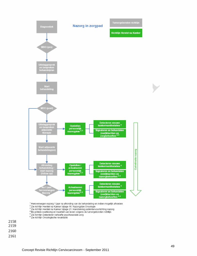
49 Concept Revisie Richtlijn Cervixcarcinoom - September 2011
2158 2159
2160
2161
50 Concept Revisie Richtlijn Cervixcarcinoom - September 2011
7.6 Seksuele dysfunctie (Evidence based tekst n.a.v. uitgangsvraag 6: Seksuele dysfunctie) 2162 Literatuurbespreking 2163 Seksueel functioneren kan worden onderscheiden in verschillende domeinen: verlangen, opwinding, 2164 orgasme, satisfactie en pijn. Er zijn weinig studies gedaan naar de invloed van preventie/behandeling van 2165 seksuele dysfunctie na behandeling van cervixcarcinoom. Seksueel disfunctioneren na behandeling voor 2166 cervix carcinoom beslaat alle domeinen van seksueel functioneren. Echter bij bestudering van de literatuur 2167 lijkt er een verband te bestaan tussen verminderd verlangen (libidoverlies) en het optreden van 2168 dyspareunie (pijn bij coitus). Omdat de ovaria doorgaans in situ blijven en de clitorisregio en innervatie niet 2169 worden aangetast is er geen fysiologische verklaring voor verlies van libido of orgasticiteit . Aannemelijker 2170 is het, dat als gevolg van de frequent gerapporteerde pijnlijke coitus, secundair daaraan libidoverlies, 2171 verminderde lubricatie en satisfactie optreden. 2172 Ongeveer 65% van de vrouwen ervaart in één of meer seksuele domeinen problemen (Scottish 2173 Intercollegiate Guidelines Network 2008). In verband met de hoge incidentie van seksuele problematiek na 2174 behandeling voor cervixcarcinoom, behoort de behandelaar dit onderwerp, bij voorkeur zowel voor het 2175 starten van de behandeling als tijdens de follow up, bespreekbaar te maken. 2176 Voor het literatuuronderzoek naar behandelingsmogelijkheden bij seksueel disfunctioneren is de SIGN 2177 richtlijn uit 2008 als uitgangspunt genomen(Scottish Intercollegiate Guidelines Network 2008). Daarnaast is 2178 een literatuuronderzoek uitgevoerd beginnend na de zoekdatum van de SIGN-richtlijn (2005). Drie 2179 systematische beoordelingen werden gevonden. De behandelingsmogelijkheden zijn onder te verdelen in 2180 psycho-educatie, medicatie en vaginale dilatatie. 2181 2182 Aanbevelingen 2183 De werkgroep is van mening dat iedere patiënt (en haar partner) gecounseld moet worden omtrent de 2184 mogelijke seksuele gevolgen van behandeling voor cervixcarcinoom. 2185 2186 De werkgroep is van mening dat met patiënten besproken moet worden dat pijnloos, eventueel niet-2187 coïtaal, vrijen een (tijdelijke) oplossing kan zijn, zodat de zin in seks behouden blijft. 2188 2189 De werkgroep is van mening dat psycho-educatie, medicatie en vaginale dilatatie alleen effectief zijn als 2190 deze interventies geïntegreerd worden toegepast. 2191 2192 2193 7.6.1 Psycho-educatie (Evidence based tekst n.a.v. uitgangsvraag 6: Seksuele dysfunctie) 2194 Literatuurbespreking 2195 Psycho-educatie bestaat uit interventies zoals counselling en psychotherapie (Miles, Candy et al. 2007). 2196 SIGN adviseert het aanbieden van ondersteunde sessies aan patiënten met cervixcarcinoom. Deze 2197 aanbeveling is gebaseerd op één RCT (Petersen and Quinlivan 2002), welke heeft gekeken naar angst en 2198 depressie in het algemeen, echter zonder specifieke aandacht voor seksueel disfunctioneren. 2199 Een literatuurupdate werd uitgevoerd, beginnend na de zoekdatum van de SIGN-richtlijn (2005) (Scottish 2200 Intercollegiate Guidelines Network 2008). Drie systematische revies werden gevonden. Eén systematische 2201 review gericht op psychoseksueel disfunctioneren na behandeling wegens een gynaecologische maligniteit, 2202 omvatte data van 5 studies met in totaal 413 patiënten en 5 verschillende interventies waaronder 3 2203 interventies op het gebied van psycho-educatie (Flynn, Kew et al. 2009): specialistische verpleegkundige 2204 interventie (Maughan and Clarke 2001); psychoeducationele groepstherapie (Robinson, Faris et al. 1999) 2205 en (echt)paar coping interventie (Scott, Halford et al. 2004). Alle studies waren van slechte 2206 methodologische kwaliteit. 2207 2208 De studie van Maughan gericht op specialistische verpleegkundige interventie, bestaande uit een 2209 preoperatieve consultatie van een gynaecologisch oncologische verpleegkundige met een specifieke 2210 psychoseksuele opleiding, en uit gemiddeld 3 postoperatieve thuisvisites, toonde geen significante 2211 verbetering op de onderzochte schalen voor seksueel functioneren (Maughan and Clarke 2001). Er leek 2212 een gering positief effect op te treden wat betreft hervatting van seksuele activiteit bij voordien seksueel 2213 actieve patiënten, maar dat effect was niet statistisch significant (OR 0,63, 95% CI 0,17-2,36). In de 2214 interventiegroep waren significant minder vrouwen (2/10 versus 9/10) met afgenomen tevredenheid over 2215 de coïtus bij 6 maanden na de operatie (OR 0,03, 95% CI 0,00-0,37). 2216 De studie van Robinson is gericht op psycho-educationele groepstherapie, bestaande uit 2 groepssessies 2217 van 90 minuten die waren gebaseerd op het IMB-model (Information-Motivation-Behavioural Skills model: 2218 informatie-motivatie-gedragsvaardigheden-model) (Robinson, Faris et al. 1999). Hoewel de SIGN richtlijn 2219
51 Concept Revisie Richtlijn Cervixcarcinoom - September 2011
hierop een aanbeveling baseert, liet deze studie alleen een verbetering van compliance met vaginale 2220 dilatatie zien en had deze interventie geen significante invloed op seksueel functioneren. 2221 De studie van Scott bestond uit een sessie met patiënt en partner (Scott, Halford et al. 2004). Ook deze 2222 studie liet geen significante invloed op seksueel functioneren zien. 2223 2224 Het literatuuronderzoek naar primaire studies na de zoekdatum van de twee meest recente Cochrane-2225 beoordelingen (2008), leverde 2 kleine studies op van lage kwaliteit, maar ze worden hier nog kort 2226 beschreven. 2227 De studie van Brotto et al. gericht op psycho-educationele interventie voor seksuele problemen bij 2228 patiënten met een gynaecologische maligniteit, toonde een significant positieve invloed op seksueel 2229 gebied wat betreft verlangen, opwinding, orgasme, bevrediging, onwelbevinden/pijn, en op somberheid en 2230 algemeen welbevinden, en een minder significante toename van fysiologische genitale opwinding en 2231 bewuste genitale opwinding (Brotto, Heiman et al. 2008). De kwaliteit van de psycho-educationele 2232 informatie werd bij feedback als zeer gebruiksvriendelijk, duidelijk en nuttig beoordeeld. Het betrof echter 2233 een pre- versus posttreatment studie zonder controlegroep, die daarom werd afgeweze. 2234 De studie van Bodurka et al. gericht op seksueel functioneren en levenskwaliteit bij 88 patiënten met nieuw 2235 gediagnosticeerd cervixcarcinoom van stadium ≥ IB1, werd afgewezen wegens lage kwaliteit (Bodurka, 2236 Sun et al. 2011). 2237 2238 2239 Conclusies 2240 Er zijn geen aanwijzingen dat psycho-educatie alleen een significant gunstig effect heeft op het seksueel 2241 functioneren van vrouwen na behandeling van cervixcarcinoom. 2242 Niveau 2: (Flynn, Kew et al. 2009) (B) 2243 2244 Overige overwegingen 2245 De patiënte moet gemotiveerd zijn voor psycho-educatie (patiënten perspectief). Professionals moeten 2246 bekwaam zijn in het geven van psycho-educatie of doorverwijzen naar een bekwaam persoon 2247 (professioneel perspectief). 2248 2249 Aanbevelingen 2250 De werkgroep is van mening dat vrouwen en hun eventuele partners standaard gewezen behoren te 2251 worden op de mogelijkheid van doorverwijzing naar een gespecialiseerde seksuoloog dan wel 2252 psychotherapeut naast de andere interventies ter behandeling van seksueel disfunctioneren. 2253 2254 2255 7.6.2 Medicatie (Evidence based tekst n.a.v. uitgangsvraag 6: Seksuele dysfunctie) 2256 Literatuurbespreking 2257 SIGN 2008 stelt dat lokale oestrogenen of benzydamine-douches ter verlichting van vaginale complicaties 2258 na radiotherapie overwogen kunnen worden. Deze aanbeveling is gebaseerd op een Cochrane-2259 beoordeling uit 2009 door Denton et al. (Denton and Maher 2003) 2260 2261 Benzydamine douches 2262 In deze Cochrane review worden twee kleine, oude RCTs beschreven die benzydamine douches 0.1% 2263 2dd gedurende 14 dagen vergeleken met placebo (Bentivoglio and Diani 1981; Volterrani, Tana et al. 2264 1987). Benzydamine is een prostaglandine synthetaseremmer, in Nederland verkrijgbaar als crème in een 2265 3% dosering. Het heeft anti-inflammatoire en daardoor antipyrethische en analgetische eigenschappen. 2266 Het is onderzocht als behandeling van acute vaginitisklachten kort na pelviene en/of vaginale 2267 brachytherapie. Uitkomstmaten in beide studies zijn gelijk: subjectief (klachtenscore van patiënte) en semi-2268 objectief: (beeld bij kolposcopie). De OR voor verbetering van acute vaginitisklachten voor 2269 bezydaminedouches is 19.29 resp 8.5 in de studies (Bentivoglio and Diani 1981; Volterrani, Tana et al. 2270 1987). Er is mogelijk sprake van bias, want in de benzydamine groep in de studie van Volteranni hadden 2271 tweemaal zoveel patiënten tevoren brachytherapie gehad en een hogere subjectieve beginscore 2272 (Volterrani, Tana et al. 1987). Verder merkt de werkgroep op dat deze medicatie alleen is beschreven als 2273 kortdurende behandeling van de acute vaginitis en er geen literatuur is over het effect op seksueel 2274 functioneren. Er is geen histologisch bewijs van het farmacologisch effect op het vaginaepitheel. 2275 2276 2277 2278

52 Concept Revisie Richtlijn Cervixcarcinoom - September 2011
Lokale oestrogenen 2279 Oestriol heeft wel aantoonbaar een farmacologisch effect op het vaginaepitheel (Gupta, Ozel et al. 2008). 2280 De Cochrane-review door Flynn et al (Flynn, Kew et al. 2009), gericht op psychoseksueel disfunctioneren 2281 na behandeling wegens een gynaecologische maligniteit, omvatte 1 studie over vaginale dienestrol-2282 toediening (Pitkin and VanVoorhis 1971). In deze studie werd een positief resultaat op korte termijn 2283 gemeld van vaginale dienestrol-toediening bij patiënten na bekkenradiotherapie. In de interventiegroep 2284 maakten minder vrouwen melding van dyspareunie: 6/44 in de interventiegroep en 16/49 in de 2285 placebogroep. De auteurs van de primaire studie concludeerden dat dit verschil statistisch niet significant 2286 was bij de chi-kwadraattest (p=0,09), maar Flynn et al. analyseerde de data opnieuw en kwamen tot de 2287 conclusie dat het verschil statistisch significant was met een OR (Odds Ratio: kansverhouding) van 0,33 2288 (95% CI 0,11-0,93). 26 patiënten in de interventiegroep waren seksueel actief, waarvan slechts 6 patiënten 2289 melding maakten van milde dyspareunie. In de controlegroep waren 30 patiënten seksueel actief, waarvan 2290 16 patiënten melding maakten van dyspareunie. 6 van deze 16 noemden de dyspareunie ernstig. 2291 Afzonderlijke analyse van de seksueel actieve patiënten met dyspareunie als effectmaatstaf, toonde een 2292 meer uitgesproken behandelingseffect (OR 0,26, 95% CI 0,08-0,84). 2293 Een Cochrane-beoordeling door Miles et al (Miles, Candy et al. 2007), gericht op interventie voor seksuele 2294 problemen na kanker in het algemeen, identificeerde ook de studie van Pitkin (Pitkin and VanVoorhis 2295 1971). 2296 2297 Conclusies 2298 Het is aannemelijk dat benzydamine douches effect hebben bij acute vaginitis kort na pelviene en/of 2299 vaginale brachytherapie. Er is echter geen literatuur over het effect van benzydamine douches op seksueel 2300 functioneren. 2301 Niveau 2: (Denton and Maher 2003) (B) 2302 2303 Het is aannemelijk dat er in geval van een postmenopauzale status, een gunstig effect bestaat van lokale 2304 oestrogeentoediening ter preventie van dyspareunie en ter bestrijding van vaginale stenose na 2305 radiotherapie bij cervixcarcinoom, 2306 Niveau 2: (Scottish Intercollegiate Guidelines Network 2008) (B); (Flynn, Kew et al. 2009) (B) 2307 2308 Overige overwegingen 2309 Benzydamine douches 2310 Er is in Nederland geen ervaring is met benzydamine douches 0.1% zoals beschreven in de studies 2311 (professioneel perspectief) en benzydamine is in Nederland alleen verkrijgbaar als een 3% crème 2312 (organisatorische factor). 2313 2314 Lokale oestrogeentoediening 2315 Oestriol heeft, in tegenstelling tot oestradiol, met name een lokaal effect (veiligheid). 2316 2317 Het is onbekend of oestriol toegevoegde waarde heeft bij vrouwen die al gesubsititueerd worden na de 2318 radiotherapie, of hun ovariele functie behouden. Indien de patiënte andere hormoongevoelige tumoren 2319 (mamma of endometriumcarcinom) heeft is oestrogeen behandeling in principe gecontra-indiceerd 2320 (veiligheid). 2321 2322 Voor compliance is counseling door de professional nodig, die verder gaat dan een eenmalig consult 2323 (professioneel perspectief), omdat seksualiteit een complex gegeven is waar fysieke, relationele en 2324 psychologische items een rol spelen. 2325 2326 Aanbevelingen 2327 Benzydamine-douches worden niet geadviseerd bij patiënten na radiotherapie voor cervixcarcinoom. 2328 2329 Er wordt geadviseerd om in geval van een postmenopauzale situatie bij patiënten die niet gesubstitueerd 2330 worden, een 6 weekse kuur van lokaal oestriol (ovules) voor te schrijven om de regeneratie van 2331 beschadigd vagina epitheel te bevorderen. Dit regime kan zo nodig herhaald worden. 2332 2333 2334 2335 2336 2337
53 Concept Revisie Richtlijn Cervixcarcinoom - September 2011
7.6.3 Vaginale dilatatie (Evidence based tekst n.a.v. uitgangsvraag 6: Seksuele dysfunctie) 2338 Literatuurbespreking 2339 Op het gebied van fysieke interventies adviseert SIGN 2008 om patiënten te instrueren om zelf regelmatig 2340 een vaginale stent of dilatator in te brengen, ter preventie van vaginale verkleving of obliteratie na 2341 radiotherapie (Scottish Intercollegiate Guidelines Network 2008). Deze aanbeveling is gebaseerd op een 2342 Cochrane-beoordeling door Denton et al. en gericht op het behouden van coïtus mogelijkheden (Denton 2343 and Maher 2003). 2344 Een Cochrane-beoordeling door Miles et al. gericht op vaginale dilatatortherapie bij patiënten met 2345 bekkenradiotherapie, identificeerde 2 gerandomiseerde studies van lage kwaliteit die lieten zien dat meer 2346 patiënten dilatatie accepteerden na psycho-educationele interventies waarbij dilatatie werd aangeprezen 2347 (Miles and Johnson 2010). In de ene studie werd echter het seksuele functioneren niet gemeten (Jeffries, 2348 Robinson et al. 2006), en in de andere studie werden geen verschillen gevonden in de scores voor 2349 seksueel functioneren (Robinson, Faris et al. 1999). Miles et al. namen ook observationele studies op in 2350 hun beoordeling, maar geen enkele van deze studies toonde aan dat vaginale dilatatie het seksuele 2351 functioneren verbetert. Ook werden enkele casussen geïdentificeerd waarbij sprake was van vaginale 2352 fistelvorming of psychische morbiditeit. Miles et al. concludeerden dat dilatatie gedurende, of binnen korte 2353 tijd na radiotherapie, beschadigingen kan veroorzaken. Bovendien ontbreekt het bewijs dat dilatatie 2354 stenose voorkomt, en dat routinedilatatie gedurende de behandeling positief uitwerkt. Zij veronderstellen 2355 dat dilatatie nuttig zou kunnen zijn tegen late vaginale complicaties wanneer ontstekingsreacties tot rust 2356 zijn gekomen. Deze bevindingen komen echter niet overeen met de aanbevelingen in de SIGN-richtlijn en 2357 in de meeste andere richtlijnen. 2358 De RCT van Robinson gericht op psycho-educationele groepstherapie, bestaande uit 2 groepssessies die 2359 waren gebaseerd op het IMB-model (Information-Motivation-Behavioural Skills model: informatie-motivatie-2360 gedragsvaardigheden-model) liet een verbetering van de compliance voor vaginale dilatatie zien 2361 (Robinson, Faris et al. 1999). 2362 Gezien het ontbreken van goede literatuur werd door de werkgroep een kleine enquête gehouden onder 2363 de afdelingen radiotherapie in Nederland. Van de 13 respondenten adviseerde men in 8 instituten het 2364 gebruik van (vaseline)tampons, 3 afdelingen adviseerden pelotes, 2 afdelingen soms tampons, soms 2365 pelotes. Op 1 afdeling werd soms een vibrator geadviseerd. Start was meestal enkele weken na afronden 2366 van de bestraling, 2-3 maal per week gedurende 1-3 uren (tampons). 2367 2368 Conclusies 2369 Er zijn tegenstrijdige aanwijzingen omtrent de bewijsvoering van systematische vaginale dilatatie ter 2370 preventie van vaginale adhesie vorming na radiotherapie. 2371 Niveau 3: (Scottish Intercollegiate Guidelines Network 2008) (C); (Miles and Johnson 2010) (C) 2372 2373 De werkgroep is van mening dat het 1-2 maal per week diep vaginaal inbrengen van een vaseline tampon 2374 een goede, niet traumatische, mechanische methode is ter preventie van vaginale adhesie vorming en 2375 voorkoming van verkorting van de vagina. Bijkomend effect is mogelijk dat het zelfvertrouwen van 2376 patiënten gunstig kan worden beïnvloed. 2377 Niveau 4: mening van deskundigen 2378 2379 Er zijn aanwijzingen dat begeleiding zinvol is ter verbetering van de compliance bij vaginale pelote therapie 2380 Niveau 3: (Scottish Intercollegiate Guidelines Network 2008) (A2) 2381 2382 Vaginale dilatatie met een pelote kan leiden tot vaginale fistelvorming, rectumbeschadiging en psychische 2383 morbiditeit (Miles and Johnson 2010) (veiligheid). 2384 2385 Vaginale dilatatie met een pelote, tampon of vibrator kan bespreekbaar gemaakt worden door de 2386 professional (professioneel perspectief) en kan tevens een functie hebben bij het herwinnen van 2387 zelfvertrouwen van patiënten die na een korte of langere periode coïtus willen hervatten. Echter de 2388 therapietrouw is matig (patiëntenperspectief). 2389 2390 Aanbevelingen 2391 Er kan op grond van gebrek aan literatuur geen aanbeveling worden gegeven omtrent het toepassen van 2392 systematische vaginale dilatatie met pelote therapie na radiotherapie. Indien het wordt toegepast wordt 2393 geadviseerd de patiënte te begeleiden om de therapietrouw van de behandeling te bevorderen. 2394 2395
54 Concept Revisie Richtlijn Cervixcarcinoom - September 2011
De werkgroep is van mening dat het 1-2 maal per week inbrengen van een vaginale vaseline tampon een 2396 zinvolle preventie van vaginale adhesies kan zijn. Met name worden vaginale vaseline tampons 2397 aanbevolen om de periode tussen de bestraling en het hervatten van coïtus te overbruggen. Bij patiënten 2398 die emotionele weerstand ervaren bij het gebruik van vaseline tampons en die geen coïtus wens hebben, 2399 is het niet bezwaarlijk om hiermee stoppen. 2400 2401 De werkgroep is van mening dat na de behandeling van cervixcarcinoom, seksualiteit tijdens de follow-up 2402 ter sprake gebracht moet worden (door gynaecoloog-oncoloog, radiotherapeut of oncologie 2403 verpleegkundige). 2404 2405
2406
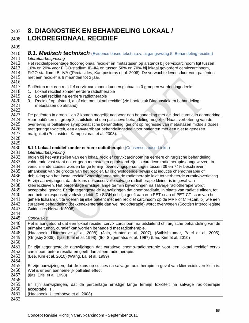
55 Concept Revisie Richtlijn Cervixcarcinoom - September 2011
8. DIAGNOSTIEK EN BEHANDELING LOKAAL / 2407
LOKOREGIONAAL RECIDIEF 2408
2409
8.1. Medisch technisch (Evidence based tekst n.a.v. uitgangsvraag 5: Behandeling recidief) 2410 Literatuurbespreking 2411 Het recidiefpercentage (locoregionaal recidief en metastasen op afstand) bij cervixcarcinoom ligt tussen 2412 10% en 20% voor FIGO-stadium IB–IIA en tussen 50% en 70% bij lokaal gevorderd cervixcarcinoom, 2413 FIGO-stadium IIB–IVA ((Pectasides, Kamposioras et al. 2008). De verwachte levensduur voor patiënten 2414 met een recidief is 6 maanden tot 2 jaar. 2415 2416 Patiënten met een recidief cervix carcinoom kunnen globaal in 3 groepen worden ingedeeld: 2417 1. Lokaal recidief zonder eerdere radiotherapie 2418 2. Lokaal recidief na eerdere radiotherapie 2419 3. Recidief op afstand, al of niet met lokaal recidief (zie hoofdstuk Diagnostiek en behandeling 2420
metastasen op afstand) 2421 2422 De patiënten in groep 1 en 2 komen mogelijk nog voor een behandeling met als doel curatie in aanmerking. 2423 Voor patiënten uit groep 3 is uitsluitend een palliatieve behandeling mogelijk. Naast verbetering van de 2424 overleving is palliatieve symptomatische behandeling, gericht op regressie van metastasen middels doses 2425 met geringe toxiciteit, een aanvaardbaar behandelingsdoel voor patiënten met een niet te genezen 2426 maligniteit (Pectasides, Kamposioras et al. 2008). 2427 2428 2429 8.1.1 Lokaal recidief zonder eerdere radiotherapie (Consensus based tekst) 2430 Literatuurbespreking 2431 Indien bij het vaststellen van een lokaal recidief cervixcarcinoom na eerdere chirurgische behandeling 2432 voldoende vast staat dat er geen metastasen op afstand zijn, is curatieve radiotherapie aangewezen. In 2433 verschillende studies worden lange termijn overlevingspercentages tussen 39 en 74% beschreven, 2434 afhankelijk van de grootte van het recidief. Er is onvoldoende bewijs dat inductie chemotherapie of 2435 debulking van het locaal recidief voorafgaande aan de radiotherapie leidt tot verbeterde curatie/overleving. 2436 Er zijn aanwijzingen, dat de kans op succesvolle salvage radiotherapie kleiner is in geval van 2437 klierrecidieven. Het percentage ernstige lange termijn bijwerkingen na salvage radiotherapie wordt 2438 acceptabel geacht. Er zijn tegengestelde aanwijzingen dat chemoradiatie, in plaats van radiatie alleen, tot 2439 een betere response/overleving leidt. De SIGN richtlijn geeft aan een PET-scan of PET-CT-scan van het 2440 gehele lichaam uit te voeren bij elke patiënt met een recidief carcinoom op de MRI- of CT-scan, bij wie een 2441 curatieve behandeling (bekkenexenteratie dan wel radiotherapie) wordt overwogen (Scottish Intercollegiate 2442 Guidelines Network 2008). 2443 2444 Conclusies 2445 Het is aangetoond dat een lokaal recidief cervix carcinoom na uitsluitend chirurgische behandeling van de 2446 primaire tumor, curatief kan worden behandeld met radiotherapie. 2447 (Haasbeek, Uitterhoeve et al. 2008), (Jain, Hunter et al. 2007), (Saibishkumar, Patel et al. 2005), 2448 (Grigsby 2005), (Ijaz, Eifel et al. 1998), (Ito, Shigematsu et al. 1997) (Lee, Kim et al. 2010) 2449 2450 Er zijn tegengestelde aanwijzingen dat curatieve chemo-radiotherapie voor een lokaal recidief cervix 2451 carcinoom betere resultaten geeft dan alleen radiotherapie. 2452 (Lee, Kim et al. 2010) (Wang, Lai et al. 1999) 2453 2454 Er zijn aanwijzingen, dat de kans op succes na salvage radiotherapie in geval van klierrecidieven klein is. 2455 Wel is er een aannemelijk palliatief effect. 2456 (Ijaz, Eifel et al. 1998) 2457 2458 Er zijn aanwijzingen, dat de percentage ernstige lange termijn toxiciteit na salvage radiotherapie 2459 acceptabel is . 2460 (Haasbeek, Uitterhoeve et al. 2008) 2461 2462

56 Concept Revisie Richtlijn Cervixcarcinoom - September 2011
Overige overwegingen 2463 De lange termijn bijwerkingen na salvage radiotherapie zijn acceptabel (veiligheid). Salvage radiotherapie 2464 geeft een tweede kans op curatie met kans op een betere kwaliteit van leven (patiëntenperspectief). 2465 Salvage radiotherapie vraagt geen andere vaardigheden dan primaire radiotherapie (professioneel 2466 perspectief). 2467 2468 Aanbevelingen 2469 Bij het vaststellen van een lokaal recidief cervixcarcinoom na uitsluitend chirurgische behandeling van de 2470 primaire tumor, wordt radiotherapie met curatieve intentie geadviseerd. Chemoradiatie kan worden 2471 overwogen. Bij een vaginaal recidief is de kans op succes hoger dan bij een klierrecidief. 2472 2473 Er wordt geadviseerd een PET/(CT) scan uit te voeren bij patiënten waarbij een recidief cervixcarcinoom is 2474 aangetoond met een MRI of CT scan en die in aanmerking komen voor curatieve radiotherapie. 2475 2476 2477 8.1.2 Lokaal recidief na eerdere radiotherapie (Evidence based tekst n.a.v. uitgangsvraag 5: 2478 Behandeling recidief) 2479 Literatuurbespreking 2480
Indien bij vaststelling van een lokaal recidief cervix carcinoom na eerdere radiotherapeutische behandeling 2481 voldoende vast staat dat er geen afstands metastasen zijn, is curatieve therapie in de vorm van een 2482 bekkenexenteratie aangewezen. Deze salvage chirurgie dient alleen te worden uitgevoerd bij patiënten 2483 met een recidief cervixcarcinoom dat zich beperkt tot het centrale bekken, zoals vastgesteld met MRI of 2484 CT scan (SIGN 2008). De werkgroep is van mening dat er in het geval van kleine lokale recidieven salvage 2485 re-irradiatie in vorm van brachytherapie gebruikt kan worden, indien er contra-indicaties voor operatie 2486 bestaan (Badakh and Grover 2009). Een PET-scan of PET-CT-scan van het gehele lichaam dient te 2487 worden uitgevoerd bij elke patiënt met een recidief carcinoom op de MRI- of CT-scan, bij wie een curatieve 2488 behandeling (bekkenexenteratie dan wel radiotherapie) wordt overwogen (Scottish Intercollegiate 2489 Guidelines Network 2008). 2490 2491 Conclusies 2492 Er zijn aanwijzingen dat bekkenexenteratie een potentieel curatieve optie is voor recidief cervixcarcinoom, 2493 op voorwaarde dat het recidief zich beperkt tot het centrale bekken. 2494 Niveau 3: (Scottish Intercollegiate Guidelines Network 2008) (C) 2495 2496 Het is aannemelijk dat de selectie van operabele patiënten kan worden geoptimaliseerd met een 2497 preoperatieve PET-CT of PET-scan wanneer het recidief of de persisterende aandoening is aangetoond 2498 met een MRI of CT scan en histologisch bevestigd. 2499 Niveau 2: (Scottish Intercollegiate Guidelines Network 2008) (B) 2500 2501 De werkgroep is van mening dat herbestraling middels brachytherapie in geval van een klein vaginaal 2502 recidief zinvol is indien er een contra-indicatie bestaat voor operatie. 2503 Niveau 4: Mening van de werkgroepleden 2504 2505
Overige overwegingen 2506 Een bekkenexcenteratie is een reële behandeloptie in een goed geselecteerde patientengroep en kan de 2507 patient uitzicht op curatie bieden. Dit komt de prognose en overleving ten goede, echter de kwaliteit van 2508 leven kan zowel positief als negatief beinvloedt worden door een dergelijk grote ingreep 2509 (patientenperspectief). De ingreep kan wel als veilig beschouwd worden (veiligheid), indien deskundig en 2510 in gespecialiseerde centra uitgevoerd (professioneel perspectief). Dit vergt wel centralisatie van dergelijk 2511 grote ingrepen in gespecialiseerde centra (organisatie). De ingreep is maatschappelijk aanvaard, over het 2512 kostenaspect kunnen geen gevalideerde uitspraken worden gedaan (kosteneffectiviteit). 2513 2514 Aanbevelingen 2515 Er wordt geadviseerd om bij patiënten die reeds radiotherapie hebben ondergaan en die een recidief 2516 hebben beperkt tot het centrale bekken, een bekkenexenteratie uit te voeren, indien de patiente dit wenst 2517 en de klinische conditie dit toelaat. 2518 2519

57 Concept Revisie Richtlijn Cervixcarcinoom - September 2011
Er dient een preoperatieve PET/(CT) scan uitgevoerd te worden bij patiënten waarbij recidief 2520 cervixcarcinoom is aangetoond met een MRI of CT scan en die in aanmerking komen voor een 2521 bekkenexenteratie. 2522 2523 De werkgroep is van mening dat in geval van een klein vaginaal recidief re-irradiatie middels 2524 brachytherapie kan worden overwogen indien er contra-indicaties bestaan voor operatie. 2525 2526 2527 2528
8.2 Voorlichting en communicatie (Redactionele update) 2529 Literatuurbespreking 2530 Patiënte en haar naasten worden geïnformeerd over de (on)mogelijkheden voor behandeling en palliatieve 2531 ondersteuning. 2532 De KWF kankerbestrijding folder Baarmoederhalskanker is hier te downloaden. 2533 De NVOG voorlichtingsfolder Baarmoederhalskanker is hier te downloaden. 2534 De stichting OLIJF voor vrouwen met gynaecologische kanker geeft informatie en verzorgt lotgenoten 2535 contact. 2536 Met patiënte 2537 Nagegaan wordt of patiënte goed is geïnformeerd over de palliatieve mogelijkheden. 2538 Nagegaan wordt of duidelijk is wie de hoofdbehandelaar is. 2539 2540 2541
8.3 Continuïteit van zorg (Redactionele update) 2542 Literatuurbespreking 2543 Intramuraal 2544 Een eventueel behandelplan wordt vastgesteld in de oncologiebespreking, waarbij besproken wordt wie 2545 welk deel van de behandeling op zich neemt en wie de regie heeft. Zo nodig wordt overlegd met het 2546 pijnteam en het psychosociale team over verdere hulp. 2547 2548 Literatuurbespreking 2549 Transmuraal 2550 Met de huisarts worden afspraken gemaakt over de benodigde zorg. Als de recidiefbehandeling bestaat uit 2551 een grote chirurgische ingreep, zal de zorg in eerste instantie gegeven worden door het tertiaire centrum. 2552 Afhankelijk van het beloop na de ingreep wordt afgesproken door wie de verdere zorg verleend zal worden. 2553 Goede afstemming is noodzakelijk. Bij patiënten die behandeld worden met primaire radiotherapie zal de 2554 zorg tijdens en direct na de behandeling gegeven worden door medewerkers van de afdeling radiotherapie, 2555 maar de ondersteunende zorg kan voor een belangrijk deel in de eerste lijn worden verleend. 2556 2557 Recidiefbehandeling met chemotherapie wordt door de medisch oncoloog gegeven. Ondersteuning komt 2558 dan van de oncologieverpleegkundige in het ziekenhuis, maar zonodig ook van de huisarts en 2559 wijkverpleegkundige. Patiënten die palliatieve zorg krijgen worden grotendeels in de eerste lijn of door de 2560 gynaecoloog behandeld. Soms is consultatie van een specialist palliatieve zorg nodig, of is tijdelijk opname 2561 in een instelling voor palliatieve zorg gewenst. In alle behandelsituaties is dus communicatie tussen intra- 2562 en extramuraal een belangrijke vereiste voor een goede continuïteit van zorg. Daarbij zijn ook duidelijke 2563 afspraken nodig over de coördinatie en de regie. 2564 2565 2566
8.4 Spreiding en concentratie, infrastructuur (Redactionele update) 2567 Literatuurbespreking 2568 De aard van de infrastructuur hangt sterk af van de aard van de behandeling en verschilt dus sterk van 2569 situatie tot situatie. Voor het uitvoeren van een exenteratie is een team van ervaren experts nodig op zowel 2570 gynaecologisch, urologisch, chirurgisch, anesthesiologisch, specialistisch-verpleegkundig en 2571 psychoseksueel gebied. Een exenteratie hoort daarom alleen in een tertiair centrum verricht te worden. De 2572 meeste huisartsen en wijkverpleegkundigen hebben ervaring met het geven van palliatieve zorg. Bij 2573 moeilijk te behandelen symptomen is het gewenst dat dit makkelijk en vlot overlegd kan worden met een 2574 specialist op het gebied van palliatieve zorg en dat eventueel kortdurend opname in een instelling voor 2575 palliatieve zorg mogelijk is. Voor intensieve zorg thuis moeten gespecialiseerde zorgverleners 2576 ingeschakeld kunnen worden. 2577
2578
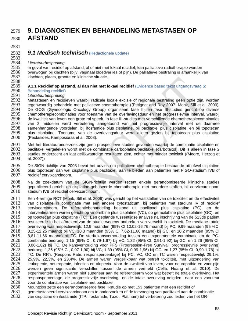
58 Concept Revisie Richtlijn Cervixcarcinoom - September 2011
9. DIAGNOSTIEK EN BEHANDELING METASTASEN OP 2579
AFSTAND 2580
2581
9.1 Medisch technisch (Redactionele update) 2582 2583 Literatuurbespreking 2584 In geval van recidief op afstand, al of niet met lokaal recidief, kan palliatieve radiotherapie worden 2585 overwogen bij klachten (bijv. vaginaal bloedverlies of pijn). De palliatieve bestraling is afhankelijk van 2586 klachten, plaats, grootte en klinische situatie. 2587 2588 9.1.1 Recidief op afstand, al dan niet met lokaal recidief (Evidence based tekst uitgangsvraag 5: 2589 Behandeling recidief) 2590 Literatuurbespreking 2591 Metastasen en recidieven waarbij radicale locale excisie of regionale bestraling geen optie zijn, worden 2592 tegenwoordig behandeld met palliatieve chemotherapie ((Petignat and Roy 2007; Monk, Sill et al. 2009). 2593 De GOG (Gynecologic Oncology Group) organiseert fase II- en fase III-studies gericht op diverse 2594 chemotherapiecombinaties voor toename van de overlevingsduur en het progressievrije interval, waarbij 2595 de kwaliteit van leven een grote rol speelt. In fase III-studies met verschillende chemotherapiecombinaties 2596 van 2 middelen werd verbetering aangetoond van het progressievrije interval met de daarmee 2597 samenhangende voordelen, bij ifosfamide plus cisplatine, bij paclitaxel plus cisplatine, en bij topotecan 2598 plus cisplatine. Toename van de overlevingsduur werd alleen gezien bij topotecan plus cisplatine 2599 (Pectasides, Kamposioras et al. 2008). 2600
Met het literatuuronderzoek zijn geen prospectieve studies gevonden waarbij de combinatie cisplatine en 2601 paclitaxel vergeleken wordt met de combinatie carboplatine/paclitaxel (carbotaxol). Dit is alleen in fase 2 2602 studies onderzocht en laat gelijkwaardige resultaten zien, echter met minder toxiciteit ((Moore, Herzog et 2603 al. 2007)) 2604
De SIGN-richtlijn van 2008 bevat het advies om palliatieve chemotherapie bestaande uit ofwel cisplatine 2605 plus topotecan dan wel cisplatine plus paclitaxel, aan te bieden aan patiënten met FIGO-stadium IVB of 2606 recidief cervixcarcinoom. 2607
Na de zoekdatum van de SIGN-richtlijn werden recent enkele gerandomiseerde klinische studies 2608 gepubliceerd gericht op cisplatine-gebaseerde chemotherapie met meerdere stoffen, bij cervixcarcinoom 2609 stadium IVB of recidief cervixcarcinoom. 2610
Een 4-armige RCT (Monk, Sill et al. 2009) was gericht op het vaststellen van de toxiciteit en de effectiviteit 2611 van cisplatine in combinatie met een andere cytostaticum, bij patiënten met stadium IV of recidief 2612 cervixcarcinoom. De referentiebehandeling bestond uit paclitaxel plus cisplatine (PC), en de 2613 interventiearmen waren gericht op vinorelbine plus cisplatine (VC), op gemcitabine plus cisplatine (GC), en 2614 op topotecan plus cisplatine (TC). Een geplande tussentijdse analyse na inschrijving van de 513de patiënt 2615 resulteerde in het afbreken van de studie wegens ontbreken van verschil in toxiciteit. De mediane totale 2616 overleving was respectievelijk: 12,9 maanden (95% CI 10,02-16,76 maand) bij PC; 9,99 maanden (95 %CI 2617 8,25-12,25 maand) bij VC; 10,3 maanden (95% CI 7,62-11,60 maand) bij GC; en 10,2 maanden (95% CI 2618 8,61-11,66 maand) bij TC. De sterftekansverhouding tussen een experimentele combinatie en de PC-2619 combinatie bedroeg: 1,15 (95% CI, 0,79-1,67) bij VC; 1,32 (95% CI, 0,91-1,92) bij GC; en 1,26 (95% CI, 2620 0,86-1,82) bij TC. De kansverhouding voor PFS (Progression-Free Survival: progressievrije overleving) 2621 bedroeg: 1,36 (95% CI, 0,97-1,90) bij VC; 1,39 (95% CI, 0,99-1,96) bij GC; en 1,27 (95% CI, 0,90-1,78) bij 2622 TC. De RR's (Respons Rate: responspercentage) bij PC, VC, GC en TC waren respectievelijk 29,1%, 2623 25,9%, 22,3%, en 23,4%. De armen waren vergelijkbaar wat betreft toxiciteit, met uitzondering van 2624 leukopenie, neutropenie, infecties en alopecia. Voor de kwaliteit van leven, voor neuropathie en voor pijn 2625 werden geen significante verschillen tussen de armen vermeld (Cella, Huang et al. 2010). De 2626 experimentele armen waren niet superieur aan de referentiearm voor wat betreft de totale overleving. Het 2627 responspercentage, de progressievrije overleving en de totale overleving neigden naar een voorkeur 2628 voor de combinatie van cisplatine met paclitaxel. 2629 Mountzios zette een gerandomiseerde fase II-studie op met 153 patiënten met een recidief of 2630 gemetastaseerd cervixcarcinoom om te onderzoeken of de toevoeging van paclitaxel aan de combinatie 2631 van cisplatine en ifosfamide (ITP: Ifosfamide, Taxol, Platinum) tot verbetering zou leiden van het OR-2632

59 Concept Revisie Richtlijn Cervixcarcinoom - September 2011
percentage (Objectieve Respons), de PFS (Progression-Free Survival: progressievrije overleving) en de 2633 OS (Overall Survival: totale overleving) (Mountzios, Dimopoulos et al. 2009). Het OR-percentage was 2634 significant hoger in de ITP-groep (59% versus 33%, p=0,002). De mediane PFS was 7,9 maanden in de 2635 ITP-arm en 6,3 maanden in de IP-arm (p=0,023). De mediane OS was 15,4 maanden in de ITP-arm en 2636 13,2 maanden in de IP-arm (p=0,048). Multivariate analyse leverde de volgende kansverhoudingen op 2637 voor ITP t.o.v. IP: 0,70 voor terugval of progressie (p=0,046) en 0,75 voor sterfte (p=0,124). De 2638 neurotoxiciteit nam toe in 43% van de patiënten met ITP en in 11% van de patiënten met IP (p<0,001). 2639 2640 Conclusies 2641 Het is aannemelijk dat combinatietherapie leidt tot verbetering van het responspercentage, de 2642 progressievrije overleving en de totale overleving bij stadium IVb gemetastaseerd of recidief 2643 cervixcarcinoom, vergeleken met cisplatine-monochemotherapie. Combinatiechemotherapieën leiden tot 2644 een toename van de toxiciteit, hoewel niet significant tot een afname van de levenskwaliteit 2645 Niveau 2: (Monk, Sill et al. 2009) (B); (Cella, Huang et al. 2010) (B); (Hirte, Strychowsky et al. 2006) (A2) 2646 2647 Er zijn aanwijzingen dat de behandelresultaten van paclitaxel plus cisplatine beter zijn dan die van 2648 topotecan plus cisplatine, zonder toename van de toxiciteit. 2649 Niveau 3: (Monk, Sill et al. 2009) (B) 2650 2651 De werkgroep is van mening dat combinatie carboplatine en paclitaxel) gelijkwaardige resultaten geeft als 2652 andere combinatie therapieen maar met minder toxititeit. 2653 Niveau 4: mening van de werkgroepleden 2654 2655 Overige overwegingen 2656 Bij de overweging tot starten met palliatieve chemotherapie dient de toxiciteit van vorige behandelingen, 2657 alsmede de verwachtte palliatie op recidief gerelateerde klachten meegenomen te worden (veiligheid en 2658 patiëntenperspectief). Combinatie chemotherapie met platinum combinaties vraagt geen extra 2659 vaardigheden van een internist oncoloog (professioneel perspectief). 2660 2661 Aanbevelingen 2662
Bij het vaststellen van afstandsmetastasen, al of niet met lokaal recidief, wordt combinatie chemotherapie 2663 met carboplatine en paclitaxel geadviseerd. 2664 2665 2666
9.2 Voorlichting en communicatie (Redactionele update) 2667 2668 Literatuurbespreking 2669 Patiënte en haar naasten worden geïnformeerd over de (on)mogelijkheden voor behandeling en palliatieve 2670 ondersteuning. Is de patiënte geïnformeerd over de palliatieve mogelijkheden? Is duidelijk wie de 2671 hoofdbehandelaar is? 2672 De KWF kankerbestrijding folder Baarmoederhalskanker is hier te downloaden. 2673 De NVOG voorlichtingsfolder Baarmoederhalskanker is hier te downloaden. 2674 De stichting OLIJF voor vrouwen met gynaecologische kanker geeft informatie en verzorgt lotgenoten 2675 contact. 2676 www.sibopmaat.nl is een website met patiënteninformatie over bijwerkingen van medicijnen die gebruikt 2677 worden ter behandeling van kanker. 2678 2679 2680
9.3 Continuïteit van zorg (Redactionele update) 2681 2682 Literatuurbespreking 2683 Intramuraal 2684 Een eventueel behandelplan wordt vastgesteld in de oncologiebespreking, waarbij besproken wordt wie 2685 welk deel van de behandeling op zich neemt en wie de regie heeft. Zo nodig wordt overlegd met het 2686 pijnteam en het psychosociale team over verdere hulp. 2687 2688 Transmuraal 2689 Met de huisarts worden afspraken gemaakt over de benodigde zorg. 2690
2691

60 Concept Revisie Richtlijn Cervixcarcinoom - September 2011
10. PALLIATIEVE ZORG (Richtlijntekst) 2692 Literatuurbespreking 2693 In de palliatieve fase kan het detecteren van behoefte aan psychosociale zorg en het zo nodig verlenen 2694 van psychosociale zorg de kwaliteit van leven (ook doen) verbeteren. In de richtlijn Detecteren behoefte 2695 psychosociale zorg (NVPO, 2009) wordt een signaleringsinstrument (de Lastmeter) aanbevolen. 2696 Signalering van klachten dient bij voorkeur eens in de drie maanden plaats te vinden. Zie verder richtlijn 2697 Detecteren behoefte psychosociale zorg. Om te bepalen of oncologische revalidatie een geschikte 2698 interventie is voor de patiënt met klachten kan de Lastmeter aangevuld worden met de VAS 2699 vermoeidheidlijst en de Patiënt Specifieke Klachtenlijst. Er zijn speciale oncologische 2700 revalidatieprogramma‟s die gericht zijn op de ziektegerichte- en symptoomgerichte fasen van palliatie. In 2701 het revalidatieprogramma staan de persoonlijke doelen en voorkeuren van de patiënt (en zijn naasten) 2702 centraal. Er kan gestreefd worden naar preventie en behandeling van symptomen enerzijds en 2703 optimaliseren van de kwaliteit van leven anderzijds. Het streven naar behoud van fysieke functies zoals 2704 traplopen kan hierin essentieel zijn. Voor patiënten die gaandeweg door progressieve ziekte „uit het 2705 programma vallen‟ is het aan te raden een beperktere versie van het aanbod thuis te faciliteren om ook in 2706 de terminale fase van de effecten van bekrachting in wat nog wel kan (empowerment) te kunnen 2707 profiteren. Zie verder de richtlijn Oncologische revalidatie (IKNL 2010). 2708 Voor de richtlijnen palliatieve zorg zie www.pallialine.nl. 2709 2710 2711 2712 2713
2714
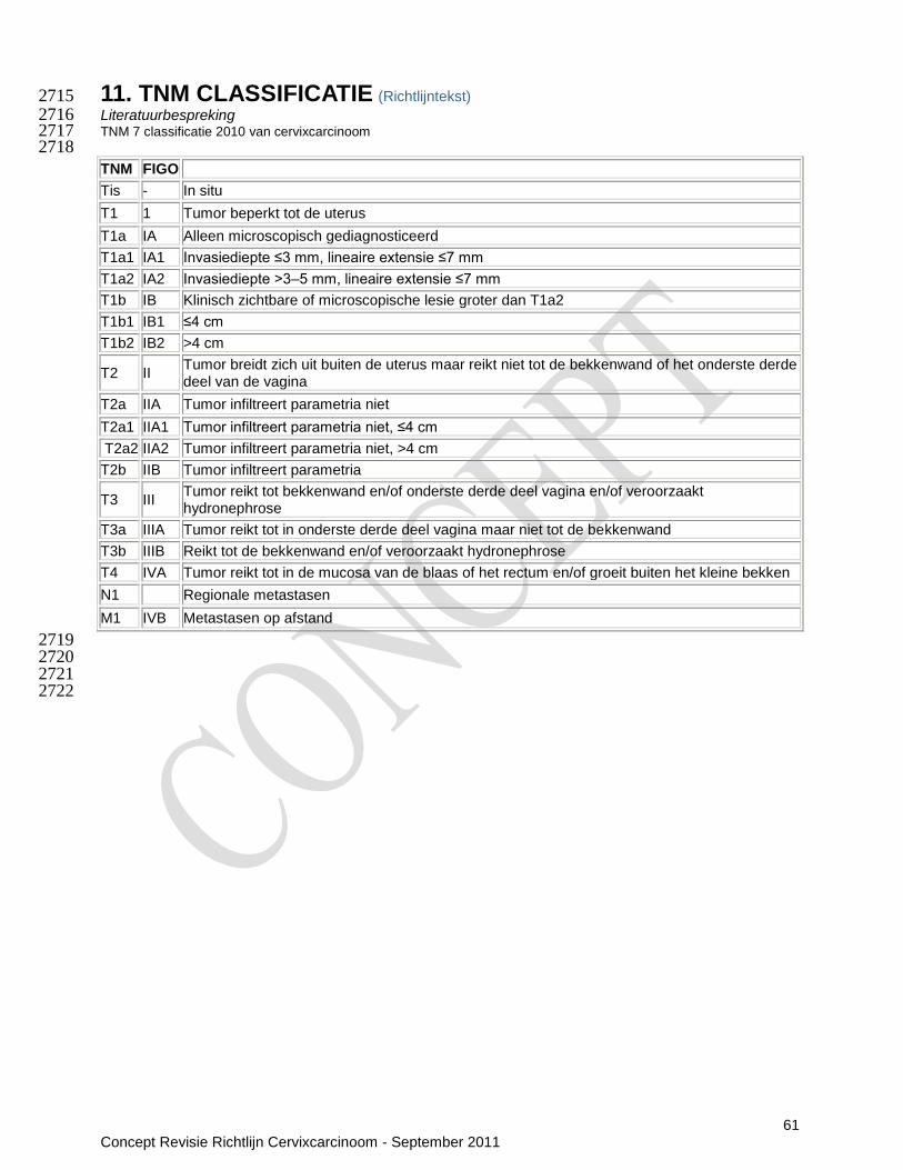
61 Concept Revisie Richtlijn Cervixcarcinoom - September 2011
11. TNM CLASSIFICATIE (Richtlijntekst) 2715 Literatuurbespreking 2716 TNM 7 classificatie 2010 van cervixcarcinoom 2717 2718
TNM FIGO
Tis - In situ
T1 1 Tumor beperkt tot de uterus
T1a IA Alleen microscopisch gediagnosticeerd
T1a1 IA1 Invasiediepte ≤3 mm, lineaire extensie ≤7 mm
T1a2 IA2 Invasiediepte ˃3–5 mm, lineaire extensie ≤7 mm
T1b IB Klinisch zichtbare of microscopische lesie groter dan T1a2
T1b1 IB1 ≤4 cm
T1b2 IB2 >4 cm
T2 II Tumor breidt zich uit buiten de uterus maar reikt niet tot de bekkenwand of het onderste derde deel van de vagina
T2a IIA Tumor infiltreert parametria niet
T2a1 IIA1 Tumor infiltreert parametria niet, ≤4 cm
T2a2 IIA2 Tumor infiltreert parametria niet, >4 cm
T2b IIB Tumor infiltreert parametria
T3 III Tumor reikt tot bekkenwand en/of onderste derde deel vagina en/of veroorzaakt hydronephrose
T3a IIIA Tumor reikt tot in onderste derde deel vagina maar niet tot de bekkenwand
T3b IIIB Reikt tot de bekkenwand en/of veroorzaakt hydronephrose
T4 IVA Tumor reikt tot in de mucosa van de blaas of het rectum en/of groeit buiten het kleine bekken
N1 Regionale metastasen
M1 IVB Metastasen op afstand
2719 2720 2721
2722

62 Concept Revisie Richtlijn Cervixcarcinoom - September 2011
12. SAMENVATTING 2723 2724
Hier komt een link naar het samenvattingskaartje van de richtlijn cervixcarcinoom. 2725 2726

63 Concept Revisie Richtlijn Cervixcarcinoom - September 2011
13. Bijlagen 2727
Bijlage 1 Samenstelling werkgroep 2728
Alle werkgroepleden zijn afgevaardigd namens wetenschappelijke verenigingen en hebben daarmee het 2729 mandaat voor hun inbreng (zie bijlage 2). Bij de samenstelling van de werkgroep is rekening gehouden 2730 met landelijke spreiding, inbreng van betrokkenen uit zowel academische als algemene 2731 ziekenhuizen/instellingen en vertegenwoordiging van de verschillende verenigingen/ disciplines. De 2732 patiëntenvereniging is eveneens vertegenwoordigd. 2733 2734
Bijlage 2 Werkgroepleden 2735
Werkgroepleden 2736 Mw. dr. K.N. Gaarenstroom, gynaecologisch oncoloog, Leids Universitair Medisch Centrum, voorzitter 2737 Mw. I. ter Borch, oncologieverpleegkundige afdeling gynaecologie, Medisch Spectrum Twente te 2738
Enschede 2739 Dhr. dr. R.L.M. Bekkers, gynaecologisch oncoloog, Universitair Medisch Centrum Nijmegen 2740 Dhr. V.P.M. Cliteur, patholoog, Westfriesgasthuis te Hoorn 2741 Dhr. prof. dr. G.J. Fleuren, patholoog, Leids Universitair Medisch Centrum 2742 Mw. C.J. Grimbergen, oncologieverpleegkundige afdeling gynaecologie , Leids Universitair Medisch 2743
Centrum 2744 Mw. R. Jensma, patiëntenvertegenwoordiger, bestuurslid Olijf 2745 Mw. dr. I.M. Jürgenliemk-Schulz, radiotherapeut-oncoloog, Universitair Medisch Centrum Utrecht 2746 Mw. prof. dr. G.G. Kenter, gynaecologisch oncoloog, Centrum Gynaecologische Oncologie 2747
Amsterdam 2748 Mw. drs. E.A.G. Lammerink, AIOS gynaecologie, Universitair Medisch Centrum Groningen 2749 Mw. drs. J.H.F. Leemhuis, socioloog, richtlijndeskundige namens NFK 2750 Mw. prof. M.J.E. Mourits, gynaecologisch oncoloog, Universitair Medisch Centrum Groningen 2751 Mw. dr. E. Pras, radiotherapeut-oncoloog, Universitair Medisch Centrum Groningen 2752 Mw. dr. A.M. Spijkerboer, radioloog, Academisch Medisch Centrum Universiteit van Amsterdam 2753 Dhr. dr. J. van der Velden, gynaecologisch oncoloog, Academisch Medisch Centrum Universiteit van 2754
Amsterdam 2755 Mw. dr. P.O. Witteveen, internist-oncoloog, Universitair Medisch Centrum Utrecht 2756 2757 Leden werkgroep richtlijn 2010 2758 Mw. dr. W.J. van Driel, gynaecologisch oncoloog, voorzitter 2759 M.E.L. van der Burg, internist-oncoloog 2760 C.L. Creutzberg, radiotherapeut-oncoloog 2761 H. Hollema, patholoog 2762 I.M. Jürgenliemk-Schulz, radiotherapeut-oncoloog 2763 G.G. Kenter, gynaecologisch oncoloog 2764 R.F.P.M. Kruitwagen, gynaecologisch oncoloog 2765 L.C.H.W. Lutgens, radiotherapeut-oncoloog 2766 L.F.A.G. Massuger, gynaecologisch oncoloog 2767 M.J.E. Mourits, gynaecologisch oncoloog 2768 P.B. Ottevanger, internist-oncoloog 2769 E. Pras, radiotherapeut-oncoloog 2770 B.F.M. Slangen, gynaecologisch oncoloog 2771 E.M. van der Steen-Banasik, radiotherapeut-oncoloog 2772 B. van Triest, radiotherapeut-oncoloog 2773 J. van der Velden, gynaecologisch oncoloog 2774 P.O. Witteveen, internist-oncoloog 2775 2776

64 Concept Revisie Richtlijn Cervixcarcinoom - September 2011
Bijlage 3 Onafhankelijkheid werkgroepleden 2777
Alle leden van de richtlijnwerkgroep hebben verklaard onafhankelijk gehandeld te hebben bij het opstellen 2778 van de richtlijn. 2779 2780
Bijlage 4 Betrokken en autoriserende verenigingen 2781
Initiatief 2782 Commissie Richtlijnen Gynaecologische Oncologie (CRGO) 2783 2784 Organisatie 2785 Integraal Kankercentrum Nederland (IKNL) 2786 2787 Betrokken verenigingen 2788 Landelijk Platform Radiotherapeuten Gynaecologische Tumoren (LPRGT) 2789 Nederlandse Federatie van Kankerpatiëntenorganisaties (NFK) 2790 Nederlandse Internisten Vereniging (NIV) 2791 Nederlandse Vereniging voor Medische Oncologie (NVMO) 2792 Nederlands Vereniging voor Obstetrie & Gynaecologie (NVOG) 2793 Nederlandse Vereniging voor Pathologie (NVVP) 2794 Nederlandse Vereniging voor Radiologie (NVvR) 2795 Nederlandse Vereniging voor Radiotherapie en Oncologie (NVRO) 2796 Stichting Olijf, Netwerk van vrouwen met gynaecologische kanker 2797 Vereniging van Verpleegkundigen & Verzorgenden Nederland 2798 Werkgroep Oncologische Gynaecologie (WOG)/Peiler Oncologie 2799 2800 Autoriserende verenigingen 2801 Nederlands Vereniging voor Obstetrie & Gynaecologie (NVOG) 2802 Nederlandse Internisten Vereniging (NIV) 2803 Nederlandse Vereniging voor Pathologie (NVVP) 2804 Nederlandse Vereniging voor Radiotherapie en Oncologie (NVRO) 2805 Nederlandse Vereniging voor Radiologie (NVvR) 2806 Vereniging van Verpleegkundigen & Verzorgenden Nederland 2807 De volgende verenigingen stemmen in met de inhoud 2808 Nederlandse Federatie van Kankerpatiëntenorganisaties (NFK) 2809 Nederlands Huisartsen Genootschap (NHG) 2810 2811
Bijlage 5 Wetenschappelijke onderbouwing 2812
Elk hoofdstuk van de richtlijn is volgens een vast stramien opgebouwd, namelijk literatuurbespreking, 2813 conclusies, overwegingen en aanbevelingen. De antwoorden op de uitgangsvragen (derhalve de 2814 aanbevelingen in deze richtlijn) zijn voor zover mogelijk gebaseerd op gepubliceerd wetenschappelijk 2815 onderzoek. 2816 2817 Zoekstrategie 2818 Voor uitgangsvraag 1 en 4 werd gezocht in de databases Medline, Embase en de Cochrane database of 2819 systematic reviews naar artikelen in het Engels en Nederlands. 2820 2821 Om de richtlijn Herstel na kanker te integreren in de richtlijn cervixcarcinoom zijn bij uitgangsvraag 4 nog 2 2822 subvragen nader uitgezocht: 2823 4.1 Is de behandelingeffectiviteit hoger naarmate het recidief eerder wordt gediagnosticeerd? 2824 4.2 Welke diagnostiek is het meest geschikt om behandelbare nieuwe kankermanifestaties vroeg en 2825 acuraat te diagnosticeren? 2826 Voor het beantwoorden van bovenstaande subvragen is het systematische review van Elit als 2827 uitgangspunt genomen. 2828 2829 Voor uitgangsvraag 2, 3, 5 en 6 is de ADAPTE methodologie gebruikt. In eerste instantie zijn bestaande 2830 (inter)nationale richtlijnen gezocht in Medline, National Guideline Clearinghouse en websites van 2831 oncologische organisaties. De gevonden richtlijnen werden door middel van het AGREE instument 2832 beoordeeld op hun kwaliteit en geselecteerd. 2833

65 Concept Revisie Richtlijn Cervixcarcinoom - September 2011
Voor uitgangsvraag 2, 3, 5 en 6 is de richtlijn „management of cervical cancer‟ van the Scottish 2834 Intercollegiate Guidelines Network (SIGN) geselecteerd waarvan de literatuursearch in 2005 is uitgevoerd. 2835 Voor uitgangsvraag 5 is tevens de richtlijn „Chemotherapy for Recurrent, Metastatic, or Persistent Cervical 2836 Cancer‟ geselecteerd van Cancer Care Ontario (CCO) waarvan de literatuursearch in 2006 is uitgevoerd. 2837 Vervolgens werd gezocht in de databases Medline, Embase en de Cochrane database of systematic 2838 reviews naar artikelen in het Engels en Nederlands vanaf de datum van het literatuuronderzoek van de 2839 SIGN richtlijn (2005). 2840 2841 Voor iedere vraag werd een aparte zoekstrategie gebruikt (zie bijlage 9) 2842 2843 Selectie 2844 Aanbevelingen werden gebaseerd op „beste evidence' en daarom werd naast selectie op relevantie tevens 2845 geselecteerd op bewijskracht. Hiervoor werd gebruik gemaakt van de volgende hiërarchische indeling van 2846 studiedesigns gebaseerd op bewijskracht: 2847 1. Meta-analyses en systematische reviews; 2848 2. Gerandomiseerde gecontroleerde studies (RCT's); 2849 3. Gecontroleerde studies (CT's). 2850 Waar deze niet voorhanden waren werd verder gezocht naar vergelijkend cohortonderzoek, vergelijkende 2851 patiënt controle-onderzoeken of niet-vergelijkend onderzoek. 2852 2853 Critical appraisal 2854 De geselecteerde artikelen zijn vervolgens beoordeeld op kwaliteit van het onderzoek en gegradeerd naar 2855 mate van bewijs (zie bijlage 6). Voor de evidence tabellen (zie bijlage 10). De mate van bewijskracht en 2856 niveau van bewijs zijn in de conclusies van de verschillende hoofdstukken weergegeven. De belangrijkste 2857 literatuur waarop de conclusies zijn gebaseerd is daarbij vermeld. 2858 2859 Oorspronkelijke richtlijn versus revisie 2860 Aangezien deze richtlijn teksten bevat uit zowel de richtlijn uit het jaar 2010 als uit de revisie van de richtlijn 2861 in het jaar 2011 staat bij elke paragraaf weergegeven wat voor soort tekst het betreft: 2862 - Evidence based tekst: hierbij is een systematisch literatuuronderzoek verricht voor een bepaalde 2863
uitgangsvraag. De tekst naar aanleiding van dit literatuuronderzoek is nieuw. 2864 - Consensus based tekst: hierbij is geen systematisch literatuuronderzoek uitgevoerd. Er wordt naar 2865
studies gerefereerd die door de werkgroepleden zelf zijn aangedragen. 2866 - Redactionele update: hierbij is de tekst uit het jaar 2010 behouden en op basis van kennis en ervaring 2867
indien nodig aangepast. Er is geen nieuw systematisch literatuuronderzoek uitgevoerd. 2868 2869
2870
Bijlage 6 Indeling van onderzoeksresultaten naar mate van bewijskracht 2871
2872 Tabel 1: Niveau van bewijskracht van de conclusie op basis van het aan de conclusie ten 2873 grondslag liggend bewijs 2874 Niveau van bewijs
Conclusie gebaseerd op Formulering
1 Eén systematische review (A1) of tenminste 2 onafhankelijk van elkaar uitgevoerde onderzoeken van niveau A1 of A2
Het is aangetoond dat…; men dient…
2 Tenminste 2 onafhankelijk van elkaar uitgevoerde onderzoeken van niveau B
Het is aannemelijk dat…; men zou…moeten…
3 Eén onderzoek van niveau A2, B of C Er zijn aanwijzingen dat…; men kan…
4 Mening van deskundigen (bijvoorbeeld de werkgroepleden)
De werkgroep is van mening dat…
2875 Tabel 2: Indeling van methodologische kwaliteit van individuele studies 2876 Voor artikelen betreffende interventie
A1 Systematische reviews die tenminste enkele onderzoeken van A2-niveau betreffen, waarbij de resultaten van afzonderlijke onderzoeken consistent zijn.

66 Concept Revisie Richtlijn Cervixcarcinoom - September 2011
A2 Gerandomiseerd vergelijkend klinisch onderzoek van goede kwaliteit en met voldoende omvang en consistentie.
B Gerandomiseerde klinische trials van matige kwaliteit of onvoldoende omvang of ander vergelijkend onderzoek (niet-gerandomiseerd: vergelijkend cohortonderzoek, patiënt-controle-onderzoek)
C Niet-vergelijkend onderzoek
D Mening van deskundigen (bijvoorbeeld de werkgroepleden)
2877 Voor artikelen betreffende diagnostiek
A1 Onderzoek naar de effecten van diagnostiek op klinische uitkomsten bij een prospectief gevolgde goed gedefinieerde patiëntengroep met een tevoren gedefinieerd beleid op grond van de te onderzoeken testuitslagen, of besliskundig onderzoek naar de effecten van diagnostiek op klinische uitkomsten, waarbij resultaten van onderzoek van A2-niveau als basis worden gebruikt en voldoende rekening wordt gehouden met onderlinge afhankelijkheid van diagnostische tests
A2 Onderzoek ten opzichte van een referentietest, waarbij van tevoren criteria zijn gedefinieerd voor de te onderzoeken test en voor een referentietest, met een goede beschrijving van de test en de onderzochte klinische populatie; het moet een voldoende grote serie van opeenvolgende patiënten betreffen, er moet gebruik zijn gemaakt van tevoren gedefinieerde afkapwaarden en de resultaten van de test en de „gouden standaard' moeten onafhankelijk zijn beoordeeld. Bij situaties waarbij multipele, diagnostische tests een rol spelen, is er in principe een onderlinge afhankelijkheid en dient de analyse hierop te zijn aangepast, bijvoorbeeld met logistische regressie
B Vergelijking met een referentietest, beschrijving van de onderzochte test en populatie, maar niet de kenmerken die verder onder niveau A staan genoemd
C Niet-vergelijkend onderzoek
D Mening van deskundigen, bijvoorbeeld de werkgroepleden
2878
2879 2880
2881
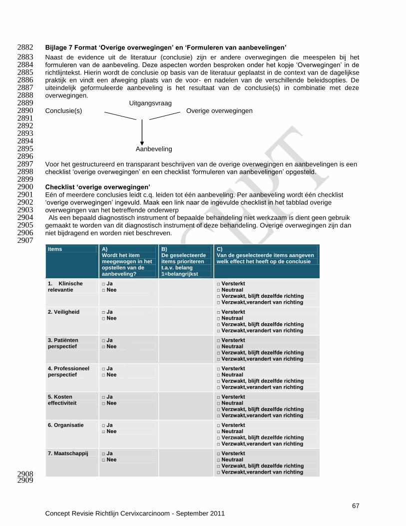
67 Concept Revisie Richtlijn Cervixcarcinoom - September 2011
Bijlage 7 Format ‘Overige overwegingen’ en ‘Formuleren van aanbevelingen’ 2882
Naast de evidence uit de literatuur (conclusie) zijn er andere overwegingen die meespelen bij het 2883 formuleren van de aanbeveling. Deze aspecten worden besproken onder het kopje „Overwegingen‟ in de 2884 richtlijntekst. Hierin wordt de conclusie op basis van de literatuur geplaatst in de context van de dagelijkse 2885 praktijk en vindt een afweging plaats van de voor- en nadelen van de verschillende beleidsopties. De 2886 uiteindelijk geformuleerde aanbeveling is het resultaat van de conclusie(s) in combinatie met deze 2887 overwegingen. 2888
Uitgangsvraag 2889 Conclusie(s) Overige overwegingen 2890 2891 2892 2893 2894
Aanbeveling 2895 2896 Voor het gestructureerd en transparant beschrijven van de overige overwegingen en aanbevelingen is een 2897 checklist „overige overwegingen‟ en een checklist „formuleren van aanbevelingen‟ opgesteld. 2898 2899 Checklist ‘overige overwegingen’ 2900 Eén of meerdere conclusies leidt c.q. leiden tot één aanbeveling. Per aanbeveling wordt één checklist 2901 „overige overwegingen‟ ingevuld. Maak een link naar de ingevulde checklist in het tabblad overige 2902 overwegingen van het betreffende onderwerp 2903 Als een bepaald diagnostisch instrument of bepaalde behandeling niet werkzaam is dient geen gebruik 2904 gemaakt te worden van dit diagnostisch instrument of deze behandeling. Overige overwegingen zijn dan 2905 niet bijdragend en worden niet beschreven. 2906 2907
Items A) Wordt het item meegewogen in het opstellen van de aanbeveling?
B) De geselecteerde items prioriteren t.a.v. belang 1=belangrijkst
C) Van de geselecteerde items aangeven welk effect het heeft op de conclusie
1. Klinische relevantie
□ Ja □ Nee
□ Versterkt □ Neutraal □ Verzwakt, blijft dezelfde richting □ Verzwakt,verandert van richting
2. Veiligheid □ Ja □ Nee
□ Versterkt □ Neutraal □ Verzwakt, blijft dezelfde richting □ Verzwakt,verandert van richting
3. Patiënten perspectief
□ Ja □ Nee
□ Versterkt □ Neutraal □ Verzwakt, blijft dezelfde richting □ Verzwakt,verandert van richting
4. Professioneel perspectief
□ Ja □ Nee
□ Versterkt □ Neutraal □ Verzwakt, blijft dezelfde richting □ Verzwakt,verandert van richting
5. Kosten effectiviteit
□ Ja □ Nee
□ Versterkt □ Neutraal □ Verzwakt, blijft dezelfde richting □ Verzwakt,verandert van richting
6. Organisatie □ Ja □ Nee
□ Versterkt □ Neutraal □ Verzwakt, blijft dezelfde richting □ Verzwakt,verandert van richting
7. Maatschappij □ Ja □ Nee
□ Versterkt □ Neutraal □ Verzwakt, blijft dezelfde richting □ Verzwakt,verandert van richting 2908
2909

68 Concept Revisie Richtlijn Cervixcarcinoom - September 2011
Beschrijving welke aspecten van de geselecteerde items ‘overige overwegingen’ van belang zijn en hoe:
1. Klinische relevantie:
2. Veiligheid:
3. Patiënten perspectief:
4. Professioneel perspectief:
5. Kosteneffectiviteit:
6. Organisatie:
7. Maatschappij:
2910 Uitleg items checklist ‘overige overwegingen’ 2911 1. Klinische relevantie: bepalen door professionals en evt. literatuuronderzoek 2912 - Sterkte van het effect vergeleken met geen interventie 2913 - Consistentie van het beschikbare bewijs uit de verschillende studies 2914 - Generaliseerbaarheid 2915 - Voorbeeld: een bepaalde behandeling kan een significante verbetering van symptomen geven die 2916
patiënten echter niet als zodanig ervaren, dus klinisch niet relevant 2917 2918
2. Veiligheidsissues: bepalen door professionals en evt. literatuuronderzoek 2919 - Bijwerkingen, risico‟s of complicaties op korte en lange termijn 2920 - Gebruik bij co-morbiditeit / gelijktijdig gebruik medicatie en/of interventie 2921 - Voorbeeld: fysieke comorbiditeit kan bepaalde behandelingen uitsluiten. 2922 2923 3. Patiëntenperspectief: bepalen door professionals, patiënten en eventueel literatuuronderzoek 2924 - Voorkeuren / te verwachten compliance / te verwachten tevredenheid / kwaliteit van leven 2925 - Voorbeeld: diagnostiek of behandeling waarvoor minder ziekenhuisbezoek nodig is; behandeling die 2926
makkelijker toe te dienen is; behandeling die sneller effect laat zien 2927 2928 4. Professioneel perspectief: bepalen door professionals 2929 - Kennis en ervaring met technieken/therapie 2930 - Risico‟s die professional loopt bij het toepassen van de interventie / tijdbesparing of het verlies aan tijd 2931
door het invoeren van de interventie 2932 - Houding, normen en waarden van de professional 2933 - Voorbeeld: een nieuwe techniek die nog niet alle professionals beheersen. 2934 2935 5. Kosteneffectiviteit: bepalen door professionals, indien gewenst en mogelijk kosteneffectiviteits-analyse 2936
door expert 2937 - Kosteneffectiviteit in verhouding tot bestaande ingrepen/behandelingen voor deze ziekte 2938 2939 6. Organisatorische factoren: bepalen door professionals 2940 - De beschikbaarheid/aanwezigheid van faciliteiten & medicijnen 2941 - De wijze waarop de organisatie van de zorg aangeboden dient te worden / grootte van de verandering 2942
in de organisatie-zorgproces / infrastructuur voor implementatie 2943 - Voorbeeld: een bepaalde diagnostiek of behandeling kan alleen in bepaalde centra worden uitgevoerd 2944
in verband met de aanwezigheid van faciliteiten zoals een PET scan. 2945 2946 7. Maatschappelijke factoren: bepalen door professionals 2947 - Vergoeding door verzekeraars / Industriële belangen / Ethische overwegingen / Juridische 2948
overwegingen / Politieke en strategische consequenties 2949 - Voorbeeld: indien twee behandelingen even effectief zijn waarvan één behandeling wordt vergoed, zal 2950
deze laatste behandeling worden aanbevolen. 2951 2952 2953 2954 2955 2956
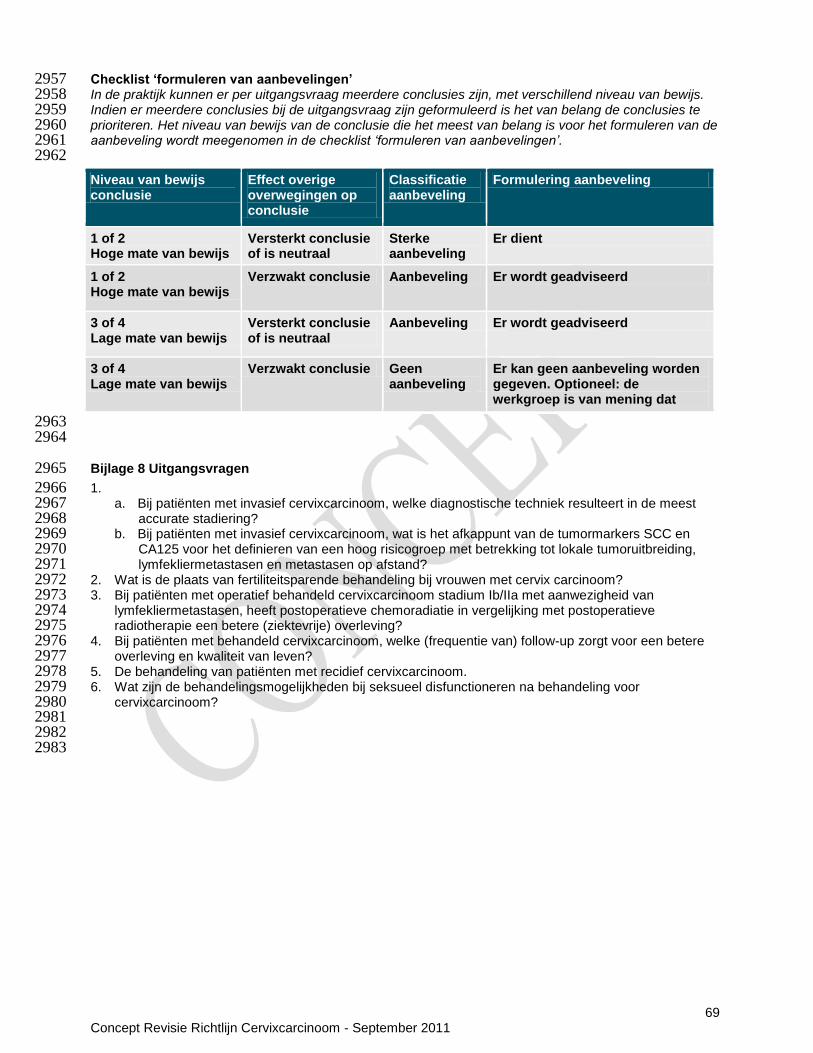
69 Concept Revisie Richtlijn Cervixcarcinoom - September 2011
Checklist ‘formuleren van aanbevelingen’ 2957 In de praktijk kunnen er per uitgangsvraag meerdere conclusies zijn, met verschillend niveau van bewijs. 2958 Indien er meerdere conclusies bij de uitgangsvraag zijn geformuleerd is het van belang de conclusies te 2959 prioriteren. Het niveau van bewijs van de conclusie die het meest van belang is voor het formuleren van de 2960 aanbeveling wordt meegenomen in de checklist ‘formuleren van aanbevelingen’. 2961 2962
2963 2964
Bijlage 8 Uitgangsvragen 2965
1. 2966 a. Bij patiënten met invasief cervixcarcinoom, welke diagnostische techniek resulteert in de meest 2967
accurate stadiering? 2968 b. Bij patiënten met invasief cervixcarcinoom, wat is het afkappunt van de tumormarkers SCC en 2969
CA125 voor het definieren van een hoog risicogroep met betrekking tot lokale tumoruitbreiding, 2970 lymfekliermetastasen en metastasen op afstand? 2971
2. Wat is de plaats van fertiliteitsparende behandeling bij vrouwen met cervix carcinoom? 2972 3. Bij patiënten met operatief behandeld cervixcarcinoom stadium Ib/IIa met aanwezigheid van 2973
lymfekliermetastasen, heeft postoperatieve chemoradiatie in vergelijking met postoperatieve 2974 radiotherapie een betere (ziektevrije) overleving? 2975
4. Bij patiënten met behandeld cervixcarcinoom, welke (frequentie van) follow-up zorgt voor een betere 2976 overleving en kwaliteit van leven? 2977
5. De behandeling van patiënten met recidief cervixcarcinoom. 2978 6. Wat zijn de behandelingsmogelijkheden bij seksueel disfunctioneren na behandeling voor 2979
cervixcarcinoom? 2980 2981 2982
2983
Niveau van bewijs conclusie
Effect overige overwegingen op conclusie
Classificatie aanbeveling
Formulering aanbeveling
1 of 2 Hoge mate van bewijs
Versterkt conclusie of is neutraal
Sterke aanbeveling
Er dient
1 of 2 Hoge mate van bewijs
Verzwakt conclusie Aanbeveling Er wordt geadviseerd
3 of 4 Lage mate van bewijs
Versterkt conclusie of is neutraal
Aanbeveling Er wordt geadviseerd
3 of 4 Lage mate van bewijs
Verzwakt conclusie Geen aanbeveling
Er kan geen aanbeveling worden gegeven. Optioneel: de werkgroep is van mening dat
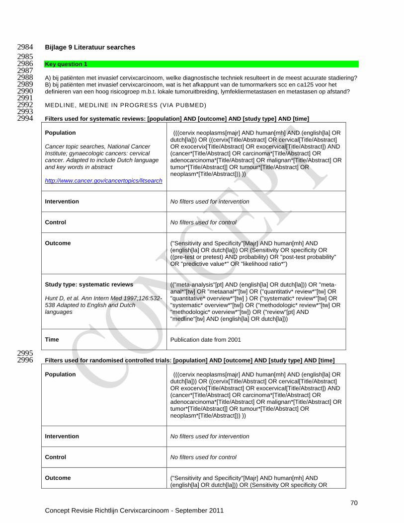
70 Concept Revisie Richtlijn Cervixcarcinoom - September 2011
Bijlage 9 Literatuur searches 2984
2985 Key question 1 2986 2987 A) bij patiënten met invasief cervixcarcinoom, welke diagnostische techniek resulteert in de meest acuurate stadiering? 2988 B) bij patiënten met invasief cervixcarcinoom, wat is het afkappunt van de tumormarkers scc en ca125 voor het 2989 definieren van een hoog risicogroep m.b.t. lokale tumoruitbreiding, lymfekliermetastasen en metastasen op afstand? 2990 2991 MEDLINE, MEDLINE IN PROGRESS (VIA PUBMED) 2992 2993 Filters used for systematic reviews: [population] AND [outcome] AND [study type] AND [time] 2994 Population Cancer topic searches, National Cancer Institute; gynaecologic cancers: cervical cancer. Adapted to include Dutch language and key words in abstract http://www.cancer.gov/cancertopics/litsearch
(((cervix neoplasms[majr] AND human[mh] AND (english[la] OR dutch[la])) OR ((cervix[Title/Abstract] OR cervical[Title/Abstract] OR exocervix[Title/Abstract] OR exocervical[Title/Abstract]) AND (cancer*[Title/Abstract] OR carcinoma*[Title/Abstract] OR adenocarcinoma*[Title/Abstract] OR malignan*[Title/Abstract] OR tumor*[Title/Abstract]] OR tumour*[Title/Abstract] OR neoplasm*[Title/Abstract])) ))
Intervention
No filters used for intervention
Control
No filters used for control
Outcome
("Sensitivity and Specificity"[Majr] AND human[mh] AND (english[la] OR dutch[la])) OR (Sensitivity OR specificity OR ((pre-test or pretest) AND probability) OR "post-test probability" OR "predictive value*" OR "likelihood ratio*")
Study type: systematic reviews Hunt D, et al. Ann Intern Med 1997;126:532-538 Adapted to English and Dutch languages
(("meta-analysis"[pt] AND (english[la] OR dutch[la])) OR "meta-anal*"[tw] OR "metaanal*"[tw] OR ("quantitativ* review*"[tw] OR "quantitative* overview*"[tw] ) OR ("systematic* review*"[tw] OR "systematic* overview*"[tw]) OR ("methodologic* review*"[tw] OR "methodologic* overview*"[tw]) OR ("review"[pt] AND "medline"[tw] AND (english[la] OR dutch[la]))
Time
Publication date from 2001
2995 Filters used for randomised controlled trials: [population] AND [outcome] AND [study type] AND [time] 2996 Population
(((cervix neoplasms[majr] AND human[mh] AND (english[la] OR dutch[la])) OR ((cervix[Title/Abstract] OR cervical[Title/Abstract] OR exocervix[Title/Abstract] OR exocervical[Title/Abstract]) AND (cancer*[Title/Abstract] OR carcinoma*[Title/Abstract] OR adenocarcinoma*[Title/Abstract] OR malignan*[Title/Abstract] OR tumor*[Title/Abstract]] OR tumour*[Title/Abstract] OR neoplasm*[Title/Abstract])) ))
Intervention
No filters used for intervention
Control
No filters used for control
Outcome
("Sensitivity and Specificity"[Majr] AND human[mh] AND (english[la] OR dutch[la])) OR (Sensitivity OR specificity OR

71 Concept Revisie Richtlijn Cervixcarcinoom - September 2011
((pre-test or pretest) AND probability) OR "post-test probability" OR "predictive value*" OR "likelihood ratio*")
Study type: randomised controlled trials
Cochrane highly sensitive search strategy adapted to include non-indexed records and non-therapeutic studies
(randomized controlled trial[pt] OR controlled clinical trial[pt] NOT (animals[mh] NOT (animals[mh] AND humans [mh]))) OR (random*[tiab] OR trial[tiab] OR groups[tiab])
Time
Publication date from 2001
2997 Filters used for observational studies: ([population] AND [outcome]) NOT ([SR] OR [RCT] OR [NOT]) AND [time] 2998 Population
(((cervix neoplasms[majr] AND human[mh] AND (english[la] OR dutch[la])) OR ((cervix[Title/Abstract] OR cervical[Title/Abstract] OR exocervix[Title/Abstract] OR exocervical[Title/Abstract]) AND (cancer*[Title/Abstract] OR carcinoma*[Title/Abstract] OR adenocarcinoma*[Title/Abstract] OR malignan*[Title/Abstract] OR tumor*[Title/Abstract]] OR tumour*[Title/Abstract] OR neoplasm*[Title/Abstract])) ))
Control
No filters used for control
Outcome
("Sensitivity and Specificity"[Majr] AND human[mh] AND (english[la] OR dutch[la])) OR (Sensitivity OR specificity OR ((pre-test or pretest) AND probability) OR "post-test probability" OR "predictive value*" OR "likelihood ratio*")
Study type: observational studies
NOT (SR OR RCTs)
NOT
"Head and Neck Neoplasms"[Majr] OR "Mass Screening"[Majr] OR "Papillomavirus Infections"[Majr] OR HPV[ti]
Time
Publication date from 2001
2999 Pubmed SCC and CA125: (([population] and ([SCC] OR [CA-125])) NOT [NOT]) AND [time] 3000 Population
(((cervix neoplasms[majr] AND human[mh] AND (english[la] OR dutch[la])) OR ((cervix[Title/Abstract] OR cervical[Title/Abstract] OR exocervix[Title/Abstract] OR exocervical[Title/Abstract]) AND (cancer*[Title/Abstract] OR carcinoma*[Title/Abstract] OR adenocarcinoma*[Title/Abstract] OR malignan*[Title/Abstract] OR tumor*[Title/Abstract]] OR tumour*[Title/Abstract] OR neoplasm*[Title/Abstract])) ))
Intervention: SCC
"squamous cell carcinoma-related antigen" [Supplementary Concept] OR SCC*[Title/Abstract]
Intervention: CA-125
"CA-125 Antigen"[Mesh] OR CA125[Title/Abstract] OR CA 125[Title/Abstract] OR CA-125[Title/Abstract]
NOT
"Head and Neck Neoplasms"[Majr] OR "Mass Screening"[Majr]

72 Concept Revisie Richtlijn Cervixcarcinoom - September 2011
OR "Papillomavirus Infections"[Majr] OR HPV[ti]
Time
Publication date from 2001
3001 EMBASE (VIA OVID) 3002 Filters used for systematic reviews: [population] AND [outcome] AND [study type: systematic reviews] AND [time and 3003 language] 3004 Filters used for RCTs: [population] AND [outcome] AND [study type: RCT] AND [time and language] 3005 Filters used for observational studies: (([population] AND [outcome]) NOT ([study type: systematic reviews] OR [study 3006 type: RCT] OR [NOT])) AND [time and language] 3007 3008 Population KCE
1. ((cervix or cervical) adj5 (neoplas$ or cancer$ or tumo$ or
metasta$ or malign$ or $carcin$)).mp. 2. exp Uterine Cervical Neoplasms/ 3. 1 or 2
Outcome
Diagnostic accuracy KCE, SIGN
1. exp "Sensitivity and Specificity"/ 2. sensitivity.tw. 3. specificity.tw. 4. ((pre-test or pretest) adj probability).tw. 5. post-test probability.tw. 6. predictive value$.tw. 7. likelihood ratio$.tw. 8. or/1-7
Study type: systematic reviews
1. exp Meta Analysis/ 2. ((meta adj analy$) or metaanalys$).tw. 3. (systematic adj (review$1 or overview$1)).tw. 4. or/1-3 5. cancerlit.ab. 6. cochrane.ab. 7. embase.ab. 8. (psychlit or psyclit).ab. 9. (psychinfo or psycinfo).ab. 10. (cinahl or cinhal).ab. 11. science citation index.ab. 12. bids.ab. 13. or/5-12 14. reference lists.ab. 15. bibliograph$.ab. 16. hand-search$.ab. 17. manual search$.ab. 18. relevant journals.ab. 19. or/14-18 20. data extraction.ab. 21. selection criteria.ab. 22. 20 or 21 23. review.pt. 24. 22 and 23 25. letter.pt. 26. editorial.pt. 27. animal/ 28. human/ 29. 27 not (27 and 28) 30. or/25-26,29 31. 4 or 13 or 19 or 24 32. 31 not 30
Study type: randomised controlled trials
1. Randomized controlled trials/ 2. Randomized controlled trial.pt. 3. Random allocation/

73 Concept Revisie Richtlijn Cervixcarcinoom - September 2011
4. Double blind method/ 5. Single blind method/ 6. Clinical trial.pt. 7. exp clinical trials/ 8. or/1-7 9. (clinic$ adj trial$1).tw. 10. ((singl$ or doubl$ or treb$ or tripl$) adj (blind$3 or
mask$3)).tw. 11. Placebos/ 12. Placebo$.tw. 13. Randomly allocated.tw. 14. (allocated adj2 random).tw. 15. or/9-14 16. 8 or 15 17. Case report.tw. 18. Letter.pt. 19. Historical article.pt. 20. Review of reported cases.pt. 21. Review, multicase.pt. 22. or/17-21 23. 16 not 22
Study type: observational studies
1. mass screening/ 2. papilloma virus/ 3. hpv.ti. 4. or/1-3
Time and language
limit [search] to ((dutch or english) and yr="2001 -Current")
3009 Filters used: [population] and ([SCC] OR [CA-125]) AND [time and language] 3010 Population
KCE
1. ((cervix or cervical) adj5 (neoplas$ or cancer$ or tumo$ or
metasta$ or malign$ or $carcin$)).mp. 2. exp Uterine Cervical Neoplasms/ 3. 1 or 2
Intervention: SCC
1. exp squamous cell carcinoma antigen/ 2. scc$.ti,ab. 3. 1 or 2
Intervention: CA-125
1. exp CA 125 antigen/ 1. CA125.ti,ab. 2. Ca 125.ti,ab. 3. Ca-125.ti,ab. 4. Or/1-4
Time and language
limit [search] to (human and (dutch or english) and yr="2001 -Current")
3011 CDSR (VIA THE COCHRANE LIBRARY) 3012 The Cochrane Database of Systematic Reviews was browsed by topic: 3013
a. cancer -> gynaecological-> cervix 3014 b. cancer -> gynaecological-> other 3015 c. cancer -> gynaecological-> supportive care 3016 d. cancer -> generic cancer care 3017 e. cancer -> palliative and supportive care -> organization of care 3018 f. cancer -> palliative and supportive care -> specific malignancies 3019
3020

74 Concept Revisie Richtlijn Cervixcarcinoom - September 2011
3021 Key question 2 3022 Wat is de plaats van fertiliteitsparende behandeling bij vrouwen met cervix carcinoom? 3023 3024 3025 OVID Medline 3026 1 ((cervix or cervical) adj5 (neoplas$ or cancer$ or tumo$ or metasta$ or malign$ or $carcin$)).mp. (67243) 3027 2 exp Uterine Cervical Neoplasms/ (51197) 3028 3 1 or 2 (67243) 3029 4 animals/ not humans/ (3469993) 3030 5 3 not 4 (66502) 3031 6 Infertility, Female/pc [Prevention & Control] (587) 3032 7 (fertility adj5 (preserv* or sparing or saving)).mp. (1922) 3033 8 (preserv* adj5 reproduc*).mp. (486) 3034 9 exp Fertility/ (28645) 3035 10 exp Pregnancy/ (647427) 3036 11 trachelectomy.mp. (263) 3037 12 cervicectomy.mp. (10) 3038 13 exp Conization/ (535) 3039 14 6 or 7 or 8 or 9 or 10 or 11 or 12 or 13 (668780) 3040 15 5 and 14 (3932) 3041 16 limit 15 to (yr="2005 - 2011" and (dutch or english or french)) (731) 3042 3043 EMBASE 3044 #1
'uterine cervix cancer'/exp OR 'uterine cervix carcinoma'/exp OR (cervi* NEAR/5 neoplas*):ab,ti OR (cervi* NEAR/5 cancer*):ab,ti OR (cervi* NEAR/5 carcin*):ab,ti OR (cervi* NEAR/5 tumo*):ab,ti OR (cervi* NEAR/5 metasta*):ab,ti OR (cervi* NEAR/5 malig*):ab,ti
75753
#2 'fertility'/de AND (preserv* OR sparing OR saving) 2201
#3 preserv* AND reproduc* 31978
#4 'pregnancy'/exp 531523
#5 'fertility'/exp
44770
#6 Trachelectomy
425
#7 Cervicectomy
76
#8
'conization'/exp 1559
#9 #5 OR #6 OR #2 OR #3 OR #4 OR #7 OR #8
590643
#10 #1 AND #9
3901
#11 #10 AND (2005:py OR 2006:py OR 2007:py OR 2008:py OR 2009:py OR 2010:py OR 2011:py)
1340
3045 CDSR 3046 MesH-term: Uterine Cervical Neoplasms/ 1 protocol 3047 3048 3049 3050 3051 3052 Key question 3 3053 Bij patiënten met operatief behandeld cervixcarcinoom stadium IB/IIA met aanwezigheid van 3054 lymfekliermetastasen, heeft postoperatieve chemoradiatie in vergelijking met postoperatieve radiotherapie 3055 een betere (ziektevrije) overleving? 3056 3057 OVID MEDLINE 3058 1 exp Uterine Cervical Neoplasms/ (51414) 3059

75 Concept Revisie Richtlijn Cervixcarcinoom - September 2011
2 exp Cervix Uteri/ or cervi*.mp. (183324) 3060 3 exp Adenocarcinoma/ (240316) 3061 4 adenocarcinoma*.mp. (148998) 3062 5 exp Carcinoma, Adenosquamous/ (1231) 3063 6 adenosquamous carcinoma*.mp. (1242) 3064 7 or/3-6 (262933) 3065 8 2 and 7 (9506) 3066 9 (cervi$ adj5 neoplas$).tw. (7277) 3067 10 (cervi$ adj5 cancer$).tw. (27685) 3068 11 (cervi$ adj5 carcin$).tw. (18629) 3069 12 (cervi$ adj5 tumo$).tw. (4809) 3070 13 (cervi$ adj5 metasta$).tw. (4502) 3071 14 (cervi$ adj5 malig$).tw. (2037) 3072 15 or/9-14 (50778) 3073 16 1 or 8 or 15 (68957) 3074 17 Radiotherapy/ (31785) 3075 18 concurrent chemoradiotherapy.tw. (930) 3076 19 exp Combined Modality Therapy/ (171035) 3077 20 exp radiotherapy/ (116659) 3078 21 chemoradiotherapy.tw. (5340) 3079 22 or/17-21 (255944) 3080 23 16 and 22 (8116) 3081 24 meta-analysis.mp,pt. or review.pt. or search:.tw. (1719535) 3082 25 randomized controlled trial.pt. (303438) 3083 26 controlled clinical trial.pt. (82120) 3084 27 randomized.ab. (210624) 3085 28 placebo.ab. (123291) 3086 29 clinical trials as topic.sh. (153308) 3087 30 randomly.ab. (153000) 3088 31 trial.ti. (90333) 3089 32 24 or 25 or 26 or 27 or 28 or 29 or 30 or 31 (2330111) 3090 33 exp animals/ not humans.sh. (3563185) 3091 34 32 not 33 (2141418) 3092 35 23 and 34 (1515) 3093 36 limit 35 to (yr="2005 - 2011" and (dutch or english or french)) (374) 3094 3095 EMBASE 3096 Neoadjuvant treatment
#1. 'uterine cervix cancer'/exp OR 'uterine cervix carcinoma'/exp OR (cervi* NEAR/5 neoplas*):ab,ti OR (cervi* NEAR/5 cancer*):ab,ti OR (cervi* NEAR/5 carcin*):ab,ti OR (cervi* NEAR/5 tumo*):ab,ti OR (cervi* NEAR/5 metasta*):ab,ti OR (cervi* NEAR/5 malig*):ab,ti
76 080
#2. 'concomitant chemoradiotherapy' 477
#3. 'multimodality cancer therapy'/exp OR 'multimodality cancer therapy' 49 898
#4. 'adjuvant chemotherapy'/exp OR 'adjuvant chemotherapy' 15 269
#5.
'radiotherapy'/exp OR 'radiotherapy' 289 860
#6.
„'adjuvant'/exp AND 'radiotherapy'/exp 1 735
#7. #2 OR #3 OR #4 OR #5 OR #6 340 009
#8. #1 AND #7
11 251
#9. #8 AND ([cochrane review]/lim OR [meta analysis]/lim OR [randomized controlled trial]/lim OR [systematic review]/lim) AND ([article]/lim OR [article in press]/lim) AND ([dutch]/lim OR [english]/lim OR [french]/lim) AND [2005-2011]/py
158
3097 CDSR 3098 MesH-term: Uterine Cervical Neoplasms/ 3099 3100
3101
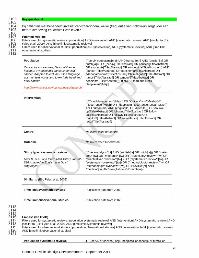
76 Concept Revisie Richtlijn Cervixcarcinoom - September 2011
Key question 4 3102 3103 Bij patiënten met behandeld invasief cervixcarcinoom, welke (frequentie van) follow-up zorgt voor een 3104 betere overleving en kwaliteit van leven? 3105 3106 Pubmed medline 3107 Filters used for systematic reviews: [population] AND [intervention] AND [systematic reviews] AND [similar to (Elit, 3108 Fyles et al. 2009)] AND [time limit systematic reviews] 3109 Filters used for observational studies: [population] AND [intervention] NOT [systematic reviews] AND [time limit 3110 observational studies] 3111 3112
Population
Cancer topic searches, National Cancer Institute; gynaecologic cancers: cervical cancer. Adapted to include Dutch language, abstract text words and to exclude head and neck cancer http://www.cancer.gov/cancertopics/litsearch
(((cervix neoplasms[majr] AND human[mh] AND (english[la] OR dutch[la])) OR ((cervix[Title/Abstract] OR cervical[Title/Abstract] OR exocervix[Title/Abstract] OR exocervical[Title/Abstract]) AND (cancer*[Title/Abstract] OR carcinoma*[Title/Abstract] OR adenocarcinoma*[Title/Abstract] OR malignan*[Title/Abstract] OR tumor*[Title/Abstract]] OR tumour*[Title/Abstract] OR neoplasm*[Title/Abstract])) )) NOT "Head and Neck Neoplasms"[Majr]
Intervention
(("Case Management"[Mesh] OR "Office Visits"[Mesh] OR "Recurrence"[Mesh] OR "Neoplasm Recurrence, Local"[Mesh]) AND human[mh] AND (english[la] OR dutch[la])) OR (follow-up[Title/Abstract] OR followup[Title/Abstract] OR follow up[Title/Abstract] OR follow$[Title/Abstract] OR monitor$[Title/Abstract] OR surveillance[Title/Abstract] OR recur[Title/Abstract])
Control
No filters used for control
Outcome
No filters used for outcome
Study type: systematic reviews Hunt D, et al. Ann Intern Med 1997;126:532-538 Adapted to English and Dutch languages
(("meta-analysis"[pt] AND (english[la] OR dutch[la])) OR "meta-anal*"[tw] OR "metaanal*"[tw] OR ("quantitativ* review*"[tw] OR "quantitative* overview*"[tw] ) OR ("systematic* review*"[tw] OR "systematic* overview*"[tw]) OR ("methodologic* review*"[tw] OR "methodologic* overview*"[tw]) OR ("review"[pt] AND "medline"[tw] AND (english[la] OR dutch[la]))
Similar to (Elit, Fyles et al. 2009)
Time limit systematic reviews
Publication date from 2001
Time limit observational studies
Publication date from 2007
3113 3114 3115 Embase (via OVID) 3116 Filters used for systematic reviews: [population systematic reviews] AND [intervention] AND [systematic reviews] AND 3117 [similar to (Elit, Fyles et al. 2009)] AND [time limit systematic reviews] 3118 Filters used for observational studies: [population observational studies] AND [intervention] NOT [systematic reviews] 3119 AND [time limit observational studies] 3120 3121
Population systematic reviews
1. ((cervix or cervical) adj5 (neoplas$ or cancer$ or tumo$ or

77 Concept Revisie Richtlijn Cervixcarcinoom - September 2011
KCE
metasta$ or malign$ or $carcin$)).mp. 2. exp Uterine Cervical Neoplasms/ 3. 1 or 2
Population observational studies Adapted to exclude head and neck cancer
1. ((cervix or cervical) adj5 (neoplas$ or cancer$ or tumo$ or
metasta$ or malign$ or $carcin$)).mp. 2. exp Uterine Cervical Neoplasms/ 3. 1 or 2 4. exp "head and neck tumor"/ or exp "head and neck
carcinoma"/ or exp "head and neck cancer"/ 5. 3 not 4
Intervention
1. Follow-Up Studies/ 2. follow-up.ti,ab. 3. followup.ti,ab. 4. follow up.ti,ab. 5. Follow$.ti,ab. 6. Monitor$.ti,ab. 7. surveillance.ti,ab. 8. Recur$.ti,ab. 9. exp tumor recurrence/ 10. exp recurrent disease/ 11. or/1-10 12. letter.pt. 13. editorial.pt 14. Case report.tw. 15. Or/12-14 16. 11 not 15
Control
No filters used for outcome
Study type: systematic reviews
1. exp Meta Analysis/ 2. ((meta adj analy$) or metaanalys$).tw. 3. (systematic adj (review$1 or overview$1)).tw. 4. or/1-3 5. cancerlit.ab. 6. cochrane.ab. 7. embase.ab. 8. (psychlit or psyclit).ab. 9. (psychinfo or psycinfo).ab. 10. (cinahl or cinhal).ab. 11. science citation index.ab. 12. bids.ab. 13. or/5-12 14. reference lists.ab. 15. bibliograph$.ab. 16. hand-search$.ab. 17. manual search$.ab. 18. relevant journals.ab. 19. or/14-18 20. data extraction.ab. 21. selection criteria.ab. 22. 20 or 21 23. review.pt. 24. 22 and 23 25. letter.pt. 26. editorial.pt. 27. animal/ 28. human/ 29. 27 not (27 and 28) 30. or/25-26,29 31. 4 or 13 or 19 or 24

78 Concept Revisie Richtlijn Cervixcarcinoom - September 2011
32. 31 not 30
Similar to (Elit, Fyles et al. 2009)
Time and language limit systematic reviews
limit [search] to (human and (dutch or english) and yr="2001 -Current")
Time and language limit observational studies
limit [search] to (human and (dutch or english) and yr="2007 -Current")
3122 CDSR (via The Cochrane Library) 3123 The Cochrane Database of Systematic Reviews was browsed by topic: 3124
a. cancer -> gynaecological-> cervix 3125 b. cancer -> gynaecological-> other 3126 c. cancer -> gynaecological-> supportive care 3127 d. cancer -> generic cancer care 3128 e. cancer -> palliative and supportive care -> organization of care 3129 f. cancer -> palliative and supportive care -> specific malignancies 3130
3131 3132 3133 Key question 4.1 3134 Is de behandeleffectiviteit hoger naarmate het recidief eerder wordt gediagnosticeerd? 3135 3136 Strategy 3137 Because of budget and time restrictions it was decided not to search for studies that might answer this key question 3138 directly. An indirect approach was used: Elit et al. reported eight studies that reported survival results separately for 3139 patiënts who were symptomatic versus those who were asymptomatic at the time of recurrence detection. Four of 3140 these studies reported survival for both asymptomatic and symptomatic patiënts, and were published from 1995 3141 onwards (table 4 in Elit et al. ). These four studies would be evaluated in more detail. 3142 3143 These four studies were: 3144
- (Bodurka-Bevers, Morris et al. 2000) 3145 - (Lim, Howells et al. 2004) 3146 - (Morice, Deyrolle et al. 2004) 3147 - (Sartori, Pasinetti et al. 2007) 3148
3149 Key question 4.2 3150 Welke diagnostiek is het meest geschikt om behandelbare nieuwe kankermanifestaties vroeg en accuraat te 3151 diagnosticeren? 3152 3153 This question was answered in part through key question 4. However, the search and selection there focused on 3154 diagnostic accuracy, which overemphasized technical diagnostics, over clinical interview and physical examination. 3155 Therefore, the ten most recent observational studies from the systematic review by Elit et al. were evaluated in more 3156 detail to extract information on physical examination&/medical history detected recurrence, symptomatic vs. 3157 asymptomatic recurrence, site of recurrence. 3158 3159 Table 1. Ten most recent studies from (Elit, Fyles et al. 2009) 3160 Reference In or excluded?
(Ansink, de Barros Lopes et al. 1996) Included
(Bodurka-Bevers, Morris et al. 2000) Included
(Duyn, Van Eijkeren et al. 2002) Included
(Esajas, Duk et al. 2001) Included
(Lim, Howells et al. 2004) Included
(Morice, Deyrolle et al. 2004) Included
(Rintala, Rantanen et al. 1997)
Excluded: does not contain information on the value of any diagnostic tool other than PAP smear
(Samlal, Van Der Velden et al. 1998) Included
(Sartori, Pasinetti et al. 2007) Included
(Zola, Fuso et al. 2007) Included
3161 Key question 5 3162
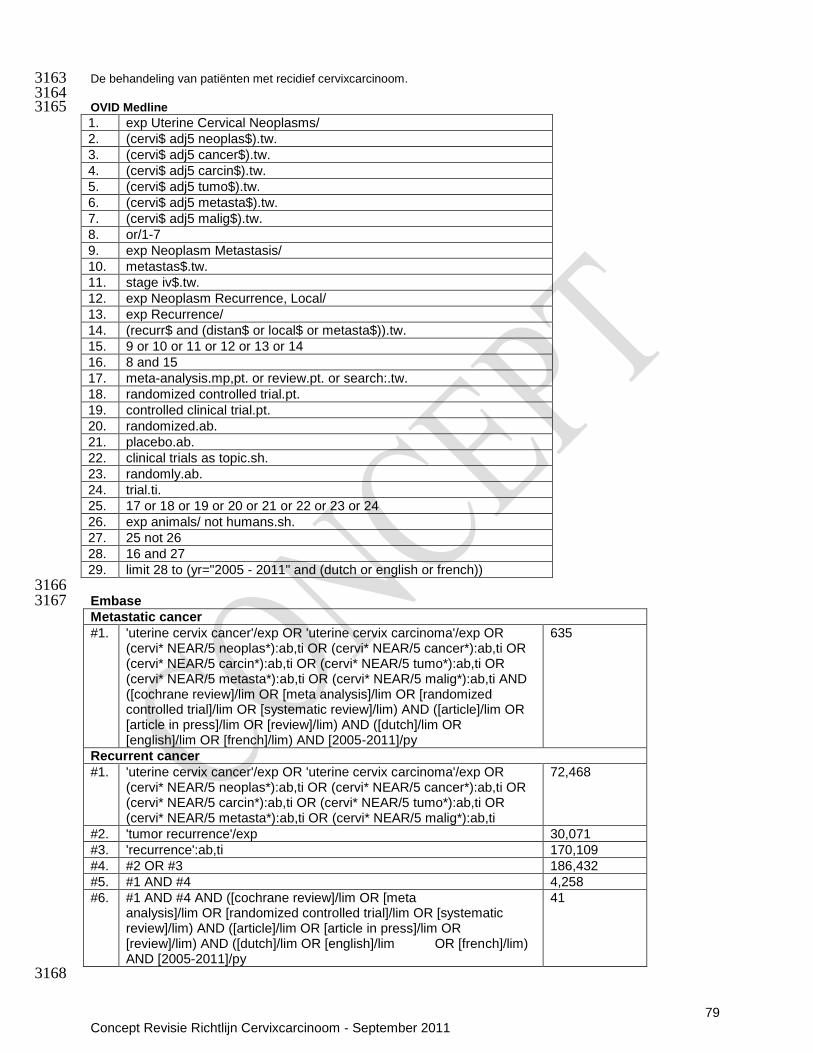
79 Concept Revisie Richtlijn Cervixcarcinoom - September 2011
De behandeling van patiënten met recidief cervixcarcinoom. 3163 3164 OVID Medline 3165 1. exp Uterine Cervical Neoplasms/
2. (cervi$ adj5 neoplas$).tw.
3. (cervi$ adj5 cancer$).tw.
4. (cervi$ adj5 carcin$).tw.
5. (cervi$ adj5 tumo$).tw.
6. (cervi$ adj5 metasta$).tw.
7. (cervi$ adj5 malig$).tw.
8. or/1-7
9. exp Neoplasm Metastasis/
10. metastas$.tw.
11. stage iv$.tw.
12. exp Neoplasm Recurrence, Local/
13. exp Recurrence/
14. (recurr$ and (distan$ or local$ or metasta$)).tw.
15. 9 or 10 or 11 or 12 or 13 or 14
16. 8 and 15
17. meta-analysis.mp,pt. or review.pt. or search:.tw.
18. randomized controlled trial.pt.
19. controlled clinical trial.pt.
20. randomized.ab.
21. placebo.ab.
22. clinical trials as topic.sh.
23. randomly.ab.
24. trial.ti.
25. 17 or 18 or 19 or 20 or 21 or 22 or 23 or 24
26. exp animals/ not humans.sh.
27. 25 not 26
28. 16 and 27
29. limit 28 to (yr="2005 - 2011" and (dutch or english or french))
3166 Embase 3167 Metastatic cancer
#1. 'uterine cervix cancer'/exp OR 'uterine cervix carcinoma'/exp OR (cervi* NEAR/5 neoplas*):ab,ti OR (cervi* NEAR/5 cancer*):ab,ti OR (cervi* NEAR/5 carcin*):ab,ti OR (cervi* NEAR/5 tumo*):ab,ti OR (cervi* NEAR/5 metasta*):ab,ti OR (cervi* NEAR/5 malig*):ab,ti AND ([cochrane review]/lim OR [meta analysis]/lim OR [randomized controlled trial]/lim OR [systematic review]/lim) AND ([article]/lim OR [article in press]/lim OR [review]/lim) AND ([dutch]/lim OR [english]/lim OR [french]/lim) AND [2005-2011]/py
635
Recurrent cancer
#1.
'uterine cervix cancer'/exp OR 'uterine cervix carcinoma'/exp OR (cervi* NEAR/5 neoplas*):ab,ti OR (cervi* NEAR/5 cancer*):ab,ti OR (cervi* NEAR/5 carcin*):ab,ti OR (cervi* NEAR/5 tumo*):ab,ti OR (cervi* NEAR/5 metasta*):ab,ti OR (cervi* NEAR/5 malig*):ab,ti
72,468
#2. 'tumor recurrence'/exp 30,071
#3. 'recurrence':ab,ti 170,109
#4. #2 OR #3 186,432
#5. #1 AND #4 4,258
#6. #1 AND #4 AND ([cochrane review]/lim OR [meta analysis]/lim OR [randomized controlled trial]/lim OR [systematic review]/lim) AND ([article]/lim OR [article in press]/lim OR [review]/lim) AND ([dutch]/lim OR [english]/lim OR [french]/lim) AND [2005-2011]/py
41
3168
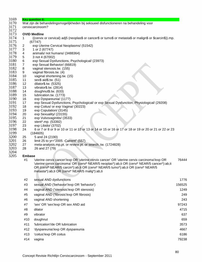
80 Concept Revisie Richtlijn Cervixcarcinoom - September 2011
Key question 6: 3169 Wat zijn de behandelingsmogelijkheden bij seksueel disfunctioneren na behandeling voor 3170 cervixcarcinoom? 3171 3172 OVID Medline 3173 1 ((cervix or cervical) adj5 (neoplas$ or cancer$ or tumo$ or metasta$ or malign$ or $carcin$)).mp. 3174
(67747) 3175 2 exp Uterine Cervical Neoplasms/ (51542) 3176 3 1 or 2 (67747) 3177 4 animals/ not humans/ (3488364) 3178 5 3 not 4 (67002) 3179 6 exp Sexual Dysfunctions, Psychological/ (23973) 3180 7 exp Sexual Behavior/ (66819) 3181 8 vaginal stenosis.tw. (155) 3182 9 vaginal fibrosis.tw. (4) 3183 10 vaginal shortening.tw. (15) 3184 11 sex$ aid$.tw. (51) 3185 12 dilator$.tw. (5325) 3186 13 vibrator$.tw. (2814) 3187 14 doughnut$.tw. (633) 3188 15 lubrication.tw. (1773) 3189 16 exp Dyspareunia/ (1177) 3190 17 exp Sexual Dysfunctions, Psychological/ or exp Sexual Dysfunction, Physiological/ (29208) 3191 18 exp Coitus/ or exp Vagina/ (30223) 3192 19 exp Copulation/ (3145) 3193 20 exp Sexuality/ (23155) 3194 21 exp Vulvovaginitis/ (3533) 3195 22 stent*.mp. (53392) 3196 23 exp Libido/ (3702) 3197 24 6 or 7 or 8 or 9 or 10 or 11 or 12 or 13 or 14 or 15 or 16 or 17 or 18 or 19 or 20 or 21 or 22 or 23 3198
(184605) 3199 25 5 and 24 (2190) 3200 26 limit 25 to yr="2005 -Current" (557) 3201 27 meta-analysis.mp,pt. or review.pt. or search:.tw. (1724828) 3202 28 26 and 27 (79) 3203 3204 Embase 3205 #1 'uterine cervix cancer'/exp OR 'uterine cervix cancer' OR 'uterine cervix carcinoma'/exp OR
'uterine cervix carcinoma' OR (cervi* NEAR/5 neoplas*):ab,ti OR (cervi* NEAR/5 cancer*):ab,ti OR (cervi* NEAR/5 carcin*):ab,ti OR (cervi* NEAR/5 tumo*):ab,ti OR (cervi* NEAR/5 metasta*):ab,ti OR (cervi* NEAR/5 malig*):ab,ti
76444
#2 sexual AND dysfunctions 1776
#3 sexual AND ('behavior'/exp OR 'behavior') 156525
#4 vaginal AND ('stenosis'/exp OR stenosis) 1249
#5 vaginal AND ('fibrosis'/exp OR fibrosis) 249
#6 vaginal AND shortening 243
#7 'sex' OR 'sex'/exp OR sex AND aid 97243
#8 dilator 4715
#9 vibrator 637
#10 doughnut 659
#11 'lubrication'/de OR lubrication 3573
#12 'dyspareunia'/exp OR dyspareunia 4667
#13 'coitus'/exp OR coitus 6186
#14 vagina 79238

81 Concept Revisie Richtlijn Cervixcarcinoom - September 2011
#15 copulation 4338
#16 'sexuality'/exp OR sexuality 138405
#17 vulvovaginitis 2128
#18 stent* 80679
#19 #2 OR #3 OR #4 OR #5 OR #6 OR #7 OR #8 OR #9 OR #10 OR #11 OR #12 OR #13 OR #14 OR #15 OR #16 OR #17 OR #18
444108
#20 #1 AND #19 9639
#21 #20 AND (2005:py OR 2006:py OR 2007:py OR 2008:py OR 2009:py OR 2010:py OR 2011:py)
3365
#22 #20 AND (2005:py OR 2006:py OR 2007:py OR 2008:py OR 2009:py OR 2010:py OR 2011:py) AND 'review'/it CDSR
MesH-term: Uterine Cervical Neoplasms/ 4 reviews, 3 retained
589

82 Concept Revisie Richtlijn Cervixcarcinoom - September 2011
Bijlage 10 Evidence tabellen
Vraag 1a Bij patiënten met invasief cervixcarcinoom, welke diagnostische techniek resulteert in de meest accurate stadiëring? Systematic reviews
Study ID Method Patiënt characteristics Intervention(s) Results primary outcome Results secondary and other outcomes
Critical appraisal of review quality
(Bipat, Glas et al. 2003)
Design: systematic review with meta-analysis Funding: not reported on Search date:1985-2002 Searched databases: Medline and Embase Included study designs: not reported on 57 included studies
Eligibility criteria: English or German language studies; sample size ≥10 patiënts; histopathology as reference standard; sufficient data to construct a 2x2 contingency table Exclusion criteria: data reported elsewhere in more detail Patiënt characteristics: cervical carcinoma
Index test: CT, MRI or both Reference standard: histopathology
Meta-analysed (N=9) accuracy of CT to detect parametrial invasion: Se 55% (95%CI 44-66); Sp 76% (visual inspection forest plot) Meta-analysed (N=52) accuracy of MRI to detect parametrial invasion: Se 74% (95%CI 68-79); Sp 85% (visual inspection forest plot) Meta-analysed (N=3) accuracy of CT to detect bladder invasion: Se 64% (95%CI 39-82); Sp 73% (95%CI 52-87) Meta-analysed (N=16) accuracy of MRI to detect bladder invasion: Se 75% (95%CI 66-83); Sp 91% (95%CI 83-95) Meta-analysed (N=2) accuracy of CT to detect rectum invasion: Se 45% (95%CI 20-73); Sp 94% (visual inspection forest plot) Meta-analysed (N=9) accuracy of MRI to detect rectum invasion: Se 71% (95%CI 53-83); Sp 83% (visual inspection forest plot)
The sensitivity for parametrial invasion by MRI was significant higher compared with CT (P=0.0027) The sensitivity for bladder invasion and rectum invasion by MRI were higher compared with CT but these differences were not statistically significant The sensitivity for lymph node involvement by MRI was significantly higher compared to CT (P=0.047) Subgroup analyses for methodological criteria, coil usage, T1 vs. T2, type of magnetic field, year of publication or sample size did not reveal differences in accuracy. No data on cervical angulation technique
Level of evidence: B 30 prospective studies, 14 retrospective studies and in 13 studies data collection was unknown Blinded assessment in 29 studies; non blinded assessment in 28 studies Complete verification in 43 studies; partial verification in 14 studies Characteristics from the original studies were not described; clinical heterogeneity unknown Statistical heterogeneity partially taken into account Included studies: Primary studies: 53 studies published before 2001 Studies published from 2001 onwards: (Narayan, Hicks et al. 2001) (Reinhardt, Ehritt-Braun et al. 2001) (Sheu, Chang et al. 2001) (Wang, Wong et al. 2001)

83 Concept Revisie Richtlijn Cervixcarcinoom - September 2011
Study ID Method Patiënt characteristics Intervention(s) Results primary outcome Results secondary and other outcomes
Critical appraisal of review quality
Meta-analysed (N=17) accuracy of CT to detect lymph node (unspecified) metastasis: Se 43% (95%CI 37-57); Sp 94% (visual inspection forest plot) Meta-analysed (N=25) accuracy of MRI to detect lymph node (unspecified) metastasis: Se 60% (95%CI 52-68); Sp 93% (visual inspection forest plot)
(Havrilesky, Kulasingam et al. 2005)
Design: systematic review with meta-analysis Funding: Centers for Medicare and Medicaid Services Search date:1966-2003 Searched databases: Medline Included study designs: observational studies
Eligibility criteria: English language studies reporting primary data and published in a peer review journal with 12 or more included patiënts Patiënt characteristics: newly diagnosed cervical cancer
Index test: CT, MRI or PET Reference standard: histology or follow-up
Meta-analysed (N=2) accuracy of CT to detect pelvic lymph node metastasis (reference: histology or follow-up): Se 47% (95%CI: 21–73), Sp not enough data to calculate Meta-analysed (N=2) accuracy of MRI to detect pelvic lymph node metastasis (reference: histology or follow-up): Se 72% (95%CI: 53–87), Sp 96% (95%CI: 92–98) Meta-analysed (N=4) accuracy of PET to detect pelvic lymph node metastasis (reference: histology or follow-up): Se 79% (95%CI: 65–90), Sp 99 (95%CI: 96–99) Single study accuracy of MRI to detect para-aortic lymph node metastasis (reference: histology): Se 67% (95%CI: 9-99), Sp 100%
No data on T1 vs. T2 weighted or contrast enhanced MRI
Level of evidence: B Included studies were small (none included over 50 patiënts) and none reported blinded assessment Three studies were in selected subgroups, e.g. patiënts had to have negative MRI or CT findings Two studies used differential verification (histology or follow-up) Statistical heterogeneity was not assessed and there was clinical heterogeneity Included studies: 15 Primary studies:
11 studies published before 2001 (Belhocine, Thille et al. 2002) (Lin, Hung et al. 2003) (Reinhardt, Ehritt-Braun et
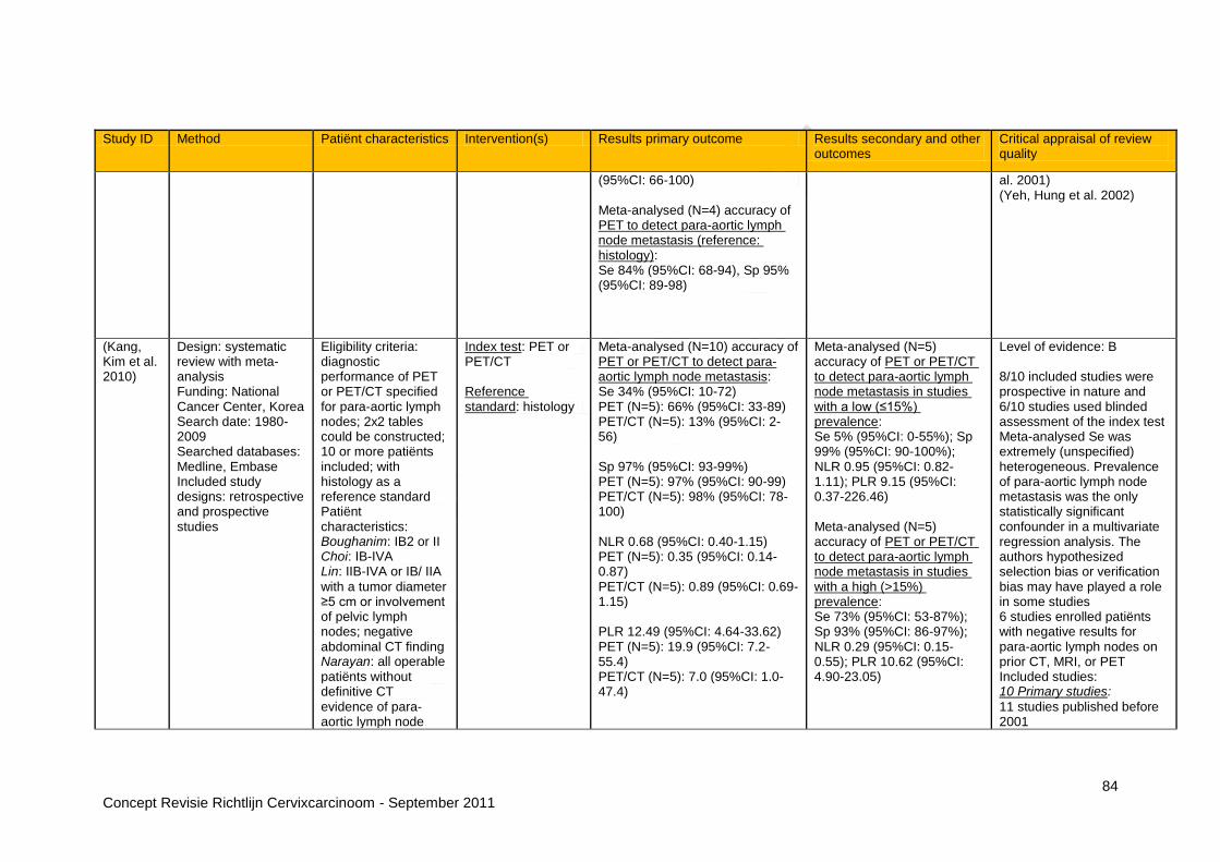
84 Concept Revisie Richtlijn Cervixcarcinoom - September 2011
Study ID Method Patiënt characteristics Intervention(s) Results primary outcome Results secondary and other outcomes
Critical appraisal of review quality
(95%CI: 66-100) Meta-analysed (N=4) accuracy of PET to detect para-aortic lymph node metastasis (reference: histology): Se 84% (95%CI: 68-94), Sp 95% (95%CI: 89-98)
al. 2001) (Yeh, Hung et al. 2002)
(Kang, Kim et al. 2010)
Design: systematic review with meta-analysis Funding: National Cancer Center, Korea Search date: 1980-2009 Searched databases: Medline, Embase Included study designs: retrospective and prospective studies
Eligibility criteria: diagnostic performance of PET or PET/CT specified for para-aortic lymph nodes; 2x2 tables could be constructed; 10 or more patiënts included; with histology as a reference standard Patiënt characteristics: Boughanim: IB2 or II Choi: IB-IVA Lin: IIB-IVA or IB/ IIA
with a tumor diameter ≥5 cm or involvement of pelvic lymph nodes; negative abdominal CT finding Narayan: all operable patiënts without definitive CT evidence of para-aortic lymph node
Index test: PET or PET/CT Reference standard: histology
Meta-analysed (N=10) accuracy of PET or PET/CT to detect para-aortic lymph node metastasis: Se 34% (95%CI: 10-72) PET (N=5): 66% (95%CI: 33-89) PET/CT (N=5): 13% (95%CI: 2-56) Sp 97% (95%CI: 93-99%) PET (N=5): 97% (95%CI: 90-99) PET/CT (N=5): 98% (95%CI: 78-100) NLR 0.68 (95%CI: 0.40-1.15) PET (N=5): 0.35 (95%CI: 0.14-0.87) PET/CT (N=5): 0.89 (95%CI: 0.69-1.15) PLR 12.49 (95%CI: 4.64-33.62) PET (N=5): 19.9 (95%CI: 7.2-55.4) PET/CT (N=5): 7.0 (95%CI: 1.0-47.4)
Meta-analysed (N=5) accuracy of PET or PET/CT to detect para-aortic lymph node metastasis in studies with a low (≤15%) prevalence: Se 5% (95%CI: 0-55%); Sp 99% (95%CI: 90-100%); NLR 0.95 (95%CI: 0.82-1.11); PLR 9.15 (95%CI: 0.37-226.46) Meta-analysed (N=5) accuracy of PET or PET/CT to detect para-aortic lymph node metastasis in studies with a high (>15%) prevalence: Se 73% (95%CI: 53-87%); Sp 93% (95%CI: 86-97%); NLR 0.29 (95%CI: 0.15-0.55); PLR 10.62 (95%CI: 4.90-23.05)
Level of evidence: B 8/10 included studies were prospective in nature and 6/10 studies used blinded assessment of the index test Meta-analysed Se was extremely (unspecified) heterogeneous. Prevalence of para-aortic lymph node metastasis was the only statistically significant confounder in a multivariate regression analysis. The authors hypothesized selection bias or verification bias may have played a role in some studies 6 studies enrolled patiënts with negative results for para-aortic lymph nodes on prior CT, MRI, or PET Included studies: 10 Primary studies:
11 studies published before 2001

85 Concept Revisie Richtlijn Cervixcarcinoom - September 2011
Study ID Method Patiënt characteristics Intervention(s) Results primary outcome Results secondary and other outcomes
Critical appraisal of review quality
metastasis Reinhardt: cervical cancer patiënts Roh:IB-IVA Vergote: IB2-IIIB
cervical cancer without para-aortic lymph node metastasis on PET and CT or PET/CT Wright: IA2-IIA Yildirim: locally advanced cervical cancer with negative CT findings for para-aortic lymph node metastasis Disease prevalence: a meta-analysed 14.2% of patiënts across all studies had para-aortic lymph node metastases
(Boughanim, Leboulleux et al. 2008) (Choi, Roh et al. 2006) (Lin, Hung et al. 2003) (Narayan, Hicks et al. 2001) (Reinhardt, Ehritt-Braun et al. 2001) (Roh, Seo et al. 2005) (Vergote, Tsolakidis et al. 2008) (Wright, Dehdashti et al. 2005) (Yildirim, Sehirali et al. 2008)
(van de Lande, Torrenga et al. 2007)
Design: systematic review with meta-analysis Funding: not reported on Search date: July 2006 Searched databases: Medline, Embase Included study designs: SNB studies
Eligibility criteria: majority (N80%) of included patiënts with early stage cervical cancer (FIGO I-IIA); English language studies; ≥ 10 patiënts included; sufficient data to reconstruct 2x2 tables Patiënt characteristics: see eligibility
Index test: SNB (Technetium, blue dye or both) Reference standard: histology
Meta-analysed (N=21) accuracy of SNB to detect lymph node metastasis: Se 89% (95%CI: 83-94) Technetium (N=5) 92% (95%CI: 79-98) Blue dye (N=4) 81% (95%CI: 67-92) Both (N=12) 92% (95%CI: 84-98)
Sentinel node detection rate: Technetium (N=7) 88% (95%CI: 82-92) Blue dye (N=5) 84% (95%CI: 79-89) Both (N=13) 97% (95%CI: 95-98)
Level of evidence: B Study quality: none of the studies used masked assessment of the reference standard; 19/22 studies were prospective; 17/22 studies used consecutive patiënts Meagre description of patiënt characteristics Included studies: Primary studies: 2 studies published before 2001

86 Concept Revisie Richtlijn Cervixcarcinoom - September 2011
Study ID Method Patiënt characteristics Intervention(s) Results primary outcome Results secondary and other outcomes
Critical appraisal of review quality
(Angioli, Palaia et al. 2005) (Barranger, Cortez et al. 2004) (Chung, Kim et al. 2003) (Di Stefano, Acquaviva et al. 2005) (Gil-Moreno, Diaz-Feijoo et al. 2005) (Hubalewska, Sowa-Staszczak et al. 2003) (Lambaudie, Collinet et al. 2003) (Lantzsch, Wolters et al. 2001) (Levenback, Coleman et al. 2002) (Li, Zhang et al. 2004) (Lin, Tzeng et al. 2005) (Malur, Krause et al. 2001) (Marchiole, Buenerd et al. 2004) (Martinez-Palones, Gil-Moreno et al. 2004) (Niikura, Okamura et al. 2004) (Pijpers, Buist et al. 2004) (Rhim, Park et al. 2002) (Rob, Strnad et al. 2005) (Roca, Caresia et al. 2005) (Silva, Silva-Filho et al. 2005) (van Dam, Hauspy et al. 2003)
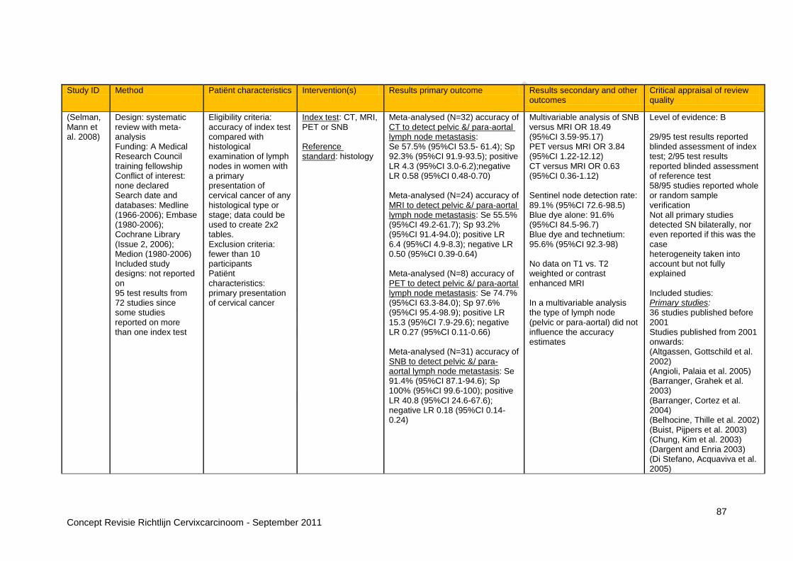
87 Concept Revisie Richtlijn Cervixcarcinoom - September 2011
Study ID Method Patiënt characteristics Intervention(s) Results primary outcome Results secondary and other outcomes
Critical appraisal of review quality
(Selman, Mann et al. 2008)
Design: systematic review with meta-analysis Funding: A Medical Research Council training fellowship Conflict of interest: none declared Search date and databases: Medline (1966-2006); Embase (1980-2006); Cochrane Library (Issue 2, 2006); Medion (1980-2006) Included study designs: not reported on 95 test results from 72 studies since some studies reported on more than one index test
Eligibility criteria: accuracy of index test compared with histological examination of lymph nodes in women with a primary presentation of cervical cancer of any histological type or stage; data could be used to create 2x2 tables. Exclusion criteria: fewer than 10 participants Patiënt characteristics: primary presentation of cervical cancer
Index test: CT, MRI, PET or SNB Reference standard: histology
Meta-analysed (N=32) accuracy of CT to detect pelvic &/ para-aortal lymph node metastasis: Se 57.5% (95%CI 53.5- 61.4); Sp 92.3% (95%CI 91.9-93.5); positive LR 4.3 (95%CI 3.0-6.2);negative LR 0.58 (95%CI 0.48-0.70) Meta-analysed (N=24) accuracy of MRI to detect pelvic &/ para-aortal lymph node metastasis: Se 55.5% (95%CI 49.2-61.7); Sp 93.2% (95%CI 91.4-94.0); positive LR 6.4 (95%CI 4.9-8.3); negative LR 0.50 (95%CI 0.39-0.64) Meta-analysed (N=8) accuracy of PET to detect pelvic &/ para-aortal lymph node metastasis: Se 74.7% (95%CI 63.3-84.0); Sp 97.6% (95%CI 95.4-98.9); positive LR 15.3 (95%CI 7.9-29.6); negative LR 0.27 (95%CI 0.11-0.66) Meta-analysed (N=31) accuracy of SNB to detect pelvic &/ para-aortal lymph node metastasis: Se 91.4% (95%CI 87.1-94.6); Sp 100% (95%CI 99.6-100); positive LR 40.8 (95%CI 24.6-67.6); negative LR 0.18 (95%CI 0.14-0.24)
Multivariable analysis of SNB versus MRI OR 18.49 (95%CI 3.59-95.17) PET versus MRI OR 3.84 (95%CI 1.22-12.12) CT versus MRI OR 0.63 (95%CI 0.36-1.12) Sentinel node detection rate: 89.1% (95%CI 72.6-98.5) Blue dye alone: 91.6% (95%CI 84.5-96.7) Blue dye and technetium: 95.6% (95%CI 92.3-98) No data on T1 vs. T2 weighted or contrast enhanced MRI In a multivariable analysis the type of lymph node (pelvic or para-aortal) did not influence the accuracy estimates
Level of evidence: B 29/95 test results reported blinded assessment of index test; 2/95 test results reported blinded assessment of reference test 58/95 studies reported whole or random sample verification Not all primary studies detected SN bilaterally, nor even reported if this was the case heterogeneity taken into account but not fully explained Included studies: Primary studies: 36 studies published before 2001 Studies published from 2001 onwards: (Altgassen, Gottschild et al. 2002) (Angioli, Palaia et al. 2005) (Barranger, Grahek et al. 2003) (Barranger, Cortez et al. 2004) (Belhocine, Thille et al. 2002) (Buist, Pijpers et al. 2003) (Chung, Kim et al. 2003) (Dargent and Enria 2003) (Di Stefano, Acquaviva et al. 2005)

88 Concept Revisie Richtlijn Cervixcarcinoom - September 2011
Study ID Method Patiënt characteristics Intervention(s) Results primary outcome Results secondary and other outcomes
Critical appraisal of review quality
(Gil-Moreno, Diaz-Feijoo et al. 2005) (Hertel, Kohler et al. 2002) (Hubalewska, Sowa-Staszczak et al. 2003) (Kokka, Vorgias et al. 2003) (Kuhnel, Horn et al. 2001) (Lambaudie, Collinet et al. 2003) (Lantzsch, Wolters et al. 2001) (Levenback, Coleman et al. 2002) (Li, Zhang et al. 2004) (Lin, Hung et al. 2003) (Lin, Tzeng et al. 2005) (Malur, Krause et al. 2001) (Marchiole, Buenerd et al. 2004) (Martinez-Palones, Gil-Moreno et al. 2004) (Niikura, Okamura et al. 2004) (Ozsarlak, Tjalma et al. 2003) (Pijpers, Buist et al. 2004) (Reinhardt, Ehritt-Braun et al. 2001) (Rhim, Park et al. 2002) (Rob, Strnad et al. 2005) (Roh, Seo et al. 2005) (Silva, Silva-Filho et al. 2005) (van Dam, Hauspy et al. 2003) (Vorgias, Katsoulis et al. 2002)

89 Concept Revisie Richtlijn Cervixcarcinoom - September 2011
Study ID Method Patiënt characteristics Intervention(s) Results primary outcome Results secondary and other outcomes
Critical appraisal of review quality
(Wang, Wong et al. 2001) (Wang, Sun et al. 2004) (Yeh, Hung et al. 2002)
Abbreviations: CI: confidence interval; NLR: negative likelihood ratio; NPV: negative predictive value; PLR: positive likelihood ratio; PPV: positive predictive value; Se: sensitivity, SE: spin echo; SN: sentinel node; SNB: sentinel node biopsy; Sp: specificity; y: year Observational studies
Study ID Method Patiënt characteristics Intervention(s) Results primary outcome
Results secondary and other outcomes
Critical appraisal of study quality
(Akata, Kerimoglu et al. 2005)
Design: prospective cohort study Source of funding: not reported on Conflict of interest: not reported on Setting: University School of Medicine, Ankara, Turkey Sample size: N=28 Duration: not reported on
Eligibility criteria: referred for MRI after histological confirmation of cervical cancer; able to receive vaginal contrast medium; surgical or pathological staging Patiënt characteristics: mean age 53.4 y; stage IA:4%; IB:43%; IIA:18%; IIB:29%; IIIB:4%; IVB:4%
Index tests: MRI without and with vaginal opacification Reference standard: histopathology
Accuracy of MRI without vaginal opacification to detect stage I: Se 54%; Sp 60%; PPV 54%; NPV 60% Accuracy of MRI without vaginal opacification to detect ≥ stage IIA: Se 67% Accuracy of MRI with vaginal opacification to detect stage I: Se 100% Accuracy of MRI with vaginal opacification to detect ≥ stage IIA: Se
Level of evidence: B Dropouts: none reported Consecutive patiënts Exclusion of 20 patiënts who did not undergo surgery: partial verification bias exclusion of 2 patiënts who could not receive vaginal contrast medium Blinded assessment of index test; blinded assessment of reference test not reported on
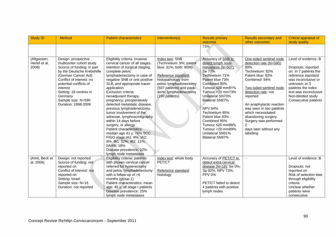
90 Concept Revisie Richtlijn Cervixcarcinoom - September 2011
Study ID Method Patiënt characteristics Intervention(s) Results primary outcome
Results secondary and other outcomes
Critical appraisal of study quality
73%
(Altgassen, Hertel et al. 2008)
Design: prospective multicenter cohort study Source of funding: in part by the Deutsche Krebshilfe (German Cancer Aid) Conflict of interest: no potential conflicts of interest Setting: 18 centres in Germany Sample size: N=590 Duration: 1998-2006
Eligibility criteria: invasive cervical cancer of all stages, intention of surgical staging, complete pelvic lymphadenectomy in case of negative SNB or one positive SLB, and appropriate tracer application. Exclusion criteria: neoadjuvant therapy, pregnancy, preoperatively detected metastatic disease, previous lymphadenectomy, tumor involvement of the adnexae, lymphoscintigraphy within 14 days before surgery, or allergy Patiënt characteristics: median age 41 y; 76% SCC. FIGO stage IA1: 8%; IA2: 8%; IB1: 52%; IB2: 11%; IIA/IIB: 18% Disease prevalence: 22% lymph node metastases
Index test: SNB (Technetium: 9%; patent blue: 31%; both: 60%) Reference standard: histopathology from pelvic lymphadenectomy (507 patiënts) and para-aortic lymphadenectomy (190 patiënts)
Accuracy of SNB to detect lymph node metastasis (N=507): Se 77% Technetium 71% Patent blue 73% Combined 80% Tumour ≤20 mm91% Tumour >20 mm73% Unilateral SN70% Bilateral SN87% NPV 94% Technetium 95% Patent blue 93% Combined 95% Tumour ≤20 mm99% Tumour >20 mm89% Unilateral SN91% Bilateral SN97%
One-sided sentinel node detection rate (N=590): 89% Technetium: 82% Patent blue: 82% Combined: 94% Two-sided sentinel node detection rate: not reported An anaphylactic reaction was seen in two patiënts which necessitated abandoning surgery. Surgery was performed 2 days later without any labelling
Level of evidence: B Dropouts: reported on. In 7 patiënts the reference standard was inconclusive or unknown; in 3 patiënts the index test was inconclusive Non-blinded study Consecutive patiënts
(Amit, Beck et al. 2006)
Design: not reported Source of funding: not reported on Conflict of interest: not reported on Setting: Israel Sample size: N=16 Duration: not reported
Eligibility criteria: patiënts with proven cervical cancer referred for hysterectomy and pelvic lymphadenectomy with a follow-up of >6 months (group 1) Patiënt characteristics: mean age: 45 y; all stage I patiënts Disease prevalence: 25% lymph node metastases
Index test: whole body PET/CT Reference standard: histology
Accuracy of PET/CT to detect extra cervical disease (N=16): Se 0%; Sp 92%, NPV 73%; PPV 0% PET/CT failed to detect 4 patiënts with positive lymph nodes
- Level of evidence: B Dropouts: not reported on Risk of selection bias through eligibility criteria Unclear whether patiënts were consecutive

91 Concept Revisie Richtlijn Cervixcarcinoom - September 2011
Study ID Method Patiënt characteristics Intervention(s) Results primary outcome
Results secondary and other outcomes
Critical appraisal of study quality
Blinded assessment not reported
(Bentivegna, Uzan et al. 2010)
Design: retrospective cohort study Source of funding: not reported on Conflict of interest: not reported on Setting: Gustave Roussy Institute, Vullejuif, University Paris Sud, France Sample size: N= 16 Duration: 2005-2008
Eligibility criteria: early-stage (<4 cm) cervical cancer (stage IB1) with MRI and PET/CT imaging before surgery including at least a pelvic lymphadenectomy Patiënt characteristics: median age 43 y; 61% SCC; 17% AC; 17% ASC; 14 patiënts underwent preoperative utero-vaginal brachytherapy; 2 patiënts underwent upfront surgery Prevalence of disease: 12.5% lymph node metastasis
Index tests: PET/CT Reference standard: histopathology from pelvic lymphadenectomy (N=16) and para-aortic lymphadenectomy (N=1)
Accuracy of PET/CT to detect lymph node metastasis bilateral (N=16): Se 0%, NPV 88%
- Level of evidence: B Consecutive patiënts Blinded assessment not reported on Long interval between index and reference test in which 14/16 patiënts had brachytherapy Selection bias through eligibility criteria
(Bjurberg, Kjellen et al. 2007)
Design: prospective study Source of funding: Berta Kamprad‟s Foundation for Research and Treatment of Cancer, Gunnar Nilsson‟s Cancer Foundation, The Donations Fund of Lund University Hospital and The Swedish Cancer Foundation Conflict of interest: not reported on Setting: Lund University Hospital, Sweden Sample size: N=17 Duration: 2004-2006
Eligibility criteria: locally advanced cervical cancer FIGO stage IB2-IVB scheduled for radical radiotherapy , with or without concomitant cisplatin, with curative intent (group 2 in article) Patiënt characteristics: mean age 56 y; 82% SCC; 12% AC; 6% ASC. FIGO stage IB2: 6%; stage IIA: 6%; stage IIB: 71%; stage IIIB: 6%; stage IVA: 12% Disease prevalence: 29% metastases not detected during routine work-up
Index test: PET Reference standard: histology
Accuracy of PET to detect metastasis not detected by a routine staging procedure, including CT and MRI (N=17): Se 83%; Sp 100%; NPV 92%; PPV 100%
- Level of evidence: B Dropouts: none Consecutive patiënts Blinded assessment not reported The one false negative finding was detected by CT 5 weeks after PET (and then confirmed by histology)
(Chao, Ho et al. 2008)
Design: prospective cohort study
Eligibility criteria: newly diagnosed SCC cervical
Index test: PET (N=38) or PET/CT (N=9)
Accuracy of PET or PET/CT to detect para-
PET or PET/CT had positive clinical impact
Level of evidence: B

92 Concept Revisie Richtlijn Cervixcarcinoom - September 2011
Study ID Method Patiënt characteristics Intervention(s) Results primary outcome
Results secondary and other outcomes
Critical appraisal of study quality
Source of funding: research grants from Chang Gung Memorial Hospital Conflict of interest: no conflicts of interest to declare Setting: Chang Gung Memorial Hospital, Taiwan Sample size: N=47 Duration: 2001-2007
cancer patiënts with a suspicion of para-aortic lymph node metastasis based on CT/MRI, or inguinal or supraclavicular lymph node metastasis based on palpation or CT/MR; scheduled for curative-intent treatment such as concurrent chemoradiation or surgery Exclusion criteria: no previous cytotoxic therapy; concomitant or a past history of malignancy; histology-proven metastasis to pleura, peritoneum, mediastinal lymph node, lung, bone or liver parenchyma; malignant ascites or pleural effusion; intolerable to extended field irradiation Patiënt characteristics: mean age 55 y; FIGO stage I/IIA: 17%; IIB/IV: 83% Prevalence of disease: para-aortic lymph node metastasis: 79%; inguinal lymph node metastasis: 11%; supraclavicular lymph node metastasis: 26%; bone metastasis: 2%; other distant non skeletal sites: 4%
Reference standard: CT/MRI, biopsy or follow-up through imaging including PET
aortal lymph node metastasis (N=47): Se 97%; Sp 90%; NPV 90%; PPV 97% Accuracy of PET or PET/CT to detect inguinal lymph node metastasis (N=47): Se 80%; Sp 86%; NPV 97%; PPV 40% Accuracy of PET or PET/CT to detect supraclavicular lymph node metastasis (N=47): Se 85%; Sp 100%; NPV 94%; PPV 100% Accuracy of PET or PET/CT to detect bone metastasis (N=47): Se 100%; Sp 98%; NPV 100%; PPV 50% Accuracy of PET or PET/CT to detect other distant non skeletal sites (N=47): Se 100%; Sp 91%; NPV 100%; PPV 33%
in 21 of the 47 study patiënts, in 23 it had no impact, and in three it had negative impact. Positive impact included disclosing additional curable sites (n=8), down-staging (n=6), offering metabolic biopsy (n=4) or change to palliation (n=3)
Dropouts: not reported Unclear whether patiënts were consecutive Only patiënts with suspected metastasis to para-aortal, inguinal or supraclavicular lymph nodes included Blinded assessment not reported on Differential verification Incorporation bias Indeterminate PET or PET/CT positive findings were tentatively included with the false positives, as indeterminate lesions were defined as ´a biopsy of the lesion of interest was not feasible or yielded a negative result and a second assessment of both CT-MRI and PET imaging showed persistent regression or remission of the lesion after definitive treatment´
(Choi, Kim et Design: prospective cohort Eligibility criteria: Index test: MRI Accuracy of MRI to Region-based analysis Level of evidence: B

93 Concept Revisie Richtlijn Cervixcarcinoom - September 2011
Study ID Method Patiënt characteristics Intervention(s) Results primary outcome
Results secondary and other outcomes
Critical appraisal of study quality
al. 2004)
study Source of funding: 2001 BK21 Project for Medicine, Dentistry, and Pharmacie Conflict of interest: not reported on Setting: Seoul National University Hospital, Seoul, Korea Sample size: N=115 Duration: January 2000 - June 2003
cervical carcinoma with proven histopathological staging and operable state Patiënt characteristics: mean age 52.3 y Prevalence of disease: 20.2% vaginal invasion
Reference standard: histopathology
detect vaginal invasion (N=114): Se 87%; Sp79%; PPV 51%; NPV 96%
of parametria and pelvic lymph node metastasis
Dropouts: 1 patiënt didn‟t undergo surgery due to confirmation of bladder invasion by cystoscopy Consecutive patiënts Exclusion of patiënts that did not have histopathological confirmation: partial verification bias Blinded assessment of index test; blinded assessment of reference test not reported on
(Choi, Roh et al. 2006)
Design: prospective cohort study Source of funding: National Cancer Center Conflict of interest: not reported on Setting: Research Institute and Hospital, National Cancer Center, Goyang. Korea Sample size: N=22 Duration: 2003-2005
Eligibility criteria: untreated patiënts with histopathologically confirmed FIGO stage IB-IVA invasive cervical carcinoma as determined by conventional workup that included MRI and PET/CT scans; no contraindications to the surgical procedure; no evidence of distant metastases; Eastern Cooperative Oncology Group performance status of 0-1 Exclusion criteria: patiënts who did not want to undergo PET/CT or laparoscopic lymphadenectomy (N=63) and tumours other than SCC
Index tests: MRI (T2 weighted, contrast enhanced) and PET/CT Reference standard: histopathology
Accuracy of MRI to detect PALN + pelvic lymph node metastasis (N=22): Se 39%; Sp 44%; PPV 50%; NPV 33% Accuracy 41% Accuracy of PET/CT to detect PALN + pelvic lymph node metastasis (N=22): Se 77%; Sp 56%; PPV 71%; NPV 63%; Accuracy 68%
- Level of evidence: B Dropouts: none reported Consecutive patiënts Selective subgroup: high prevalence of lymph node metastasis Blinded assessment of index and reference test

94 Concept Revisie Richtlijn Cervixcarcinoom - September 2011
Study ID Method Patiënt characteristics Intervention(s) Results primary outcome
Results secondary and other outcomes
Critical appraisal of study quality
(N=10) Patiënt characteristics: mean age 50 y; 100% SCC Prevalence of disease: 59% lymph nodes metastasis
(Chou, Chang et al. 2006)
Design: prospective study Source of funding: National Science Council and the Institute of Nuclear Energy Research, Chang Gung Memorial Hospital Conflict of interest: no conflicts of interest to declare Setting: Chang Gung Memorial Hospital, Taiwan Sample size: N=60 Duration: not reported
Eligibility criteria: cervical cancer patiënts scheduled for radical hysterectomy and pelvic lymphadenectomy; SCC≤ 4 cm by MRI or AD or ASC of any size; age 18-70 y Exclusion criteria: small-cell carcinoma; MRI showed suspicious lymph nodes; histologically proven metastasis to lymph nodes; ever received radiotherapy and/or chemotherapy for cervical cancer; history of allergy to the radiotracer; a previous diagnosis of cancer other than non-melanoma skin cancer; pregnancy Patiënt characteristics: median age: 48 y; 60% SCC; 33% AC; 7% ASC. FIGO staging: IA2: 2%; IB1: 90%; IB2: 5%; IIA:3% Disease prevalence: 18% lymph node metastases
Index test: PET Reference standard: histology
Accuracy of PET to detect pelvic lymph node metastasis not detected by MRI (N=60): Se 10%, Sp 94%, NPV 84%, PPV 25%
- Level of evidence: B No dropouts Unclear if patiënts were consecutive Blinded assessment not reported Selective subgroup of MRI negative patiënts
(Chung, Kang et al. 2007)
Design: retrospective cohort study Source of funding: Korea Health 21 R&D Project, Ministry of Health & Welfare, Republic of Korea Conflict of interest: none
Eligibility criteria: histologically confirmed invasive carcinoma of the uterine cervix; FIGO stage IA, IB, IIA or IIB; no medical or surgical contra-indications to the primary treatment of
Index tests: MRI (T2 weighted, no contrast enhancement) Reference standard: histology
Accuracy of MRI to detect parametrial invasion (N=119): Se 100%; Sp 89%; PPV 62%; NPV 100%; accuracy 91%;
Cervical angulation not reported
Level of evidence: B Consecutive patiënts Blinded assessment of index test; blinded assessment of reference test not
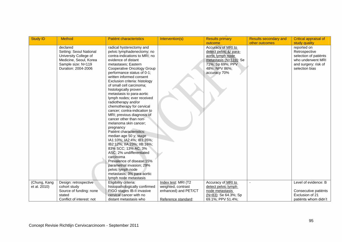
95 Concept Revisie Richtlijn Cervixcarcinoom - September 2011
Study ID Method Patiënt characteristics Intervention(s) Results primary outcome
Results secondary and other outcomes
Critical appraisal of study quality
declared Setting: Seoul National University College of Medicine, Seoul, Korea Sample size: N=119 Duration: 2004-2006
radical hysterectomy and pelvic lymphadenectomy; no contra-indications to MRI; no evidence of distant metastases; Eastern Cooperative Oncology Group performance status of 0-1; written informed consent Exclusion criteria: histology of small cell carcinoma; histologically proven metastasis to para-aortic lymph nodes; ever received radiotherapy and/or chemotherapy for cervical cancer; contra-indication to MRI; previous diagnosis of cancer other than non-melanoma skin cancer; pregnancy Patiënt characteristics: median age 50 y; stage IA1:10%; IA2:4%; IB1:35%; IB2:12%; IIA:23%; IIB:16%; 83% SCC; 13% AC; 3% ASC; 2% undifferentiated carcinoma Prevalence of disease:15% parametrial invasion; 29% pelvic lymph node metastasis; 3% para-aortic lymph node metastasis
Accuracy of MRI to detect pelvic &/ para-aortic lymph node metastasis (N=119): Se 71%; Sp 69%; PPV 48%; NPV 86%; accuracy 70%
reported on Retrospective selection of patiënts who underwent MRI and surgery: risk of selection bias
(Chung, Kang et al. 2010)
Design: retrospective cohort study Source of funding: none stated Conflict of interest: not
Eligibility criteria: histopathologically confirmed FIGO stages IB-II invasive cervical cancer with no distant metastasis who
Index test: MRI (T2 weighted, contrast enhanced) and PET/CT Reference standard:
Accuracy of MRI to detect pelvic lymph node metastasis (N=83): Se 64.3%; Sp 69.1%; PPV 51.4%;
- Level of evidence: B Consecutive patiënts Exclusion of 21 patiënts whom didn‟t
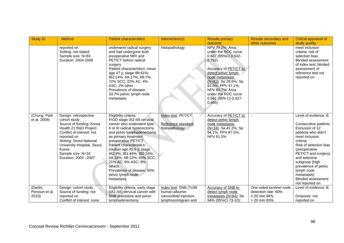
96 Concept Revisie Richtlijn Cervixcarcinoom - September 2011
Study ID Method Patiënt characteristics Intervention(s) Results primary outcome
Results secondary and other outcomes
Critical appraisal of study quality
reported on Setting: not stated Sample size: N=83 Duration: 2004-2008
underwent radical surgery and had undergone both preoperative MRI and PET/CT before radical surgery Patiënt characteristics: mean age 47 y; stage IBI:61%; IB2:14%; IIA:17%; IIB:7%; 72% SCC; 22% AC; 4% ASC; 2% other Prevalence of disease: 33.7% pelvic lymph node metastasis
histopathology NPV 79.2%; Area under the ROC curve 0.667 (95%CI 0.542-0.792) Accuracy of PET/CT to detect pelvic lymph node metastasis (N=83): Se 28.6%; Sp 83.6%; PPV 47.1%; NPV 69.7%; Area under the ROC curve 0.561 (95% CI 0.427-0.695)
meet inclusion criteria: risk of selection bias Blinded assessment of index test; blinded assessment of reference test not reported on
(Chung, Park et al. 2009)
Design: retrospective cohort study Source of funding: Korea Health 21 R&D Project Conflict of interest: not reported on Setting: Seoul National University Hospital, Seoul, Korea Sample size: N=34 Duration: 2003 –2007
Eligibility criteria: FIGO stage IA2-IIB cervical cancer who underwent type II or III radical hysterectomy and pelvic lymphadenectomy as primary treatment; preoperative PET/CT Patiënt characteristics: median age 45.5 y; stage IA2:3%; IB1:44%; IB2:24%; IIA:18%; IIB:12%; 65% SCC; 21% AC; 6% ASC; 9% others Prevalence of disease: 50% pelvic lymph node metastasis
Index test: PET/CT Reference standard: histopathology
Accuracy of PET/CT to detect pelvic lymph node metastasis (N=34): Se 41.2%; Sp 94.1%; PPV 87.5%; NPV 61.5%
- Level of evidence: B Consecutive patiënts Exclusion of 12 patiënts who didn‟t meet inclusion criteria Risk of selection bias (preoperative PET/CT and surgery) and selective subgroup (high prevalence of pelvic lymph node metastasis) Blinded assessment not reported on
(Darlin, Persson et al. 2010)
Design: cohort study Source of funding: not reported on Conflict of interest: none
Eligibility criteria: early stage (IA1-IIA) cervical cancer with SNB procedure and pelvic lymphadenectomy
Index test: SNB (Tc99 human-albumin nanocolloid injection, lymphoscintigram and
Accuracy of SNB to detect lymph node metastasis (N=94): Se 94% (95%CI 73-10);
One-sided sentinel node detection rate: 90% ≤ 20 mm 94% ˃ 20 mm 83%
Level of evidence: B Dropouts: not reported on
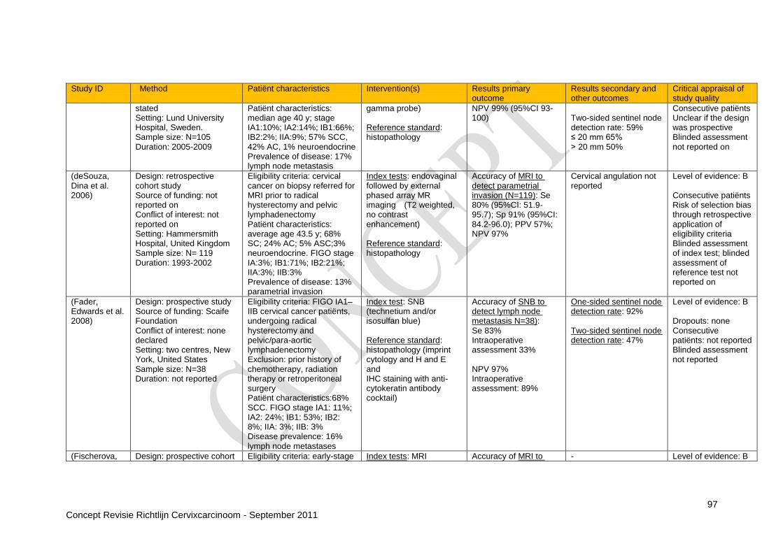
97 Concept Revisie Richtlijn Cervixcarcinoom - September 2011
Study ID Method Patiënt characteristics Intervention(s) Results primary outcome
Results secondary and other outcomes
Critical appraisal of study quality
stated Setting: Lund University Hospital, Sweden. Sample size: N=105 Duration: 2005-2009
Patiënt characteristics: median age 40 y; stage IA1:10%; IA2:14%; IB1:66%; IB2:2%; IIA:9%; 57% SCC, 42% AC, 1% neuroendocrine Prevalence of disease: 17% lymph node metastasis
gamma probe) Reference standard: histopathology
NPV 99% (95%CI 93-100)
Two-sided sentinel node detection rate: 59% ≤ 20 mm 65% ˃ 20 mm 50%
Consecutive patiënts Unclear if the design was prospective Blinded assessment not reported on
(deSouza, Dina et al. 2006)
Design: retrospective cohort study Source of funding: not reported on Conflict of interest: not reported on Setting: Hammersmith Hospital, United Kingdom Sample size: N= 119 Duration: 1993-2002
Eligibility criteria: cervical cancer on biopsy referred for MRI prior to radical hysterectomy and pelvic lymphadenectomy Patiënt characteristics: average age 43.5 y; 68% SC; 24% AC; 5% ASC;3% neuroendocrine. FIGO stage IA:3%; IB1:71%; IB2:21%; IIA:3%; IIB:3% Prevalence of disease: 13% parametrial invasion
Index tests: endovaginal followed by external phased array MR imaging (T2 weighted, no contrast enhancement) Reference standard: histopathology
Accuracy of MRI to detect parametrial invasion (N=119): Se 80% (95%CI: 51.9-95.7); Sp 91% (95%CI: 84.2-96.0); PPV 57%; NPV 97%
Cervical angulation not reported
Level of evidence: B Consecutive patiënts Risk of selection bias through retrospective application of eligibility criteria Blinded assessment of index test; blinded assessment of reference test not reported on
(Fader, Edwards et al. 2008)
Design: prospective study Source of funding: Scaife Foundation Conflict of interest: none declared Setting: two centres, New York, United States Sample size: N=38 Duration: not reported
Eligibility criteria: FIGO IA1–IIB cervical cancer patiënts, undergoing radical hysterectomy and pelvic/para-aortic lymphadenectomy Exclusion: prior history of chemotherapy, radiation therapy or retroperitoneal surgery Patiënt characteristics:68% SCC. FIGO stage IA1: 11%; IA2: 24%; IB1: 53%; IB2: 8%; IIA: 3%; IIB: 3% Disease prevalence: 16% lymph node metastases
Index test: SNB (technetium and/or isosulfan blue) Reference standard: histopathology (imprint cytology and H and E and IHC staining with anti-cytokeratin antibody cocktail)
Accuracy of SNB to detect lymph node metastasis N=38): Se 83% Intraoperative assessment 33% NPV 97% Intraoperative assessment: 89%
One-sided sentinel node detection rate: 92% Two-sided sentinel node detection rate: 47%
Level of evidence: B Dropouts: none Consecutive patiënts: not reported Blinded assessment not reported
(Fischerova, Design: prospective cohort Eligibility criteria: early-stage Index tests: MRI Accuracy of MRI to - Level of evidence: B
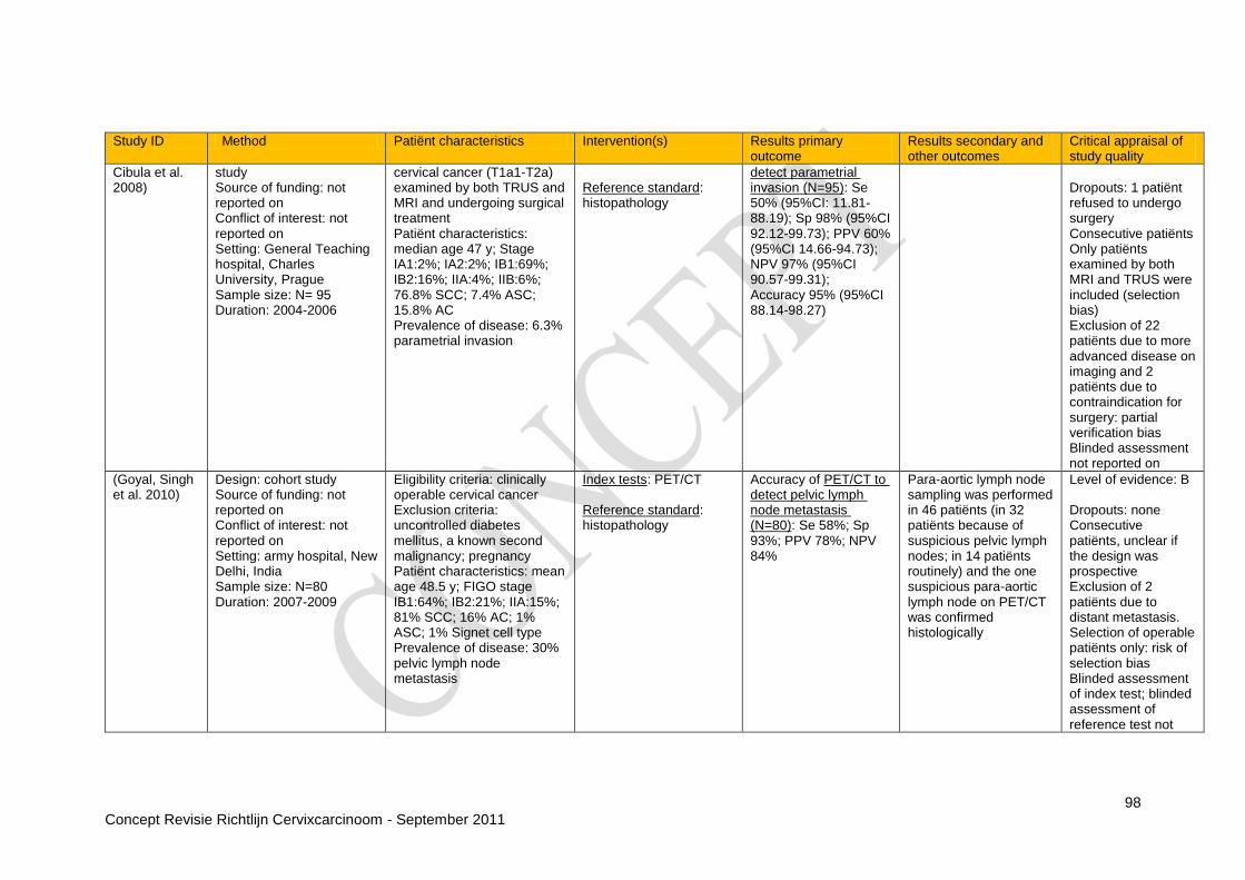
98 Concept Revisie Richtlijn Cervixcarcinoom - September 2011
Study ID Method Patiënt characteristics Intervention(s) Results primary outcome
Results secondary and other outcomes
Critical appraisal of study quality
Cibula et al. 2008)
study Source of funding: not reported on Conflict of interest: not reported on Setting: General Teaching hospital, Charles University, Prague Sample size: N= 95 Duration: 2004-2006
cervical cancer (T1a1-T2a) examined by both TRUS and MRI and undergoing surgical treatment Patiënt characteristics: median age 47 y; Stage IA1:2%; IA2:2%; IB1:69%; IB2:16%; IIA:4%; IIB:6%; 76.8% SCC; 7.4% ASC; 15.8% AC Prevalence of disease: 6.3% parametrial invasion
Reference standard: histopathology
detect parametrial invasion (N=95): Se 50% (95%CI: 11.81-88.19); Sp 98% (95%CI 92.12-99.73); PPV 60% (95%CI 14.66-94.73); NPV 97% (95%CI 90.57-99.31); Accuracy 95% (95%CI 88.14-98.27)
Dropouts: 1 patiënt refused to undergo surgery Consecutive patiënts Only patiënts examined by both MRI and TRUS were included (selection bias) Exclusion of 22 patiënts due to more advanced disease on imaging and 2 patiënts due to contraindication for surgery: partial verification bias Blinded assessment not reported on
(Goyal, Singh et al. 2010)
Design: cohort study Source of funding: not reported on Conflict of interest: not reported on Setting: army hospital, New Delhi, India Sample size: N=80 Duration: 2007-2009
Eligibility criteria: clinically operable cervical cancer Exclusion criteria: uncontrolled diabetes mellitus, a known second malignancy; pregnancy Patiënt characteristics: mean age 48.5 y; FIGO stage IB1:64%; IB2:21%; IIA:15%; 81% SCC; 16% AC; 1% ASC; 1% Signet cell type Prevalence of disease: 30% pelvic lymph node metastasis
Index tests: PET/CT Reference standard: histopathology
Accuracy of PET/CT to detect pelvic lymph node metastasis (N=80): Se 58%; Sp 93%; PPV 78%; NPV 84%
Para-aortic lymph node sampling was performed in 46 patiënts (in 32 patiënts because of suspicious pelvic lymph nodes; in 14 patiënts routinely) and the one suspicious para-aortic lymph node on PET/CT was confirmed histologically
Level of evidence: B Dropouts: none Consecutive patiënts, unclear if the design was prospective Exclusion of 2 patiënts due to distant metastasis. Selection of operable patiënts only: risk of selection bias Blinded assessment of index test; blinded assessment of reference test not
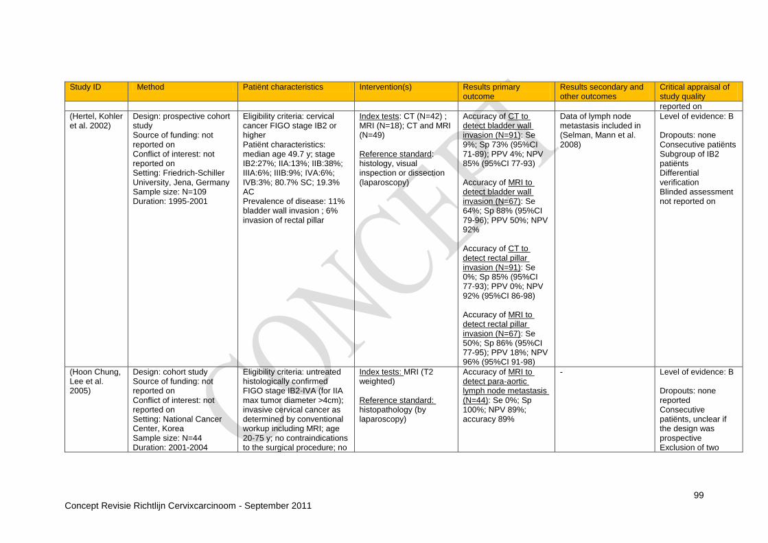
99 Concept Revisie Richtlijn Cervixcarcinoom - September 2011
Study ID Method Patiënt characteristics Intervention(s) Results primary outcome
Results secondary and other outcomes
Critical appraisal of study quality
reported on
(Hertel, Kohler et al. 2002)
Design: prospective cohort study Source of funding: not reported on Conflict of interest: not reported on Setting: Friedrich-Schiller University, Jena, Germany Sample size: N=109 Duration: 1995-2001
Eligibility criteria: cervical cancer FIGO stage IB2 or higher Patiënt characteristics: median age 49.7 y; stage IB2:27%; IIA:13%; IIB:38%; IIIA:6%; IIIB:9%; IVA:6%; IVB:3%; 80.7% SC; 19.3% AC Prevalence of disease: 11% bladder wall invasion ; 6% invasion of rectal pillar
Index tests: CT (N=42) ; MRI (N=18); CT and MRI (N=49) Reference standard: histology, visual inspection or dissection (laparoscopy)
Accuracy of CT to detect bladder wall invasion (N=91): Se 9%; Sp 73% (95%CI 71-89); PPV 4%; NPV 85% (95%CI 77-93) Accuracy of MRI to detect bladder wall invasion (N=67): Se 64%; Sp 88% (95%CI 79-96); PPV 50%; NPV 92% Accuracy of CT to detect rectal pillar invasion (N=91): Se 0%; Sp 85% (95%CI 77-93); PPV 0%; NPV 92% (95%CI 86-98) Accuracy of MRI to detect rectal pillar invasion (N=67): Se 50%; Sp 86% (95%CI 77-95); PPV 18%; NPV 96% (95%CI 91-98)
Data of lymph node metastasis included in (Selman, Mann et al. 2008)
Level of evidence: B Dropouts: none Consecutive patiënts Subgroup of IB2 patiënts Differential verification Blinded assessment not reported on
(Hoon Chung, Lee et al. 2005)
Design: cohort study Source of funding: not reported on Conflict of interest: not reported on Setting: National Cancer Center, Korea Sample size: N=44 Duration: 2001-2004
Eligibility criteria: untreated histologically confirmed FIGO stage IB2-IVA (for IIA max tumor diameter >4cm); invasive cervical cancer as determined by conventional workup including MRI; age 20-75 y; no contraindications to the surgical procedure; no
Index tests: MRI (T2 weighted) Reference standard: histopathology (by laparoscopy)
Accuracy of MRI to detect para-aortic lymph node metastasis (N=44): Se 0%; Sp 100%; NPV 89%; accuracy 89%
-
Level of evidence: B Dropouts: none reported Consecutive patiënts, unclear if the design was prospective Exclusion of two

100 Concept Revisie Richtlijn Cervixcarcinoom - September 2011
Study ID Method Patiënt characteristics Intervention(s) Results primary outcome
Results secondary and other outcomes
Critical appraisal of study quality
evidence of distant metastasis; Eastern Cooperative Oncology Group performance status 0 or 1; informed consent Patiënt characteristics: median age 48.0 y (23-72); Stage IB2: 9.1%; IIA:6.8%; IIB:77%; IIIB:7%. 82% SCC; 11% AC; 7% ASC
patiënts due to severe intra-abdominal adhesion related to previous surgery No blinded assessment of index and reference test
(Hori, Kim et al. 2009)
Design: prospective cohort study Source of funding: not reported on Conflict of interest: no financial relationship disclosed Setting: Osaka University Hospital Sample size: N=31 Duration: 2006-2007
Eligibility criteria: biopsy-proved untreated cervical carcinoma Patiënt characteristics: mean age 51.1 y; 71% SCC; 13% mucinous AC; 6% serous AC; 6% endometrioid AC; 3% ASCC. Stage IA1:6%; IA2:3%; IB1:55%; IB2:10%; IIA:13%; IIB:13% Prevalence of disease: 13% parametrial invasion; 19% vaginal invasion; 23% lymph node metastasis
Index tests: 3.0-T MRI and 1.5-T MRI (T2 weighted, contrast enhanced with cervical angulation) Reference standard: Histopathology or follow-up CT
Accuracy of 3.0-T MRI to detect parametrial invasion (N=31): Se 75% (95%CI 33-100); Sp 70% (95%CI 53-88); PPV 27% (95%CI 1-54); NPV 95% (95%CI 85-100); accuracy 71% (95%CI 55-87); area under ROC curve=0.82 Accuracy of 1.5-T MRI to detect parametrial invasion (N=31): Se 75% (95%CI 33-100); Sp 70% (95%CI 53-88); PPV 27% (95%CI 1-54); NPV 95% (95%CI 85-100); accuracy 71% (95%CI 55-87); area under ROC curve 0.85 Accuracy of 3.0-T MRI to detect vaginal invasion (N=31): Se 67% (95%CI 29-100); Sp 68% (95%CI 50-86);
There were no significant differences between 3.0- and 1.5-T MR imaging in terms of the areas under the ROC curve, sensitivity or specificity (p>0.5 for all comparisons)
Level of evidence: B Consecutive patiënts Dropouts:16 patiënts did not agree to be included in the study: risk of selection bias 12 patiënts were excluded due to radiation therapy Differential verification: 1 patiënt did not undergo pelvic lymphadenectomy and follow-up CT findings 6 months after surgery were used as reference standard Blinded assessment of index test; blinded assessment of reference standard not reported on Data from reader 1 presented, data from

101 Concept Revisie Richtlijn Cervixcarcinoom - September 2011
Study ID Method Patiënt characteristics Intervention(s) Results primary outcome
Results secondary and other outcomes
Critical appraisal of study quality
PPV 33% (95%CI 7-60); NPV 89% (95%CI 76-100); accuracy 68% (95%CI 51-84); area under ROC curve 0.62 Accuracy of 1.5-T MRI to detect vaginal invasion (N=31): Se 67% (95%CI 29-100); Sp 72% (95%CI 54-90); PPV 36% (95%CI 8-65); NPV 90% (95%CI 77-100); accuracy 71% (95%CI 55-87); area under ROC curve 0.67 Accuracy of 3.0-T MRI to detect pelvic lymph node metastasis (N=31): Se 57% (95%CI 20-94); Sp 83% (95%CI 68-98); PPV 50% (95%CI 15-85); NPV 87% (95%CI 73-100); accuracy 77% (95%CI 63-92); area under ROC curve 0.72 Accuracy of 1.5-T MRI to detect pelvic lymph node metastasis (N=31): Se 57% (95%CI 20-94); Sp 88% (95%CI 74-100); PPV 57% (95%CI 20-94); NPV 88% (95%CI 74-100); accuracy 81%
reader 2 also shown in article
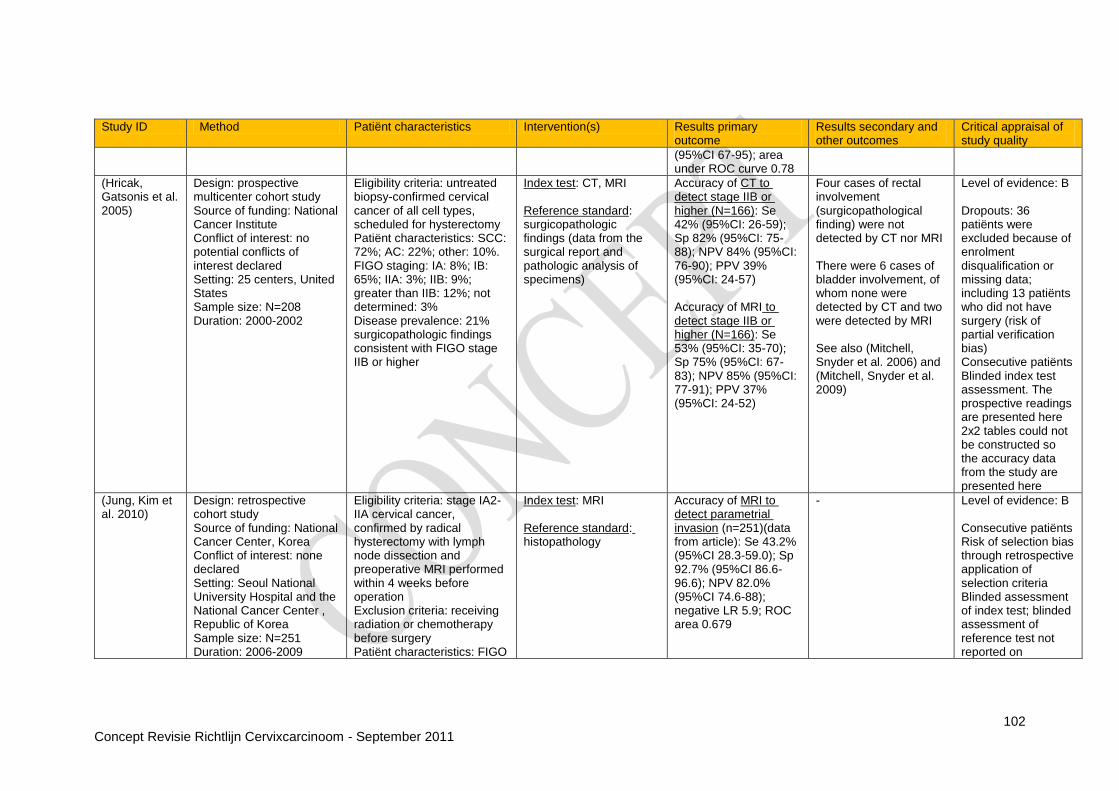
102 Concept Revisie Richtlijn Cervixcarcinoom - September 2011
Study ID Method Patiënt characteristics Intervention(s) Results primary outcome
Results secondary and other outcomes
Critical appraisal of study quality
(95%CI 67-95); area under ROC curve 0.78
(Hricak, Gatsonis et al. 2005)
Design: prospective multicenter cohort study Source of funding: National Cancer Institute Conflict of interest: no potential conflicts of interest declared Setting: 25 centers, United States Sample size: N=208 Duration: 2000-2002
Eligibility criteria: untreated biopsy-confirmed cervical cancer of all cell types, scheduled for hysterectomy Patiënt characteristics: SCC: 72%; AC: 22%; other: 10%. FIGO staging: IA: 8%; IB: 65%; IIA: 3%; IIB: 9%; greater than IIB: 12%; not determined: 3% Disease prevalence: 21% surgicopathologic findings consistent with FIGO stage IIB or higher
Index test: CT, MRI Reference standard: surgicopathologic findings (data from the surgical report and pathologic analysis of specimens)
Accuracy of CT to detect stage IIB or higher (N=166): Se 42% (95%CI: 26-59); Sp 82% (95%CI: 75-88); NPV 84% (95%CI: 76-90); PPV 39% (95%CI: 24-57) Accuracy of MRI to detect stage IIB or higher (N=166): Se 53% (95%CI: 35-70); Sp 75% (95%CI: 67-83); NPV 85% (95%CI: 77-91); PPV 37% (95%CI: 24-52)
Four cases of rectal involvement (surgicopathological finding) were not detected by CT nor MRI There were 6 cases of bladder involvement, of whom none were detected by CT and two were detected by MRI See also (Mitchell, Snyder et al. 2006) and (Mitchell, Snyder et al. 2009)
Level of evidence: B Dropouts: 36 patiënts were excluded because of enrolment disqualification or missing data; including 13 patiënts who did not have surgery (risk of partial verification bias) Consecutive patiënts Blinded index test assessment. The prospective readings are presented here 2x2 tables could not be constructed so the accuracy data from the study are presented here
(Jung, Kim et al. 2010)
Design: retrospective cohort study Source of funding: National Cancer Center, Korea Conflict of interest: none declared Setting: Seoul National University Hospital and the National Cancer Center , Republic of Korea Sample size: N=251 Duration: 2006-2009
Eligibility criteria: stage IA2-IIA cervical cancer, confirmed by radical hysterectomy with lymph node dissection and preoperative MRI performed within 4 weeks before operation Exclusion criteria: receiving radiation or chemotherapy before surgery Patiënt characteristics: FIGO
Index test: MRI Reference standard: histopathology
Accuracy of MRI to detect parametrial invasion (n=251)(data from article): Se 43.2% (95%CI 28.3-59.0); Sp 92.7% (95%CI 86.6-96.6); NPV 82.0% (95%CI 74.6-88); negative LR 5.9; ROC area 0.679
- Level of evidence: B Consecutive patiënts Risk of selection bias through retrospective application of selection criteria Blinded assessment of index test; blinded assessment of reference test not reported on

103 Concept Revisie Richtlijn Cervixcarcinoom - September 2011
Study ID Method Patiënt characteristics Intervention(s) Results primary outcome
Results secondary and other outcomes
Critical appraisal of study quality
stage IA2:3%; IB1:75%; IB2:6%; IIA:15%; 72% SCC; 28% AC/ASC. Prevalence of disease: 18% parametrial invasion
A 2x2 table could not be constructed
(Kim, Choi et al. 2009)
Design: retrospective cohort study Source of funding: not reported on Conflict of interest: none declared Setting: Republic of Korea Sample size: N=79 Duration: 2001-2007
Eligibility criteria: untreated histopathologically confirmed FIGO stage IB-IVA cervical cancer determined by conventional work-up that included MRI; no contraindication to surgical procedure; no evidence of distant metastasis; Eastern Cooperative Oncology Group performance status of 0-1. Exclusion criteria: small cell carcinoma and patiënts who did not undergo laparoscopic lymph node dissection Patiënt characteristics: not reported on Prevalence of disease: 38% metastatic lymph nodes
Index test: PET/CT and MRI/PET Reference standard: histopathology
Accuracy of PET/CT to detect para-aortic + pelvic lymph node metastasis (N=79): Se 47%; Sp 71%; PPV 50%; NPV 69%; ROC area 0.690 (95%CI :0.650-0.728) Accuracy of MRI/PET to detect para-aortic + pelvic lymph node metastasis (N=79): Se 57%; Sp 67%; PPV 52%; NPV 72%; ROC area 0.735 (0.696-0.771)
- Level of evidence: B Consecutive patiënts Retrospective selection of patiënts with a MRI and PET/CT prior to surgery (risk of selection bias) Blinded assessment of index test; blinded assessment of reference standard not reported on
(Kokka, Vorgias et al. 2003)
Design: retrospective study of medical records Source of funding: not reported on Conflict of interest: not reported on Setting: Metaxa Memorial Cancer Hospital, Peiras Greece Sample size: N=309 Duration: 1986-2000
Eligibility criteria: early (IB-IIA clinical FIGO) untreated cervical cancer patiënts who completed pre-treatment evaluation at the institution Exclusion criteria: neoadjuvant treatment and/or radiotherapy Patiënt characteristics:mean age: 48 y; SCC: 86%; AC: 11%; ASC: 2%. FIGO surgicopathological staging IB: 65%; IIA: 34%; IV: 1%
Index test: CT Reference standard: histology or visual inspection (cystoscopy and/or urine cytology, barium enema with sigmoidoscopy)
Accuracy of CT to detect urinary tract invasion (N=309): Se 100%; Sp 99.7%; NPV 100%; PPV 75% Accuracy of CT to detect gastrointestinal tract invasion (N=307): Se 50%; Sp 99.7%; NPV 99.7%; PPV 50%
Accuracy of CT to detect lymph node metastasis: included in (Selman, Mann et al. 2008)
Level of evidence: B Consecutive patiënts Risk of selection bias through selection criteria Blinded assessment not reported Differential verification, and partial verification (307/309) for gastrointestinal tract

104 Concept Revisie Richtlijn Cervixcarcinoom - September 2011
Study ID Method Patiënt characteristics Intervention(s) Results primary outcome
Results secondary and other outcomes
Critical appraisal of study quality
Disease prevalence: urinary tract invasion: 1%; gastrointestinal tract invasion: 0.3%
invasion
(Leblanc, Gauthier et al. 2011)
Design: retrospective multicenter study Source of funding: not reported on Conflict of interest: not reported on Setting: 5 French tertiary-care centers Sample size: N=125 Duration: 2004-2008
Eligibility criteria: primary stage IB2-IVA locally advanced cervical cancer or a centropelvic recurrence after chemoradiation for locally advanced cervical cancer, without evidence of distant metastasis and enlarged para-aortic nodes (>10mm) at abdominopelvic MRI with or without CT scan Patiënt characteristics: median age 48.3 y; 87% SCC; 11% AC; 2% clear cell. Stage IB2:34%; IIA:7%; IIB:37%; IIIA:2%; IIIB:10%; IVA:8%; centropelvic recurrence: 2% Prevalence of disease:17% para-aortic lymph node metastasis
Index tests: PET completed by CT or hybrid PET/CT Reference standard: histopathology
Accuracy of PET/CT to detect para-aortic lymph node metastasis (N=125): Se 33%; Sp 94%; PPV 54%; NPV 88%
Accuracy of PET/CT to detect para-aortic lymph node metastasis ≤ 5 mm (data form article): Se 22%; Sp 91%; PPV 15%; NPV 94% Accuracy of PET/CT to detect para-aortic lymph node metastasis >5 mm (data form article): Se 42%; Sp 93%; PPV 38%; NPV 94%
Level of evidence: B Unclear if patiënts were consecutive Blinded assessment of index test; blinded assessment of reference test not reported on Selection bias through retrospective application of selection criteria 2/125 patiënts had centropelvic recurrence
(Liu, Yen et al. 2009)
Design: retrospective study Source of funding: not reported on Conflict of interest: no conflicts of interest declared Setting: Chang Gung Memorial Hospital, Taiwan Sample size: N=165 Duration: not reported
Eligibility criteria: invasive cervical cancer patiënts (stage III/IV or positive lymph node metastasis) who had had PET and either CT or MRI performed within 30 days Exclusion: history of other malignancy; follow-up <180 days after PET (except those who died of disease within 180 days)
Index tests: CT and PET (N=40), MRI and PET (N=146) Reference standard: PET and either CT or MRI positive, along with a concordant clinical course of progression, with or without a transient response to palliative treatment. In case of
CT and PET group: accuracy of CT to detect hematogenous bone metastasis (N=40): Se 25%; Sp 100%; NPV 92%; PPV 100% CT and PET group: accuracy of PET to detect hematogenous bone metastasis
- Level of evidence: B Not reported whether patiënts were consecutive Risk of selection bias through retrospective application of selection criteria Blinded index test assessment Differential

105 Concept Revisie Richtlijn Cervixcarcinoom - September 2011
Study ID Method Patiënt characteristics Intervention(s) Results primary outcome
Results secondary and other outcomes
Critical appraisal of study quality
Patiënt characteristics: not described Disease prevalence: 7.3% hematogenous bone metastases
discordant imaging findings a bone biopsy was done if there would be clinical implications; otherwise visceral metastasis on imaging or new evidence of hematogenous bone metastasis within 180 days was considered proof
(N=40): Se 100%; Sp 100%; NPV 100%; PPV 100% MRI and PET group: accuracy of MRI to detect hematogenous bone metastasis (N=146): Se 80%; Sp 99%; NPV 99%; PPV 80% MRI and PET group: accuracy of PET to detect hematogenous bone metastasis (N=146): Se 100%; Sp 99%; NPV 100%; PPV 91%
verification Incorporation bias
(Loft, Berthelsen et al. 2007)
Design: prospective cohort study Source of funding: donation by The John and Birthe Meyer Doundation Conflict of interest: not reported on Setting: Rigshospitalet Copenhagen University Hospital, Denmark Sample size: N=119 Duration: 2002-2005
Eligibility criteria: newly diagnosed cervical cancer stage ≥1B Exclusion criteria: current previous or malignant disease of another type; diabetes mellitus; pregnancy; claustrophobia; extreme obesity or other reasons due to which the PET/CT scan could not be performed Patiënt characteristics: stage IB1:24%; IB2:3%; 2A:6%; 2B:26%; 3A:1%; 3B:36%; 4A:4%; 82% SCC; 11% AC; 8% others Disease prevalence: para-aortal lymph node
Index test: PET/CT Reference standard: To detect para-aortal lymph node metastasis: histopathology (N=12) and other imaging modalities or follow-up (N=107) To detect distant metastasis: histopathology (N=11) and other imaging modalities or follow-up (N=108)
Accuracy of PET/CT to detect para-aortal lymph node metastasis (N=119): Se 100%; Sp 99%; PPV 94%; NPV 100% Accuracy of PET/CT to detect distant metastasis (N=119): Se 100%; Sp 94%; PPV 53%; NPV 100%
- Level of evidence: B Dropouts: none reported Consecutive patiënts 1 patiënt who had had a hysterectomy prior to PET/CT was excluded Differential verification: histology, other imaging modalities and follow-up Partial verification: 14 patiënts who had positive pelvic lymph nodes on PET and

106 Concept Revisie Richtlijn Cervixcarcinoom - September 2011
Study ID Method Patiënt characteristics Intervention(s) Results primary outcome
Results secondary and other outcomes
Critical appraisal of study quality
metastasis: 13%; distant metastasis: 8%
were not examined histologically were excluded for calculations concerning pelvic lymph node status Incorporation bias in some patiënts Blinded assessment not reported on
(Manfredi, Gui et al. 2009)
Design: prospective cohort study Source of funding: not reported on Conflict of interest: not reported on Setting: Italy Sample size: N=53 Duration: not reported on
Eligibility criteria: localised cervical carcinoma (FIGO stage <IIB) studied by MRI Patiënt characteristics: mean age 47.3 y; 73% SCC; 18% AC; 9% ASC. Grade I:9%; Grade II:32%; Grade III:59%. Prevalence of disease: 6% vaginal invasion; 13% tumour extension to the internal os
Index test: MRI Reference standard: histopathology
Accuracy of MRI to detect vaginal invasion (N=53): Se 67%; Sp 92%; PPV 33%; NPV 98%; accuracy 91% Accuracy of MRI to detect tumour extension to the internal os (N=53): Se 86%; Sp 93%; PPV 67%; NPV 98%;accuracy 92%
Lesion based analysis of pelvic and lumbo-aortic lymph node metastasis Accuracy of MRI to detect stromal invasion: data for 2x2 table were not shown
Level of evidence: B Consecutive patiënts Dropouts: 2 patiënts who refused MRI were not included in analysis: risk of selection bias Exclusion of 5 patiënts due to locally advanced disease on MRI (partial verification) No blinded assessment of index and reference test
(Matsushita, Kurata et al. 2001)
Design: retrospective cohort study Source of funding: not reported on Conflict of interest: not reported on Setting: Niigata University Faculty of Medicine, Niigata, Japan Sample size: N=23 Duration: 1991-2000
Eligibility criteria: primary adenocarcinoma of the uterine cervix and MRI before surgery Patiënt characteristics: mean age 53 y; stage IB:65%; IIB:26%; IIIB:4%; IVB:4%; 35% adenoma malignum; 30% endometrioid adenocarcinoma; 22% ASC; 9% clear cell carcinoma; 4%
Index test: T2-weighted MRI Reference standard: histopathology
Accuracy of MRI to detect parametrial invasion (N=23): Se 60%; Sp 100%; PPV 100%; NPV 90%
- Level of evidence: B Consecutive patiënts Risk of selection bias through retrospective design where MRI is needed to be included Blinded assessment of index test; blinded assessment of

107 Concept Revisie Richtlijn Cervixcarcinoom - September 2011
Study ID Method Patiënt characteristics Intervention(s) Results primary outcome
Results secondary and other outcomes
Critical appraisal of study quality
serous AC; 6 patiënts received neoadjuvant chemotherapy Prevalence of disease: 22% parametrial invasion
reference test not reported on
(Mitchell, Snyder et al. 2006)
Design: prospective multicenter cohort study Source of funding: National Cancer Institute Conflict of interest: no potential conflicts of interest declared Setting: 25 centers, United States Sample size: N=208 Duration: 2000-2002
Eligibility criteria: untreated biopsy-confirmed cervical cancer of all cell types, scheduled for hysterectomy Patiënt characteristics: SCC: 72%; AC: 22%; other: 10%. FIGO staging: IA: 8%; IB: 65%; IIA: 3%; IIB: 9%; greater than IIB: 12%; not determined: 3% Disease prevalence: uterine involvement: 17% (32/172); stromal invasion: 44% had shallow (≤5mm) invasion, 22% had deep (>5mm) invasion
Index tests: CT, MRI Reference standard: histology
Accuracy of CT to detect uterine involvement (N=na): AUC 0.66 Accuracy of MRI to detect uterine involvement (N=na): AUC 0.80
Agreement between CT and histology on the tumor size: kappa coefficient 0.18 (95%CI: 0.06-0.30); weighted kappa coefficient 0.32 (95%CI: 0.20-0.43) Agreement between MRI and histology on the tumor size: kappa coefficient 0.30 (95%CI: 0.18-0.42); weighted kappa coefficient 0.41 (95%CI: 0.30-0.53) Accuracy of CT to detect stromal invasion (N=119): Se 29% (95%CI: 21-39); Sp 79% (95%CI: 59-92); NPV 23% (95%CI: 15-33); PPV 83% (95%CI: 67-94) Accuracy of MRI to detect stromal invasion (N=102): Se 65% (95%CI: 54-74); Sp 43% (95%CI: 23-66); NPV 23% (95%CI: 12-39); PPV 82% (95%CI: 72-
Level of evidence: B Dropouts: 36 patiënts were excluded because of enrolment disqualification or missing data including 13 patiënts who did not have surgery (risk of partial verification bias) Consecutive patiënts Blinded index test assessment. The prospective readings are presented here 2x2 tables could not be constructed so the accuracy data from the study are presented here
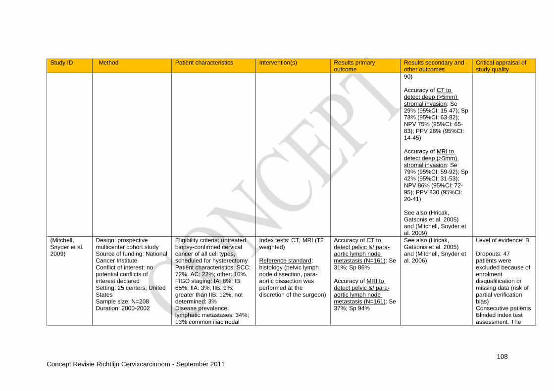
108 Concept Revisie Richtlijn Cervixcarcinoom - September 2011
Study ID Method Patiënt characteristics Intervention(s) Results primary outcome
Results secondary and other outcomes
Critical appraisal of study quality
90) Accuracy of CT to detect deep (>5mm) stromal invasion: Se 29% (95%CI: 15-47); Sp 73% (95%CI: 63-82); NPV 75% (95%CI: 65-83); PPV 28% (95%CI: 14-45) Accuracy of MRI to detect deep (>5mm) stromal invasion: Se 79% (95%CI: 59-92); Sp 42% (95%CI: 31-53); NPV 86% (95%CI: 72-95); PPV 830 (95%CI: 20-41) See also (Hricak, Gatsonis et al. 2005) and (Mitchell, Snyder et al. 2009)
(Mitchell, Snyder et al. 2009)
Design: prospective multicenter cohort study Source of funding: National Cancer Institute Conflict of interest: no potential conflicts of interest declared Setting: 25 centers, United States Sample size: N=208 Duration: 2000-2002
Eligibility criteria: untreated biopsy-confirmed cervical cancer of all cell types, scheduled for hysterectomy Patiënt characteristics: SCC: 72%; AC: 22%; other: 10%. FIGO staging: IA: 8%; IB: 65%; IIA: 3%; IIB: 9%; greater than IIB: 12%; not determined: 3% Disease prevalence: lymphatic metastases: 34%; 13% common iliac nodal
Index tests: CT, MRI (T2 weighted) Reference standard: histology (pelvic lymph node dissection, para-aortic dissection was performed at the discretion of the surgeon)
Accuracy of CT to detect pelvic &/ para-aortic lymph node metastasis (N=161): Se 31%; Sp 86% Accuracy of MRI to detect pelvic &/ para-aortic lymph node metastasis (N=161): Se 37%; Sp 94%
See also (Hricak, Gatsonis et al. 2005) and (Mitchell, Snyder et al. 2006)
Level of evidence: B Dropouts: 47 patiënts were excluded because of enrolment disqualification or missing data (risk of partial verification bias) Consecutive patiënts Blinded index test assessment. The

109 Concept Revisie Richtlijn Cervixcarcinoom - September 2011
Study ID Method Patiënt characteristics Intervention(s) Results primary outcome
Results secondary and other outcomes
Critical appraisal of study quality
metastases; 9% para-aortic nodal metastases
prospective readings are presented here 2x2 tables could not be constructed so the accuracy data from the study are presented here
(Nam, Huh et al. 2010)
Design: retrospective cohort study Source of funding: not reported on Conflict of interest: not reported on Setting: Samsung Medical Centre, Seoul, South Korea Sample size: N=92 Duration: 1997-2007
Eligibility criteria: uterine cervical cancer patiënts (FIGO stage IIB-IVA), treated with radiotherapy with a curative intent, with or without chemotherapy, who had a pre-treatment MRI Patiënt characteristics: 37% > 60 y of age; 95% SCC; 5% AC. All patiënts were FIGO stage IIB-IVA Disease prevalence: 15% mucosal bladder invasion
Index test: MRI Reference standard: cystoscopy
Accuracy of MRI to detect bladder invasion (defined as wall invasion or mucosal invasion) (N=92): Se 93%; Sp 63%; NPV 98%; PPV 31% Accuracy of MRI to detect bladder invasion (defined as mucosal invasion) (N=92): Se 93%; Sp 94%; NPV 99%; PPV 72%
- Level of evidence: B Dropouts: 4 patiënts were excluded because of missing follow-up data and 1 patiënt because of hysterectomy after radiotherapy Consecutive patiënts Risk of selection bias through selection criteria Blinded assessment not reported
(Oberoi, Vohra et al. 2002)
Design: retrospective cohort study Source of funding: not reported on Conflict of interest: not reported on Setting: Rajiv Gandhi Cancer Institute & Research Centre, New Delhi, India Sample size: N=105 Duration: 1997-2001
Eligibility criteria: surgery for primary carcinoma of the cervix Histological diagnosis of cervix carcinoma; FIGO stage IB or higher Patiënt characteristics: mean age 51 y; FIGO stage IB: 68%; IIA: 7%; IIB: 18%; IIIA: 6%; IVA: 2%; 98% SCC; 2% AC Prevalence of disease: parametrium invasion: 19%; vaginal invasion upper 2/3
rd:
18%; vaginal invasion lower
Index tests: MRI (T2 weighted) Reference standard: histopathology
Accuracy of MRI to detect stage IIB or higher (N=105): Se 85%; Sp; 92%; NPV: 92%; PPV: 79% Accuracy of MRI to detect parametrium invasion (N=105): Se 87%; Sp 93%; PPV 77%; NPV 96%; accuracy 91% Accuracy of MRI to detect vaginal invasion
Accuracy of MRI to detect surgico-pathological staging: stage IB: 90%; IIA: 57%; IIB: 84%; IIIA: 83%; IVA: 100%
Level of evidence: B Consecutive patiënts Risk of selection bias through retrospective selection of patiënts with available index test Blinded assessment of index and reference test not reported on

110 Concept Revisie Richtlijn Cervixcarcinoom - September 2011
Study ID Method Patiënt characteristics Intervention(s) Results primary outcome
Results secondary and other outcomes
Critical appraisal of study quality
1/3 rd: 7%; bladder invasion: 2%; rectum invasion: 1%; pelvic lymph node metastasis: 13%
upper 2/3rd
(N=105): Se 83%; Sp 94%; PPV 79%; NPV 95%; accuracy 91% Accuracy of MRI to detect vaginal invasion lower 1/3
rd (N=105): Se
78%; Sp 100%; PPV 100%; NPV 98%; accuracy 98% Accuracy of MRI to detect bladder invasion (N=105): Se 100%; Sp 100%; PPV 100%; NPV 100%; accuracy 100% Accuracy of MRI to detect rectum invasion (N=105): Se 100%; Sp 100%; PPV 100%; NPV 100%; accuracy 100% Accuracy of MRI to detect pelvic lymph node metastasis (N=105): Se 67%; Sp 96%; PPV 82%; NPV 92%; accuracy 90%
(Ramirez, Jhingran et al. 2010)
Design: prospective study Source of funding: supported in part by a Cancer Center Support Grant Conflict of interest: not reported on
Eligibility criteria: stage IB2-IVA cervical cancer and a candidate for treatment with radiotherapy and concurrent chemotherapy; no evidence of para-aortic lymphadenopathy (all nodes
Index tests: PET/CT Reference standard: histology (laparoscopic staging)
The accuracy of PET/CT to detect para-aortic lymph node metastasis in CT or MRI negative patiënts (N=60): Se 36%; Sp 96%; NPV 83%; PPV
There was one conversion from laparoscopy to laparotomy because of uncontrolled bleeding The median length of
Level of evidence: B Dropouts: 1; another 4 patiënts were excluded because of too high blood glucose levels or
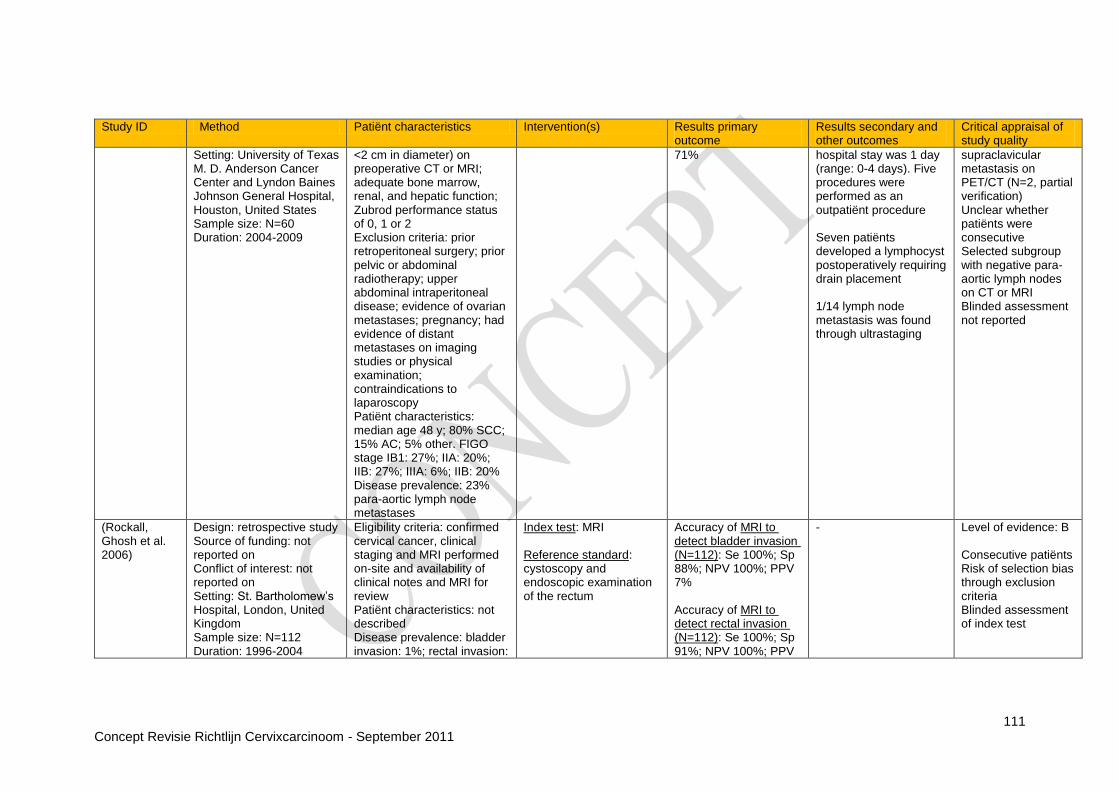
111 Concept Revisie Richtlijn Cervixcarcinoom - September 2011
Study ID Method Patiënt characteristics Intervention(s) Results primary outcome
Results secondary and other outcomes
Critical appraisal of study quality
Setting: University of Texas M. D. Anderson Cancer Center and Lyndon Baines Johnson General Hospital, Houston, United States Sample size: N=60 Duration: 2004-2009
<2 cm in diameter) on preoperative CT or MRI; adequate bone marrow, renal, and hepatic function; Zubrod performance status of 0, 1 or 2 Exclusion criteria: prior retroperitoneal surgery; prior pelvic or abdominal radiotherapy; upper abdominal intraperitoneal disease; evidence of ovarian metastases; pregnancy; had evidence of distant metastases on imaging studies or physical examination; contraindications to laparoscopy Patiënt characteristics: median age 48 y; 80% SCC; 15% AC; 5% other. FIGO stage IB1: 27%; IIA: 20%; IIB: 27%; IIIA: 6%; IIB: 20% Disease prevalence: 23% para-aortic lymph node metastases
71% hospital stay was 1 day (range: 0-4 days). Five procedures were performed as an outpatiënt procedure Seven patiënts developed a lymphocyst postoperatively requiring drain placement 1/14 lymph node metastasis was found through ultrastaging
supraclavicular metastasis on PET/CT (N=2, partial verification) Unclear whether patiënts were consecutive Selected subgroup with negative para-aortic lymph nodes on CT or MRI Blinded assessment not reported
(Rockall, Ghosh et al. 2006)
Design: retrospective study Source of funding: not reported on Conflict of interest: not reported on Setting: St. Bartholomew‟s Hospital, London, United Kingdom Sample size: N=112 Duration: 1996-2004
Eligibility criteria: confirmed cervical cancer, clinical staging and MRI performed on-site and availability of clinical notes and MRI for review Patiënt characteristics: not described Disease prevalence: bladder invasion: 1%; rectal invasion:
Index test: MRI Reference standard: cystoscopy and endoscopic examination of the rectum
Accuracy of MRI to detect bladder invasion (N=112): Se 100%; Sp 88%; NPV 100%; PPV 7% Accuracy of MRI to detect rectal invasion (N=112): Se 100%; Sp 91%; NPV 100%; PPV
- Level of evidence: B Consecutive patiënts Risk of selection bias through exclusion criteria Blinded assessment of index test

112 Concept Revisie Richtlijn Cervixcarcinoom - September 2011
Study ID Method Patiënt characteristics Intervention(s) Results primary outcome
Results secondary and other outcomes
Critical appraisal of study quality
2% 17%
(Sahdev, Sohaib et al. 2007)
Design: retrospective study Source of funding: not reported on Conflict of interest: not reported on Setting: St. Bartholomew‟s Hospital, London, United Kingdom Sample size: N=150 Duration: 1995-2005
Eligibility criteria: early cervical cancer Exclusion: 223 patiënts were excluded because they had advanced cervical carcinoma involving parametrium or pelvic sidewall on imaging (including MRI) and/or on clinical examination, or if the MRI was inadequate Patiënt characteristics: 63% underwent radical hysterectomy (mean age 34 y) and 37% underwent radical vaginal trachelectomy (mean age 23 y) Disease prevalence: 12% internal os involvement; 5% myometrial invasion; 2% parametrial invasion; 13% lymph node metastases
Index test: MRI (T2 weighted) Reference standard: histology
Accuracy of MRI to detect involvement of the internal os (N=150): Se 90%; Sp 98%; NPV 98%; PPV 86% Accuracy of MRI to detect myometrial invasion (N=150): Se 100%; Sp 99%; NPV 100%; PPV 88% Agreement between MRI and histologic staging (N=150): kappa 0.56 (95%CI: 0.34–0.60) Accuracy of MRI to detect pelvic lymph node metastasis (N=150): Se 37%; Sp 95%; NPV 94%; PPV 39%
Accuracy of MRI to evaluate tumor size: mean difference in tumor size histology-MRI: -9 mm; 95% limits of agreement were 212.6-113 mm, thus, 95% of all tumors were within 13 mm of histologic size
Level of evidence: B Consecutive patiënts Risk of selection bias through exclusion criteria (81 patiënts were excluded) leaves a selective subgroup Blinded assessment of index test
(Sandvik, Jensen et al. 2011)
Design: retrospective cohort study Source of funding: not reported on Conflict of interest: none stated Setting: Herlev Hospital, Denmark Sample size: N=117
Eligibility criteria: Patiënt characteristics: 75% SCC; 17% AD; 6% ASC; 2% other. FIGO stage IA: 13%; IB: 43%; IIB: 24%; IIIB: 13%; IVA: 2%; IVB: 4% Disease prevalence: 14% (5/36)
Index test: PET/CT Reference standard: histology (biopsy for distant metastasis)
Accuracy of PET/CT to detect pelvic lymph node metastasis (N=36): Se 20%; Sp 90%; NPV 88%; PPV 25%
Accuracy of PET/CT to detect lymph node metastasis (unspecified), distant metastasis or other malignancies (N=42): Se 50%; Sp 91%; NPV 88%; PPV 57%
Level of evidence: B Consecutive patiënts 34 patiënts with early stage disease did not have PET because of limited capacity and were excluded (selection bias)
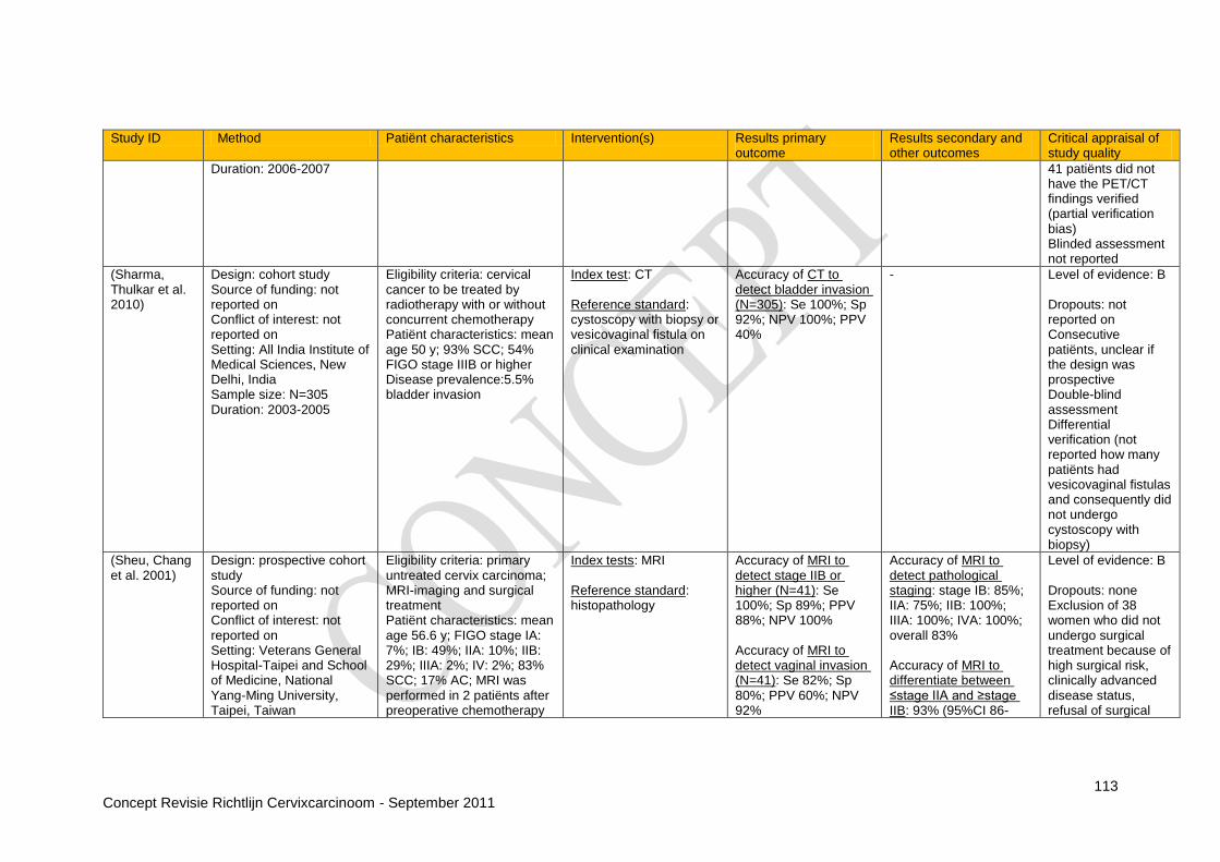
113 Concept Revisie Richtlijn Cervixcarcinoom - September 2011
Study ID Method Patiënt characteristics Intervention(s) Results primary outcome
Results secondary and other outcomes
Critical appraisal of study quality
Duration: 2006-2007 41 patiënts did not have the PET/CT findings verified (partial verification bias) Blinded assessment not reported
(Sharma, Thulkar et al. 2010)
Design: cohort study Source of funding: not reported on Conflict of interest: not reported on Setting: All India Institute of Medical Sciences, New Delhi, India Sample size: N=305 Duration: 2003-2005
Eligibility criteria: cervical cancer to be treated by radiotherapy with or without concurrent chemotherapy Patiënt characteristics: mean age 50 y; 93% SCC; 54% FIGO stage IIIB or higher Disease prevalence:5.5% bladder invasion
Index test: CT Reference standard: cystoscopy with biopsy or vesicovaginal fistula on clinical examination
Accuracy of CT to detect bladder invasion (N=305): Se 100%; Sp 92%; NPV 100%; PPV 40%
- Level of evidence: B Dropouts: not reported on Consecutive patiënts, unclear if the design was prospective Double-blind assessment Differential verification (not reported how many patiënts had vesicovaginal fistulas and consequently did not undergo cystoscopy with biopsy)
(Sheu, Chang et al. 2001)
Design: prospective cohort study Source of funding: not reported on Conflict of interest: not reported on Setting: Veterans General Hospital-Taipei and School of Medicine, National Yang-Ming University, Taipei, Taiwan
Eligibility criteria: primary untreated cervix carcinoma; MRI-imaging and surgical treatment Patiënt characteristics: mean age 56.6 y; FIGO stage IA: 7%; IB: 49%; IIA: 10%; IIB: 29%; IIIA: 2%; IV: 2%; 83% SCC; 17% AC; MRI was performed in 2 patiënts after preoperative chemotherapy
Index tests: MRI Reference standard: histopathology
Accuracy of MRI to detect stage IIB or higher (N=41): Se 100%; Sp 89%; PPV 88%; NPV 100% Accuracy of MRI to detect vaginal invasion (N=41): Se 82%; Sp 80%; PPV 60%; NPV 92%
Accuracy of MRI to detect pathological staging: stage IB: 85%; IIA: 75%; IIB: 100%; IIIA: 100%; IVA: 100%; overall 83% Accuracy of MRI to differentiate between ≤stage IIA and ≥stage IIB: 93% (95%CI 86-
Level of evidence: B Dropouts: none Exclusion of 38 women who did not undergo surgical treatment because of high surgical risk, clinically advanced disease status, refusal of surgical

114 Concept Revisie Richtlijn Cervixcarcinoom - September 2011
Study ID Method Patiënt characteristics Intervention(s) Results primary outcome
Results secondary and other outcomes
Critical appraisal of study quality
Sample size: N=41 Duration: 1996-1999
and in 4 patiënts after preoperative radiation therapy Prevalence of disease: 27% vaginal invasion
100) Accuracy of MRI to detect tumor size > 1cm: Se 94%; Sp 57%; PPV 91%; NPV 67% Accuracy of MRI to detect tumor size < 0.5 cm: Se 71%; Sp 91%; PPV 63%; NPV 92% Data of parametrial invasion and lymph node metastases in (Bipat, Glas et al. 2003)
intervention (preferred radiotherapy) or did not undergo MRI due to pacemaker implantation, intracranial vascular clips or claustrophobia (partial verification bias) Consecutive patiënts Blinded assessment of index test; blinded assessment of reference test not reported on
(Sironi, Bellomi et al. 2002)
Design: prospective cohort study Source of funding: not reported on Conflict of interest: not reported on Setting: University of Milan, Italy Sample size: N=73 Duration: not reported
Eligibility criteria: invasive cervical carcinoma clinically considered < 3cm and confined to the cervix (FIGO stage IB1) Exclusion criteria: contraindications to MRI or use of intravenous gadolinium Mean age: 47 y Disease prevalence:37% parametrial invasion
Index test: MRI (FSE T2-W, SE T1-W Gd, SE T1-W Gd FS) Reference standard: histopathology
Accuracy of MRI FSE T2-W to detect parametrial invasion (N=73): Se 79% (95%CI: 61-84); Sp 81% (95%CI: 68-89); NPV 95% (95%CI: 75-98); PPV 73% (95%CI: 61-86) Accuracy of MRI SE T1-W Gd to detect parametrial invasion (N=73): Se 71% (95%CI: 59-83); Sp 23% (95%CI: -8-31); NPV 77% (95%CI: 59-79); PPV 53% (95%CI: 32-68)
- Level of evidence: A2 Dropouts: 8 patiënts were excluded because they refused surgery Subgroup of IB1 patiënts Consecutive patiënts 2x2 tables could not be constructed: the reported outcomes are presented Double-blind assessment

115 Concept Revisie Richtlijn Cervixcarcinoom - September 2011
Study ID Method Patiënt characteristics Intervention(s) Results primary outcome
Results secondary and other outcomes
Critical appraisal of study quality
Accuracy of MRI SE T1-W Gd FS to detect parametrial invasion (N=73): Se 68% (95%CI: 59-79); Sp 63% (95%CI: 49-73); NPV 82% (95%CI: 73-92); PPV 74% (95%CI: 65-84)
(Sironi, Buda et al. 2006)
Design: prospective cohort study Source of funding: not reported on Conflict of interest: none stated Setting: Milan, Italy Sample size: N=47 Duration: 2003-2004
Eligibility criteria: hisopathologically confirmed diagnosis of primary cervical carcinoma Exclusion criteria: FIGO stage IIB or higher; relative contraindications to PET scanning Patiënt characteristics: mean age 45.3 y; FIGO stage IA1: 9%; IB1: 74%; IB2: 17%; 79% SCC; 21% AC; 6 patiënts with stage IB2 underwent neoadjuvant chemotherapy before PET/CT Prevalence of disease: 32% pelvic lymph node metastasis
Index test: PET/CT Reference standard: histopathology
Accuracy of PET/CT to detect pelvic lymph node metastasis (N=47): Se 73% (95%CI 48.0-89.1); Sp 97% (95%CI 84.3-99.4); PPV 92%; NPV 89%; accuracy 89%
- Level of evidence: B Dropouts: none Consecutive patiënts Double-blind assessment
(Testa, Ludovisi et al. 2009)
Design: prospective cohort study Source of funding: partially supported by I.R.I.S-PCR-OGONLUS Conflict of interest: not reported on Setting: Catholic University, Rome, Italy
Eligibility criteria: patiënts with early cervical cancer planned for primary surgery and patiënts with locally advanced cervical cancer planned for surgery after neoadjuvant treatment Exclusion: patiënts who underwent a cervical cone
Index test: MRI (T2 weighted) Reference standard: histopathology
Accuracy of MRI to detect parametrial invasion (N=68): Se 40%; Sp 89%; NPV 95%; PPV 22% Accuracy of MRI to detect vaginal invasion (N=68): Se 0%; Sp
Mean difference between histopathological measurements and MRI measurements of the craniocaudal diameter of the tumor: 1.49 mm (95% CI: −1.41 to 4.40 mm,
Level of evidence: B Dropouts: seven patiënts were not operated on because of no response to neoadjuvant treatment (partial verification bias)
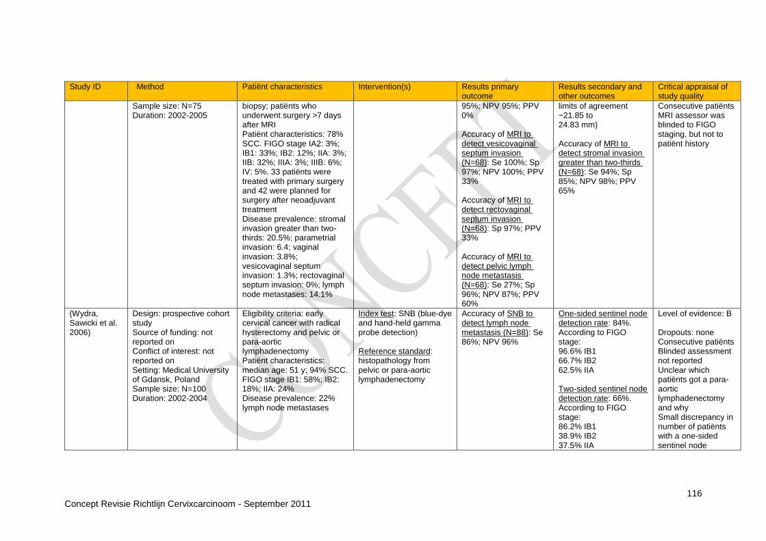
116 Concept Revisie Richtlijn Cervixcarcinoom - September 2011
Study ID Method Patiënt characteristics Intervention(s) Results primary outcome
Results secondary and other outcomes
Critical appraisal of study quality
Sample size: N=75 Duration: 2002-2005
biopsy; patiënts who underwent surgery >7 days after MRI Patiënt characteristics: 78% SCC. FIGO stage IA2: 3%; IB1: 33%; IB2: 12%; IIA: 3%; IIB: 32%; IIIA: 3%; IIIB: 6%; IV: 5%. 33 patiënts were treated with primary surgery and 42 were planned for surgery after neoadjuvant treatment Disease prevalence: stromal invasion greater than two-thirds: 20.5%; parametrial invasion: 6.4; vaginal invasion: 3.8%; vesicovaginal septum invasion: 1.3%; rectovaginal septum invasion: 0%; lymph node metastases: 14.1%
95%; NPV 95%; PPV 0% Accuracy of MRI to detect vesicovaginal septum invasion (N=68): Se 100%; Sp 97%; NPV 100%; PPV 33% Accuracy of MRI to detect rectovaginal septum invasion (N=68): Sp 97%; PPV 33% Accuracy of MRI to detect pelvic lymph node metastasis (N=68): Se 27%; Sp 96%; NPV 87%; PPV 60%
limits of agreement −21.85 to 24.83 mm) Accuracy of MRI to detect stromal invasion greater than two-thirds (N=68): Se 94%; Sp 85%; NPV 98%; PPV 65%
Consecutive patiënts MRI assessor was blinded to FIGO staging, but not to patiënt history
(Wydra, Sawicki et al. 2006)
Design: prospective cohort study Source of funding: not reported on Conflict of interest: not reported on Setting: Medical University of Gdansk, Poland Sample size: N=100 Duration: 2002-2004
Eligibility criteria: early cervical cancer with radical hysterectomy and pelvic or para-aortic lymphadenectomy Patiënt characteristics: median age: 51 y; 94% SCC. FIGO stage IB1: 58%; IB2: 18%; IIA: 24% Disease prevalence: 22% lymph node metastases
Index test: SNB (blue-dye and hand-held gamma probe detection) Reference standard: histopathology from pelvic or para-aortic lymphadenectomy
Accuracy of SNB to detect lymph node metastasis (N=88): Se 86%; NPV 96%
One-sided sentinel node detection rate: 84%. According to FIGO stage: 96.6% IB1 66.7% IB2 62.5% IIA Two-sided sentinel node detection rate: 66%. According to FIGO stage: 86.2% IB1 38.9% IB2 37.5% IIA
Level of evidence: B Dropouts: none Consecutive patiënts Blinded assessment not reported Unclear which patiënts got a para-aortic lymphadenectomy and why Small discrepancy in number of patiënts with a one-sided sentinel node
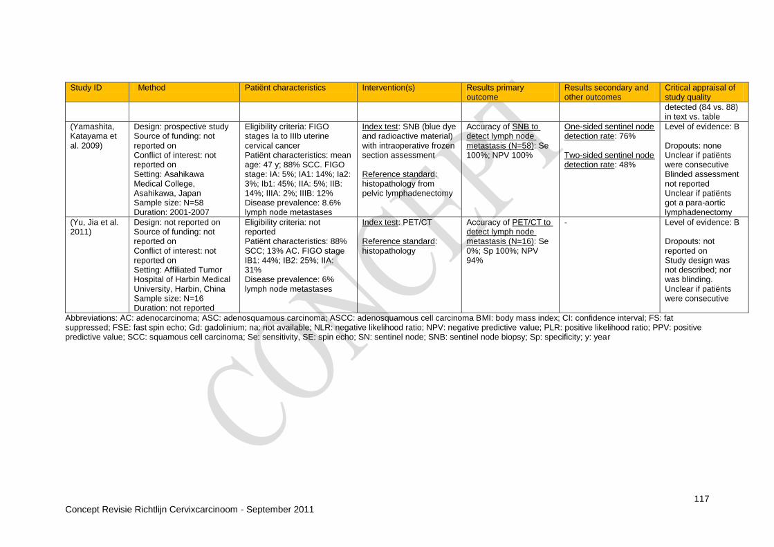
117 Concept Revisie Richtlijn Cervixcarcinoom - September 2011
Study ID Method Patiënt characteristics Intervention(s) Results primary outcome
Results secondary and other outcomes
Critical appraisal of study quality
detected (84 vs. 88) in text vs. table
(Yamashita, Katayama et al. 2009)
Design: prospective study Source of funding: not reported on Conflict of interest: not reported on Setting: Asahikawa Medical College, Asahikawa, Japan Sample size: N=58 Duration: 2001-2007
Eligibility criteria: FIGO stages Ia to IIIb uterine cervical cancer Patiënt characteristics: mean age: 47 y; 88% SCC. FIGO stage: IA: 5%; IA1: 14%; Ia2: 3%; Ib1: 45%; IIA: 5%; IIB: 14%; IIIA: 2%; IIIB: 12% Disease prevalence: 8.6% lymph node metastases
Index test: SNB (blue dye and radioactive material) with intraoperative frozen section assessment Reference standard: histopathology from pelvic lymphadenectomy
Accuracy of SNB to detect lymph node metastasis (N=58): Se 100%; NPV 100%
One-sided sentinel node detection rate: 76% Two-sided sentinel node detection rate: 48%
Level of evidence: B Dropouts: none Unclear if patiënts were consecutive Blinded assessment not reported Unclear if patiënts got a para-aortic lymphadenectomy
(Yu, Jia et al. 2011)
Design: not reported on Source of funding: not reported on Conflict of interest: not reported on Setting: Affiliated Tumor Hospital of Harbin Medical University, Harbin, China Sample size: N=16 Duration: not reported
Eligibility criteria: not reported Patiënt characteristics: 88% SCC; 13% AC. FIGO stage IB1: 44%; IB2: 25%; IIA: 31% Disease prevalence: 6% lymph node metastases
Index test: PET/CT Reference standard: histopathology
Accuracy of PET/CT to detect lymph node metastasis (N=16): Se 0%; Sp 100%; NPV 94%
- Level of evidence: B Dropouts: not reported on Study design was not described; nor was blinding. Unclear if patiënts were consecutive
Abbreviations: AC: adenocarcinoma; ASC: adenosquamous carcinoma; ASCC: adenosquamous cell carcinoma BMI: body mass index; CI: confidence interval; FS: fat suppressed; FSE: fast spin echo; Gd: gadolinium; na: not available; NLR: negative likelihood ratio; NPV: negative predictive value; PLR: positive likelihood ratio; PPV: positive predictive value; SCC: squamous cell carcinoma; Se: sensitivity, SE: spin echo; SN: sentinel node; SNB: sentinel node biopsy; Sp: specificity; y: year

118 Concept Revisie Richtlijn Cervixcarcinoom - September 2011
Vraag 1B Bij patiënten met invasief cervixcarcinoom, wat is het afkappunt van de tumormarkers SCC en CA125 voor het definiëren van een hoog risicogroep m.b.t. locale tumoruitbreiding, lymfekliermetastasen en metastasen op afstand? Observational studies
Study ID Method Patiënt characteristics Intervention(s) Results primary outcome
Results secondary and other outcomes
Critical appraisal of study quality
(Bender, Sorosky et al. 2003)
Design: retrospective cohort study Source of funding: not reported on Conflicts of interest: not reported on Setting: University of Iowa Sample size: N=73 Duration: 1986-1998
Eligibility criteria: cervical adenocarcinoma or adenosquamous carcinoma treated at this centre, and a pre-treatment CA 125 (50% of all patiënts treated) Patiënt characteristics:mean age 45 y; 93% AC; 86% with low (I- IIA) FIGO stage; 52 patiënts underwent surgery and were included in the primary outcome Disease prevalence: 17% lymph node metastases
Index test: serum CA 125 Reference standard: histology
Accuracy of CA 125≥30 U/mL to predict positive lymph nodes (N=50): Se 67%; Sp 84%; NPV 92%; PPV 46%
- Level of evidence: B Consecutive patiënts; risk of selection bias through eligibility criteria Partial verification: only patiënts with a radical hysterectomy could be verified by histology Uses the 1995 FIGO criteria The cut-off level for CA 125 was not predefined
(Chen, Liang et al. 2008)
Design: prospective study Source of funding: not reported on Conflicts of interest: not reported on Setting: China Medical University Hospital, China Sample size: N=148 Duration: 2001-2006
Eligibility criteria: untreated stage IB2–IVA squamous cell cancer of the uterine cervix, without evidence of enlarged para-aortic lymph nodes Patiënt characteristics: not specified. All patiënts went for concurrent chemoradiation Disease prevalence: tumor size >4 cm: 91 %; 15% lymph node metastases
Index tests: SSCA Reference standard: CT (lymph node positivity) and CT or pelvic examination (tumour size)
Accuracy of SSCA ≥2.0 ng/ml to predict a tumour size ≥4cm (N=148): Se 64%; Sp 31%; NPV 8%; PPV 91% Accuracy of SSCA ≥2.0 ng/ml to predict positive lymph nodes (N=148): Se 77%; Sp 38%; NPV 91%; PPV 18%
- Level of evidence: B Dropouts: not reported Unclear if patiënts were consecutive Differential verification for the outcome tumour size The reference standard for parametrial invasion was not stated: not included as an outcome

119 Concept Revisie Richtlijn Cervixcarcinoom - September 2011
Study ID Method Patiënt characteristics Intervention(s) Results primary outcome
Results secondary and other outcomes
Critical appraisal of study quality
Concerns a subgroup of patiënts who did not receive surgery
(Kotowicz, Fuksiewicz et al. 2008)
Design: not reported Source of funding: not reported on Conflicts of interest: not reported on Setting: not stated Sample size: N=182 Duration: not stated
Eligibility criteria: not stated Patiënt characteristics: median age: 54 y; 13% AC; 87% SCC; 30% FIGO stage I-IIA Disease prevalence: 27% lymph node metastases
Index tests: CA 125, SCCA Reference standard: histology (N=55) or CT (N=127)
Accuracy of CA 125 ≥5.0 ng/ml to predict positive lymph nodes (N=182): Se 18%; Sp 60%; NPV 66%; PPV 15% Accuracy of SSCA ≥1.5 ng/ml to predict positive lymph nodes (N=182): Se 67%; Sp 84%; NPV 92%; PPV 46%
- Level of evidence: B Dropouts: not reported Design is not reported on, nor is blinding Differential verification
(Takeda, Sakuragi et al. 2002)
Design: not reported Source of funding: not reported on Conflicts of interest: not reported on Setting: single institution study, Japan Sample size: N=103 Duration: 1988-2000
Eligibility criteria: invasive squamous cell cervical carcinoma treated with radical hysterectomy at the institution and whom had a preoperative SCCA, CA-125 and CA-19-9 available Patiënt characteristics: median age 52 y; 55% FIGO stage IB; 43% stage IIB Disease prevalence:27% lymph node metastases
Index tests: either SCCA or CA 125, or both combined Reference standard: histology
Accuracy of SSCA ≥1.5 ng/ml and/or CA 125 35 U/ml to predict positive lymph nodes (N=103): Se 79%; Sp 56%; NPV 88%; PPV 40%
- Level of evidence: B Dropouts: not reported Unclear if patiënts were consecutive Design is not reported on, nor is blinding. Likely a retrospective study because preoperative tumour markers had to be available for inclusion Risk of selection bias through selection of hysterectomy patiënts with preoperative tumor markers available

120 Concept Revisie Richtlijn Cervixcarcinoom - September 2011
Study ID Method Patiënt characteristics Intervention(s) Results primary outcome
Results secondary and other outcomes
Critical appraisal of study quality
(van de Lande, Davelaar et al. 2009)
Design: not reported Source of funding: Biocare foundation Conflict of interest: no conflicts of interest to declare Setting: VU University Medical Center, Amsterdam, the Netherlands Sample size: N=91 Duration: 1996-2006
Eligibility criteria: SSC Exclusion: prior or concomitant malignancy Patiënt characteristics: mean age: 42 y; FIGO stage IB1: 79%; IB2: 11%; IIA: 10% Disease prevalence: 31% lymph node metastases
Index tests: SCCA Reference standard: histology
Accuracy of SSCA ≥1.65 ng/ml to predict positive lymph nodes in patiënts with stage IB1 (N=72): Se 53%; Sp 84%; NPV 85%; PPV 50% Accuracy of SSCA ≥1.65 ng/ml to predict positive lymph nodes in patiënts with stage IB2 or IIA (N=19): Se 63%; Sp 46%; NPV 63%; PPV 40%
- Level of evidence: No dropouts Unclear if patiënts were consecutive Design is not reported on, nor is blinding The cut off level for SSCA was not predefined Insufficient data to construct a 2x2 table; the accuracy data from the study are reported here
Abbreviations: AC: adenocarcinoma; CA 125: cancer antigen 125; NPV: negative predictive value; PPV: positive predictive value; ROC: receiver operating curve; SCC: squamous cell carcinoma; Se: sensitivity; Sp: specificity; SSC: squamous cell carcinoma; SSCA: squamous cell carcinoma antigen; y: year

121 Concept Revisie Richtlijn Cervixcarcinoom - September 2011
Vraag 2: Wat is de plaats van fertiliteitsparende behandeling bij vrouwen met cervix carcinoom?
Primary studies Conization
Reference Methodology Patiënt characteristics Intervention(s) Results primary outcome
Results secondary and other outcomes
Critical appraisal of review quality
Bisseling 2007 Retrospective cohort study Funding: not stated Setting: 2 university centres (1 in the Netherlands, 1 in Australia) Sample size: N=38 Duration: May 1987 – August 2004
Eligibility criteria: Patiënts with stage IA1 and IA2 cervical cancer Patiënt characteristics: Stage IA1: N=29, age 25-48 Stage IA2: N=9, age 30-45
Conization +/- PLND: stage IA1 N=16; stage IA2 N=2 vs. Hysterectomy +/- PLND: stage IA1 N=13; stage IA2 N=7 (8 radical hysterectomies, 8 simple (intrafascial) abdominal hysterectomies, and 4 vaginal hysterectomies)
No recurrence Excised parametria (N=8) and pelvic lymph nodes (N=17) were all free from disease
Number of women attempting to conceive not reported 18 fertility preserving treatment: 11 patiënts with 18 pregnancies resulting in 13 live births, 2 terminations and 3 spontaneous abortions
Level of evidence: very low B Average follow-up: 72 months No correction for confounders
Kim 2010 Retrospective cohort study Funding: not stated Setting: university hospital, Korea Sample size: N=108 Duration: Jan 1999 – Feb 2008
Eligibility criteria: Patiënts with stage IA1 cervical cancer undergoing conization Patiënt characteristics: Median age: 41 vs. 38 years
Simple hysterectomy (N=40) vs. Conisation (N=68)
No recurrence in hysterectomy group 7 recurrences in conization only group, all in patiënts with positive resection margins (N=40)
6 patiënts with recurrence were treated successfully with repeat conization or simple hysterectomy; pathology was primarily CIN 3 or microinvasive disease at most
Level of evidence: very low B Median follow-up: 67 months No correction for confounders 1 patiënts with recurrence lost to follow up for 6 years and subsequently
Reference Search date Recommendations Evidence base Level of evidence
SIGN 2008 2005 Women requesting fertility conservation should be offered radical trachelectomy and pelvic lymph node dissection, providing the tumour diameter is less than 2cm and no lymphatic-vascular space invasion is present
Case series C
Women with early stage disease and no LVSI (FIGO IA2 and microscopic IB1) requesting fertility conservation may be offered cold knife conisation or LLETZ combined with pelvic lymph node dissection
Case series C

122 Concept Revisie Richtlijn Cervixcarcinoom - September 2011
Reference Methodology Patiënt characteristics Intervention(s) Results primary outcome
Results secondary and other outcomes
Critical appraisal of review quality
treated with CRT for advanced disease
Lee 2009 Retrospective cohort study Funding: not stated Setting: 3 tertiary hospitals, Korea Sample size: N=75 Duration: Jan 1997 – Dec 2006
Eligibility criteria: Patiënts with stage IA1 cervical cancer undergoing conization Patiënt characteristics: Mean age: 48 vs. 35 years (p<0.05)
Simple hysterectomy (N=53) vs. Conization (N=22)
No recurrence in the hysterectomy group In the conservative group, 10 patiënts underwent repeat conization; 2 patiënts underwent hysterectomy for abnormalities at second follow-up
Number of women attempting to conceive not reported 2 pregnancies and live births in conservative group
Level of evidence: very low B Mean follow-up: 34 vs. 37 months No correction for confounders Selection bias is possible, given the difference in mean age between the 2 groups
Reynolds 2010 Retrospective cohort study Funding: one author received a grant from the National Institutes of Health Setting: 2 tertiary hospitals, US Sample size: N=66 Duration: 1983 – 2008
Eligibility criteria: Patiënts with stage IA1 (N=52) or IA2 (N=14) cervical cancer Patiënt characteristics: Median age: 39 years
Conization (IA1: N=7 and IA2: N=1) Simple hysterectomy (IA1:N=16 and IA2:N=2) Radical hysterectomy (IA1:N=29 and IA2:N=9) Radical vaginal trachelectomy (IA1:N=0 and IA2:N=2) 34 and 12 patiënts respectively also underwent PLND
No recurrences No parametrial involvement in any of the 40 patiënts who underwent radical surgery One patiënt with positive lymph nodes in the group that underwent PLND (1/46, 2.2%)
Level of evidence: very low B No correction for confounders Mean follow-up: 71 months for IA1 group vs. 80 months for IA2 group
Yahata 2010 Retrospective cohort study Funding: not stated Setting: 2 hospitals, Japan Sample size: N=27 Duration: 1990 – 2004
Eligibility criteria: Patiënts with stage IA1 cervical cancer More than 5 years follow-up Patiënt characteristics: Mean age: 43 years
Hysterectomy (N=17) 15 radical, 2 simple. vs. Conisation (N=10)
No recurrence in both groups 2 second conizations for positive margins in conservative group 2 patiënts with positive margins underwent hysterectomy (are included in hysterectomy group)
3 pregnancies and live births in conservative group
Level of evidence: very low B No correction for confounders Mean follow-up: 75 months for conization group vs. 133 months for hysterectomy group
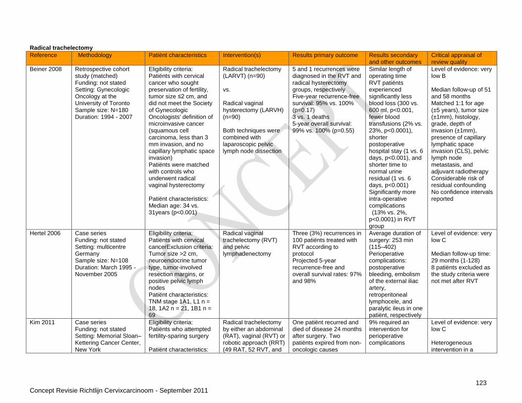
123 Concept Revisie Richtlijn Cervixcarcinoom - September 2011
Radical trachelectomy
Reference Methodology Patiënt characteristics Intervention(s) Results primary outcome Results secondary and other outcomes
Critical appraisal of review quality
Beiner 2008 Retrospective cohort study (matched) Funding: not stated Setting: Gynecologic Oncology at the University of Toronto Sample size: N=180 Duration: 1994 - 2007
Eligibility criteria: Patiënts with cervical cancer who sought preservation of fertility, tumor size ≤2 cm, and did not meet the Society of Gynecologic Oncologists' definition of microinvasive cancer (squamous cell carcinoma, less than 3 mm invasion, and no capillary lymphatic space invasion) Patiënts were matched with controls who underwent radical vaginal hysterectomy Patiënt characteristics: Median age: 34 vs. 31years (p<0.001)
Radical trachelectomy (LARVT) (n=90) vs. Radical vaginal hysterectomy (LARVH) (n=90) Both techniques were combined with laparoscopic pelvic lymph node dissection
5 and 1 recurrences were diagnosed in the RVT and radical hysterectomy groups, respectively Five-year recurrence-free survival: 95% vs. 100% (p=0.17) 3 vs. 1 deaths 5-year overall survival: 99% vs. 100% (p=0.55)
Similar length of operating time RVT patiënts experienced significantly less blood loss (300 vs. 600 ml, p<0.001, fewer blood transfusions (2% vs. 23%, p<0.0001), shorter postoperative hospital stay (1 vs. 6 days, p<0.001), and shorter time to normal urine residual (1 vs. 6 days, p<0.001) Significantly more intra-operative complications (13% vs. 2%, p<0.0001) in RVT group
Level of evidence: very low B Median follow-up of 51 and 58 months Matched 1:1 for age (±5 years), tumor size (±1mm), histology, grade, depth of invasion (±1mm), presence of capillary lymphatic space invasion (CLS), pelvic lymph node metastasis, and adjuvant radiotherapy Considerable risk of residual confounding No confidence intervals reported
Hertel 2006 Case series Funding: not stated Setting: multicentre Germany Sample size: N=108 Duration: March 1995 - November 2005
Eligibility criteria: Patiënts with cervical cancerExclusion criteria: Tumor size >2 cm, neuroendocrine tumor type, tumor-involved resection margins, or positive pelvic lymph nodes Patiënt characteristics: TNM stage 1A1, L1 n = 18, 1A2 n = 21, 1B1 n = 69
Radical vaginal trachelectomy (RVT) and pelvic lymphadenectomy
Three (3%) recurrences in 100 patiënts treated with RVT according to protocol Projected 5-year recurrence-free and overall survival rates: 97% and 98%
Average duration of surgery: 253 min (115–402) Perioperative complications: postoperative bleeding, embolism of the external iliac artery, retroperitoneal lymphocele, and paralytic ileus in one patiënt, respectively
Level of evidence: very low C Median follow-up time: 29 months (1-128) 8 patiënts excluded as the study criteria were not met after RVT
Kim 2011 Case series Funding: not stated Setting: Memorial Sloan–Kettering Cancer Center, New York
Eligibility criteria: Patiënts who attempted fertility-sparing surgery Patiënt characteristics:
Radical trachelectomy by either an abdominal (RAT), vaginal (RVT) or robotic approach (RRT) (49 RAT, 52 RVT, and
One patiënt recurred and died of disease 24 months after surgery. Two patiënts expired from non-oncologic causes
9% required an intervention for perioperative complications
Level of evidence: very low C Heterogeneous intervention in a

124 Concept Revisie Richtlijn Cervixcarcinoom - September 2011
Reference Methodology Patiënt characteristics Intervention(s) Results primary outcome Results secondary and other outcomes
Critical appraisal of review quality
Sample size: N=105 Duration: November 2001 - September 2010
Preoperative stages: 12 stage IA1 (12%), 12 stage IA2 (12%), and 81 stage IB2 (77%)
4 RRT) 35 were actively attempting conception 6-12 months after surgery 22 patiënts (63%) successfully conceived
specialised centre Median follow-up: 29 months (range 0.1–99.8)
Li 2011 Case series Funding: not stated Setting: one centre Shangai Sample size: N=64 Duration: 04/2004 - 09/2010
Eligibility criteria: Confirmed invasive cervical cancer Tumor size < 4 cm FIGO stage IA1 disease with lymph vascular space invasion, or positive surgical margin and distorted cervicovaginal anatomy after conization; or stage IA2 or IB1 disease Desire to preserve fertility Patiënt characteristics: Median age: 29.5 years (range 11-41) Stage IA1 27%, IA2 12%, IB1 61% SCC 81%
Abdominal radical trachelectomy
No recurrences 14 patiënts tumor > 2 cm 10 patiënts attempted to conceive: 2 pregnancies, 1 delivery and one ongoing
Median blood loss: 362 ml (range 100–700 ml) Median length of postoperative hospital stay: 10.14 days (range 7–21 days) Postoperative cervical stenosis: 4/64 (6.25%)
Level of evidence: very low C Median follow-up: 22.8 months (range 1–78 months)
Marchiole 2007
Retrospective cohort study Funding: not stated Setting: Sample size: N=257 Duration: December 1986 - December 2003
Eligibility criteria: FIGO stage I–IIA carcinoma of the cervix Patiënt characteristics: Mean age: 32 vs. 47 years (p<0.001) Stage IA: 24.6% vs. 18%; stage IB1: 70.3% vs. 76.3%; stage IIA: 5.1% vs. 5.8% (NS) SCC: 76.3% vs. 73.4% (NS)
Radical trachelectomy (LARVT) (n=118) vs. Radical hysterectomy (n=139) Both combined with laparoscopic pelvic lymph node dissection
Risk of recurrence: 7 cases (5.2%) in patiënts treated with LAVRT and 9 cases (6.5%) in patiënts treated with LAVRH (p=NS) 6 recurrences in the LAVRT group had a tumor, giving on 21 interventions in the group size > 2 cm
Rate of intraoperative complications: 2.5% for LAVRT and 5.8% for LAVRH, p=NS Rate of postoperative complications: 21.2% for LAVRT and 19.4% for LAVRH, p=NS
Level of evidence: low B Median follow-up: 95 months (range 31–234) for LARVT and 113 months (range 36–249) for LARVH Statistical adjustment was done for risk factors in terms of recurrence-free survival: tumor size, nodal status, LVSI, histotype, age and type of operation and did not alter conclusions;

125 Concept Revisie Richtlijn Cervixcarcinoom - September 2011
Reference Methodology Patiënt characteristics Intervention(s) Results primary outcome Results secondary and other outcomes
Critical appraisal of review quality
however, details of the adjustment were not reported
Nam 2011 Case series Funding: not stated Setting: four institutions in Korea Sample size: N=59 Duration: not mentioned
Eligibility criteria: Patiënts with early-stage cervical cancer who wanted to preserve fertility Patiënt characteristics: Median age: 29 years (range 22-44) Median tumour size: 1.8 cm
Laparoscopic radical trachelectomy
59 enrolled for LRT In 5 patiënts, LRT was abandoned because of lymph node metastasis or parametrial involvement 2 recurrences and 1 death from disease 16 patiënts attempted to conceive: 8 pregnancies, 3 healthy babies
Median estimated blood loss: 300 mL (range 50–1000) Perioperative transfusion in 15 patiënts 6 received adjuvant treatment 1 vesicovaginal fistula
Level of evidence: very low C Median follow-up was 31 months (range 7–70)
Nishio 2009 Case series Funding: not stated Setting: university hospital, Japan Sample size: N=61 Duration: September 2002 - March 2008
Eligibility criteria: Desire for fertility-sparing FIGO stage IA1 with lymph-vascular space involvement (LVSI) FIGO stage IA2 or stage IB1, no involvement of the upper endocervical canal and no Evidence of lymph node metastasis, as determined by MRI/CT Patiënt characteristics: Median age: 33 years (range 26-44) FIGO IA1 6.6%, IA2 13.1%, IB1 80.3% SCC 95.1%
Abdominal radical trachelectomy
Six recurrences (9.8%); none of the recurrences occurred in patiënts with a tumor diameter of <20 mm except in one case with adenocarcinoma among the 13 patiënts with a tumor diameter of ≥20 mm, five developed recurrent disease. Twenty-nine women attempted to conceive; four were successful. All four of these women had live births: two preterm deliveries, and two full-term deliveries
Median estimated blood loss (ml): 1160 (352–5568) Median length of stay (days): 23 (11–63) Median operative time (min): 436 (317–586) Median time to recovery of bladder dysfunction (days): 15 (7–35)
Level of evidence: very low C Median follow-up: 27 months (range 1-79 months)
Plante 2011 Case series Funding: not stated Setting: one centre in Quebec Sample size: N=140 Duration: not mentioned
Eligibility criteria: Patiënts with early-stage cervical cancer (stages IA, IB, and IIA) Desire to preserve fertility Patiënt characteristics: Median age: 31 years Stage IA2 21%, IB1 69%
Radical vaginal trachelectomy
6 recurrences (4.8%), 2 deaths (1.6%) Actuarial 5-year recurrence-free survival: 95.8% (95%CI 0.90–0.98) for the entire population, 79% (95%CI 0.49–0.93) in the group in which VRT was abandoned (p=0.001) Tumor size >2 cm was
Number of women attempting to conceive not reported 58 women conceived a total of 106 pregnancies The first- and second-trimester miscarriage rates
Level of evidence: very low C Mean follow-up was 93 months (range 4–225) RVT was abandoned in 15 patiënts (reasons not stated) Correction for confounders unclear

126 Concept Revisie Richtlijn Cervixcarcinoom - September 2011
Reference Methodology Patiënt characteristics Intervention(s) Results primary outcome Results secondary and other outcomes
Critical appraisal of review quality
SCC 56% associated with a higher risk of recurrence (p=0.002) 3 recurrences in 13 tumors > 2 cm
were 20 and 3% 77 (73%) pregnancies reached the third trimester 58 (75%) delivered at term
Shepherd 2006
Case series Funding: not stated Setting: multicentre UK Sample size: N=123 Duration: August 1994 - 2005
Eligibility criteria: Patiënts with early-stage cervical cancer Patiënt characteristics: Mean age: 30.6 (SD 4.3) Stage IA2 1.6%, IB1 98.4% SCC 67.5%
Radical vaginal trachelectomy with pelvic lymphadenectomy Eleven women (8.9%) had completion treatment. Two had completion surgery and nine had chemoradiotherapy
Three recurrences (2.7%) among the women who did not have completion treatment and two (18.2%) in those who did Sixty-three women attempted pregnancy. There were 55 pregnancies in 26 women and 28 live births in 19.
6 perioperative and 26 postoperative complications
Level of evidence: very low C fertiliteitsparende
CLS: capillary lymphatic space invasion; FIGO International Federation of Gynecology and Obstetrics; LARVT laparoscopic-assisted radical vaginal trachelectomy ; LARVH laparoscopic-assisted radical vaginal hysterectomy; LVSI: lymphatic-vascular space invasion; LLETZ: large loop excision of the transformation zone; PLND: pelvic lymph node dissection; RCT: randomized controlled trial; RH: radical hysterectomy; RVT: radical vaginal trachelectomy; RRT: Robotic Radical trachelectomy TNM: Tumor, Node, Metastasis system

127 Concept Revisie Richtlijn Cervixcarcinoom - September 2011
Vraag 3: Bij patiënten met operatief behandeld cervixcarcinoom stadium 1B/IIA met aanwezigheid van lymfekliermetastasen, heeft postoperatieve chemoradiatie in vergelijking met postoperatieve radiotherapie een betere (ziektevrije) overleving? Adjuvant chemoradiotherapy/radiotherapy
Systematic reviews
Reference Methodology Patiënt characteristics Intervention(s) Results primary outcome
Results secondary and other outcomes
Critical appraisal of review quality
Rosa 2009 Systematic Review Funding: CAPES, Brazil; Department of Health; UK NHS Cochrane Collaboration programme Grant Scheme CPG-506 Search date: January 2009 Databases: CENTRAL, Medline, EMBASE, LILACS, Biological Abstracts, CancerLit, trial registers, conference proceedings, references, experts Study designs: RCT N included studies: 3 (368 patiënts) Tattersall 1992 (n=71)(Tattersall, Ramirez et al. 1992) Peters 2000 (n=268 enrolled; 243 assessed)(Peters, Liu et al. 2000)
Eligibility criteria: Peters 2000 Patiënts with clinical stage IA2, IB, and IIA carcinoma of the cervix, initially treated with radical hysterectomy and pelvic lymphadenectomy, and who had positive pelvic lymph nodes and/or positive margins and/or microscopic involvement of the parametrium Median follow up : 42 months Protocol CE3005: Patiënts with clinical stage IB or IIA Median follow up : 29.5 months
Adjuvant platinum-based chemotherapy (in addition to radical hysterectomy, RT or both) Peters 2000: 4 cycles of CT with cisplatin and 5-fluorouracil. Protocol CE 3005: chemotherapy with bleomicin + ifosfamide + cisplatin vs. Adjuvant pelvic radiotherapy alone
CTRT vs. RT (N=2) Level of evidence: B Allocation concealment: N=1 No blinding Peters 2000 and Tattersall 1992: Kaplan-Meier plots (max duration of follow-up) Peters 2000: based on an interim analysis of the data which rejected the null hypothesis of no benefit of CT Protocol CE3005: unpublished data
Death from all causes (243 patiënts): HR 0.56 (95%CI 0.36-0.87; p=0.0096; I² 0%) in favour of chemotherapy
Disease progression (243 patiënts): HR 0.47 (0.30-0.74; p=0.0012; I² 0%) in favour of chemotherapy Grade 4 toxicity (288 patiënts): HR 5.66 (2.14-14.98; p=0.00048; I² 0%) in favour of no chemotherapy
Tattersall 1992 3 cycles of CT with Chemotherapy followed by RT vs. RT (N=1)
Reference Search date Recommendations Evidence base Level of evidence
SIGN 2008 2005 Patiënts who have undergone surgery for cervical carcinoma and have positive nodes should be considered for adjuvant treatment with concurrent chemoradiotherapy with platinum based chemotherapy
RCT‟s B
Patiënts who have undergone surgery for cervical carcinoma, have negative nodes and any two of the following risk factors should be considered for adjuvant treatment with radiotherapy, if fit enough: -greater than a third stromal invasion -lymphovascular space invasion -tumour diameter of > 4 cm
RCT‟s B
Concurrent chemoradiation should be considered in preference to radiation alone RCT‟s B

128 Concept Revisie Richtlijn Cervixcarcinoom - September 2011
Reference Methodology Patiënt characteristics Intervention(s) Results primary outcome
Results secondary and other outcomes
Critical appraisal of review quality
Protocol CE3005 - UK Clinical Trials Register 2001 (n=57 enrolled; 54 assessed)(Cancer Research UK 2001)
Patiënts with clinical stage IB (87%) and IIA (13%) carcinoma of the cervix, initially treated with radical hysterectomy and pelvic lymphadenectomy, and who had positive pelvic lymph nodes (1 - >5) Median follow up : 30 months
cisplatin, vinblastine and bleomycin followed by pelvic RT Vs. RT only
Disease progression (71 patiënts): HR 1.34 (0.24-7.66)
Abbreviations: CT: chemotherapy; CTRT: chemoradiotherapy; HR: hazard ratio; RCT: randomized controlled trial; RT: radiotherapy
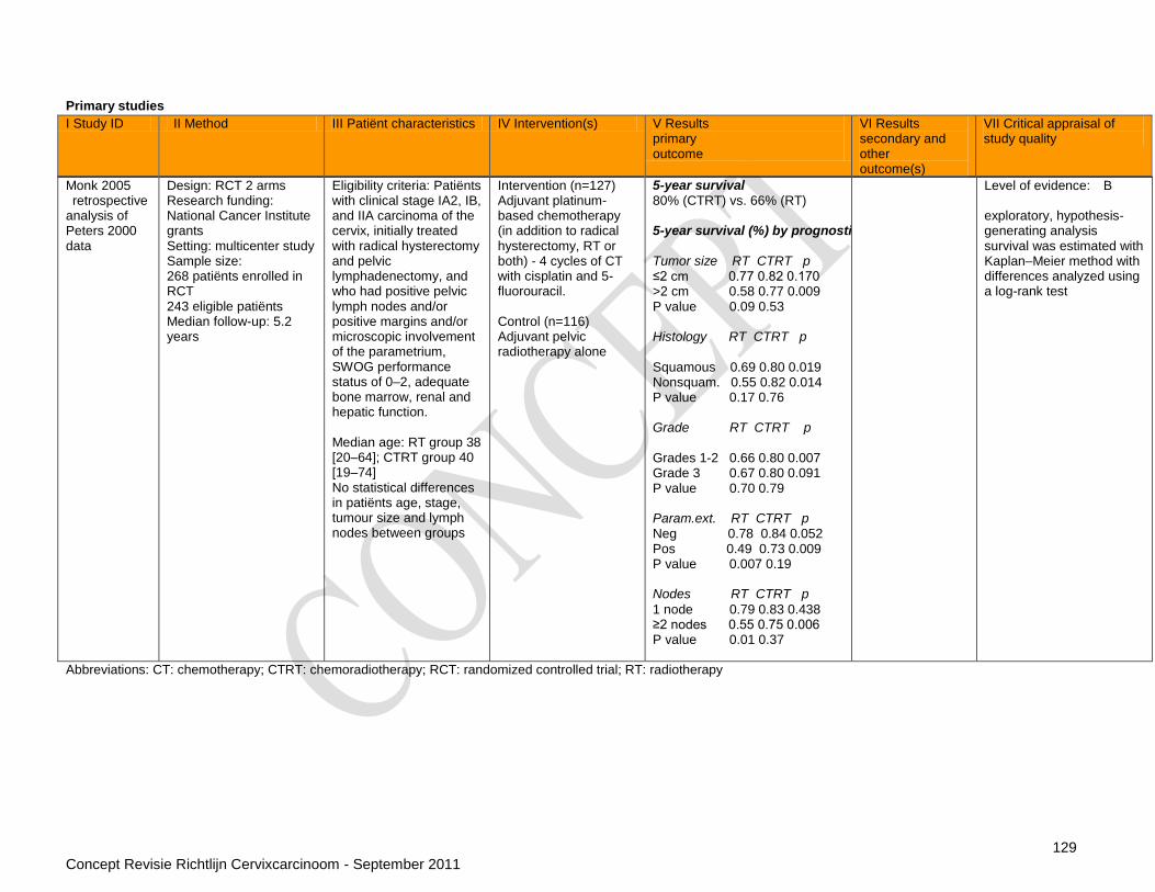
129 Concept Revisie Richtlijn Cervixcarcinoom - September 2011
Primary studies
Monk 2005 retrospective analysis of Peters 2000 data
Design: RCT 2 arms Research funding: National Cancer Institute grants Setting: multicenter study Sample size: 268 patiënts enrolled in RCT 243 eligible patiënts Median follow-up: 5.2 years
Eligibility criteria: Patiënts with clinical stage IA2, IB, and IIA carcinoma of the cervix, initially treated with radical hysterectomy and pelvic lymphadenectomy, and who had positive pelvic lymph nodes and/or positive margins and/or microscopic involvement of the parametrium, SWOG performance status of 0–2, adequate bone marrow, renal and hepatic function. Median age: RT group 38 [20–64]; CTRT group 40 [19–74] No statistical differences in patiënts age, stage, tumour size and lymph nodes between groups
Intervention (n=127) Adjuvant platinum-based chemotherapy (in addition to radical hysterectomy, RT or both) - 4 cycles of CT with cisplatin and 5-fluorouracil. Control (n=116) Adjuvant pelvic radiotherapy alone
5-year survival 80% (CTRT) vs. 66% (RT) 5-year survival (%) by prognostic factor (RT, CTRT, pvalue)
5 Tumor size RT CTRT p ≤2 cm 0.77 0.82 0.170 >2 cm 0.58 0.77 0.009 P value 0.09 0.53 10 Histology RT CTRT p Squamous 0.69 0.80 0.019 Nonsquam. 0.55 0.82 0.014 P value 0.17 0.76 15 Grade RT CTRT p Grades 1-2 0.66 0.80 0.007 Grade 3 0.67 0.80 0.091 20 P value 0.70 0.79 Param.ext. RT CTRT p Neg 0.78 0.84 0.052 Pos 0.49 0.73 0.009 25 P value 0.007 0.19 Nodes RT CTRT p
1 node 0.79 0.83 0.438 ≥2 nodes 0.55 0.75 0.006 30 P value 0.01 0.37
Level of evidence: B exploratory, hypothesis-generating analysis survival was estimated with Kaplan–Meier method with differences analyzed using a log-rank test
Abbreviations: CT: chemotherapy; CTRT: chemoradiotherapy; RCT: randomized controlled trial; RT: radiotherapy
I Study ID II Method III Patiënt characteristics IV Intervention(s) V Results primary outcome
VI Results secondary and other outcome(s)
VII Critical appraisal of study quality

130 Concept Revisie Richtlijn Cervixcarcinoom - September 2011
Vraag 4 Bij patiënten met behandeld cervixcarcinoom, welke (frequentie van) follow-up zorgt voor een betere overleving en kwaliteit van leven? Systematic reviews Study ID Method Patiënt characteristics Intervention(s) Results primary outcome Results secondary and other
outcomes Critical appraisal of review quality
(Elit, Fyles et al. 2009)
Design: systematic review
Funding: Cancer Care Ontario, Ontario Ministry of Health and Long-term Care
Conflicts of interest: none to declare
Search date:1980-November 2007
Searched databases: Medline, Embase, Cochrane databases, Canadian Medical Association Infobase, and the National Guideline Clearinghouse
Included study designs: randomized controlled trials, practice guidelines, meta-analyses, systematic reviews or cohort studies
Eligibility criteria: studies were to report data relating to follow-up programs by the method of detection, the entry criteria for the study population, survival, and the number of recurrences found during screening, or on patiënt quality of life; >25 patiënts included and English language
Patiënt characteristics: 15/17 studies included patiënts with surgical stage IB or IIA; 6/17 studies included patiënts with all stages. The majority of included patiënts were IB or IIA. Treatment received: 4/17 studies surgery only; 3/17 studies radiotherapy alone; 9/17 studies combined treatment
Index test: not applicable
Reference standard: not applicable
Follow-up visits typically occurred once every 3–4 months for the first 2 years, every 6 months for the next 3 years and then annually until year 10. There were no comparative studies on the frequency of follow-up visits Median time to recurrence ranged from 7–36 months after primary treatment. The % of recurrences detected per time period was: - 2 years 62-89% - 3 years 75-85% - 5 years 89- 99% For patiënts who were symptomatic at the time of recurrence detection, median overall survival after recurrence ranged from 8- 38 months, and for asymptomatic patiënts the range was 8 months to a median survival that was not reached after 53 months of follow-up
Rates of recurrence: ranged 8–26% - 14–57% in the pelvis - 15–61% at distant or
multiple sites Asymptomatic recurrence was detected by (in % of patiënts with asymptomatic recurrence): - physical exam 29–71% - chest x-ray 20–47% - CT 0–34% - vaginal vault cytology 0–
17% - ultrasound 0-2% - MRI 0-9% - intravenous pyelography
0% - tumour markers 0-26% - other 11-33%
Level of evidence: B (C voor uitkomst overleving)
All studies were retrospective, uncontrolled observational studies
Very heterogeneous patiënt populations
Included studies: SRs: None Primary studies: 11 published before 2001 (Duyn, Van Eijkeren et al. 2002) (Esajas, Duk et al. 2001) (Lim, Howells et al. 2004) (Morice, Deyrolle et al. 2004) (Sartori, Pasinetti et al. 2007) (Zola, Fuso et al. 2007)
(Havrilesky, Kulasingam et al. 2005)
Design: systematic review with meta-analysis
Funding: Centers for Medicare and Medicaid Services
Search date:1966-2003
Searched databases: Medline
Included study designs: observational studies
Eligibility criteria: English language studies reporting primary data and published in a peer review journal with 12 or more included patiënts
Patiënt characteristics: not reported
Disease prevalence: not reported
Index test: PET
Reference standard: histology or follow-up of 6 months or more
Meta-analysed (N=3) accuracy of PET to detect recurrence with clinical
suspicion: Se 96% (95% CI: 87–99%),
Sp 81% (95% CI: 58–94%) Meta-analysed (N=2) accuracy of PET to detect recurrence without clinical
suspicion: Se 92% (95% CI: 77–98%),
Sp 75% (95% CI: 69–80%)
Single study accuracy of PET to detect recurrence (unspecified): Se 100%, Sp 77%
Recurrence was not reported separately for locoregional or distal recurrence
Level of evidence: B
All studies were retrospective and none of them reported blinded assessment
Included studies: Primary studies: 1 published before 2001 (Belhocine, Thille et al. 2002) (Chang, Hung et al. 2004) (Nakamoto, Eisbruch et al. 2002) (Ryu, Kim et al. 2003) (Sun, Chen et al. 2001)
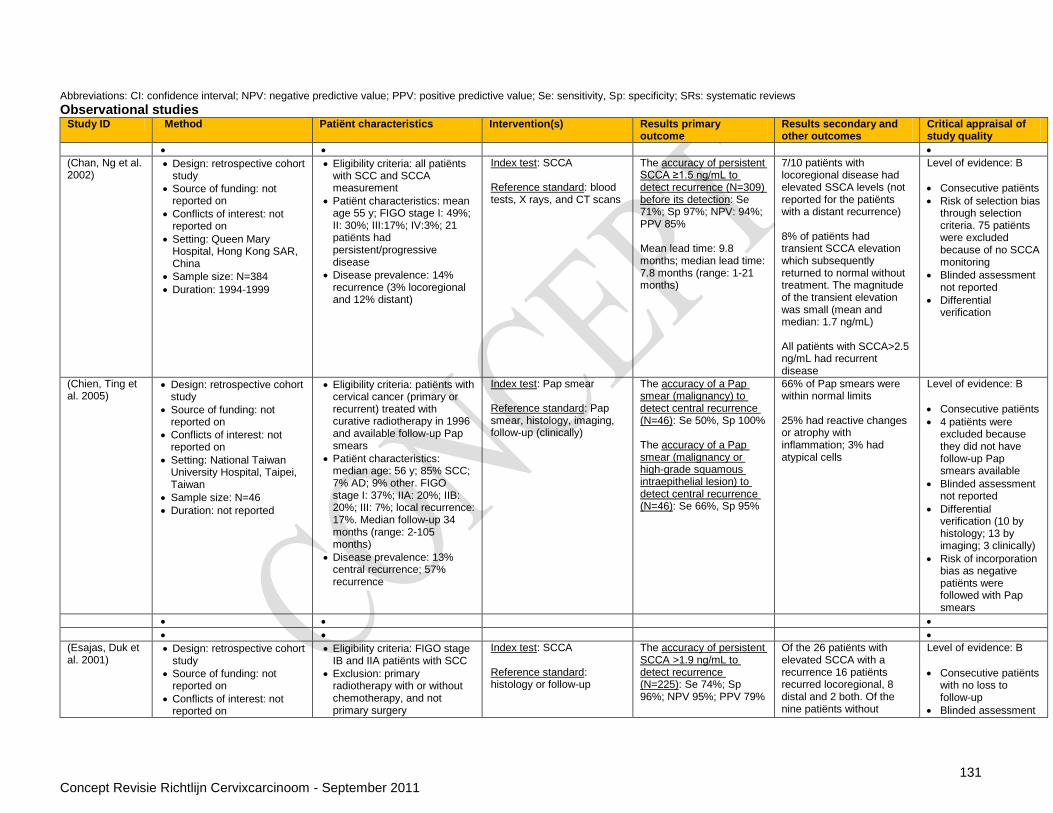
131 Concept Revisie Richtlijn Cervixcarcinoom - September 2011
Abbreviations: CI: confidence interval; NPV: negative predictive value; PPV: positive predictive value; Se: sensitivity, Sp: specificity; SRs: systematic reviews Observational studies
Study ID Method Patiënt characteristics Intervention(s) Results primary outcome
Results secondary and other outcomes
Critical appraisal of study quality
(Chan, Ng et al. 2002)
Design: retrospective cohort study
Source of funding: not reported on
Conflicts of interest: not reported on
Setting: Queen Mary Hospital, Hong Kong SAR, China
Sample size: N=384
Duration: 1994-1999
Eligibility criteria: all patiënts with SCC and SCCA measurement
Patiënt characteristics: mean age 55 y; FIGO stage I: 49%; II: 30%; III:17%; IV:3%; 21 patiënts had persistent/progressive disease
Disease prevalence: 14% recurrence (3% locoregional and 12% distant)
Index test: SCCA Reference standard: blood tests, X rays, and CT scans
The accuracy of persistent SCCA ≥1.5 ng/mL to detect recurrence (N=309) before its detection: Se 71%; Sp 97%; NPV: 94%; PPV 85% Mean lead time: 9.8 months; median lead time: 7.8 months (range: 1-21 months)
7/10 patiënts with locoregional disease had elevated SSCA levels (not reported for the patiënts with a distant recurrence) 8% of patiënts had transient SCCA elevation which subsequently returned to normal without treatment. The magnitude of the transient elevation was small (mean and median: 1.7 ng/mL) All patiënts with SCCA>2.5 ng/mL had recurrent disease
Level of evidence: B
Consecutive patiënts
Risk of selection bias through selection criteria. 75 patiënts were excluded because of no SCCA monitoring
Blinded assessment not reported
Differential verification
(Chien, Ting et al. 2005)
Design: retrospective cohort study
Source of funding: not reported on
Conflicts of interest: not reported on
Setting: National Taiwan University Hospital, Taipei, Taiwan
Sample size: N=46
Duration: not reported
Eligibility criteria: patiënts with cervical cancer (primary or recurrent) treated with curative radiotherapy in 1996 and available follow-up Pap smears
Patiënt characteristics: median age: 56 y; 85% SCC; 7% AD; 9% other. FIGO stage I: 37%; IIA: 20%; IIB: 20%; III: 7%; local recurrence: 17%. Median follow-up 34 months (range: 2-105 months)
Disease prevalence: 13% central recurrence; 57% recurrence
Index test: Pap smear Reference standard: Pap smear, histology, imaging, follow-up (clinically)
The accuracy of a Pap smear (malignancy) to detect central recurrence (N=46): Se 50%, Sp 100% The accuracy of a Pap smear (malignancy or high-grade squamous intraepithelial lesion) to detect central recurrence (N=46): Se 66%, Sp 95%
66% of Pap smears were within normal limits 25% had reactive changes or atrophy with inflammation; 3% had atypical cells
Level of evidence: B
Consecutive patiënts
4 patiënts were excluded because they did not have follow-up Pap smears available
Blinded assessment not reported
Differential verification (10 by histology; 13 by imaging; 3 clinically)
Risk of incorporation bias as negative patiënts were followed with Pap smears
(Esajas, Duk et al. 2001)
Design: retrospective cohort study
Source of funding: not reported on
Conflicts of interest: not reported on
Eligibility criteria: FIGO stage IB and IIA patiënts with SCC
Exclusion: primary radiotherapy with or without chemotherapy, and not primary surgery
Index test: SCCA Reference standard: histology or follow-up
The accuracy of persistent SCCA >1.9 ng/mL to detect recurrence (N=225): Se 74%; Sp 96%; NPV 95%; PPV 79%
Of the 26 patiënts with elevated SCCA with a recurrence 16 patiënts recurred locoregional, 8 distal and 2 both. Of the nine patiënts without
Level of evidence: B
Consecutive patiënts with no loss to follow-up
Blinded assessment

132 Concept Revisie Richtlijn Cervixcarcinoom - September 2011
Study ID Method Patiënt characteristics Intervention(s) Results primary outcome
Results secondary and other outcomes
Critical appraisal of study quality
Setting: University Hospital Groningen, the Netherlands
Sample size: N=225
Duration: 1987-1998
Patiënt characteristics: see eligibility
Disease prevalence: 16% recurrence (10% locoregional, 4% distant and 1% both)
elevated SCCA with a recurrence 6 patiënts had a locoregional recurrence, 2 patiënts had a distal recurrence and 1 a mixed recurrence 7% of patiënts had a transient rise in SCCA that normalised after 6 to 8 weeks False-positive serum SCCA could be related to benign skin disorders and to chronic obstructive pulmonary disease Also included in (Elit, Fyles et al. 2009)
not reported
Differential follow-up
(Forni, Ferrandina et al. 2007)
Design: prospective cohort study
Source of funding: not reported on
Conflicts of interest: no conflicts of interest to report
Setting: Italy
Sample size: N=135
Duration: not reported
Eligibility criteria: all patiënts treated by the same team
Patiënt characteristics: median age: 57 y; 76% primary cervical carcinoma; 24% had already experienced disease recurrence that had been successfully treated
Disease prevalence: 32% recurrence or re-recurrence (21% locoregional and 11% both locoregional and distant)
Index test: SCCA Reference standard: physical and gynecologic examination (including Papanicolaou smear and colposcopy), complete blood analysis, chest X-ray and abdominopelvic MRI or CT plus transrectal ultrasonography
The accuracy of SCCA >1.4 ng/mL to detect recurrence before symptoms (N=135): Se 79%; Sp 96%; NPV 91%; PPV 90% The accuracy of SCCA 1.4 ng/mL + gynaecologic examination to detect recurrence (N=135): Se 95%; Sp 96%; NPV 98%; PPV 91%
No difference was found in the SCCA levels according to the site of recurrence 24/28 locoregional recurrences had elevated SCCA. 10/15 patiënts with both locoregional and distant recurrence had elevated SCCA In all patiënts, the elevation of SCCA levels preceded the appearance of any signs or symptoms of disease with a mean lead time of 4.7 months The total projected cost of the standard follow- up procedure, including CT or MRI, was 3,653.4 Euros per patiënt. The projected cost of the approach using only SCCA and gynaecologic examination was 298.5 Euros per patiënt
Level of evidence: B
Dropouts: none
Consecutive patiënts
Blinded assessment not reported
Differential verification
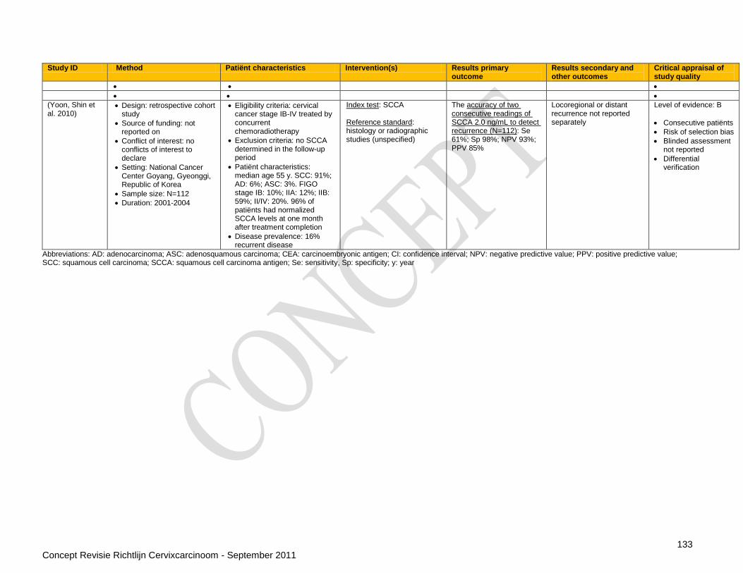
133 Concept Revisie Richtlijn Cervixcarcinoom - September 2011
Study ID Method Patiënt characteristics Intervention(s) Results primary outcome
Results secondary and other outcomes
Critical appraisal of study quality
(Yoon, Shin et al. 2010)
Design: retrospective cohort study
Source of funding: not reported on
Conflict of interest: no conflicts of interest to declare
Setting: National Cancer Center Goyang, Gyeonggi, Republic of Korea
Sample size: N=112
Duration: 2001-2004
Eligibility criteria: cervical cancer stage IB-IV treated by concurrent chemoradiotherapy
Exclusion criteria: no SCCA determined in the follow-up period
Patiënt characteristics: median age 55 y. SCC: 91%; AD: 6%; ASC: 3%. FIGO stage IB: 10%; IIA: 12%; IIB: 59%; II/IV: 20%. 96% of patiënts had normalized SCCA levels at one month after treatment completion
Disease prevalence: 16% recurrent disease
Index test: SCCA Reference standard: histology or radiographic studies (unspecified)
The accuracy of two consecutive readings of SCCA 2.0 ng/mL to detect recurrence (N=112): Se 61%; Sp 98%; NPV 93%; PPV 85%
Locoregional or distant recurrence not reported separately
Level of evidence: B
Consecutive patiënts
Risk of selection bias
Blinded assessment not reported
Differential verification
Abbreviations: AD: adenocarcinoma; ASC: adenosquamous carcinoma; CEA: carcinoembryonic antigen; CI: confidence interval; NPV: negative predictive value; PPV: positive predictive value; SCC: squamous cell carcinoma; SCCA: squamous cell carcinoma antigen; Se: sensitivity, Sp: specificity; y: year
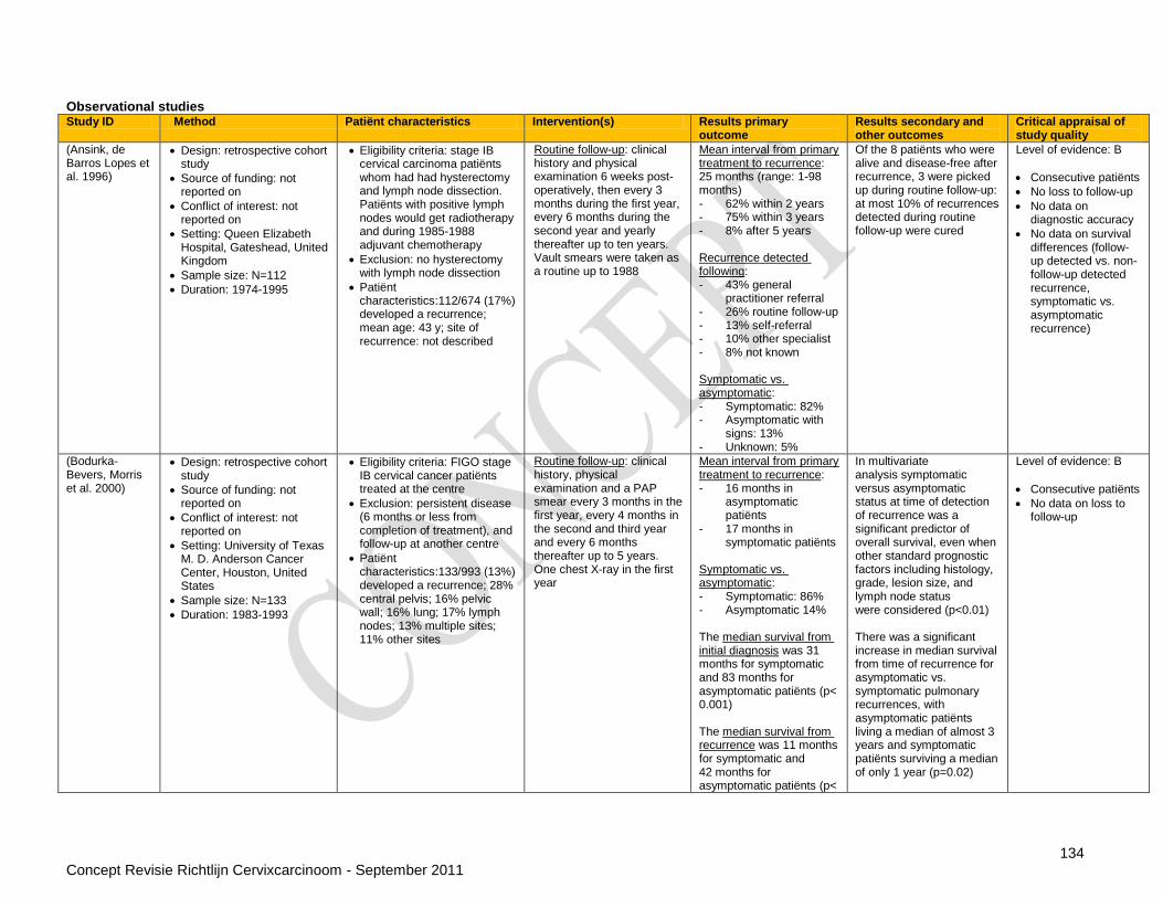
134 Concept Revisie Richtlijn Cervixcarcinoom - September 2011
Observational studies Study ID Method Patiënt characteristics Intervention(s) Results primary
outcome Results secondary and other outcomes
Critical appraisal of study quality
(Ansink, de Barros Lopes et al. 1996)
Design: retrospective cohort study
Source of funding: not reported on
Conflict of interest: not reported on
Setting: Queen Elizabeth Hospital, Gateshead, United Kingdom
Sample size: N=112
Duration: 1974-1995
Eligibility criteria: stage IB cervical carcinoma patiënts whom had had hysterectomy and lymph node dissection. Patiënts with positive lymph nodes would get radiotherapy and during 1985-1988 adjuvant chemotherapy
Exclusion: no hysterectomy with lymph node dissection
Patiënt characteristics:112/674 (17%) developed a recurrence; mean age: 43 y; site of recurrence: not described
Routine follow-up: clinical history and physical examination 6 weeks post-operatively, then every 3 months during the first year, every 6 months during the second year and yearly thereafter up to ten years. Vault smears were taken as a routine up to 1988
Mean interval from primary treatment to recurrence: 25 months (range: 1-98 months) - 62% within 2 years - 75% within 3 years - 8% after 5 years Recurrence detected following: - 43% general
practitioner referral - 26% routine follow-up - 13% self-referral - 10% other specialist - 8% not known Symptomatic vs. asymptomatic: - Symptomatic: 82% - Asymptomatic with
signs: 13% - Unknown: 5%
Of the 8 patiënts who were alive and disease-free after recurrence, 3 were picked up during routine follow-up: at most 10% of recurrences detected during routine follow-up were cured
Level of evidence: B
Consecutive patiënts
No loss to follow-up
No data on diagnostic accuracy
No data on survival differences (follow-up detected vs. non-follow-up detected recurrence, symptomatic vs. asymptomatic recurrence)
(Bodurka-Bevers, Morris et al. 2000)
Design: retrospective cohort study
Source of funding: not reported on
Conflict of interest: not reported on
Setting: University of Texas M. D. Anderson Cancer Center, Houston, United States
Sample size: N=133
Duration: 1983-1993
Eligibility criteria: FIGO stage IB cervical cancer patiënts treated at the centre
Exclusion: persistent disease (6 months or less from completion of treatment), and follow-up at another centre
Patiënt characteristics:133/993 (13%) developed a recurrence; 28% central pelvis; 16% pelvic wall; 16% lung; 17% lymph nodes; 13% multiple sites; 11% other sites
Routine follow-up: clinical history, physical examination and a PAP smear every 3 months in the first year, every 4 months in the second and third year and every 6 months thereafter up to 5 years. One chest X-ray in the first year
Mean interval from primary treatment to recurrence: - 16 months in
asymptomatic patiënts
- 17 months in symptomatic patiënts
Symptomatic vs. asymptomatic: - Symptomatic: 86% - Asymptomatic 14% The median survival from initial diagnosis was 31 months for symptomatic and 83 months for asymptomatic patiënts (p< 0.001) The median survival from recurrence was 11 months for symptomatic and 42 months for asymptomatic patiënts (p<
In multivariate analysis symptomatic versus asymptomatic status at time of detection of recurrence was a significant predictor of overall survival, even when other standard prognostic factors including histology, grade, lesion size, and lymph node status were considered (p<0.01) There was a significant increase in median survival from time of recurrence for asymptomatic vs. symptomatic pulmonary recurrences, with asymptomatic patiënts living a median of almost 3 years and symptomatic patiënts surviving a median of only 1 year (p=0.02)
Level of evidence: B
Consecutive patiënts
No data on loss to follow-up
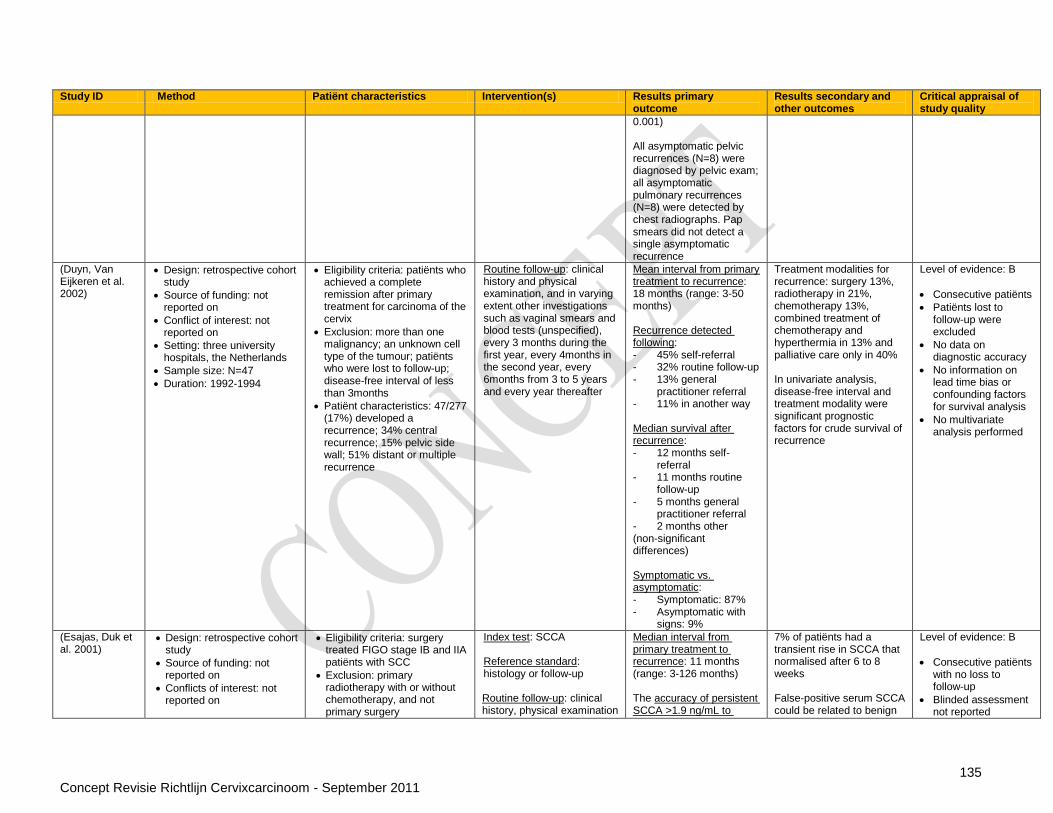
135 Concept Revisie Richtlijn Cervixcarcinoom - September 2011
Study ID Method Patiënt characteristics Intervention(s) Results primary outcome
Results secondary and other outcomes
Critical appraisal of study quality
0.001) All asymptomatic pelvic recurrences (N=8) were diagnosed by pelvic exam; all asymptomatic pulmonary recurrences (N=8) were detected by chest radiographs. Pap smears did not detect a single asymptomatic recurrence
(Duyn, Van Eijkeren et al. 2002)
Design: retrospective cohort study
Source of funding: not reported on
Conflict of interest: not reported on
Setting: three university hospitals, the Netherlands
Sample size: N=47
Duration: 1992-1994
Eligibility criteria: patiënts who achieved a complete remission after primary treatment for carcinoma of the cervix
Exclusion: more than one malignancy; an unknown cell type of the tumour; patiënts who were lost to follow-up; disease-free interval of less than 3months
Patiënt characteristics: 47/277 (17%) developed a recurrence; 34% central recurrence; 15% pelvic side wall; 51% distant or multiple recurrence
Routine follow-up: clinical history and physical examination, and in varying extent other investigations such as vaginal smears and blood tests (unspecified), every 3 months during the first year, every 4months in the second year, every 6months from 3 to 5 years and every year thereafter
Mean interval from primary treatment to recurrence: 18 months (range: 3-50 months) Recurrence detected following: - 45% self-referral - 32% routine follow-up - 13% general
practitioner referral - 11% in another way Median survival after recurrence: - 12 months self-
referral - 11 months routine
follow-up - 5 months general
practitioner referral - 2 months other (non-significant differences) Symptomatic vs. asymptomatic: - Symptomatic: 87% - Asymptomatic with
signs: 9%
Treatment modalities for recurrence: surgery 13%, radiotherapy in 21%, chemotherapy 13%, combined treatment of chemotherapy and hyperthermia in 13% and palliative care only in 40% In univariate analysis, disease-free interval and treatment modality were significant prognostic factors for crude survival of recurrence
Level of evidence: B
Consecutive patiënts
Patiënts lost to follow-up were excluded
No data on diagnostic accuracy
No information on lead time bias or confounding factors for survival analysis
No multivariate analysis performed
(Esajas, Duk et al. 2001)
Design: retrospective cohort study
Source of funding: not reported on
Conflicts of interest: not reported on
Eligibility criteria: surgery treated FIGO stage IB and IIA patiënts with SCC
Exclusion: primary radiotherapy with or without chemotherapy, and not primary surgery
Index test: SCCA Reference standard: histology or follow-up Routine follow-up: clinical history, physical examination
Median interval from primary treatment to recurrence: 11 months (range: 3-126 months) The accuracy of persistent SCCA >1.9 ng/mL to
7% of patiënts had a transient rise in SCCA that normalised after 6 to 8 weeks False-positive serum SCCA could be related to benign
Level of evidence: B
Consecutive patiënts with no loss to follow-up
Blinded assessment not reported
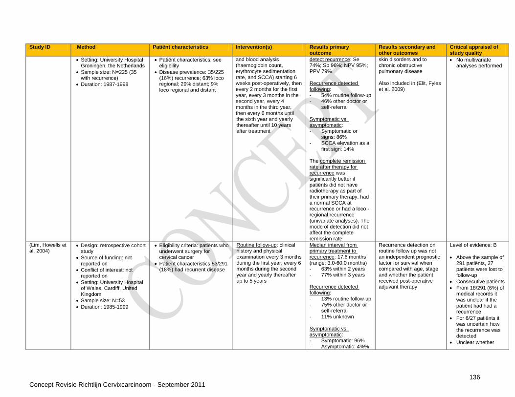
136 Concept Revisie Richtlijn Cervixcarcinoom - September 2011
Study ID Method Patiënt characteristics Intervention(s) Results primary outcome
Results secondary and other outcomes
Critical appraisal of study quality
Setting: University Hospital Groningen, the Netherlands
Sample size: N=225 (35 with recurrence)
Duration: 1987-1998
Patiënt characteristics: see eligibility
Disease prevalence: 35/225 (16%) recurrence; 63% loco regional; 29% distant; 9% loco regional and distant
and blood analysis (haemoglobin count, erythrocyte sedimentation rate, and SCCA) starting 6 weeks post-operatively, then every 2 months for the first year, every 3 months in the second year, every 4 months in the third year, then every 6 months until the sixth year and yearly thereafter until 10 years after treatment
detect recurrence: Se 74%; Sp 96%; NPV 95%; PPV 79% Recurrence detected following: - 54% routine follow-up - 46% other doctor or
self-referral Symptomatic vs. asymptomatic: - Symptomatic or
signs: 86% - SCCA elevation as a
first sign: 14% The complete remission rate after therapy for recurrence was significantly better if patiënts did not have radiotherapy as part of their primary therapy, had a normal SCCA at recurrence or had a loco -regional recurrence (univariate analyses). The mode of detection did not affect the complete remission rate
skin disorders and to chronic obstructive pulmonary disease Also included in (Elit, Fyles et al. 2009)
No multivariate analyses performed
(Lim, Howells et al. 2004)
Design: retrospective cohort study
Source of funding: not reported on
Conflict of interest: not reported on
Setting: University Hospital of Wales, Cardiff, United Kingdom
Sample size: N=53
Duration: 1985-1999
Eligibility criteria: patiënts who underwent surgery for cervical cancer
Patiënt characteristics:53/291 (18%) had recurrent disease
Routine follow-up: clinical history and physical examination every 3 months during the first year, every 6 months during the second year and yearly thereafter up to 5 years
Median interval from primary treatment to recurrence: 17.6 months (range: 3.0-60.0 months) - 63% within 2 years - 77% within 3 years Recurrence detected following: - 13% routine follow-up - 75% other doctor or
self-referral - 11% unknown Symptomatic vs. asymptomatic: - Symptomatic: 96% - Asymptomatic: 4%%
Recurrence detection on routine follow up was not an independent prognostic factor for survival when compared with age, stage and whether the patiënt received post-operative adjuvant therapy
Level of evidence: B
Above the sample of 291 patiënts, 27 patiënts were lost to follow-up
Consecutive patiënts
From 18/291 (6%) of medical records it was unclear if the patiënt had had a recurrence
For 6/27 patiënts it was uncertain how the recurrence was detected
Unclear whether
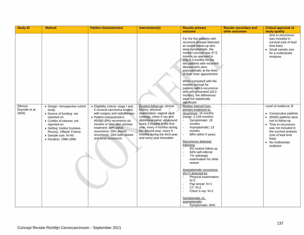
137 Concept Revisie Richtlijn Cervixcarcinoom - September 2011
Study ID Method Patiënt characteristics Intervention(s) Results primary outcome
Results secondary and other outcomes
Critical appraisal of study quality
For the five patiënts with recurrent disease detected at routine follow-up who were symptomatic, the median survival was 37.5 months as opposed to only 8.3 months for the two patiënts with recurrent disease who were asymptomatic at the time of their clinic appointment When compared with the median survival for patiënts with a recurrence who self-presented (26.0 months), the differences were not statistically significant
time to recurrence was included in survival (risk of lead time bias)
Small sample size for a multivariate analyses
(Morice, Deyrolle et al. 2004)
Design: retrospective cohort study
Source of funding: not reported on
Conflict of interest: not reported on
Setting: Institut Gustave-Roussy, Villejuif, France
Sample size: N=45
Duration: 1986-1998
Eligibility criteria: stage I and II cervical carcinoma treated with surgery and radiotherapy
Patiënt characteristics: 45/583 (8%) recurrence six months or later after primary treatment; 56% pelvic recurrence; 29% distant recurrence; 16% both distant and local recurrence
Routine follow-up: clinical history, physical examination, vaginal vault cytology, chest X-ray and abdominal pelvic ultrasound every 3 months in the first year, every 4 months during the second year, every 6 months during the third year, and every year thereafter
Median interval from primary treatment to recurrence: 16 months (range: 2-128 months) - Symptomatic: 16
months - Asymptomatic: 13
months - 89% within 5 years Recurrence detected following: - 9% routine follow-up - 84% self-referral - 7% radiologic
examination for other reason
Asymptomatic recurrence (N=7) detected by: - Physical examination:
N=2 - Pap smear: N=1 - CT: N=2 - Chest X-ray: N=2 Symptomatic vs. asymptomatic: - Symptomatic: 84%
- Level of evidence: B
Consecutive patiënts
28/583 patiënts were lost to follow-up
Time to recurrence was not included in the survival analysis (risk of lead time bias)
No multivariate analyses

138 Concept Revisie Richtlijn Cervixcarcinoom - September 2011
Study ID Method Patiënt characteristics Intervention(s) Results primary outcome
Results secondary and other outcomes
Critical appraisal of study quality
- Asymptomatic: 16% The median survival since recurrence was 14 months for symptomatic and asymptomatic patiënts
(Samlal, Van Der Velden et al. 1998)
Design: retrospective cohort study
Source of funding: not reported on
Conflict of interest: not reported on
Setting: Academic Medical Centre, Amsterdam, the Netherlands
Sample size: N=27
Duration: 1982-1991
Eligibility criteria: cervical carcinoma IB and IIA treated at the study´s centre
Patiënt characteristics: 27/271 (10%) recurrence; 52% pelvic recurrence; 48% extra-pelvic recurrence
Routine follow-up: clinical history, physical examination and vaginal vault cytology, and in patiënts who had received radiotherapy also chest X-rays and ultrasound examinations of the kidneys, every 3 months in the first year, every 6 months up to three years and every year thereafter up to 10 years after treatment. From 1989 SCCA was included in routine follow-up
Median interval from primary treatment to recurrence: 14 months (range: 3-64 months) - 77% within 3 years - 14 months for a
pelvic recurrence vs. 17 months for an extra-pelvic recurrence (p=0.03)
Recurrence detected following: - 36% routine follow-up Symptomatic vs. asymptomatic: - Symptomatic: 65% - Asymptomatic: 33% The death rate in symptomatic patiënts was 94% vs. 56% in asymptomatic patiënts (significant difference). Two patiënts who survived a pulmonary recurrence were asymptomatic
41% of all recurrences were detected through physical examination and 52% through routine follow-up radiological examination
Level of evidence: B
Consecutive patiënts
Loss to follow-up not reported
Median follow-up 60 months
No multivariate analyses performed
(Sartori, Pasinetti et al. 2007)
Design: retrospective cohort study
Source of funding: not reported on
Conflict of interest: none to declare
Setting: University of Brescia, Brescia, Italy
Sample size: N=63
Duration: 1981-2004
Eligibility criteria: stage IB–IIA cervical cancer, referred to the centre, with recurrence more than 3 months after primary treatment
Patiënt characteristics: median age 46 years (range: 18-72 years); 83% IB; 18% IIA; 57% pelvic recurrence; 43 distant recurrence
Routine follow-up: clinical history and physical examination every 3 months in the first year, every 4 months in the second year, every 6 months in the third to fifth year and annually thereafter. Pap smear from the vaginal vault and chest radiograph were taken annually
Diagnosis of recurrence was based on: - CT: 41% - Physical examination:
27% - Ultrasound: 10% - Histology: 8% - Chest X-ray: 6% - MRI: 5% - Cytology: 3% 53% of symptomatic recurrences was detected by physical examination
- Level of evidence: B
Includes the same patiënts as (Zola, Fuso et al. 2007), so only additional information is described here
Consecutive patiënts
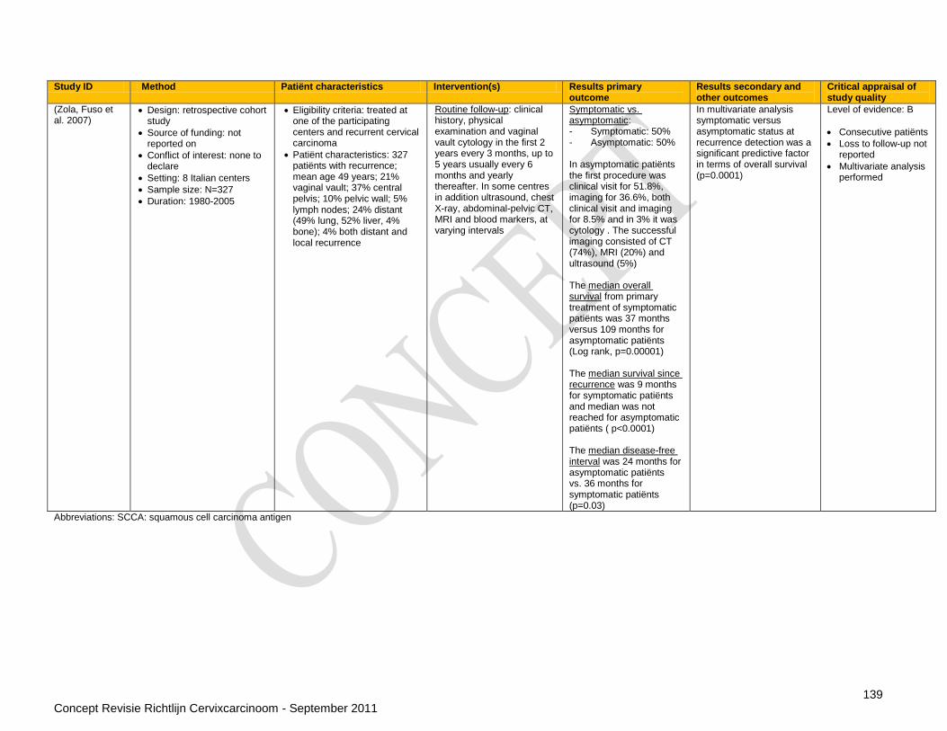
139 Concept Revisie Richtlijn Cervixcarcinoom - September 2011
Study ID Method Patiënt characteristics Intervention(s) Results primary outcome
Results secondary and other outcomes
Critical appraisal of study quality
(Zola, Fuso et al. 2007)
Design: retrospective cohort study
Source of funding: not reported on
Conflict of interest: none to declare
Setting: 8 Italian centers
Sample size: N=327
Duration: 1980-2005
Eligibility criteria: treated at one of the participating centers and recurrent cervical carcinoma
Patiënt characteristics: 327 patiënts with recurrence; mean age 49 years; 21% vaginal vault; 37% central pelvis; 10% pelvic wall; 5% lymph nodes; 24% distant (49% lung, 52% liver, 4% bone); 4% both distant and local recurrence
Routine follow-up: clinical history, physical examination and vaginal vault cytology in the first 2 years every 3 months, up to 5 years usually every 6 months and yearly thereafter. In some centres in addition ultrasound, chest X-ray, abdominal-pelvic CT, MRI and blood markers, at varying intervals
Symptomatic vs. asymptomatic: - Symptomatic: 50% - Asymptomatic: 50% In asymptomatic patiënts the first procedure was clinical visit for 51.8%, imaging for 36.6%, both clinical visit and imaging for 8.5% and in 3% it was cytology . The successful imaging consisted of CT (74%), MRI (20%) and ultrasound (5%) The median overall survival from primary treatment of symptomatic patiënts was 37 months versus 109 months for asymptomatic patiënts (Log rank, p=0.00001) The median survival since recurrence was 9 months for symptomatic patiënts and median was not reached for asymptomatic patiënts ( p<0.0001) The median disease-free interval was 24 months for asymptomatic patiënts vs. 36 months for symptomatic patiënts (p=0.03)
In multivariate analysis symptomatic versus asymptomatic status at recurrence detection was a significant predictive factor in terms of overall survival (p=0.0001)
Level of evidence: B
Consecutive patiënts
Loss to follow-up not reported
Multivariate analysis performed
Abbreviations: SCCA: squamous cell carcinoma antigen

140 Concept Revisie Richtlijn Cervixcarcinoom - September 2011
Vraag 5: De behandeling van patiënten met recidief cervixcarcinoom
Guidelines1
Treatment of recurrent, metastatic or persistent cervical cancer
Reference Search date Recommendations Evidence base Level of evidence
SIGN 2008 2005 Pelvic exenteration should be reserved as salvage surgery for women with recurrent cervical cancer in the central pelvis whose chemoradiotherapy has failed
Observational studies
C
MRI or CT should be considered initially to assess potential clinical recurrence in symptomatic patiënts
Observational studies
B
A whole body PET scan or PET-CT should be performed on all patiënts in whom recurrent or persistent disease has been demonstrated on MRI or CT and in whom salvage therapy (either pelvic exenteration or radiotherapy) is being considered
Observational studies
B
Palliative chemotherapy should be offered to women with FIGO stage IVB or recurrent cervical carcinoma, after discussion of the relative benefits and risks, with either:
cisplatin 50 mg/m² on day 1 plus topotecan 0.75 mg/m² on days 1 to 3 every 3 weeks, or
cisplatin 50 mg/m² on day 1 plus paclitaxel 135 mg/m² every 3 weeks
RCT‟s A2
CCO 2006 February 2006 It is recommended that all patiënts, particularly those who have been previously treated with cisplatin as a radiosensitizer, be offered the opportunity to participate in randomized trials, if available, that evaluate the efficacy and toxicity of other single-agent or combination chemotherapy regimens
Expert opinion D
Until further evidence becomes available, it is recommended that cisplatin in combination with topotecan should be offered to patiënts on the basis of improvements in response and survival outcomes when compared with single-agent cisplatin alone. (Note: The improvement in outcomes must be weighed against significant increases in adverse events, especially hematological toxicities, and the degree of the clinical benefit. Despite the increase in toxicity, no significant differences in quality of life were detected. Severe hematological toxicities were managed by dose modification and the use of granulocyte-colony–stimulating factors (G-CSFs) in subsequent cycles)
RCT‟s A2
1 The recommendations from the referenced guidelines are literally presented in this table. The level of evidence is attributed by the authors of this report.

141 Concept Revisie Richtlijn Cervixcarcinoom - September 2011
Additional evidence
Chemotherapy for recurrent and stage IVB
Primary studies
Study ID Method Patiënt characteristics
Intervention(s) Results primary outcome
Results secondary and other outcome(s)
Critical appraisal of study quality
Monk 2009 Cella 2010
Design: RCT 4 arms
Research funding: none
Setting: one hospital
Sample size: o 513 patiënts
enrolled in RCT with 2 arms early closure for futility
o 472 patiënts enrolled in RCT with 4 arms 434 patiënts evaluable for efficacy
Duration: 12 months
Eligibility criteria: o Stage IVB, recurrent,
or persistent cervical cancer
o Squamous, adenosquamous and adenocarcinoma
o Confirmed by biopsy or CT/MRI if lesion > 3 cm
o GOG performance status 0-1
o adequate hepatic, renal, and bone marrow function, no history of prior chemotherapy for metastatic disease
1. Intervention(s)
VC (n=108): vinorelbine 30mg/m
2 on days
1 and 8 plus cisplatin 50 mg/m
2 on day 1
every 3 weeks;
GC (n=112): gemcitabine 1,000 mg/m
2 on
days 1 and 8 plus cisplatin 50 mg/m
2 on day 1
every 3 weeks;
TC (n=111): topotecan 0.75 mg/m
2 on days 1,
2, and 3 plus cisplatin 50 mg/m
2 on day 1
every 3 weeks. 2. Comparator(s)
PC (n=103): paclitaxel 135mg/m
2 over
24 hours plus cisplatin 50mg/m
2
on day 2 every 3 weeks;
Median Overall survival PC: 12.87 months (95%CI 10.02-16.76 months, unadjusted for multiplicity). VC: 9.99 months (95%CI 8.25-12.25 months) HR 1.15 (95%CI 0.79- 1.67) GC: 10.28 months (95%CI 7.62-11.60 months) HR 1.32 (95%CI 0.91-1.92) TC: 10.25 months (95%CI 8.61-11.66 months) HR 1.26 (95%CI 0.86-1.82) Median progression-free survival PC: 5.82 months (95%CI 4.53-7.59 months, unadjusted
Toxicity Comparable rates of adverse events between arms except for : Grade 3 leucopenia: 43% (GC), 63% (PC), 68% (VC), 71% (TC) ; p<0.0001 Grade 3 neutropenia: 42% (GC), 78% (PC), 78% (VC), 83% (TC); p<0.0001 Grade 3 thrombocytopenia: 28% (GC), 7% (PC), 7.5% (VC), 35% (TC); p<0.0001 Grade 3 anaemia : 34% (GC), 17% (PC), 29% (VC), 35% (TC); p=0.02
Level of evidence: B Results critical appraisal
No information about blinding
No information about comparability of treatments

142 Concept Revisie Richtlijn Cervixcarcinoom - September 2011
Study ID Method Patiënt characteristics
Intervention(s) Results primary outcome
Results secondary and other outcome(s)
Critical appraisal of study quality
for multiplicity). VC: 3.98 months (95%CI 3.19-5.16 months) HR 1.36 (95%CI 0.97- 1.90) GC: 4.70 months (95%CI 3.58-5.59 months) HR 1.39 (95%CI 0.99-1.96) TC: 4.57 months (95%CI 3.71-5.75 months) HR 1.27 (95%CI 0.90-1.78)
Grade 3 infection: 9% (GC), 13% (PC), 7.5% (VC), 5% (TC); p=0.04 Grade 2 alopecia: 54% (PC), 9% (VC), 7% (GC) 26% (TC) (P<.0001). Quality of life No significant differences for QoL, neuropathy or pain between arms
Mountzios 2009
Design: RCT 2 arms
Setting: outpatiënt administration
Sample size: o 153 patiënts
enrolled in RCT 149 eligible patiënts
Median follow-up : 57.3 months (range 4–96 months)
Eligibility criteria: histologically documented primary metastatic or recurrent carcinoma of the uterine cervix
No previous chemotherapy for advanced disease with the exception of prior cisplatin as radiation sensitizer
ECOG performance status 0-2
Adequate hepatic, renal, and bone
1. Intervention o ITP : ifosfamide
1.5 g/m2, daily,
on days 1–3 + cisplatin 70 mg/m
2 on day 2 +
paclitaxel 175 mg/m
2 on day 1
2. Comparator o IP : ifosfamide
1.5 g/m2, daily,
on days 1–3 + cisplatin 70 mg/m
2 on day 2
Complete response ITP: 25%; 95%CI 16%-36% IP: 11%; 95%CI 5%-20% P=0.033 Partial response ITP: 34%; 95%CI 24%-46% IP: 22%, 95%CI 13%-33% P = 0.105 Overall response
ITP vs. IP: 59%
Median PFS ITP vs. IP: 7.9 (95%CI 6.1–9.8 months) vs. 6.3 months (95%CI 4.3–8.2 months), P = 0.023 Median OS ITP vs. IP: 15.4 (95%CI 8.6–22.3 months) vs.13.2 months (95%CI 10.9–15.5 months), P = 0.048
Level of evidence: B Results critical appraisal
No information about technique for randomisation
Blinding of outcome evaluation
ITT analysis
Phase II trial
No loss to follow-up
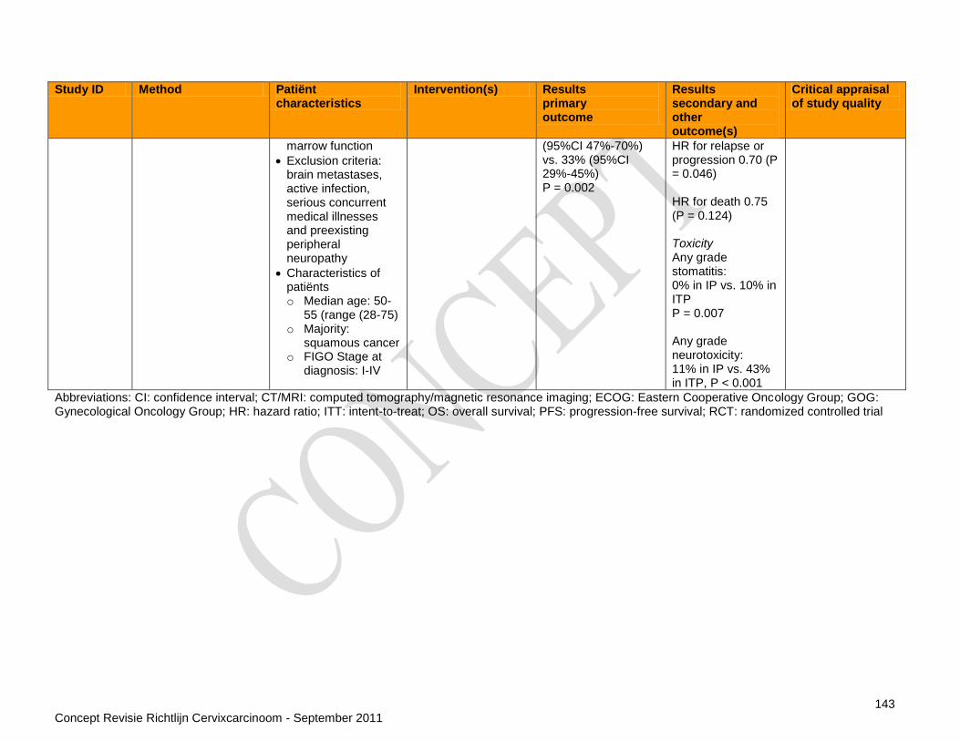
143 Concept Revisie Richtlijn Cervixcarcinoom - September 2011
Study ID Method Patiënt characteristics
Intervention(s) Results primary outcome
Results secondary and other outcome(s)
Critical appraisal of study quality
marrow function
Exclusion criteria: brain metastases, active infection, serious concurrent medical illnesses and preexisting peripheral neuropathy
Characteristics of patiënts o Median age: 50-
55 (range (28-75) o Majority:
squamous cancer o FIGO Stage at
diagnosis: I-IV
(95%CI 47%-70%) vs. 33% (95%CI 29%-45%) P = 0.002
HR for relapse or progression 0.70 (P = 0.046) HR for death 0.75 (P = 0.124) Toxicity Any grade stomatitis: 0% in IP vs. 10% in ITP P = 0.007 Any grade neurotoxicity: 11% in IP vs. 43% in ITP, P < 0.001
Abbreviations: CI: confidence interval; CT/MRI: computed tomography/magnetic resonance imaging; ECOG: Eastern Cooperative Oncology Group; GOG: Gynecological Oncology Group; HR: hazard ratio; ITT: intent-to-treat; OS: overall survival; PFS: progression-free survival; RCT: randomized controlled trial

144 Concept Revisie Richtlijn Cervixcarcinoom - September 2011
Vraag 6: Wat zijn de behandelingsmogelijkheden bij seksueel disfunctioneren na behandeling voor cervixcarcinoom Guidelines
Reference Search date Recommendations Evidence base Level of evidence
SIGN 2008 2005 Women should be offered a vaginal stent or dilator to prevent post-radiotherapy vaginal complications
Case series C
Information about female sexual function should be offered to patiënts by a relevantly trained healthcare professional using a model of care that involves addressing motivational issues and teaching behavioural skills
RCT A2
Patiënts should be offered support sessions by a designated member of their care team, as soon as possible after treatment, which my include one or more of the following:
- Relaxation - Personalised information about their disease and treatment - Emotional support and care
RCT of poor quality B
Topical oestrogens or benzydamine douches may be considered to alleviate postradiotherapy vaginal complications
RCT of poor quality B
Systematic reviews
I Study ID II Method III Patiënt characteristics IV Intervention(s) V Results primary outcome VI Results secondary outcome
VII Critical appraisal of review quality
Miles 2007 - Design: systematic review
- Sources of funding: none mentioned
- Search date: January 2007
- Searched databases: Cochrane Central Register of Controlled Trials (CENTRAL), MEDLINE, PsycINFO AMED, CINAHL National Health Service Research Register
- Included study designs: RCT
- Number of included studies: 1
- Eligibility criteria:patiënts undergoing interventions for sexual dysfunction following treatments for cancer
Vaginal oestrogen versus placebo
Sexual vaginal intercourse (self reported) (treatment N=26/44; control N=30/49) odds ratio(M-H, fixed, 95% CI) 0.91 [0.40, 2.10] Dyspareunia (self reported) (treatment N=20/26; control N=14/30) odds ratio (M-H, fixed, 95% CI) 3.81 [1.19, 12.16] Severe dyspareunia (self reported) (treatment N=0/26; control N=6/30) odds ratio (M-H, fixed, 95% CI) 0.07 [0.00, 1.33]
Level of evidence: B Most assessed interventions were for males treated for prostate carcinoma, only one study in women identified, same study as Flynn 2009 with a different presentation
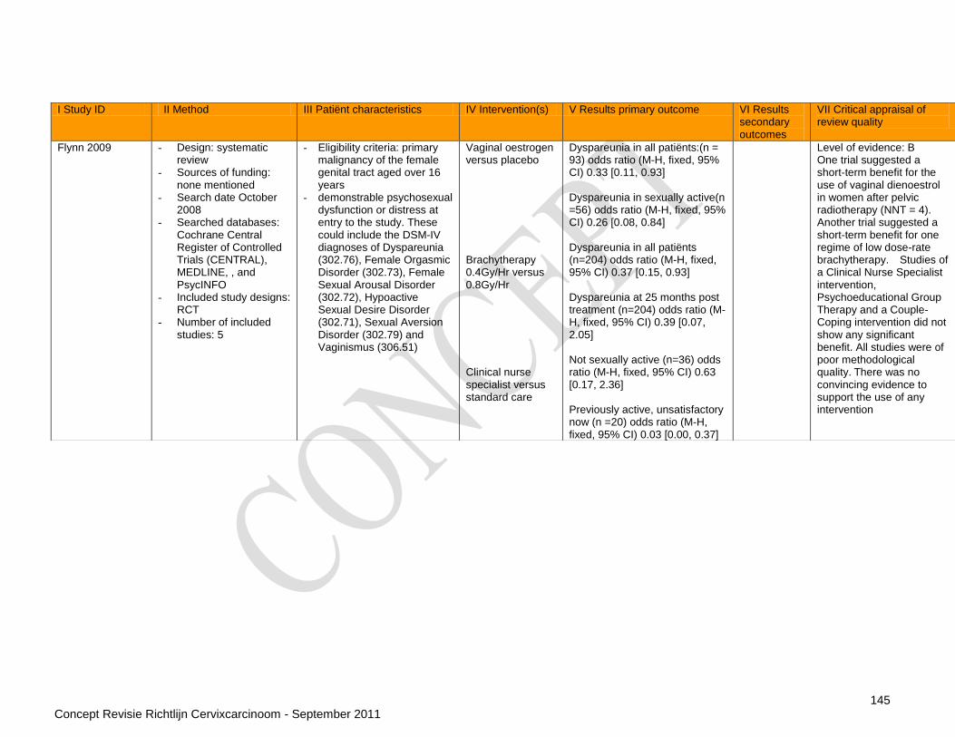
145 Concept Revisie Richtlijn Cervixcarcinoom - September 2011
I Study ID II Method III Patiënt characteristics IV Intervention(s) V Results primary outcome VI Results secondary outcomes
VII Critical appraisal of review quality
Flynn 2009 - Design: systematic review
- Sources of funding: none mentioned
- Search date October 2008
- Searched databases: Cochrane Central Register of Controlled Trials (CENTRAL), MEDLINE, , and PsycINFO
- Included study designs: RCT
- Number of included studies: 5
- Eligibility criteria: primary malignancy of the female genital tract aged over 16 years
- demonstrable psychosexual dysfunction or distress at entry to the study. These could include the DSM-IV diagnoses of Dyspareunia (302.76), Female Orgasmic Disorder (302.73), Female Sexual Arousal Disorder (302.72), Hypoactive Sexual Desire Disorder (302.71), Sexual Aversion Disorder (302.79) and Vaginismus (306.51)
Vaginal oestrogen versus placebo Brachytherapy 0.4Gy/Hr versus 0.8Gy/Hr Clinical nurse specialist versus standard care
Dyspareunia in all patiënts:(n = 93) odds ratio (M-H, fixed, 95% CI) 0.33 [0.11, 0.93] Dyspareunia in sexually active(n =56) odds ratio (M-H, fixed, 95% CI) 0.26 [0.08, 0.84] Dyspareunia in all patiënts (n=204) odds ratio (M-H, fixed, 95% CI) 0.37 [0.15, 0.93] Dyspareunia at 25 months post treatment (n=204) odds ratio (M-H, fixed, 95% CI) 0.39 [0.07, 2.05] Not sexually active (n=36) odds ratio (M-H, fixed, 95% CI) 0.63 [0.17, 2.36] Previously active, unsatisfactory now (n =20) odds ratio (M-H, fixed, 95% CI) 0.03 [0.00, 0.37]
Level of evidence: B One trial suggested a short-term benefit for the use of vaginal dienoestrol in women after pelvic radiotherapy (NNT = 4). Another trial suggested a short-term benefit for one regime of low dose-rate brachytherapy. Studies of a Clinical Nurse Specialist intervention, Psychoeducational Group Therapy and a Couple-Coping intervention did not show any significant benefit. All studies were of poor methodological quality. There was no convincing evidence to support the use of any intervention

146 Concept Revisie Richtlijn Cervixcarcinoom - September 2011
I Study ID II Method III Patiënt characteristics IV Intervention(s) V Results primary outcome
VI Results secondary outcomes
VII Critical appraisal of review quality
Miles 2010 Johnson 2010 (two reports of the same systematic review)
- Design: systematic review - Sources of funding: none
mentioned - Search date: January
2007 - Searched databases:
Cochrane Central Register of Controlled Trials (CENTRAL), MEDLINE, PsycINFO AMED, CINAHL National Health Service Research Register
- Included study designs: RCT
- Number of included studies: 1
- Eligibility criteria Patiënts undergoing interventions for sexual dysfunction following treatments for cancer
Psychoeducational support for the use of dilation Other non randomised studies
Sexual functional score after 3 months mean difference (IV, fixed, 95% CI) 0.04 [–0.03, 0.11] Sexual functional score after one year mean difference (IV, fixed, 95% CI) –0.01 [–0.07, 0.05] One comparative unmatched trial showed no advantage from inserting mitomycin C One retrospective report implied that dilation lowered stenosis rates, but the control group was not comparable
Case reports described vaginal fistulas or psychological morbidity A report of five women implied that stenosis can be treated by dilation many years after radiotherapy. One uncontrolled observational report involving 89 women showed that the median vaginal length 6–10 weeks after therapy was measured at 6 cm, but women tolerated a 9 cm measurer after 4 months of dilation experience
Level of evidence: C No RCT with a direct comparison was identified, only one small trial involving support identified Authors concluded that there was insufficient evidence supporting dilation

147 Concept Revisie Richtlijn Cervixcarcinoom - September 2011
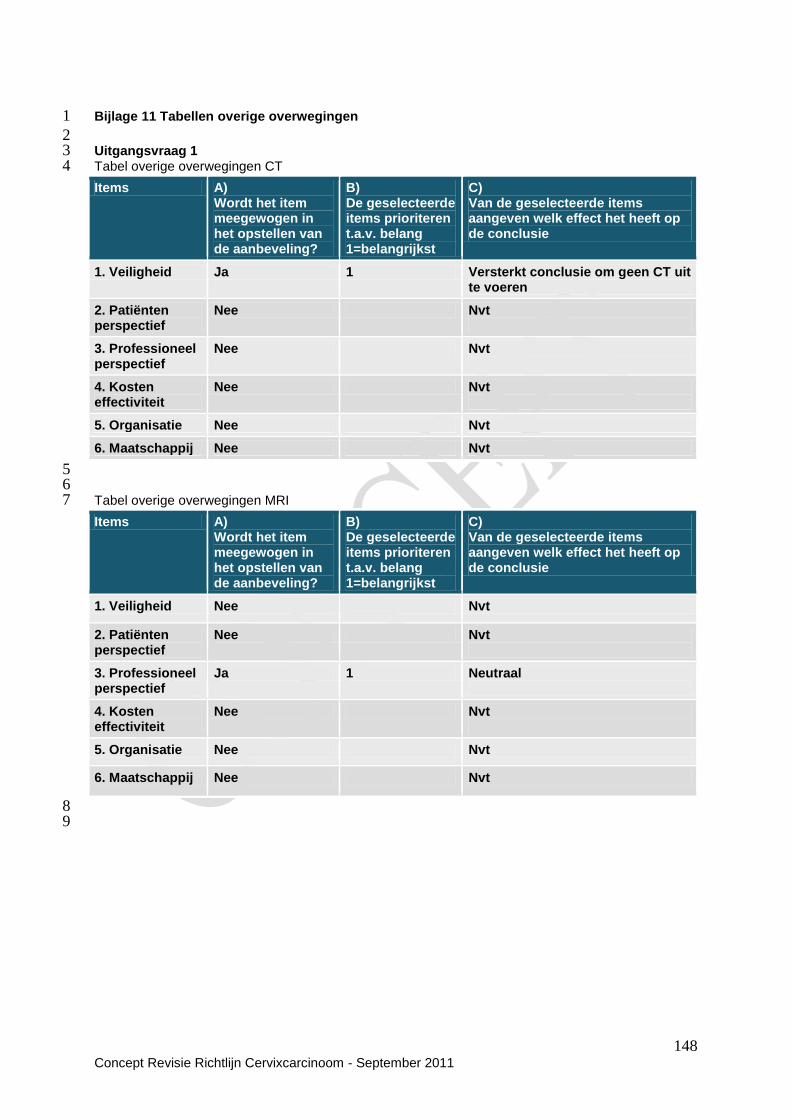
148 Concept Revisie Richtlijn Cervixcarcinoom - September 2011
Bijlage 11 Tabellen overige overwegingen 1
2 Uitgangsvraag 1 3 Tabel overige overwegingen CT 4
5 6 Tabel overige overwegingen MRI 7
8 9
Items A) Wordt het item meegewogen in het opstellen van de aanbeveling?
B) De geselecteerde items prioriteren t.a.v. belang 1=belangrijkst
C) Van de geselecteerde items aangeven welk effect het heeft op de conclusie
1. Veiligheid Ja 1 Versterkt conclusie om geen CT uit te voeren
2. Patiënten perspectief
Nee Nvt
3. Professioneel perspectief
Nee Nvt
4. Kosten effectiviteit
Nee Nvt
5. Organisatie Nee Nvt
6. Maatschappij Nee Nvt
Items A) Wordt het item meegewogen in het opstellen van de aanbeveling?
B) De geselecteerde items prioriteren t.a.v. belang 1=belangrijkst
C) Van de geselecteerde items aangeven welk effect het heeft op de conclusie
1. Veiligheid Nee Nvt
2. Patiënten perspectief
Nee Nvt
3. Professioneel perspectief
Ja 1 Neutraal
4. Kosten effectiviteit
Nee Nvt
5. Organisatie Nee Nvt
6. Maatschappij Nee Nvt

149 Concept Revisie Richtlijn Cervixcarcinoom - September 2011
Tabel overige overwegingen PET(CT) 1
2 3 Uitgangsvraag 2 4 Tabel overige overwegingen Conisatie 5
6
7
Items A) Wordt het item meegewogen in het opstellen van de aanbeveling?
B) De geselecteerde items prioriteren t.a.v. belang 1=belangrijkst
C) Van de geselecteerde items aangeven welk effect het heeft op de conclusie
1. Veiligheid Ja Verzwakt, blijft dezelfde richting
2. Patiënten perspectief
Nee Nvt
3. Professioneel perspectief
Ja 1 Neutraal
4. Kosten effectiviteit
Ja Verzwakt, blijft dezelfde richting
5. Organisatie Ja Neutraal
6. Maatschappij Nee Nvt
Items A)
Wordt het item
meegewogen in
het opstellen van
de aanbeveling?
B)
De geselecteerde
items prioriteren
t.a.v. belang
1=belangrijkst
C)
Van de geselecteerde items
aangeven welk effect het heeft op
de conclusie
1. Veiligheid Ja Versterkt
2. Patiënten
perspectief
Ja
Versterkt
3. Professioneel
perspectief
Ja
Versterkt
4. Kosten
effectiviteit
Ja
Versterkt
5. Organisatie Ja Versterkt
6. Maatschappij Ja Versterkt

150 Concept Revisie Richtlijn Cervixcarcinoom - September 2011
Tabel overige overwegingen Conisatie met pelviene lymfklierdissectie 1
2
3
Tabel overige overwegingen radicale trachelectomie 4
5
Items A) Wordt het item meegewogen in het opstellen van de aanbeveling?
B) De geselecteerde items prioriteren t.a.v. belang 1=belangrijkst
C) Van de geselecteerde items aangeven welk effect het heeft op de conclusie
1. Veiligheid Ja Neutraal
2. Patiënten perspectief
Ja Neutraal
3. Professioneel perspectief
Ja Neutraal
4. Kosten effectiviteit
Ja Neutraal
5. Organisatie Ja Neutraal
6. Maatschappij Nee Nvt
Items A) Wordt het item
meegewogen in het
opstellen van de
aanbeveling?
B) De geselecteerde
items prioriteren t.a.v.
belang 1=belangrijkst
C) Van de geselecteerde
items aangeven welk effect
het heeft op de conclusie
1. Veiligheid Ja Neutraal
2. Patiënten
perspectief
Ja
Versterkt
3. Professioneel
perspectief
Ja Verzwakt, blijft dezelfde
richting
4. Kosten
effectiviteit
Ja
Neutraal
5. Organisatie Ja Verzwakt, blijft dezelfde
richting
6. Maatschappij Nee Nvt
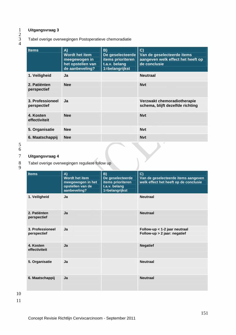
151 Concept Revisie Richtlijn Cervixcarcinoom - September 2011
Uitgangsvraag 3 1 2 Tabel overige overwegingen Postoperatieve chemoradiatie 3 4
5 6
Uitgangsvraag 4 7
Tabel overige overwegingen reguliere follow up 8 9
10
11
Items A) Wordt het item meegewogen in het opstellen van de aanbeveling?
B) De geselecteerde items prioriteren t.a.v. belang 1=belangrijkst
C) Van de geselecteerde items aangeven welk effect het heeft op de conclusie
1. Veiligheid Ja Neutraal
2. Patiënten perspectief
Nee Nvt
3. Professioneel perspectief
Ja Verzwakt chemoradiotherapie schema, blijft dezelfde richting
4. Kosten effectiviteit
Nee Nvt
5. Organisatie Nee Nvt
6. Maatschappij Nee Nvt
Items A) Wordt het item meegewogen in het opstellen van de aanbeveling?
B) De geselecteerde items prioriteren t.a.v. belang 1=belangrijkst
C) Van de geselecteerde items aangeven welk effect het heeft op de conclusie
1. Veiligheid Ja
Neutraal
2. Patiënten perspectief
Ja
Neutraal
3. Professioneel perspectief
Ja
Follow-up < 1-2 jaar neutraal Follow-up > 2 jaar: negatief
4. Kosten effectiviteit
Ja
Negatief
5. Organisatie Ja
Neutraal
6. Maatschappij Ja
Neutraal

152 Concept Revisie Richtlijn Cervixcarcinoom - September 2011
Uitgangsvraag 5 1
2 Tabel overige overwegingen Lokaal recidief zonder eerdere radiotherapie 3 4
5 Tabel overige overwegingen Lokaal recidief na eerdere bestraling 6 7
8 9 10 11 12
Items A) Wordt het item meegewogen in het opstellen van de aanbeveling?
B) De geselecteerde items prioriteren t.a.v. belang 1=belangrijkst
C) Van de geselecteerde items aangeven welk effect het heeft op de conclusie
1. Veiligheid Ja
4 Versterkt
2. Patiënten perspectief
Ja
1 Versterkt
3. Professioneel perspectief
Ja
2 Neutraal
4. Kosten effectiviteit
Ja 3 Versterkt
5. Organisatie Nee Neutraal
6. Maatschappij Nee
Neutraal
Items A) Wordt het item meegewogen in het opstellen van de aanbeveling?
B) De geselecteerde items prioriteren t.a.v. belang 1=belangrijkst
C) Van de geselecteerde items aangeven welk effect het heeft op de conclusie
1. Veiligheid Nee 5 Neutraal
2. Patiënten perspectief
Ja 1 Versterkt
3. Professioneel perspectief
Ja
2 Versterkt
4. Kosten effectiviteit
Nee 6 Neutraal
5. Organisatie Ja 4 Neutraal
6. Maatschappij Ja 3 Versterkt

153 Concept Revisie Richtlijn Cervixcarcinoom - September 2011
Tabel overige overwegingen Recidief op afstand, al dan niet met lokaal recidief 1 2
3 4 Uitgangsvraag 6 5 6 Tabel overige overwegingen medicatie 7
8
9 10 11 12 13 14 15 16 17
Items A) Wordt het item meegewogen in het opstellen van de aanbeveling?
B) De geselecteerde items prioriteren t.a.v. belang 1=belangrijkst
C) Van de geselecteerde items aangeven welk effect het heeft op de conclusie
1. Veiligheid Ja Versterkt
2. Patiënten perspectief
Ja
Versterkt
3. Professioneel perspectief
Nee Nvt
4. Kosten effectiviteit
Nee Nvt
5. Organisatie Ja
Versterkt
6. Maatschappij Nee Nvt
Items A) Wordt het item meegewogen in het opstellen van de aanbeveling?
B) De geselecteerde items prioriteren t.a.v. belang 1=belangrijkst
C) Van de geselecteerde items aangeven welk effect het heeft op de conclusie
1. Veiligheid Ja
Oestrogeentoediening: neutraal
2.Patiënten perspectief
Nee
Nvt
3.Professioneel perspectief
Ja Oestrogeentoediening: neutraal Benzydamine douches: verzwakt, blijft dezelfde richting
4. Kosten effectiviteit
Nee Nvt
5. Organisatie Nee Benzydamine douches: verzwakt, verandert van richting
6. Maatschappij Nee Nvt
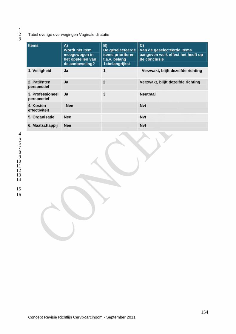
154 Concept Revisie Richtlijn Cervixcarcinoom - September 2011
1 Tabel overige overwegingen Vaginale dilatatie 2 3
4 5 6 7 8 9 10 11 12 13 14
15
16
Items A) Wordt het item meegewogen in het opstellen van de aanbeveling?
B) De geselecteerde items prioriteren t.a.v. belang 1=belangrijkst
C) Van de geselecteerde items aangeven welk effect het heeft op de conclusie
1. Veiligheid Ja
1 Verzwakt, blijft dezelfde richting
2. Patiënten perspectief
Ja
2 Verzwakt, blijft dezelfde richting
3. Professioneel perspectief
Ja 3 Neutraal
4. Kosten effectiviteit
Nee Nvt
5. Organisatie Nee Nvt
6. Maatschappij Nee Nvt
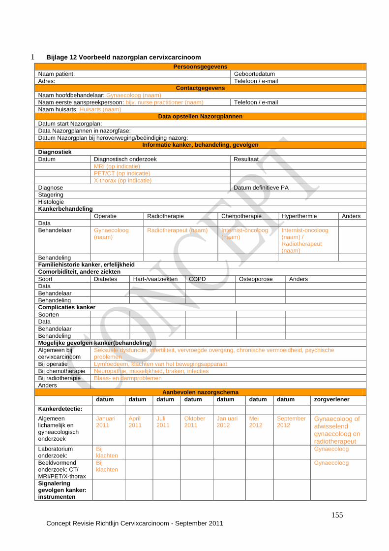
155 Concept Revisie Richtlijn Cervixcarcinoom - September 2011
Bijlage 12 Voorbeeld nazorgplan cervixcarcinoom 1
Persoonsgegevens
Naam patiënt: Geboortedatum
Adres: Telefoon / e-mail
Contactgegevens
Naam hoofdbehandelaar: Gynaecoloog (naam)
Naam eerste aanspreekpersoon: bijv. nurse practitioner (naam) Telefoon / e-mail
Naam huisarts: Huisarts (naam)
Data opstellen Nazorgplannen
Datum start Nazorgplan:
Data Nazorgplannen in nazorgfase:
Datum Nazorgplan bij heroverweging/beëindiging nazorg:
Informatie kanker, behandeling, gevolgen
Diagnostiek
Datum Diagnostisch onderzoek Resultaat
MRI (op indicatie)
PET/CT (op indicatie)
X-thorax (op indicatie)
Diagnose Datum definitieve PA
Stagering
Histologie
Kankerbehandeling
Operatie Radiotherapie Chemotherapie Hyperthermie Anders
Data
Behandelaar Gynaecoloog (naam)
Radiotherapeut (naam) Internist-oncoloog (naam)
Internist-oncoloog (naam) / Radiotherapeut (naam)
Behandeling
Familiehistorie kanker, erfelijkheid
Comorbiditeit, andere ziekten
Soort Diabetes Hart-/vaatziekten COPD Osteoporose Anders
Data
Behandelaar
Behandeling
Complicaties kanker
Soorten
Data
Behandelaar
Behandeling
Mogelijke gevolgen kanker(behandeling)
Algemeen bij cervixcarcinoom
Seksuele dysfunctie, infertiliteit, vervroegde overgang, chronische vermoeidheid, psychische problemen
Bij operatie Lymfoedeem, klachten van het bewegingsapparaat
Bij chemotherapie Neuropathie, misselijkheid, braken, infecties
Bij radiotherapie Blaas- en darmproblemen
Anders
Aanbevolen nazorgschema
datum datum datum datum datum datum datum zorgverlener
Kankerdetectie:
Algemeen lichamelijk en gyneacologisch onderzoek
Januari 2011
April 2011
Juli 2011
Oktober 2011
Jan uari 2012
Mei 2012
September 2012
Gynaecoloog of afwisselend gynaecoloog en radiotherapeut
Laboratorium onderzoek:
Bij klachten
Gynaecoloog
Beeldvormend onderzoek: CT/ MRI/PET/X-thorax
Bij klachten
Gynaecoloog
Signalering gevolgen kanker: instrumenten
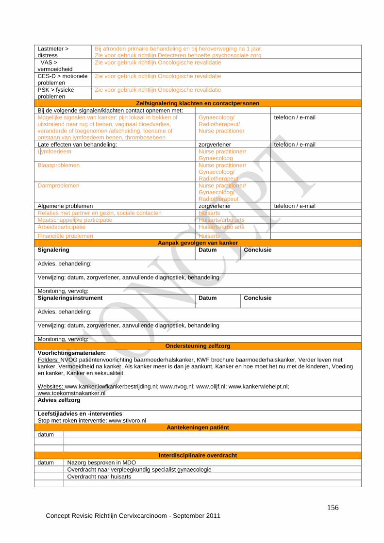
156 Concept Revisie Richtlijn Cervixcarcinoom - September 2011
Lastmeter > distress
Bij afronden primaire behandeling en bij heroverweging na 1 jaar. Zie voor gebruik richtlijn Detecteren behoefte psychosociale zorg
VAS > vermoeidheid
Zie voor gebruik richtlijn Oncologische revalidatie
CES-D > motionele problemen
Zie voor gebruik richtlijn Oncologische revalidatie
PSK > fysieke problemen
Zie voor gebruik richtlijn Oncologische revalidatie
Zelfsignalering klachten en contactpersonen
Bij de volgende signalen/klachten contact opnemen met:
Mogelijke signalen van kanker: pijn lokaal in bekken of uitstralend naar rug of benen, vaginaal bloedverlies, veranderde of toegenomen /afscheiding, toename of ontstaan van lymfoedeem benen, thrombosebeen
Gynaecoloog/ Radiotherapeut/ Nurse practitioner
telefoon / e-mail
Late effecten van behandeling: zorgverlener telefoon / e-mail
Lymfoedeem Nurse practitioner/ Gynaecoloog
Blaasproblemen Nurse practitioner/ Gynaecoloog/ Radiotherapeut
Darmproblemen Nurse practitioner/ Gynaecoloog/ Radiotherapeut
Algemene problemen zorgverlener telefoon / e-mail
Relaties met partner en gezin, sociale contacten Huisarts
Maatschappelijke participatie Huisarts/arbo arts
Arbeidsparticipatie Huisarts/arbo arts
Financiële problemen Huisarts
Aanpak gevolgen van kanker
Signalering
Datum
Conclusie
Advies, behandeling:
Verwijzing: datum, zorgverlener, aanvullende diagnostiek, behandeling
Monitoring, vervolg:
Signaleringsinstrument
Datum Conclusie
Advies, behandeling:
Verwijzing: datum, zorgverlener, aanvullende diagnostiek, behandeling
Monitoring, vervolg:
Ondersteuning zelfzorg
Voorlichtingsmaterialen:
Folders: NVOG patiëntenvoorlichting baarmoederhalskanker, KWF brochure baarmoederhalskanker, Verder leven met kanker, Vermoeidheid na kanker, Als kanker meer is dan je aankunt, Kanker en hoe moet het nu met de kinderen, Voeding en kanker, Kanker en seksualiteit. Websites: www.kanker.kwfkankerbestrijding.nl; www.nvog.nl; www.olijf.nl; www.kankerwiehelpt.nl; www.toekomstnakanker.nl
Advies zelfzorg
Leefstijladvies en -interventies
Stop met roken interventie: www.stivoro.nl
Aantekeningen patiënt
datum
Interdisciplinaire overdracht
datum Nazorg besproken in MDO
Overdracht naar verpleegkundig specialist gynaecologie
Overdracht naar huisarts

157 Concept Revisie Richtlijn Cervixcarcinoom - September 2011
1
Bijlage 13 Actualisatie 2
Voorwaarden voor revisie en beoordelingsfrequentie zijn vastgelegd in de richtlijn. De 3 geldigheidstermijn voor de richtlijn (maximaal 5 jaar na vaststelling) wordt vanuit het IKNL bewaakt. 4
158 Concept Revisie Richtlijn Cervixcarcinoom - September 2011
Om verscheidene redenen kan actualisatie eerder dan beoogd nodig zijn. Zo nodig zal de richtlijn 1 tussentijds op onderdelen worden bijgesteld. 2
Bijlage 14 Houderschap richtlijn 3
De houder van de richtlijn moet kunnen aantonen dat de richtlijn zorgvuldig en met de vereiste 4 deskundigheid tot stand is gekomen. Onder houder wordt verstaan de verenigingen van 5 beroepsbeoefenaren die de richtlijn autoriseren. 6 Het Integraal Kankercentrum Nederland draagt zorg voor het beheer en de ontsluiting van de richtlijn. 7
Bijlage 15 Juridische betekenis van richtlijnen 8
De richtlijn bevat aanbevelingen van algemene aard. Het is mogelijk dat deze aanbevelingen in een 9 individueel geval niet van toepassing zijn. Er kunnen zich feiten of omstandigheden voordoen 10 waardoor het wenselijk is dat in het belang van de patiënt van de richtlijn wordt afgeweken. 11 Wanneer van de richtlijn wordt afgeweken, dient dit beargumenteerd gedocumenteerd te worden. 12 De toepasbaarheid en de toepassing van de richtlijnen in de praktijk is de verantwoordelijkheid van de 13 behandelende arts. 14
Bijlage 16 Verantwoording 15
Het Integraal Kankercentrum Nederland bevordert dat mensen met kanker en hun naasten zo dicht 16 mogelijk bij huis toegang hebben tot een samenhangend en kwalitatief verantwoord zorgaanbod. 17 Het Integraal Kankercentrum Nederland is opgericht om behandeling, zorg en klinisch onderzoek 18 binnen de oncologie te verbeteren. Daarnaast heeft het een taak in het opzetten en ondersteunen van 19 netwerken voor palliatieve zorg. 20 21 Het Integraal Kankercentrum Nederland werkt aan multidisciplinaire richtlijnontwikkeling voor de 22 oncologische en palliatieve zorg. Naast deze ontwikkeling van richtlijnen faciliteert het Integraal 23 Kankercentrum Nederland ook het onderhoud, het beheer, de implementatie en de evaluatie van deze 24 richtlijnen. 25 26 De leidraad voor de ontwikkeling van de richtlijnen voor oncologische en palliatieve zorg is het 27 AGREE instrument. Dit instrument is gemaakt voor de beoordeling van bestaande, nieuwe en 28 herziene richtlijnen. 29 Het AGREE Instrument beoordeelt zowel de kwaliteit van de verslaglegging als de kwaliteit van 30 bepaalde aspecten van de aanbevelingen. Het beoordeelt de kans dat een richtlijn zijn gewenste doel 31 zal behalen, maar niet de daadwerkelijke impact op patiëntuitkomsten. 32 33 Het AGREE Instrument is opgebouwd uit 23 items verdeeld over zes domeinen. Elk domein beslaat 34 een aparte dimensie van kwaliteit van richtlijnen, namelijk: 35 - Onderwerp en doel betreft het doel van de richtlijn de specifieke klinische vragen waarop de 36
richtlijn een antwoord geeft en de patiëntenpopulatie waarop de richtlijn van toepassing is. 37 - Betrokkenheid van belanghebbenden richt zich op de mate waarin de richtlijn de opvattingen van 38
de beoogde gebruikers weerspiegelt. 39 - Methodologie hangt samen met het proces waarin bewijsmateriaal is verzameld en samengesteld 40
en met de gebruikte methoden om aanbevelingen op te stellen en te herzien. 41 - Helderheid en presentatie gaat over het taalgebruik en de vorm van de richtlijn. 42 - Toepassing houdt verband met de mogelijke organisatorische, gedragsmatige en financiële 43
consequenties van het toepassen van de richtlijn. 44 - Onafhankelijkheid van de opstellers betreft de onafhankelijkheid van de aanbevelingen en 45
erkenning van mogelijke conflicterende belangen van leden van de werkgroep. 46 47
Bijlage 17 Implementatie en evaluatie 48
Bij het ontwikkelen van de richtlijnen wordt rekening gehouden met de uitvoerbaarheid van de richtlijn. 49 Daarbij wordt gelet op bevorderende of belemmerende factoren. Om het gebruik in de dagelijkse 50 praktijk te bevorderen wordt een samenvattingkaart gemaakt. Daarnaast wordt de richtlijn 51 gepubliceerd op Oncoline (de website van het IKNL). Tevens wordt de richtlijn verspreid onder de 52 professionals via de (wetenschappelijke) verenigingen en de werkgroepen van het Integraal 53 Kankercentrum Nederland. 54

159 Concept Revisie Richtlijn Cervixcarcinoom - September 2011
Tijdens het ontwikkelen van de richtlijn worden indicatoren voor de evaluatie van de aanbevelingen in 1 de richtlijn opgesteld. Middels een documentatieproject kan met behulp van deze indicatoren worden 2 vastgesteld in hoeverre de richtlijn wordt nageleefd. De informatie uit het documentatieproject vormt 3 input bij de revisie van richtlijn. 4 5
Bijlage 18 Kennislacunes 6
7 Uitgangsvraag 1 8 Bij patiënten met primair invasief cervixcarcinoom, welke diagnostische techniek resulteert in 9 de meest accurate stadiering? 10 1. Wat zijn de indicaties voor het maken van een PET/CT bij cervixcarcinoom? Doelmatigheid- en 11
kosteneffectiviteitsanalyses zijn er nog niet in voldoende mate. 12 2. Geeft MRI betere informatie bij stadiering dan het onderzoek onder narcose en hoe is de 13
kosteneffectiviteit? 14 15 Uitgangsvraag 2 16 Wat is de plaats van fertiliteitsparende behandeling bij vrouwen met cervix carcinoom? 17 1. Er is geen gerandomiseerd onderzoek naar de verschillen in uitkomst tussen een radicale 18
vaginale trachelectomie en een radicale abdominale trachelectomie. De huidige kennis is 19 gebaseerd op case series. 20
2. Verder is onbekend in welke situaties een parametriumresectie achterwege kan worden gelaten. 21 Wat is het verschil in uitkomst tussen een conisatie + pelviene lymfadenectomie versus een 22 radicale trachelectomie (dus met meenemen van het parametrium) + pelviene lymfadenectomie bij 23 patiënten met een vroeg stadium cervxicarcinoom? 24
25 Uitgangsvraag 3 26 Bij patiënten met operatief behandeld cervixcarcinoom stadium Ib/IIa met aanwezigheid van 27 lymfekliermetastasen, heeft postoperatieve chemoradiatie in vergelijking met postoperatieve 28 radiotherapie een betere (ziektevrije) overleving? 29 1. Het is niet geheel duidelijk of aanvullende behandeling (zo ja, welke en in welke vorm) bij 30
patiënten na een radicale hysterectomie met een tumorgrootte < of > 2 cm en 1 of 2 positieve 31 klieren een verbetering van de overleving zal geven. Echter deze groep zal nooit voldoende groot 32 zijn voor randomisatie. 33
2. De vorm en toediening van chemotherapie toegevoegd aan de postoperatieve radiotherapie is 34 onduidelijk (concurrent, sequentieel of gecombineerd). Deze studies lopen in de Verenigde Staten. 35
36 Uitgangsvraag 4 37 Bij patienten met een behandeld cervixcarcinoom, welke (frequentie van) follow-up zorgt voor 38 een betere overleving en kwaliteit van leven? 39 Er zijn geen gerandomiseerde studies over het verschil in overleving/kwaliteit van leven tussen de 40 behandeling van asymptomatische patienten en symptomatische patienten met een 41 recidief/metastasen op afstand bij cervixcarcinoom. 42 43 Uitgangsvraag 5 44 De behandeling van patiënten met recidief cervixcarcinoom. 45 1. Het is nog onbekend of de toevoeging van biologicals aan chemotherapie bij patiënten met 46
metastasen op afstand van cervixcarcinoom, een betere overleving en kwaliteit van leven geeft 47 dan chemotherapiecombinaties alleen. Behandeling van recidief binnen bestraald gebied dient 48 verder onderzocht te worden. 49
50 Uitgangsvraag 6 51 Wat zijn de behandelingsmogelijkheden bij seksueel disfunctioneren na behandeling voor 52 cervixcarcinoom? 53 1. Er is tegenstrijdige literatuur over het effect van lokale oestrogeentoediening en benzydamine-54
douches ter preventie van dyspareunie en ter bestrijding van vaginale stenose na radiotherapie bij 55 cervixcarcinoom. Tevens ontbreekt hierbij de evidence over de toediening van oestrogenen bij 56 postmenopauzale vrouwen versus premenopauzale vrouwen. 57

160 Concept Revisie Richtlijn Cervixcarcinoom - September 2011
2. Er is tegenstrijdige literatuur over de effectiviteit van systematische vaginale dilatatie met een 1 pelote ter preventie van vaginale adhesie vorming na radiotherapie. Tevens ontbreekt literatuur 2 over de effectiviteit van vaselinge tampons na radiotherapie. 3
3. Er ontbreekt literatuur over de geïntegreerde aanpak van seksueel disfunctioneren na 4 behandeling cervixcarcinoom. 5
6

161 Concept Revisie Richtlijn Cervixcarcinoom - September 2011
Bijlage 19 Referenties 1
2 Uitgangsvraag 1 3 Bij patiënten met primair invasief cervixcarcinoom, welke diagnostische techniek resulteert in de meest 4 accurate stadiering? 5 6 Akata, D., U. Kerimoglu, et al. (2005). "Efficacy of transvaginal contrast-enhanced MRI in the early 7 staging of cervical carcinoma." European Radiology 15(8): 1727-1733. 8 Altgassen, C., H. Hertel, et al. (2008). "Multicenter validation study of the sentinel lymph node concept 9 in cervical cancer: AGO study group." Journal of Clinical Oncology 26(18): 2943-2951. 10 Amit, A., D. Beck, et al. (2006). "The role of hybrid PET/CT in the evaluation of patiënts with cervical 11 cancer." Gynecol Oncol 100(1): 65-69. 12 Bender, D. P., J. I. Sorosky, et al. (2003). "Serum CA 125 is an independent prognostic factor in 13 cervical adenocarcinoma." Am J Obstet Gynecol 189(1): 113-117. 14 Bentivegna, E., C. Uzan, et al. (2010). "Correlation between [18f]fluorodeoxyglucose positron-15 emission tomography scan and histology of pelvic nodes in early-stage cervical cancer." Anticancer 16 Res 30(3): 1029-1032. 17 Bipat, S., A. S. Glas, et al. (2003). "Computed tomography and magnetic resonance imaging in 18 staging of uterine cervical carcinoma: a systematic review." Gynecol Oncol 91(1): 59-66. 19 Bjurberg, M., E. Kjellen, et al. (2007). "FDG-PET in cervical cancer: staging, re-staging and follow-up." 20 Acta Obstet Gynecol Scand 86(11): 1385-1391. 21 Chao, A., K. C. Ho, et al. (2008). "Positron emission tomography in evaluating the feasibility of curative 22 intent in cervical cancer patiënts with limited distant lymph node metastases." Gynecol Oncol 110(2): 23 172-178. 24 Chen, S. W., J. A. Liang, et al. (2008). "Clinical implications of elevated pretreatment 25 carcinoembryonic antigen in patiënts with advanced squamous cell carcinoma of the uterine cervix." 26 Tumor Biology 29(4): 255-261. 27 Choi, H. J., J. W. Roh, et al. (2006). "Comparison of the accuracy of magnetic resonance imaging and 28 positron emission tomography/computed tomography in the presurgical detection of lymph node 29 metastases in patiënts with uterine cervical carcinoma: a prospective study." Cancer 106(4): 914-922. 30 Choi, S. H., S. H. Kim, et al. (2004). "Preoperative magnetic resonance imaging staging of uterine 31 cervical carcinoma: results of prospective study." Journal of computer assisted tomography 28(5): 32 620-627. 33 Chou, H. H., T. C. Chang, et al. (2006). "Low value of [18F]-fluoro-2-deoxy-D-glucose positron 34 emission tomography in primary staging of early-stage cervical cancer before radical hysterectomy." J 35 Clin Oncol 24(1): 123-128. 36 Chung, H. H., K. W. Kang, et al. (2010). "Role of magnetic resonance imaging and positron emission 37 tomography/computed tomography in preoperative lymph node detection of uterine cervical cancer." 38 Am J Obstet Gynecol 203(2): 156 e151-155. 39 Chung, H. H., S. B. Kang, et al. (2007). "Can preoperative MRI accurately evaluate nodal and 40 parametrial invasion in early stage cervical cancer?" Japanese journal of clinical oncology 37(5): 370-41 375. 42 Chung, H. H., N. H. Park, et al. (2009). "Role of integrated PET-CT in pelvic lymph node staging of 43 cervical cancer before radical hysterectomy." Gynecol Obstet Invest 67(1): 61-66. 44 Darlin, L., J. Persson, et al. (2010). "The sentinel node concept in early cervical cancer performs well 45 in tumors smaller than 2 cm." Gynecol Oncol 117(2): 266-269. 46 deSouza, N. M., R. Dina, et al. (2006). "Cervical cancer: value of an endovaginal coil magnetic 47 resonance imaging technique in detecting small volume disease and assessing parametrial 48 extension." Gynecol Oncol 102(1): 80-85. 49 Fader, A. N., R. P. Edwards, et al. (2008). "Sentinel lymph node biopsy in early-stage cervical cancer: 50 utility of intraoperative versus postoperative assessment." Gynecol Oncol 111(1): 13-17. 51 Fischerova, D., D. Cibula, et al. (2008). "Transrectal ultrasound and magnetic resonance imaging in 52 staging of early cervical cancer." Int J Gynecol Cancer 18(4): 766-772. 53 Goyal, B. K., H. Singh, et al. (2010). "Value of PET-CT in avoiding multimodality therapy in operable 54 cervical cancer." Int J Gynecol Cancer 20(6): 1041-1045. 55 Havrilesky, L. J., S. L. Kulasingam, et al. (2005). "FDG-PET for management of cervical and ovarian 56 cancer." Gynecol Oncol 97(1): 183-191. 57 Hertel, H., C. Kohler, et al. (2002). "Laparoscopic staging compared with imaging techniques in the 58 staging of advanced cervical cancer." Gynecologic Oncology 87(1): 46-51. 59
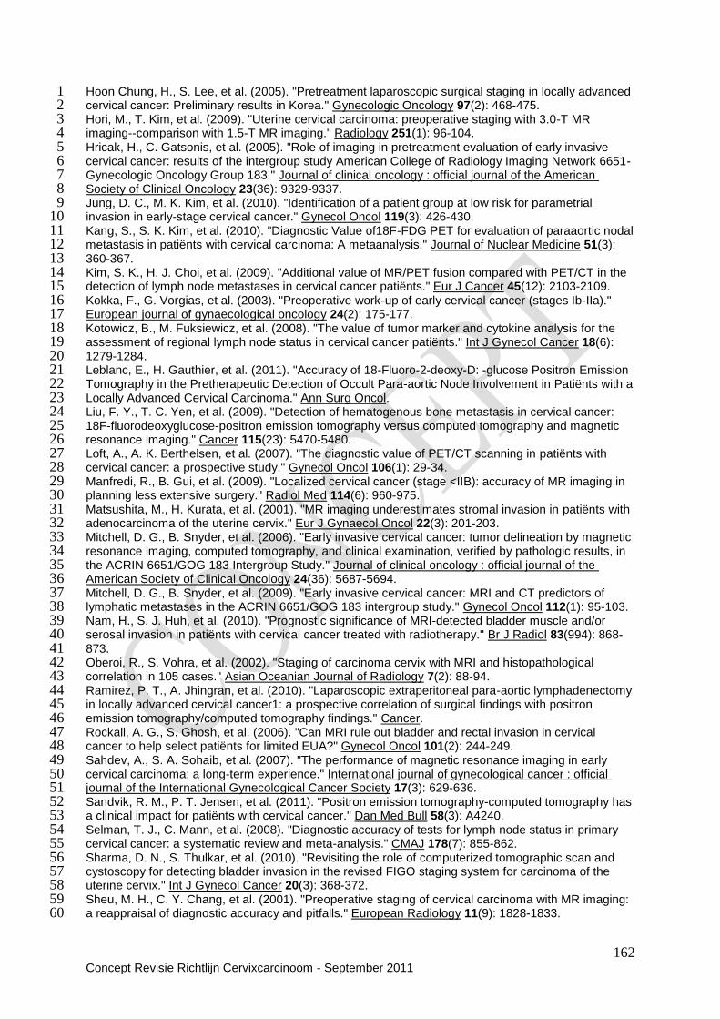
162 Concept Revisie Richtlijn Cervixcarcinoom - September 2011
Hoon Chung, H., S. Lee, et al. (2005). "Pretreatment laparoscopic surgical staging in locally advanced 1 cervical cancer: Preliminary results in Korea." Gynecologic Oncology 97(2): 468-475. 2 Hori, M., T. Kim, et al. (2009). "Uterine cervical carcinoma: preoperative staging with 3.0-T MR 3 imaging--comparison with 1.5-T MR imaging." Radiology 251(1): 96-104. 4 Hricak, H., C. Gatsonis, et al. (2005). "Role of imaging in pretreatment evaluation of early invasive 5 cervical cancer: results of the intergroup study American College of Radiology Imaging Network 6651-6 Gynecologic Oncology Group 183." Journal of clinical oncology : official journal of the American 7 Society of Clinical Oncology 23(36): 9329-9337. 8 Jung, D. C., M. K. Kim, et al. (2010). "Identification of a patiënt group at low risk for parametrial 9 invasion in early-stage cervical cancer." Gynecol Oncol 119(3): 426-430. 10 Kang, S., S. K. Kim, et al. (2010). "Diagnostic Value of18F-FDG PET for evaluation of paraaortic nodal 11 metastasis in patiënts with cervical carcinoma: A metaanalysis." Journal of Nuclear Medicine 51(3): 12 360-367. 13 Kim, S. K., H. J. Choi, et al. (2009). "Additional value of MR/PET fusion compared with PET/CT in the 14 detection of lymph node metastases in cervical cancer patiënts." Eur J Cancer 45(12): 2103-2109. 15 Kokka, F., G. Vorgias, et al. (2003). "Preoperative work-up of early cervical cancer (stages Ib-IIa)." 16 European journal of gynaecological oncology 24(2): 175-177. 17 Kotowicz, B., M. Fuksiewicz, et al. (2008). "The value of tumor marker and cytokine analysis for the 18 assessment of regional lymph node status in cervical cancer patiënts." Int J Gynecol Cancer 18(6): 19 1279-1284. 20 Leblanc, E., H. Gauthier, et al. (2011). "Accuracy of 18-Fluoro-2-deoxy-D: -glucose Positron Emission 21 Tomography in the Pretherapeutic Detection of Occult Para-aortic Node Involvement in Patiënts with a 22 Locally Advanced Cervical Carcinoma." Ann Surg Oncol. 23 Liu, F. Y., T. C. Yen, et al. (2009). "Detection of hematogenous bone metastasis in cervical cancer: 24 18F-fluorodeoxyglucose-positron emission tomography versus computed tomography and magnetic 25 resonance imaging." Cancer 115(23): 5470-5480. 26 Loft, A., A. K. Berthelsen, et al. (2007). "The diagnostic value of PET/CT scanning in patiënts with 27 cervical cancer: a prospective study." Gynecol Oncol 106(1): 29-34. 28 Manfredi, R., B. Gui, et al. (2009). "Localized cervical cancer (stage <IIB): accuracy of MR imaging in 29 planning less extensive surgery." Radiol Med 114(6): 960-975. 30 Matsushita, M., H. Kurata, et al. (2001). "MR imaging underestimates stromal invasion in patiënts with 31 adenocarcinoma of the uterine cervix." Eur J Gynaecol Oncol 22(3): 201-203. 32 Mitchell, D. G., B. Snyder, et al. (2006). "Early invasive cervical cancer: tumor delineation by magnetic 33 resonance imaging, computed tomography, and clinical examination, verified by pathologic results, in 34 the ACRIN 6651/GOG 183 Intergroup Study." Journal of clinical oncology : official journal of the 35 American Society of Clinical Oncology 24(36): 5687-5694. 36 Mitchell, D. G., B. Snyder, et al. (2009). "Early invasive cervical cancer: MRI and CT predictors of 37 lymphatic metastases in the ACRIN 6651/GOG 183 intergroup study." Gynecol Oncol 112(1): 95-103. 38 Nam, H., S. J. Huh, et al. (2010). "Prognostic significance of MRI-detected bladder muscle and/or 39 serosal invasion in patiënts with cervical cancer treated with radiotherapy." Br J Radiol 83(994): 868-40 873. 41 Oberoi, R., S. Vohra, et al. (2002). "Staging of carcinoma cervix with MRI and histopathological 42 correlation in 105 cases." Asian Oceanian Journal of Radiology 7(2): 88-94. 43 Ramirez, P. T., A. Jhingran, et al. (2010). "Laparoscopic extraperitoneal para-aortic lymphadenectomy 44 in locally advanced cervical cancer1: a prospective correlation of surgical findings with positron 45 emission tomography/computed tomography findings." Cancer. 46 Rockall, A. G., S. Ghosh, et al. (2006). "Can MRI rule out bladder and rectal invasion in cervical 47 cancer to help select patiënts for limited EUA?" Gynecol Oncol 101(2): 244-249. 48 Sahdev, A., S. A. Sohaib, et al. (2007). "The performance of magnetic resonance imaging in early 49 cervical carcinoma: a long-term experience." International journal of gynecological cancer : official 50 journal of the International Gynecological Cancer Society 17(3): 629-636. 51 Sandvik, R. M., P. T. Jensen, et al. (2011). "Positron emission tomography-computed tomography has 52 a clinical impact for patiënts with cervical cancer." Dan Med Bull 58(3): A4240. 53 Selman, T. J., C. Mann, et al. (2008). "Diagnostic accuracy of tests for lymph node status in primary 54 cervical cancer: a systematic review and meta-analysis." CMAJ 178(7): 855-862. 55 Sharma, D. N., S. Thulkar, et al. (2010). "Revisiting the role of computerized tomographic scan and 56 cystoscopy for detecting bladder invasion in the revised FIGO staging system for carcinoma of the 57 uterine cervix." Int J Gynecol Cancer 20(3): 368-372. 58 Sheu, M. H., C. Y. Chang, et al. (2001). "Preoperative staging of cervical carcinoma with MR imaging: 59 a reappraisal of diagnostic accuracy and pitfalls." European Radiology 11(9): 1828-1833. 60
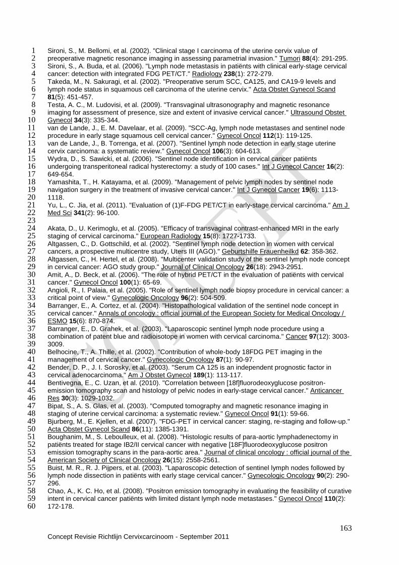
163 Concept Revisie Richtlijn Cervixcarcinoom - September 2011
Sironi, S., M. Bellomi, et al. (2002). "Clinical stage I carcinoma of the uterine cervix value of 1 preoperative magnetic resonance imaging in assessing parametrial invasion." Tumori 88(4): 291-295. 2 Sironi, S., A. Buda, et al. (2006). "Lymph node metastasis in patiënts with clinical early-stage cervical 3 cancer: detection with integrated FDG PET/CT." Radiology 238(1): 272-279. 4 Takeda, M., N. Sakuragi, et al. (2002). "Preoperative serum SCC, CA125, and CA19-9 levels and 5 lymph node status in squamous cell carcinoma of the uterine cervix." Acta Obstet Gynecol Scand 6 81(5): 451-457. 7 Testa, A. C., M. Ludovisi, et al. (2009). "Transvaginal ultrasonography and magnetic resonance 8 imaging for assessment of presence, size and extent of invasive cervical cancer." Ultrasound Obstet 9 Gynecol 34(3): 335-344. 10 van de Lande, J., E. M. Davelaar, et al. (2009). "SCC-Ag, lymph node metastases and sentinel node 11 procedure in early stage squamous cell cervical cancer." Gynecol Oncol 112(1): 119-125. 12 van de Lande, J., B. Torrenga, et al. (2007). "Sentinel lymph node detection in early stage uterine 13 cervix carcinoma: a systematic review." Gynecol Oncol 106(3): 604-613. 14 Wydra, D., S. Sawicki, et al. (2006). "Sentinel node identification in cervical cancer patiënts 15 undergoing transperitoneal radical hysterectomy: a study of 100 cases." Int J Gynecol Cancer 16(2): 16 649-654. 17 Yamashita, T., H. Katayama, et al. (2009). "Management of pelvic lymph nodes by sentinel node 18 navigation surgery in the treatment of invasive cervical cancer." Int J Gynecol Cancer 19(6): 1113-19 1118. 20 Yu, L., C. Jia, et al. (2011). "Evaluation of (1)F-FDG PET/CT in early-stage cervical carcinoma." Am J 21 Med Sci 341(2): 96-100. 22 23 Akata, D., U. Kerimoglu, et al. (2005). "Efficacy of transvaginal contrast-enhanced MRI in the early 24 staging of cervical carcinoma." European Radiology 15(8): 1727-1733. 25 Altgassen, C., D. Gottschild, et al. (2002). "Sentinel lymph node detection in women with cervical 26 cancers, a prospective multicentre study, Uters III (AGO)." Geburtshilfe Frauenheilkd 62: 358-362. 27 Altgassen, C., H. Hertel, et al. (2008). "Multicenter validation study of the sentinel lymph node concept 28 in cervical cancer: AGO study group." Journal of Clinical Oncology 26(18): 2943-2951. 29 Amit, A., D. Beck, et al. (2006). "The role of hybrid PET/CT in the evaluation of patiënts with cervical 30 cancer." Gynecol Oncol 100(1): 65-69. 31 Angioli, R., I. Palaia, et al. (2005). "Role of sentinel lymph node biopsy procedure in cervical cancer: a 32 critical point of view." Gynecologic Oncology 96(2): 504-509. 33 Barranger, E., A. Cortez, et al. (2004). "Histopathological validation of the sentinel node concept in 34 cervical cancer." Annals of oncology : official journal of the European Society for Medical Oncology / 35 ESMO 15(6): 870-874. 36 Barranger, E., D. Grahek, et al. (2003). "Laparoscopic sentinel lymph node procedure using a 37 combination of patent blue and radioisotope in women with cervical carcinoma." Cancer 97(12): 3003-38 3009. 39 Belhocine, T., A. Thille, et al. (2002). "Contribution of whole-body 18FDG PET imaging in the 40 management of cervical cancer." Gynecologic Oncology 87(1): 90-97. 41 Bender, D. P., J. I. Sorosky, et al. (2003). "Serum CA 125 is an independent prognostic factor in 42 cervical adenocarcinoma." Am J Obstet Gynecol 189(1): 113-117. 43 Bentivegna, E., C. Uzan, et al. (2010). "Correlation between [18f]fluorodeoxyglucose positron-44 emission tomography scan and histology of pelvic nodes in early-stage cervical cancer." Anticancer 45 Res 30(3): 1029-1032. 46 Bipat, S., A. S. Glas, et al. (2003). "Computed tomography and magnetic resonance imaging in 47 staging of uterine cervical carcinoma: a systematic review." Gynecol Oncol 91(1): 59-66. 48 Bjurberg, M., E. Kjellen, et al. (2007). "FDG-PET in cervical cancer: staging, re-staging and follow-up." 49 Acta Obstet Gynecol Scand 86(11): 1385-1391. 50 Boughanim, M., S. Leboulleux, et al. (2008). "Histologic results of para-aortic lymphadenectomy in 51 patiënts treated for stage IB2/II cervical cancer with negative [18F]fluorodeoxyglucose positron 52 emission tomography scans in the para-aortic area." Journal of clinical oncology : official journal of the 53 American Society of Clinical Oncology 26(15): 2558-2561. 54 Buist, M. R., R. J. Pijpers, et al. (2003). "Laparoscopic detection of sentinel lymph nodes followed by 55 lymph node dissection in patiënts with early stage cervical cancer." Gynecologic Oncology 90(2): 290-56 296. 57 Chao, A., K. C. Ho, et al. (2008). "Positron emission tomography in evaluating the feasibility of curative 58 intent in cervical cancer patiënts with limited distant lymph node metastases." Gynecol Oncol 110(2): 59 172-178. 60

164 Concept Revisie Richtlijn Cervixcarcinoom - September 2011
Chen, S. W., J. A. Liang, et al. (2008). "Clinical implications of elevated pretreatment 1 carcinoembryonic antigen in patiënts with advanced squamous cell carcinoma of the uterine cervix." 2 Tumor Biology 29(4): 255-261. 3 Choi, H. J., J. W. Roh, et al. (2006). "Comparison of the accuracy of magnetic resonance imaging and 4 positron emission tomography/computed tomography in the presurgical detection of lymph node 5 metastases in patiënts with uterine cervical carcinoma: a prospective study." Cancer 106(4): 914-922. 6 Choi, S. H., S. H. Kim, et al. (2004). "Preoperative magnetic resonance imaging staging of uterine 7 cervical carcinoma: results of prospective study." Journal of computer assisted tomography 28(5): 8 620-627. 9 Chou, H. H., T. C. Chang, et al. (2006). "Low value of [18F]-fluoro-2-deoxy-D-glucose positron 10 emission tomography in primary staging of early-stage cervical cancer before radical hysterectomy." J 11 Clin Oncol 24(1): 123-128. 12 Chung, H. H., K. W. Kang, et al. (2010). "Role of magnetic resonance imaging and positron emission 13 tomography/computed tomography in preoperative lymph node detection of uterine cervical cancer." 14 Am J Obstet Gynecol 203(2): 156 e151-155. 15 Chung, H. H., S. B. Kang, et al. (2007). "Can preoperative MRI accurately evaluate nodal and 16 parametrial invasion in early stage cervical cancer?" Japanese journal of clinical oncology 37(5): 370-17 375. 18 Chung, H. H., N. H. Park, et al. (2009). "Role of integrated PET-CT in pelvic lymph node staging of 19 cervical cancer before radical hysterectomy." Gynecol Obstet Invest 67(1): 61-66. 20 Chung, Y. A., S. H. Kim, et al. (2003). "Usefulness of lymphoscintigraphy and intraoperative gamma 21 probe detection in the identification of sentinel nodes in cervical cancer." European journal of nuclear 22 medicine and molecular imaging 30(7): 1014-1017. 23 Dargent, D. and R. Enria (2003). "Laparoscopic assessment of the sentinel lymph nodes in early 24 cervical cancer. Technique--preliminary results and future developments." Critical reviews in 25 oncology/hematology 48(3): 305-310. 26 Darlin, L., J. Persson, et al. (2010). "The sentinel node concept in early cervical cancer performs well 27 in tumors smaller than 2 cm." Gynecol Oncol 117(2): 266-269. 28 deSouza, N. M., R. Dina, et al. (2006). "Cervical cancer: value of an endovaginal coil magnetic 29 resonance imaging technique in detecting small volume disease and assessing parametrial 30 extension." Gynecol Oncol 102(1): 80-85. 31 Di Stefano, A. B., G. Acquaviva, et al. (2005). "Lymph node mapping and sentinel node detection in 32 patiënts with cervical carcinoma: a 2-year experience." Gynecologic Oncology 99(3): 671-679. 33 Fader, A. N., R. P. Edwards, et al. (2008). "Sentinel lymph node biopsy in early-stage cervical cancer: 34 utility of intraoperative versus postoperative assessment." Gynecol Oncol 111(1): 13-17. 35 Fischerova, D., D. Cibula, et al. (2008). "Transrectal ultrasound and magnetic resonance imaging in 36 staging of early cervical cancer." Int J Gynecol Cancer 18(4): 766-772. 37 Gil-Moreno, A., B. Diaz-Feijoo, et al. (2005). "Total laparoscopic radical hysterectomy with 38 intraoperative sentinel node identification in patiënts with early invasive cervical cancer." Gynecologic 39 Oncology 96(1): 187-193. 40 Goyal, B. K., H. Singh, et al. (2010). "Value of PET-CT in avoiding multimodality therapy in operable 41 cervical cancer." Int J Gynecol Cancer 20(6): 1041-1045. 42 Havrilesky, L. J., S. L. Kulasingam, et al. (2005). "FDG-PET for management of cervical and ovarian 43 cancer." Gynecol Oncol 97(1): 183-191. 44 Hertel, H., C. Kohler, et al. (2002). "Laparoscopic staging compared with imaging techniques in the 45 staging of advanced cervical cancer." Gynecologic Oncology 87(1): 46-51. 46 Hoon Chung, H., S. Lee, et al. (2005). "Pretreatment laparoscopic surgical staging in locally advanced 47 cervical cancer: Preliminary results in Korea." Gynecologic Oncology 97(2): 468-475. 48 Hori, M., T. Kim, et al. (2009). "Uterine cervical carcinoma: preoperative staging with 3.0-T MR 49 imaging--comparison with 1.5-T MR imaging." Radiology 251(1): 96-104. 50 Hricak, H., C. Gatsonis, et al. (2005). "Role of imaging in pretreatment evaluation of early invasive 51 cervical cancer: results of the intergroup study American College of Radiology Imaging Network 6651-52 Gynecologic Oncology Group 183." Journal of clinical oncology : official journal of the American 53 Society of Clinical Oncology 23(36): 9329-9337. 54 Hubalewska, A., A. Sowa-Staszczak, et al. (2003). "Use of Tc99m-nanocolloid for sentinel nodes 55 identification in cervical cancer." Nuclear medicine review. Central & Eastern Europe : journal of 56 Bulgarian, Czech, Macedonian, Polish, Romanian, Russian, Slovak, Yugoslav societies of nuclear 57 medicine and Ukrainian Society of Radiology 6(2): 127-130. 58 Jung, D. C., M. K. Kim, et al. (2010). "Identification of a patiënt group at low risk for parametrial 59 invasion in early-stage cervical cancer." Gynecol Oncol 119(3): 426-430. 60
165 Concept Revisie Richtlijn Cervixcarcinoom - September 2011
Kang, S., S. K. Kim, et al. (2010). "Diagnostic Value of18F-FDG PET for evaluation of paraaortic nodal 1 metastasis in patiënts with cervical carcinoma: A metaanalysis." Journal of Nuclear Medicine 51(3): 2 360-367. 3 Kim, S. K., H. J. Choi, et al. (2009). "Additional value of MR/PET fusion compared with PET/CT in the 4 detection of lymph node metastases in cervical cancer patiënts." Eur J Cancer 45(12): 2103-2109. 5 Kokka, F., G. Vorgias, et al. (2003). "Preoperative work-up of early cervical cancer (stages Ib-IIa)." 6 European journal of gynaecological oncology 24(2): 175-177. 7 Kotowicz, B., M. Fuksiewicz, et al. (2008). "The value of tumor marker and cytokine analysis for the 8 assessment of regional lymph node status in cervical cancer patiënts." Int J Gynecol Cancer 18(6): 9 1279-1284. 10 Kuhnel, G., L. C. Horn, et al. (2001). "[18F-FDG positron-emission-tomography in cervical carcinoma: 11 preliminary findings]." Zentralblatt fur Gynakologie 123(4): 229-235. 12 Lambaudie, E., P. Collinet, et al. (2003). "Laparoscopic identification of sentinel lymph nodes in early 13 stage cervical cancer: prospective study using a combination of patent blue dye injection and 14 technetium radiocolloid injection." Gynecologic Oncology 89(1): 84-87. 15 Lantzsch, T., M. Wolters, et al. (2001). "Sentinel node procedure in Ib cervical cancer: a preliminary 16 series." British journal of cancer 85(6): 791-794. 17 Leblanc, E., H. Gauthier, et al. (2011). "Accuracy of 18-Fluoro-2-deoxy-D: -glucose Positron Emission 18 Tomography in the Pretherapeutic Detection of Occult Para-aortic Node Involvement in Patiënts with a 19 Locally Advanced Cervical Carcinoma." Ann Surg Oncol. 20 Levenback, C., R. L. Coleman, et al. (2002). "Lymphatic mapping and sentinel node identification in 21 patiënts with cervix cancer undergoing radical hysterectomy and pelvic lymphadenectomy." Journal of 22 clinical oncology : official journal of the American Society of Clinical Oncology 20(3): 688-693. 23 Li, B., W. H. Zhang, et al. (2004). "[A pilot study on the detection of sentinel lymph nodes in early 24 stage cervical cancer]." Zhonghua fu chan ke za zhi 39(1): 4-6. 25 Li, B., W. H. Zhang, et al. (2004). "Sentinel lymph node identification in patiënts with early stage 26 cervical cancer undergoing radical hysterectomy and pelvic lymphadenectomy." Chinese medical 27 journal 117(6): 867-870. 28 Lin, W. C., Y. C. Hung, et al. (2003). "Usefulness of (18)F-fluorodeoxyglucose positron emission 29 tomography to detect para-aortic lymph nodal metastasis in advanced cervical cancer with negative 30 computed tomography findings." Gynecologic Oncology 89(1): 73-76. 31 Lin, Y. S., C. C. Tzeng, et al. (2005). "Sentinel node detection with radiocolloid lymphatic mapping in 32 early invasive cervical cancer." International journal of gynecological cancer : official journal of the 33 International Gynecological Cancer Society 15(2): 273-277. 34 Liu, F. Y., T. C. Yen, et al. (2009). "Detection of hematogenous bone metastasis in cervical cancer: 35 18F-fluorodeoxyglucose-positron emission tomography versus computed tomography and magnetic 36 resonance imaging." Cancer 115(23): 5470-5480. 37 Loft, A., A. K. Berthelsen, et al. (2007). "The diagnostic value of PET/CT scanning in patiënts with 38 cervical cancer: a prospective study." Gynecol Oncol 106(1): 29-34. 39 Malur, S., N. Krause, et al. (2001). "Sentinel lymph node detection in patiënts with cervical cancer." 40 Gynecologic Oncology 80(2): 254-257. 41 Manfredi, R., B. Gui, et al. (2009). "Localized cervical cancer (stage <IIB): accuracy of MR imaging in 42 planning less extensive surgery." Radiol Med 114(6): 960-975. 43 Marchiole, P., A. Buenerd, et al. (2004). "Sentinel lymph node biopsy is not accurate in predicting 44 lymph node status for patiënts with cervical carcinoma." Cancer 100(10): 2154-2159. 45 Martinez-Palones, J. M., A. Gil-Moreno, et al. (2004). "Intraoperative sentinel node identification in 46 early stage cervical cancer using a combination of radiolabeled albumin injection and isosulfan blue 47 dye injection." Gynecologic Oncology 92(3): 845-850. 48 Martinez, A., S. Zerdoud, et al. (2010). "Hybrid imaging by SPECT/CT for sentinel lymph node 49 detection in patiënts with cancer of the uterine cervix." Gynecol Oncol 119(3): 431-435. 50 Matsushita, M., H. Kurata, et al. (2001). "MR imaging underestimates stromal invasion in patiënts with 51 adenocarcinoma of the uterine cervix." Eur J Gynaecol Oncol 22(3): 201-203. 52 Mitchell, D. G., B. Snyder, et al. (2006). "Early invasive cervical cancer: tumor delineation by magnetic 53 resonance imaging, computed tomography, and clinical examination, verified by pathologic results, in 54 the ACRIN 6651/GOG 183 Intergroup Study." Journal of clinical oncology : official journal of the 55 American Society of Clinical Oncology 24(36): 5687-5694. 56 Mitchell, D. G., B. Snyder, et al. (2009). "Early invasive cervical cancer: MRI and CT predictors of 57 lymphatic metastases in the ACRIN 6651/GOG 183 intergroup study." Gynecol Oncol 112(1): 95-103. 58
166 Concept Revisie Richtlijn Cervixcarcinoom - September 2011
Nam, H., S. J. Huh, et al. (2010). "Prognostic significance of MRI-detected bladder muscle and/or 1 serosal invasion in patiënts with cervical cancer treated with radiotherapy." Br J Radiol 83(994): 868-2 873. 3 Narayan, K., R. J. Hicks, et al. (2001). "A comparison of MRI and PET scanning in surgically staged 4 loco-regionally advanced cervical cancer: potential impact on treatment." International journal of 5 gynecological cancer : official journal of the International Gynecological Cancer Society 11(4): 263-271. 6 Niikura, H., C. Okamura, et al. (2004). "Sentinel lymph node detection in early cervical cancer with 7 combination 99mTc phytate and patent blue." Gynecologic Oncology 94(2): 528-532. 8 Oberoi, R., S. Vohra, et al. (2002). "Staging of carcinoma cervix with MRI and histopathological 9 correlation in 105 cases." Asian Oceanian Journal of Radiology 7(2): 88-94. 10 Ozsarlak, O., W. Tjalma, et al. (2003). "The correlation of preoperative CT, MR imaging, and clinical 11 staging (FIGO) with histopathology findings in primary cervical carcinoma." European Radiology 12 13(10): 2338-2345. 13 Pijpers, R., M. R. Buist, et al. (2004). "The sentinel node in cervical cancer: scintigraphy and 14 laparoscopic gamma probe-guided biopsy." European journal of nuclear medicine and molecular 15 imaging 31(11): 1479-1486. 16 Ramirez, P. T., A. Jhingran, et al. (2010). "Laparoscopic extraperitoneal para-aortic lymphadenectomy 17 in locally advanced cervical cancer1: a prospective correlation of surgical findings with positron 18 emission tomography/computed tomography findings." Cancer. 19 Reinhardt, M. J., C. Ehritt-Braun, et al. (2001). "Metastatic lymph nodes in patiënts with cervical 20 cancer: detection with MR imaging and FDG PET." Radiology 218(3): 776-782. 21 Rhim, C. C., J. S. Park, et al. (2002). "Sentinel node biopsy as an indicator for pelvic nodes dissection 22 in early stage cervical cancer." Journal of Korean medical science 17(4): 507-511. 23 Rob, L., P. Strnad, et al. (2005). "Study of lymphatic mapping and sentinel node identification in early 24 stage cervical cancer." Gynecologic Oncology 98(2): 281-288. 25 Roca, I., A. P. Caresia, et al. (2005). "Usefulness of sentinel lymph node detection in early stages of 26 cervical cancer." European journal of nuclear medicine and molecular imaging 32(10): 1210-1216. 27 Rockall, A. G., S. Ghosh, et al. (2006). "Can MRI rule out bladder and rectal invasion in cervical 28 cancer to help select patiënts for limited EUA?" Gynecol Oncol 101(2): 244-249. 29 Roh, J. W., S. S. Seo, et al. (2005). "Role of positron emission tomography in pretreatment lymph 30 node staging of uterine cervical cancer: a prospective surgicopathologic correlation study." European 31 journal of cancer 41(14): 2086-2092. 32 Sahdev, A., S. A. Sohaib, et al. (2007). "The performance of magnetic resonance imaging in early 33 cervical carcinoma: a long-term experience." International journal of gynecological cancer : official 34 journal of the International Gynecological Cancer Society 17(3): 629-636. 35 Sandvik, R. M., P. T. Jensen, et al. (2011). "Positron emission tomography-computed tomography has 36 a clinical impact for patiënts with cervical cancer." Dan Med Bull 58(3): A4240. 37 Selman, T. J., C. Mann, et al. (2008). "Diagnostic accuracy of tests for lymph node status in primary 38 cervical cancer: a systematic review and meta-analysis." CMAJ 178(7): 855-862. 39 Sharma, D. N., S. Thulkar, et al. (2010). "Revisiting the role of computerized tomographic scan and 40 cystoscopy for detecting bladder invasion in the revised FIGO staging system for carcinoma of the 41 uterine cervix." Int J Gynecol Cancer 20(3): 368-372. 42 Sheu, M. H., C. Y. Chang, et al. (2001). "Preoperative staging of cervical carcinoma with MR imaging: 43 a reappraisal of diagnostic accuracy and pitfalls." European Radiology 11(9): 1828-1833. 44 Silva, L. B., A. L. Silva-Filho, et al. (2005). "Sentinel node detection in cervical cancer with (99m)Tc-45 phytate." Gynecologic Oncology 97(2): 588-595. 46 Sironi, S., M. Bellomi, et al. (2002). "Clinical stage I carcinoma of the uterine cervix value of 47 preoperative magnetic resonance imaging in assessing parametrial invasion." Tumori 88(4): 291-295. 48 Sironi, S., A. Buda, et al. (2006). "Lymph node metastasis in patiënts with clinical early-stage cervical 49 cancer: detection with integrated FDG PET/CT." Radiology 238(1): 272-279. 50 Takeda, M., N. Sakuragi, et al. (2002). "Preoperative serum SCC, CA125, and CA19-9 levels and 51 lymph node status in squamous cell carcinoma of the uterine cervix." Acta Obstet Gynecol Scand 52 81(5): 451-457. 53 Testa, A. C., M. Ludovisi, et al. (2009). "Transvaginal ultrasonography and magnetic resonance 54 imaging for assessment of presence, size and extent of invasive cervical cancer." Ultrasound Obstet 55 Gynecol 34(3): 335-344. 56 Tran, B. N., P. W. Grigsby, et al. (2003). "Occult supraclavicular lymph node metastasis identified by 57 FDG-PET in patiënts with carcinoma of the uterine cervix." Gynecol Oncol 90(3): 572-576. 58 van Dam, P. A., J. Hauspy, et al. (2003). "Intraoperative sentinel node identification with Technetium-59 99m-labeled nanocolloid in patiënts with cancer of the uterine cervix: a feasibility study." International 60

167 Concept Revisie Richtlijn Cervixcarcinoom - September 2011
journal of gynecological cancer : official journal of the International Gynecological Cancer Society 1 13(2): 182-186. 2 van de Lande, J., E. M. Davelaar, et al. (2009). "SCC-Ag, lymph node metastases and sentinel node 3 procedure in early stage squamous cell cervical cancer." Gynecol Oncol 112(1): 119-125. 4 van de Lande, J., B. Torrenga, et al. (2007). "Sentinel lymph node detection in early stage uterine 5 cervix carcinoma: a systematic review." Gynecol Oncol 106(3): 604-613. 6 Vergote, I., D. Tsolakidis, et al. (2008). "Value of positron emission tomography of the para-aortic 7 lymph nodes in cervical carcinoma stage IB2-IIIB." Journal of clinical oncology : official journal of the 8 American Society of Clinical Oncology 26(34): 5654-5655; author reply 5655-5657. 9 Vorgias, G., M. Katsoulis, et al. (2002). "Preoperative imaging of primary intra-abdominal 10 gynaecological malignancies. Diagnostic accuracy of CT-scan and MRI. A Greek cohort study." 11 European journal of gynaecological oncology 23(2): 139-144. 12 Wang, H. Y., J. M. Sun, et al. (2004). "[Sentinel lymph nodes detection in patiënts with cervical cancer 13 undergoing radical hysterectomy]." Zhonghua fu chan ke za zhi 39(1): 7-9. 14 Wang, L. J., Y. C. Wong, et al. (2001). "Cervical carcinoma: MR imaging with integrated 15 endorectal/phased-array coils: a pilot study." European Radiology 11(9): 1822-1827. 16 Wright, J. D., F. Dehdashti, et al. (2005). "Preoperative lymph node staging of early-stage cervical 17 carcinoma by [18F]-fluoro-2-deoxy-D-glucose-positron emission tomography." Cancer 104(11): 2484-18 2491. 19 Wydra, D., S. Sawicki, et al. (2006). "Sentinel node identification in cervical cancer patiënts 20 undergoing transperitoneal radical hysterectomy: a study of 100 cases." Int J Gynecol Cancer 16(2): 21 649-654. 22 Yamashita, T., H. Katayama, et al. (2009). "Management of pelvic lymph nodes by sentinel node 23 navigation surgery in the treatment of invasive cervical cancer." Int J Gynecol Cancer 19(6): 1113-24 1118. 25 Yeh, L. S., Y. C. Hung, et al. (2002). "Detecting para-aortic lymph nodal metastasis by positron 26 emission tomography of 18F-fluorodeoxyglucose in advanced cervical cancer with negative magnetic 27 resonance imaging findings." Oncology Reports 9(6): 1289-1292. 28 Yildirim, Y., S. Sehirali, et al. (2008). "Integrated PET/CT for the evaluation of para-aortic nodal 29 metastasis in locally advanced cervical cancer patiënts with negative conventional CT findings." 30 Gynecol Oncol 108(1): 154-159. 31 Yu, L., C. Jia, et al. (2011). "Evaluation of (1)F-FDG PET/CT in early-stage cervical carcinoma." Am J 32 Med Sci 341(2): 96-100. 33 Yuan, C. C., P. H. Wang, et al. (2002). "Both TPA and SCC-Ag levels are prognostic even in high-risk 34 stage Ib-IIa cervical carcinoma as determined by a stratification analysis." Eur J Gynaecol Oncol 35 23(1): 17-20. 36 37 38 Uitgangsvraag 2 39 Wat is de plaats van fertiliteitsparende behandeling bij vrouwen met cervix carcinoom? 40 41 42 Rob L, Skapa P, Robova H. Fertility-sparing surgery in patiënts with cervical cancer. Lancet Oncol. 2011;12(2):192-200 Rob L, Pluta M, Skapa P, Robova H. Advances in fertility-sparing surgery for cervical cancer. Expert Rev Anticancer Ther. 2010;10(7):1101-14 Gien LT, Covens A. Fertility-sparing options for early stage cervical cancer. Gynecol Oncol. 2010;117(2):350-7 Martin A, et al. J Minim Invasive Gynecol 17(1):37-41 - Laparoscopic nerve-sparing radical trachelectomy: surgical technique and outcome Ramirez PT, et al. Gynecol Oncol 116(3):512-5 - Safety and feasibility of robotic radical trachelectomy in patiënts with early-stage cervical cancer Yao T, et al. Eur J Obstet Gynecol Reprod Biol 151(1):77-81 - The functional reconstruction of fertility-sparing radical abdominal trachelectomy for early stage cervical carcinoma
2005 Obstet. Gynecol. Surv. 60(11):729-730 - Vaginal radical trachelectomy: A valuable fertility-preserving option in the management of early-stage cervical cancer, a series of 50 pregnancies and review of the literature Akiba Y, et al. 2005 J. Obstet. Gynaecol. Res. 31(3):252-256 - Is laser conization adequate for therapeutic excision of adenocarcinoma in situ of the uterine cervix? Germann N, et al. 2005 Ann. Oncol. 16(3):397-402 - Management and clinical outcomes of pregnant
168 Concept Revisie Richtlijn Cervixcarcinoom - September 2011
patiënts with invasive cervical cancer
Massuger L, et al. 2005 Ned. Tijdschr. Obstet. Gynaecol. 118(9):205-207 - Radical trachelectomy in early stage cervical cancer: An overview of the first results from Nijmegen
Plante M, et al. 2005 Gynecol. Oncol. 98(1):3-10 - Vaginal radical trachelectomy: A valuable fertility-preserving option in the management of early-stage cervical cancer. A series of 50 pregnancies and review of the literature Ungar L, et al. 2005 BJOG Int. J. Obstet. Gynaecol. 112(3):366-369 - Abdominal radical trachelectomy: A fertility-preserving option for women with early cervical cancer
Alexander-Sefre F, et al. 2006 Gynecol. Oncol. 101(3):450-454 - Surgical morbidity associated with radical trachelectomy and radical hysterectomy Bafghi A, et al. 2006 J. Gynecol. Obstet. Biol. Reprod. 35(7):696-701 - Radical trachelectomy: From the laparoscopic approach to the vaginal route Fambrini M, et al. 2006 Int. J. Gynecol. Obstet. 95(3):292-293 - Laser cylindrical excision for cervical adenocarcinoma in situ Farthing A 2006 BJOG Int. J. Obstet. Gynaecol. 113(2):129-134 - Conserving fertility in the management of gynaecological cancers Hertel H, et al. 2006 Onkologe 12(9):895-900 - Preservation of fertility in women with early carcinoma of the cervix Shen K, et al. 2006 Zhonghua Fu Chan Ke Za Zhi 41(4):222-225 - Analysis of 16 patiënts with early cervical cancer treated by laparoscopic vaginal radical trachelectomy
Ungar L, et al. 2006 Obstet. Gynecol. 108(3 II):811-814 - Abdominal radical trachelectomy during pregnancy to preserve pregnancy and fertility Abu-Rustum NR, et al. 2007 Gynecol Oncol 104(2 Suppl 1):56-9 - Fertility-sparing radical abdominal trachelectomy for cervical carcinoma.[Erratum appears in Gynecol Oncol. 2007 Dec;107(3):599]
Fambrini M, et al. 2007 Int. J. Gynecol. Cancer 17(1):127-131 - Feasibility and outcome of laser CO2 conization performed within the 18th week of gestation Naik R, et al. 2007 BJOG 114(8):958-63 - Conservative surgical management of small-volume stage IB1 cervical cancer Rob L, et al. 2007 Int. J. Gynecol. Cancer 17(1):304-310 - Less radical fertility-sparing surgery than radical trachelectomy in early cervical cancer Sagili H 2007 BJOG Int. J. Obstet. Gynaecol. 114(1):115-116 - Regarding radical vaginal trachelectomy as a fertility-sparing procedure in women with early-stage cervical cancer-cumulative pregnancy rate in a series of 123 women 2 Sonoda Y, et al. 2007 Gynecol. Oncol. 104(2 SUPPL.):50-55 - Radical vaginal trachelectomy and laparoscopic pelvic lymphadenectomy for early-stage cervical cancer in patiënts who desire to preserve fertility Yamaguchi H, et al. 2007 Int J Gynecol Cancer 17(2):455-9 - Clinical efficacy of conservative laser therapy for early-stage cervical cancer Abu-Rustum NR, et al. 2008 Gynecol Oncol 111(2):261-4 - Surgical and pathologic outcomes of fertility-sparing radical abdominal trachelectomy for FIGO stage IB1 cervical cancer
Chen Y, et al. 2008 Eur. J. Obstet. Gynecol. Reprod. Biol. 136(1):90-93 - A fertility-preserving option in early cervical carcinoma: Laparoscopy-assisted vaginal radical trachelectomy and pelvic lymphadenectomy Bull-Phelps SL, Garner EIO, Walsh CS, Gehrig PA, Miller DS, Schorge JO. Fertility-sparing surgery in 101 women with adenocarcinoma in situ of the cervix. Gynecol. Oncol. 2007;107(2):316-9.
Diaz JP, et al. 2008 Gynecol. Oncol. 111(2):255-260 - Oncologic outcome of fertility-sparing radical trachelectomy versus radical hysterectomy for stage IB1 cervical carcinoma 2007 Maneo A, et al. 2008 Gynecol. Oncol. 111(3):438-443 - Neoadjuvant chemotherapy and conservative surgery for stage IB1 cervical cancer Milliken DA, et al. 2008 Curr. Opin. Oncol. 20(5):575-580 - Fertility preserving surgery for carcinoma of the cervix Pahisa J, et al. 2008 Gynecol. Oncol. 110(3 SUPPL.2):S29-S32 - Vaginal approaches to fertility-sparing surgery in invasive cervical cancer Pareja FR, et al. 2008 Gynecol Oncol 111(3):555-60 - Abdominal radical trachelectomy for invasive cervical cancer: a case series and literature review
Rob L, et al. 2008 Gynecol. Oncol. 111(2 SUPPL.):S116-S120 - A less radical treatment option to the fertility-sparing radical trachelectomy in patiënts with stage I cervical cancer
Shepherd JH, et al. 2008 Clin. Oncol. 20(6):395-400 - Conservative Surgery for Carcinoma of the Cervix

169 Concept Revisie Richtlijn Cervixcarcinoom - September 2011
Sonoda Y, et al. 2008 Expert Rev. Obstet. Gynecol. 3(2):211-221 - Fertility-sparing surgery in gynecologic oncology Sonoda Y, et al. 2008 Gynecol. Oncol. 108(1):214-219 - Initial experience with Dargent's operation: The radical vaginal trachelectomy Tulandi T, et al. 2008 Obstet. Gynecol. 112(5):1160-1172 - Preservation of female fertility: An essential progress Burnett AF 2009 Gynecol. Oncol. 112(2):S8 - Surgical film: Robotic radical trachelectomy and pelvic lymphandenctomy in early cervical cancer Burnett AF, et al. 2009 J. Minimally Invasive Gynecol. 16(5):569-572 - Robotic Radical Trachelectomy for Preservation of Fertility in Early Cervical Cancer: Case Series and Description of Technique
Cibula D, et al. 2009 Int. J. Gynecol. Cancer 19(8):1407-1411 - Abdominal radical trachelectomy in fertility-sparing treatment of early-stage cervical cancer Costa S, et al. 2009 Int. J. Gynecol. Cancer 19(1):33-38 - Outcome of conservatively treated microinvasive squamous cell carcinoma of the uterine cervix during a 10-year follow-up
Ditto A, et al. 2009 Gynecol. Surg. 6(S204 - Ultraconservative fertility-sparing surgery in early cervical cancer Einstein MH, et al. 2009 Gynecol. Oncol. 112(1):73-77 - Radical vaginal versus abdominal trachelectomy for stage IB1 cervical cancer: A comparison of surgical and pathologic outcomes
Jeong-Yeo P, et al. 2009 Int. J. Gynecol. Obstet. 107(S714-S715 - Laparoscopic radical trachelectomy in young women with early cervical cancer Jeremic K, et al. 2009 Eur. J. Gynaecol. Oncol. 30(3):309-312 - Radical abdominal trachelectomy in managing early cervical invasion Lee SW, et al. 2009 Acta Obstet. Gynecol. Scand. 88(2):209-215 - The efficacy of conservative management after conization in patiënts with stage IA1 microinvasive cervical carcinoma
Noehr B, et al. 2009 Obstet. Gynecol. 114(6):1232-1238 - Depth of cervical cone removed by loop electrosurgical excision procedure and subsequent risk of spontaneous preterm delivery
Noyes N, et al. 2009 Gynecol. Oncol. 114(1):117-120 - Options in the management of fertility-related issues after radical trachelectomy in patiënts with early cervical cancer
Park NY, et al. 2009 J. Laparoendosc. Adv. Surg. Techn. 19(1):53-58 - Total laparoscopic nerve-sparing radical trachelectomy Persson J, et al. 2009 Int. J. Gynecol. Obstet. 107(S399 - Robot-assisted abdominal laparoscopic radical trachelectomy Smrkolj S, et al. 2009 Int. J. Gynecol. Obstet. 107(S343-S344 - The conservative management of patiënts with microinvasive cervical cancer Stefanovic A, et al. 2009 Int. J. Gynecol. Obstet. 107(S348-S349 - Radical trachelectomy in treatment of early stages of cervical cancer Abu-Rustum NR, et al. 2010 JNCCN J. Nat. Compr. Cancer Netw. 8(12):1435-1438 - Fertility-sparing surgery in early-stage cervical cancer: Indications and applications
Hasenbein K, et al. 2010 Arch. Gynecol. Obstet. 282(S189-S190 - Invasive cervical cancer during pregnancy: Laparoscopic nodal evaluation and neoadjuvant chemotherapy with cisplatin before oncologic treatment delay Nishio H, et al. 2010 Obstet. Gynecol. Surv. 65(1):19-20 - Abdominal radical trachelectomy as a fertility-sparing procedure in women with early stage cervical cancer in a series of 61 women
Okumura N, et al. 2010 Hum. Reprod. 25(i268-i269 - Fecundity and perinatological outcome after abdominal radical trachelectomy in 80 women with early-stage cervical cancer
Park JY, et al. 2010 BJOG Int. J. Obstet. Gynaecol. 117(8):1032-1033 - Fertility-sparing surgery for young women with early-stage cervical cancer Wright JD, et al. 2010 Obstet. Gynecol. 115(3):585-590 - Fertility-conserving surgery for young women with stage IA1 cervical cancer: Safety and access Eskander RN, et al. 2011 Am. J. Obstet. Gynecol. - Fertility preserving options in patiënts with gynecologic malignancies Fagotti A, et al. 2011 Fertil. Steril. 95(3):1109-1112 - Excisional cone as fertility-sparing treatment in early-stage cervical cancer Han L, et al. 2011 Int. J. Gynecol. Obstet. 112(2):149-153 - Systematic comparison of radical vaginal trachelectomy and radical hysterectomy in the treatment of early-stage cervical cancer
Helpman L, et al. 2011 Gynecol. Oncol. 120(S15-S16 - Adenocarcinoma of the cervix: Is radical vaginal trachelectomy safe? Kim M, et al. 2011 Gynecol. Oncol. 120(S126-S127 - Criteria for the safety of less radical

170 Concept Revisie Richtlijn Cervixcarcinoom - September 2011
trachelectomy in early-stage cervical cancer: A multicenter study
Nick A, et al. 2011 Gynecol. Oncol. 120(S127-S128 - Fertility-sparing surgery for treatment of early-stage cervical cancer: Open versus robotic radical trachelectomy
Sonoda Y, et al. 2011 Gynecol. Oncol. 120(S110-S111 - Oncologic outcomes of nullcut throughnull radical trachelectomy for early-stage cervical cancer
Scottish Intercollegiate Guidelines Network. Management of cervical cancer. A national clinical 1 guideline. Edinburgh: SIGN; 2008. 2 Rob L, Skapa P, Robova H. Fertility-sparing surgery in patiënts with cervical cancer. Lancet Oncol. 3 2011;12(2):192-200. 4 Rob L, Pluta M, Skapa P, Robova H. Advances in fertility-sparing surgery for cervical cancer. Expert 5 Rev Anticancer Ther. 2010;10(7):1101-14. 6 Gien LT, Covens A. Fertility-sparing options for early stage cervical cancer. Gynecol Oncol. 7 2010;117(2):350-7. 8 Bisseling KCHM, Bekkers RLM, Rome RM, Quinn MA. Treatment of microinvasive adenocarcinoma of 9 the uterine cervix: a retrospective study and review of the literature. Gynecol Oncol. 2007;107(3):424-10 30. 11 Hefler LA, Polterauer S, Schneitter A, Concin N, Hofstetter G, Bentz E, et al. Repeat surgery in 12 patiënts with cervical cancer stage FIGO IA1: a series of 156 cases and a review of the literature. 13 Anticancer Res. 2010;30(2):565-8. 14 Kim WY, Chang S-J, Chang K-H, Yoo S-C, Ryu H-S. Conservative management of stage IA1 15 squamous cell carcinoma of the cervix with positive resection margins after conization. Int J Gynaecol 16 Obstet. 2010;109(2):110-2. 17 Lee SW, Kim YM, Son WS, You HJ, Kim DY, Kim JH, et al. The efficacy of conservative management 18 after conization in patiënts with stage IA1 microinvasive cervical carcinoma. Acta Obstet. Gynecol. 19 Scand. 2009;88(2):209-15. 20 Reynolds EA, Tierney K, Keeney GL, Felix JC, Weaver AL, Roman LD, et al. Analysis of outcomes of 21 microinvasive adenocarcinoma of the uterine cervix by treatment type. Obstet Gynecol. 22 2010;116(5):1150-7. 23 Yahata T, Nishino K, Kashima K, Sekine M, Fujita K, Sasagawa M, et al. Conservative treatment of 24 stage IA1 adenocarcinoma of the uterine cervix with a long-term follow-up. Int J Gynecol Cancer. 25 2010;20(6):1063-6. 26 Bull-Phelps SL, Garner EIO, Walsh CS, Gehrig PA, Miller DS, Schorge JO. Fertility-sparing surgery in 27 101 women with adenocarcinoma in situ of the cervix. Gynecol. Oncol. 2007;107(2):316-9. 28 Marchiole P, Benchaib M, Buenerd A, Lazlo E, Dargent D, Mathevet P. Oncological safety of 29 laparoscopic-assisted vaginal radical trachelectomy (LARVT or Dargent's operation): A comparative 30 study with laparoscopic-assisted vaginal radical hysterectomy (LARVH). Gynecol. Oncol. 31 2007;106(1):132-41. 32 Beiner ME, Hauspy J, Rosen B, Murphy J, Laframboise S, Nofech-Mozes S, et al. Radical vaginal 33 trachelectomy vs. radical hysterectomy for small early stage cervical cancer: A matched case-control 34 study. Gynecol. Oncol. 2008;110(2):168-71. 35 Plante M, Gregoire J, Renaud M, Roy M. The vaginal radical trachelectomy: An update of a series of 36 125 cases and 106 pregnancies. Gynecol. Oncol. 2011;120:S29. 37 Hertel H, Kohler C, Grund D, Hillemanns P, Possover M, Michels W, et al. Radical vaginal 38 trachelectomy (RVT) combined with laparoscopic pelvic lymphadenectomy: Prospective multicenter 39 study of 100 patiënts with early cervical cancer. Gynecol. Oncol. 2006;103(2):506-11. 40 Shepherd JH, Spencer C, Herod J, Ind TE. Radical vaginal trachelectomy as a fertility-sparing 41 procedure in women with early-stage cervical cancer-cumulative pregnancy rate in a series of 123 42 women. BJOG. 2006;113(6):719-24. 43 Nishio H, Fujii T, Kameyama K, Susumu N, Nakamura M, Iwata T, et al. Abdominal radical 44 trachelectomy as a fertility-sparing procedure in women with early-stage cervical cancer in a series of 45 61 women. Gynecol. Oncol. 2009;115(1):51-5. 46 Kim C, Abu-Rustum N, Chi D, Gardner G, Leitao M, Barakat R, et al. Oncologic outcomes of radical 47 trachelectomy at a single institution. Gynecol. Oncol. 2011;120:S110. 48 Kim WY, Chang SJ, Chang KH, Yoo SC, Chun M, Ryu HS. Treatment patterns and outcomes in bulky 49 stage IB2 cervical cancer patiënts: A single institution's experience over 14 years. Gynecol. Obstet. 50 Invest. 2011;71(1):19-23. 51 Rob L, Pluta M, Strnad P, Hrehorcak M, Chmel R, Skapa P, et al. A less radical treatment option to 52 the fertility-sparing radical trachelectomy in patiënts with stage I cervical cancer. Gynecol. Oncol. 53 2008;111(2 SUPPL.):S116-S20. 54

171 Concept Revisie Richtlijn Cervixcarcinoom - September 2011
Maneo A, Chiari S, Bonazzi C, Mangioni C. Neoadjuvant chemotherapy and conservative surgery for 1 stage IB1 cervical cancer. Gynecol. Oncol. 2008;111(3):438-43. 2 Li J, Li Z, Wang H, Zang R, Zhou Y, Ju X, Ke G, Wu X. Radical abdominal trachelectomy for cervical 3 malignancies: surgical, oncological and fertility outcomes in 62 patiënts. Gynecol Oncol. 2011 Jun 4 1;121(3):565-70. 5 J. Nam1, W. Joo2, S. Chang3, H. Yoo2, J. Park1, M. Jeong4, D. Kim1, J. Kim1, Y. Kim1, Y. Kim1 6 Multicenter study on fertility-sparing laparoscopic radical trachelectomy Gynecologic Oncology 7 Volume 120, Supplement 1, March 2011, Page S110 8 93. Kenter GG, Heintz AP. Surgical treatment of low stage cervical carcinoma: back to the old days? 9 Int J Gynaecol Cancer 2002;12(5):429-34 10 99. Koliopoulos G, Sotiriadis A, Kyrgiou M, Martin-Hirsch P, Makrydimas G, Paraskevaidis E. 11 Conservative surgical methods for FIGO stage IA2 12 squamous cervical carcinoma and their role in preserving women's fertility. Gynecol Oncol 13 2004;93(2):469-73. 14 100. Kwon JS, Case AM. Effects of cancer treatment on reproduction and fertility. J Obstet Gynaecol 15 Can 2002;24(8):619-27. 16 101. Bernardini M, Barrett J, Seaward G, Covens A. Pregnancy outcomes in patiënts after radical 17 trachelectomy. Am J Obstet Gynecol 18 2003;189(5):1378-82. 19 102. C ovens A, Shaw P, Murphy J, DePetrillo D, Lickrish G, Laframboise S, et al. Is radical 20 trachelectomy a safe alternative to radical hysterectomy for patiënts with stage IA-B carcinoma of the 21 cervix? Cancer 1999;86(11):2273-9. 22 103. Dargent D, Martin X, Sacchetoni A, Mathevet P. Laparoscopic vaginal radical trachelectomy: A 23 treatment to preserve the fertility of cervical 24 carcinoma patiënts. Cancer 2000;88(8):1877-82. 25 104. M cHale MT, Le TD, Burger RA, Gu M, Rutgers JL, Monk BJ. Fertility sparing treatment for in situ 26 and early invasive adenocarcinoma of the 27 cervix. Obstet Gynecol 2001;98(5 Pt 1):726-31. 28 105. P lante M, Renaud MC , Hoskins IA, Roy M. Vaginal radical trachelectomy:A valuable fertility-29 preserving option in the management of early-stage cervical cancer. A series of 50 pregnancies and 30 review of the literature. Gynecol Oncol 2005;98(1):3-10. 31 106. Hertel H, Kohler C, Grund D, Hillemanns P, Possover M, Michels W, et al. Radical vaginal 32 trachelectomy (RV T) combined with laparoscopic 33 pelvic lymphadenectomy: prospective multicenter study of 100 patiënts with early cervical cancer. 34 Gynecol Oncol 2006;103(2):506-11. 35 107. P lante M, Renaud MC , Francois H, Roy M. Vaginal radical trachelectomy:an oncologically safe 36 fertility-preserving surgery. An updated series of 72 cases and review of the literature. Gynecol Oncol 37 2004;94(3):614-23. 38 108. Shepherd JH, Spencer C, Herod J, Ind TE. Radical vaginal trachelectomy as a fertility-sparing 39 procedure in women with early-stage cervical 40 cancer-cumulative pregnancy rate in a series of 123 women. BJOG 2006;113(6):719-24. 41 109. M ota F. Microinvasive squamous carcinoma of the cervix: treatment modalities. Acta Obstet 42 Gynecol Scand 2003;82(6):505-9. 43 110. Stegeman M, Louwen M, van der Velden J, ten Kate FJ, den Bakker MA, Burger CW, et al. The 44 incidence of parametrial tumor involvement 45 in select patiënts with early cervix cancer is too low to justify parametrectomy. Gynecol Oncol 46 2007;105(2):475-80. 47 111. Naik R, Cross P, Nayar A, Mayadevi S, Lopes A, Godfrey K, et al.Conservative surgical 48 management of small-volume stage IB1 cervical 49 cancer. BJOG 2007;114(8):958-63. 50 112. Kyrgiou M, Koliopoulos G, Martin-Hirsch P, Arbyn M, Prendiville W, Paraskevaidis E. Obstetric 51 outcomes after conservative treatment for 52 intraepithelial or early invasive cervical lesions: systematic review and meta-analysis. Lancet 53 2006;367(9509):489-98. 54 113. Hertel H, Kohler C, Michels W, Possover M, Tozzi R, Schneider A. Laparoscopic-assisted radical 55 vaginal hysterectomy (LARV H): 56 prospective evaluation of 200 patiënts with cervical cancer. Gynecol Oncol 2003;90(3):505-11. 57 114. M alur S, Possover M, Schneider A. Laparoscopically assisted radical vaginal vs radical 58 abdominal hysterectomy type II in patiënts with 59 cervical cancer. Surg Endosc 2001;15(3):289-92. 60
172 Concept Revisie Richtlijn Cervixcarcinoom - September 2011
115. Nam JH, Kim JH, Kim DY, Kim MK, Yoo HJ, Kim YM , et al. Comparative study of laparoscopico-1 vaginal radical hysterectomy and abdominal 2 radical hysterectomy in patiënts with early cervical cancer. Gynecol Oncol 2004;92(1):277-83. 3 116. Tan AL, Mackintosh AR, Bhat R. Laparoscopic pelvic lymphadenectomy and radical vaginal 4 hysterectomy vs. radical abdominal hysterectomy and pelvic lymphadenectomy: A feasibility study. 5 Gynaecol Endosc 1999;8(3):149-52. 6 117. Trimbos JB, Franchi M, Zanaboni F, Velden JVD, Vergote I. 'State of the art' of radical 7 hysterectomy; current practice in European oncology 8 centres. Eur J Cancer 2004;40(3):375-8. 9 10 11 Uitgangsvraag 3: 12 Bij patiënten met operatief behandeld cervixcarcinoom stadium Ib/Iia met aanwezigheid van 13 lymfekliermetastasen, heeft postoperatieve chemoradiatie in vergelijking met postoperatieve 14 radiotherapie een betere (ziektevrije) overleving? 15
Petignat P, Roy M. Diagnosis and management of cervical cancer. BMJ. 2007;335(7623):765-8. 16 Scottish Intercollegiate Guidelines Network. Management of cervical cancer. A national clinical 17 guideline. Edinburgh: SIGN; 2008. 18 Rosa DD, Medeiros LR, Edelweiss MI, Bozzetti MC, Pohlmann PR, Stein AT, et al. Adjuvant platinum-19 based chemotherapy for early stage cervical cancer. Cochrane Database of Systematic Reviews. 20 2009;3(3):CD005342. 21 Cancer Research UK. A prospective randomised trial of adjuvant chemotherapy in node positive early 22 stage carcinoma of the cervix - Protocol CE3005. 2001. 23 Peters WA, 3rd, Liu PY, Barrett RJ, 2nd, Stock RJ, Monk BJ, Berek JS, et al. Concurrent 24 chemotherapy and pelvic radiation therapy compared with pelvic radiation therapy alone as adjuvant 25 therapy after radical surgery in high-risk early-stage cancer of the cervix. J Clin Oncol. 26 2000;18(8):1606-13. 27 Tattersall MH, Ramirez C, Coppleson M. A randomized trial of adjuvant chemotherapy after radical 28 hysterectomy in stage Ib-IIa cervical cancer patiënts with pelvic lymph node metastases. Gynecol 29 Oncol. 1992;46(2):176-81. 30 Monk BJ, Wang J, Im S, Stock RJ, Peters WA, 3rd, Liu PY, et al. Rethinking the use of radiation and 31 chemotherapy after radical hysterectomy: a clinical-pathologic analysis of a Gynecologic Oncology 32 Group/Southwest Oncology Group/Radiation Therapy Oncology Group trial. Gynecol Oncol. 33 2005;96(3):721-8. 34
35
Uitgangsvraag 4: 36 Bij patiënten met behandeld cervixcarcinoom, welke (frequentie van) follow-up zorgt voor een betere 37 overleving en kwaliteit van leven? 38 39 Maher EJ, Denton A. Survivorship, Late Effects and Cancer of the Cervix. Clinical Oncology 40 2008:20:479-487 41 8 - Burgess C 42 Burgess C, Cornelius V, Love S, Graham J, Richards M, Ramirez A. Depression and anxiety in 43 women with 44 early breast cancer: five year observational cohort study. BMJ. 2005; 330(7493): 702.Epub2005feb.4 45 10 - Carlson LE 46 Carlson LE, Bultz BD. Cancer distress screening. Needs, models, and methods. J Psychosom 47 Research. 48 2003; 55(5): 403-9. 49 13 - Craig CE 50 Craig CE. Long term care planning for cancer survivors: a health services research agenda. J Cancer 51 Surviv. 2007, 1: 64-74 52 17 - Demark-Wahnefried W 53 Demark-Wahnefried W, Pinto BM, Gritz ER. Promoting health and physical functioning among cancer 54 survivors: potential for prevention and questions that remain. J Clin Oncol. 2006; 24(32): 5125-31. 55 18 - Detmar S 56 Detmar S, Muller M, Schornagel J, Wever L, Aaronson N. Health-related quality of life assessments 57 and 58 patiënt-physician communicaton; a randomised clinical trial. JAMA. 2002; 288(23): 3027-34. 59
173 Concept Revisie Richtlijn Cervixcarcinoom - September 2011
19 - Ferrell BR 1 Ferrell BR, Winn R. Medical and nursing education and training opportunities to improve cancer 2 survivorship care. J Clin Oncol. 2006; 24(32); 5142-8. 3 22 - Gezondheidsraad 4 Gezondheidsraad. Nacontrole in de oncologie. Doelen onderscheiden, inhoud onderbouwen. Den 5 Haag: 6 Gezondheidsraad 2007; publicatienr. 2007/10. 7 23 - Gezondheidsraad 8 Gezondheidsraad.Taakherschikking in de Gezondheidszorg. Inzet van praktijk assistenten en 9 –ondersteuners, nurse practitioners en physician assistants. Den Haag; Gezondheidsraad 2008; 10 publicatienr. 2008/25. 11 33 - Hoffman B 12 Hoffman B, Stovall E. Survivorship perspective and advocacy. J Clin Oncol. 2006; 24(32): 5154-9. 13 44 - McCool J 14 McCool J, Morris J. Focus of doctor-patiënt communication in follow-up consultations for patiënts 15 treated 16 surgically for colorectal cancer. J Manag Med. 1999; 13 (2-3): 169-77. 17 49 - Oeffinger KC 18 Oeffinger KC, McCabe MS. Models for delivering survivorship care. J Clin Oncol. 2006; 24(32): 5117- 19 52 - Parker PA 20 Parker PA, Youssef A, Walker S, Basen-Engquist K, Cohen L, Gritz ER, Wei QX, Robb GL. Short-term 21 and 22 long-term psychosocial adjustment and quality of life in women undergoing different surgical 23 procedures for 24 breast cancer. Ann Surg Oncol. 14(11): 3078-89. 25 63 - Stanton AL 26 Stanton AL. Psychosocial concerns and interventions for cancer survivors. J Clin Oncol. 2006; 24(32): 27 5132-7. 28 64 - Stommel M 29 Stommel M, Kurtz ME, Kurtz JC, Given CW, Given BA. A longitudinal analysis of the course of 30 depressive 31 symptomatology in geriatric patiënts with cancer of the breast, colon, lung, or prostate. Health Psychol. 32 2004; 23(6): 564-73. 33 65 - Van Weert E 34 Van Weert E, Hoekstra-Weebers J, Grol B, Otter R, Arendzen HJ, Postema K, Sanderman R, van der 35 Schans C. A multidimensional cancer rehabilitation program for cancer survivors: effectiveness on 36 health-related quality of life. J Psychosom Res. 2005; 58(6): 485-96. 37 70 - Wagner EH 38 Wagner EH, Bennett SM, Austin BT, Greene SM, Schaefer JK, Vonkorff M. Finding common ground: 39 patiënt-centeredness and evidence based chronic illness care. J. Altern Complement Med. 2005; 40 11(1): 87-15. 41 Maher EJ, Denton A. Survivorship, Late Effects and Cancer of the Cervix. Clinical Oncology 42 2008;20:479-487 43 Olijf, Netwerk voor vrouwen met gynaecologische aKanker. Kwaliteitscriteria voor de zorg, vanuit het 44 perspectief van vrouwen met gynaecologische kanker. November 2010 45 Goncalves V. Long-term quality of life in gynecological cancer survivors. Current Opinion in Obstetrics 46 and Gynecology 2010:22;30-35 47 Amit, A., D. Beck, et al. (2006). "The role of hybrid PET/CT in the evaluation of patiënts with cervical 48 cancer." Gynecol Oncol 100(1): 65-69. 49 Babar, S., A. Rockall, et al. (2007). "Magnetic resonance imaging appearances of recurrent cervical 50 carcinoma." Int J Gynecol Cancer 17(3): 637-645. 51 Badaracco, G., A. Savarese, et al. (2010). "Persistence of HPV after radio-chemotherapy in locally 52 advanced cervical cancer." Oncol Rep 23(4): 1093-1099. 53 Bjurberg, M., E. Kjellen, et al. (2007). "FDG-PET in cervical cancer: staging, re-staging and follow-up." 54 Acta Obstet Gynecol Scand 86(11): 1385-1391. 55 Brooks, R. A., J. S. Rader, et al. (2009). "Surveillance FDG-PET detection of asymptomatic 56 recurrences in patiënts." Obstetrical and Gynecological Survey 64(7): 457-458. 57 Brooks, R. A., J. S. Rader, et al. (2009). "Surveillance FDG-PET detection of asymptomatic 58 recurrences in patiënts with cervical cancer." Gynecol Oncol 112(1): 104-109. 59
174 Concept Revisie Richtlijn Cervixcarcinoom - September 2011
Cairns, M., K. S. Cuschieri, et al. (2010). "High-risk HPV genotyping in the follow-up of women treated 1 conservatively for microinvasive cervical cancer." Int J Gynecol Cancer 20(1): 154-157. 2 Chan, Y. M., T. Y. Ng, et al. (2002). "Monitoring of serum squamous cell carcinoma antigen levels in 3 invasive cervical cancer: is it cost-effective?" Gynecol Oncol 84(1): 7-11. 4 Chang, T. C., K. S. Law, et al. (2004). "Positron emission tomography for unexplained elevation of 5 serum squamous cell carcinoma antigen levels during follow-up for patiënts with cervical 6 malignancies: a phase II study." Cancer 101(1): 164-171. 7 Chang, W. C., Y. C. Hung, et al. (2004). "Usefulness of FDG-PET to detect recurrent cervical cancer 8 based on asymptomatically elevated tumor marker serum levels--a preliminary report." Cancer Invest 9 22(2): 180-184. 10 Chien, C. R., L. L. Ting, et al. (2005). "Post-radiation Pap smear for Chinese patiënts with cervical 11 cancer: a ten-year follow-up." Eur J Gynaecol Oncol 26(6): 619-622. 12 Chung, H. H., H. Jo, et al. (2007). "Clinical impact of integrated PET/CT on the management of 13 suspected cervical cancer recurrence." Gynecol Oncol 104(3): 529-534. 14 Chung, H. H., S. K. Kim, et al. (2006). "Clinical impact of FDG-PET imaging in post-therapy 15 surveillance of uterine cervical cancer: from diagnosis to prognosis." Gynecol Oncol 103(1): 165-170. 16 Duyn, A., M. Van Eijkeren, et al. (2002). "Recurrent cervical cancer: detection and prognosis." Acta 17 Obstet Gynecol Scand 81(8): 759-763. 18 Eiriksson, L. R. and A. Covens (2011). "Follow-up after chemoradiation for cervical cancer: Why?" 19 Cancer. 20 Elit, L., A. W. Fyles, et al. (2009). "Follow-up for women after treatment for cervical cancer: a 21 systematic review." Gynecol Oncol 114(3): 528-535. 22 Elit, L., A. W. Fyles, et al. (2010). "Follow-up for women after treatment for cervical cancer." Current 23 Oncology 17(3): 65-69. 24 Engin, G., S. Kucucuk, et al. (2011). "Correlation of clinical and MRI staging in cervical carcinoma 25 treated with radiation therapy: a single-center experience." Diagn Interv Radiol 17(1): 44-51. 26 Esajas, M. D., J. M. Duk, et al. (2001). "Clinical value of routine serum squamous cell carcinoma 27 antigen in follow-up of patiënts with early-stage cervical cancer." J Clin Oncol 19(19): 3960-3966. 28 Farghaly, S. A. (2007). "Distant malignant recurrence after laparoscopically assisted radical vaginal 29 trachelectomy in patiënts with early uterine cervical cancer." Am J Obstet Gynecol 196(2): e22; author 30 reply e22-23. 31 Feratovic, R., S. N. Lewin, et al. (2008). "Cytologic findings after fertility-sparing radical 32 trachelectomy." Cancer 114(1): 1-6. 33 Forni, F., G. Ferrandina, et al. (2007). "Squamous cell carcinoma antigen in follow-up of cervical 34 cancer treated with radiotherapy: evaluation of cost-effectiveness." Int J Radiat Oncol Biol Phys 69(4): 35 1145-1149. 36 Gadducci, A., R. Tana, et al. (2008). "The serum assay of tumour markers in the prognostic evaluation, 37 treatment monitoring and follow-up of patiënts with cervical cancer: a review of the literature." Crit Rev 38 Oncol Hematol 66(1): 10-20. 39 Ghorab, Z., N. Ismiil, et al. (2009). "Postradical vaginal trachelectomy follow-up by isthmic-vaginal 40 smear cytology: a 13-year audit." Diagn Cytopathol 37(9): 641-646. 41 Gottwald, L., W. Lech, et al. (2009). "Transvaginal doppler sonography for assessment the response 42 to radiotherapy in locally advanced squamous cervical cancer: A preliminary study." Archives of 43 Medical Science 5(3): 459-464. 44 Haie-Meder, C., P. Morice, et al. (2010). "Cervical cancer: ESMO Clinical Practice Guidelines for 45 diagnosis, treatment and follow-up." Ann Oncol 21 Suppl 5: v37-40. 46 Havrilesky, L. J., S. L. Kulasingam, et al. (2005). "FDG-PET for management of cervical and ovarian 47 cancer." Gynecol Oncol 97(1): 183-191. 48 Havrilesky, L. J., T. Z. Wong, et al. (2003). "The role of PET scanning in the detection of recurrent 49 cervical cancer." Gynecol Oncol 90(1): 186-190. 50 Hung, Y. C., Y. C. Shiau, et al. (2002). "Early predicting recurrent cervical cancer with combination of 51 tissue polypeptide specific antigen (TPS) and squamous cell carcinoma antigen (SCC)." Neoplasma 52 49(6): 415-417. 53 Husain, A., T. Akhurst, et al. (2007). "A prospective study of the accuracy of 18Fluorodeoxyglucose 54 positron emission tomography (18FDG PET) in identifying sites of metastasis prior to pelvic 55 exenteration." Gynecol Oncol 106(1): 177-180. 56 Jao, M. S., T. C. Chang, et al. (2010). "Long-term follow up of cervical cancer patiënts with 57 unexplained squamous cell carcinoma antigen elevation after post-therapy surveillance using positron 58 emission tomography." J Obstet Gynaecol Res 36(5): 1003-1008. 59
175 Concept Revisie Richtlijn Cervixcarcinoom - September 2011
Kang, S., J. Y. Park, et al. (2011). "Pelvic lymph node status assessed by 18F-fluorodeoxyglucose 1 positron emission tomography predicts low-risk group for distant recurrence in locally advanced 2 cervical cancer: A prospective study." International Journal of Radiation Oncology Biology Physics 3 79(3): 788-793. 4 Kew, F. M., A. P. Roberts, et al. (2005). "The role of routine follow-up after gynecological malignancy." 5 International Journal of Gynecological Cancer 15(3): 413-419. 6 Kitajima, K., K. Murakami, et al. (2009). "Performance of integrated FDG-PET/contrast-enhanced CT 7 in the diagnosis of recurrent uterine cancer: comparison with PET and enhanced CT." Eur J Nucl Med 8 Mol Imaging 36(3): 362-372. 9 Kitajima, K., K. Murakami, et al. (2008). "Performance of FDG-PET/CT for diagnosis of recurrent 10 uterine cervical cancer." Eur Radiol 18(10): 2040-2047. 11 Kitajima, K., K. Suzuki, et al. (2010). "Low-dose non-enhanced CT versus full-dose contrast-enhanced 12 CT in integrated PET/CT studies for the diagnosis of uterine cancer recurrence." Eur J Nucl Med Mol 13 Imaging 37(8): 1490-1498. 14 Kumar, R. and A. Alavi (2004). "PET imaging in gynecologic malignancies." Radiologic Clinics of North 15 America 42(6): 1155-1167. 16 Liang, J. A., C. H. Kao, et al. (2003). "The role of thallium-201 whole body scan with pelvic SPECT in 17 patiënts with uterine cervical cancer treated by radiation therapy: a preliminary report." Ann Nucl Med 18 17(7): 613-617. 19 Lim, K. C., R. E. Howells, et al. (2004). "The role of clinical follow up in early stage cervical cancer in 20 South Wales." BJOG 111(12): 1444-1448. 21 Lin, C. T., T. C. Yen, et al. (2006). "Role of [18F]fluoro-2-deoxy-D-glucose positron emission 22 tomography in re-recurrent cervical cancer." Int J Gynecol Cancer 16(6): 1994-2003. 23 Liu, F. Y., T. C. Yen, et al. (2009). "Detection of hematogenous bone metastasis in cervical cancer: 24 18F-fluorodeoxyglucose-positron emission tomography versus computed tomography and magnetic 25 resonance imaging." Cancer 115(23): 5470-5480. 26 Magne, N., C. Chargari, et al. (2008). "New trends in the evaluation and treatment of cervix cancer: 27 the role of FDG-PET." Cancer Treat Rev 34(8): 671-681. 28 Manfredi, R., S. Baltieri, et al. (2008). "Recurrent uterine cancer after surgery: magnetic resonance 29 imaging patterns and their changes after concomitant chemoradiation." Radiol Med 113(8): 1143-1156. 30 Mittra, E., T. El-Maghraby, et al. (2009). "Efficacy of (18)F-FDG PET/CT in the evaluation of patiënts 31 with recurrent cervical carcinoma." Eur J Nucl Med Mol Imaging. 32 Nijhuis, E. R., A. G. van der Zee, et al. (2006). "Gynecologic examination and cervical biopsies after 33 (chemo) radiation for cervical cancer to identify patiënts eligible for salvage surgery." Int J Radiat 34 Oncol Biol Phys 66(3): 699-705. 35 Ogino, I., H. Nakayama, et al. (2005). "The curative role of radiotherapy in patiënts with isolated para-36 aortic node recurrence from cervical cancer and value of squamous cell carcinoma antigen for early 37 detection." Int J Gynecol Cancer 15(4): 630-638. 38 Pallardy, A., C. Bodet-Milin, et al. (2010). "Clinical and survival impact of FDG PET in patiënts with 39 suspicion of recurrent cervical carcinoma." Eur J Nucl Med Mol Imaging 37(7): 1270-1278. 40 Roijer, E., H. W. de Bruijn, et al. (2006). "Squamous cell carcinoma antigen isoforms in serum from 41 cervical cancer patiënts." Tumour Biol 27(3): 142-152. 42 Roy, D., A. Kulkarni, et al. (2008). "Transrectal ultrasound-guided biopsy of recurrent cervical 43 carcinoma." Br J Radiol 81(971): 902-906. 44 Ryu, S. Y., M. H. Kim, et al. (2003). "Detection of early recurrence with 18F-FDG PET in patiënts with 45 cervical cancer." J Nucl Med 44(3): 347-352. 46 Sakurai, H., Y. Suzuki, et al. (2006). "FDG-PET in the detection of recurrence of uterine cervical 47 carcinoma following radiation therapy--tumor volume and FDG uptake value." Gynecol Oncol 100(3): 48 601-607. 49 Sartori, E., B. Pasinetti, et al. (2007). "Pattern of failure and value of follow-up procedures in 50 endometrial and cervical cancer patiënts." Gynecol Oncol 107(1 Suppl 1): S241-247. 51 Sauder, K., D. C. Wilbur, et al. (2009). "An approach to post-radical trachelectomy vaginal-isthmus 52 cytology." Diagn Cytopathol 37(6): 437-442. 53 Sheng, X., X. Du, et al. (2009). "Clinical value of serum HMGB1 levels in early detection of recurrent 54 squamous cell carcinoma of uterine cervix: comparison with serum SCCA, CYFRA21-1, and CEA 55 levels." Croat Med J 50(5): 455-464. 56 Simcock, B. and M. Shafi (2007). "Invasive cancer of the cervix." Obstetrics, Gynaecology and 57 Reproductive Medicine 17(6): 181-187. 58 Singh, N., E. Titmuss, et al. (2004). "A review of post-trachelectomy isthmic and vaginal smear 59 cytology." Cytopathology 15(2): 97-103. 60
176 Concept Revisie Richtlijn Cervixcarcinoom - September 2011
Singh, N., E. Titmuss, et al. (2005). "Routine cervical cytology has no role after primary 1 chemoradiation for cervical cancer." Cytopathology 16(6): 315-316. 2 Singh, R. K., S. Maulik, et al. (2006). "Human papillomavirus prevalence in postradiotherapy uterine 3 cervical carcinoma patiënts: Correlation with recurrence of the disease." International Journal of 4 Gynecological Cancer 16(3): 1048-1054. 5 Sironi, S., M. Picchio, et al. (2007). "Post-therapy surveillance of patiënts with uterine cancers: value 6 of integrated FDG PET/CT in the detection of recurrence." Eur J Nucl Med Mol Imaging 34(4): 472-479. 7 Siva, S., A. Herschtal, et al. (2011). "Impact of post-therapy positron emission tomography on 8 prognostic stratification and surveillance after chemoradiotherapy for cervical cancer." Cancer. 9 Song, Y. J., J. Y. Kim, et al. (2010). "Persistent human papillomavirus DNA is associated with local 10 recurrence after radiotherapy of uterine cervical cancer." Int J Cancer. 11 Stokes-Lampard, H., S. Wilson, et al. (2006). "Vaginal vault smears after hysterectomy for reasons 12 other than malignancy: a systematic review of the literature." BJOG 113(12): 1354-1365. 13 Su, S. S., T. C. Che, et al. (2001). "Value of whole body 18F-fluoro-2-deoxyglucose positron emission 14 tomography in the evaluation of recurrent cervical cancer." Anticancer Research 21(4 B): 2957-2961. 15 Sun, S. S., T. C. Chen, et al. (2001). "Value of whole body 18F-fluoro-2-deoxyglucose positron 16 emission tomography in the evaluation of recurrent cervical cancer." Anticancer Res 21(4B): 2957-17 2961. 18 Tangtrakul, S., V. Linasmita, et al. (2002). "Detection of residual disease by cytology in patiënts with 19 cervical intraepithelial neoplasia III post-large loop excision of the transformation zone." Journal of 20 Obstetrics and Gynaecology Research 28(2): 95-98. 21 Unger, J. B., J. J. Ivy, et al. (2004). "Detection of recurrent cervical cancer by whole-body FDG PET 22 scan in asymptomatic and symptomatic women." Gynecol Oncol 94(1): 212-216. 23 van der Veldt, A. A., M. R. Buist, et al. (2008). "Clarifying the diagnosis of clinically suspected 24 recurrence of cervical cancer: impact of 18F-FDG PET." J Nucl Med 49(12): 1936-1943. 25 van der Veldt, A. A., L. Hooft, et al. (2006). "Microvessel density and p53 in detecting cervical cancer 26 by FDG PET in cases of suspected recurrence." Eur J Nucl Med Mol Imaging 33(12): 1408-1416. 27 Venturoli, S., S. Ambretti, et al. (2008). "Correlation of high-risk human papillomavirus genotypes 28 persistence and risk of residual or recurrent cervical disease after surgical treatment." J Med Virol 29 80(8): 1434-1440. 30 Yen, T. C., C. H. Lai, et al. (2006). "Comparative benefits and limitations of 18F-FDG PET and CT-31 MRI in documented or suspected recurrent cervical cancer." Eur J Nucl Med Mol Imaging 33(12): 32 1399-1407. 33 Yen, T. C., L. C. See, et al. (2004). "Defining the priority of using 18F-FDG PET for recurrent cervical 34 cancer." J Nucl Med 45(10): 1632-1639. 35 Yoon, S. M., K. H. Shin, et al. (2007). "The clinical values of squamous cell carcinoma antigen and 36 carcinoembryonic antigen in patiënts with cervical cancer treated with concurrent chemoradiotherapy." 37 Int J Gynecol Cancer 17(4): 872-878. 38 Yoon, S. M., K. H. Shin, et al. (2010). "Use of serum squamous cell carcinoma antigen for follow-up 39 monitoring of cervical cancer patiënts who were treated by concurrent chemoradiotherapy." Radiat 40 Oncol 5: 78. 41 Zanagnolo, V., L. Ming, et al. (2009). "Surveillance procedures for patiënts with cervical carcinoma: a 42 review of the literature." Int J Gynecol Cancer 19(2): 194-201. 43 Zanagnolo, V., L. A. Minig, et al. (2009). "Surveillance procedures for patiënts for cervical carcinoma: 44 a review of the literature." Int J Gynecol Cancer 19(3): 306-313. 45 Zannoni, G. F. and V. G. Vellone (2008). "Accuracy of Papanicolaou smears in cervical cancer 46 patiënts treated with radiochemotherapy followed by radical surgery." Am J Clin Pathol 130(5): 787-47 794. 48 Zola, P., L. Fuso, et al. (2007). "Follow-up strategies in gynecological oncology: Searching 49 appropriateness." International Journal of Gynecological Cancer 17(6): 1186-1193. 50 Zola, P., L. Fuso, et al. (2007). "Could follow-up different modalities play a role in asymptomatic 51 cervical cancer relapses diagnosis? An Italian multicenter retrospective analysis." Gynecol Oncol 52 107(1 Suppl 1): S150-154. 53
54
Ansink, A., A. de Barros Lopes, et al. (1996). "Recurrent stage IB cervical carcinoma: evaluation of the 55 effectiveness of routine follow up surveillance." British journal of obstetrics and gynaecology 103(11): 56 1156-1158. 57 Bodurka-Bevers, D., M. Morris, et al. (2000). "Posttherapy surveillance of women with cervical cancer: 58 an outcomes analysis." Gynecologic oncology 78(2): 187-193. 59
177 Concept Revisie Richtlijn Cervixcarcinoom - September 2011
Duyn, A., M. Van Eijkeren, et al. (2002). "Recurrent cervical cancer: detection and prognosis." Acta 1 Obstet Gynecol Scand 81(8): 759-763. 2 Elit, L., A. W. Fyles, et al. (2009). "Follow-up for women after treatment for cervical cancer: a 3 systematic review." Gynecol Oncol 114(3): 528-535. 4 Esajas, M. D., J. M. Duk, et al. (2001). "Clinical value of routine serum squamous cell carcinoma 5 antigen in follow-up of patiënts with early-stage cervical cancer." J Clin Oncol 19(19): 3960-3966. 6 Lim, K. C., R. E. Howells, et al. (2004). "The role of clinical follow up in early stage cervical cancer in 7 South Wales." BJOG 111(12): 1444-1448. 8 Morice, P., C. Deyrolle, et al. (2004). "Value of routine follow-up procedures for patiënts with stage I/II 9 cervical cancer treated with combined surgery-radiation therapy." Annals of oncology : official journal 10 of the European Society for Medical Oncology / ESMO 15(2): 218-223. 11 Rintala, M. A., V. T. Rantanen, et al. (1997). "PAP smear after radiation therapy for cervical 12 carcinoma." Anticancer research 17(5B): 3747-3750. 13 Samlal, R. A., J. Van Der Velden, et al. (1998). "Recurrent cervical carcinoma after radical 14 hysterectomy: an analysis of clinical aspects and prognosis." International journal of gynecological 15 cancer : official journal of the International Gynecological Cancer Society 8(1): 78-84. 16 Sartori, E., B. Pasinetti, et al. (2007). "Pattern of failure and value of follow-up procedures in 17 endometrial and cervical cancer patiënts." Gynecol Oncol 107(1 Suppl 1): S241-247. 18 Zola, P., L. Fuso, et al. (2007). "Could follow-up different modalities play a role in asymptomatic 19 cervical cancer relapses diagnosis? An Italian multicenter retrospective analysis." Gynecol Oncol 20 107(1 Suppl 1): S150-154. 21 22 Chan, Y. M., T. Y. Ng, et al. (2002). "Monitoring of serum squamous cell carcinoma antigen levels in 23 invasive cervical cancer: is it cost-effective?" Gynecol Oncol 84(1): 7-11. 24 Chien, C. R., L. L. Ting, et al. (2005). "Post-radiation Pap smear for Chinese patiënts with cervical 25 cancer: a ten-year follow-up." Eur J Gynaecol Oncol 26(6): 619-622. 26 Elit, L., A. W. Fyles, et al. (2009). "Follow-up for women after treatment for cervical cancer: a 27 systematic review." Gynecol Oncol 114(3): 528-535. 28 Esajas, M. D., J. M. Duk, et al. (2001). "Clinical value of routine serum squamous cell carcinoma 29 antigen in follow-up of patiënts with early-stage cervical cancer." J Clin Oncol 19(19): 3960-3966. 30 Forni, F., G. Ferrandina, et al. (2007). "Squamous cell carcinoma antigen in follow-up of cervical 31 cancer treated with radiotherapy: evaluation of cost-effectiveness." Int J Radiat Oncol Biol Phys 69(4): 32 1145-1149. 33 Ghorab, Z., N. Ismiil, et al. (2009). "Postradical vaginal trachelectomy follow-up by isthmic-vaginal 34 smear cytology: a 13-year audit." Diagn Cytopathol 37(9): 641-646. 35 Havrilesky, L. J., S. L. Kulasingam, et al. (2005). "FDG-PET for management of cervical and ovarian 36 cancer." Gynecol Oncol 97(1): 183-191. 37 Liu, F. Y., T. C. Yen, et al. (2009). "Detection of hematogenous bone metastasis in cervical cancer: 38 18F-fluorodeoxyglucose-positron emission tomography versus computed tomography and magnetic 39 resonance imaging." Cancer 115(23): 5470-5480. 40 Yoon, S. M., K. H. Shin, et al. (2010). "Use of serum squamous cell carcinoma antigen for follow-up 41 monitoring of cervical cancer patiënts who were treated by concurrent chemoradiotherapy." Radiat 42 Oncol 5: 78. 43 44 Ansink, A., A. de Barros Lopes, et al. (1996). "Recurrent stage IB cervical carcinoma: evaluation of the 45 effectiveness of routine follow up surveillance." British journal of obstetrics and gynaecology 103(11): 46 1156-1158. 47 Bodurka-Bevers, D., M. Morris, et al. (2000). "Posttherapy surveillance of women with cervical cancer: 48 an outcomes analysis." Gynecologic oncology 78(2): 187-193. 49 Duyn, A., M. Van Eijkeren, et al. (2002). "Recurrent cervical cancer: detection and prognosis." Acta 50 Obstet Gynecol Scand 81(8): 759-763. 51 Elit, L., A. W. Fyles, et al. (2009). "Follow-up for women after treatment for cervical cancer: a 52 systematic review." Gynecol Oncol 114(3): 528-535. 53 Esajas, M. D., J. M. Duk, et al. (2001). "Clinical value of routine serum squamous cell carcinoma 54 antigen in follow-up of patiënts with early-stage cervical cancer." J Clin Oncol 19(19): 3960-3966. 55 Lim, K. C., R. E. Howells, et al. (2004). "The role of clinical follow up in early stage cervical cancer in 56 South Wales." BJOG 111(12): 1444-1448. 57 Morice, P., C. Deyrolle, et al. (2004). "Value of routine follow-up procedures for patiënts with stage I/II 58 cervical cancer treated with combined surgery-radiation therapy." Annals of oncology : official journal 59 of the European Society for Medical Oncology / ESMO 15(2): 218-223. 60
178 Concept Revisie Richtlijn Cervixcarcinoom - September 2011
Samlal, R. A., J. Van Der Velden, et al. (1998). "Recurrent cervical carcinoma after radical 1 hysterectomy: an analysis of clinical aspects and prognosis." International journal of gynecological 2 cancer : official journal of the International Gynecological Cancer Society 8(1): 78-84. 3 Sartori, E., B. Pasinetti, et al. (2007). "Pattern of failure and value of follow-up procedures in 4 endometrial and cervical cancer patiënts." Gynecol Oncol 107(1 Suppl 1): S241-247. 5 Zola, P., L. Fuso, et al. (2007). "Could follow-up different modalities play a role in asymptomatic 6 cervical cancer relapses diagnosis? An Italian multicenter retrospective analysis." Gynecol Oncol 7 107(1 Suppl 1): S150-154. 8 9 Belhocine, T., A. Thille, et al. (2002). "Contribution of whole-body 18FDG PET imaging in the 10 management of cervical cancer." Gynecologic Oncology 87(1): 90-97. 11 Chan, Y. M., T. Y. Ng, et al. (2002). "Monitoring of serum squamous cell carcinoma antigen levels in 12 invasive cervical cancer: is it cost-effective?" Gynecol Oncol 84(1): 7-11. 13 Chang, W. C., Y. C. Hung, et al. (2004). "Usefulness of FDG-PET to detect recurrent cervical cancer 14 based on asymptomatically elevated tumor marker serum levels--a preliminary report." Cancer Invest 15 22(2): 180-184. 16 Chien, C. R., L. L. Ting, et al. (2005). "Post-radiation Pap smear for Chinese patiënts with cervical 17 cancer: a ten-year follow-up." Eur J Gynaecol Oncol 26(6): 619-622. 18 Duyn, A., M. Van Eijkeren, et al. (2002). "Recurrent cervical cancer: detection and prognosis." Acta 19 Obstet Gynecol Scand 81(8): 759-763. 20 Elit, L., A. W. Fyles, et al. (2009). "Follow-up for women after treatment for cervical cancer: a 21 systematic review." Gynecol Oncol 114(3): 528-535. 22 Esajas, M. D., J. M. Duk, et al. (2001). "Clinical value of routine serum squamous cell carcinoma 23 antigen in follow-up of patiënts with early-stage cervical cancer." J Clin Oncol 19(19): 3960-3966. 24 Forni, F., G. Ferrandina, et al. (2007). "Squamous cell carcinoma antigen in follow-up of cervical 25 cancer treated with radiotherapy: evaluation of cost-effectiveness." Int J Radiat Oncol Biol Phys 69(4): 26 1145-1149. 27 Havrilesky, L. J., S. L. Kulasingam, et al. (2005). "FDG-PET for management of cervical and ovarian 28 cancer." Gynecol Oncol 97(1): 183-191. 29 Lim, K. C., R. E. Howells, et al. (2004). "The role of clinical follow up in early stage cervical cancer in 30 South Wales." BJOG 111(12): 1444-1448. 31 Morice, P., C. Deyrolle, et al. (2004). "Value of routine follow-up procedures for patiënts with stage I/II 32 cervical cancer treated with combined surgery-radiation therapy." Annals of oncology : official journal 33 of the European Society for Medical Oncology / ESMO 15(2): 218-223. 34 Nakamoto, Y., A. Eisbruch, et al. (2002). "Prognostic value of positron emission tomography using F-35 18-fluorodeoxyglucose in patiënts with cervical cancer undergoing radiotherapy." Gynecologic 36 Oncology 84(2): 289-295. 37 Ryu, S. Y., M. H. Kim, et al. (2003). "Detection of early recurrence with 18F-FDG PET in patiënts with 38 cervical cancer." J Nucl Med 44(3): 347-352. 39 Sartori, E., B. Pasinetti, et al. (2007). "Pattern of failure and value of follow-up procedures in 40 endometrial and cervical cancer patiënts." Gynecol Oncol 107(1 Suppl 1): S241-247. 41 Sun, S. S., T. C. Chen, et al. (2001). "Value of whole body 18F-fluoro-2-deoxyglucose positron 42 emission tomography in the evaluation of recurrent cervical cancer." Anticancer Res 21(4B): 2957-43 2961. 44 Yoon, S. M., K. H. Shin, et al. (2010). "Use of serum squamous cell carcinoma antigen for follow-up 45 monitoring of cervical cancer patiënts who were treated by concurrent chemoradiotherapy." Radiat 46 Oncol 5: 78. 47 Zola, P., L. Fuso, et al. (2007). "Could follow-up different modalities play a role in asymptomatic 48 cervical cancer relapses diagnosis? An Italian multicenter retrospective analysis." Gynecol Oncol 49 107(1 Suppl 1): S150-154. 50 51 Ansink, A., A. de Barros Lopes, et al. (1996). "Recurrent stage IB cervical carcinoma: evaluation of the 52 effectiveness of routine follow up surveillance." British journal of obstetrics and gynaecology 103(11): 53 1156-1158. 54 Bodurka-Bevers, D., M. Morris, et al. (2000). "Posttherapy surveillance of women with cervical cancer: 55 an outcomes analysis." Gynecologic oncology 78(2): 187-193. 56 Duyn, A., M. Van Eijkeren, et al. (2002). "Recurrent cervical cancer: detection and prognosis." Acta 57 Obstet Gynecol Scand 81(8): 759-763. 58 Elit, L., A. W. Fyles, et al. (2009). "Follow-up for women after treatment for cervical cancer: a 59 systematic review." Gynecol Oncol 114(3): 528-535. 60
179 Concept Revisie Richtlijn Cervixcarcinoom - September 2011
Esajas, M. D., J. M. Duk, et al. (2001). "Clinical value of routine serum squamous cell carcinoma 1 antigen in follow-up of patiënts with early-stage cervical cancer." J Clin Oncol 19(19): 3960-3966. 2 Lim, K. C., R. E. Howells, et al. (2004). "The role of clinical follow up in early stage cervical cancer in 3 South Wales." BJOG 111(12): 1444-1448. 4 Morice, P., C. Deyrolle, et al. (2004). "Value of routine follow-up procedures for patiënts with stage I/II 5 cervical cancer treated with combined surgery-radiation therapy." Annals of oncology : official journal 6 of the European Society for Medical Oncology / ESMO 15(2): 218-223. 7 Samlal, R. A., J. Van Der Velden, et al. (1998). "Recurrent cervical carcinoma after radical 8 hysterectomy: an analysis of clinical aspects and prognosis." International journal of gynecological 9 cancer : official journal of the International Gynecological Cancer Society 8(1): 78-84. 10 Sartori, E., B. Pasinetti, et al. (2007). "Pattern of failure and value of follow-up procedures in 11 endometrial and cervical cancer patiënts." Gynecol Oncol 107(1 Suppl 1): S241-247. 12 Zola, P., L. Fuso, et al. (2007). "Could follow-up different modalities play a role in asymptomatic 13 cervical cancer relapses diagnosis? An Italian multicenter retrospective analysis." Gynecol Oncol 14 107(1 Suppl 1): S150-154. 15 16 17 18 Uitgangsvraag 5: 19 De behandeling van patiënten met recidief cervixcarcinoom 20
21 Pectasides D, Kamposioras K, Papaxoinis G, Pectasides E. Chemotherapy for recurrent cervical 22 cancer. Cancer Treat Rev. 2008;34(7):603-13. 23 Scottish Intercollegiate Guidelines Network. Management of cervical cancer. A national clinical 24 guideline. Edinburgh: SIGN; 2008. 25 Monk BJ, Sill MW, McMeekin DS, Cohn DE, Ramondetta LM, Boardman CH, et al. Phase III trial of 26 four cisplatin-containing doublet combinations in stage IVB, recurrent, or persistent cervical carcinoma: 27 a Gynecologic Oncology Group study. J Clin Oncol. 2009;27(28):4649-55. 28 Petignat P, Roy M. Diagnosis and management of cervical cancer. BMJ. 2007;335(7623):765-8. 29 Cella D, Huang HQ, Monk BJ, Wenzel L, Benda J, McMeekin DS, et al. Health-related quality of life 30 outcomes associated with four cisplatin-based doublet chemotherapy regimens for stage IVB recurrent 31 or persistent cervical cancer: a Gynecologic Oncology Group study. Gynecol Oncol. 2010;119(3):531-32 7. 33 Mountzios G, Dimopoulos MA, Bamias A, Vourli G, Kalofonos H, Aravantinos G, et al. Randomized 34 multicenter phase II trial of cisplatin and ifosfamide with or without paclitaxel in recurrent or metastatic 35 carcinoma of the uterine cervix: a Hellenic Cooperative Oncology Group (HeCOG) study. Ann Oncol. 36 2009;20(8):1362-8. 37 Hirte H, Strychowsky J, Oliver T, Fung-Kee-Fung M, Elit L, Oza A, et al. Chemotherapy for Recurrent, 38 Metastatic, or Persistent Cervical Cancer: A Clinical Practice Guideline. 2006. Evidence-Based Series 39 #4-20: Section 1 40 41 Haasbeek CJ, Uitterhoeve AL, van der Velden J, González DG, Stalpers LJ.Long-term results of 42 salvage radiotherapy for the treatment of recurrent cervical carcinoma after prior surgery. Radiother 43 Oncol. 2008 Nov;89(2):197-204. Epub 2008 Jan 30. 44 Jain P, Hunter RD, Livsey JE, Coyle C, Swindell R, Davidson SE. Salvaging locoregional recurrence 45 with radiotherapy after surgery in early cervical cancer. Clin Oncol (R Coll Radiol). 2007 46 Dec;19(10):763-8. 47 Saibishkumar EP, Patel FD, Ghoshal S, Kumar V, Karunanidhi G, Sharma SC. Results of salvage 48 radiotherapy after inadequate surgery in invasive cervical carcinoma patiënts: a retrospective analysis. 49 Int J Radiat Oncol Biol Phys. 2005 Nov 1;63(3):828-33. Epub 2005 Jun 13. 50 Grigsby PW. Radiotherapy for pelvic recurrence after radical hysterectomy for cervical cancer. Radiat 51 Med. 2005 Aug;23(5):327-30. 52 Ijaz T, Eifel PJ, Burke T, Oswald MJ. Radiation therapy of pelvic recurrence after radical hysterectomy 53 for cervical carcinoma. Gynecol Oncol. 1998 Aug;70(2):241-6. 54 Ito H, Shigematsu N, Kawada T, Kubo A, Isobe K, Hara R, Yasuda S, Aruga T,Ogata H. Radiotherapy 55 for centrally recurrent cervical cancer of the vaginal stump following hysterectomy. Gynecol Oncol. 56 1997 Nov;67(2):154-61. 57 58
180 Concept Revisie Richtlijn Cervixcarcinoom - September 2011
Lee YS, Kim YS, Kim JH, Ahn SD, Lee SW, Shin SS, Nam JH, Kim YT, Kim YM, Kim JH, Choi 1 EK.Feasibility and outcome of concurrent chemoradiotherapy for recurrent cervical carcinoma after 2 initial surgery.Tumori. 2010 Jul-Aug;96(4):553-9. 3 Wang CJ, Lai CH, Huang HJ, Hong JH, Chou HH, Huang KG, Lin JD.Recurrent cervical carcinoma 4 after primary radical surgery.Am J Obstet Gynecol. 1999 Sep;181(3):518-24. 5 Badakh DK, Grover AH. Reirradiation with high-dose-rate remote afterloading brachytherapy implant 6 in patiënts with locally recurrent or residual cervical carcinoma. J Cancer Res Ther. 2009 Jan-7 Mar;5(1):24-30. PubMed PMID: 19293485. 8 Pectasidis Int J Gyn Cancer 2009 119(4) 777-81, Moore KN Gynecol Oncol 2007 105(2) 299-303 9 10 11 Uitgangsvraag 6: 12 Wat zijn de behandelingsmogelijkheden bij seksueel disfunctioneren na behandeling voor 13 cervixcarcinoom? 14 15 Scottish Intercollegiate Guidelines Network, Management of cervical cancer. A national clinical 16 guideline SIGN, Editor. 2008, SIGN: Edinburgh. 17 Miles, C.L., et al., Interventions for sexual dysfunction following treatments for cancer. Cochrane 18 Database Syst Rev, 2007(4): p. CD005540 19 Petersen, R.W. and J.A. Quinlivan, Preventing anxiety and depression in gynaecological cancer: a 20 randomised controlled trial. BJOG, 2002. 109(4): p. 386-94 21 Flynn, P., F. Kew, and S.R. Kisely, Interventions for psychosexual dysfunction in women treated for 22 gynaecological malignancy. Cochrane Database Syst Rev, 2009(2): p. CD004708 23 Maughan, K. and C. Clarke, The effect of a clinical nurse specialist in gynaecological oncology on 24 quality of life and sexuality. J Clin Nurs, 2001. 10(2): p. 221-9 25 Robinson, J.W., P.D. Faris, and C.B. Scott, Psychoeducational group increases vaginal dilation for 26 younger women and reduces sexual fears for women of all ages with gynecological carcinoma treated 27 with radiotherapy. Int J Radiat Oncol Biol Phys, 1999. 44(3): p. 497-506 28 Scott, J.L., W.K. Halford, and B.G. Ward, United we stand? The effects of a couple-coping intervention 29 on adjustment to early stage breast or gynecological cancer. J Consult Clin Psychol, 2004. 72(6): p. 30 1122-35 31 Brotto, L.A., et al., A psychoeducational intervention for sexual dysfunction in women with gynecologic 32 cancer. Arch Sex Behav, 2008. 37(2): p. 317-29 33 Bentivoglio, G. and F. Diani, Use of topical benzydamine in gynecology. Clin Exp Obstet Gynecol, 34 1981. 8(3): p. 103-10 35 Volterrani, F., S. Tana, and N. Trenti, Topical benzydamine in the treatment of vaginal radiomucositis. 36 Int J Tissue React, 1987. 9(2): p. 169-71 37 Gupta, P., et al., The effect of transdermal and vaginal estrogen therapy on markers of 38 postmenopausal estrogen status. Menopause, 2008. 15(1): p. 94-7 39 Pitkin, R.M. and L.W. VanVoorhis, Postirradiation vaginitis. An evaluation of prophylaxis with topical 40 estrogen. Radiology, 1971. 99(2): p. 417-21 41 Miles, T. and N. Johnson, Vaginal dilator therapy for women receiving pelvic radiotherapy. Cochrane 42 Database Syst Rev, 2010(9): p. CD007291 43 Jeffries, S.A., et al., An effective group psychoeducational intervention for improving compliance with 44 vaginal dilation: a randomized controlled trial. Int J Radiat Oncol Biol Phys, 2006. 65(2): p. 404-11 45
46
47
48
181 Concept Revisie Richtlijn Cervixcarcinoom - September 2011
Extra referenties 1 Akata, D., U. Kerimoglu, et al. (2005). "Efficacy of transvaginal contrast-enhanced MRI in 2
the early staging of cervical carcinoma." European Radiology 15(8): 1727-1733. 3
Altgassen, C., D. Gottschild, et al. (2002). "Sentinel lymph node detection in women with 4
cervical cancers, a prospective multicentre study, Uters III (AGO)." Geburtshilfe 5
Frauenheilkd 62: 358-362. 6
Altgassen, C., H. Hertel, et al. (2008). "Multicenter validation study of the sentinel lymph 7
node concept in cervical cancer: AGO study group." Journal of Clinical Oncology 8
26(18): 2943-2951. 9
Amit, A., D. Beck, et al. (2006). "The role of hybrid PET/CT in the evaluation of patients 10
with cervical cancer." Gynecol Oncol 100(1): 65-69. 11
Angioli, R., I. Palaia, et al. (2005). "Role of sentinel lymph node biopsy procedure in cervical 12
cancer: a critical point of view." Gynecologic Oncology 96(2): 504-509. 13
Ansink, A., A. de Barros Lopes, et al. (1996). "Recurrent stage IB cervical carcinoma: 14
evaluation of the effectiveness of routine follow up surveillance." British journal of 15
obstetrics and gynaecology 103(11): 1156-1158. 16
Badakh, D. K. and A. H. Grover (2009). "Reirradiation with high-dose-rate remote 17
afterloading brachytherapy implant in patients with locally recurrent or residual 18
cervical carcinoma." J Cancer Res Ther 5(1): 24-30. 19
Barranger, E., A. Cortez, et al. (2004). "Histopathological validation of the sentinel node 20
concept in cervical cancer." Annals of oncology : official journal of the European 21
Society for Medical Oncology / ESMO 15(6): 870-874. 22
Barranger, E., D. Grahek, et al. (2003). "Laparoscopic sentinel lymph node procedure using a 23
combination of patent blue and radioisotope in women with cervical carcinoma." 24
Cancer 97(12): 3003-3009. 25
Beiner, M. E., J. Hauspy, et al. (2008). "Radical vaginal trachelectomy vs. radical 26
hysterectomy for small early stage cervical cancer: A matched case-control study." 27
Gynecol. Oncol. 110(2): 168-171. 28
Belhocine, T., A. Thille, et al. (2002). "Contribution of whole-body 18FDG PET imaging in 29
the management of cervical cancer." Gynecologic Oncology 87(1): 90-97. 30
Bender, D. P., J. I. Sorosky, et al. (2003). "Serum CA 125 is an independent prognostic factor 31
in cervical adenocarcinoma." Am J Obstet Gynecol 189(1): 113-117. 32
Bentivegna, E., C. Uzan, et al. (2010). "Correlation between [18f]fluorodeoxyglucose 33
positron-emission tomography scan and histology of pelvic nodes in early-stage 34
cervical cancer." Anticancer Res 30(3): 1029-1032. 35
Bentivoglio, G. and F. Diani (1981). "Use of topical benzydamine in gynecology." Clin Exp 36
Obstet Gynecol 8(3): 103-110. 37
Bernardini, M., J. Barrett, et al. (2003). "Pregnancy outcomes in patients after radical 38
trachelectomy." Am J Obstet Gynecol 189(5): 1378-1382. 39
Bipat, S., A. S. Glas, et al. (2003). "Computed tomography and magnetic resonance imaging 40
in staging of uterine cervical carcinoma: a systematic review." Gynecol Oncol 91(1): 41
59-66. 42
Bisseling, K. C. H. M., R. L. M. Bekkers, et al. (2007). "Treatment of microinvasive 43
adenocarcinoma of the uterine cervix: a retrospective study and review of the 44
literature." Gynecol Oncol 107(3): 424-430. 45
Bjurberg, M., E. Kjellen, et al. (2007). "FDG-PET in cervical cancer: staging, re-staging and 46
follow-up." Acta Obstet Gynecol Scand 86(11): 1385-1391. 47
Bodurka-Bevers, D., M. Morris, et al. (2000). "Posttherapy surveillance of women with 48
cervical cancer: an outcomes analysis." Gynecologic Oncology 78(2): 187-193. 49
182 Concept Revisie Richtlijn Cervixcarcinoom - September 2011
Bodurka, D., C. Sun, et al. (2011). "A longitudinal evaluation of sexual functioning and 1
quality of life in cervical cancer survivors." Gynecol. Oncol. 120: S81-S82. 2
Boughanim, M., S. Leboulleux, et al. (2008). "Histologic results of para-aortic 3
lymphadenectomy in patients treated for stage IB2/II cervical cancer with negative 4
[18F]fluorodeoxyglucose positron emission tomography scans in the para-aortic area." 5
Journal of clinical oncology : official journal of the American Society of Clinical 6
Oncology 26(15): 2558-2561. 7
Brooks, R. A., J. S. Rader, et al. (2009). "Surveillance FDG-PET detection of asymptomatic 8
recurrences in patients with cervical cancer." Gynecol Oncol 112(1): 104-109. 9
Brotto, L. A., J. R. Heiman, et al. (2008). "A psychoeducational intervention for sexual 10
dysfunction in women with gynecologic cancer." Arch Sex Behav 37(2): 317-329. 11
Buist, M. R., R. J. Pijpers, et al. (2003). "Laparoscopic detection of sentinel lymph nodes 12
followed by lymph node dissection in patients with early stage cervical cancer." 13
Gynecologic Oncology 90(2): 290-296. 14
Cancer Research UK (2001). A prospective randomised trial of adjuvant chemotherapy in 15
node positive early stage carcinoma of the cervix - Protocol CE3005. UK Clinical 16
Trials Register. 17
Cella, D., H. Q. Huang, et al. (2010). "Health-related quality of life outcomes associated with 18
four cisplatin-based doublet chemotherapy regimens for stage IVB recurrent or 19
persistent cervical cancer: a Gynecologic Oncology Group study." Gynecol Oncol 20
119(3): 531-537. 21
Chan, Y. M., T. Y. Ng, et al. (2002). "Monitoring of serum squamous cell carcinoma antigen 22
levels in invasive cervical cancer: is it cost-effective?" Gynecol Oncol 84(1): 7-11. 23
Chang, W. C., Y. C. Hung, et al. (2004). "Usefulness of FDG-PET to detect recurrent cervical 24
cancer based on asymptomatically elevated tumor marker serum levels--a preliminary 25
report." Cancer Invest 22(2): 180-184. 26
Chao, A., K. C. Ho, et al. (2008). "Positron emission tomography in evaluating the feasibility 27
of curative intent in cervical cancer patients with limited distant lymph node 28
metastases." Gynecol Oncol 110(2): 172-178. 29
Chen, S. W., J. A. Liang, et al. (2008). "Clinical implications of elevated pretreatment 30
carcinoembryonic antigen in patients with advanced squamous cell carcinoma of the 31
uterine cervix." Tumor Biology 29(4): 255-261. 32
Chien, C. R., L. L. Ting, et al. (2005). "Post-radiation Pap smear for Chinese patients with 33
cervical cancer: a ten-year follow-up." Eur J Gynaecol Oncol 26(6): 619-622. 34
Choi, H. J., J. W. Roh, et al. (2006). "Comparison of the accuracy of magnetic resonance 35
imaging and positron emission tomography/computed tomography in the presurgical 36
detection of lymph node metastases in patients with uterine cervical carcinoma: a 37
prospective study." Cancer 106(4): 914-922. 38
Choi, S. H., S. H. Kim, et al. (2004). "Preoperative magnetic resonance imaging staging of 39
uterine cervical carcinoma: results of prospective study." Journal of computer assisted 40
tomography 28(5): 620-627. 41
Chou, H. H., T. C. Chang, et al. (2006). "Low value of [18F]-fluoro-2-deoxy-D-glucose 42
positron emission tomography in primary staging of early-stage cervical cancer before 43
radical hysterectomy." J Clin Oncol 24(1): 123-128. 44
Chung, H. H., K. W. Kang, et al. (2010). "Role of magnetic resonance imaging and positron 45
emission tomography/computed tomography in preoperative lymph node detection of 46
uterine cervical cancer." Am J Obstet Gynecol 203(2): 156 e151-155. 47
Chung, H. H., S. B. Kang, et al. (2007). "Can preoperative MRI accurately evaluate nodal and 48
parametrial invasion in early stage cervical cancer?" Japanese journal of clinical 49
oncology 37(5): 370-375. 50
183 Concept Revisie Richtlijn Cervixcarcinoom - September 2011
Chung, H. H., S. K. Kim, et al. (2006). "Clinical impact of FDG-PET imaging in post-therapy 1
surveillance of uterine cervical cancer: from diagnosis to prognosis." Gynecol Oncol 2
103(1): 165-170. 3
Chung, H. H., N. H. Park, et al. (2009). "Role of integrated PET-CT in pelvic lymph node 4
staging of cervical cancer before radical hysterectomy." Gynecol Obstet Invest 67(1): 5
61-66. 6
Chung, Y. A., S. H. Kim, et al. (2003). "Usefulness of lymphoscintigraphy and intraoperative 7
gamma probe detection in the identification of sentinel nodes in cervical cancer." 8
European journal of nuclear medicine and molecular imaging 30(7): 1014-1017. 9
Covens, A., P. Shaw, et al. (1999). "Is radical trachelectomy a safe alternative to radical 10
hysterectomy for patients with stage IA-B carcinoma of the cervix?" Cancer 86(11): 11
2273-2279. 12
Dargent, D. and R. Enria (2003). "Laparoscopic assessment of the sentinel lymph nodes in 13
early cervical cancer. Technique--preliminary results and future developments." 14
Critical reviews in oncology/hematology 48(3): 305-310. 15
Dargent, D., X. Martin, et al. (2000). "Laparoscopic vaginal radical trachelectomy: a 16
treatment to preserve the fertility of cervical carcinoma patients." Cancer 88(8): 1877-17
1882. 18
Darlin, L., J. Persson, et al. (2010). "The sentinel node concept in early cervical cancer 19
performs well in tumors smaller than 2 cm." Gynecol Oncol 117(2): 266-269. 20
Denton, A. S. and E. J. Maher (2003). "Interventions for the physical aspects of sexual 21
dysfunction in women following pelvic radiotherapy." Cochrane Database Syst 22
Rev(1): CD003750. 23
deSouza, N. M., R. Dina, et al. (2006). "Cervical cancer: value of an endovaginal coil 24
magnetic resonance imaging technique in detecting small volume disease and 25
assessing parametrial extension." Gynecol Oncol 102(1): 80-85. 26
Di Stefano, A. B., G. Acquaviva, et al. (2005). "Lymph node mapping and sentinel node 27
detection in patients with cervical carcinoma: a 2-year experience." Gynecologic 28
Oncology 99(3): 671-679. 29
Duyn, A., M. Van Eijkeren, et al. (2002). "Recurrent cervical cancer: detection and 30
prognosis." Acta Obstet Gynecol Scand 81(8): 759-763. 31
Elit, L., A. W. Fyles, et al. (2009). "Follow-up for women after treatment for cervical cancer: 32
a systematic review." Gynecol Oncol 114(3): 528-535. 33
Esajas, M. D., J. M. Duk, et al. (2001). "Clinical value of routine serum squamous cell 34
carcinoma antigen in follow-up of patients with early-stage cervical cancer." J Clin 35
Oncol 19(19): 3960-3966. 36
Fader, A. N., R. P. Edwards, et al. (2008). "Sentinel lymph node biopsy in early-stage cervical 37
cancer: utility of intraoperative versus postoperative assessment." Gynecol Oncol 38
111(1): 13-17. 39
Fischerova, D., D. Cibula, et al. (2008). "Transrectal ultrasound and magnetic resonance 40
imaging in staging of early cervical cancer." Int J Gynecol Cancer 18(4): 766-772. 41
Flynn, P., F. Kew, et al. (2009). "Interventions for psychosexual dysfunction in women 42
treated for gynaecological malignancy." Cochrane Database Syst Rev(2): CD004708. 43
Forni, F., G. Ferrandina, et al. (2007). "Squamous cell carcinoma antigen in follow-up of 44
cervical cancer treated with radiotherapy: evaluation of cost-effectiveness." Int J 45
Radiat Oncol Biol Phys 69(4): 1145-1149. 46
Gien, L. T. and A. Covens (2010). "Fertility-sparing options for early stage cervical cancer." 47
Gynecol Oncol 117(2): 350-357. 48

184 Concept Revisie Richtlijn Cervixcarcinoom - September 2011
Gil-Moreno, A., B. Diaz-Feijoo, et al. (2005). "Total laparoscopic radical hysterectomy with 1
intraoperative sentinel node identification in patients with early invasive cervical 2
cancer." Gynecologic Oncology 96(1): 187-193. 3
Goncalves, V. (2010). "Long-term quality of life in gynecological cancer survivors." Curr 4
Opin Obstet Gynecol 22(1): 30-35. 5
Goyal, B. K., H. Singh, et al. (2010). "Value of PET-CT in avoiding multimodality therapy in 6
operable cervical cancer." Int J Gynecol Cancer 20(6): 1041-1045. 7
Grigsby, P. W. (2005). "Radiotherapy for pelvic recurrence after radical hysterectomy for 8
cervical cancer." Radiat Med 23(5): 327-330. 9
Gupta, P., B. Ozel, et al. (2008). "The effect of transdermal and vaginal estrogen therapy on 10
markers of postmenopausal estrogen status." Menopause 15(1): 94-97. 11
Haasbeek, C. J., A. L. Uitterhoeve, et al. (2008). "Long-term results of salvage radiotherapy 12
for the treatment of recurrent cervical carcinoma after prior surgery." Radiother Oncol 13
89(2): 197-204. 14
Havrilesky, L. J., S. L. Kulasingam, et al. (2005). "FDG-PET for management of cervical and 15
ovarian cancer." Gynecol Oncol 97(1): 183-191. 16
Hefler, L. A., S. Polterauer, et al. (2010). "Repeat surgery in patients with cervical cancer 17
stage FIGO IA1: a series of 156 cases and a review of the literature." Anticancer Res 18
30(2): 565-568. 19
Hertel, H., C. Kohler, et al. (2002). "Laparoscopic staging compared with imaging techniques 20
in the staging of advanced cervical cancer." Gynecologic Oncology 87(1): 46-51. 21
Hertel, H., C. Kohler, et al. (2006). "Radical vaginal trachelectomy (RVT) combined with 22
laparoscopic pelvic lymphadenectomy: Prospective multicenter study of 100 patients 23
with early cervical cancer." Gynecol. Oncol. 103(2): 506-511. 24
Hertel, H., C. Kohler, et al. (2006). "Radical vaginal trachelectomy (RVT) combined with 25
laparoscopic pelvic lymphadenectomy: prospective multicenter study of 100 patients 26
with early cervical cancer." Gynecol Oncol 103(2): 506-511. 27
Hirte, H., J. Strychowsky, et al. (2006). Chemotherapy for Recurrent, Metastatic, or Persistent 28
Cervical Cancer: A Clinical Practice Guideline. Evidence-Based Series #4-20: Section 29
1. P. i. E.-B. C. P. a. C. C. O. (CCO). 30
Hoon Chung, H., S. Lee, et al. (2005). "Pretreatment laparoscopic surgical staging in locally 31
advanced cervical cancer: Preliminary results in Korea." Gynecologic Oncology 97(2): 32
468-475. 33
Hori, M., T. Kim, et al. (2009). "Uterine cervical carcinoma: preoperative staging with 3.0-T 34
MR imaging--comparison with 1.5-T MR imaging." Radiology 251(1): 96-104. 35
Hricak, H., C. Gatsonis, et al. (2005). "Role of imaging in pretreatment evaluation of early 36
invasive cervical cancer: results of the intergroup study American College of 37
Radiology Imaging Network 6651-Gynecologic Oncology Group 183." Journal of 38
clinical oncology : official journal of the American Society of Clinical Oncology 39
23(36): 9329-9337. 40
Hubalewska, A., A. Sowa-Staszczak, et al. (2003). "Use of Tc99m-nanocolloid for sentinel 41
nodes identification in cervical cancer." Nuclear medicine review. Central & Eastern 42
Europe : journal of Bulgarian, Czech, Macedonian, Polish, Romanian, Russian, Slovak, 43
Yugoslav societies of nuclear medicine and Ukrainian Society of Radiology 6(2): 127-44
130. 45
Ijaz, T., P. J. Eifel, et al. (1998). "Radiation therapy of pelvic recurrence after radical 46
hysterectomy for cervical carcinoma." Gynecol Oncol 70(2): 241-246. 47
Ito, H., N. Shigematsu, et al. (1997). "Radiotherapy for centrally recurrent cervical cancer of 48
the vaginal stump following hysterectomy." Gynecol Oncol 67(2): 154-161. 49

185 Concept Revisie Richtlijn Cervixcarcinoom - September 2011
Jain, P., R. D. Hunter, et al. (2007). "Salvaging locoregional recurrence with radiotherapy 1
after surgery in early cervical cancer." Clin Oncol (R Coll Radiol) 19(10): 763-768. 2
Jeffries, S. A., J. W. Robinson, et al. (2006). "An effective group psychoeducational 3
intervention for improving compliance with vaginal dilation: a randomized controlled 4
trial." Int J Radiat Oncol Biol Phys 65(2): 404-411. 5
Jung, D. C., M. K. Kim, et al. (2010). "Identification of a patient group at low risk for 6
parametrial invasion in early-stage cervical cancer." Gynecol Oncol 119(3): 426-430. 7
Kang, S., S. K. Kim, et al. (2010). "Diagnostic Value of18F-FDG PET for evaluation of 8
paraaortic nodal metastasis in patients with cervical carcinoma: A metaanalysis." 9
Journal of Nuclear Medicine 51(3): 360-367. 10
Kim, C., N. Abu-Rustum, et al. (2011). "Oncologic outcomes of radical trachelectomy at a 11
single institution." Gynecol. Oncol. 120: S110. 12
Kim, S. K., H. J. Choi, et al. (2009). "Additional value of MR/PET fusion compared with 13
PET/CT in the detection of lymph node metastases in cervical cancer patients." Eur J 14
Cancer 45(12): 2103-2109. 15
Kim, W. Y., S.-J. Chang, et al. (2010). "Conservative management of stage IA1 squamous 16
cell carcinoma of the cervix with positive resection margins after conization." Int J 17
Gynaecol Obstet 109(2): 110-112. 18
Kim, W. Y., S. J. Chang, et al. (2011). "Treatment patterns and outcomes in bulky stage IB2 19
cervical cancer patients: A single institution's experience over 14 years." Gynecol. 20
Obstet. Invest. 71(1): 19-23. 21
Kitajima, K., K. Murakami, et al. (2008). "Performance of FDG-PET/CT for diagnosis of 22
recurrent uterine cervical cancer." Eur Radiol 18(10): 2040-2047. 23
Kokka, F., G. Vorgias, et al. (2003). "Preoperative work-up of early cervical cancer (stages 24
Ib-IIa)." European journal of gynaecological oncology 24(2): 175-177. 25
Koliopoulos, G., A. Sotiriadis, et al. (2004). "Conservative surgical methods for FIGO stage 26
IA2 squamous cervical carcinoma and their role in preserving women's fertility." 27
Gynecol Oncol 93(2): 469-473. 28
Kotowicz, B., M. Fuksiewicz, et al. (2008). "The value of tumor marker and cytokine analysis 29
for the assessment of regional lymph node status in cervical cancer patients." Int J 30
Gynecol Cancer 18(6): 1279-1284. 31
Kuhnel, G., L. C. Horn, et al. (2001). "[18F-FDG positron-emission-tomography in cervical 32
carcinoma: preliminary findings]." Zentralblatt fur Gynakologie 123(4): 229-235. 33
Kwon, J. S. and A. M. Case (2002). "Effects of cancer treatment on reproduction and 34
fertility." J Obstet Gynaecol Can 24(8): 619-627. 35
Kyrgiou, M., G. Koliopoulos, et al. (2006). "Obstetric outcomes after conservative treatment 36
for intraepithelial or early invasive cervical lesions: systematic review and meta-37
analysis." Lancet 367(9509): 489-498. 38
Lambaudie, E., P. Collinet, et al. (2003). "Laparoscopic identification of sentinel lymph nodes 39
in early stage cervical cancer: prospective study using a combination of patent blue 40
dye injection and technetium radiocolloid injection." Gynecologic Oncology 89(1): 41
84-87. 42
Lantzsch, T., M. Wolters, et al. (2001). "Sentinel node procedure in Ib cervical cancer: a 43
preliminary series." British journal of cancer 85(6): 791-794. 44
Leblanc, E., H. Gauthier, et al. (2011). "Accuracy of 18-Fluoro-2-deoxy-D: -glucose Positron 45
Emission Tomography in the Pretherapeutic Detection of Occult Para-aortic Node 46
Involvement in Patients with a Locally Advanced Cervical Carcinoma." Ann Surg 47
Oncol. 48

186 Concept Revisie Richtlijn Cervixcarcinoom - September 2011
Lee, S. W., Y. M. Kim, et al. (2009). "The efficacy of conservative management after 1
conization in patients with stage IA1 microinvasive cervical carcinoma." Acta Obstet. 2
Gynecol. Scand. 88(2): 209-215. 3
Lee, S. W., Y. M. Kim, et al. (2009). "The efficacy of conservative management after 4
conization in patients with stage IA1 microinvasive cervical carcinoma." Acta Obstet 5
Gynecol Scand 88(2): 209-215. 6
Lee, Y. S., Y. S. Kim, et al. (2010). "Feasibility and outcome of concurrent 7
chemoradiotherapy for recurrent cervical carcinoma after initial surgery." Tumori 8
96(4): 553-559. 9
Levenback, C., R. L. Coleman, et al. (2002). "Lymphatic mapping and sentinel node 10
identification in patients with cervix cancer undergoing radical hysterectomy and 11
pelvic lymphadenectomy." Journal of clinical oncology : official journal of the 12
American Society of Clinical Oncology 20(3): 688-693. 13
Li, B., W. H. Zhang, et al. (2004). "[A pilot study on the detection of sentinel lymph nodes in 14
early stage cervical cancer]." Zhonghua fu chan ke za zhi 39(1): 4-6. 15
Li, B., W. H. Zhang, et al. (2004). "Sentinel lymph node identification in patients with early 16
stage cervical cancer undergoing radical hysterectomy and pelvic lymphadenectomy." 17
Chinese medical journal 117(6): 867-870. 18
Li, J., Z. Li, et al. (2011). "Radical abdominal trachelectomy for cervical malignancies: 19
Surgical, oncological and fertility outcomes in 62 patients." Gynecol. Oncol. 20
Lim, K. C., R. E. Howells, et al. (2004). "The role of clinical follow up in early stage cervical 21
cancer in South Wales." BJOG 111(12): 1444-1448. 22
Lin, W. C., Y. C. Hung, et al. (2003). "Usefulness of (18)F-fluorodeoxyglucose positron 23
emission tomography to detect para-aortic lymph nodal metastasis in advanced 24
cervical cancer with negative computed tomography findings." Gynecologic Oncology 25
89(1): 73-76. 26
Lin, Y. S., C. C. Tzeng, et al. (2005). "Sentinel node detection with radiocolloid lymphatic 27
mapping in early invasive cervical cancer." International journal of gynecological 28
cancer : official journal of the International Gynecological Cancer Society 15(2): 273-29
277. 30
Liu, F. Y., T. C. Yen, et al. (2009). "Detection of hematogenous bone metastasis in cervical 31
cancer: 18F-fluorodeoxyglucose-positron emission tomography versus computed 32
tomography and magnetic resonance imaging." Cancer 115(23): 5470-5480. 33
Loft, A., A. K. Berthelsen, et al. (2007). "The diagnostic value of PET/CT scanning in 34
patients with cervical cancer: a prospective study." Gynecol Oncol 106(1): 29-34. 35
Lutgens, L., J. van der Zee, et al. (2010). "Combined use of hyperthermia and radiation 36
therapy for treating locally advanced cervical carcinoma." Cochrane Database Syst 37
Rev(1): CD006377. 38
Maher, E. J. and A. Denton (2008). "Survivorship, late effects and cancer of the cervix." Clin 39
Oncol (R Coll Radiol) 20(6): 479-487. 40
Malur, S., N. Krause, et al. (2001). "Sentinel lymph node detection in patients with cervical 41
cancer." Gynecologic Oncology 80(2): 254-257. 42
Maneo, A., S. Chiari, et al. (2008). "Neoadjuvant chemotherapy and conservative surgery for 43
stage IB1 cervical cancer." Gynecol Oncol 111(3): 438-443. 44
Manfredi, R., B. Gui, et al. (2009). "Localized cervical cancer (stage <IIB): accuracy of MR 45
imaging in planning less extensive surgery." Radiol Med 114(6): 960-975. 46
Marchiole, P., M. Benchaib, et al. (2007). "Oncological safety of laparoscopic-assisted 47
vaginal radical trachelectomy (LARVT or Dargent's operation): A comparative study 48
with laparoscopic-assisted vaginal radical hysterectomy (LARVH)." Gynecol. Oncol. 49
106(1): 132-141. 50

187 Concept Revisie Richtlijn Cervixcarcinoom - September 2011
Marchiole, P., A. Buenerd, et al. (2004). "Sentinel lymph node biopsy is not accurate in 1
predicting lymph node status for patients with cervical carcinoma." Cancer 100(10): 2
2154-2159. 3
Martinez-Palones, J. M., A. Gil-Moreno, et al. (2004). "Intraoperative sentinel node 4
identification in early stage cervical cancer using a combination of radiolabeled 5
albumin injection and isosulfan blue dye injection." Gynecologic Oncology 92(3): 6
845-850. 7
Matsushita, M., H. Kurata, et al. (2001). "MR imaging underestimates stromal invasion in 8
patients with adenocarcinoma of the uterine cervix." Eur J Gynaecol Oncol 22(3): 9
201-203. 10
Maughan, K. and C. Clarke (2001). "The effect of a clinical nurse specialist in gynaecological 11
oncology on quality of life and sexuality." J Clin Nurs 10(2): 221-229. 12
McHale, M. T., T. D. Le, et al. (2001). "Fertility sparing treatment for in situ and early 13
invasive adenocarcinoma of the cervix." Obstet Gynecol 98(5 Pt 1): 726-731. 14
Miles, C. L., B. Candy, et al. (2007). "Interventions for sexual dysfunction following 15
treatments for cancer." Cochrane Database Syst Rev(4): CD005540. 16
Miles, T. and N. Johnson (2010). "Vaginal dilator therapy for women receiving pelvic 17
radiotherapy." Cochrane Database Syst Rev(9): CD007291. 18
Mitchell, D. G., B. Snyder, et al. (2006). "Early invasive cervical cancer: tumor delineation by 19
magnetic resonance imaging, computed tomography, and clinical examination, 20
verified by pathologic results, in the ACRIN 6651/GOG 183 Intergroup Study." 21
Journal of clinical oncology : official journal of the American Society of Clinical 22
Oncology 24(36): 5687-5694. 23
Mitchell, D. G., B. Snyder, et al. (2009). "Early invasive cervical cancer: MRI and CT 24
predictors of lymphatic metastases in the ACRIN 6651/GOG 183 intergroup study." 25
Gynecol Oncol 112(1): 95-103. 26
Monk, B. J., M. W. Sill, et al. (2009). "Phase III trial of four cisplatin-containing doublet 27
combinations in stage IVB, recurrent, or persistent cervical carcinoma: a Gynecologic 28
Oncology Group study." J Clin Oncol 27(28): 4649-4655. 29
Monk, B. J., J. Wang, et al. (2005). "Rethinking the use of radiation and chemotherapy after 30
radical hysterectomy: a clinical-pathologic analysis of a Gynecologic Oncology 31
Group/Southwest Oncology Group/Radiation Therapy Oncology Group trial." 32
Gynecol Oncol 96(3): 721-728. 33
Moore, K. N., T. J. Herzog, et al. (2007). "A comparison of cisplatin/paclitaxel and 34
carboplatin/paclitaxel in stage IVB, recurrent or persistent cervical cancer." Gynecol 35
Oncol 105(2): 299-303. 36
Morice, P., C. Deyrolle, et al. (2004). "Value of routine follow-up procedures for patients 37
with stage I/II cervical cancer treated with combined surgery-radiation therapy." 38
Annals of oncology : official journal of the European Society for Medical Oncology / 39
ESMO 15(2): 218-223. 40
Mota, F. (2003). "Microinvasive squamous carcinoma of the cervix: treatment modalities." 41
Acta Obstet Gynecol Scand 82(6): 505-509. 42
Mountzios, G., M. A. Dimopoulos, et al. (2009). "Randomized multicenter phase II trial of 43
cisplatin and ifosfamide with or without paclitaxel in recurrent or metastatic 44
carcinoma of the uterine cervix: a Hellenic Cooperative Oncology Group (HeCOG) 45
study." Ann Oncol 20(8): 1362-1368. 46
Naik, R., P. Cross, et al. (2007). "Conservative surgical management of small-volume stage 47
IB1 cervical cancer." BJOG 114(8): 958-963. 48

188 Concept Revisie Richtlijn Cervixcarcinoom - September 2011
Nakamoto, Y., A. Eisbruch, et al. (2002). "Prognostic value of positron emission tomography 1
using F-18-fluorodeoxyglucose in patients with cervical cancer undergoing 2
radiotherapy." Gynecologic Oncology 84(2): 289-295. 3
Nam, H., S. J. Huh, et al. (2010). "Prognostic significance of MRI-detected bladder muscle 4
and/or serosal invasion in patients with cervical cancer treated with radiotherapy." Br J 5
Radiol 83(994): 868-873. 6
Narayan, K., R. J. Hicks, et al. (2001). "A comparison of MRI and PET scanning in surgically 7
staged loco-regionally advanced cervical cancer: potential impact on treatment." 8
International journal of gynecological cancer : official journal of the International 9
Gynecological Cancer Society 11(4): 263-271. 10
Niikura, H., C. Okamura, et al. (2004). "Sentinel lymph node detection in early cervical 11
cancer with combination 99mTc phytate and patent blue." Gynecologic Oncology 12
94(2): 528-532. 13
Nijhuis, E. R., A. G. van der Zee, et al. (2006). "Gynecologic examination and cervical 14
biopsies after (chemo) radiation for cervical cancer to identify patients eligible for 15
salvage surgery." Int J Radiat Oncol Biol Phys 66(3): 699-705. 16
Nishio, H., T. Fujii, et al. (2009). "Abdominal radical trachelectomy as a fertility-sparing 17
procedure in women with early-stage cervical cancer in a series of 61 women." 18
Gynecol Oncol 115(1): 51-55. 19
Nishio, H., T. Fujii, et al. (2009). "Abdominal radical trachelectomy as a fertility-sparing 20
procedure in women with early-stage cervical cancer in a series of 61 women." 21
Gynecol. Oncol. 115(1): 51-55. 22
Oberoi, R., S. Vohra, et al. (2002). "Staging of carcinoma cervix with MRI and 23
histopathological correlation in 105 cases." Asian Oceanian Journal of Radiology 7(2): 24
88-94. 25
Ozsarlak, O., W. Tjalma, et al. (2003). "The correlation of preoperative CT, MR imaging, and 26
clinical staging (FIGO) with histopathology findings in primary cervical carcinoma." 27
European Radiology 13(10): 2338-2345. 28
Park, J. Y., D. Y. Kim, et al. "Outcomes after radical hysterectomy according to tumor size 29
divided by 2-cm interval in patients with early cervical cancer." Ann Oncol 22(1): 59-30
67. 31
Pectasides, D., K. Kamposioras, et al. (2008). "Chemotherapy for recurrent cervical cancer." 32
Cancer Treat Rev 34(7): 603-613. 33
Peters, W. A., 3rd, P. Y. Liu, et al. (2000). "Concurrent chemotherapy and pelvic radiation 34
therapy compared with pelvic radiation therapy alone as adjuvant therapy after radical 35
surgery in high-risk early-stage cancer of the cervix." J Clin Oncol 18(8): 1606-1613. 36
Petersen, R. W. and J. A. Quinlivan (2002). "Preventing anxiety and depression in 37
gynaecological cancer: a randomised controlled trial." BJOG 109(4): 386-394. 38
Petignat, P. and M. Roy (2007). "Diagnosis and management of cervical cancer." BMJ 39
335(7623): 765-768. 40
Pijpers, R., M. R. Buist, et al. (2004). "The sentinel node in cervical cancer: scintigraphy and 41
laparoscopic gamma probe-guided biopsy." European journal of nuclear medicine and 42
molecular imaging 31(11): 1479-1486. 43
Pitkin, R. M. and L. W. VanVoorhis (1971). "Postirradiation vaginitis. An evaluation of 44
prophylaxis with topical estrogen." Radiology 99(2): 417-421. 45
Plante, M., J. Gregoire, et al. (2011). "The vaginal radical trachelectomy: An update of a 46
series of 125 cases and 106 pregnancies." Gynecol. Oncol. 120: S29. 47
Plante, M., M. C. Renaud, et al. (2004). "Vaginal radical trachelectomy: an oncologically safe 48
fertility-preserving surgery. An updated series of 72 cases and review of the 49
literature." Gynecol Oncol 94(3): 614-623. 50

189 Concept Revisie Richtlijn Cervixcarcinoom - September 2011
Plante, M., M. C. Renaud, et al. (2005). "Vaginal radical trachelectomy: a valuable fertility-1
preserving option in the management of early-stage cervical cancer. A series of 50 2
pregnancies and review of the literature." Gynecol Oncol 98(1): 3-10. 3
Ploch, E. (1987). "Hormonal replacement therapy in patients after cervical cancer treatment." 4
Gynecol Oncol 26(2): 169-177. 5
Ramirez, P. T., A. Jhingran, et al. (2010). "Laparoscopic extraperitoneal para-aortic 6
lymphadenectomy in locally advanced cervical cancer1: a prospective correlation of 7
surgical findings with positron emission tomography/computed tomography findings." 8
Cancer. 9
Reinhardt, M. J., C. Ehritt-Braun, et al. (2001). "Metastatic lymph nodes in patients with 10
cervical cancer: detection with MR imaging and FDG PET." Radiology 218(3): 776-11
782. 12
Reynolds, E. A., K. Tierney, et al. (2010). "Analysis of outcomes of microinvasive 13
adenocarcinoma of the uterine cervix by treatment type." Obstet Gynecol 116(5): 14
1150-1157. 15
Rhim, C. C., J. S. Park, et al. (2002). "Sentinel node biopsy as an indicator for pelvic nodes 16
dissection in early stage cervical cancer." Journal of Korean medical science 17(4): 17
507-511. 18
Rintala, M. A., V. T. Rantanen, et al. (1997). "PAP smear after radiation therapy for cervical 19
carcinoma." Anticancer research 17(5B): 3747-3750. 20
Rob, L., M. Pluta, et al. (2010). "Advances in fertility-sparing surgery for cervical cancer." 21
Expert Rev Anticancer Ther 10(7): 1101-1114. 22
Rob, L., M. Pluta, et al. (2008). "A less radical treatment option to the fertility-sparing radical 23
trachelectomy in patients with stage I cervical cancer." Gynecol Oncol 111(2 Suppl): 24
S116-120. 25
Rob, L., P. Skapa, et al. (2011). "Fertility-sparing surgery in patients with cervical cancer." 26
Lancet Oncol 12(2): 192-200. 27
Rob, L., P. Strnad, et al. (2005). "Study of lymphatic mapping and sentinel node identification 28
in early stage cervical cancer." Gynecologic Oncology 98(2): 281-288. 29
Robinson, J. W., P. D. Faris, et al. (1999). "Psychoeducational group increases vaginal 30
dilation for younger women and reduces sexual fears for women of all ages with 31
gynecological carcinoma treated with radiotherapy." Int J Radiat Oncol Biol Phys 32
44(3): 497-506. 33
Roca, I., A. P. Caresia, et al. (2005). "Usefulness of sentinel lymph node detection in early 34
stages of cervical cancer." European journal of nuclear medicine and molecular 35
imaging 32(10): 1210-1216. 36
Rockall, A. G., S. Ghosh, et al. (2006). "Can MRI rule out bladder and rectal invasion in 37
cervical cancer to help select patients for limited EUA?" Gynecol Oncol 101(2): 244-38
249. 39
Roh, J. W., S. S. Seo, et al. (2005). "Role of positron emission tomography in pretreatment 40
lymph node staging of uterine cervical cancer: a prospective surgicopathologic 41
correlation study." European journal of cancer 41(14): 2086-2092. 42
Rosa, D. D., L. R. Medeiros, et al. (2009). "Adjuvant platinum-based chemotherapy for early 43
stage cervical cancer." Cochrane Database of Systematic Reviews 3(3): CD005342. 44
Ryu, S. Y., M. H. Kim, et al. (2003). "Detection of early recurrence with 18F-FDG PET in 45
patients with cervical cancer." J Nucl Med 44(3): 347-352. 46
Sahdev, A., S. A. Sohaib, et al. (2007). "The performance of magnetic resonance imaging in 47
early cervical carcinoma: a long-term experience." International journal of 48
gynecological cancer : official journal of the International Gynecological Cancer 49
Society 17(3): 629-636. 50

190 Concept Revisie Richtlijn Cervixcarcinoom - September 2011
Saibishkumar, E. P., F. D. Patel, et al. (2005). "Results of salvage radiotherapy after 1
inadequate surgery in invasive cervical carcinoma patients: a retrospective analysis." 2
Int J Radiat Oncol Biol Phys 63(3): 828-833. 3
Samlal, R. A., J. Van Der Velden, et al. (1998). "Recurrent cervical carcinoma after radical 4
hysterectomy: an analysis of clinical aspects and prognosis." International journal of 5
gynecological cancer : official journal of the International Gynecological Cancer 6
Society 8(1): 78-84. 7
Sandvik, R. M., P. T. Jensen, et al. (2011). "Positron emission tomography-computed 8
tomography has a clinical impact for patients with cervical cancer." Dan Med Bull 9
58(3): A4240. 10
Sartori, E., B. Pasinetti, et al. (2007). "Pattern of failure and value of follow-up procedures in 11
endometrial and cervical cancer patients." Gynecol Oncol 107(1 Suppl 1): S241-247. 12
Scott, J. L., W. K. Halford, et al. (2004). "United we stand? The effects of a couple-coping 13
intervention on adjustment to early stage breast or gynecological cancer." J Consult 14
Clin Psychol 72(6): 1122-1135. 15
Scottish Intercollegiate Guidelines Network (2008). Management of cervical cancer. A 16
national clinical guideline. SIGN. Edinburgh, SIGN. 17
Sedlis, A., B. N. Bundy, et al. (1999). "A randomized trial of pelvic radiation therapy versus 18
no further therapy in selected patients with stage IB carcinoma of the cervix after 19
radical hysterectomy and pelvic lymphadenectomy: A Gynecologic Oncology Group 20
Study." Gynecol Oncol 73(2): 177-183. 21
Selman, T. J., C. Mann, et al. (2008). "Diagnostic accuracy of tests for lymph node status in 22
primary cervical cancer: a systematic review and meta-analysis." CMAJ 178(7): 855-23
862. 24
Sharma, D. N., S. Thulkar, et al. (2010). "Revisiting the role of computerized tomographic 25
scan and cystoscopy for detecting bladder invasion in the revised FIGO staging system 26
for carcinoma of the uterine cervix." Int J Gynecol Cancer 20(3): 368-372. 27
Shepherd, J. H., C. Spencer, et al. (2006). "Radical vaginal trachelectomy as a fertility-sparing 28
procedure in women with early-stage cervical cancer-cumulative pregnancy rate in a 29
series of 123 women." BJOG 113(6): 719-724. 30
Sheu, M. H., C. Y. Chang, et al. (2001). "Preoperative staging of cervical carcinoma with MR 31
imaging: a reappraisal of diagnostic accuracy and pitfalls." European Radiology 11(9): 32
1828-1833. 33
Silva, L. B., A. L. Silva-Filho, et al. (2005). "Sentinel node detection in cervical cancer with 34
(99m)Tc-phytate." Gynecologic Oncology 97(2): 588-595. 35
Sironi, S., M. Bellomi, et al. (2002). "Clinical stage I carcinoma of the uterine cervix value of 36
preoperative magnetic resonance imaging in assessing parametrial invasion." Tumori 37
88(4): 291-295. 38
Sironi, S., A. Buda, et al. (2006). "Lymph node metastasis in patients with clinical early-stage 39
cervical cancer: detection with integrated FDG PET/CT." Radiology 238(1): 272-279. 40
Stegeman, M., M. Louwen, et al. (2007). "The incidence of parametrial tumor involvement in 41
select patients with early cervix cancer is too low to justify parametrectomy." Gynecol 42
Oncol 105(2): 475-480. 43
Sun, S. S., T. C. Chen, et al. (2001). "Value of whole body 18F-fluoro-2-deoxyglucose 44
positron emission tomography in the evaluation of recurrent cervical cancer." 45
Anticancer Res 21(4B): 2957-2961. 46
Suprasert, P., J. Srisomboon, et al. "Twelve years experience with radical hysterectomy and 47
pelvic lymphadenectomy in early stage cervical cancer." J Obstet Gynaecol 30(3): 48
294-298. 49
191 Concept Revisie Richtlijn Cervixcarcinoom - September 2011
Takeda, M., N. Sakuragi, et al. (2002). "Preoperative serum SCC, CA125, and CA19-9 levels 1
and lymph node status in squamous cell carcinoma of the uterine cervix." Acta Obstet 2
Gynecol Scand 81(5): 451-457. 3
Tattersall, M. H., C. Ramirez, et al. (1992). "A randomized trial of adjuvant chemotherapy 4
after radical hysterectomy in stage Ib-IIa cervical cancer patients with pelvic lymph 5
node metastases." Gynecol Oncol 46(2): 176-181. 6
Testa, A. C., M. Ludovisi, et al. (2009). "Transvaginal ultrasonography and magnetic 7
resonance imaging for assessment of presence, size and extent of invasive cervical 8
cancer." Ultrasound Obstet Gynecol 34(3): 335-344. 9
van Dam, P. A., J. Hauspy, et al. (2003). "Intraoperative sentinel node identification with 10
Technetium-99m-labeled nanocolloid in patients with cancer of the uterine cervix: a 11
feasibility study." International journal of gynecological cancer : official journal of the 12
International Gynecological Cancer Society 13(2): 182-186. 13
van de Lande, J., E. M. Davelaar, et al. (2009). "SCC-Ag, lymph node metastases and sentinel 14
node procedure in early stage squamous cell cervical cancer." Gynecol Oncol 112(1): 15
119-125. 16
van de Lande, J., B. Torrenga, et al. (2007). "Sentinel lymph node detection in early stage 17
uterine cervix carcinoma: a systematic review." Gynecol Oncol 106(3): 604-613. 18
Vergote, I., D. Tsolakidis, et al. (2008). "Value of positron emission tomography of the para-19
aortic lymph nodes in cervical carcinoma stage IB2-IIIB." Journal of clinical 20
oncology : official journal of the American Society of Clinical Oncology 26(34): 21
5654-5655; author reply 5655-5657. 22
Volterrani, F., S. Tana, et al. (1987). "Topical benzydamine in the treatment of vaginal 23
radiomucositis." Int J Tissue React 9(2): 169-171. 24
Vorgias, G., M. Katsoulis, et al. (2002). "Preoperative imaging of primary intra-abdominal 25
gynaecological malignancies. Diagnostic accuracy of CT-scan and MRI. A Greek 26
cohort study." European journal of gynaecological oncology 23(2): 139-144. 27
Wang, H. Y., J. M. Sun, et al. (2004). "[Sentinel lymph nodes detection in patients with 28
cervical cancer undergoing radical hysterectomy]." Zhonghua fu chan ke za zhi 39(1): 29
7-9. 30
Wang, L. J., Y. C. Wong, et al. (2001). "Cervical carcinoma: MR imaging with integrated 31
endorectal/phased-array coils: a pilot study." European Radiology 11(9): 1822-1827. 32
Wright, J. D., F. Dehdashti, et al. (2005). "Preoperative lymph node staging of early-stage 33
cervical carcinoma by [18F]-fluoro-2-deoxy-D-glucose-positron emission 34
tomography." Cancer 104(11): 2484-2491. 35
Wydra, D., S. Sawicki, et al. (2006). "Sentinel node identification in cervical cancer patients 36
undergoing transperitoneal radical hysterectomy: a study of 100 cases." Int J Gynecol 37
Cancer 16(2): 649-654. 38
Yahata, T., K. Nishino, et al. (2010). "Conservative treatment of stage IA1 adenocarcinoma of 39
the uterine cervix with a long-term follow-up." Int J Gynecol Cancer 20(6): 1063-1066. 40
Yamashita, T., H. Katayama, et al. (2009). "Management of pelvic lymph nodes by sentinel 41
node navigation surgery in the treatment of invasive cervical cancer." Int J Gynecol 42
Cancer 19(6): 1113-1118. 43
Yeh, L. S., Y. C. Hung, et al. (2002). "Detecting para-aortic lymph nodal metastasis by 44
positron emission tomography of 18F-fluorodeoxyglucose in advanced cervical cancer 45
with negative magnetic resonance imaging findings." Oncology Reports 9(6): 1289-46
1292. 47
Yildirim, Y., S. Sehirali, et al. (2008). "Integrated PET/CT for the evaluation of para-aortic 48
nodal metastasis in locally advanced cervical cancer patients with negative 49
conventional CT findings." Gynecol Oncol 108(1): 154-159. 50
192 Concept Revisie Richtlijn Cervixcarcinoom - September 2011
Yoon, S. M., K. H. Shin, et al. (2010). "Use of serum squamous cell carcinoma antigen for 1
follow-up monitoring of cervical cancer patients who were treated by concurrent 2
chemoradiotherapy." Radiat Oncol 5: 78. 3
Yu, L., C. Jia, et al. (2011). "Evaluation of (1)F-FDG PET/CT in early-stage cervical 4
carcinoma." Am J Med Sci 341(2): 96-100. 5
Yuan, C. C., P. H. Wang, et al. (2002). "Both TPA and SCC-Ag levels are prognostic even in 6
high-risk stage Ib-IIa cervical carcinoma as determined by a stratification analysis." 7
Eur J Gynaecol Oncol 23(1): 17-20. 8
Zola, P., L. Fuso, et al. (2007). "Could follow-up different modalities play a role in 9
asymptomatic cervical cancer relapses diagnosis? An Italian multicenter retrospective 10
analysis." Gynecol Oncol 107(1 Suppl 1): S150-154. 11
12
13


![Richtlijn Schildklierfunctiestoornissen Revisie 2012...8 [Revisie 2012]II.3 131Itherapie bij Graves’ hyperthyreoïdie [Revisie 2012]II.3.1 Wanneer is radioactief jodium als secundaire](https://static.fdocument.pub/doc/165x107/610e9f38f5872506c464226b/richtlijn-schildklierfunctiestoornissen-revisie-2012-8-revisie-2012ii3-131itherapie.jpg)