COLLECTING CANCER DATA: HEMATOPOIETIC AND LYMPHOID...
32
Hematopoietic and Lymphoid Neoplasms 11/6/14 NAACCR 2014-2015 Webinar Series 1 COLLECTING CANCER DATA: HEMATOPOIETIC AND LYMPHOID NEOPLASMS Jim Hofferkamp, CTR ([email protected]) Shannon Vann, CTR ([email protected]) Q&A • Please submit all questions concerning webinar content through the Q&A panel. Reminder: • If you have participants watching this webinar at your site, please collect their names and emails. • We will be distributing a Q&A document in about one week. This document will fully answer questions asked during the webinar and will contain any corrections that we may discover after the webinar. 2 FABULOUS PRIZES 3
Transcript of COLLECTING CANCER DATA: HEMATOPOIETIC AND LYMPHOID...
Hematopoietic and Lymphoid Neoplasms
11/6/14
NAACCR 2014-2015 Webinar Series 1
COLLECTING CANCER DATA: HEMATOPOIETIC AND LYMPHOID
NEOPLASMS
Jim Hofferkamp, CTR ([email protected])Shannon Vann, CTR ([email protected])
Q&A
• Please submit all questions concerning webinar content through the Q&A panel.
Reminder:
• If you have participants watching this webinar at your site, please collect their names and emails.• We will be distributing a Q&A document in about one week. This document will fully answer questions asked during the webinar and will contain any corrections that we may discover after the webinar.
2
FABULOUS PRIZES
3
Hematopoietic and Lymphoid Neoplasms
11/6/14
NAACCR 2014-2015 Webinar Series 2
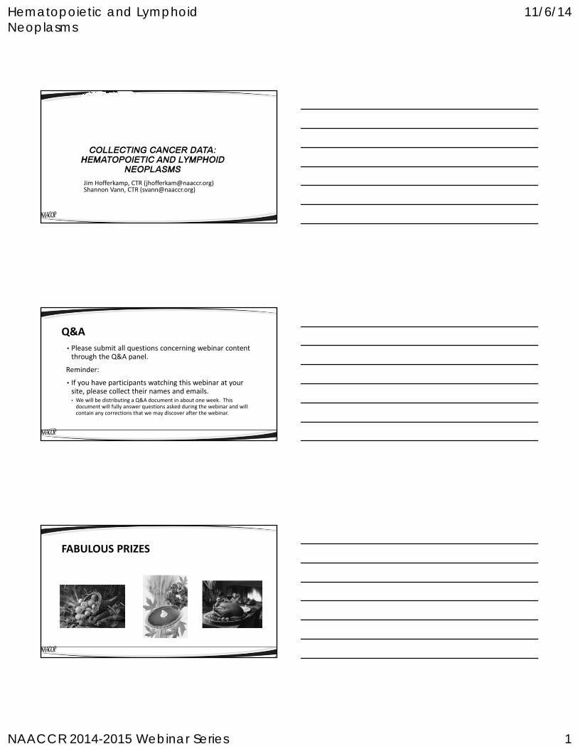
HEMATOPOIETIC AND LYMPHOID DATABASE AND MANUAL
• Determine reportability
• Determine multiple primaries
• Assign primary site
• Assign histology
• Assign grade
4
DIAGNOSTIC CONFIRMATION• Microscopically confirmed• 1: Positive histology• Tissue specimen• Bone marrow specimen• CBC, WBC, peripheral blood smear for leukemia only
• 2: Positive cytology• 3: Positive histology PLUS:• Positive immunophenotyping AND/OR• Positive genetic studies
• 4: Positive microscopic confirmation, method not specified
5
DIAGNOSTIC CONFIRMATION
• Not microscopically confirmed• 5: Positive laboratory test/marker study
• 6: Direct visualization without microscopic confirmation
• 7: Radiology and other imaging techniques without microscopic confirmation
• 8: Clinical diagnosis only (other than 5, 6, or 7)
• 9: Unknown whether or not microscopically confirmed
6
Hematopoietic and Lymphoid Neoplasms
11/6/14
NAACCR 2014-2015 Webinar Series 3
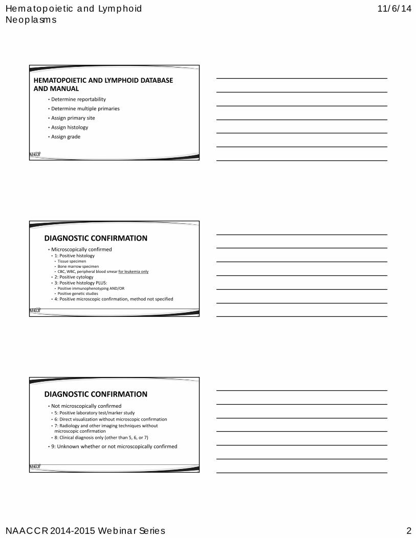
POP QUIZ
• Patient presents with unexplained weight loss, chronic fatigue, and bruising. Peripheral blood smear showed chronic myeloid leukemia.
• What is the code for diagnostic confirmation?• 1 Positive histology
7
POP QUIZ
• Bone marrow biopsy: B lymphoblastic leukemia
• FISH: Most likely represents a hyperdiploid clone
• What is the code for diagnostic confirmation?• 1 Positive histology
8
POP QUIZ• Tonsillectomy and adenoidectomy path: Follicular lymphoma of the tonsil
• FISH: BCL2 gene rearrangements; follicular lymphoma grade 2
• What is the code for diagnostic confirmation? • 3 Positive histology PLUS:• Positive immunophenotyping AND/OR• Positive genetic studies
9

Hematopoietic and Lymphoid Neoplasms
11/6/14
NAACCR 2014-2015 Webinar Series 4
POP QUIZ• PET scan: Malignant adenopathy of mediastinal and retroperitoneal lymph nodes consistent with lymphoma.
• Patient refused any further work‐up or treatment because of other serious co‐morbidities.
• What is the code for diagnostic confirmation?• 7 Radiology and other imaging techniques without microscopic confirmation
10
POP QUIZ
• Bone marrow biopsy: Negative
• Cytogenetics: Loss of chromosome 7
• Discharge diagnosis: Myeloproliferative neoplasm, unclassifiable
• What is the code for diagnostic confirmation?• 8: Clinical diagnosis only (other than 5, 6, or 7)
11
AMBIGUOUS TERMINOLOGYREPORTABILITY
• Apparently
• Appears
• Comparable with
See page 20 of your manual for a full list
• Do not report cases diagnosed only by ambiguous cytology (cytology diagnosis preceded by ambiguous term)
HISTOLOGY
• Do not use ambiguous terms to code a specific histology
12
Hematopoietic and Lymphoid Neoplasms
11/6/14
NAACCR 2014-2015 Webinar Series 5
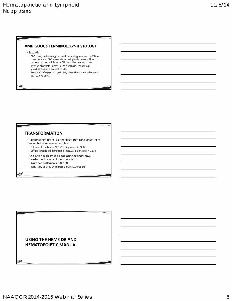
AMBIGUOUS TERMINOLOGY‐HISTOLOGY
• Exception• CBC done, no histology or provisional diagnosis on the CBC or smear reports. CBC states abnormal lymphocytosis. Flow cytometry compatible with CLL. No other workup done.
• Per the abstractor notes in the database, “abnormal lymphocytosis” is present in CLL.
• Assign histology for CLL (9823/3) since there is no other code that can be used.
13
TRANSFORMATION
• A chronic neoplasm is a neoplasm that can transform to an acute/more severe neoplasm• Follicular Lymphoma (9695/3) diagnosed in 2012
• Diffuse large B‐cell lymphoma (9680/3) diagnosed in 2014
• An acute neoplasm is a neoplasm that may have transformed from a chronic neoplasm• Acute myeloid leukemia (9861/3)
• Refractory anemia with ring sideroblasts (9982/3)
14
USING THE HEME DB AND HEMATOPOIETIC MANUAL
15
Hematopoietic and Lymphoid Neoplasms
11/6/14
NAACCR 2014-2015 Webinar Series 6
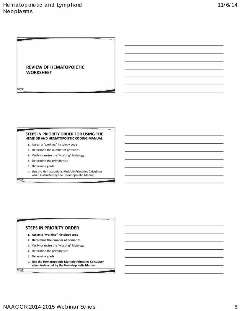
REVIEW OF HEMATOPOIETIC WORKSHEET
16
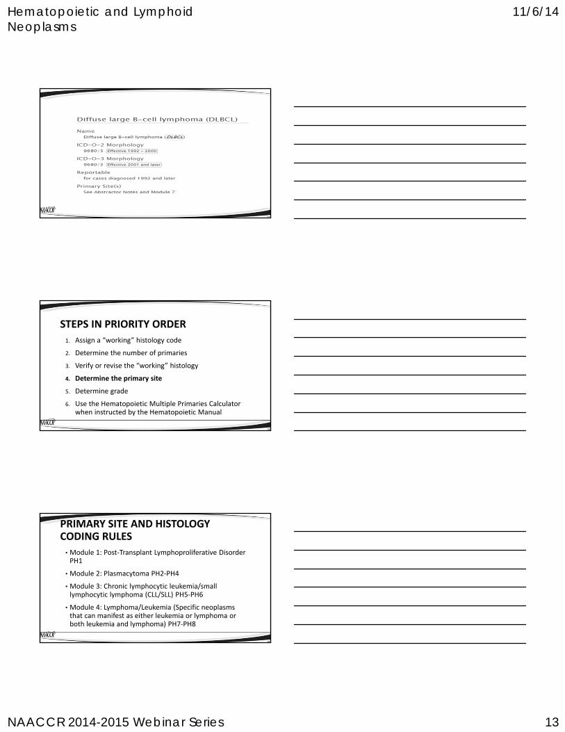
STEPS IN PRIORITY ORDER FOR USING THE HEME DB AND HEMATOPOIETIC CODING MANUAL
1. Assign a “working” histology code
2. Determine the number of primaries
3. Verify or revise the “working” histology
4. Determine the primary site
5. Determine grade
6. Use the Hematopoietic Multiple Primaries Calculator when instructed by the Hematopoietic Manual
17
STEPS IN PRIORITY ORDER
1. Assign a “working” histology code
2. Determine the number of primaries
3. Verify or revise the “working” histology
4. Determine the primary site
5. Determine grade
6. Use the Hematopoietic Multiple Primaries Calculator when instructed by the Hematopoietic Manual
18
Hematopoietic and Lymphoid Neoplasms
11/6/14
NAACCR 2014-2015 Webinar Series 7
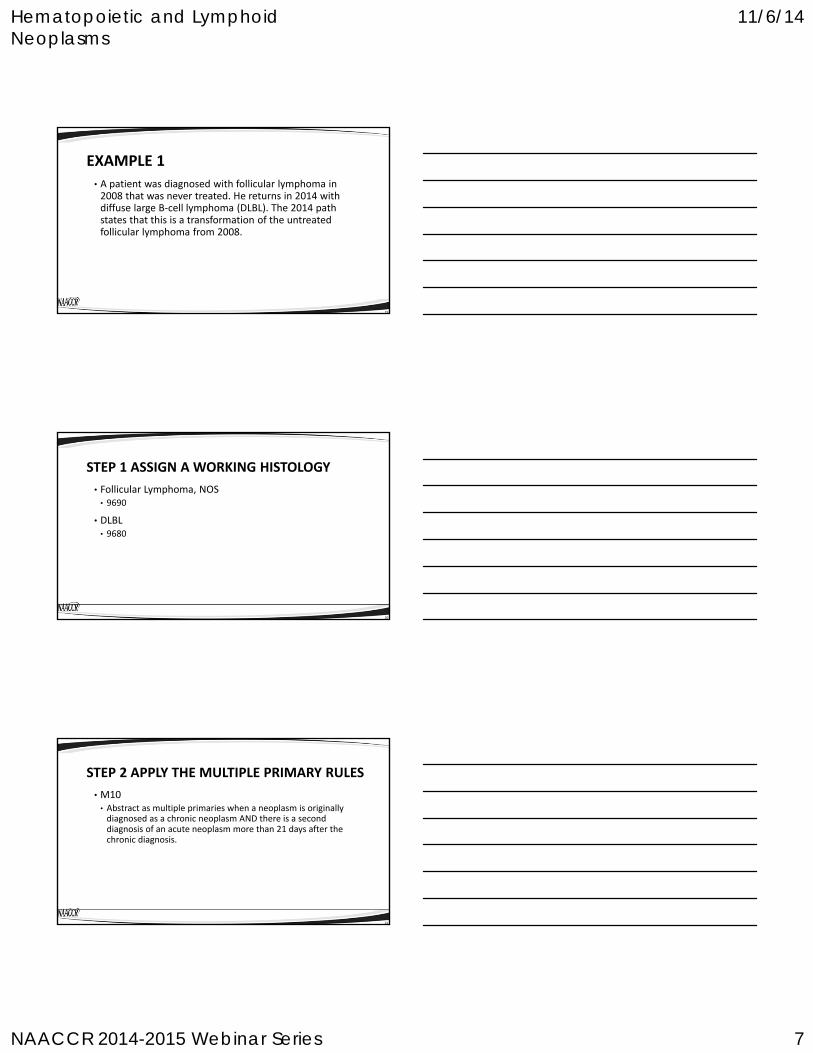
EXAMPLE 1
• A patient was diagnosed with follicular lymphoma in 2008 that was never treated. He returns in 2014 with diffuse large B‐cell lymphoma (DLBL). The 2014 path states that this is a transformation of the untreated follicular lymphoma from 2008.
19
STEP 1 ASSIGN A WORKING HISTOLOGY
• Follicular Lymphoma, NOS• 9690
• DLBL• 9680
20
STEP 2 APPLY THE MULTIPLE PRIMARY RULES
• M10• Abstract as multiple primaries when a neoplasm is originally diagnosed as a chronic neoplasm AND there is a second diagnosis of an acute neoplasm more than 21 days after the chronic diagnosis.
21
Hematopoietic and Lymphoid Neoplasms
11/6/14
NAACCR 2014-2015 Webinar Series 8
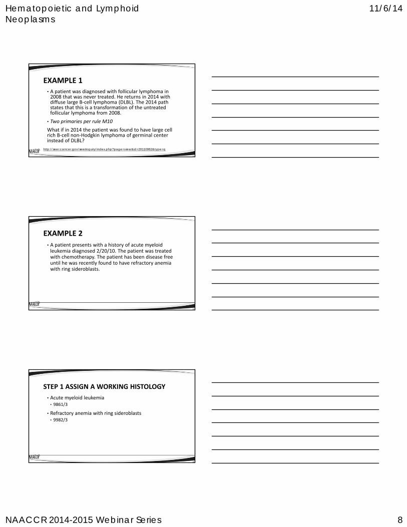
EXAMPLE 1• A patient was diagnosed with follicular lymphoma in 2008 that was never treated. He returns in 2014 with diffuse large B‐cell lymphoma (DLBL). The 2014 path states that this is a transformation of the untreated follicular lymphoma from 2008.
• Two primaries per rule M10
What if in 2014 the patient was found to have large cell rich B‐cell non‐Hodgkin lymphoma of germinal center instead of DLBL?
http://seer.cancer.gov/seerinquiry/index.php?page=view&id=20110062&type=q
22
EXAMPLE 2
• A patient presents with a history of acute myeloid leukemia diagnosed 2/20/10. The patient was treated with chemotherapy. The patient has been disease free until he was recently found to have refractory anemia with ring sideroblasts.
23
STEP 1 ASSIGN A WORKING HISTOLOGY
• Acute myeloid leukemia• 9861/3
• Refractory anemia with ring sideroblasts• 9982/3
24
Hematopoietic and Lymphoid Neoplasms
11/6/14
NAACCR 2014-2015 Webinar Series 9
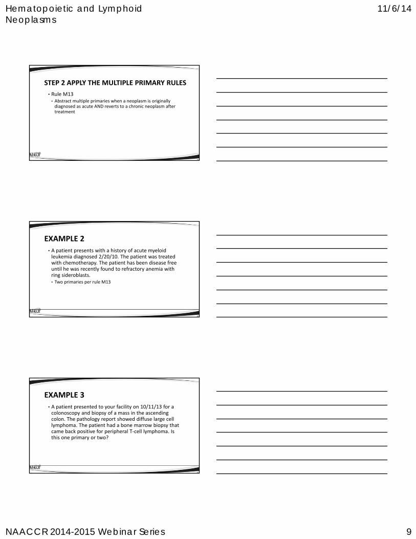
STEP 2 APPLY THE MULTIPLE PRIMARY RULES
• Rule M13 • Abstract multiple primaries when a neoplasm is originally diagnosed as acute AND reverts to a chronic neoplasm after treatment
25
EXAMPLE 2
• A patient presents with a history of acute myeloid leukemia diagnosed 2/20/10. The patient was treated with chemotherapy. The patient has been disease free until he was recently found to refractory anemia with ring sideroblasts.• Two primaries per rule M13
26
EXAMPLE 3
• A patient presented to your facility on 10/11/13 for a colonoscopy and biopsy of a mass in the ascending colon. The pathology report showed diffuse large cell lymphoma. The patient had a bone marrow biopsy that came back positive for peripheral T‐cell lymphoma. Is this one primary or two?
27
Hematopoietic and Lymphoid Neoplasms
11/6/14
NAACCR 2014-2015 Webinar Series 10
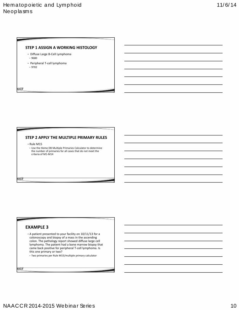
STEP 1 ASSIGN A WORKING HISTOLOGY
• Diffuse Large B‐Cell Lymphoma • 9680
• Peripheral T‐cell lymphoma• 9702
28
STEP 2 APPLY THE MULTIPLE PRIMARY RULES
• Rule M15• Use the Heme DB Multiple Primaries Calculator to determine the number of primaries for all cases that do not meet the criteria of M1‐M14
29
EXAMPLE 3
• A patient presented to your facility on 10/11/13 for a colonoscopy and biopsy of a mass in the ascending colon. The pathology report showed diffuse large cell lymphoma. The patient had a bone marrow biopsy that came back positive for peripheral T‐cell lymphoma. Is this one primary or two?• Two primaries per Rule M15/multiple primary calculator
30

Hematopoietic and Lymphoid Neoplasms
11/6/14
NAACCR 2014-2015 Webinar Series 11
STEPS IN PRIORITY ORDER
1. Assign a “working” histology code
2. Determine the number of primaries
3. Verify or revise the “working” histology
4. Determine the primary site
5. Determine grade
6. Use the Hematopoietic Multiple Primaries Calculator when instructed by the Hematopoietic Manual
31
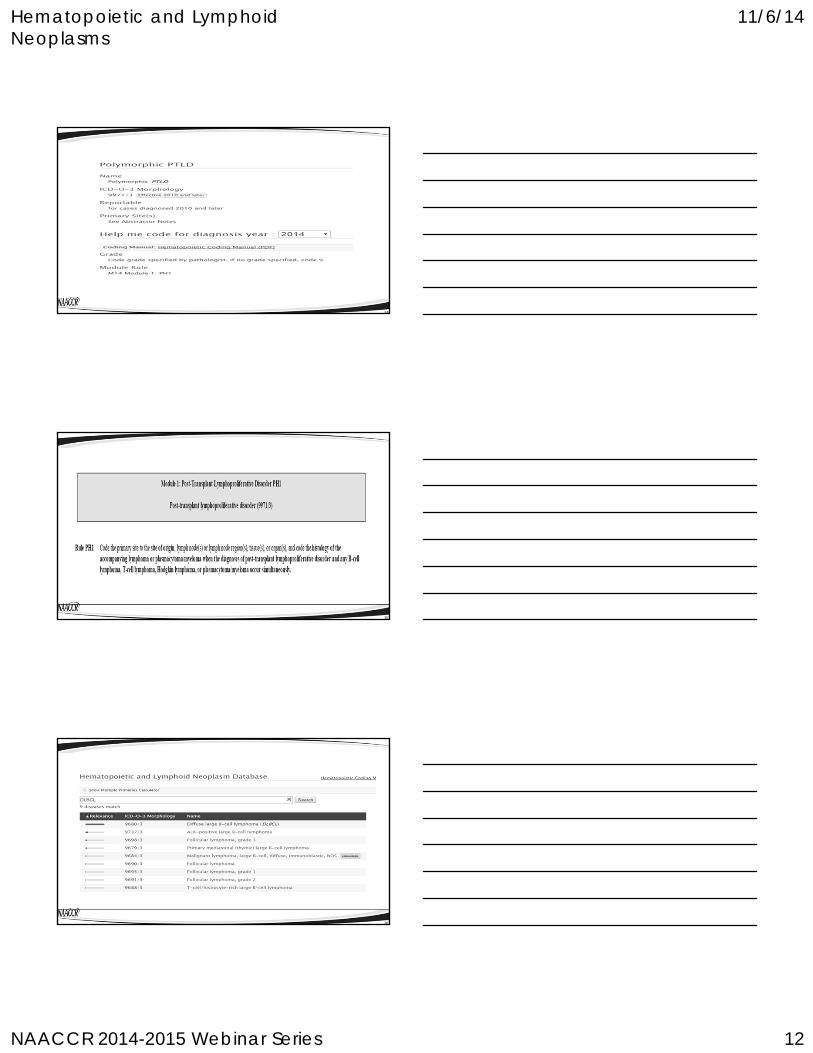
STEPS IN PRIORITY ORDER
• Example• Patient has history of liver transplant.
• Lymphadenopathy of axillary, mediastinal, and hilar nodes
• Axillary lymph node biopsy: Post‐transplant lymphoproliferative disorder (PLTD)
• Cytogenetics: Translocations involving c‐MYC, BCL6, and IgH genes; PLTD and diffuse large b‐cell lymphoma (DLBCL)
32
33
Hematopoietic and Lymphoid Neoplasms
11/6/14
NAACCR 2014-2015 Webinar Series 12
34
35
36
Hematopoietic and Lymphoid Neoplasms
11/6/14
NAACCR 2014-2015 Webinar Series 13
37
STEPS IN PRIORITY ORDER
1. Assign a “working” histology code
2. Determine the number of primaries
3. Verify or revise the “working” histology
4. Determine the primary site
5. Determine grade
6. Use the Hematopoietic Multiple Primaries Calculator when instructed by the Hematopoietic Manual
38
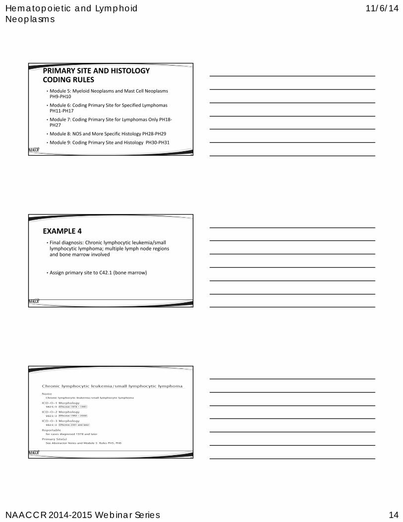
PRIMARY SITE AND HISTOLOGY CODING RULES
• Module 1: Post‐Transplant Lymphoproliferative Disorder PH1
• Module 2: Plasmacytoma PH2‐PH4
• Module 3: Chronic lymphocytic leukemia/small lymphocytic lymphoma (CLL/SLL) PH5‐PH6
• Module 4: Lymphoma/Leukemia (Specific neoplasms that can manifest as either leukemia or lymphoma or both leukemia and lymphoma) PH7‐PH8
39
Hematopoietic and Lymphoid Neoplasms
11/6/14
NAACCR 2014-2015 Webinar Series 14
PRIMARY SITE AND HISTOLOGY CODING RULES• Module 5: Myeloid Neoplasms and Mast Cell Neoplasms PH9‐PH10
• Module 6: Coding Primary Site for Specified Lymphomas PH11‐PH17
• Module 7: Coding Primary Site for Lymphomas Only PH18‐PH27
• Module 8: NOS and More Specific Histology PH28‐PH29
• Module 9: Coding Primary Site and Histology PH30‐PH31
40
EXAMPLE 4
• Final diagnosis: Chronic lymphocytic leukemia/small lymphocytic lymphoma; multiple lymph node regions and bone marrow involved
• Assign primary site to C42.1 (bone marrow)
41
42
Hematopoietic and Lymphoid Neoplasms
11/6/14
NAACCR 2014-2015 Webinar Series 15
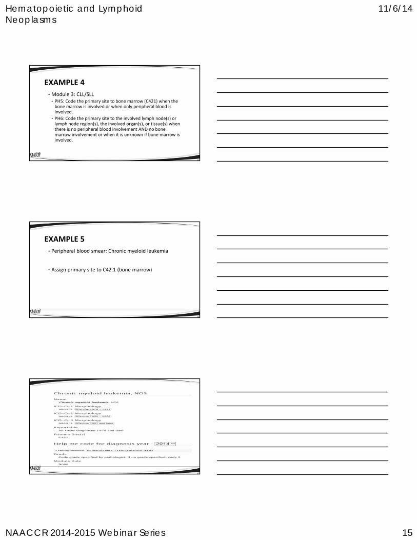
EXAMPLE 4
• Module 3: CLL/SLL• PH5: Code the primary site to bone marrow (C421) when the bone marrow is involved or when only peripheral blood is involved.
• PH6: Code the primary site to the involved lymph node(s) or lymph node region(s), the involved organ(s), or tissue(s) when there is no peripheral blood involvement AND no bone marrow involvement or when it is unknown if bone marrow is involved.
43
EXAMPLE 5
• Peripheral blood smear: Chronic myeloid leukemia
• Assign primary site to C42.1 (bone marrow)
44
45
Hematopoietic and Lymphoid Neoplasms
11/6/14
NAACCR 2014-2015 Webinar Series 16
EXAMPLE 5
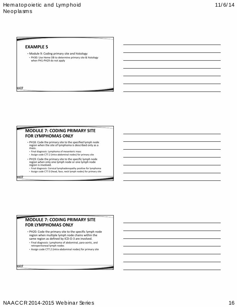
• Module 9: Coding primary site and histology • PH30: Use Heme DB to determine primary site & histology when PH1‐PH29 do not apply
46
MODULE 7: CODING PRIMARY SITE FOR LYMPHOMAS ONLY• PH18: Code the primary site to the specified lymph node region when the site of lymphoma is described only as a mass.• Final diagnosis: Lymphoma of mesenteric mass• Assign code C77.2 (intra‐abdominal nodes) for primary site
• PH19: Code the primary site to the specific lymph node region when only one lymph node or one lymph node region is involved.• Final diagnosis: Cervical lymphadenopathy positive for lymphoma• Assign code C77.0 (head, face, neck lymph nodes) for primary site
47
MODULE 7: CODING PRIMARY SITE FOR LYMPHOMAS ONLY
• PH20: Code the primary site to the specific lymph node region when multiple lymph node chains within the same region as defined by ICD‐O‐3 are involved.• Final diagnosis: Lymphoma of abdominal, para‐aortic, and retroperitoneal lymph nodes
• Assign code C77.2 (intra‐abdominal nodes) for primary site
48
Hematopoietic and Lymphoid Neoplasms
11/6/14
NAACCR 2014-2015 Webinar Series 17
MODULE 7: CODING PRIMARY SITE FOR LYMPHOMAS ONLY
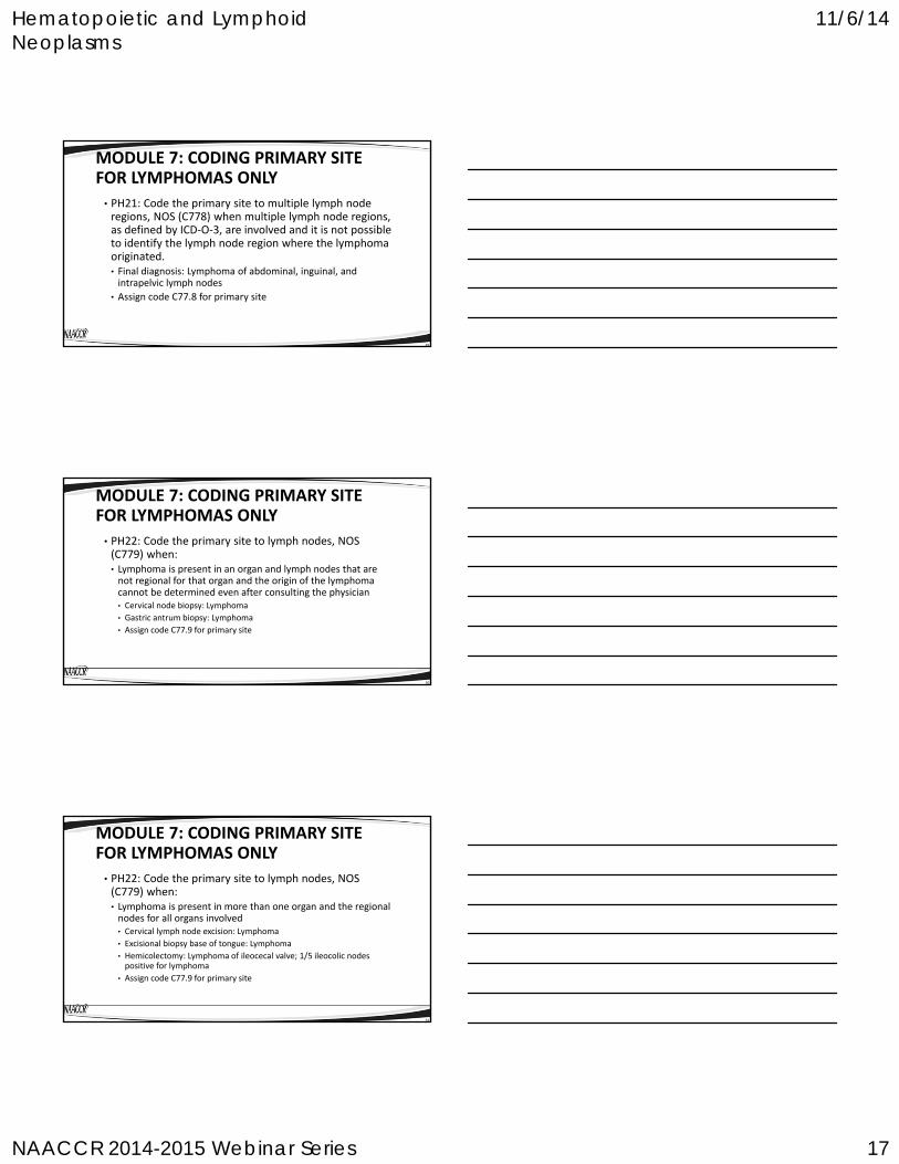
• PH21: Code the primary site to multiple lymph node regions, NOS (C778) when multiple lymph node regions, as defined by ICD‐O‐3, are involved and it is not possible to identify the lymph node region where the lymphoma originated.• Final diagnosis: Lymphoma of abdominal, inguinal, and intrapelvic lymph nodes
• Assign code C77.8 for primary site
49
MODULE 7: CODING PRIMARY SITE FOR LYMPHOMAS ONLY
• PH22: Code the primary site to lymph nodes, NOS (C779) when:• Lymphoma is present in an organ and lymph nodes that are not regional for that organ and the origin of the lymphoma cannot be determined even after consulting the physician • Cervical node biopsy: Lymphoma
• Gastric antrum biopsy: Lymphoma
• Assign code C77.9 for primary site
50
MODULE 7: CODING PRIMARY SITE FOR LYMPHOMAS ONLY
• PH22: Code the primary site to lymph nodes, NOS (C779) when:• Lymphoma is present in more than one organ and the regional nodes for all organs involved • Cervical lymph node excision: Lymphoma
• Excisional biopsy base of tongue: Lymphoma
• Hemicolectomy: Lymphoma of ileocecal valve; 1/5 ileocolic nodes positive for lymphoma
• Assign code C77.9 for primary site
51
Hematopoietic and Lymphoid Neoplasms
11/6/14
NAACCR 2014-2015 Webinar Series 18
MODULE 7: CODING PRIMARY SITE FOR LYMPHOMAS ONLY
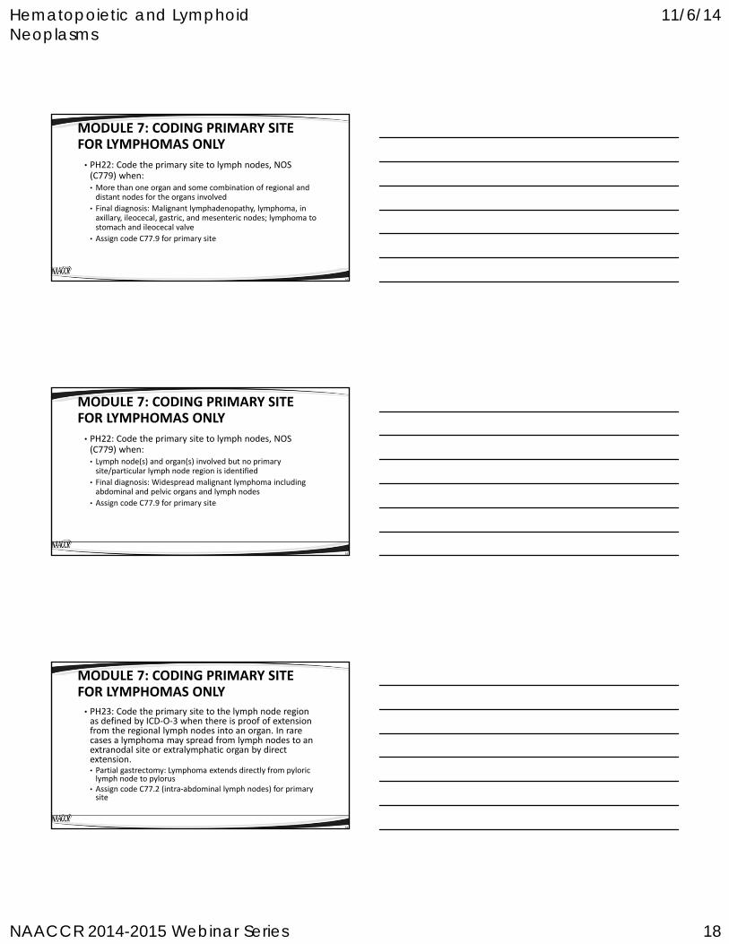
• PH22: Code the primary site to lymph nodes, NOS (C779) when:• More than one organ and some combination of regional and distant nodes for the organs involved
• Final diagnosis: Malignant lymphadenopathy, lymphoma, in axillary, ileocecal, gastric, and mesenteric nodes; lymphoma to stomach and ileocecal valve
• Assign code C77.9 for primary site
52
MODULE 7: CODING PRIMARY SITE FOR LYMPHOMAS ONLY
• PH22: Code the primary site to lymph nodes, NOS (C779) when:• Lymph node(s) and organ(s) involved but no primary site/particular lymph node region is identified
• Final diagnosis: Widespread malignant lymphoma including abdominal and pelvic organs and lymph nodes
• Assign code C77.9 for primary site
53
MODULE 7: CODING PRIMARY SITE FOR LYMPHOMAS ONLY• PH23: Code the primary site to the lymph node region as defined by ICD‐O‐3 when there is proof of extension from the regional lymph nodes into an organ. In rare cases a lymphoma may spread from lymph nodes to an extranodal site or extralymphatic organ by direct extension.• Partial gastrectomy: Lymphoma extends directly from pyloric lymph node to pylorus
• Assign code C77.2 (intra‐abdominal lymph nodes) for primary site
54
Hematopoietic and Lymphoid Neoplasms
11/6/14
NAACCR 2014-2015 Webinar Series 19
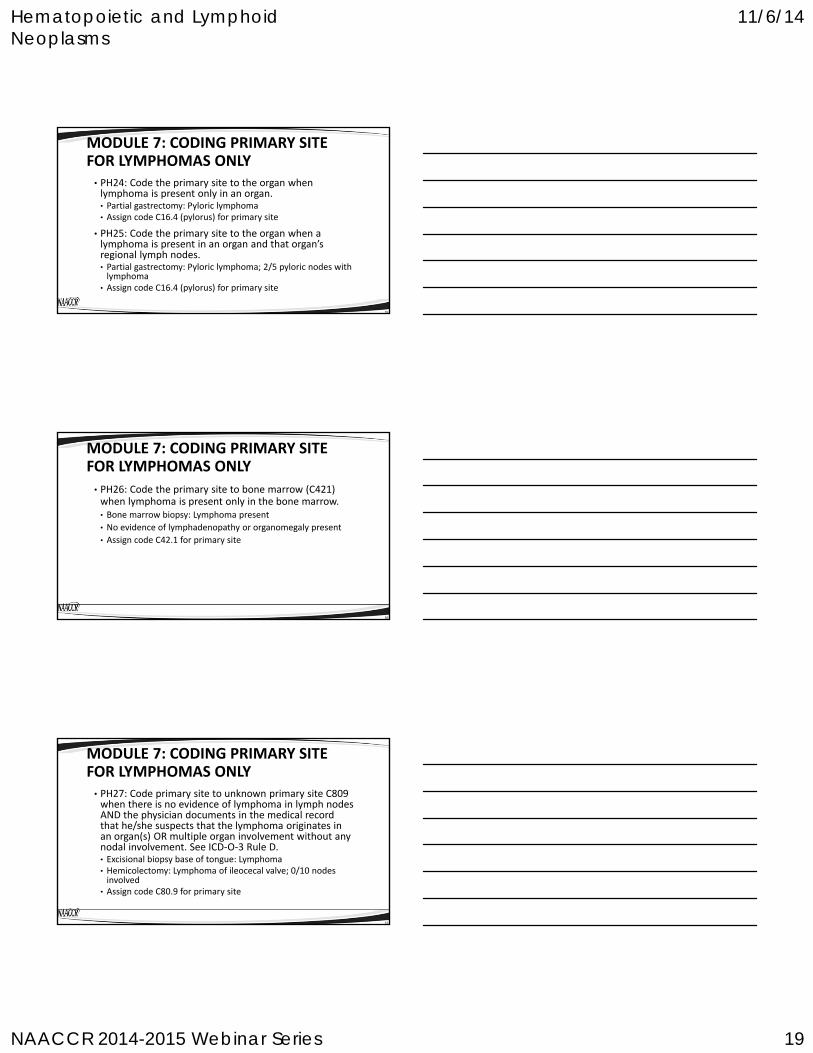
MODULE 7: CODING PRIMARY SITE FOR LYMPHOMAS ONLY• PH24: Code the primary site to the organ when lymphoma is present only in an organ.• Partial gastrectomy: Pyloric lymphoma• Assign code C16.4 (pylorus) for primary site
• PH25: Code the primary site to the organ when a lymphoma is present in an organ and that organ’s regional lymph nodes.• Partial gastrectomy: Pyloric lymphoma; 2/5 pyloric nodes with lymphoma
• Assign code C16.4 (pylorus) for primary site
55
MODULE 7: CODING PRIMARY SITE FOR LYMPHOMAS ONLY
• PH26: Code the primary site to bone marrow (C421) when lymphoma is present only in the bone marrow.• Bone marrow biopsy: Lymphoma present
• No evidence of lymphadenopathy or organomegaly present
• Assign code C42.1 for primary site
56
MODULE 7: CODING PRIMARY SITE FOR LYMPHOMAS ONLY• PH27: Code primary site to unknown primary site C809 when there is no evidence of lymphoma in lymph nodes AND the physician documents in the medical record that he/she suspects that the lymphoma originates in an organ(s) OR multiple organ involvement without any nodal involvement. See ICD‐O‐3 Rule D.• Excisional biopsy base of tongue: Lymphoma• Hemicolectomy: Lymphoma of ileocecal valve; 0/10 nodes involved
• Assign code C80.9 for primary site
57
Hematopoietic and Lymphoid Neoplasms
11/6/14
NAACCR 2014-2015 Webinar Series 20
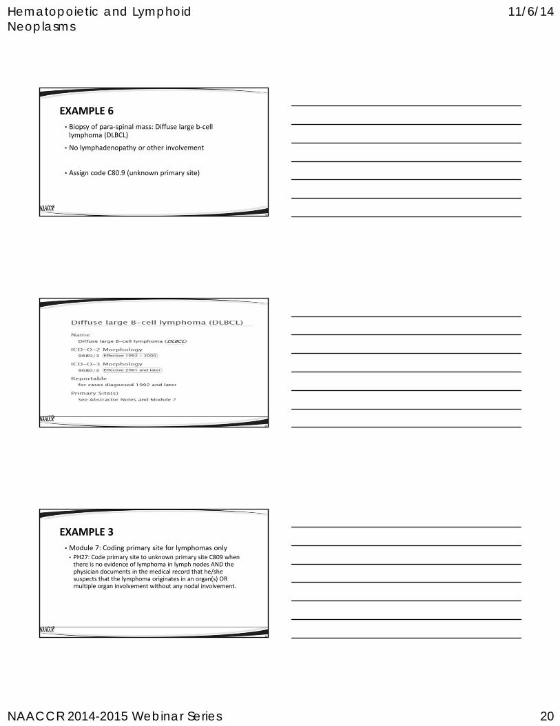
EXAMPLE 6
• Biopsy of para‐spinal mass: Diffuse large b‐cell lymphoma (DLBCL)
• No lymphadenopathy or other involvement
• Assign code C80.9 (unknown primary site)
58
59
EXAMPLE 3
• Module 7: Coding primary site for lymphomas only• PH27: Code primary site to unknown primary site C809 when there is no evidence of lymphoma in lymph nodes AND the physician documents in the medical record that he/she suspects that the lymphoma originates in an organ(s) OR multiple organ involvement without any nodal involvement.
60
Hematopoietic and Lymphoid Neoplasms
11/6/14
NAACCR 2014-2015 Webinar Series 21
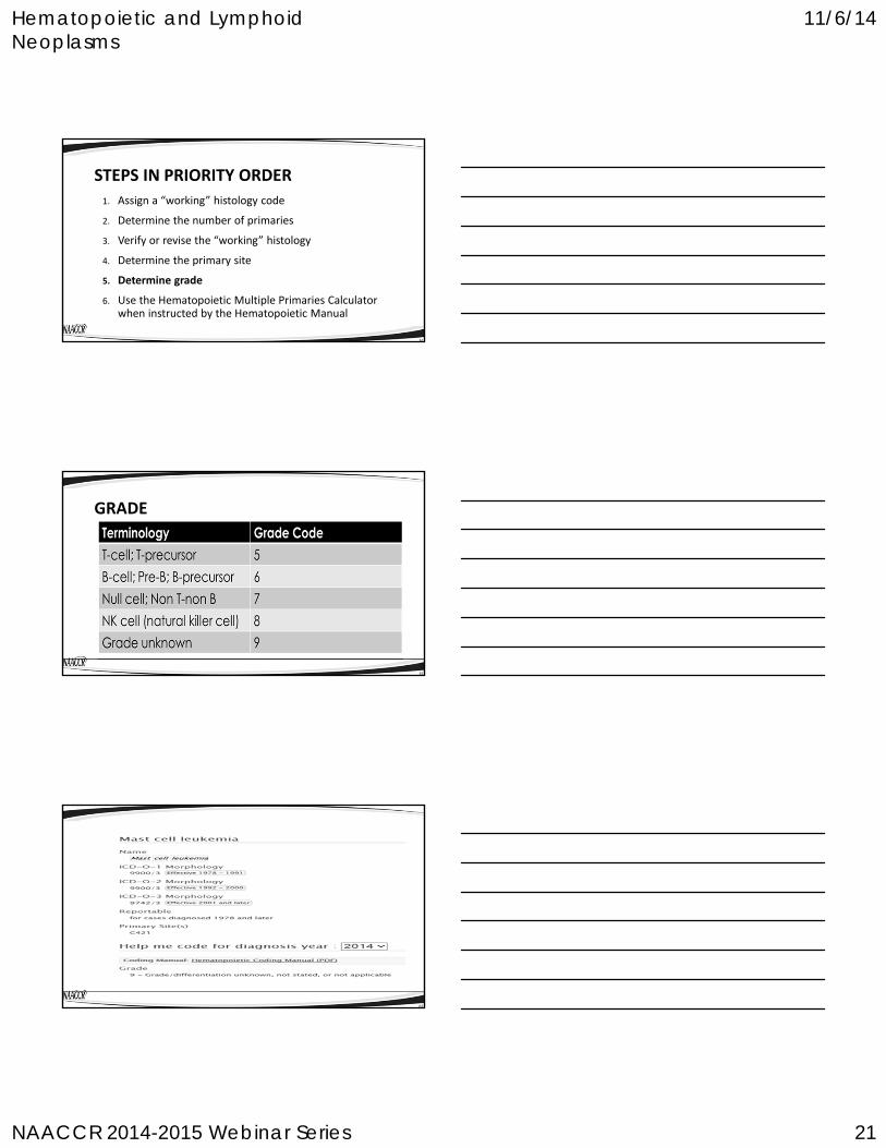
STEPS IN PRIORITY ORDER
1. Assign a “working” histology code
2. Determine the number of primaries
3. Verify or revise the “working” histology
4. Determine the primary site
5. Determine grade
6. Use the Hematopoietic Multiple Primaries Calculator when instructed by the Hematopoietic Manual
61
GRADE
62
63
Hematopoietic and Lymphoid Neoplasms
11/6/14
NAACCR 2014-2015 Webinar Series 22
64
65
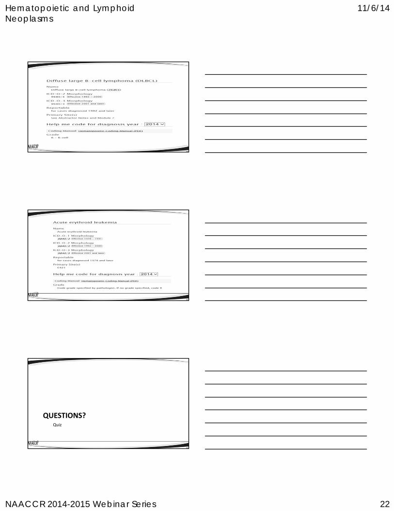
QUESTIONS?Quiz
66
Hematopoietic and Lymphoid Neoplasms
11/6/14
NAACCR 2014-2015 Webinar Series 23
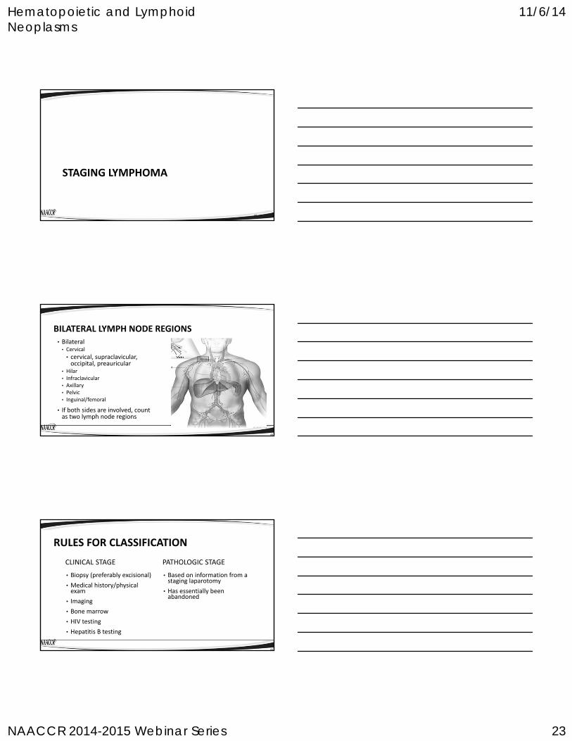
STAGING LYMPHOMA
67
BILATERAL LYMPH NODE REGIONS• Bilateral• Cervical
• cervical, supraclavicular, occipital, preauricular
• Hilar• Infraclavicular• Axillary• Pelvic• Inguinal/femoral
• If both sides are involved, count as two lymph node regions
68
RULES FOR CLASSIFICATION
CLINICAL STAGE
• Biopsy (preferably excisional)
• Medical history/physical exam
• Imaging
• Bone marrow
• HIV testing
• Hepatitis B testing
PATHOLOGIC STAGE
• Based on information from a staging laparotomy
• Has essentially been abandoned
69
Hematopoietic and Lymphoid Neoplasms
11/6/14
NAACCR 2014-2015 Webinar Series 24
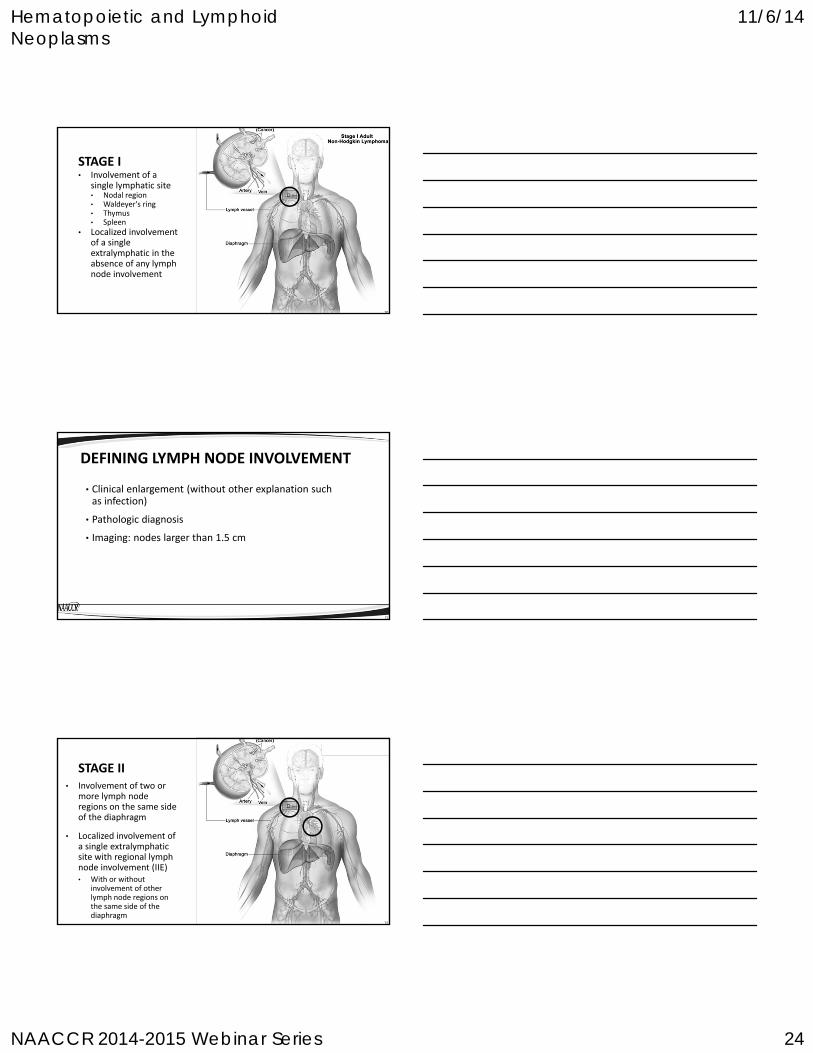
STAGE I• Involvement of a
single lymphatic site• Nodal region• Waldeyer's ring• Thymus• Spleen
• Localized involvement of a single extralymphatic in the absence of any lymph node involvement
70
DEFINING LYMPH NODE INVOLVEMENT
• Clinical enlargement (without other explanation such as infection)
• Pathologic diagnosis
• Imaging: nodes larger than 1.5 cm
71
STAGE II• Involvement of two or
more lymph node regions on the same side of the diaphragm
• Localized involvement of a single extralymphatic site with regional lymph node involvement (IIE)• With or without
involvement of other lymph node regions on the same side of the diaphragm
72
Hematopoietic and Lymphoid Neoplasms
11/6/14
NAACCR 2014-2015 Webinar Series 25
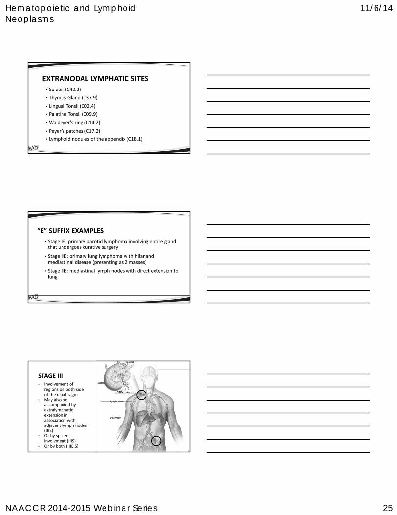
EXTRANODAL LYMPHATIC SITES• Spleen (C42.2)
• Thymus Gland (C37.9)
• Lingual Tonsil (C02.4)
• Palatine Tonsil (C09.9)
• Waldeyer's ring (C14.2)
• Peyer’s patches (C17.2)
• Lymphoid nodules of the appendix (C18.1)
73
“E” SUFFIX EXAMPLES
• Stage IE: primary parotid lymphoma involving entire gland that undergoes curative surgery
• Stage IIE: primary lung lymphoma with hilar and mediastinal disease (presenting as 2 masses)
• Stage IIE: mediastinal lymph nodes with direct extension to lung
74
STAGE III• Involvement of
regions on both side of the diaphragm
• May also be accompanied by extralymphatic extension in association with adjacent lymph nodes (IIIE)
• Or by spleen involvment (IIIS)
• Or by both (IIIE,S)75
Hematopoietic and Lymphoid Neoplasms
11/6/14
NAACCR 2014-2015 Webinar Series 26
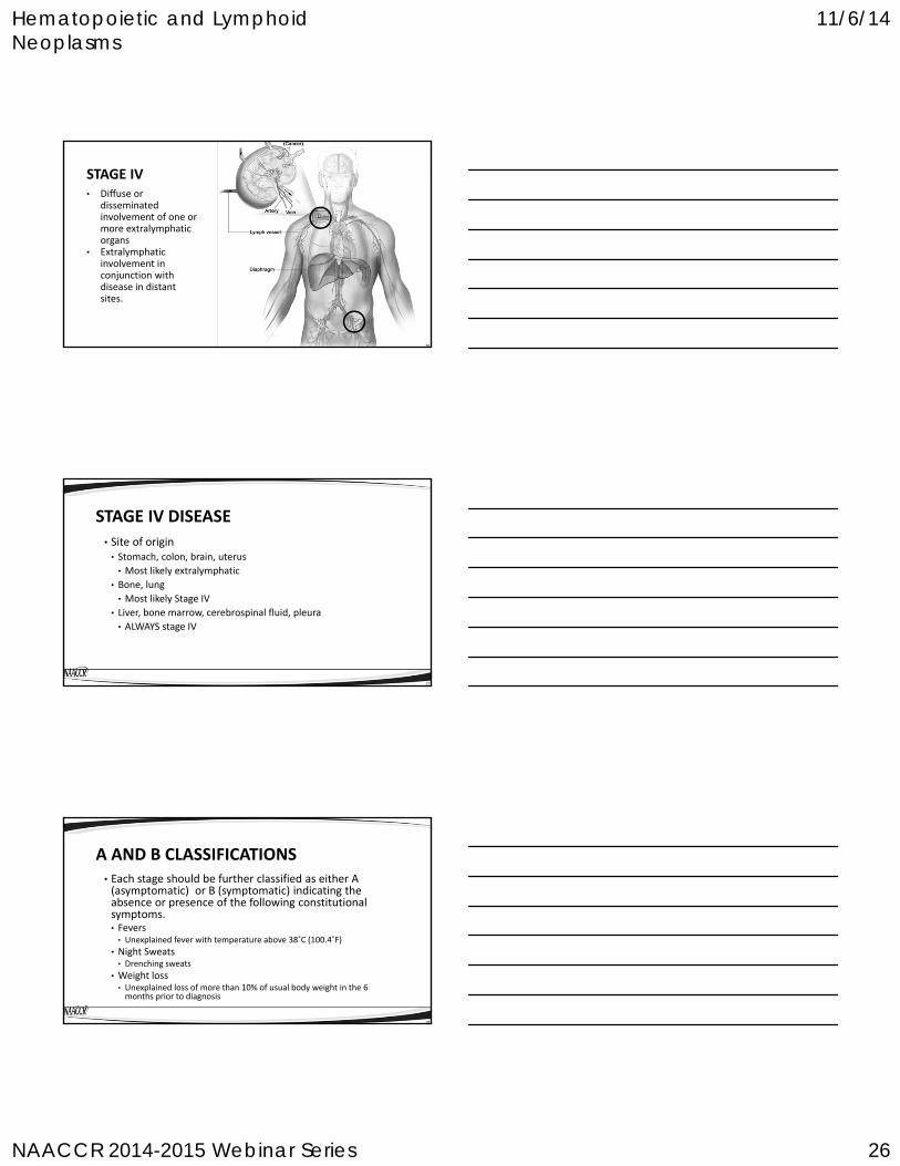
STAGE IV• Diffuse or
disseminated involvement of one or more extralymphatic organs
• Extralymphatic involvement in conjunction with disease in distant sites.
76
STAGE IV DISEASE
• Site of origin• Stomach, colon, brain, uterus
• Most likely extralymphatic
• Bone, lung
• Most likely Stage IV
• Liver, bone marrow, cerebrospinal fluid, pleura
• ALWAYS stage IV
77
A AND B CLASSIFICATIONS• Each stage should be further classified as either A (asymptomatic) or B (symptomatic) indicating the absence or presence of the following constitutional symptoms.• Fevers• Unexplained fever with temperature above 38˚C (100.4˚F)
• Night Sweats• Drenching sweats
• Weight loss• Unexplained loss of more than 10% of usual body weight in the 6 months prior to diagnosis
78
Hematopoietic and Lymphoid Neoplasms
11/6/14
NAACCR 2014-2015 Webinar Series 27
SUMMARY STAGE 2000Hematopoietic, Reticuloendothelial, Immunoproliferative, and Myeoproliferative Neoplasms
79
SUMMARY STAGE 2000
• Hematopoietic, reticuloendothelial, immunoproliferative, and myeoproliferative neoplasms• 1: Localized• Isolated/monostotic/single/solitary/unifocal
• 7: Distant• Polyostotic; disease disseminated at diagnosis
• 9: Death certificate only
80
SUMMARY STAGE 2000
• Hematopoietic, reticuloendothelial, immunoproliferative, and myeoproliferative neoplasms• Assign code 1, 7, or 9• 9731/3, 9734/3
• 9740/3, 9750/3, 9755/3, 9756/3, 9757/3, 9758/3, 9764/3, 9930/3
• Assign code 1 unless death certificate only (code 9)• 9751/3
• Assign all other listed histologies code 7 unless death certificate only (code 9)
81
Hematopoietic and Lymphoid Neoplasms
11/6/14
NAACCR 2014-2015 Webinar Series 28
SUMMARY STAGE 2000Hodgkin and Non‐Hodgkin Lymphomas of All Sites
82
SUMMARY STAGE 2000: LYMPHOMA
• 1 Localized• Stage I: Involvement of a single lymph node region
• Stage IE• Localized involvement of a single extralymphatic organ/site
• Multifocal involvement of one extralymphatic organ/site
• Stage IS: Localized involvement of spleen only
83
SUMMARY STAGE 2000: LYMPHOMA
• 5 Regional NOS• Stage II: Involvement of two or more lymph node regions on the SAME side of the diaphragm
• Stage IIE
• Stage IIS
• Stage IIES: Involvement of spleen PLUS localized involvement of a single extralymphatic organ/site BELOW the diaphragm WITH/WITHOUT involvement of lymph node(s) BELOW the diaphragm
84

Hematopoietic and Lymphoid Neoplasms
11/6/14
NAACCR 2014-2015 Webinar Series 29
SUMMARY STAGE 2000: LYMPHOMA• 7 Distant• Stage III: Involvement of lymph node regions on BOTH sides of the diaphragm
• Stage IIIE: Involvement of an extralymphatic organ or site PLUS involvement of lymph node(s) on the OPPOSITE side of the diaphragm
• Stage IIIES: • Involvement of the spleen PLUS involvement of lymph node region(s) ABOVE the diaphragm PLUS involvement of a single extralymphatic organ/site on either side of the diaphragm
• Involvement of the spleen PLUS a single extralymphatic organ/site ABOVE the diaphragm WITH OR WITHOUT involvement of lymph node(s)
85
SUMMARY STAGE 2000: LYMPHOMA
• 7 Distant (continued)• Stage IV: • Disseminated involvement of ONE OR MORE extralymphatic organ(s)/site(s)
• (Multifocal) involvement of MORE THAN ONE extralymphatic organ/site
• Metastases• Bone marrow
• Liver
• 9 Unstaged; not stated
86
COLLABORATIVE STAGE DATA COLLECTION SYSTEM (CSV02.05)
Lymphoma
87
Hematopoietic and Lymphoid Neoplasms
11/6/14
NAACCR 2014-2015 Webinar Series 30
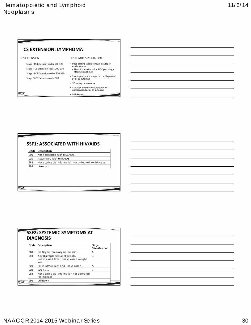
CS EXTENSION: LYMPHOMA
CS EXTENSION
• Stage I CS Extension codes 100‐120
• Stage II CS Extension codes 200‐230
• Stage III CS Extension codes 300‐330
• Stage IV CS Extension code 800
CS TUMOR SIZE EXT/EVAL
• 0‐No staging laparotomy; no autopsy evidence used• Used if the criteria for AJCC pathologic staging is not met
• 2‐Autopsy(tumor suspected or diagnosed prior to autopsy)
• 3‐Staging Laparotomy
• 8‐Autopsy (tumor unsuspected or undiagnosed prior to autopsy)
• 9 Unknown88
SSF1: ASSOCIATED WITH HIV/AIDSCode Description000 Not associated with HIV/AIDS010 Associated with HIV/AIDS988 Not applicable: Information not collected for this case999 Unknown
89
SSF2: SYSTEMIC SYMPTOMS AT DIAGNOSIS
Code Description Stage Classification
000 No B symptoms (asymptomatic) A010 Any B symptoms: Night sweats,
unexplained fever, unexplained weightloss
B
020 Pruritus (recurrent and unexplained) A030 020 + 010 B988 Not applicable: Information not collected
for this case999 Unknown
90
Hematopoietic and Lymphoid Neoplasms
11/6/14
NAACCR 2014-2015 Webinar Series 31
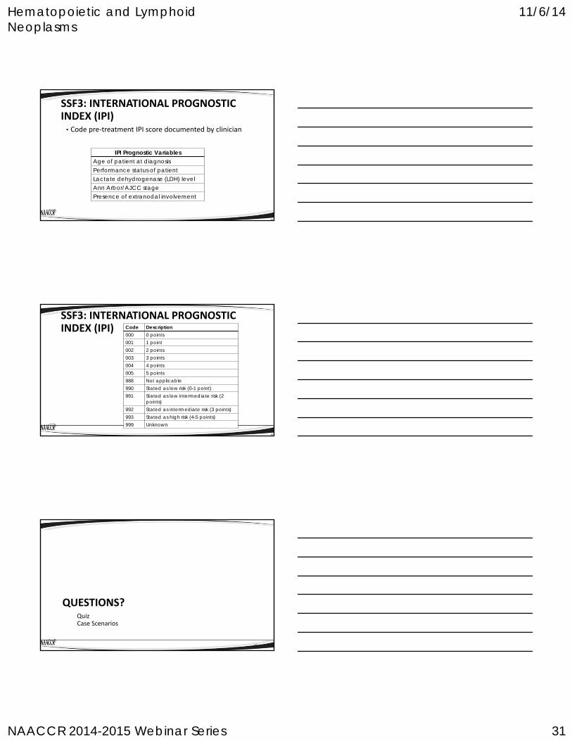
SSF3: INTERNATIONAL PROGNOSTIC INDEX (IPI)• Code pre‐treatment IPI score documented by clinician
IPI Prognostic VariablesAge of patient at diagnosisPerformance status of patientLactate dehydrogenase (LDH) levelAnn Arbor/AJCC stagePresence of extranodal involvement
91
SSF3: INTERNATIONAL PROGNOSTIC INDEX (IPI) Code Description
000 0 points001 1 point002 2 points003 3 points004 4 points005 5 points988 Not applicable990 Stated as low risk (0-1 point)991 Stated as low intermediate risk (2
points)992 Stated as intermediate risk (3 points)993 Stated as high risk (4-5 points)999 Unknown
92
Quiz Case Scenarios
QUESTIONS?
93
Hematopoietic and Lymphoid Neoplasms
11/6/14
NAACCR 2014-2015 Webinar Series 32
COMING UP…• Using the Multiple Primary and Histology (MP/H) Coding Rules• 12/4/14
• Collecting Cancer Data: Testis• 1/8/15
94
AND THE WINNERS ARE…..
95
CE CERTIFICATE QUIZ/SURVEY
• Phrase
• Linkhttp://www.surveygizmo.com/s3/1879750/Hematopoietics‐2014
96