Clinical Study High-Resolution En Face Images of ...
13
Hindawi Publishing Corporation BioMed Research International Volume 2013, Article ID 676803, 12 pages http://dx.doi.org/10.1155/2013/676803 Clinical Study High-Resolution En Face Images of Microcystic Macular Edema in Patients with Autosomal Dominant Optic Atrophy Kiyoko Gocho, 1 Sachiko Kikuchi, 1 Takenori Kabuto, 1 Shuhei Kameya, 1 Kei Shinoda, 2 Atsushi Mizota, 2 Kunihiko Yamaki, 1 and Hiroshi Takahashi 3 1 Department of Ophthalmology, Nippon Medical School Chiba Hokusoh Hospital, 1715 Kamagari, Inzai, Chiba 270-1694, Japan 2 Department of Ophthalmology, Teikyo University School of Medicine, 2-11-1 Kaga, Itabashi-ku, Tokyo 173-8605, Japan 3 Department of Ophthalmology, Nippon Medical School, 1-1-5 Sendagi, Bunkyo-ku, Tokyo 113-8602, Japan Correspondence should be addressed to Shuhei Kameya; [email protected] Received 7 August 2013; Revised 21 October 2013; Accepted 4 November 2013 Academic Editor: Marcela Votruba Copyright © 2013 Kiyoko Gocho et al. is is an open access article distributed under the Creative Commons Attribution License, which permits unrestricted use, distribution, and reproduction in any medium, provided the original work is properly cited. e purpose of this study was to investigate the characteristics of microcystic macular edema (MME) determined from the en face images obtained by an adaptive optics (AO) fundus camera in patients with autosomal dominant optic atrophy (ADOA) and to try to determine the mechanisms underlying the degeneration of the inner retinal cells and RNFL by using the advantage of AO. Six patients from 4 families with ADOA underwent detailed ophthalmic examinations including spectral domain optical coherence tomography (SD-OCT). Mutational screening of all coding and flanking intron sequences of the OPA1 gene was performed by DNA sequencing. SD-OCT showed a severe reduction in the retinal nerve fiber layer (RNFL) thickness in all patients. A new splicing defect and two new frameshiſt mutations with premature termination of the Opa1 protein were identified in three families. A reported nonsense mutation was identified in one family. SD-OCT of one patient showed MME in the inner nuclear layer (INL) of the retina. AO images showed microcysts in the en face images of the INL. Our data indicate that AO is a useful method to identify MME in neurodegenerative diseases and may also help determine the mechanisms underlying the degeneration of the inner retinal cells and RNFL. 1. Introduction Autosomal dominant optic atrophy (ADOA; MIM no. 165500), also known as Kjer’s disease [1], is the most common hereditary ocular neuropathy with a prevalence of 1/12,000– 1/50,000 [2–4]. ADOA is characterized by a decrease in the visual acuity that develops in childhood, temporal palor of the optic discs, centrocecal scotoma, and color vision defects [5, 6]. Histopathological studies of human eyes with ADOA showed diffuse atrophy of the retinal ganglion cell (RGC) layer that predominated in the central retina [7, 8]. ADOA has considerable intra- and interfamilial clinical variability with incomplete penetrance estimated to be about 90% in the familial forms of the disease [9]. Mutations in the optic atrophy 1 gene, OPA1 (MIM no. 605290), located on chromosome 3q28-q29, are responsible for about 60–80% of the cases of ADOA [10–12]. OPA1 encodes a mitochondrial dynamin-related GTPase, which is anchored to the mitochondrial inner membrane [13, 14]. Although the Opa1 protein is ubiquitously expressed in human tissues, a strong expression of the Opa1 protein has been reported in the RGC layer [15]. e Opa1 protein has multiple functions and plays a key role in the fusion of mitochondria and thus in organizing the mitochondrial network [13, 14]. e other functions of the Opa1 protein are related to oxidative phosphorylation, maintenance of the membrane potential [11, 16, 17], maintenance of mtDNA [18, 19], organizing the cristae, and control of mitochondrial apoptosis through the compartmentalization of cytochrome C[17, 20]. Mutations of the OPA1 gene result in a loss of function in most ADOA patients indicating that haploinsuf- ficiency is involved in the pathomechanism of the disease [21]. However, to date, there is no clear evidence to suggest a further role for the OPA1 gene in the degeneration of RGCs in ADOA. Recently, Barboni et al. detected “microcystic macu- lar edema (MME)” in the inner nuclear layer (INL) of
Transcript of Clinical Study High-Resolution En Face Images of ...

Hindawi Publishing CorporationBioMed Research InternationalVolume 2013 Article ID 676803 12 pageshttpdxdoiorg1011552013676803
Clinical StudyHigh-Resolution En Face Images of Microcystic Macular Edemain Patients with Autosomal Dominant Optic Atrophy
Kiyoko Gocho1 Sachiko Kikuchi1 Takenori Kabuto1 Shuhei Kameya1 Kei Shinoda2
Atsushi Mizota2 Kunihiko Yamaki1 and Hiroshi Takahashi3
1 Department of Ophthalmology Nippon Medical School Chiba Hokusoh Hospital 1715 Kamagari Inzai Chiba 270-1694 Japan2Department of Ophthalmology Teikyo University School of Medicine 2-11-1 Kaga Itabashi-ku Tokyo 173-8605 Japan3Department of Ophthalmology Nippon Medical School 1-1-5 Sendagi Bunkyo-ku Tokyo 113-8602 Japan
Correspondence should be addressed to Shuhei Kameya shuheiknmsacjp
Received 7 August 2013 Revised 21 October 2013 Accepted 4 November 2013
Academic Editor Marcela Votruba
Copyright copy 2013 Kiyoko Gocho et al This is an open access article distributed under the Creative Commons Attribution Licensewhich permits unrestricted use distribution and reproduction in any medium provided the original work is properly cited
The purpose of this study was to investigate the characteristics of microcystic macular edema (MME) determined from the en faceimages obtained by an adaptive optics (AO) fundus camera in patients with autosomal dominant optic atrophy (ADOA) and to tryto determine the mechanisms underlying the degeneration of the inner retinal cells and RNFL by using the advantage of AO Sixpatients from 4 families with ADOA underwent detailed ophthalmic examinations including spectral domain optical coherencetomography (SD-OCT)Mutational screening of all coding and flanking intron sequences of theOPA1 gene was performed byDNAsequencing SD-OCT showed a severe reduction in the retinal nerve fiber layer (RNFL) thickness in all patients A new splicingdefect and two new frameshift mutations with premature termination of the Opa1 protein were identified in three families Areported nonsense mutation was identified in one family SD-OCT of one patient showedMME in the inner nuclear layer (INL) ofthe retina AO images showed microcysts in the en face images of the INL Our data indicate that AO is a useful method to identifyMME in neurodegenerative diseases andmay also help determine the mechanisms underlying the degeneration of the inner retinalcells and RNFL
1 IntroductionAutosomal dominant optic atrophy (ADOA MIM no165500) also known as Kjerrsquos disease [1] is the most commonhereditary ocular neuropathy with a prevalence of 112000ndash150000 [2ndash4] ADOA is characterized by a decrease in thevisual acuity that develops in childhood temporal palor ofthe optic discs centrocecal scotoma and color vision defects[5 6] Histopathological studies of human eyes with ADOAshowed diffuse atrophy of the retinal ganglion cell (RGC)layer that predominated in the central retina [7 8]
ADOA has considerable intra- and interfamilial clinicalvariability with incomplete penetrance estimated to be about90 in the familial forms of the disease [9] Mutations in theoptic atrophy 1 gene OPA1 (MIM no 605290) located onchromosome 3q28-q29 are responsible for about 60ndash80 ofthe cases of ADOA [10ndash12]
OPA1 encodes a mitochondrial dynamin-related GTPasewhich is anchored to the mitochondrial inner membrane
[13 14] Although the Opa1 protein is ubiquitously expressedin human tissues a strong expression of the Opa1 proteinhas been reported in the RGC layer [15] The Opa1 proteinhas multiple functions and plays a key role in the fusionof mitochondria and thus in organizing the mitochondrialnetwork [13 14] The other functions of the Opa1 proteinare related to oxidative phosphorylation maintenance of themembrane potential [11 16 17] maintenance of mtDNA[18 19] organizing the cristae and control of mitochondrialapoptosis through the compartmentalization of cytochromeC [17 20] Mutations of the OPA1 gene result in a loss offunction in most ADOA patients indicating that haploinsuf-ficiency is involved in the pathomechanism of the disease[21] However to date there is no clear evidence to suggesta further role for the OPA1 gene in the degeneration of RGCsin ADOA
Recently Barboni et al detected ldquomicrocystic macu-lar edema (MME)rdquo in the inner nuclear layer (INL) of
2 BioMed Research International
patients with Leberrsquos hereditary optic neuropathy (LHON)and ADOA [22] The INL is predominantly made up of thenuclei of the horizontal bipolar and amacrine cells MMEwas originally identified in patients with multiple sclerosis(MS) by Gelfand et al and it was characterized by cysticlacunar areas of hyporeflectivity with clear boundaries inthe spectral domain optical coherence tomographic (SD-OCT) images [23] They suggested that MME represented abreakdown of the blood-retina barrier caused by subclinicaluveitis or retinitis Abegg et al noted similar changes in a caseof compressive optic neuropathy due to a glioma but theysuggested retrograde transsynaptic degeneration as the causeofMME [24] Balk et al noted similar characteristics in a caseof recurrent optic neuritis not due tomultiple sclerosis addinginflammation as a possible cause of MME [25]
Adaptive optics (AO) technology has enabled cliniciansto view the retina with high microscopic lateral resolution[26 27] This technique has been used to analyze the conephotoreceptor mosaic in eyes with inherited retinal degen-erations [28 29] It has also been used to analyze the innerretinal layers for example the retinal nerve fiber layer [30]However this new technology has not been used to analyzethe inner layers of the retina in patients with MME AO hasa transverse resolution of approximately 16 120583m compared tocommercial OCT systems with a resolution of approximately15 120583m This higher resolution should help in detecting andevaluating en face images of MME
Thus the purpose of this study was to investigate thecharacteristics of MME determined from the en face imagesobtained by an AO fundus camera in patients with ADOAand also to try to determine the mechanisms underlying thedegeneration of the inner retinal cells and RNFL by AO Toaccomplish this 6 patients from4 familieswith theOPA1 genewere studied
2 Methods
The protocol of this study conformed to the tenets of theDeclaration of Helsinki and was approved by the Institu-tional Review Board of the Nippon Medical School Sixconsecutive cases of ADOA patients from 4 families whovisited Nippon Medical School Chiba Hokusoh Hospitalfrom December 2010 through April 2013 were studied Awritten informed consent was obtained from the six patientsafter an explanation of the nature and possible complicationsof the experimental protocol
21 Clinical Examinations The ophthalmological examina-tions included measurements of the best-corrected visualacuity (BCVA) determination of the refractive error (spher-ical equivalent) slit-lamp biomicroscopy ophthalmoscopyfundus photography fluorescein angiography (FA) perime-try SD-OCT infrared imaging and full-field electroretinog-raphy (ERG) The visual fields were obtained by Goldmanperimetry andHumphrey Visual Field Analyzer (Model 745iCarl Zeiss Meditec Inc Dublin California) The Swedishinteractive threshold algorithm standard strategy was usedwith program 30-2 of the Humphrey Visual Field AnalyzerColor vision was evaluated with the Farnsworth Panel D-15
SD-OCT (Carl Zeiss Meditec) images were obtained fromall of the patients The B-scan retinal images were composedof 27000s consecutive A-scans acquired through the centerof the macula horizontally for Figures 5(b) and 8(a) Inall patients the fixation was centered on the macula ForFigure 8(b) we moved a horizontal scan line manually tothe area containing the MME detected by AO with centeredpatient fixation For RNFL thickness analysis we performeda vertical SD-OCT scan at about 1mm from the edge of opticdisc with centered fixation The total scan depth was 2mmthe axial resolution was 5 120583m and transverse resolution was15 120583mThe images presented are 6-mm-long scans except forFigure 5(b) which has been cut to fit AO images The 512 times128 Macular Cube scan protocol was used to obtain the enface OCT images With this protocol 128 cross-sectional B-scan images were obtained each composed of 512 A-scansIn all patients fixation was centered on the macula Full-fieldscotopic and photopic ERGswere recorded using an extendedtesting protocol incorporating the International Society forClinical Electrophysiology of Vision standards [31]
22 Genetic Testing Blood samples were collected from thepatients and genomic DNA was isolated from peripheralwhite blood cells with a blood DNA isolation kit (NucleoSpinBlood XL Macherey Nagel Germany) The DNA was usedas a template to amplify the OPA1 gene Coding regionsand flanking introns of the OPA1 gene were amplified bypolymerase chain reaction (PCR) with published primers[32] The PCR products were purified (ExoSAP-IT USBCorp USA) and both strands of the gene were sequencedwith an automated sequencer (Bio Matrix Research ChibaJapan)
RT-PCR was used to amplify the cDNAs of OPA1 ThemRNAs were obtained from peripheral white blood cellswith the TRIzol reagent (Invitrogen CA USA) and templatecDNAs were generated with random hexamer primers Wedesigned exon-spanning primer pairs and used them toamplify exon 18 to exon 20 of the OPA1 cDNA They are for-ward primer (51015840-GTTGAACAACAGGCTGATAG-31015840) andreverse primer (51015840-GCTTGATATCCACTGTGGTG-31015840) Therecovered DNAs were subcloned into the StrataClone PCRcloning vector (Stratagene CA USA) Plasmid DNAs from20 positive clones were purified with the Qiagen PlasmidPurification Kit (Qiagen CA USA) and sequenced with anautomated sequencer (Bio Matrix Research Chiba Japan)
23 Adaptive Optics (AO) Flood Illumination Image Acqui-sition Fundus images were obtained with an infrared AOretinal camera (rtx1 Imagine Eyes Orsay France) [33]This system was used in earlier investigations to imageindividual cone photoreceptors [27 29 34 35] and otherretinal structures [27 36] In our study the AO instrumentilluminated a 4-degree square field of the retina with 850 nminfrared flashes to acquire en face images of the retina with atransverse optical resolution of 250 line pairsmm SuccessiveAO images were taken at adjacent retinal locations with anangular spacing of 2 degrees in the horizontal and verticaldirections This procedure allowed for a horizontal andvertical overlap of at least 2 degree between successive images
BioMed Research International 3
Table 1 Summary of the clinical data of patients with ADOA
Patient ID Sex Age BCVAa (ODOS) Visual field Disc appearance Temporal RNFLbthinning
1-II-1 F 35 008007 Centrocecal scotoma Temporal palor Yes2-II-1 M 39 0304 Centrocecal scotoma Temporal palor Yes3-III-1 F 43 0204 Blind spot enlargement Temporal palor Yes4-II-1 M 52 0507 Blind spot enlargement Temporal palor Yes4-III-1 F 20 0706 Blind spot enlargement Temporal palor Yes4-III-2 M 18 0302 Centrocecal scotoma Temporal palor YesaBest corrected visual acuity (decimal)bRetinal nerve fiber layer
Prior to each acquisition the focusing depth was adjusted tothe inner nuclear layer The resulting images were stitchedtogether by superimposing retinal vessel landmarks withan image editing software (GIMP The GIMP DevelopmentTeam Image J National Institute of Health Bethesda MD)The size of each pixel was typically 08 120583mwhen calculated atthe retinal plane and the values were adjusted for variationsin the axial length of the eye [37] We also analyzed normalcontrols and patients with advanced glaucoma to determinewhether MME was present They were 50 normal controlsand 5 advanced glaucomatous retinas There were 27 menand 23 women whose age ranged from 18 to 57 years (mean381 plusmn 83 years) in this normal control group There were3 men and 2 women whose age ranged from 37 to 57 years(mean 468 plusmn 65 years) in the glaucoma groupThe focusingdepth was adjusted to the INL
3 Results
31 Clinical Findings We studied 6 patients from 4 familieswith ADOA (Figure 1) and the clinical characteristics ofthese 6 patients are summarized in Table 1 The decimalBCVA of all patients was reduced with a range from 07 to007TheGoldmann kinetic visual fields showed a centrocecalscotoma in three patients and a blind spot enlargement inthe other three patients Temporal optic disc palor was seenin all patients Ito et al reported that the retinal nerve fiberlayer (RNFL) in the macular area of patients with ADOAwassignificantly thinner than that in control subjects by SD-OCT[38] They also showed that the RNFL in the temporal areasof circular scans around the optic disc was almost lost whilethe nasal areas were relatively well preserved
We performed a vertical SD-OCT scan at about 1mmfrom the edge of optic discThe results in the ADOA patientsshowed that the temporal RNFL was very thin in all of thepatients (Figure 2) FA did not show any leakage in Patients1-II-1 and 2-II-1 (data not shown) We did not perform FA onthe other 4 patients
The clinical findings of a representative case are shownin Figure 3 (Patient 1-II-1) Fundus examinations showedtemporal palor of the optic discs (Figure 3(a)) A centrocecalscotoma was observed in the Goldmann kinetic visual fieldstest (Figure 3(b)) Panel D-15 showed that the confusionpattern was consistent with a tritan axis blue-yellow defectin each eye (Figure 3(c)) The a- and b-waves of the scotopic
Family 1
I
II
III
I
II
I
II
III
I
II
III
Family 3 Family 4
Family 2
11
2
1
11
Unaffected
Affected
Vision abnormality
Figure 1 Pedigrees of the four families of six ADOA patientsAffected patients are shown with solid symbols and unaffected withopen symbols In family 3 two members who may have had visionabnormalities are shown with gray symbols We were not able toexamine them
and photopic full-field ERGs were of normal amplitudesTheamplitudes of the photopic negative response (PhNR) of thecone ERGs which is believed to originate from inner retinallayers have been reported to be reduced in ADOA patients[39] In this case the PhNR of the cone ERG was decreasedand the peak of the PhNR was a positive potential relative tothe baseline (Figure 3(d))
32 Molecular Genetic Findings We identified one alreadyreported pathogenic mutation and three new mutations inthe four families (Table 2) Patient 1-II-1 was found to have anew heterozygous G to Amutation at position minus1 of intron 18that is likely to abolish the 31015840 splice acceptor site (c1771-1GgtAFigures 4(a) and 4(b)) The family history revealed no othermembers including her parents with any eye disease Wecould not test the genetics in other family members becauseshe was not willing to have them tested Although this
4 BioMed Research International
(a) (b) (c) (d)
(e) (f) (g) (h)
(i) (j) (k) (l)
(m) (n)Figure 2 Retinal nerve fiber layer thickness analysis on spectral-domain optical coherence tomography (SD-OCT) images of the eyes in anormal control and in the ADOA patients Infrared (IR) reflectance images (a c e g i k m) and SD-OCT images (b d f h j l n) are shownThe green vertical lines in the IR images indicate localization of scanned line to obtain the SD-OCT images SD-OCT scan was performedfrom lower to upper retina Images obtained from normal control (a b) Patient 1-II-1 (c d) Patient 2-II-1 (e f) Patient 3-III-1 (g h) Patient4-II-1 (i j) Patient 4-III-1 (k l) and Patient 4-III-2 (m n) are shown Arrows indicate the temporal region of their optic disc Note that theRNFL thickness (yellow arrowheads) of normal control is thick enough to measure in the temporal region of optic disc while that of allADOA patients is almost absent and appears as a thin line
Table 2 Summary of the mutations of OPA1 gene
Patient ID Nucleotidechange Consequencea Domain Location Reference
1-II-1 c1771-1GgtA pN591GfsX18(splicing defect) Dynamin central region Boundary of intron 18-
exon 19 This study
2-II-1 c1899delT pI633MfsX12 Dynamin central region Exon 20 This study
3-III-1 c1096CgtT pR366X GTPase domain Exon 11 Alexander et al 2000[10]
4-II-1 c1102delTpR368GfsX4 GTPase domain Exon 11 This study4-III-1 Same as above
4-III-2 Same as aboveaReference sequence NM 0155602
mutation has never been reported a mutation at position minus2of intron 18 (c1771-2AgtG)has been reported to be pathogenicwith a splicing defect [40]
To investigate the impact of the splice acceptor sitemutation we analyzed OPA1 transcripts expressed in thewhite blood cells from this patient Two distinct RT-PCRproducts were obtained from the patient (data not shown)To separate themutant transcripts fromwild-type transcriptsthe RT-PCR products were cloned into a cloning vectorTwenty clones from the patient were sequenced to verifythe inserts and 5 of them showed truncated inserts with askipping of exon 19 (Figures 4(c)ndash4(e)) This skipping wouldyield a truncated proteinwith a premature termination codondue to a frameshift (pN591GfsX18)
Patient 2-II-1was found to have a newheterozygous singlebase-pair deletion at position 1899 (c1899delT) This wouldyield a truncated proteinwith a premature termination codondue to a frameshift (pI633MfsX12) We could not test thegenetic changes in other family members because they werenot willing to have them tested Although this mutation isa novel mutation two small deletion mutations within thesame exon (c1881 1882delAG c1892 1893delAT) have beenreported as pathogenic mutations for ADOA [40 41]
Patient 3-III-1 was found to have a reported nonsensemutation A heterozygous C to T mutation at position 1096(c1096CgtT) directly changed an arginine at amino acidposition 366 to a stop codon (R366X) This mutation wasconfirmed to be pathogenic by several studies [10 42 43]
BioMed Research International 5
(a)
OS OD
(b)
OD
P P1 1
2 23 4 5
6
7
3 4 56
7
8
9
101112
13
8
9
101112
1314
1514
15
Prot
ane
Prot
anD
euta
neD
euta
n
TritaneTritan Pr
otan
ePr
otan
Deu
tane
Deu
tan
TritaneTritan
OS
(c)
OD OS
Rod
Combinedrod and cone
Cone
OP
30Hzflicker
100120583V
100120583V
100120583V
25ms
50120583V
50120583V
10ms
10ms
10ms
10ms
(d)
Figure 3The clinical findings of Patient 1-II-1 (a) Fundus photograph of the patient showing temporal palor of the optic discs (b) Goldmannkinetic visual fields showing bilateral centrocecal scotoma (c) Panel D-15 shows that the confusion pattern is consistent with tritan (blue-yellow defect) axis in each eye (d) Rod combined rod-cone cone oscillatory potentials and 30-Hz flicker full-field electroretinograms(ERGs) are shown Photopic negative response (PhNR) of cone ERGs is reduced and the peak of the PhNR is a positive potential relative tothe baseline (dotted line) Arrows indicate PhNR
Patients 4-II-1 4-III-1 and 4-III-2 from the same familywere found to have a new heterozygous single base-pairdeletion at amino acid position 1102 (c1102delT) that wouldyield a truncated proteinwith a premature termination codondue to a frameshift (pR368GfsX4)
All three new mutations identified caused a frameshiftwith premature termination codon These mutations wouldlikely be pathogenic by the mechanism of haploinsufficiencyas reported [21]
33 High-Resolution Imaging of Microcystic Macular Edema(MME) by SD-OCT and Adaptive Optics The SD-OCTimages of Patient 1-II-1 showed cystic lacunar areas ofhyporeflectivity with clear boundaries or MME which werecompatiblewith the characteristics suggested byGelfand et al(Figure 5(b)) [23] Wolff et al [44] reported that microcystscould also be observed using en face OCT imaging En faceOCT imaging of Patient 1-II-1 revealed that cysts were locatedin the superior nasal and inferior macular quadrants inboth eyes (Figures 6(a) and 6(b)) AO imaging obtainedfrom the same area showed high-resolution en face imagesof the microcysts in the inner layer of the retina (Figures 5(d)
and 5(e)) They were of various sizes and appeared as darkreflectance areas outlined by hyperreflective regions Mostwere oval shaped (Figures 5(d) and 5(e)) Similar structureswere not found in more than 50 normal controls and 5patientswith advanced glaucoma in our departmentWehavenot examined a patient with MS or optic nerve atrophy otherthan those with ADOA
Abegg et al and Wolff et al have reported that an areawith MME is seen with different patterns as hyporeflectiveregions in the IR images in the perimacular area [24 44]The IR images of our ADOA cases also had ring-shapedhyporeflective regions in the perimacular area (Figures 5(a)7(b) and 7(c)) The hyporeflective region in the IR imagecoincided well with the area containing the microcysticstructures in the AO images (Figures 7(a)ndash7(d))
B-scan and en face SD-OCT images of Patient 2-II-1 didnot show the MME clearly however the IR image had acrescent-shaped perimacular hyporeflective region (Figures6(c) 6(d) and 8(a)ndash8(d)) In the crescent-shaped area theen face AO image showed microcystic structures (Figures8(e)ndash8(g)) The number of microcysts was fewer in Patient2-II-1 than in Patient 1-II-1 however the clarity of the
6 BioMed Research International
A A AA A A AAG G G G TTTT CA AA AA AAG G GGT T T TC
Exon 11
Mutant c 1102delA(k)
A G T T C A T T a g g cc a a tt
Exon 19 Intron 18
Wild type
A G T T C A T T a g g cc a a tt
t
Exon 19 Intron 18
Mutant c 1771-1GgtA
A A AG G GT A A AGT TT CCCC
Wild type mRNAExon 18 Exon 19
A AG G GT A A AG G G G TTC C C
Mutant mRNAExon 18 Exon 20
Ex18 Ex19gtmiddot middot middotag gtmiddot middot middotag
gtmiddot middot middotaggtmiddot middot middotaa
Ex20
Ex18 Ex19 Ex20
Wild type mRNA
Mutant mRNA
A AAAAA GGG TT TTC A TC
Wild type Exon 20
A AAAAA GGG TT TTC A TCA AA A A AGGG TT TC A TC
Mutant c 1899delT Exon 20
AAAA G GGG TT TT C AAGC
Exon 11
Wild type
AAAA G GGG TT TT C AAGC
TExon 11
A A AA A A AAG G G G TTTT C
Exon 11
Wild type
(a)
(b)
(c)
(d)
(e)
(f)
(g)
(h)
(i)
(j)
Figure 4 Molecular genetic findings of the ADOA patients ((a) and (b)) Sequence chromatograms of the wild-type allele and the mutantallele (Patient 1-II-1) are shown In the mutant allele (b) a heterozygous C to T (reverse strand) mutation indicated by a vertical arrow isshown at the minus1 position of intron 18 (c1771-1GgtA) ((c) and (d)) Sequence chromatograms of the wild-type and the mutant (Patient 1-II-1)cDNAs from white blood cells are shown Entire exon 19 is skipped in the mutant mRNA (d) Skipping exon 19 leads to a deletion of 77 bp ofmRNA of OPA1 gene and a resulting frameshift in the product (pN591GfsX18) (e) Schematic diagram of the splicing error in Patient 1-II-1is shown As a result of G to A mutation at position minus1 of intron 18 whole exon 19 is skipped in the mutant gene ((f) and (g)) Sequencechromatograms of the wild-type allele and the mutant allele (Patient 2-II-1) are shown In the mutant allele (g) a heterozygous one base-pairdeletion indicated by a vertical arrow can be seen (c1899delT) ((h) and (i)) Sequence chromatograms of the wild-type allele and the mutantallele (Patient 3-III-1) are shown In the mutant allele (i) a heterozygous C to Tmutation indicated by a vertical arrow is shown (c1096CgtT)((j) and (k)) Sequence chromatograms of the wild-type allele and the mutant allele (Patient 4-II-1) are shown In the mutant allele (g) aheterozygous one base-pair deletion indicated by a vertical arrow is shown (c1102delA)
BioMed Research International 7
(a)
(b)
(c)
(d)
(e)Figure 5 The localization and the structure of microcystic macular edema in Patient 1-II-1 (a) An infrared image of the macular region ofthe patient The box outlined in green lines shows the area scanned to obtain the OCT image in (b) A white box indicates the area shown in(c) and (d) An orange box indicates the area shown in (e) A polygonal area outlined in yellow is the area shown in Figure 6(a) (b) SD-OCTimage of the patient shows cystic lacunar areas of hyporeflectivity with clear boundaries in the nasal region The RNFL is almost lost in thisarea Arrows indicate the edge of the area outlined in orange in (a) and (d) (c) Magnified infrared image outlined in white in (a) is shownA green line indicates the area scanned to obtain OCT image (b) (d) Montage of AO image corresponding to area (c) is shown Note thatretinal blood vessels are shown in exactly the same region in the images (c) and (d) (e) Magnified AO image outlined in orange in (a) and(d) is shown The AO image shows various size dark reflectance areas outlined by hyperreflective region and most are oval shaped Bars in(d) and (e) indicate 200 120583m
8 BioMed Research International
(a) (b) (c) (d)
Figure 6 En faceOCT images of Patient 1-II-1 and 2-II-1 En face SD-OCT images of the eyes in patient 1-II-1 ((a) and (b)) and 2-II-1((c) and(d)) are shown Images from right eyes ((a) and (c)) and left eyes ((b) and (d)) are shown En face OCT imaging reveals the presence of thecysts in patient 1-II-1 En face OCT images of the patient 2-II-1 did not show cysts clearly Bars in (d) indicates 500 120583m
(a)
(b) (c) (d)
Figure 7The AO and IR images of Patient 1-II-1 (a) Montage of AO images of the patient is shownThemicrocystic structures are observedas perimacular rings ((b) and (c)) Infrared (IR) images of the case show a hyporeflective region with perimacular ring shape Perimacularring shape is outlined by dotted yellow line (c) (d) Minimized image of that shown in (a) The area with microcystic structures is outlinedin dotted yellow line Note that the hyporeflective region in the IR image and the area containing microcystic structure in AO image are wellmatched
BioMed Research International 9
(a)
(b)
(c)
(d)
(e)
(f)
(g)
Figure 8TheOCT IR and AO images of Patient 2-II-1 ((a) and (b)) SD-OCT images of the patient do not showmicrocystic macular edemaclearly The RNFL is very thin in this area The scan lines to obtain these images are shown in (d) ((c) and (d)) IR images of the patient areshown The IR image has a crescent shaped perimacular hyporeflectance region outlined in yellow dotted line The green and orange linesindicate the scan lines to obtain SD-OCT images of (a) and (b) respectively The orange scan line overlaps the region outlined in (f) (e) IRimage superimposed on AO image is shown (f) Montage of AO images of the patient outlined area in (d) is shown A small number of themicrocystic structures are observed in the image (g) Magnified AO image outlined in orange in (e) is shownThe AO image has various sizedark reflectance areas outlined by hyperreflective region as observed in Patient 1-II-1 Bars in (e) and (f) indicate 200 120583m
microcysts was the same in these two patients (Figures 5(e)and 8(g))
Although we analyzed the other 4 patients extensivelywe did not find microcystic structures in their AO imagesThe refractive error and axial length were not significantlydifferent in all six patients
4 DiscussionOver 200 mutations in the OPA1 gene have been identifiedin patients with ADOA (HGMD professional Institute ofMedical Genetics in Cardiff) Approximately one-half of theOPA1mutations lead to premature termination codons fromnonsense mutations or frameshifts from small insertions
10 BioMed Research International
deletions or splice site mutations [45] These truncatedmRNAs are unstable and get degraded by specific pathwaysthat is nonsense-mediated mRNA decay which are in-builtprotective cellular mechanisms against mutant proteins withpossible dominant negative effect [42 46 47] The reducedOpa1 protein expression levels observed in these reportedcases support the role of haploinsufficiency in ADOA Theseresults strongly suggest that the three new heterozygousmutations with premature termination codon identified inthis study are pathogenic
Gelfand et al reported that MME was associated withlower visual acuity and a thinner RNFL in patients withMS [23] In our cases Patient 1-II-1 with the poorest BCVAhad the clearest MME in her SD-OCT and AO imagesOur vertical SD-OCT image between the optic disc andmacular region showed that the temporal RNFL was almostcompletely absent in all of the patients However amongthese patients Patient 1-II-1 had the thinnest RNFL in theperipheral region of the vertical scan Our data are consistentwith the hypothesis that the degree of MME is related to thedisease severity
Gelfand et al hypothesized that the presence of MMEwas associated with a breakdown of the blood-retinal barrier[23] However Barboni et al noted that patients with LHONand DOA do not have any fluorescein leakage as expected forthe noninflammatory status of their disease [22] Our resultsalso showed that genetically identified ADOA patients withMME do not have any signs of leakage from their retinalvessels
MME has been detected in the INL of the retina withchiasmal glioma [24] It is highly unlikely that the MME ina patient with brain tumor is due to inflammation of theretina and optic nerve Thus Abegg et al hypothesized thatthe MME in the INL was due to retrograde transsynapticdegeneration [24]
It is well established that retrograde transsynaptic degen-eration can occur in the human central nervous system [4849] VanBuren observed atrophy of the RGC following a rightoccipital lobectomy inmonkeys [50] Recently Jindahra et alpresented evidence of retrograde trans-synaptic degenerationof RGCs identified by SD-OCT following both congenitaland acquired lesions of the retrogeniculate visual pathwayin humans [51] In addition Green et al reported that theneurodegenerative changes caused by retrograde transsynap-tic degeneration in a patient with MS were seen not onlyin the RNFL and ganglion cell layer but also in the INLof their retina [52] Their histopathological study showedprominent atrophy of the INL in 40 of the eyes sufferingfrom MS and none of the control eyes They also recognizedthat the severity of the INL atrophy appeared to be relatedto the severity of RGC atrophy Similar INL vacuoles havebeen observed histopathologically in rhesus monkeys withidiopathic optic atrophy [53] Combining these observationswith our observations we suggest the possibility that the darkregions observed in the en face AO images of our ADOApatients are areas of degenerated horizontal bipolar andamacrine cells in the INL caused by retrograde transsynapticdegeneration
The en face MME structures detected by AO were alsofound in another ADOA patient who did not show MMEclearly in the en face and cross-sectional OCT images Theseobservations indicate that AO might be useful in identifyingMME in other neurodegenerative diseases and may also behelpful in determining the mechanisms underlying RGC andINL degeneration
Our study has a number of limitations We identifiedthe en face MME in patients with ADOA however thereare several other diseases that have MME in their SD-OCT images for example MS recurrent optic neuritisneuromyelitis optica LHON and chiasmal glioma We needto investigate the en face MME structures in patients withsuch diseases to identify whether they also show the en faceMME in their INL and to compare their features to thoseof en face MME seen in our patient with ADOA It willprobably be helpful in clarifying the pathomechanisms of thedegeneration of inner retinal cell degeneration to investigateseveral diseases with different etiology
We have found MME in the INL of the ADOA patientsbut it is important to note that only in two patients Thecross-sectional nature of our study did not allow us todraw conclusions regarding the evolution of MME in ADOAand the other diseases To address these issues systematiclongitudinal studies incorporating detailed ophthalmologicassessments in large cohort are needed and may help deter-mine the mechanisms involved in the development of MMEAlthough the controls in our study including those withadvanced glaucoma did not show MME in their en face AOimages we need to determine why patients with advancedglaucoma did not show en face MME despite the RGC lossWe cannot explain why we did not find MME in the other4 ADOA patients in this study It may be related to thedisease severity however patient 2-II-1 showed comparableperipheral RNFL thickness and better BCVA compared to theother ADOA patients withoutMME Some other factors maybe needed for MME to develop
In conclusion our findings showed that genetically iden-tified ADOA patients without any sign of inflammation canhave MME in the INL of the retina Our data indicate thatthe disease severity may be associated with the presence ofMME in the INL as reported because we found the clearestMME in the patients with poorest BCVA although someother factors may be needed for MME to develop otherthan disease severity Our findings indicate that there is apossibility that retrograde trans synaptic degeneration couldcause severe damages in horizontal bipolar and amacrinecells in the INL after the optic nerve atrophy Further studiesare needed and these findings will probably be helpfulin clarifying the pathology of the degeneration of innerretinal cells by retrograde transsynaptic degeneration inpatients with optic nerve atrophies and in developing newtherapies
Conflict of Interests
The authors declare that they have no conflict of interestsassociated with this paper
BioMed Research International 11
Acknowledgment
The authors thank Dr Duco Hamsaki of the Bascom PalmerEye Institute University of Miami School of MedicineMiami FL USA for discussions and editing their paper
References
[1] P Kjer ldquoInfantile optic atrophy with dominant mode of inher-itance a clinical and genetic study of 19 Danish familiesrdquo ActaOphthalmologica vol 164 pp 1ndash147 1959
[2] H Eiberg B Kjer P Kjer and T Rosenberg ldquoDominant opticatrophy (OPA1) mapped to chromosome 3q region Linkageanalysisrdquo Human Molecular Genetics vol 3 no 6 pp 977ndash9801994
[3] B Kjer H Eiberg P Kjer and T Rosenberg ldquoDominant opticatrophy mapped to chromosome 3q region II Clinical andepidemiological aspectsrdquo Acta Ophthalmologica Scandinavicavol 74 no 1 pp 3ndash7 1996
[4] WM LyleGenetic Risks A Reference for Eye Care PractitionersUniversity of Waterloo Press Ontario Canada 1990
[5] J B Caldwell R O Howard and L A Riggs ldquoDominantjuvenile optic atrophy A study in two families and review ofhereditary disease in childhoodrdquo Archives of Ophthalmologyvol 85 no 2 pp 133ndash147 1971
[6] C S Hoyt ldquoAutosomal dominant optic atrophy A spectrum ofdisabilityrdquo Ophthalmology vol 87 no 3 pp 245ndash251 1980
[7] P B Johnston R N Gaster V C Smith and R C Tripathi ldquoAclinicopathologic study of autosomal dominant optic atrophyrdquoAmerican Journal of Ophthalmology vol 88 no 5 pp 868ndash8751979
[8] P Kjer O A Jensen and L Klinken ldquoHistopathology of eyeoptic nerve and brain in a case of dominant optic atrophyrdquoActaOphthalmologica vol 61 no 2 pp 300ndash312 1983
[9] A C Cohn C Toomes C Potter et al ldquoAutosomal dominantoptic atrophy penetrance and expressivity in patients withOPA1 mutationsrdquo American Journal of Ophthalmology vol 143no 4 pp 656ndash662 2007
[10] C AlexanderM Votruba U E A Pesch et al ldquoOPA1 encodinga dynamin-related GTPase is mutated in autosomal dominantoptic atrophy linked to chromosome 3q28rdquoNatureGenetics vol26 no 2 pp 211ndash215 2000
[11] P Amati-Bonneau A Guichet A Olichon et al ldquoOPA1 R445Hmutation in optic atrophy associated with sensorineural deaf-nessrdquo Annals of Neurology vol 58 no 6 pp 958ndash963 2005
[12] C Delettre G Lenaers J-M Griffoin et al ldquoNuclear geneOPA1 encoding a mitochondrial dynamin-related protein ismutated in dominant optic atrophyrdquo Nature Genetics vol 26no 2 pp 207ndash210 2000
[13] C Delettre G Lenaers L Pelloquin P Belenguer and C PHamel ldquoOPA1 (Kjer type) dominant optic atrophy a novelmitochondrial diseaserdquoMolecularGenetics andMetabolism vol75 no 2 pp 97ndash107 2002
[14] A Olichon E Guillou C Delettre et al ldquoMitochondrial dyna-mics and disease OPA1rdquo Biochimica et Biophysica Acta vol1763 no 5-6 pp 500ndash509 2006
[15] U E A Pesch J E Fries S Bette et al ldquoOPA1 the disease genefor autosomal dominant optic atrophy is specifically expre-ssed in ganglion cells and intrinsic neurons of the retinardquo Inve-stigative Ophthalmology and Visual Science vol 45 no 11 pp4217ndash4225 2004
[16] R Lodi C Tonon M L Valentino et al ldquoDeficit of in vivomitochondrial ATPproduction inOPA1-related dominant opticatrophyrdquo Annals of Neurology vol 56 no 5 pp 719ndash723 2004
[17] A Olichon L Baricault N Gas et al ldquoLoss of OPA1 perturbatesthe mitochondrial inner membrane structure and integrityleading to cytochrome c release and apoptosisrdquo Journal ofBiological Chemistry vol 278 no 10 pp 7743ndash7746 2003
[18] P Amati-Bonneau M L Valentino P Reynier et al ldquoOPA1mutations induce mitochondrial DNA instability and opticatrophy rsquoplusrsquo phenotypesrdquo Brain vol 131 no 2 pp 338ndash3512008
[19] G Hudson P Amati-Bonneau E L Blakely et al ldquoMutationof OPA1 causes dominant optic atrophy with external ophthal-moplegia ataxia deafness and multiple mitochondrial DNAdeletions a novel disorder of mtDNAmaintenancerdquo Brain vol131 no 2 pp 329ndash337 2008
[20] C Frezza S Cipolat O Martins de Brito et al ldquoOPA1 controlsapoptotic cristae remodeling independently from mitochon-drial fusionrdquo Cell vol 126 no 1 pp 177ndash189 2006
[21] N J Marchbank J E Craig J P Leek et al ldquoDeletion of theOPA1 gene in a dominant optic atrophy family evidence thathaploinsufficiency is the cause of diseaserdquo Journal of MedicalGenetics vol 39 no 8 p e47 2002
[22] P Barboni V Carelli G Savini M Carbonelli C La MorgiaandA A Sadun ldquoMicrocysticmacular degeneration fromopticneuropathy not inflammatory not trans-synaptic degenera-tionrdquo Brain vol 135 article e239 2013
[23] J M Gelfand R Nolan D M Schwartz J Graves and A JGreen ldquoMicrocystic macular oedema in multiple sclerosis isassociated with disease severityrdquo Brain vol 135 pp 1786ndash17932012
[24] M Abegg M Zinkernagel and S Wolf ldquoMicrocystic maculardegeneration from optic neuropathyrdquo Brain vol 135 articlee225 2012
[25] L J Balk J Killestein C H Polman B M Uitdehaag and APetzold ldquoMicrocystic macular oedema confirmed but not spe-cific for multiple sclerosisrdquo Brain vol 135 article e226 2012
[26] J Liang D R Williams and D T Miller ldquoSupernormal visionand high-resolution retinal imaging through adaptive opticsrdquoJournal of the Optical Society of America A vol 14 no 11 pp2884ndash2892 1997
[27] M Lombardo S Serrao N Devaney M Parravano and GLombardo ldquoAdaptive optics technology for High-Resolutionretinal imagingrdquo Sensors vol 13 pp 334ndash366 2012
[28] J L Duncan Y Zhang J Gandhi et al ldquoHigh-resolution ima-ging with adaptive optics in patients with inherited retinaldegenerationrdquo Investigative Ophthalmology and Visual Sciencevol 48 no 7 pp 3283ndash3291 2007
[29] N Tojo T Nakamura C Fuchizawa T Oiwake and A Hay-ashi ldquoAdaptive optics fundus images of cone photoreceptorsin the macula of patients with retinitis pigmentosardquo ClinicalOphthalmology vol 7 pp 203ndash210 2013
[30] K Takayama S Ooto M Hangai et al ldquoHigh-resolution ima-ging of the retinal nerve fiber layer in normal eyes using ada-ptive optics scanning laser ophthalmoscopyrdquo PLoS ONE vol 7no 3 Article ID e33158 2012
[31] G E Holder M G Brigell M Hawlina T Meigen V Vae-gan and M Bach ldquoISCEV standard for clinical pattern ele-ctroretinographymdash2007 updaterdquo Documenta Ophthalmologicavol 114 no 3 pp 111ndash116 2007
12 BioMed Research International
[32] Y Mashima K Yamada M Wakakura et al ldquoSpectrum ofpathogenic mitochondrial DNAmutations and clinical featuresin Japanese families with Leberrsquos hereditary optic neuropathyrdquoCurrent Eye Research vol 17 no 4 pp 403ndash408 1998
[33] C Viard K Nakashima B Lamory M Paques X Levecq andN Chateau ldquoImaging microscopic structures in pathologicalretinas using a flood-illumination adaptive optics retinal cam-erardquo inOphthalmic Technologies XXI vol 7885 of Proceedings ofSPIE January 2011
[34] M Lombardo G Lombardo P Ducoli and S Serrao ldquoAdaptiveoptics photoreceptor imagingrdquo Ophthalmology vol 119 pp1498ndash1498 2012
[35] M Lombardo S Serrao P Ducoli and G Lombardo ldquoVaria-tions in image optical quality of the eye and the sampling limit ofresolution of the conemosaic with axial length in young adultsrdquoJournal of Cataract amp Refractive Surgery vol 38 pp 1147ndash11552012
[36] I Audo M El Sanharawi C Vignal-Clermont et al ldquoFovealdamage in habitual poppers usersrdquo Archives of Ophthalmologyvol 129 no 6 pp 703ndash708 2011
[37] A G Bennett A R Rudnicka and D F Edgar ldquoImprovementson Littmannrsquos method of determining the size of retinal featuresby fundus photographyrdquoGraefersquos Archive for Clinical and Exper-imental Ophthalmology vol 232 no 6 pp 361ndash367 1994
[38] Y Ito M Nakamura T Yamakoshi J Lin H Yatsuya andH Terasaki ldquoReduction of inner retinal thickness in patientswith autosomal dominant optic atrophy associated with OPA1mutationsrdquo Investigative Ophthalmology and Visual Science vol48 no 9 pp 4079ndash4086 2007
[39] K Miyata M Nakamura M Kondo et al ldquoReduction ofoscillatory potentials andphotopic negative response in patientswith autosomal dominant optic atrophy with OPA1 mutationsrdquoInvestigative Ophthalmology and Visual Science vol 48 no 2pp 820ndash824 2007
[40] M Ferre D Bonneau D Milea et al ldquoMolecular screeningof 980 cases of suspected hereditary optic neuropathy with areport on 77 novel OPA1 mutationsrdquo Human Mutation vol 30no 7 pp E692ndashE705 2009
[41] O Baris C Delettre P Amati-Bonneau et al ldquoFourteen novelOPA1 mutations in autosomal dominant optic atrophy incl-uding two de novomutations in sporadic optic atrophyrdquoHumanmutation vol 21 no 6 p 656 2003
[42] U E A Pesch B Leo-Kottler S Mayer et al ldquoOPA1 muta-tions in patients with autosomal dominant optic atrophy andevidence for semi-dominant inheritancerdquo Human MolecularGenetics vol 10 no 13 pp 1359ndash1368 2001
[43] C Delettre J-M Griffoin J Kaplan et al ldquoMutation spectrumand splicing variants in the OPA1 generdquo Human Genetics vol109 no 6 pp 584ndash591 2001
[44] B Wolff C Basdekidou V Vasseur M Mauget-Faysse J ASahel and C Vignal ldquoRetinal inner nuclear layer microcysticchanges in optic nerve atrophy a novel spectral-domain OCTfindingrdquo Retina vol 33 no 10 pp 2133ndash2138 2013
[45] M Ferre P Amati-Bonneau Y Tourmen Y Malthiery andP Reynier ldquoeOPA1 an online database for OPA1 mutationsrdquoHuman mutation vol 25 no 5 pp 423ndash428 2005
[46] S Schimpf S Schaich and B Wissinger ldquoActivation of crypticsplice sites is a frequent splicing defect mechanism caused bymutations in exon and intron sequences of the OPA1 generdquoHuman Genetics vol 118 no 6 pp 767ndash771 2006
[47] S Schimpf N Fuhrmann S Schaich and B Wissinger ldquoCom-prehensive cDNA study and quantitative transcript analysis
of mutant OPA1 transcripts containing premature terminationcodonsrdquo Human Mutation vol 29 no 1 pp 106ndash112 2008
[48] G Holmes and T G Stewart ldquoOn the connection of the inferiorolives with the cerebellum in manrdquo Brain vol 31 no 1 pp 125ndash137 1908
[49] AW CampbellHistological Studies on the Localisation of Cere-bral Function Cambridge University Press Cambridge MassUSA 1905
[50] JMVanBuren ldquoTrans-synaptic retrograde degeneration in thevisual system of primatesrdquo Journal of Neurology Neurosurgeryand Psychiatry vol 26 pp 402ndash409 1963
[51] P Jindahra A Petrie and G T Plant ldquoRetrograde trans-syna-ptic retinal ganglion cell loss identified by optical coherencetomographyrdquo Brain vol 132 no 3 pp 628ndash634 2009
[52] A J Green S McQuaid S L Hauser I V Allen and R LynessldquoOcular pathology in multiple sclerosis retinal atrophy andinflammation irrespective of disease durationrdquo Brain vol 133no 6 pp 1591ndash1601 2010
[53] B Fortune L Wang B V Bui C F Burgoyne and G A CioffildquoIdiopathic bilateral optic atrophy in the rhesus macaquerdquoInvestigative Ophthalmology and Visual Science vol 46 no 11pp 3943ndash3956 2005
Submit your manuscripts athttpwwwhindawicom
Stem CellsInternational
Hindawi Publishing Corporationhttpwwwhindawicom Volume 2014
Hindawi Publishing Corporationhttpwwwhindawicom Volume 2014
MEDIATORSINFLAMMATION
of
Hindawi Publishing Corporationhttpwwwhindawicom Volume 2014
Behavioural Neurology
EndocrinologyInternational Journal of
Hindawi Publishing Corporationhttpwwwhindawicom Volume 2014
Hindawi Publishing Corporationhttpwwwhindawicom Volume 2014
Disease Markers
Hindawi Publishing Corporationhttpwwwhindawicom Volume 2014
BioMed Research International
OncologyJournal of
Hindawi Publishing Corporationhttpwwwhindawicom Volume 2014
Hindawi Publishing Corporationhttpwwwhindawicom Volume 2014
Oxidative Medicine and Cellular Longevity
Hindawi Publishing Corporationhttpwwwhindawicom Volume 2014
PPAR Research
The Scientific World JournalHindawi Publishing Corporation httpwwwhindawicom Volume 2014
Immunology ResearchHindawi Publishing Corporationhttpwwwhindawicom Volume 2014
Journal of
ObesityJournal of
Hindawi Publishing Corporationhttpwwwhindawicom Volume 2014
Hindawi Publishing Corporationhttpwwwhindawicom Volume 2014
Computational and Mathematical Methods in Medicine
OphthalmologyJournal of
Hindawi Publishing Corporationhttpwwwhindawicom Volume 2014
Diabetes ResearchJournal of
Hindawi Publishing Corporationhttpwwwhindawicom Volume 2014
Hindawi Publishing Corporationhttpwwwhindawicom Volume 2014
Research and TreatmentAIDS
Hindawi Publishing Corporationhttpwwwhindawicom Volume 2014
Gastroenterology Research and Practice
Hindawi Publishing Corporationhttpwwwhindawicom Volume 2014
Parkinsonrsquos Disease
Evidence-Based Complementary and Alternative Medicine
Volume 2014Hindawi Publishing Corporationhttpwwwhindawicom
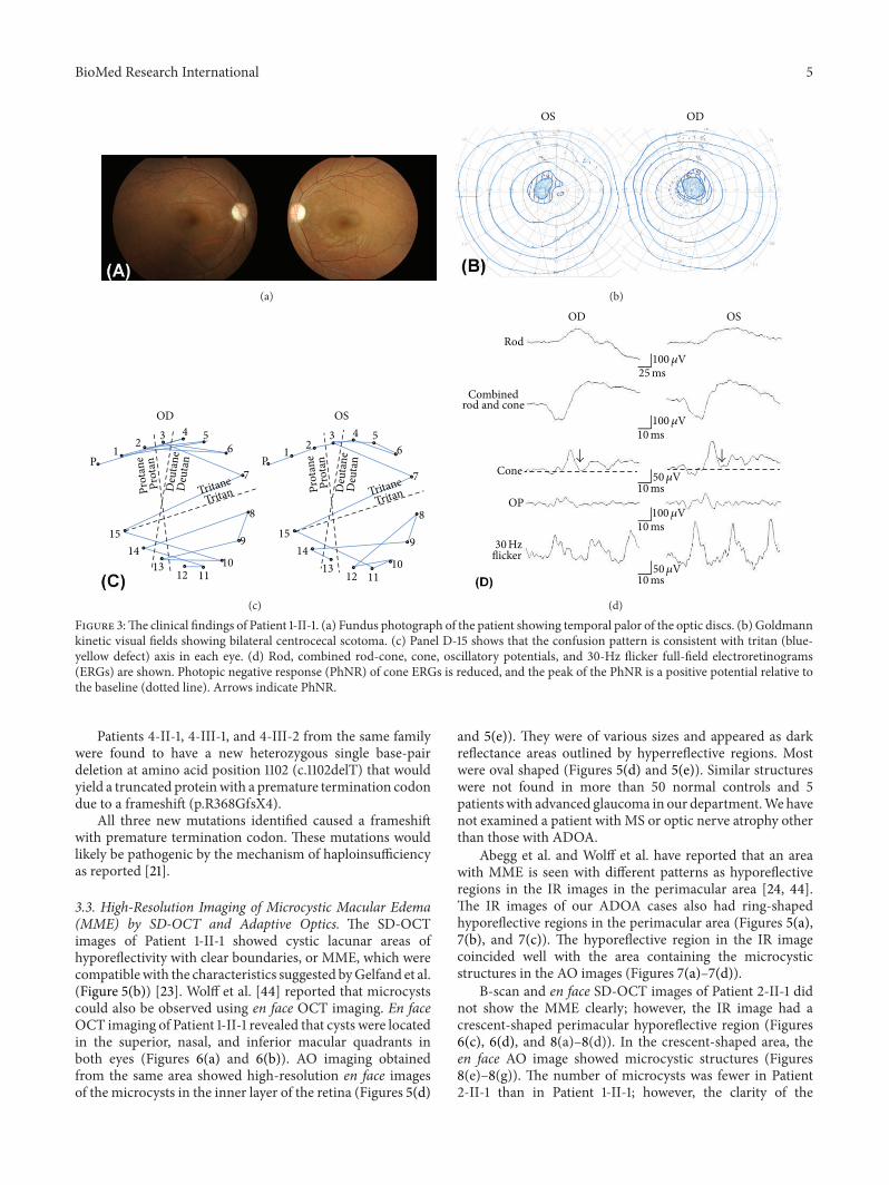