Clinical Outcomes of Intracerebral Hemorrhage in Hemodialysis Patients · 2017-04-26 · Clinical...
18
Clinical Outcomes of Intracere in Hemodialysis Patients 著者 Sakamoto Noriaki, Ishikawa Eiich Kazuyasu, Uemae Yoji, Komatsu Yoji Akira journal or publication title World neurosurgery volume 81 number 3-4 page range 538-542 year 2014 権利 (C) 2014 Elsevier Inc. NOTICE: this is t author’s version of a work that for publication in World neuros resulting from the publishing peer review, editing, correctio formatting, and other quality c mechanisms may not be reflecte document. Changes may have been work since it was submitted for definitive version was subsequ in World neurosurgery, 81, 3-4, 2014, 10.1016/j.wneu.2013.10.033 URL http://hdl.handle.net/2241/00121638 doi: 10.1016/j.wneu.2013.10.033
Transcript of Clinical Outcomes of Intracerebral Hemorrhage in Hemodialysis Patients · 2017-04-26 · Clinical...
Clinical Outcomes of Intracerebral Hemorrhagein Hemodialysis Patients
著者 Sakamoto Noriaki, Ishikawa Eiichi, AokiKazuyasu, Uemae Yoji, Komatsu Yoji, MatsumuraAkira
journal orpublication title
World neurosurgery
volume 81number 3-4page range 538-542year 2014権利 (C) 2014 Elsevier Inc. NOTICE: this is the
author’s version of a work that was acceptedfor publication in World neurosurgery. Changesresulting from the publishing process, such aspeer review, editing, corrections, structuralformatting, and other quality controlmechanisms may not be reflected in thisdocument. Changes may have been made to thiswork since it was submitted for publication. Adefinitive version was subsequently publishedin World neurosurgery, 81, 3-4, 2014, DOI:10.1016/j.wneu.2013.10.033
URL http://hdl.handle.net/2241/00121638doi: 10.1016/j.wneu.2013.10.033
Clinical outcomes of intracerebral hemorrhage in
hemodialysis patients
Noriaki Sakamoto a, c, Eiichi Ishikawa c, Kazuyasu Aoki a, b, Yoji Uemae a, Yoji Komatsu a, c,
Akira Matsumura c
a Department of Neurosurgery, Hitachi General Hospital, Ibaraki
b Department of Neurosurgery, Nerima Hikarigaoka Hospital, Tokyo
c Department of Neurosurgery, Faculty of Medicine, University of Tsukuba, Ibaraki
Key words: Intracerebral hemorrhage, Hemodialysis, Chronic renal failure, Hypertension
Running head on left-hand page: N. Sakamoto et al.
Running title on right-hand page: Intracerebral hemorrhage in hemodialysis patients
Address reprint requests to: Eiichi Ishikawa, MD, PhD, Department of Neurosurgery, Faculty
of Medicine, Graduate School of Comprehensive Human Sciences, University of Tsukuba,
1-1-1 Tennodai, Tsukuba, Ibaraki 305-8575, Japan. e-mail: [email protected]
1
Introduction
Chronic renal failure (CRF) is a worldwide public health problem that is associated with
a high risk of occurrence of cardiovascular events [9,19]. Hemodialysis (HD) is performed in
more than 90% of patients in advanced stages of CRF. The number of HD patients in Japan
has increased from 1624.1 per million at the end of 2000 to 2279.5 per million in 2009. The
total number of HD patients exceeded 281,996 in 2009, the highest rate since 1983 [17]. The
trend is similar in other countries, the total number of HD patients in the United States
increasing from 281,355 in 2000 to 415,013 in 2010 (United States Renal Data System)
[4,26]. Several studies have indicated that the incidence of ischemic and hemorrhagic
events in the intracranial region (strokes) in CRF patients is high [1,7,8,10,12,14-
16,18,21-23,27]. A single-center study in Japan showed that the frequency of intracerebral
hemorrhage (ICH, 52% of 151 patients) in HD patients was higher than that of cerebral
infarction (CI, 41%) between 1980 and 1996, while the rate of ICH (29%) between 1997 and
2002 was lower than that of CI (68%) [23]. Intensive control of hypertension, diabetes and
hyperlipidemia may have reduced the incidence of ICH. However, the clinical status of HD
patients with ICH remains severe, ICH being a common cause of death in HD patients. The
incidence of death due to ICH is 2- to 3-fold higher than that due to CI [8,10,12,15].
Here, we retrospectively investigated 5 years worth of clinical data from ICH patients
treated with or without HD at our institution. We reveal the differences in the clinical courses
of HD and non-HD patients and identify the risk factors for poor outcomes in ICH patients
undergoing HD.
Patients and Methods
We conducted a single-center retrospective study based upon a review of medical records.
2
The records of 366 consecutive patients with non-traumatic spontaneous ICH who were
admitted to the neurosurgical department of Hitachi General Hospital between January 2007
and December 2011 were examined. Patients with ICH due to secondary causes, such as
ruptured aneurysm, primary ischemic stroke, and arteriovenous malformation or tumor, were
excluded from this study. In all cases, computed tomography (CT) scan was performed on
admission. Magnetic resonance imaging (MRI) was performed on admission or after surgery
for all patients with ICH except for deceased patients whose condition was rapidly
deteriorating.
The following clinical information, including baseline characteristics, was collected: age,
gender, location/side of the hematoma, volume of the hematoma, presence of
intraventricular hemorrhage (IVH), microbleeds (MBs) on MRI scans, modified Rankin scale
(mRS) scores at admission/discharge, type of surgical operation, use of antihypertensive
and anti-diabetic drugs and antiplatelets/anticoagulants, primary renal disease, current
history of HD, and the day of the week on which the ICH occurred in relation to HD. The
hematoma volume in each case was determined by one of the authors (N.S.) from CT scans
obtained at the onset as follows: the maximum transverse diameter × the maximal
antero-posterior diameter × the maximal supero-inferior diameter × 1/2. Presence of IVH
was also assessed using CT scans. Hematoma volume in the lateral ventricle was not
included in the calculation of ‘volume of hematoma’ in this study.
The values are expressed as means ± SD. Differences in patient data were evaluated
using univariate logistic analysis as well as χ2 test, Fisher’s test or Student t test. P values
less than 0.05 were considered to indicate statistical significance. All calculations were
performed using JMP 5 software (SAA Corp., USA). Multivariate logistic analysis was
performed for data in which p values were less than 0.1 on univariate logistic analyses.
Differences were considered statistically significant if the p value was <0.05 in this analysis
3
as well. In the logistic analyses, continuous variables were dichotomized in terms of their
mean or median values.
Results
Comparisons between HD and non-HD patients
Table 1 shows the clinical characteristics of the patients in the study. A total of 366 patients
were admitted to our hospital with a diagnosis of ICH during the study period. They were
divided into two groups: 32 patients (9%) with CRF who received HD (HD group) and 334
patients who did not receive HD treatment (non-HD group). All the CRF patients in this study
were on HD. Surgical hematoma evacuation was performed in 25% of the HD patients and in
13.5% of the non-HD patients, while a hematoma drain was inserted into the lateral ventricle
of 6% and 7.5% of HD and non-HD patients, respectively. There were no differences in
patient age, gender, laterality of hematoma and surgical procedure between the two groups.
The HD group had higher rates of hematomas in the basal ganglia (84% in the HD group vs.
57% in the non-HD group, p<0.05), IVH (53% vs. 34%, p<0.05), use of antihypertensive
drugs (72% vs. 32%, p<0.01), use of anti-diabetic drugs (41% vs. 20%, p<0.01) and use of
antiplatelets/anticoagulants (41% vs. 17%, p<0.01). The mortality rate (mRS VI) was higher
in the HD group (44%) than in the non-HD group (21%). There were no significant
differences in the existence of cerebral MBs between the two groups, as seen on MRI (p>0.1,
Fisher direct test). As seen in Table 1, univariate logistic analysis showed that hematoma
location, presence of IVH, use of antihypertensive drugs, use of anti-diabetic drugs, use of
antiplatelets/anticoagulants, patient mortality, and the number of patients with mRS ≥5 were
significantly different between HD and non-HD groups. Similar results were also shown
using the χ2 test or Fisher’s test (data not shown). Multivariate logistic analysis showed that
4
hematoma location, use of antihypertensive drugs, use of antiplatelets/anticoagulants, and
number of patients with mRS ≥5 were different between the two groups (Table 1).
Analysis of patients who had died (mRS VI) in both groups indicated significant differences
in the number of hematomas in the basal ganglia (86% vs. 45%, p<0.01), the use of
antihypertensive drugs (50% vs. 21%: p<0.01) and the use of antiplatelets/anticoagulants
(36% vs. 12%, p<0.05). There were no significant differences in the use of anti-diabetic
drugs and incidence of IVH between the two groups.
Analyses of HD patients
Next, detailed data from the 32 patients on HD was analyzed. The causes of CRF were
diabetic nephropathy (44%), glomerulonephritis (19%), cystic kidney disease (3%), sclerosis
(3%), renal carcinoma (3%) and unknown etiology (28%). On admission, 13 patients (41%)
received antiplatelets/anticoagulants for maintenance of shunt patency (31.2%), history of
previous angina pectoris (2 cases), or history of previous cerebral infarction (1 case).
The condition of 24 of the patients (75%) was rated as being serious (mRS V) at the time
of hospitalization, and antihypertensive drugs were administered to 23 of these patients.
Mean systolic blood pressure values on admission in patients with and without
antihypertensive drugs before admission were 190.0+/-43.2 and 166.3+/-53.1 mmHg
(p>0.05, Student t test), respectively. Hematoma drain placement in the lateral ventricle was
performed in 2 patients, both of who survived, while surgical hematoma evacuation was
performed for 6 patients who survived and 2 patients who had a poor outcome (mRS VI).
The final outcomes were mRS VI (44%) and V (25%). As shown in Figure 1, most patients’
conditions did not improve throughout hospitalization. We analyzed the risk factors for poor
outcomes (mRS VI). As shown in Table 2, univariate logistic analysis demonstrated
significant differences between the patients who did and did not survive in terms of GCS
5
scores on admission, mRS scores on admission, hematoma volume, presence of IVH, and
the use of antihypertensive drugs. Multivariate logistic analysis, however, showed no
significant prognostic factor, indicating that the results of the analysis should only be used as
an advisory, since the analyses were performed in a small sized sample. Older patients
tended to have higher mortality rates than younger patients, although the difference between
the two groups was not significant. There was no difference in systolic blood pressure
between poor outcome patients and other HD patients (179.8+/- 59.8 mmHg (n=13) vs.
185.0+/- 35.0 (n=16), p>0.1, Student t test), or in HD duration between poor outcome
patients and other HD patients (7.1 +/- 5.4 years (n=13) vs. 7.7 +/- 4.3 (n=15), p>0.1,
Student t test).
As shown in Figure 2, no significant difference was observed between mortality (mRS VI)
due to ICH on dialysis days (10 cases, 37%) and non-dialysis days (17 cases, 63%) in 27
cases with data regarding the timing of the ICH (p>0.05, Fisher’s method). In these cases,
26% of the ICH occurred before the HD procedure on an HD day, which was a higher rate
than during or after the HD procedure (11%) on the dialysis day.
Discussion
On admission, the condition of ICH patients on HD in this study was frequently classified
as being serious. The prognostic factors associated with mortality were GCS on admission
<8, hematoma volume >50 ml, the presence of IVH, and lack of antihypertensive drug usage.
In previous papers, the overall 30-day mortality rate in ICH patients with advanced CRF has
been shown to be 30 - 83% [7,16]. In one of the studies, the prognostic factors
independently associated with mortality were as follows: GCS scores, old age, systolic blood
pressure, ICH volume ≥30 ml, presence of IVH, and high serum glucose [7]. In another
6
paper, additional factors indicating poor prognosis were level of consciousness on admission,
the size and shape of the hematoma, prothrombin time, and fibrin degradation product level
[14]. In general, cerebral MBs detected on T2-weighted MRI scans are closely related to ICH
[11,13,20,24], although our results could not corroborate this since our data included only
ICH patients. A previous paper showed that 35% of CRF patients had MBs. Factors
associated with MBs were male sex, old age, hypertension and worsening of CRF, although
MBs did not show a correlation with the duration of HD treatment [25].
In our study, antihypertensive drugs were used more frequently by CRF patients than
non-CRF patients. CRF patients frequently have renal hypertension, which may cause ICH.
The odds ratio for ICH was 2.44 in the CRF group when compared to the non-CRF group
[22]. However, the use of antiplatelets/anticoagulants did not significantly predict poorer
outcomes. Therefore, patients should strictly control their hypertension and should not
hesitate to take antiplatelets/anticoagulants as they do not pose a risk for ICH. A limitation of
the present study is that the results were obtained from retrospective and non-controlled
data. However, various analyses, including univariate and multivariate logistic regression
analyses showed similar results. Hence, we believe that these results are generally reliable.
As shown in Figure 2, ICH frequently occurred on a non-dialysis day. HD in CRF
patients is usually scheduled three times a week, and some reports show that the
occurrence of disease is related to the day of HD. For example, sudden cardiac death
events occur more often on Mondays and Wednesdays [2]. The occurrence rates of acute
myocardial infarction, heart failure and stroke are high when the interval between HD
treatments is two days [5], This suggests that medical practitioners should be careful to
screen HD patients for ICH on non-dialysis days.
In our study, mortality in ICH patients on HD was associated with lack of
antihypertensive drug usage. Most patients who do not use antihypertensive drugs are
7
patients with absent or mild hypertension, although there may be some cases of drug
withdrawal in patients with severe hypertension. Interestingly, previous studies regarding risk
factors for poor outcomes in non-traumatic ICH also did not contain a history of hypertension
[3, 6]. For instance, one of these studies suggests that the factors independently associated
with 30-day mortality are GCS scores, age ≥80 years, infratentorial origin of ICH, ICH
volume and the presence of IVH [6]. In this previous study, the incidence of hypertension in
the poor outcome group (43%) was lower than that in the other ICH group (73%) [6], similar
to the present results. We speculate that severe ICH depends upon factors other than
hypertension (such as existence of microaneurysms). Hence, the occurrence of severe ICH
in both HD and non-HD patients should be considered independently of hypertension.
In conclusion, multivariate logistic analysis showed that hematoma location, use of
antihypertensive drugs, use of antiplatelets/anticoagulants, and patient outcome were
significantly different between HD and non-HD groups. On the other hand, use of
antihypertensive drugs or antiplatelets/anticoagulants did not worsen the outcome of the ICH
patients on HD. Thus, in HD patients, the possibility of severe ICH should be considered
regardless of the use of antihypertensive drugs or antiplatelets/anticoagulants. In this study,
ICH frequently occurred before the HD procedure on an HD day or on an interval day.
Conflict of Interest statement
All authors declare no conflicts of interest.
8
References
1. Anavekar NS, McMurray JJ, Velazquez EJ, Solomon SD, Kober L, Rouleau JL, White
HD, Nordlander R, Maggioni A, Dickstein K, Zelenkofske S, Leimberger JD, Califf RM,
Pfeffer MA. Relation between renal dysfunction and cardiovascular outcomes after
myocardial infarction. N Engl J Med. 2004;351:1285-95
2. Bleyer AJ, Russell GB, Satko SG. Sudden and cardiac death rates in hemodialysis
patients. Kidney Int. 1999;55:1553-9
3. Cheung RT, Zou LY. Use of the original, modified, or new intracerebral hemorrhage
score to predict mortality and morbidity after intracerebral hemorrhage. Stroke.
2003;34:1717-22.
4. Collins AJ, Foley RN, Herzog C, Chavers B, Gilbertson D, Herzog C, Ishani A,
Johansen K, Kasiske B, Kutner N, Liu J, St Peter W, Ding S, Guo H, Kats A, Lamb K, Li S,
Li S, Roberts T, Skeans M, Snyder J, Solid C, Thompson B, Weinhandl E, Xiong H, Yusuf
A, Zaun D, Arko C, Chen SC, Daniels F, Ebben J, Frazier E, Hanzlik C, Johnson R,
Sheets D, Wang X, Forrest B, Constantini E, Everson S, Eggers P, Agodoa L. US Renal
Data System 2012 Annual Data Report. Am J Kidney Dis. 2013;61(1 Suppl 1):A7
5. Foley RN, Gilbertson DT, Murray T, Collins AJ. Long interdialytic interval and mortality
among patients receiving hemodialysis. N Engl J Med. 2011;365:1099-107.
6. Hemphill JC 3rd, Bonovich DC, Besmertis L, Manley GT, Johnston SC. The ICH score:
a simple, reliable grading scale for intracerebral hemorrhage. Stroke. 2001;32:891-7.
7. Huang BR, Liao CC, Huang WH, Hsu YH, Hsu JC, Yen HC, Lin CL. Prognostic factors
of spontaneous intracerebral hemorrhage in hemodialysis patients and predictors of
30-day mortality. Intern Med J. 2008;38:568-74.
9
8. Iseki K, Fukiyama K; Okawa Dialysis Study (OKIDS) Group. Clinical demographics and
long-term prognosis after stroke in patients on chronic hemodialysis. The Okinawa
Dialysis Study (OKIDS) Group. Nephrol Dial Transplant. 2000;15:1808-13
9. Iseki K, Fukiyama K. Predictors of stroke in patients receiving chronic
hemodialysis. Kidney Int. 1996;50:1672–5.
10. Iseki K, Kinjo K, Kimura Y, Osawa A, Fukiyama K. Evidence for high risk of cerebral
hemorrhage in chronic dialysis patients. Kidney Int. 1993;44:1086-90
11. Kato H, Izumiyama M, Izumiyama K, Takahashi A, Itoyama Y. Silent cerebral
microbleeds on T2*-weighted MRI: correlation with stroke subtype, stroke recurrence,
and leukoaraiosis. Stroke. 2002;33:1536-40
12. Kawamura M, Fijimoto S, Hisanaga S, Yamamoto Y, Eto T. Incidence, outcome, and
risk factors of cerebrovascular events in patients undergoing maintenance hemodialysis.
Am J Kidney Dis. 1998;31:991-6
13. Kinoshita T, Okudera T, Tamura H, Ogawa T, Hatazawa J. Assessment of lacunar
hemorrhage associated with hypertensive stroke by echo-planar gradient-echo
T2*-weighted MRI. Stroke. 2000;31:1646-50
14. Miyahara K, Murata H, Abe H. Predictors of intracranial hematoma enlargement in
patients undergoing hemodialysis. Neurol Med Chir (Tokyo). 2007;47:47-51
15. Molshatzki N, Orion D, Tsabari R, Schwammenthal Y, Merzeliak O, Toashi M, Tanne D.
Chronic kidney disease in patients with acute intracerebral hemorrhage: association with
large hematoma volume and poor outcome. Cerebrovasc Dis. 2011;31:271-7.
16. Murakami M, Hamasaki T, Kimura S, Maruyama D, Kakita K. Clinical features and
management of intracranial hemorrhage in patients undergoing maintenance dialysis
therapy. Neurol Med Chir (Tokyo). 2004;44:225-32
10
17. Nakai S, Iseki K, Itami N, Ogata S, Kazama JJ, Kimata N, Shigematsu T, Shinoda T,
Shoji T, Suzuki K, Taniguchi M, Tsuchida K, Nakamoto H, Nishi H, Hashimoto S,
Hasegawa T, Hanafusa N, Hamano T, Fujii N, Masakane I, Marubayashi S, Morita O,
Yamagata K, Wakai K, Wada A, Watanabe Y, Tsubakihara Y. Overview of regular
dialysis treatment in Japan (as of 31 December 2009). Ther Apher Dial. 2012;16:11-53.
18. Nakayama K, Tannir NM, Liu P, Wathen JK, Cheng YC, Champlin RE, Ueno NT.
Natural history of metastatic renal cell carcinoma in patients who underwent consultation
for allogeneic hematopoietic stem cell transplantation. Biol Blood Marrow Transplant.
2007;13:975-85
19. Ninomiya T, Kiyohara Y, Kubo M, Tanizaki Y, Doi Y, Okubo K, Wakugawa Y, Hata J,
Oishi Y, Shikata K, Yonemoto K, Hirakata H, Iida M. Chronic kidney disease and
cardiovascular disease in a general Japanese population: the Hisayama Study. Kidney
Int. 2005;68:228-36.
20. Offenbacher H, Fazekas F, Schmidt R, Koch M, Fazekas G, Kapeller P. MR of cerebral
abnormalities concomitant with primary intracerebral hematomas. AJNR Am J
Neuroradiol. 1996;17:573-8
21. Ovbiagele B. Chronic kidney disease and risk of death during hospitalization for stroke.
J Neurol Sci. 2011;301:46-50.
22. Thrift AG, Evans RG, Donnan GA. Hypertension and the risk of intracerebral
hemorrhage: special considerations in patients with renal disease. Nephrol Dial
Transplant. 1999;14:2291-2
23. Toyoda K, Fujii K, Fujimi S, Kumai Y, Tsuchimochi H, Ibayashi S, Iida M. Stroke in
patients on maintenance hemodialysis: a 22-year single-center study. Am J Kidney Dis.
2005;45:1058-66
11
24. Tsushima Y, Tanizaki Y, Aoki J, Endo K. MR detection of microhemorrhages in
neurologically healthy adults. Neuroradiology. 2002;44:31-6
25. Watanabe A. Cerebral microbleeds and intracerebral hemorrhages in patients on
maintenance hemodialysis. J Stroke Cerebrovasc Dis. 2007;16:30-3.
26. Xue JL, Ma JZ, Louis TA, Collins AJ. Forecast of the number of patients with end-stage
renal disease in the United States to the year 2010. J Am Soc Nephrol. 2001;12:2753-8.
27. Yahalom G, Schwartz R, Schwammenthal Y, Merzeliak O, Toashi M, Orion D, Sela BA,
Tanne D. Chronic kidney disease and clinical outcome in patients with acute stroke.
Stroke. 2009;40:1296-303.
Figure legends
Figure 1. Modified Rankin Scale scores in intracerebral hemorrhage (ICH) patients on
admission and on discharge
Figure 2 Relationship between ICH occurrence and the timing of the hemodialysis (HD)
procedure. Black and white bars indicate the frequencies of living and deceased patients,
respectively.
12
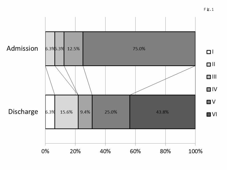
Fig. 1
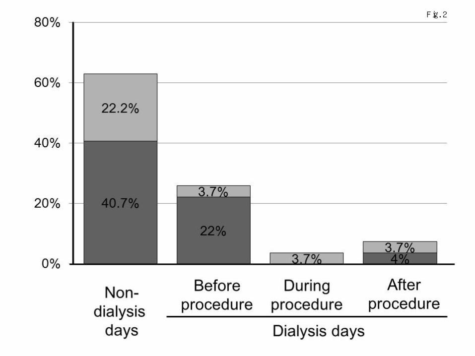
Fig. 2
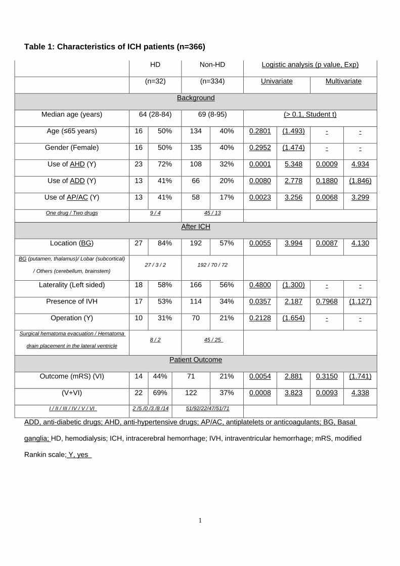
Table 1: Characteristics of ICH patients (n=366)
HD Non-HD Logistic analysis (p value, Exp)
(n=32) (n=334) Univariate Multivariate
Background
Median age (years) 64 (28-84) 69 (8-95) (> 0.1, Student t)
Age (≤65 years) 16 50% 134 40% 0.2801 (1.493) - -
Gender (Female) 16 50% 135 40% 0.2952 (1.474) - -
Use of AHD (Y) 23 72% 108 32% 0.0001 5.348 0.0009 4.934
Use of ADD (Y) 13 41% 66 20% 0.0080 2.778 0.1880 (1.846)
Use of AP/AC (Y) 13 41% 58 17% 0.0023 3.256 0.0068 3.299
One drug / Two drugs 9 / 4 45 / 13
After ICH
Location (BG) 27 84% 192 57% 0.0055 3.994 0.0087 4.130
BG (putamen, thalamus)/ Lobar (subcortical)
/ Others (cerebellum, brainstem) 27 / 3 / 2 192 / 70 / 72
Laterality (Left sided) 18 58% 166 56% 0.4800 (1.300) - -
Presence of IVH 17 53% 114 34% 0.0357 2.187 0.7968 (1.127)
Operation (Y) 10 31% 70 21% 0.2128 (1.654) - -
Surgical hematoma evacuation / Hematoma
drain placement in the lateral ventricle 8 / 2 45 / 25
Patient Outcome
Outcome (mRS) (VI) 14 44% 71 21% 0.0054 2.881 0.3150 (1.741)
(V+VI) 22 69% 122 37% 0.0008 3.823 0.0093 4.338
I / II / III / IV / V / VI 2 /5 /0 /3 /8 /14 51/92/22/47/51/71
ADD, anti-diabetic drugs; AHD, anti-hypertensive drugs; AP/AC, antiplatelets or anticoagulants; BG, Basal
ganglia; HD, hemodialysis; ICH, intracerebral hemorrhage; IVH, intraventricular hemorrhage; mRS, modified
Rankin scale; Y, yes
1
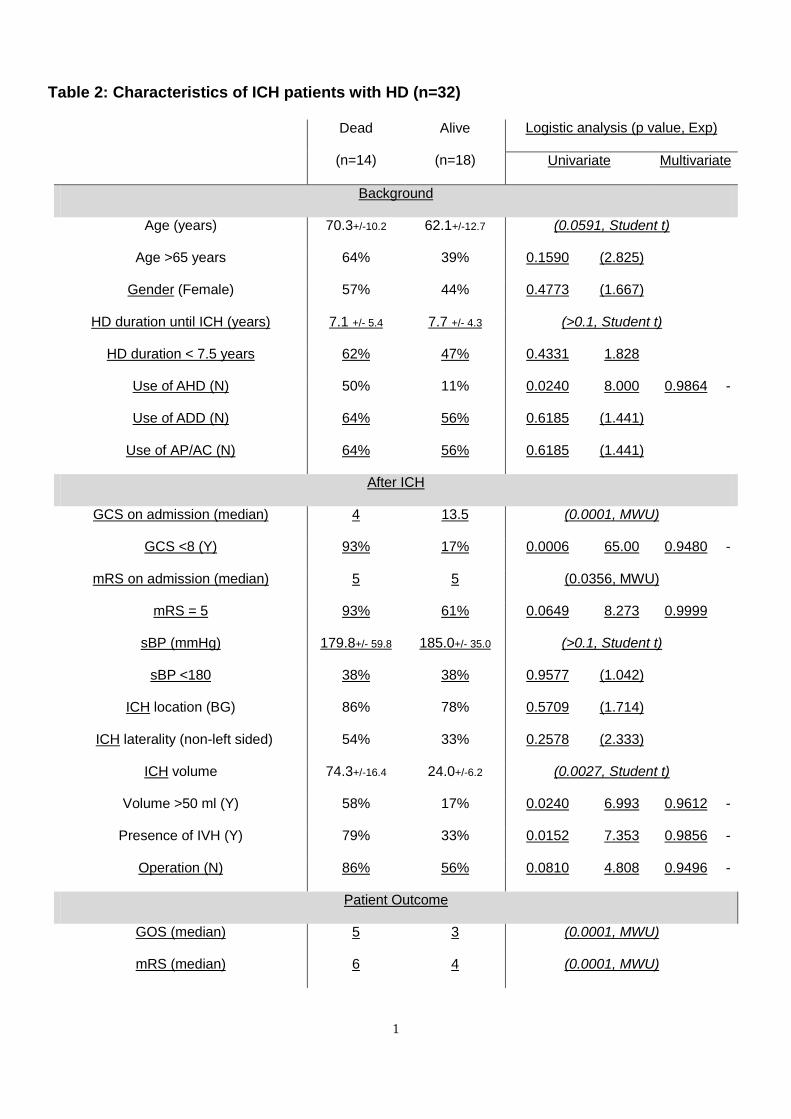
Table 2: Characteristics of ICH patients with HD (n=32)
Dead
(n=14)
Alive
(n=18)
Logistic analysis (p value, Exp)
Univariate Multivariate
Background
Age (years) 70.3+/-10.2 62.1+/-12.7 (0.0591, Student t)
Age >65 years 64% 39% 0.1590 (2.825)
Gender (Female) 57% 44% 0.4773 (1.667)
HD duration until ICH (years) 7.1 +/- 5.4 7.7 +/- 4.3 (>0.1, Student t)
HD duration < 7.5 years 62% 47% 0.4331 1.828
Use of AHD (N) 50% 11% 0.0240 8.000 0.9864 -
Use of ADD (N) 64% 56% 0.6185 (1.441)
Use of AP/AC (N) 64% 56% 0.6185 (1.441)
After ICH
GCS on admission (median) 4 13.5 (0.0001, MWU)
GCS <8 (Y) 93% 17% 0.0006 65.00 0.9480 -
mRS on admission (median) 5 5 (0.0356, MWU)
mRS = 5 93% 61% 0.0649 8.273 0.9999
sBP (mmHg) 179.8+/- 59.8 185.0+/- 35.0 (>0.1, Student t)
sBP <180 38% 38% 0.9577 (1.042)
ICH location (BG) 86% 78% 0.5709 (1.714)
ICH laterality (non-left sided) 54% 33% 0.2578 (2.333)
ICH volume 74.3+/-16.4 24.0+/-6.2 (0.0027, Student t)
Volume >50 ml (Y) 58% 17% 0.0240 6.993 0.9612 -
Presence of IVH (Y) 79% 33% 0.0152 7.353 0.9856 -
Operation (N) 86% 56% 0.0810 4.808 0.9496 -
Patient Outcome
GOS (median) 5 3 (0.0001, MWU)
mRS (median) 6 4 (0.0001, MWU)
1

ADD, anti-diabetic drugs; AHD, anti-hypertensive drugs; AP/AC, anti-platelets or anti-coagulants; BG, basal
ganglia; GCS, Glasgow Come Scale; GOS, Glasgow Outcome Scale; ICH, intracerebral hemorrhage; IVH,
intraventricular hemorrhage; mRS, modified Rankin scale; MWU, Mann-Whitney U test; N, no; sBP, Systolic
Blood pressure on admission; Y, yes.
2