Class I and Class II Chitin Synthases Are Involved in ... · advancing, was removed and mounted on...
12
EUKARYOTIC CELL, June 2005, p. 1125–1136 Vol. 4, No. 6 1535-9778/05/$08.000 doi:10.1128/EC.4.6.1125–1136.2005 Copyright © 2005, American Society for Microbiology. All Rights Reserved. Class I and Class II Chitin Synthases Are Involved in Septum Formation in the Filamentous Fungus Aspergillus nidulans Masayuki Ichinomiya, 1 † Emi Yamada, 1 † Shuichi Yamashita, 2 Akinori Ohta, 1 and Hiroyuki Horiuchi 1 * Department of Biotechnology 1 and Department of Agricultural and Environmental Biology, 2 The University of Tokyo, Tokyo 113-8657, Japan Received 25 November 2004/Accepted 29 March 2005 The class II and class I chitin synthases of the filamentous fungus Aspergillus nidulans are encoded by chsA and chsC, respectively. Previously, we presented several lines of evidence suggesting that ChsA and ChsC have overlapping functions in maintaining cell wall integrity. In order to determine the functions of these chitin synthases, we employed electron and fluorescence microscopy and investigated in detail the cell wall of a chsA chsC double mutant (AC mutant) along with the localization of ChsA and ChsC. In the lateral cell wall of the AC mutant, electron-transparent regions were thickened. Septa of the AC mutant were aberrantly thick and had a large pore. Some septa were located abnormally close to adjacent septa. A functional hemagglutinin (HA)-tagged ChsA (HA-ChsA) and a functional FLAG-tagged ChsC (FLAG-ChsC) were each localized to a subset of septation sites. Comparison with the localization pattern of actin, which is known to localize at forming septa, suggested that ChsA and ChsC transiently exist at the septation sites during and shortly after septum formation. Double staining of HA-ChsA and FLAG-ChsC indicated that their localizations were not identical but partly overlapped at the septation sites. Fluorescence of FLAG-ChsC, but not of HA-ChsA, was also observed at hyphal tips. These data indicate that ChsA and ChsC share overlapping roles in septum formation. Chitin, a polymer of -1,4-linked N-acetylglucosamine, is one of the major cell wall constituents in many filamentous fungi. Chitin synthases are membrane proteins that catalyze the polymerization of N-acetylglucosamine. To date, many chitin synthase genes have been identified from various fungi, and the polypeptides deduced from these genes have been divided into six classes (32, 37). Studies in the budding yeast Saccharomyces cerevisiae have revealed the localization and specific functions of its three chitin synthases (Chs1, Chs2, and Chs3) (5, 34). In this organism, chitin is concentrated mostly in septa. Chs3, a class IV chitin synthase, is required for chitin ring formation at the base of emerging buds and for chitin synthesis in the lateral cell wall during vegetative growth. Chs3 is found at the plasma membrane and in an intracellular com- partment called the chitosome, and its correct localization re- quires the functioning of several proteins (5, 32, 34, 37). Chs2, a class II enzyme, synthesizes chitin in primary septa. Chs2 appears at a late stage of mitosis and is localized to the sep- tation site; it is degraded immediately after septum formation (7, 36). Chs1, a class I enzyme, repairs the weakened cell walls of daughter cells after their separation from mother cells; this separation is executed by the activity of a chitinase that digests the primary septa. Chs1 exists at a constant level throughout the cell cycle and is reported to be present on the plasma membrane and chitosomes (60), although its precise localiza- tion has not been reported. In our previous work, we identified five chitin synthase genes, chsA, chsB, chsC, chsD, and csmA, that encode chitin synthases of classes II, III, I, IV, and V, respectively, in As- pergillus nidulans (12, 29, 30, 58). A search through the A. nidulans genome database (http://www-genome.wi.mit.edu /annotation/fungi/aspergillus/) identified eight chitin synthase genes, including chsA-chsD and csmA. Elimination or reduc- tion of the expression of chsB or csmA caused obvious defects in hyphal growth and asexual development (3, 18, 21, 43, 58). Analogous observations have also been reported in other fila- mentous fungi (2, 23, 25, 31, 59). Deletion mutants of chsA, chsC, or chsD exhibited no clear morphological differences from the wild-type strain (29, 30, 58). Several combinations of double mutants have also been constructed in A. nidulans (9, 13, 18, 20, 21, 29). Among these, chsA chsC double mutants (AC mutants) showed remarkable synergistic defects in hy- phal growth as well as in sexual and asexual development in comparison with the single mutants (13). Although the growth rate of the AC mutants under ordinary conditions was similar to that of the wild-type strain, the growing edge of their colo- nies was irregular and their hyphae were occasionally lysed. The growth rate was reduced by chitin-binding dyes, salts (1.2 M NaCl or KCl), or a detergent (0.005% sodium dodecyl sulfate [SDS]). Conidial production by the AC mutants was less than 0.01% of that by the wild-type strain due to the reduced number and altered morphology of conidiophores. The wild-type conidiophore consists of a stalk, a vesicle, two tiers of sterigmata (metulae and phialides), and conidia (1). Although the AC mutants formed normal conidiophore ves- icles at the tips of conidiophore stalks, they produced chains of sterigmata and occasionally reproduced conidiophores upon vesicles, which are termed secondary conidiophores (13). In this study, we investigated the cell wall ultrastructure of * Corresponding author. Mailing address: 1-1-1 Yayoi, Bunkyo-ku, Tokyo 113-8657, Japan. Phone: 81-3-5841-5170. Fax: 81-3-5841-8015. E-mail: [email protected]. † M.I. and E.Y. contributed equally to this work. 1125 by on June 14, 2008 ec.asm.org Downloaded from
Transcript of Class I and Class II Chitin Synthases Are Involved in ... · advancing, was removed and mounted on...
EUKARYOTIC CELL, June 2005, p. 1125–1136 Vol. 4, No. 61535-9778/05/$08.00�0 doi:10.1128/EC.4.6.1125–1136.2005Copyright © 2005, American Society for Microbiology. All Rights Reserved.
Class I and Class II Chitin Synthases Are Involved in SeptumFormation in the Filamentous Fungus Aspergillus nidulans
Masayuki Ichinomiya,1† Emi Yamada,1† Shuichi Yamashita,2Akinori Ohta,1 and Hiroyuki Horiuchi1*
Department of Biotechnology1 and Department of Agricultural and Environmental Biology,2
The University of Tokyo, Tokyo 113-8657, Japan
Received 25 November 2004/Accepted 29 March 2005
The class II and class I chitin synthases of the filamentous fungus Aspergillus nidulans are encoded by chsAand chsC, respectively. Previously, we presented several lines of evidence suggesting that ChsA and ChsC haveoverlapping functions in maintaining cell wall integrity. In order to determine the functions of these chitinsynthases, we employed electron and fluorescence microscopy and investigated in detail the cell wall of a �chsA�chsC double mutant (�AC mutant) along with the localization of ChsA and ChsC. In the lateral cell wall ofthe �AC mutant, electron-transparent regions were thickened. Septa of the �AC mutant were aberrantly thickand had a large pore. Some septa were located abnormally close to adjacent septa. A functional hemagglutinin(HA)-tagged ChsA (HA-ChsA) and a functional FLAG-tagged ChsC (FLAG-ChsC) were each localized to asubset of septation sites. Comparison with the localization pattern of actin, which is known to localize atforming septa, suggested that ChsA and ChsC transiently exist at the septation sites during and shortly afterseptum formation. Double staining of HA-ChsA and FLAG-ChsC indicated that their localizations were notidentical but partly overlapped at the septation sites. Fluorescence of FLAG-ChsC, but not of HA-ChsA, wasalso observed at hyphal tips. These data indicate that ChsA and ChsC share overlapping roles in septumformation.
Chitin, a polymer of �-1,4-linked N-acetylglucosamine, isone of the major cell wall constituents in many filamentousfungi. Chitin synthases are membrane proteins that catalyzethe polymerization of N-acetylglucosamine. To date, manychitin synthase genes have been identified from various fungi,and the polypeptides deduced from these genes have beendivided into six classes (32, 37). Studies in the budding yeastSaccharomyces cerevisiae have revealed the localization andspecific functions of its three chitin synthases (Chs1, Chs2, andChs3) (5, 34). In this organism, chitin is concentrated mostly insepta. Chs3, a class IV chitin synthase, is required for chitinring formation at the base of emerging buds and for chitinsynthesis in the lateral cell wall during vegetative growth. Chs3is found at the plasma membrane and in an intracellular com-partment called the chitosome, and its correct localization re-quires the functioning of several proteins (5, 32, 34, 37). Chs2,a class II enzyme, synthesizes chitin in primary septa. Chs2appears at a late stage of mitosis and is localized to the sep-tation site; it is degraded immediately after septum formation(7, 36). Chs1, a class I enzyme, repairs the weakened cell wallsof daughter cells after their separation from mother cells; thisseparation is executed by the activity of a chitinase that digeststhe primary septa. Chs1 exists at a constant level throughoutthe cell cycle and is reported to be present on the plasmamembrane and chitosomes (60), although its precise localiza-tion has not been reported.
In our previous work, we identified five chitin synthase
genes, chsA, chsB, chsC, chsD, and csmA, that encode chitinsynthases of classes II, III, I, IV, and V, respectively, in As-pergillus nidulans (12, 29, 30, 58). A search through the A.nidulans genome database (http://www-genome.wi.mit.edu/annotation/fungi/aspergillus/) identified eight chitin synthasegenes, including chsA-chsD and csmA. Elimination or reduc-tion of the expression of chsB or csmA caused obvious defectsin hyphal growth and asexual development (3, 18, 21, 43, 58).Analogous observations have also been reported in other fila-mentous fungi (2, 23, 25, 31, 59). Deletion mutants of chsA,chsC, or chsD exhibited no clear morphological differencesfrom the wild-type strain (29, 30, 58). Several combinations ofdouble mutants have also been constructed in A. nidulans (9,13, 18, 20, 21, 29). Among these, �chsA �chsC double mutants(�AC mutants) showed remarkable synergistic defects in hy-phal growth as well as in sexual and asexual development incomparison with the single mutants (13). Although the growthrate of the �AC mutants under ordinary conditions was similarto that of the wild-type strain, the growing edge of their colo-nies was irregular and their hyphae were occasionally lysed.The growth rate was reduced by chitin-binding dyes, salts (1.2M NaCl or KCl), or a detergent (0.005% sodium dodecylsulfate [SDS]). Conidial production by the �AC mutants wasless than 0.01% of that by the wild-type strain due to thereduced number and altered morphology of conidiophores.The wild-type conidiophore consists of a stalk, a vesicle, twotiers of sterigmata (metulae and phialides), and conidia (1).Although the �AC mutants formed normal conidiophore ves-icles at the tips of conidiophore stalks, they produced chains ofsterigmata and occasionally reproduced conidiophores uponvesicles, which are termed secondary conidiophores (13).
In this study, we investigated the cell wall ultrastructure of
* Corresponding author. Mailing address: 1-1-1 Yayoi, Bunkyo-ku,Tokyo 113-8657, Japan. Phone: 81-3-5841-5170. Fax: 81-3-5841-8015.E-mail: [email protected].
† M.I. and E.Y. contributed equally to this work.
1125
by on June 14, 2008 ec.asm
.orgD
ownloaded from
the �AC mutant by electron microscopy and identified defectsin both the lateral wall construction and septum formation. Weconstructed strains that expressed epitope-tagged ChsA and/orChsC. These fusion proteins were functional, and either ofthem was found at septation sites when their subcellular local-ization was examined. Double staining of these fusion proteinssuggested that their localization at the septation sites partiallyoverlapped. ChsC, but not ChsA, was found at hyphal tips.These results suggest that ChsA and ChsC have overlappingfunctions in proper septum formation in A. nidulans.
MATERIALS AND METHODS
Strains and growth conditions. The A. nidulans strains used in this study arelisted in Table 1. Strains with the same genotype (e.g., AC-7 and AC-8) showedindistinguishable phenotypes. Standard genetic manipulation techniques wereused for A. nidulans (24, 33, 35). Strains were cultured at 37°C unless otherwisementioned. Minimal medium (MMG) was prepared as described previously byRowlands and Turner (38), except that glucose was added to 2% in MMG. Y2Gmedium (0.5% Bacto yeast extract, 2% D-glucose, 0.1% [vol/vol] trace elementssolution [38]) was also used. To support the growth of pyrG89 strains, we sup-plemented media with 5 mM uridine and 10 mM uracil. For the growth of argB2,biA1, and pyroA4 mutants, 0.20 mg/ml arginine, 0.02 �g/ml biotin, and 0.50 �g/mlpyridoxin, respectively, were added in MMG. Solid media were prepared byadding 1.5% agar. Although �AC mutants produced few conidiophores onMMG or Y2G plates, they produced a much larger number of conidiophores onplates supplemented with a moderate amount of osmotic stabilizers, such as 0.6M sorbitol or sucrose (M. Ichinomiya et al., unpublished data). Those conidio-phores containing a small number of conidia were scraped from plates and usedfor liquid cultivation.
Transmission electron microscopy. Fixation, embedding, and examination ofsamples with transmission electron microscopy were carried out as describedpreviously (18). Mycelia were fixed in 5% glutaraldehyde diluted in 0.1 Mphosphate buffer (pH 7.0) and in 1% buffered osmium tetroxide (pH 7.0).
Specimens were embedded in epoxy resin. Samples were prepared from thewild-type strain and the chs deletion mutants grown on MMG plates for 96 h.
Fluorescence microscopy. Conidia were spread on a thin MMG plate andincubated for 12 h. A small piece of agar containing germlings was subjected tofixation and staining as described previously by Harris et al. (16), except for theconcentration of reagents used for the staining. Germlings of the wild-type strain,�chsA mutant (�A mutant), or �chsC mutant (�C mutant) were stained with asolution containing 500 ng/ml 4�,6-diamidino-2-phenylindole (DAPI) (Wako)and 12 �g/ml calcofluor white (CFW) (Fluorescent Brightener 28, F-6259;Sigma). For �AC mutants, 6 �g/ml CFW was used. Germlings were observedusing an Olympus BX52. Images were taken with an ORCA-ER charge-coupled-device camera (HAMAMATSU) and analyzed by AQUACOSMOS software(HAMAMATSU).
Counting of septa. Conidia of each strain were inoculated onto a thin MMGplate and grown for 72 h. A small piece of agar, on which the colony edge wasadvancing, was removed and mounted on a slide glass. Septa were visualized byusing 0.01% CFW. The number of septa formed in 200 �m of a hypha wascounted. Apical parts of hyphae containing the three septa closest to the hyphaltip or containing foot cells of conidiophores were not subjected to measurement.A total of 60 hyphae from three colonies of each strain were examined, and theaverage and standard deviations were calculated.
DNA manipulation and construction of plasmids. Standard techniques wereused for DNA manipulation and construction of plasmids (39). PCR was per-formed using an Expand High Fidelity PCR system (Roche Diagnostics) orKOD-Plus (TOYOBO). DNA sequences were determined using ABI PRISMBigDye Terminator, version 3.0, Cycle Sequencing Ready Reaction kit and ABIPRISM 310 genetic analyzer (Applied Biosystems).
pHBS, which contains the chsA gene and the argB gene, was constructed byinserting the 1.7-kb BamHI-SphI fragment of pSS1 into the HindIII site on themultiple-cloning site of pchsA (29). A 0.2-kb hemagglutinin (HA)-encodingsequence was PCR amplified from pHA6 (47) using primers 6�HA.Sma.F (5�-GGCCC GGGTA CCCAT ACGAT G-3� [italics indicate the SmaI site]) and6�HA.Sma.R (5�-CCCCC GGGGC TAGCG TAATC TG-3� [italics indicatethe SmaI site]). The fragment was digested with SmaI and inserted into the SmaIsite on the chsA-encoding region of pHBS, yielding pHBSBXHA. p3XFLAG-myc-CMV-26 expression vector (E6401; Sigma) contains a sequence encoding
TABLE 1. A. nidulans strains used in this study
Strain Genotypeb Source
FGSC A26 biA1 FGSCa
ABPU/AU biA1 pyrG89 wA3 argB2 pyroA4 [pSS1][pP1] 29A-4 biA1 pyrG89 wA3 argB2 pyroA4 �chsA::argB 29C2-11/A-16 biA1 pyrG89 wA3 argB2 pyroA4 �chsC::pyr-4 [pSS1] 13AC-7, -8 biA1 pyrG89 wA3 argB2 pyroA4 �chsA::argB �chsC::pyr-4 13ABPUS14 biA1 pyrG89 wA3 argB2 sC114 pyroA4 This study///-8, -10 biA1 pyrG89 wA3 argB2 sC114 pyroA4 [pUCPYR1][pUSC][pSS1] 56ABPUS////-7 biA1 pyrG89 wA3 argB2 sC114 pyroA4 [pUCPYR1][pUSC][pSS1][pUCPYROA] This studyA�PG/sC-9 biA1 pyrG89 wA3 argB2 sC114 pyroA4 �chsA::pyrG [pUSC] 56A�PG/sC/A-1 biA1 pyrG89 wA3 argB2 sC114 pyroA4 �chsA::pyrG [pUSC][pSS1] 56A�PG/sC/A/P-1, -12 biA1 pyrG89 wA3 argB2 sC114 pyroA4 �chsA::pyrG [pUSC][pSS1] [pUCPYROA] This studyC�sC/PG/A-1 biA1 pyrG89 wA3 argB2 sC114 pyroA4 �chsC::sC [pUCPYR1][pSS1] 56�ACa4 biA1 pyrG89 wA3 argB2 sC114 pyroA4 �chsA::pyrG �chsC::sC 56�AC/A-4 biA1 pyrG89 wA3 argB2 sC114 pyroA4 �chsA::pyrG �chsC::sC [pSS1] 56�AC/pHBS-4, -5, -6 biA1 pyrG89 wA3 argB2 sC114 pyroA4 �chsA::pyrG �chsC::sC chsA(p)::chsA::argB This study�AC/pHBSBXHA-7, -8 biA1 pyrG89 wA3 argB2 sC114 pyroA4 �chsA::pyrG �chsC::sC chsA(p)::HA-chsA::argB This studyA�PG/sC/pHBSBXHA-2, -3, -9 biA1 pyrG89 wA3 argB2 sC114 pyroA4 �chsA::pyrG chsA(p)::HA-chsA::argB [pUSC] This studyC�sC/PG/A/FLAGC-1, -2, -4 biA1 pyrG89 wA3 argB2 sC114 pyroA4 �chsC::sC chsC(p)::FLAG-chsC::pyroA
[pUCPYR1][pSS1]This study
�AC/A/P-2, -5, -8 biA1 pyrG89 wA3 argB2 sC114 pyroA4 �chsA::pyrG �chsC::sC [pSS1][pUCPYROA] This study�AC/FLAGC-2, -4 biA1 pyrG89 wA3 argB2 sC114 pyroA4 �chsA::pyrG �chsC::sC chsC(p)::FLAG-chsC::pyroA This study�AC/A/FLAGC-2, -5 biA1 pyrG89 wA3 argB2 sC114 pyroA4 �chsA::pyrG �chsC::sC chsC(p)::FLAG-chsC::pyroA
[pSS1]This study
�AC/ChsC-1, -3 biA1 pyrG89 wA3 argB2 sC114 pyroA4 �chsA::pyrG �chsC::sC chsC(p)::chsC::pyroA This study�AC/A/ChsC-8, -9, -13 biA1 pyrG89 wA3 argB2 sC114 pyroA4 �chsA::pyrG �chsC::sC chsC(p)::chsC::pyroA [pSS1] This study�AC/HAA/FLAGC-1, -2 biA1 pyrG89 wA3 argB2 sC114 pyroA4 �chsA::pyrG �chsC::sC chsA(p)::HA-chsA::argB
chsC(p)::FLAG-chsC::pyroAThis study
a Fungal Genetics Stock Center, Kansas City, Kans.b The plasmids in brackets were digested with a restriction enzyme that has the sole recognition site on the auxotrophic marker. The linearized plasmid was used for
transformation.
1126 ICHINOMIYA ET AL. EUKARYOT. CELL
by on June 14, 2008 ec.asm
.orgD
ownloaded from
three repeats of the FLAG epitope. The FLAG-encoding sequence was PCRamplified from p3XFLAG-myc-CMV-26 using primers FLAG-F (5�-GGAATTCAGA ATTAA CCATG GACTA C-3� [italics indicate the EcoRI site]) andFLAG-R (5�-GAAGA TCTCG CAAGC TTGTC ATCGT CATC-3� [italics in-dicate the BglII site]). The PCR products and p3XFLAG-myc-CMV-26 weredigested with BglII and EcoRI and ligated, yielding p6XFLAG. A sequenceencoding six repeats of the FLAG epitope was PCR amplified using primers6�FF (5�-CCGCT CGAGT GAACC GTCAG AATTA ACC-3� [italics indicatethe XhoI site]) and 6�FR (5�-CCGCT CGAGA TATCA GATCT CGCAA G-3�[italics indicate the XhoI site]). The fragment was digested with XhoI andinserted into the XhoI site of pchsC, yielding pchsC-X-6XFLAG. The pyroA-encoding sequence was PCR amplified from FGSC A26 total DNA using primerspyroA1s (5�-GGCTG CAGAA GTGCG CG-3� [italics indicate the PstI site]) andpyroA2684as (5�-CCGGA TCCAG GAGTA TACG-3� [italics indicate theBamHI site]). The PCR products were digested with BamHI and PstI and ligatedinto the BamHI and PstI sites of pUC118, yielding pUCPYROA. A 2.7-kbBamHI-PstI fragment of pUCPYROA was inserted into the KpnI site of pchsC-X-6XFLAG and pchsC, yielding pC6XF-pyroA and pCpyroA, respectively. Bysequencing, it was confirmed that for every cloned PCR product, no mutationwas introduced into the sequence.
Construction of A. nidulans strains by transformation. All strains constructedin this study are derived from ABPUS14. Plasmids used in transformation werepHBS, pSS1, pHBSBXHA, pUCPYROA, pCpyroA, and pC6XF-pyroA.
�ACa4 was transformed by pHBS [containing chsA(p)::chsA], pHBSBXHA[containing chsA(p)::HA-chsA], or pSS1 (containing only the argB marker). Asingle copy of the plasmids was integrated into the genomic argB locus, which wasconfirmed by Southern analysis probed with a 1.1-kb fragment of pSS1. pHBSwas linearized by digestion with HindIII and used for transformation. TotalDNAs prepared from transformants were digested with XbaI and subjected toSouthern analysis. A 3.0-kb band was detected in the wild-type strain. In sometransformants, 2.5- and 12.1-kb bands were detected. Three of them were des-ignated �AC/pHBS-4, �AC/pHBS-5, and �AC/pHBS-6. pHBSBXHA was lin-earized by digestion with HindIII and used for transformation. Total DNAsprepared from transformants were digested with XbaI and subjected to Southernanalysis. In some transformants, 2.5- and 12.3-kb bands were detected. Two ofthem were designated �AC/pHBSBXHA-7 and �AC/pHBSBXHA-8. pSS1 waslinearized by digestion with BglII and used for transformation. Two transfor-mants that presented 2.5- and 5.4-kb bands were designated �AC/A-4 and�AC/A-6.
�AC/A/P-2, �AC/A/P-5, and �AC/A/P-8 were constructed by transforming�AC/A-4 with pUCPYROA. Integration of pUCPYROA into the genomic py-roA locus was confirmed by detection of 4.5- and 10.7-kb bands instead of the9.4-kb wild-type pyroA band in Southern hybridization probed with a 1.2-kbEcoRV fragment of pUCPYROA against total DNAs digested with HindIII.
�AC/ChsC-1 and �AC/ChsC-3 were constructed by transforming �ACa4 withpCpyroA [containing chsC(p)::chsC] linearized by digestion with MluI. Integra-tion of pCpyroA into the genomic pyroA locus was confirmed by detection of 6.5-and 10.7-kb bands instead of the 9.4-kb wild-type pyroA band in Southern hy-bridization probed with a 1.2-kb EcoRV fragment of pUCPYROA against totalDNAs digested with HindIII. �AC/A/ChsC-8, �AC/A/ChsC-9, and �AC/A/ChsC-13 were constructed by transforming �AC/ChsC-1 with pSS1 as describedabove.
�AC/FLAGC-2 and �AC/FLAGC-4 were constructed by transforming�ACa4 with pCX6F-pyroA [containing chsC(p)::FLAG-chsC] linearized by di-gestion with MluI. Integration of pC6XF-pyroA into the genomic pyroA locuswas confirmed by detection of 6.5- and 10.7-kb bands instead of the 9.4-kbwild-type pyroA band in Southern hybridization probed with a 1.2-kb EcoRVfragment of pUCpyroA against total DNAs digested with HindIII. �AC/A/FLAGC-2 and �AC/A/FLAGC-5 were constructed by transforming �AC/ChsC-1 with pSS1 as described above.
�AC/HAA/FLAGC-1 and �AC/HAA/FLAGC-2, strains that express bothHA-tagged ChsA (HA-ChsA) and FLAG-tagged ChsC (FLAG-ChsC), wereconstructed from �AC/pHBSBXHA-7 by transforming with pC6XF-pyroA asdescribed above.
ABPUS////-7 was obtained by transforming ///-10 with pUCPYROA as de-scribed above.
A�PG/sC/A/P-1 and A�PG/sC/A/P-12 were obtained by transforming A�PG/sC/A-1 with pUCPYROA as described above. A�PG/sC/pHBSBXHA-2, A�PG/sC/pHBSBXHA-3, and A�PG/sC/pHBSBXHA-9 were obtained by transformingA�PG/sC-9 with pHBSBXHA as described above.
C�sC/PG/A/FLAGC-1, C�sC/PG/A/FLAGC-2, and C�sC/PG/A/FLAGC-4were obtained by transforming with pC6XF-pyroA as described above.
Examination of production of conidia. Conidia were point inoculated at thecenter of MMG plates, which were incubated for 72 h. The conidia produced bya colony were scraped thoroughly and suspended in H2O containing 0.01%Tween 20. The number of conidia was counted using a hemocytometer. Colonydiameter was also measured. Data were taken from five colonies for each strainto determine the average and standard deviation of conidia number and colonydiameter.
Northern analysis. Total RNA isolation and Northern analysis were per-formed as described previously (20). Total RNAs were prepared from myceliacultured for 20 h in 50 ml of MMG. To detect chsA mRNA, a 1.25-kb ClaIfragment from pAM was used.
Preparation of A. nidulans cell lysate and Western analysis. Approximately 5� 107 conidia were inoculated into 50 ml of MMG and were cultured for 20 h.Preparation of cell lysates and fractionation by centrifugation were performed asdescribed previously (47). Proteins were extracted in an extraction buffer (25 mMTris-HCl [pH 7.3], 137 mM NaCl, 5 �l/ml protease inhibitor cocktail [P 8215;Sigma]) or another buffer (10 mM Tris-HCl [pH 8.2], 0.8 M sucrose, 5 �l/mlprotease inhibitor cocktail). Protein content was determined using Bio-Radprotein assay dye reagent and bovine serum albumin as a standard. Samplescontaining 10 to 50 �g protein were boiled in 1� sample buffer (58 mM Tris-HCl[pH 6.8], 5% glycerol, 2% SDS, 1.55% dithiothreitol, 0.002% bromophenol blue)for 3 min and electrophoresed on 6% or 7.5% SDS-polyacrylamide gel. Proteinswere transferred to Hybond ECL nitrocellurose membrane (Amersham Bio-sciences). HA-ChsA was detected using a mouse anti-HA monoclonal antibody,HA.11 (BAbCO), at a 1:1,000 dilution and an anti-mouse immunoglobulin G(IgG), horseradish peroxidase-linked antibody (Cell Signaling Technology) at a1:5,000 dilution. FLAG-ChsC was detected using a mouse anti-FLAG M2 mono-clonal antibody (F 3165; Sigma) at a 1:1,000 dilution. Signals were detected usingECL Western blotting detection system (Amersham Bioscience).
Indirect immunofluorescence microscopy. Indirect immunofluorescence wasperformed as described previously by Harris et al. (16) or Momany (26) with thefollowing modifications. Conidia (1 � 106 to 5 � 106) were inoculated into 10 mlof MMG and incubated for 12 to 14 or 20 h. Cell wall digestion was performedwith a digestion solution (20 mg/ml Yatalase [TaKaRa], 5 mM EGTA [pH 7], 10mg/ml egg white [Sigma]) for 15 to 30 min at 30°C or in another solution (30mg/ml Yatalase, 3 mg/ml lysing enzyme [L 2265; Sigma], and 10 mg/ml egg whitein phosphate-buffered saline]) for 10 min at 37°C. Cell wall digestion of AC-7 (a�AC mutant) was performed with a digestion solution (10 mg/ml Yatalase[TaKaRa], 5 mM EGTA [pH 7], 10 mg/ml egg white [Sigma]) for 15 min at 30°C.To detect �-tubulin and actin, a mouse anti-�-tubulin monoclonal antibody, DM1A (1:250) (T 9026; Sigma), and a rabbit anti-actin antibody (1:250) (A 2066;Sigma), respectively, were used. To detect HA-ChsA, a mouse anti-HA mono-clonal antibody (1:1,000; BAbCO) was used. To detect FLAG-ChsC, a mouseanti-FLAG M2 monoclonal antibody (1:800) (F 3165; Sigma) or a rabbit anti-FLAG polyclonal antibody (1:800) (F 7425; Sigma) was used. As secondaryantibodies, an anti-mouse IgG fluorescein isothiocyanate-conjugated antibodyfrom goat (1:100) (F 0257; Sigma) and an anti-rabbit IgG Cy3-conjugated anti-body from sheep (1:400) (C2306; Sigma) were used. Germlings were observedusing an Olympus BX52. Images were taken with an ORCA-ER charge-coupled-device camera (HAMAMATSU) and analyzed by AQUACOSMOS software(HAMAMATSU).
RESULTS
Cell wall and septal ultrastructures of the chsA chsC doublemutant. Since our previous results suggested a cell wall defectin the �AC mutants, we examined the cell wall structure of the�AC mutant in detail by transmission electron microscopy(Fig. 1). In the wild-type strain, the lateral cell wall was ob-served as a layer of moderate electron density and uniformthickness (Fig. 1A). Longitudinal sectioning of hyphae re-vealed septa of uniform thickness. When hyphae were sec-tioned at the center of septa, we observed a central pore (datanot shown), an observation consistent with previous reports byother researchers (27). �chsA or �chsC single mutants (�A or�C mutants) did not display different appearances in their cellwall and septa in comparison with those of the wild-type strain(data not shown). In contrast, the cell wall of the �AC mutantexhibited several marked abnormalities (Fig. 1B to D). The
VOL. 4, 2005 TWO CHITIN SYNTHASES OF A. NIDULANS 1127
by on June 14, 2008 ec.asm
.orgD
ownloaded from
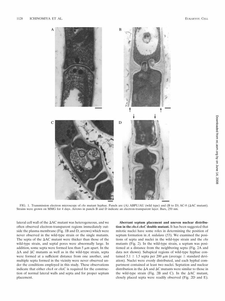
lateral cell wall of the �AC mutant was heterogeneous, and weoften observed electron-transparent regions immediately out-side the plasma membrane (Fig. 1B and D, arrows) which werenever observed in the wild-type strain or the single mutants.The septa of the �AC mutant were thicker than those of thewild-type strain, and septal pores were abnormally large. Inaddition, some septa were formed less than 5 �m apart. In the�A and �C mutants as well as in the wild-type strain, septawere formed at a sufficient distance from one another, andmultiple septa formed in the vicinity were never observed un-der the conditions employed in this study. These observationsindicate that either chsA or chsC is required for the construc-tion of normal lateral walls and septa and for proper septumplacement.
Aberrant septum placement and uneven nuclear distribu-tion in the chsA chsC double mutant. It has been suggested thatmitotic nuclei have some roles in determining the position ofseptum formation in A. nidulans (53). We examined the posi-tions of septa and nuclei in the wild-type strain and the chsmutants (Fig. 2). In the wild-type strain, a septum was posi-tioned at a distance from the neighboring septa (Fig. 2A anddata not shown). Subapical regions of wild-type hyphae con-tained 5.1 � 1.5 septa per 200 �m (average � standard devi-ation). Nuclei were evenly distributed, and each hyphal com-partment contained at least two nuclei. Septation and nucleardistribution in the �A and �C mutants were similar to those inthe wild-type strain (Fig. 2B and C). In the �AC mutant,closely placed septa were readily observed (Fig. 2D and E).
FIG. 1. Transmission electron microscopy of chs mutant hyphae. Panels are (A) ABPU/AU (wild type) and (B to D) AC-8 (�AC mutant).Strains were grown on MMG for 4 days. Arrows in panels B and D indicate an electron-transparent layer. Bars, 250 nm.
1128 ICHINOMIYA ET AL. EUKARYOT. CELL
by on June 14, 2008 ec.asm
.orgD
ownloaded from
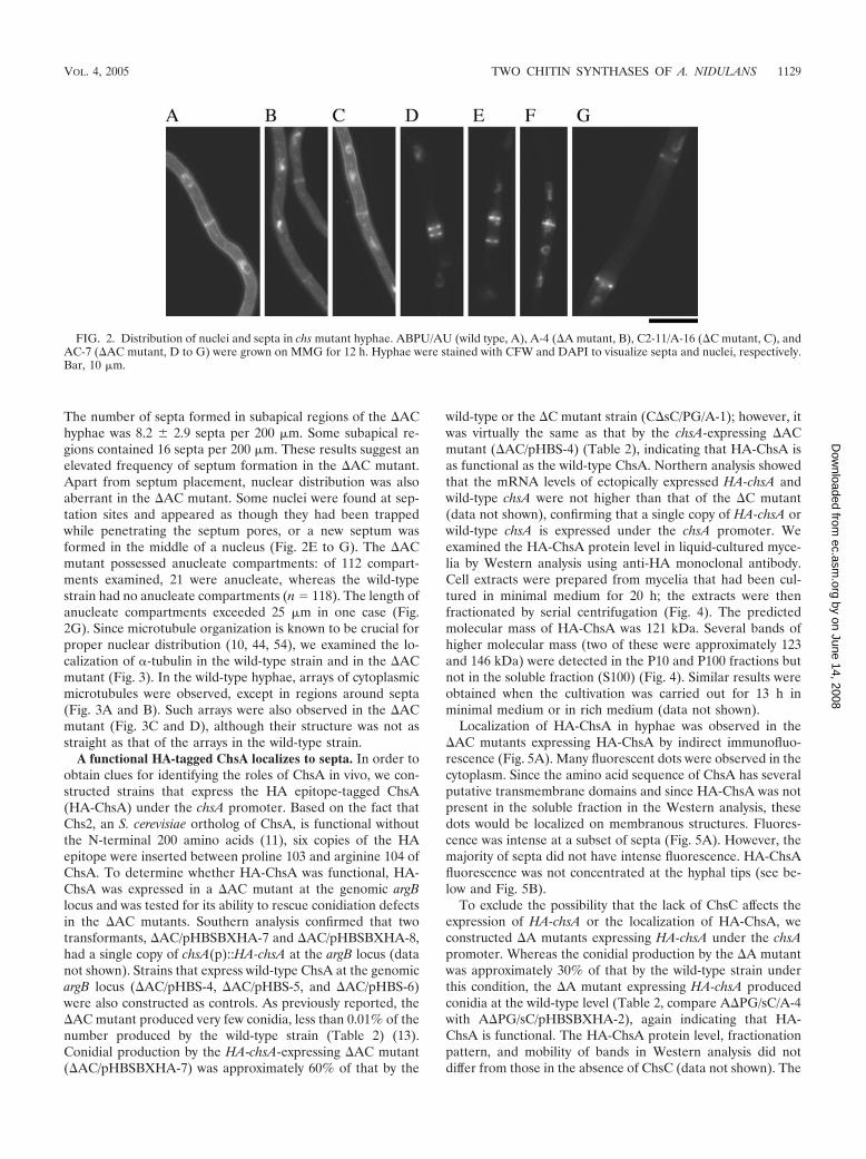
The number of septa formed in subapical regions of the �AChyphae was 8.2 � 2.9 septa per 200 �m. Some subapical re-gions contained 16 septa per 200 �m. These results suggest anelevated frequency of septum formation in the �AC mutant.Apart from septum placement, nuclear distribution was alsoaberrant in the �AC mutant. Some nuclei were found at sep-tation sites and appeared as though they had been trappedwhile penetrating the septum pores, or a new septum wasformed in the middle of a nucleus (Fig. 2E to G). The �ACmutant possessed anucleate compartments: of 112 compart-ments examined, 21 were anucleate, whereas the wild-typestrain had no anucleate compartments (n 118). The length ofanucleate compartments exceeded 25 �m in one case (Fig.2G). Since microtubule organization is known to be crucial forproper nuclear distribution (10, 44, 54), we examined the lo-calization of �-tubulin in the wild-type strain and in the �ACmutant (Fig. 3). In the wild-type hyphae, arrays of cytoplasmicmicrotubules were observed, except in regions around septa(Fig. 3A and B). Such arrays were also observed in the �ACmutant (Fig. 3C and D), although their structure was not asstraight as that of the arrays in the wild-type strain.
A functional HA-tagged ChsA localizes to septa. In order toobtain clues for identifying the roles of ChsA in vivo, we con-structed strains that express the HA epitope-tagged ChsA(HA-ChsA) under the chsA promoter. Based on the fact thatChs2, an S. cerevisiae ortholog of ChsA, is functional withoutthe N-terminal 200 amino acids (11), six copies of the HAepitope were inserted between proline 103 and arginine 104 ofChsA. To determine whether HA-ChsA was functional, HA-ChsA was expressed in a �AC mutant at the genomic argBlocus and was tested for its ability to rescue conidiation defectsin the �AC mutants. Southern analysis confirmed that twotransformants, �AC/pHBSBXHA-7 and �AC/pHBSBXHA-8,had a single copy of chsA(p)::HA-chsA at the argB locus (datanot shown). Strains that express wild-type ChsA at the genomicargB locus (�AC/pHBS-4, �AC/pHBS-5, and �AC/pHBS-6)were also constructed as controls. As previously reported, the�AC mutant produced very few conidia, less than 0.01% of thenumber produced by the wild-type strain (Table 2) (13).Conidial production by the HA-chsA-expressing �AC mutant(�AC/pHBSBXHA-7) was approximately 60% of that by the
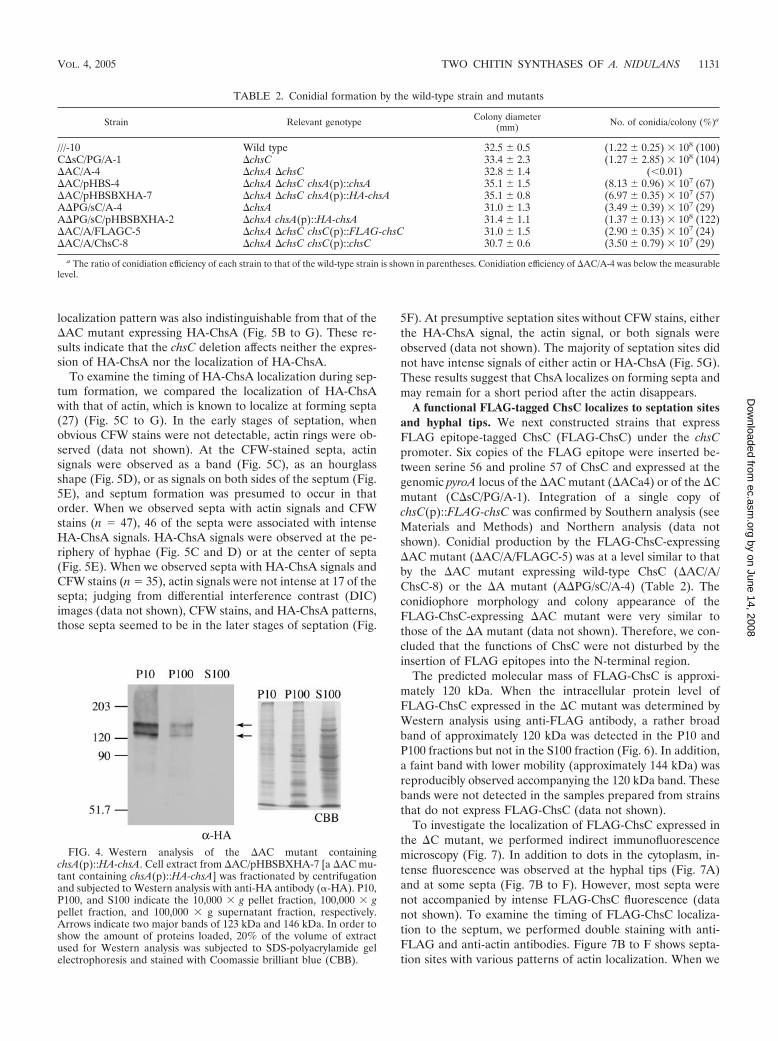
wild-type or the �C mutant strain (C�sC/PG/A-1); however, itwas virtually the same as that by the chsA-expressing �ACmutant (�AC/pHBS-4) (Table 2), indicating that HA-ChsA isas functional as the wild-type ChsA. Northern analysis showedthat the mRNA levels of ectopically expressed HA-chsA andwild-type chsA were not higher than that of the �C mutant(data not shown), confirming that a single copy of HA-chsA orwild-type chsA is expressed under the chsA promoter. Weexamined the HA-ChsA protein level in liquid-cultured myce-lia by Western analysis using anti-HA monoclonal antibody.Cell extracts were prepared from mycelia that had been cul-tured in minimal medium for 20 h; the extracts were thenfractionated by serial centrifugation (Fig. 4). The predictedmolecular mass of HA-ChsA was 121 kDa. Several bands ofhigher molecular mass (two of these were approximately 123and 146 kDa) were detected in the P10 and P100 fractions butnot in the soluble fraction (S100) (Fig. 4). Similar results wereobtained when the cultivation was carried out for 13 h inminimal medium or in rich medium (data not shown).
Localization of HA-ChsA in hyphae was observed in the�AC mutants expressing HA-ChsA by indirect immunofluo-rescence (Fig. 5A). Many fluorescent dots were observed in thecytoplasm. Since the amino acid sequence of ChsA has severalputative transmembrane domains and since HA-ChsA was notpresent in the soluble fraction in the Western analysis, thesedots would be localized on membranous structures. Fluores-cence was intense at a subset of septa (Fig. 5A). However, themajority of septa did not have intense fluorescence. HA-ChsAfluorescence was not concentrated at the hyphal tips (see be-low and Fig. 5B).
To exclude the possibility that the lack of ChsC affects theexpression of HA-chsA or the localization of HA-ChsA, weconstructed �A mutants expressing HA-chsA under the chsApromoter. Whereas the conidial production by the �A mutantwas approximately 30% of that by the wild-type strain underthis condition, the �A mutant expressing HA-chsA producedconidia at the wild-type level (Table 2, compare A�PG/sC/A-4with A�PG/sC/pHBSBXHA-2), again indicating that HA-ChsA is functional. The HA-ChsA protein level, fractionationpattern, and mobility of bands in Western analysis did notdiffer from those in the absence of ChsC (data not shown). The
FIG. 2. Distribution of nuclei and septa in chs mutant hyphae. ABPU/AU (wild type, A), A-4 (�A mutant, B), C2-11/A-16 (�C mutant, C), andAC-7 (�AC mutant, D to G) were grown on MMG for 12 h. Hyphae were stained with CFW and DAPI to visualize septa and nuclei, respectively.Bar, 10 �m.
VOL. 4, 2005 TWO CHITIN SYNTHASES OF A. NIDULANS 1129
by on June 14, 2008 ec.asm
.orgD
ownloaded from

FIG. 3. Microtubule organization in the wild-type strain and the �AC mutant. ABPU/AU (A and B) and AC-7 (C and D) grown on MMG for13 h were subjected to indirect immunofluorescence microscopy using anti-actin antibody (�-actin) and anti-�-tubulin antibody (�-tubulin).Hyphae were also stained with DAPI and CFW. The arrowhead in panel D indicates the septum with a microtubule passing through. Bars, 10 �m.
1130 ICHINOMIYA ET AL. EUKARYOT. CELL
by on June 14, 2008 ec.asm
.orgD
ownloaded from
localization pattern was also indistinguishable from that of the�AC mutant expressing HA-ChsA (Fig. 5B to G). These re-sults indicate that the chsC deletion affects neither the expres-sion of HA-ChsA nor the localization of HA-ChsA.
To examine the timing of HA-ChsA localization during sep-tum formation, we compared the localization of HA-ChsAwith that of actin, which is known to localize at forming septa(27) (Fig. 5C to G). In the early stages of septation, whenobvious CFW stains were not detectable, actin rings were ob-served (data not shown). At the CFW-stained septa, actinsignals were observed as a band (Fig. 5C), as an hourglassshape (Fig. 5D), or as signals on both sides of the septum (Fig.5E), and septum formation was presumed to occur in thatorder. When we observed septa with actin signals and CFWstains (n 47), 46 of the septa were associated with intenseHA-ChsA signals. HA-ChsA signals were observed at the pe-riphery of hyphae (Fig. 5C and D) or at the center of septa(Fig. 5E). When we observed septa with HA-ChsA signals andCFW stains (n 35), actin signals were not intense at 17 of thesepta; judging from differential interference contrast (DIC)images (data not shown), CFW stains, and HA-ChsA patterns,those septa seemed to be in the later stages of septation (Fig.
5F). At presumptive septation sites without CFW stains, eitherthe HA-ChsA signal, the actin signal, or both signals wereobserved (data not shown). The majority of septation sites didnot have intense signals of either actin or HA-ChsA (Fig. 5G).These results suggest that ChsA localizes on forming septa andmay remain for a short period after the actin disappears.
A functional FLAG-tagged ChsC localizes to septation sitesand hyphal tips. We next constructed strains that expressFLAG epitope-tagged ChsC (FLAG-ChsC) under the chsCpromoter. Six copies of the FLAG epitope were inserted be-tween serine 56 and proline 57 of ChsC and expressed at thegenomic pyroA locus of the �AC mutant (�ACa4) or of the �Cmutant (C�sC/PG/A-1). Integration of a single copy ofchsC(p)::FLAG-chsC was confirmed by Southern analysis (seeMaterials and Methods) and Northern analysis (data notshown). Conidial production by the FLAG-ChsC-expressing�AC mutant (�AC/A/FLAGC-5) was at a level similar to thatby the �AC mutant expressing wild-type ChsC (�AC/A/ChsC-8) or the �A mutant (A�PG/sC/A-4) (Table 2). Theconidiophore morphology and colony appearance of theFLAG-ChsC-expressing �AC mutant were very similar tothose of the �A mutant (data not shown). Therefore, we con-cluded that the functions of ChsC were not disturbed by theinsertion of FLAG epitopes into the N-terminal region.
The predicted molecular mass of FLAG-ChsC is approxi-mately 120 kDa. When the intracellular protein level ofFLAG-ChsC expressed in the �C mutant was determined byWestern analysis using anti-FLAG antibody, a rather broadband of approximately 120 kDa was detected in the P10 andP100 fractions but not in the S100 fraction (Fig. 6). In addition,a faint band with lower mobility (approximately 144 kDa) wasreproducibly observed accompanying the 120 kDa band. Thesebands were not detected in the samples prepared from strainsthat do not express FLAG-ChsC (data not shown).
To investigate the localization of FLAG-ChsC expressed inthe �C mutant, we performed indirect immunofluorescencemicroscopy (Fig. 7). In addition to dots in the cytoplasm, in-tense fluorescence was observed at the hyphal tips (Fig. 7A)and at some septa (Fig. 7B to F). However, most septa werenot accompanied by intense FLAG-ChsC fluorescence (datanot shown). To examine the timing of FLAG-ChsC localiza-tion to the septum, we performed double staining with anti-FLAG and anti-actin antibodies. Figure 7B to F shows septa-tion sites with various patterns of actin localization. When we
FIG. 4. Western analysis of the �AC mutant containingchsA(p)::HA-chsA. Cell extract from �AC/pHBSBXHA-7 [a �AC mu-tant containing chsA(p)::HA-chsA] was fractionated by centrifugationand subjected to Western analysis with anti-HA antibody (�-HA). P10,P100, and S100 indicate the 10,000 � g pellet fraction, 100,000 � gpellet fraction, and 100,000 � g supernatant fraction, respectively.Arrows indicate two major bands of 123 kDa and 146 kDa. In order toshow the amount of proteins loaded, 20% of the volume of extractused for Western analysis was subjected to SDS-polyacrylamide gelelectrophoresis and stained with Coomassie brilliant blue (CBB).
TABLE 2. Conidial formation by the wild-type strain and mutants
Strain Relevant genotype Colony diameter(mm) No. of conidia/colony (%)a
///-10 Wild type 32.5 � 0.5 (1.22 � 0.25) � 108 (100)C�sC/PG/A-1 �chsC 33.4 � 2.3 (1.27 � 2.85) � 108 (104)�AC/A-4 �chsA �chsC 32.8 � 1.4 (0.01)�AC/pHBS-4 �chsA �chsC chsA(p)::chsA 35.1 � 1.5 (8.13 � 0.96) � 107 (67)�AC/pHBSBXHA-7 �chsA �chsC chsA(p)::HA-chsA 35.1 � 0.8 (6.97 � 0.35) � 107 (57)A�PG/sC/A-4 �chsA 31.0 � 1.3 (3.49 � 0.39) � 107 (29)A�PG/sC/pHBSBXHA-2 �chsA chsA(p)::HA-chsA 31.4 � 1.1 (1.37 � 0.13) � 108 (122)�AC/A/FLAGC-5 �chsA �chsC chsC(p)::FLAG-chsC 31.0 � 1.5 (2.90 � 0.35) � 107 (24)�AC/A/ChsC-8 �chsA �chsC chsC(p)::chsC 30.7 � 0.6 (3.50 � 0.79) � 107 (29)
a The ratio of conidiation efficiency of each strain to that of the wild-type strain is shown in parentheses. Conidiation efficiency of �AC/A-4 was below the measurablelevel.
VOL. 4, 2005 TWO CHITIN SYNTHASES OF A. NIDULANS 1131
by on June 14, 2008 ec.asm
.orgD
ownloaded from

FIG. 5. Localization of HA-ChsA. (A) �AC/pHBSBXHA-7 [�AC mutant containing chsA(p)::HA-chsA] was grown for 12 h and subjected toindirect immunofluorescence microscopy using anti-HA antibody (�-HA). Cells were also stained with CFW. A DIC image is also shown. (B toG) A�PG/sC/pHBSBXHA-2 [�A mutant containing chsA(p)::HA-chsA] was double stained with anti-actin antibody (�-actin) and anti-HAantibody (�-HA). Corresponding DIC or CFW-stained images are shown. The arrowhead in panel C indicates a faintly CFW-stained chitin ring.Bars, 10 �m.
1132 ICHINOMIYA ET AL. EUKARYOT. CELL
by on June 14, 2008 ec.asm
.orgD
ownloaded from
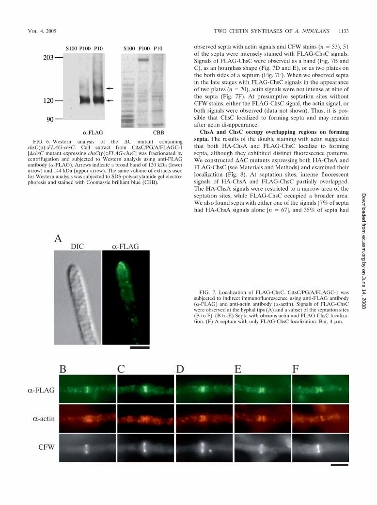
observed septa with actin signals and CFW stains (n 53), 51of the septa were intensely stained with FLAG-ChsC signals.Signals of FLAG-ChsC were observed as a band (Fig. 7B andC), as an hourglass shape (Fig. 7D and E), or as two plates onthe both sides of a septum (Fig. 7F). When we observed septain the late stages with FLAG-ChsC signals in the appearanceof two plates (n 20), actin signals were not intense at nine ofthe septa (Fig. 7F). At presumptive septation sites withoutCFW stains, either the FLAG-ChsC signal, the actin signal, orboth signals were observed (data not shown). Thus, it is pos-sible that ChsC localized to forming septa and may remainafter actin disappearance.
ChsA and ChsC occupy overlapping regions on formingsepta. The results of the double staining with actin suggestedthat both HA-ChsA and FLAG-ChsC localize to formingsepta, although they exhibited distinct fluorescence patterns.We constructed �AC mutants expressing both HA-ChsA andFLAG-ChsC (see Materials and Methods) and examined theirlocalization (Fig. 8). At septation sites, intense fluorescentsignals of HA-ChsA and FLAG-ChsC partially overlapped.The HA-ChsA signals were restricted to a narrow area of theseptation sites, while FLAG-ChsC occupied a broader area.We also found septa with either one of the signals (7% of septahad HA-ChsA signals alone [n 67], and 35% of septa had
FIG. 6. Western analysis of the �C mutant containingchsC(p)::FLAG-chsC. Cell extract from C�sC/PG/A/FLAGC-1[�chsC mutant expressing chsC(p)::FLAG-chsC] was fractionated bycentrifugation and subjected to Western analysis using anti-FLAGantibody (�-FLAG). Arrows indicate a broad band of 120 kDa (lowerarrow) and 144 kDa (upper arrow). The same volume of extracts usedfor Western analysis was subjected to SDS-polyacrylamide gel electro-phoresis and stained with Coomassie brilliant blue (CBB).
FIG. 7. Localization of FLAG-ChsC. C�sC/PG/A/FLAGC-1 wassubjected to indirect immunofluorescence using anti-FLAG antibody(�-FLAG) and anti-actin antibody (�-actin). Signals of FLAG-ChsCwere observed at the hyphal tips (A) and a subset of the septation sites(B to F). (B to E) Septa with obvious actin and FLAG-ChsC localiza-tion. (F) A septum with only FLAG-ChsC localization. Bar, 4 �m.
VOL. 4, 2005 TWO CHITIN SYNTHASES OF A. NIDULANS 1133
by on June 14, 2008 ec.asm
.orgD
ownloaded from
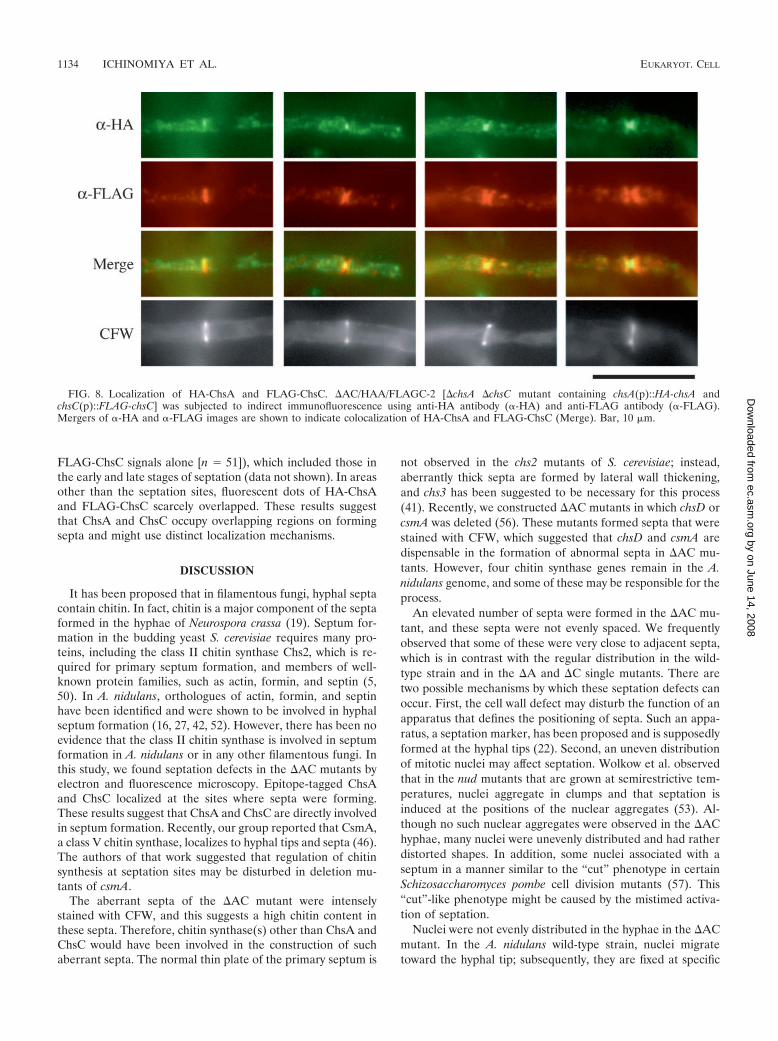
FLAG-ChsC signals alone [n 51]), which included those inthe early and late stages of septation (data not shown). In areasother than the septation sites, fluorescent dots of HA-ChsAand FLAG-ChsC scarcely overlapped. These results suggestthat ChsA and ChsC occupy overlapping regions on formingsepta and might use distinct localization mechanisms.
DISCUSSION
It has been proposed that in filamentous fungi, hyphal septacontain chitin. In fact, chitin is a major component of the septaformed in the hyphae of Neurospora crassa (19). Septum for-mation in the budding yeast S. cerevisiae requires many pro-teins, including the class II chitin synthase Chs2, which is re-quired for primary septum formation, and members of well-known protein families, such as actin, formin, and septin (5,50). In A. nidulans, orthologues of actin, formin, and septinhave been identified and were shown to be involved in hyphalseptum formation (16, 27, 42, 52). However, there has been noevidence that the class II chitin synthase is involved in septumformation in A. nidulans or in any other filamentous fungi. Inthis study, we found septation defects in the �AC mutants byelectron and fluorescence microscopy. Epitope-tagged ChsAand ChsC localized at the sites where septa were forming.These results suggest that ChsA and ChsC are directly involvedin septum formation. Recently, our group reported that CsmA,a class V chitin synthase, localizes to hyphal tips and septa (46).The authors of that work suggested that regulation of chitinsynthesis at septation sites may be disturbed in deletion mu-tants of csmA.
The aberrant septa of the �AC mutant were intenselystained with CFW, and this suggests a high chitin content inthese septa. Therefore, chitin synthase(s) other than ChsA andChsC would have been involved in the construction of suchaberrant septa. The normal thin plate of the primary septum is
not observed in the chs2 mutants of S. cerevisiae; instead,aberrantly thick septa are formed by lateral wall thickening,and chs3 has been suggested to be necessary for this process(41). Recently, we constructed �AC mutants in which chsD orcsmA was deleted (56). These mutants formed septa that werestained with CFW, which suggested that chsD and csmA aredispensable in the formation of abnormal septa in �AC mu-tants. However, four chitin synthase genes remain in the A.nidulans genome, and some of these may be responsible for theprocess.
An elevated number of septa were formed in the �AC mu-tant, and these septa were not evenly spaced. We frequentlyobserved that some of these were very close to adjacent septa,which is in contrast with the regular distribution in the wild-type strain and in the �A and �C single mutants. There aretwo possible mechanisms by which these septation defects canoccur. First, the cell wall defect may disturb the function of anapparatus that defines the positioning of septa. Such an appa-ratus, a septation marker, has been proposed and is supposedlyformed at the hyphal tips (22). Second, an uneven distributionof mitotic nuclei may affect septation. Wolkow et al. observedthat in the nud mutants that are grown at semirestrictive tem-peratures, nuclei aggregate in clumps and that septation isinduced at the positions of the nuclear aggregates (53). Al-though no such nuclear aggregates were observed in the �AChyphae, many nuclei were unevenly distributed and had ratherdistorted shapes. In addition, some nuclei associated with aseptum in a manner similar to the “cut” phenotype in certainSchizosaccharomyces pombe cell division mutants (57). This“cut”-like phenotype might be caused by the mistimed activa-tion of septation.
Nuclei were not evenly distributed in the hyphae in the �ACmutant. In the A. nidulans wild-type strain, nuclei migratetoward the hyphal tip; subsequently, they are fixed at specific
FIG. 8. Localization of HA-ChsA and FLAG-ChsC. �AC/HAA/FLAGC-2 [�chsA �chsC mutant containing chsA(p)::HA-chsA andchsC(p)::FLAG-chsC] was subjected to indirect immunofluorescence using anti-HA antibody (�-HA) and anti-FLAG antibody (�-FLAG).Mergers of �-HA and �-FLAG images are shown to indicate colocalization of HA-ChsA and FLAG-ChsC (Merge). Bar, 10 �m.
1134 ICHINOMIYA ET AL. EUKARYOT. CELL
by on June 14, 2008 ec.asm
.orgD
ownloaded from
positions (45). We prefer the idea that the fixation process isdisturbed, because nuclei did migrate through hyphae and be-cause microtubules were present in the �AC mutant, thoughthey were less polarized than in the wild-type strain. ApsA andApsB are believed to be involved in the regulation of thisfixation process (10, 44, 54). Since ApsA was shown to localizeat the hyphal cortex (45), it is possible that certain machinerythat regulates nuclear positioning and possibly interacts withApsA and ApsB exists at a similar cortical location. If this isthe case, its function and/or localization may be disturbed bythe cell wall defects in the �AC mutant, which were observedin Fig. 1. Interestingly, a deletion mutant of the nudC gene,which has been identified as necessary for normal nucleardistribution (55), has been shown to have cell wall defects (6).This result, together with ours, supports the hypothesis that thefungal cell wall might provide rigid supports for nuclei tomigrate to and stay at specific positions in the hyphae.
When HA-ChsA-expressing strains were subjected to West-ern analysis, we detected several bands with molecular masseshigher than 121 kDa, the predicted molecular mass of HA-ChsA (Fig. 4). It is possible that ChsA is posttranslationallymodified. To date, it has been shown that chitin synthases of S.cerevisiae receive posttranslational modifications. Chs3 is gly-cosylated (40, 48) and phosphorylated (49). Chs1, Chs2, andChs3 are ubiquitination substrates at the plasma membrane(17). The smeared FLAG-ChsC low-mobility band might begenerated by posttranslational modification(s), although it isnot as prominent as that of ChsA. Alternatively, these higher-molecular-mass bands might be artifacts. Cos et al. reportedthat two bands (160 and 230 kDa) were detected in cells ex-pressing HA-tagged Chs3, the predicted size of which was 131kDa in S. cerevisiae, and they considered the 230-kDa band tobe a running artifact (8). A smeared high-molecular-mass bandis also observed in the analysis of class III chitin synthase inWangiella dermatitidis (51).
HA-ChsA and FLAG-ChsC localized to forming septa andpartially colocalized there. This suggests that they construct anoverlapping fraction of chitin on septa, which would explainthe synthetic defect in the septum formation of the �AC mu-tants. However, we could not observe their colocalization at asubset of septa. Since the lack of ChsA or ChsC did not changethe localization of the other, and since their colocalization wasnot observed in the cytoplasmic space, it is possible that theirlocalization and disappearance at septation sites may be inde-pendent of each other. Such independence might mean thattheir localization and disappearance occur at different timing.Colocalization was not observed at the hyphal tips, either;localization of ChsC at the hyphal tips is consistent with itsinvolvement in vegetative growth, which was suggested fromthe analysis of chsB chsC double mutants (20). The transportand retention mechanisms of chitin synthases to different hy-phal regions in filamentous fungi remain to be clarified. Iden-tification of proteins interacting with ChsA or ChsC wouldhelp in understanding their transport and retention mecha-nisms.
Although we identified septation defects in the �AC mutantin this study, this mutant also exhibited other defects (13). It ispossible that the overlap of functions between ChsA and ChsCis not restricted to septum formation; it may extend to themaintenance of cell wall integrity and to conidiation. Alterna-
tively, the primary defect in the �AC mutants might occur inseptation during hyphal growth, and other phenotypes mightbe caused indirectly. Although several temperature-sensitivemutants that failed to form septa (sep mutants) have beenisolated and characterized in A. nidulans (4, 14–16, 28, 42),whether they have cell wall defects like the �AC mutants hasnot been examined. Further investigations are required to elu-cidate how ChsA and ChsC are involved in the maintenance ofcell wall integrity and in conidiation.
ACKNOWLEDGMENTS
This work was supported by a grant-in-aid for Scientific Researchfrom the Ministry of Education, Culture, Sports, Science, and Tech-nology of Japan and partly by a grant from Bio-oriented TechnologyResearch Advancement Institution.
This work was performed using the facilities of the BiotechnologyResearch Center of the University of Tokyo.
REFERENCES
1. Adams, T. H., J. K. Wieser, and J. H. Yu. 1998. Asexual sporulation inAspergillus nidulans. Microbiol. Mol. Biol. Rev. 62:35–54.
2. Aufauvre-Brown, A., E. Mellado, N. A. Gow, and D. W. Holden. 1997. As-pergillus fumigatus chsE: a gene related to CHS3 of Saccharomyces cerevisiaeand important for hyphal growth and conidiophore development but notpathogenicity. Fungal Genet. Biol. 21:141–152.
3. Borgia, P. T., N. Iartchouk, P. J. Riggle, K. R. Winter, Y. Koltin, and C. E.Bulawa. 1996. The chsB gene of Aspergillus nidulans is necessary for normalhyphal growth and development. Fungal Genet. Biol. 20:193–203.
4. Bruno, K. S., J. L. Morrell, J. E. Hamer, and C. J. Staiger. 2001. SEPH, aCdc7p orthologue from Aspergillus nidulans, functions upstream of actin ringformation during cytokinesis. Mol. Microbiol. 42:3–12.
5. Cabib, E., D. H. Roh, M. Schmidt, L. B. Crotti, and A. Varma. 2001. Theyeast cell wall and septum as paradigms of cell growth and morphogenesis.J. Biol. Chem. 276:19679–19682.
6. Chiu, Y. H., X. Xiang, A. L. Dawe, and N. R. Morris. 1997. Deletion of nudC,a nuclear migration gene of Aspergillus nidulans, causes morphological andcell wall abnormalities and is lethal. Mol. Biol. Cell 8:1735–1749.
7. Chuang, J. S., and R. W. Schekman. 1996. Differential trafficking and timedlocalization of two chitin synthase proteins, Chs2p and Chs3p. J. Cell Biol.135:597–610.
8. Cos, T., R. A. Ford, J. A. Trilla, A. Duran, E. Cabib, and C. Roncero. 1998.Molecular analysis of Chs3p participation in chitin synthase III activity. Eur.J. Biochem. 256:419–426.
9. Culp, D. W., C. L. Dodge, Y. Miao, L. Li, D. Sag-Ozkal, and P. T. Borgia.2000. The chsA gene from Aspergillus nidulans is necessary for maximalconidiation. FEMS Microbiol. Lett. 182:349–353.
10. Fischer, R. 1999. Nuclear movement in filamentous fungi. FEMS Microbiol.Rev. 23:39–68.
11. Ford, R. A., J. A. Shaw, and E. Cabib. 1996. Yeast chitin synthases 1 and 2consist of a non-homologous and dispensable N-terminal region and of ahomologous moiety essential for function. Mol. Gen. Genet. 252:420–428.
12. Fujiwara, M., H. Horiuchi, A. Ohta, and M. Takagi. 1997. A novel fungalgene encoding chitin synthase with a myosin motor-like domain. Biochem.Biophys. Res. Commun. 236:75–78.
13. Fujiwara, M., M. Ichinomiya, T. Motoyama, H. Horiuchi, A. Ohta, and M.Takagi. 2000. Evidence that the Aspergillus nidulans class I and class II chitinsynthase genes, chsC and chsA, share critical roles in hyphal wall integrityand conidiophore development. J. Biochem. (Tokyo) 127:359–366.
14. Harris, S. D., and J. E. Hamer. 1995. sepB: an Aspergillus nidulans geneinvolved in chromosome segregation and the initiation of cytokinesis. EMBOJ. 14:5244–5257.
15. Harris, S. D., L. Hamer, K. E. Sharpless, and J. E. Hamer. 1997. TheAspergillus nidulans sepA gene encodes an FH1/2 protein involved in cyto-kinesis and the maintenance of cellular polarity. EMBO J. 16:3474–3483.
16. Harris, S. D., J. L. Morrell, and J. E. Hamer. 1994. Identification andcharacterization of Aspergillus nidulans mutants defective in cytokinesis. Ge-netics 136:517–532.
17. Hitchcock, A. L., K. Auld, S. P. Gygi, and P. A. Silver. 2003. A subset ofmembrane-associated proteins is ubiquitinated in response to mutations inthe endoplasmic reticulum degradation machinery. Proc. Natl. Acad. Sci.USA 100:12735–12740.
18. Horiuchi, H., M. Fujiwara, S. Yamashita, A. Ohta, and M. Takagi. 1999.Proliferation of intrahyphal hyphae caused by disruption of csmA, whichencodes a class V chitin synthase with a myosin motor-like domain in As-pergillus nidulans. J. Bacteriol. 181:3721–3729.
19. Hunsley, D., and G. W. Gooday. 1974. The structure and development ofsepta in Neurospora crassa. Protoplasma 82:125–146.
VOL. 4, 2005 TWO CHITIN SYNTHASES OF A. NIDULANS 1135
by on June 14, 2008 ec.asm
.orgD
ownloaded from
20. Ichinomiya, M., H. Horiuchi, and A. Ohta. 2002. Different functions of theclass I and class II chitin synthase genes, chsC and chsA, are revealed by therepression of the chsB expression of Aspergillus nidulans. Curr. Genet. 41:51–58.
21. Ichinomiya, M., T. Motoyama, M. Fujiwara, M. Takagi, H. Horiuchi, and A.Ohta. 2002. Repression of chsB expression reveals the functional importanceof class IV chitin synthase gene chsD in hyphal growth and conidiation ofAspergillus nidulans. Microbiology 148:1335–1347.
22. Kaminskyj, S. G. 2000. Septum position is marked at the tip of Aspergillusnidulans hyphae. Fungal Genet. Biol. 31:105–113.
23. Madrid, M. P., A. Di Pietro, and M. I. Roncero. 2003. Class V chitin synthasedetermines pathogenesis in the vascular wilt fungus Fusarium oxysporum andmediates resistance to plant defence compounds. Mol. Microbiol. 47:257–266.
24. May, G. S., C. A. McGoldrick, C. L. Holt, and S. H. Denison. 1992. ThebimB3 mutation of Aspergillus nidulans uncouples DNA replication from thecompletion of mitosis. J. Biol. Chem. 267:15737–15743.
25. Mellado, E., A. Aufauvre-Brown, N. A. Gow, and D. W. Holden. 1996. TheAspergillus fumigatus chsC and chsG genes encode class III chitin synthaseswith different functions. Mol. Microbiol. 20:667–679.
26. Momany, M. 2001. Cell biology of the duplication cycle in fungi, p. 119–125.In N. Talbot (ed.), Molecular and cellular biology of filamentous fungi.Oxford University Press, Oxford, United Kingdom.
27. Momany, M., and J. E. Hamer. 1997. Relationship of actin, microtubules,and crosswall synthesis during septation in Aspergillus nidulans. Cell Motil.Cytoskel. 38:373–384.
28. Morris, N. R. 1976. A temperature-sensitive mutant of Aspergillus nidulansreversibly blocked in nuclear division. Exp. Cell Res. 98:204–210.
29. Motoyama, T., M. Fujiwara, N. Kojima, H. Horiuchi, A. Ohta, and M.Takagi. 1996. The Aspergillus nidulans genes chsA and chsD encode chitinsynthases which have redundant functions in conidia formation. Mol. Gen.Genet. 251:442–450 (Corrected and republished 253:520–528, 1997.)
30. Motoyama, T., N. Kojima, H. Horiuchi, A. Ohta, and M. Takagi. 1994.Isolation of a chitin synthase gene (chsC) of Aspergillus nidulans. Biosci.Biotechnol. Biochem. 58:2254–2257.
31. Muller, C., C. M. Hjort, K. Hansen, and J. Nielsen. 2002. Altering theexpression of two chitin synthase genes differentially affects the growth andmorphology of Aspergillus oryzae. Microbiology 148:4025–4033.
32. Munro, C. A., and N. A. Gow. 2001. Chitin synthesis in human pathogenicfungi. Med. Mycol. 39:41–53.
33. Oakley, C. E., C. F. Weil, P. L. Kretz, and B. R. Oakley. 1987. Cloning of theriboB locus of Aspergillus nidulans. Gene 53:293–298.
34. Orlean, P. 1997. Biogenesis of yeast wall and surface components. In J. R.Pringle, J. R. Broach, and E. W. Jones (ed.), The molecular and cellularbiology of the yeast Saccharomyces cerevisiae: cell cycle and cell biology, vol.3. Cold Spring Harbor Laboratory Press, Cold Spring Harbor, N.Y.
35. Pontecorvo, G., J. A. Roper, L. M. Hemmons, K. D. MacDonald, and A. W. J.Bufton. 1953. The genetics of Aspergillus nidulans. Adv. Genet. 5:141–238.
36. Roh, D. H., B. Bowers, M. Schmidt, and E. Cabib. 2002. The septationapparatus, an autonomous system in budding yeast. Mol. Biol. Cell 13:2747–2759.
37. Roncero, C. 2002. The genetic complexity of chitin synthesis in fungi. Curr.Genet. 41:367–378.
38. Rowlands, R. T., and G. Turner. 1973. Nuclear and extranuclear inheritanceof oligomycin resistance in Aspergillus nidulans. Mol. Gen. Genet. 126:201–216.
39. Sambrook, J., E. F. Fritsch, and T. Maniatis. 1989. Molecular cloning: alaboratory manual, 2nd ed. Cold Spring Harbor Laboratory Press, ColdSpring Harbor, N.Y.
40. Santos, B., and M. Snyder. 1997. Targeting of chitin synthase 3 to polarizedgrowth sites in yeast requires Chs5p and Myo2p. J. Cell Biol. 136:95–110.
41. Schmidt, M., B. Bowers, A. Varma, D. H. Roh, and E. Cabib. 2002. Inbudding yeast, contraction of the actomyosin ring and formation of theprimary septum at cytokinesis depend on each other. J. Cell Sci. 115:293–302.
42. Sharpless, K. E., and S. D. Harris. 2002. Functional characterization andlocalization of the Aspergillus nidulans formin SEPA. Mol. Biol. Cell 13:469–479.
43. Specht, C. A., Y. Liu, P. W. Robbins, C. E. Bulawa, N. Iartchouk, K. R.Winter, P. J. Riggle, J. C. Rhodes, C. L. Dodge, D. W. Culp, and P. T. Borgia.1996. The chsD and chsE genes of Aspergillus nidulans and their roles inchitin synthesis. Fungal Genet. Biol. 20:153–167.
44. Suelmann, R., and R. Fischer. 2000. Nuclear migration in fungi—differentmotors at work. Res. Microbiol. 151:247–254.
45. Suelmann, R., N. Sievers, and R. Fischer. 1997. Nuclear traffic in fungalhyphae: in vivo study of nuclear migration and positioning in Aspergillusnidulans. Mol. Microbiol. 25:757–769.
46. Takeshita, N., A. Ohta, and H. Horiuchi. 2005. CsmA, a class V chitinsynthase with a myosin motor-like domain, is localized through direct inter-action with the actin cytoskeleton in Aspergillus nidulans. Mol. Biol. Cell16:1961–1970.
47. Takeshita, N., A. Ohta, and H. Horiuchi. 2002. csmA, a gene encoding a classV chitin synthase with a myosin motor-like domain of Aspergillus nidulans, istranslated as a single polypeptide and regulated in response to osmoticconditions. Biochem. Biophys. Res. Commun. 298:103–109.
48. Trilla, J. A., A. Duran, and C. Roncero. 1999. Chs7p, a new protein involvedin the control of protein export from the endoplasmic reticulum that isspecifically engaged in the regulation of chitin synthesis in Saccharomycescerevisiae. J. Cell Biol. 145:1153–1163.
49. Valdivia, R. H., and R. Schekman. 2003. The yeasts Rho1p and Pkc1pregulate the transport of chitin synthase III (Chs3p) from internal stores tothe plasma membrane. Proc. Natl. Acad. Sci. USA 100:10287–10292.
50. Walther, A., and J. Wendland. 2003. Septation and cytokinesis in fungi.Fungal Genet. Biol. 40:187–196.
51. Wang, Z., and P. J. Szaniszlo. 2000. WdCHS3, a gene that encodes a class IIIchitin synthase in Wangiella (Exophiala) dermatitidis, is expressed differen-tially under stress conditions. J. Bacteriol. 182:874–881.
52. Westfall, P. J., and M. Momany. 2002. Aspergillus nidulans septin AspB playspre- and postmitotic roles in septum, branch, and conidiophore develop-ment. Mol. Biol. Cell 13:110–118.
53. Wolkow, T. D., S. D. Harris, and J. E. Hamer. 1996. Cytokinesis in Aspergillusnidulans is controlled by cell size, nuclear positioning and mitosis. J. Cell Sci.109:2179–2188.
54. Xiang, X., and R. Fischer. 2004. Nuclear migration and positioning in fila-mentous fungi. Fungal Genet. Biol. 41:411–419.
55. Xiang, X., and N. R. Morris. 1999. Hyphal tip growth and nuclear migration.Curr. Opin. Microbiol. 2:636–640.
56. Yamada, E., M. Ichinomiya, A. Ohta, and H. Horiuchi. 2005. The class Vchitin synthase gene csmA is crucial for the growth of the chsA chsC doublemutant in Aspergillus nidulans. Biosci. Biotechnol. Biochem. 69:87–97.
57. Yanagida, M. 1998. Fission yeast cut mutations revisited: control of an-aphase. Trends Cell Biol. 8:144–149.
58. Yanai, K., N. Kojima, N. Takaya, H. Horiuchi, A. Ohta, and M. Takagi. 1994.Isolation and characterization of two chitin synthase genes from Aspergillusnidulans. Biosci. Biotechnol. Biochem. 58:1828–1835.
59. Yarden, O., and C. Yanofsky. 1991. Chitin synthase 1 plays a major role incell wall biogenesis in Neurospora crassa. Genes Dev. 5:2420–2430.
60. Ziman, M., J. S. Chuang, and R. W. Schekman. 1996. Chs1p and Chs3p, twoproteins involved in chitin synthesis, populate a compartment of the Saccha-romyces cerevisiae endocytic pathway. Mol. Biol. Cell 7:1909–1919.
1136 ICHINOMIYA ET AL. EUKARYOT. CELL
by on June 14, 2008 ec.asm
.orgD
ownloaded from